wear characteristics of current aesthetic dental
TRANSCRIPT
Available online at www.sciencedirect.com
www.elsevier.com/locate/jmbbm
j o u r n a l o f t h e m e c h a n i c a l b e h a v i o r o f b i o m e d i c a l m a t e r i a l s 2 0 ( 2 0 1 3 ) 1 1 3 – 1 2 5
1751-6161/$ - see frohttp://dx.doi.org/10
nCorresponding autE-mail address:
Research Paper
Wear characteristics of current aesthetic dental restorativeCAD/CAM materials: Two-body wear, gloss retention,roughness and Martens hardness
Werner H. Mormanna,n, Bogna Stawarczykb, Andreas Endera, Beatrice Senerc,Thomas Attinc, Albert Mehla
aDepartment of Computer-Assisted Restorative Dentistry, Center of Dental Medicine, University of Zurich, Plattenstrasse 11, 8032 Zurich,
SwitzerlandbDepartment of Prosthodontics, Dental School, Ludwig-Maximilians-University Munich, Goethestrasse 70, 80336 Munich, GermanycClinic of Preventive Dentistry, Periodontology and Cariology, Center of Dental Medicine, University of Zurich, Plattenstrasse 11, 8032 Zurich,
Switzerland
a r t i c l e i n f o
Article history:
Received 19 October 2012
Accepted 6 January 2013
Available online 23 January 2013
Keywords:
CAD/CAM-materials
Two-body wear
Toothbrushing wear
Gloss
Gloss retention
Roughness
Martens hardness
nt matter & 2013 Elsevie.1016/j.jmbbm.2013.01.00
hor. Tel.: þ41 44 634 33 60werner.moermann@zzm
a b s t r a c t
Objectives: This study determined the two-body wear and toothbrushing wear parameters,
including gloss and roughness measurements and additionally Martens hardness, of nine
aesthetic CAD/CAM materials, one direct resin-based nanocomposite plus that of human
enamel as a control group.
Materials and methods: Two-body wear was investigated in a computer-controlled chewing
simulator (1.2 million loadings, 49 N at 1.7 Hz; 3000 thermocycles 5/50 1C). Each of the 11
groups consisted of 12 specimens and 12 enamel antagonists. Quantitative analysis of
wear was carried out with a 3D-surface analyser. Gloss and roughness measurements were
evaluated using a glossmeter and an inductive surface profilometer before and after
abrasive toothbrushing of machine-polished specimens. Additionally Martens hardness
was measured. Statistically significant differences were calculated with one-way ANOVA
(analysis of variance).
Results: Statistically significant differences were found for two-body wear, gloss, surface
roughness and hardness. Zirconium dioxide ceramics showed no material wear and low
wear of the enamel antagonist. Two-body wear of CAD/CAM-silicate and -lithium disilicate
ceramics, -hybrid ceramics and -nanocomposite as well as direct nanocomposite did not
differ significantly from that of human enamel. Temporary polymers showed significantly
higher material wear than permanent materials. Abrasive toothbrushing significantly
reduced gloss and increased roughness of all materials except zirconium dioxide ceramics.
Gloss retention was highest with zirconium dioxide ceramics, silicate ceramics, hybrid
ceramics and nanocomposites. Temporary polymers showed least gloss retention. Martens
hardness differed significantly among ceramics, between ceramics and composites, and
between resin composites and acrylic block materials as well.
Conclusions: All permanent aesthetic CAD/CAM block materials tested behave similarly or
better with respect to two-body wear and toothbrushing wear than human enamel, which
r Ltd. All rights reserved.3
; fax: þ41 44 634 43 08..uzh.ch (W.H. Mormann).
j o u r n a l o f t h e m e c h a n i c a l b e h a v i o r o f b i o m e d i c a l m a t e r i a l s 2 0 ( 2 0 1 3 ) 1 1 3 – 1 2 5114
is not true for temporary polymer CAD/CAM block materials. Ceramics show the best gloss
retention compared to hybrid ceramics, composites and acrylic polymers.
& 2013 Elsevier Ltd. All rights reserved.
1. Introduction
Currently available is a range of ceramic- and polymer-based
aesthetic block materials with flexural strengths below
200 MPa, which can be one-step-CAD/CAM processed in
minutes by the dentist, even for large preparations, including
the replacement of cusps to a fitting permanent restoration,
while the patient is seated in the chair (Datzmann, 1996;
Kunzelmann et al., 2007; Magne and Knezevic, 2009;
Mormann, 2004). In contrast, the high-strength lithium dis-
ilicate and translucent zirconium dioxide ceramics, aesthetic
CAD/CAM block ceramics, need a two-step work process in
the dental laboratory consisting of computer aided design
and machining as a first step and an additional heat treat-
ment as the second step to reach their final high strength
(Bindl et al., 2003; Fischer et al., 2011a; Sirona, 2011). Anyway,
industrially pre-fabricated block ceramics appear to be more
structurally reliable for dental applications than ceramic
materials which are manually processed under dental labora-
tory conditions, although CAD/CAM procedures may induce
surface and subsurface flaws that may adversely affect this
property (Tinschert et al., 2000).
The reason for polishing temporary as well as permanent
CAD/CAM restorations is to eliminate surface defects caused
by machining and to establish high gloss and low roughness
on the external surfaces. The critical roughness threshold for
plaque formation has been reported to be 0.2 mm (Teughels
et al., 2006). A smooth surface adds to the patient’s comfort,
as already a surface roughness in the order of 0.3 mm can be
detected by the tip of the patient’s tongue (Jones et al., 2004).
Additionally, polishing generates the aesthetically pleasing
glossy appearance like natural enamel. Gloss retention, that
is, wear resistance, of dental materials towards abrasive
action, such as toothbrushing, depends on their structure
and is considered an attribute of longevity and quality of the
material, particularly of direct resin composites (Da Costa
et al., 2010; Ferracane, 2011; Heintze et al., 2006; Lee et al.,
2010). Little is known of whether and how the structural
reliability offered by block pre-fabrication manifests itself by
the wear performance of the established, newly developed
and experimental ceramic- as well as polymer-based aes-
thetic CAD/CAM block materials.
The group of one-step CAD–CAM materials for permanent
restorations comprises feldspathic silicate ceramics (Datzmann,
1996), feldspar-based leucite reinforced glass ceramics (Chen
et al., 1999; Tinschert et al., 2000), a newly developed resin-
based block nanocomposite (3M ESPE, 2011), an experimen-
tal isofiller resin-based composite with ‘nano additives’
(Lendenmann and Wanner, 2011) as well as a novel interpene-
trating network ceramic (Bojemuller and Coldea, 2012; He and
Swain, 2011). For temporary restorations, microfilled acrylate
polymer blocks (Baltzer and Kaufmann-Jinoian, 2007) and
unfilled polymethyl methacrylate blocks (Wanner, 2010) are
used. The one-step CAD/CAM restorations are mostly manually
polished while the two-step CAD/CAM restorations generated
from lithium disilicate glass ceramics (Bindl et al., 2003; Kurbad
and Reichel, 2005; Wiedhahn, 2007) or from translucent zirco-
nium dioxide ceramics (Sirona, 2011; Vollbrecht, 2007) are
normally glazed or veneered but can also be polished with
standard procedures (Kurbad and Reichel, 2005; Sirona, 2011;
Preis et al., 2012; Wiedhahn, 2007).
Tooth wear is a complex cumulative and irreversible
process with a multifactorial aetiology (Mehta et al., 2012).
Information on tooth wear in occlusal contact areas is
critical, since loss of occlusal support changes the vertical
dimension and the anatomy of the occlusal surface, which
can induce parafunction (Ramfjord and Ash, 1983). In a three-
year clinical study, CAD/CAM generated composite crowns
showed preservation of occlusal anatomic form of 26.5% only
versus 96% for ceramic crowns (Vanoorbeek et al., 2010). A
recent analysis mentions excess wear and loosening as the
major clinical weaknesses of composite crowns (Kelly, 2011).
Notwithstanding recent structural improvements, wear of
resin-based materials may also be an issue if used for large
restorations comprising cusp replacement and multiple
restorations in a quadrant (Ferracane, 2011; Kramer et al.,
2009). Ideally, a restoration should have wear resistance
similar to that of enamel (Lambrechts et al., 1989). The
normal vertical loss of enamel from physiological wear
was estimated to be approximately 20–38 mm per annum
(Lambrechts et al., 1989). Vertical loss of the enamel of
opposing teeth caused by ceramic crowns is not statistically
different from the vertical loss of enamel at contralateral
control teeth in one- and three-year clinical studies (Esquivel-
Upshaw et al., 2012; Suputtamongkol et al., 2008). In vitro
simulation of the wear performance of emerging new mate-
rials, especially in the form of CAD/CAM block materials, still
appears to be practical for ranking different types of materi-
als, despite the limitations of laboratory methods, notably the
Zurich masticator used in this study, to mimic live conditions
(Heintze et al., 2012; Lambrechts et al., 2006).
As the objective of the present investigation, two-body
wear is evaluated in the contact area on discs of established
and novel aesthetic CAD/CAM materials, as well as on ena-
mel, using excised palatal cusps of the upper first molars as
the antagonist stylus in a computer-controlled masticator
(Gohring et al., 2002, 2003; Krejci et al., 1990a, 1990b, 1990c,
1992, 1999; Krejci and Lutz, 1990; Lutz et al., 1992; Lutz and
Krejci, 2000). Based on the above-mentioned information from
the literature, the tested null-hypothesis was that the estab-
lished and the new aesthetic CAD/CAM materials, whether
ceramic- or polymer-based, show two-body wear similar to
that of enamel and that the loss of vertical dimension is
similar.
Toothbrushing is a common cause of abrasion leading to
physical wear of the tooth surface through a mechanical
j o u r n a l o f t h e m e c h a n i c a l b e h a v i o r o f b i o m e d i c a l m a t e r i a l s 2 0 ( 2 0 1 3 ) 1 1 3 – 1 2 5 115
process independent of occlusion (Mehta et al., 2012). Tooth-
brushing influences the wear of enamel and of restorations,
mainly by the abrasivity of the toothpaste slurry and by the
structure of the restorative materials (Da Costa et al., 2010;
Lee et al., 2010; Wiegand et al., 2008). Toothbrushing wear
effects are assessed by gloss and roughness measurements
(Da Costa et al., 2010; Lee et al., 2010). In the present
investigation, the null-hypothesis was that gloss retention
and roughness after abrasive toothbrushing show the same
results for enamel and all investigated CAD/CAM materials.
Martens hardness (Fischer et al., 2011b; Shahdad et al.,
2007; Stawarczyk et al., 2011) is investigated to relate hard-
ness to contact wear as well as to the effects of toothbrushing
wear. The null-hypothesis was to test whether all of the
aesthetic CAD/CAM materials and enamel show the same
hardness.
2. Material and methods
Human enamel (EN) as the control, aesthetic CAD/CAM block
materials, viz. two high-strength ceramics, translucent zirco-
nium dioxide ‘inCoris TZI’ (IN) and lithium disilicate ‘IPS
e.max CAD’ (EX), two silicate ceramics ‘IPS Empress CAD’ (EC)
and ‘Vita Mark II’ (VM), an interpenetrating network ceramic
‘Vita Enamic’ (VE), two resin-based block composites ‘Lava
Ultimate’ (LU) and ‘Experimental Composite’ (EP) as well as
one direct nanocomposite ‘Filtek Supreme XTE’ (FI) and two
acrylic polymer-based temporary block materials ‘Telio CAD’
(TC) and ‘CAD-Temp’ CA) were tested in this study (Table 1).
2.1. Specimen preparation
Enamel (EN) specimens (n¼12; control) were prepared from
the mesio-buccal cusp slope of caries-free, extracted man-
dibular first molars, which had been stored in a 0.1% thymol
solution for up to one year. The buccal slope of the mesial
cusp was excised from the crowns with manual cuts, using
Table 1 – Type of specimens, brands, code, number (N) of specmaterials tested for two-body wear.
Specimens Brands Code N
Human enamel – EN 12
Zirconium dioxide ceramics inCoris TZI IN 12
Lithium disilicate glass
ceramics
IPS e.max CAD EX 12
Leucite glass ceramics Empress CAD EC 12
Feldspathic ceramics VITA Mark II VM 12
Hybrid ceramics VITA Enamic VE 12
Nanocomposite CAD/CAM
block
Lava Ultimate LU 12
Nanocomposite direct light
cure
Filtek XTE FI 12
Composite with nano
additives
Experimental
Composite
EP 12
Unfilled PMMA Telio CAD TC 12
Microfilled acrylate polymer CAD-Temp CA 12
a handpiece and diamond-coated burs, trimmed, placed in
the centre of the cavity of custom-made stainless steel
specimen carriers (Ø 14 mm, depth 4 mm; custom-made
device at the University of Zurich, Switzerland) and secured
with amalgam (Dispersalloy, LOT 010425, Caulk Dentsply,
Milford, DE). The enamel surface was positioned parallel to
the top edge of the specimen carrier to provide a horizontal
enamel surface for the antagonist to make contact.
Twelve test specimens each were prepared from size ‘14’
blocks (14�12�18 mm) of materials EX, EC, VM, VE, LU, EP as
well as from size ‘40’ blocks (40�19�15) of IN, TC and CA.
Slices (12�14 mm2 respectively 19�15 mm2) of 4 mm thick-
ness were cut from the blocks with a saw microtome (SP1600,
Leica Microsystems, Basel Switzerland). The circumference of
the cut slices was manually adjusted to fit the specimen
carrier cavity and slices were embedded into the specimen
carrier using self-cure acrylic resin (ScandiQuick, SCAN DIA,
Hagen, Germany). The direct nanocomposite (FI) was filled
into the cavity of the specimen carrier in two layers of 2 mm
thickness each and five overlapping areas of each layer were
light cured 40 s each with a standard LED lamp (Bluephase
Ivoclar Vivadent) equipped with an 8 mm diameter light tip
and an actual irradiation output of 700 MW/cm2 followed
by 2 min irradiation in a light oven (Spectramat, Ivoclar
Vivadent). The surfaces of all specimens were flattened
and polished under water-cooling in a polishing machine
with P180, P500, P1200, P2400 and P4000 SiC paper at
150 rpm (Struers Waterproof Silicon Carbide Paper FEPA
P]180-4000; Pedemax Planopol 2, Struers-Metalog, Copenha-
gen, Denmark).
As antagonists for each group, 12 mesio-buccal cusps of
caries-free extracted upper first molars, which had been
stored in 0.1% thymol aqueous solution up to one year, were
excised and embedded into stainless steel carriers (cavity:
Ø 8 mm, depth 2 mm; custom-made device at the University
of Zurich, Switzerland) with amalgam (Dispersalloy, LOT
010425). Each of the 11 groups consisted of 12 specimens
and 12 antagonists.
imens per group, batch numbers and manufacturers of the
Batch Manufacturers
Excised molar
enamel
Extracted teeth
2011271569 Sirona, Bensheim, Germany
K05592 Ivoclar Vivadent, Schaan,
Liechtenstein
P50773 Ivoclar Vivadent
24750 VITA Zahnfabrik, Bad Sackingen,
Germany
AVS-V0471 VITA Zahnfabrik
34-8700-2348-7 3M ESPE, Seefeld, Germany
N261532/N194075 3M ESPE
PM0384 Ivoclar Vivadent
46429 Ivoclar Vivadent
12430 VITA Zahnfabrik
j o u r n a l o f t h e m e c h a n i c a l b e h a v i o r o f b i o m e d i c a l m a t e r i a l s 2 0 ( 2 0 1 3 ) 1 1 3 – 1 2 5116
2.2. Quantitative and qualitative analysisof two-body wear
Thermo-mechanical loading in a computer-controlled masti-
cator (CoCoM 2, custom- made device at the University of
Zurich, Switzerland) comprised occlusal loading of 49 N at
1.67 Hz and simultaneous thermal stress with temperature
changes between 5 1C and 50 1C every 120 s (Krejci et al.,
1990a, 1990b, 1990c). After the loading phase, the specimens
showing the least and the highest wear of every group,
including their antagonists, were selected and then dried
and sputtered with gold (Sputter SCD 030, Balzers Union,
Balzers, FL). For the qualitative characterisation of wear
patterns, the selected specimens were visually examined
under a scanning electron microscope (SEM) (Carl Zeiss Supra
50VP FESEM, Carl Zeiss, Oberkochen, Germany).
Quantitative analysis of two-body wear on specimens and
their antagonists was carried out before and after loading
in a 3D-surface analyser (3DS, PPK, Zurich, Switzerland). The
custom-made surface analyser consisted of a calliper needle
(mechanical probe) and a measuring table scanning surface,
positioned in the contact area every 100 mm in x and y
totalling 100 measuring points per mm2, with a resolution
of 1 mm in x, y and z. Before starting the loading test, the
coordinates of reference points of the surveyor’s table, of the
specimen and of the antagonist carriers, as well as of the
occlusal contact points, were saved and exact repositioning
of specimens as well as of antagonists in the 3D surface
analyser after loading was allowed. The size of the measuring
field was set to 9 mm2 for specimens and 2.25 mm2 for
antagonists respectively. To prevent antagonists from drying,
tape (Tesa, Beiersdorf, Hamburg, Germany) was luted circu-
larly to the carriers and filled with tap water. Substance loss
was calculated, overlaying the scanning data with congruent
points and subtracting initial measurements from the final
measurements. Wear in a test object was defined as the
largest vertical loss of substance found to have occurred in
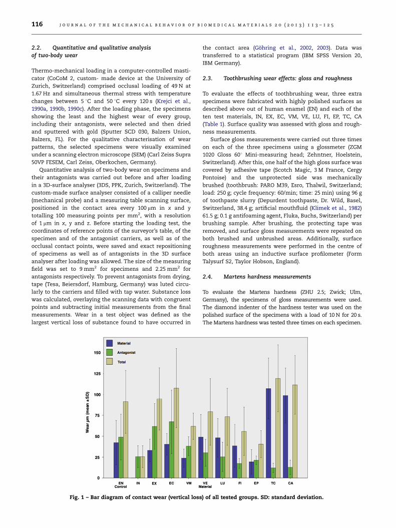
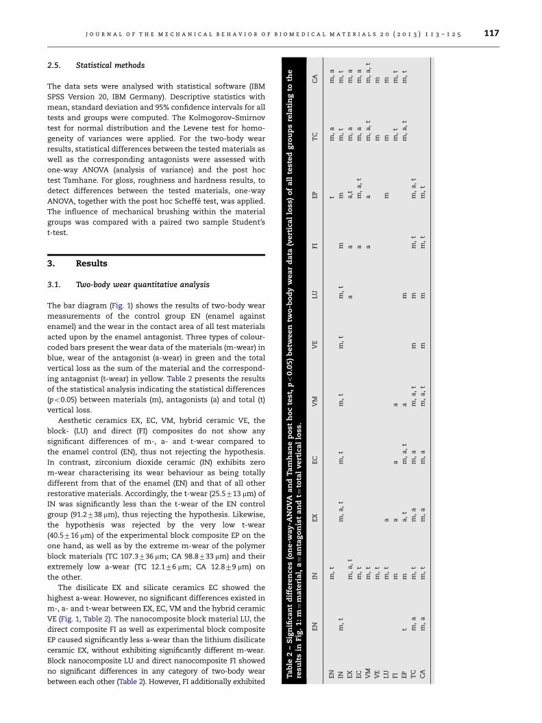
Fig. 1 – Bar diagram of contact wear (vertical loss
the contact area (Gohring et al., 2002, 2003). Data was
transferred to a statistical program (IBM SPSS Version 20,
IBM Germany).
2.3. Toothbrushing wear effects: gloss and roughness
To evaluate the effects of toothbrushing wear, three extra
specimens were fabricated with highly polished surfaces as
described above out of human enamel (EN) and each of the
ten test materials, IN, EX, EC, VM, VE, LU, FI, EP, TC, CA
(Table 1). Surface quality was assessed with gloss and rough-
ness measurements.
Surface gloss measurements were carried out three times
on each of the three specimens using a glossmeter (ZGM
1020 Gloss 601 Mini-measuring head; Zehntner, Hoelstein,
Switzerland). After this, one half of the high gloss surface was
covered by adhesive tape (Scotch Magic, 3 M France, Cergy
Pontoise) and the unprotected side was mechanically
brushed (toothbrush: PARO M39, Esro, Thalwil, Switzerland;
load: 250 g; cycle frequency: 60/min; time: 25 min) using 96 g
of toothpaste slurry (Depurdent toothpaste, Dr. Wild, Basel,
Switzerland, 38.4 g; artificial mouthfluid (Klimek et al., 1982)
61.5 g; 0.1 g antifoaming agent, Fluka, Buchs, Switzerland) per
brushing sample. After brushing, the protecting tape was
removed, and surface gloss measurements were repeated on
both brushed and unbrushed areas. Additionally, surface
roughness measurements were performed in the centre of
both areas using an inductive surface profilometer (Form
Talysurf S2, Taylor Hobson, England).
2.4. Martens hardness measurements
To evaluate the Martens hardness (ZHU 2.5; Zwick; Ulm,
Germany), the specimens of gloss measurements were used.
The diamond indenter of the hardness tester was used on the
polished surface of the specimens with a load of 10 N for 20 s.
The Martens hardness was tested three times on each specimen.
) of all tested groups. SD: standard deviation.
rtic
al
loss
)o
fa
llte
sted
gro
up
sre
lati
ng
toth
e
EP
TC
CA
tm
,a
m,
a
mm
,t
m,
t
a,t
m,
am
,a
m,
a,
tm
,a
m,
a
am
,a
,t
m,
a,
t
mm
mm
m
m,
tm
,t
m,
a,
tm
,t
tm
,a
,t
tm
,t
j o u r n a l o f t h e m e c h a n i c a l b e h a v i o r o f b i o m e d i c a l m a t e r i a l s 2 0 ( 2 0 1 3 ) 1 1 3 – 1 2 5 117
2.5. Statistical methods
The data sets were analysed with statistical software (IBM
SPSS Version 20, IBM Germany). Descriptive statistics with
mean, standard deviation and 95% confidence intervals for all
tests and groups were computed. The Kolmogorov–Smirnov
test for normal distribution and the Levene test for homo-
geneity of variances were applied. For the two-body wear
results, statistical differences between the tested materials as
well as the corresponding antagonists were assessed with
one-way ANOVA (analysis of variance) and the post hoc
test Tamhane. For gloss, roughness and hardness results, to
detect differences between the tested materials, one-way
ANOVA, together with the post hoc Scheffe test, was applied.
The influence of mechanical brushing within the material
groups was compared with a paired two sample Student’s
t-test.
Ta
ble
2–
Sig
nifi
can
td
iffe
ren
ces
(on
e-w
ay-A
NO
VA
an
dT
am
ha
ne
po
sth
oc
test
,po
0.0
5)
betw
een
two
-bo
dy
wea
rd
ata
(ve
resu
lts
inFig
.1
:m¼
mate
ria
l,a¼
an
tag
on
ist
an
dt¼
tota
lvert
ica
llo
ss.
EN
INE
XE
CV
MV
ELU
FI
EN
m,
t
INm
,t
m,
a,
tm
,t
m,
tm
,t
m,
tm
EX
m,
a,
ta
a
EC
m,
ta
VM
m,
ta
VE
m,
t
LU
m,
ta
FI
ma
aa
EP
tm
a,
tm
,a
,t
am
TC
m,
am
,t
m,
am
,a
m,
a,
tm
mm
,
CA
m,
am
,t
m,
am
,a
m,
a,
tm
mm
,
3. Results
3.1. Two-body wear quantitative analysis
The bar diagram (Fig. 1) shows the results of two-body wear
measurements of the control group EN (enamel against
enamel) and the wear in the contact area of all test materials
acted upon by the enamel antagonist. Three types of colour-
coded bars present the wear data of the materials (m-wear) in
blue, wear of the antagonist (a-wear) in green and the total
vertical loss as the sum of the material and the correspond-
ing antagonist (t-wear) in yellow. Table 2 presents the results
of the statistical analysis indicating the statistical differences
(po0.05) between materials (m), antagonists (a) and total (t)
vertical loss.
Aesthetic ceramics EX, EC, VM, hybrid ceramic VE, the
block- (LU) and direct (FI) composites do not show any
significant differences of m-, a- and t-wear compared to
the enamel control (EN), thus not rejecting the hypothesis.
In contrast, zirconium dioxide ceramic (IN) exhibits zero
m-wear characterising its wear behaviour as being totally
different from that of the enamel (EN) and that of all other
restorative materials. Accordingly, the t-wear (25.5713 mm) of
IN was significantly less than the t-wear of the EN control
group (91.2738 mm), thus rejecting the hypothesis. Likewise,
the hypothesis was rejected by the very low t-wear
(40.5716 mm) of the experimental block composite EP on the
one hand, as well as by the extreme m-wear of the polymer
block materials (TC 107.3736 mm; CA 98.8733 mm) and their
extremely low a-wear (TC 12.176 mm; CA 12.879 mm) on
the other.
The disilicate EX and silicate ceramics EC showed the
highest a-wear. However, no significant differences existed in
m-, a- and t-wear between EX, EC, VM and the hybrid ceramic
VE (Fig. 1, Table 2). The nanocomposite block material LU, the
direct composite FI as well as experimental block composite
EP caused significantly less a-wear than the lithium disilicate
ceramic EX, without exhibiting significantly different m-wear.
Block nanocomposite LU and direct nanocomposite FI showed
no significant differences in any category of two-body wear
between each other (Table 2). However, FI additionally exhibited
j o u r n a l o f t h e m e c h a n i c a l b e h a v i o r o f b i o m e d i c a l m a t e r i a l s 2 0 ( 2 0 1 3 ) 1 1 3 – 1 2 5118
significantly lower a-wear compared to both silicate ceramics
EC and VM without showing significantly different m-wear
(Fig. 1, Table 2). The wear characteristics of hybrid ceramic VE
and nanocomposite LU as well as FI appear similar with respect
to the m-, a- and t-wear data (Fig. 1).
The experimental block composite EP showed significantly
less a-wear than the ceramics EX, EC and VM as well as less
m-wear than EC and LU. The temporary acrylic polymers TC
and CA form a group with significantly higher m-wear than
enamel and all ceramic and composite permanent materials
as well.
3.2. Two-body wear qualitative analysis
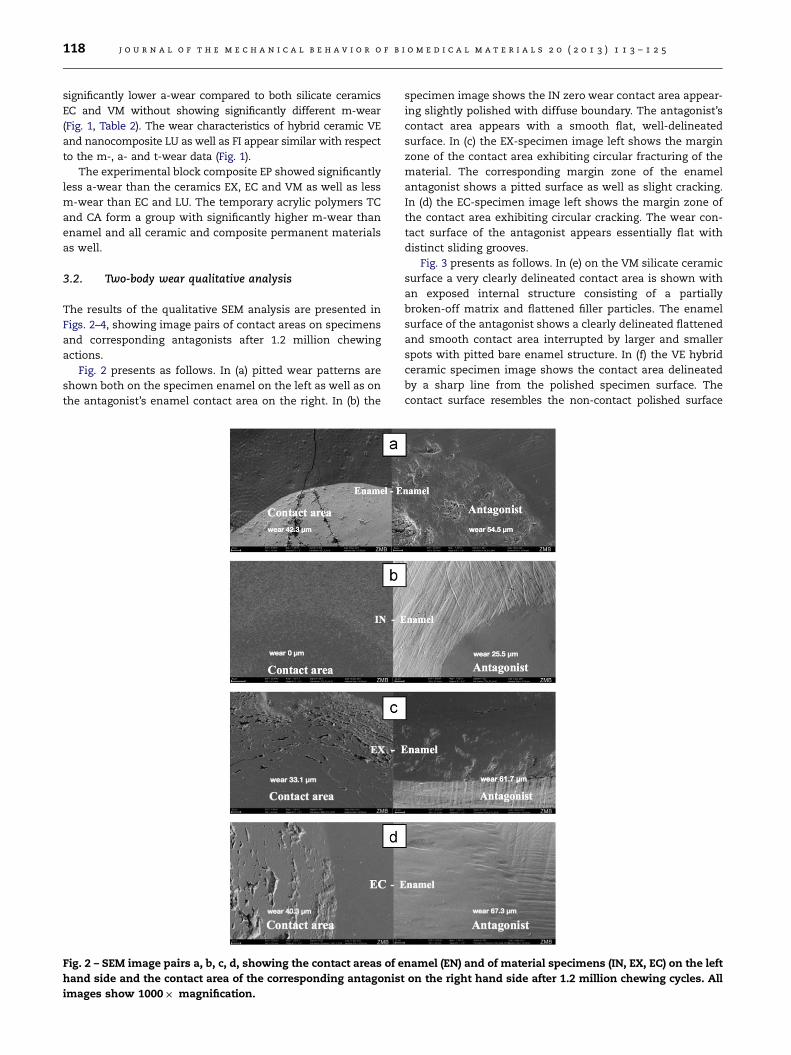
The results of the qualitative SEM analysis are presented in
Figs. 2–4, showing image pairs of contact areas on specimens
and corresponding antagonists after 1.2 million chewing
actions.
Fig. 2 presents as follows. In (a) pitted wear patterns are
shown both on the specimen enamel on the left as well as on
the antagonist’s enamel contact area on the right. In (b) the
Fig. 2 – SEM image pairs a, b, c, d, showing the contact areas of e
hand side and the contact area of the corresponding antagonis
images show 1000� magnification.
specimen image shows the IN zero wear contact area appear-
ing slightly polished with diffuse boundary. The antagonist’s
contact area appears with a smooth flat, well-delineated
surface. In (c) the EX-specimen image left shows the margin
zone of the contact area exhibiting circular fracturing of the
material. The corresponding margin zone of the enamel
antagonist shows a pitted surface as well as slight cracking.
In (d) the EC-specimen image left shows the margin zone of
the contact area exhibiting circular cracking. The wear con-
tact surface of the antagonist appears essentially flat with
distinct sliding grooves.
Fig. 3 presents as follows. In (e) on the VM silicate ceramic
surface a very clearly delineated contact area is shown with
an exposed internal structure consisting of a partially
broken-off matrix and flattened filler particles. The enamel
surface of the antagonist shows a clearly delineated flattened
and smooth contact area interrupted by larger and smaller
spots with pitted bare enamel structure. In (f) the VE hybrid
ceramic specimen image shows the contact area delineated
by a sharp line from the polished specimen surface. The
contact surface resembles the non-contact polished surface
namel (EN) and of material specimens (IN, EX, EC) on the left
t on the right hand side after 1.2 million chewing cycles. All

Fig. 3 – SEM image pairs e, f, g, h, showing the contact areas of material specimens (VM, VE, LU, FI) on the left hand side and
the contact area of the corresponding antagonist on the right hand side after 1.2 million chewing cycles. All images show
1000� magnification.
j o u r n a l o f t h e m e c h a n i c a l b e h a v i o r o f b i o m e d i c a l m a t e r i a l s 2 0 ( 2 0 1 3 ) 1 1 3 – 1 2 5 119
with only slight pitting at the internal side of the boundary line
and in the central contact area. The contact surface of the
enamel antagonist looks smooth with only minor pitting. In (g)
the contact area of the LU CAD/CAM nanocomposite specimen
shows slight pitting inside the demarcation line and fine
microcracks running across the contact area. The corresponding
contact surface on the enamel antagonist appears essentially
smooth. In (h) the contact area of the FI direct nanocomposite
specimen shows the margin zone of the contact area exhibiting
circular cracking as well as some microporosity and pitting. The
wear contact surface of the antagonist appears essentially flat
with distinct sliding grooves.
Fig. 4 presents in (i) the EP experimental CAD/CAM
composite contact area with blurred boundaries. Within the
contact area the structure of the material shows filler parti-
cles of different sizes, only slight pitting, as well as scarce
micropores and a single lost filler particle cavity. The surface
of the enamel antagonist appears smooth. In (k) the contact
area of the TC CAD/CAM unfilled polymer material shows
a circular crater-like formation accompanied by circular
parallel-running crack lines. On the antagonist, practically
no evident wear effect can be seen. In (l) the contact area of
the CA CAD/CAM microfilled polymer material shows circular
step-like fracturing material. On the antagonist the contact
area is slightly visible on the cusp tip at low magnification.
3.3. Gloss
The results of the gloss measurements are presented in
Table 3 as gloss units (GU). Machine polishing created a high
gloss value of 53 GU on enamel (EN) and similar high gloss
values between 57 GU on lithium disilicate (EX) and 50 GU on
unfilled polymer (TC). The gloss value of zirconium dioxide
ceramic (IN) of 128 GU was completely out of the range of that
of the enamel control (EN) and of all other permanent as well
as provisional restorative materials.
Abrasive toothbrushing reduced the gloss value of enamel
(EN) significantly by 53% while the gloss of zirconium dioxide
ceramic (IN) was actually slightly increased. Brushing did not
significantly change the gloss of lithium disilicate (EX) and
caused only slight gloss reductions on silicate ceramics as well.
Brushing caused significant moderate decrease of gloss of
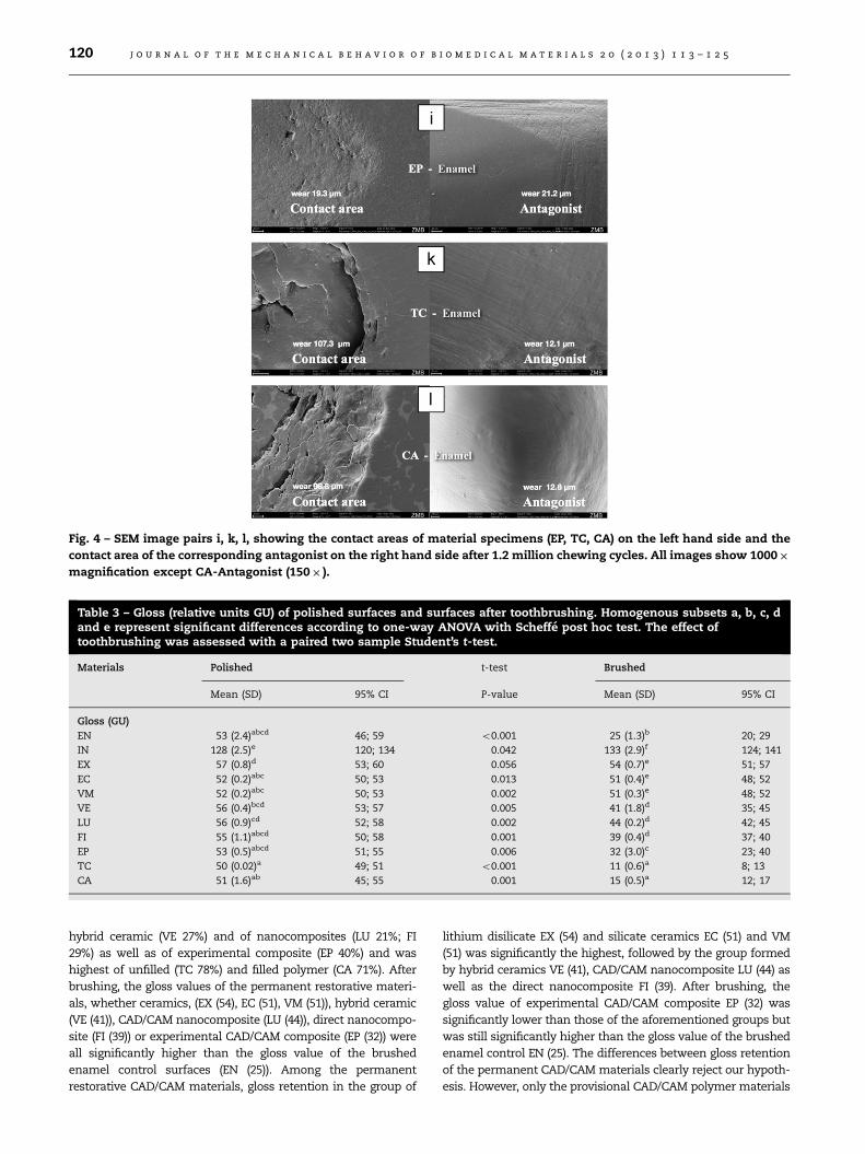
Fig. 4 – SEM image pairs i, k, l, showing the contact areas of material specimens (EP, TC, CA) on the left hand side and the
contact area of the corresponding antagonist on the right hand side after 1.2 million chewing cycles. All images show 1000�
magnification except CA-Antagonist (150� ).
Table 3 – Gloss (relative units GU) of polished surfaces and surfaces after toothbrushing. Homogenous subsets a, b, c, dand e represent significant differences according to one-way ANOVA with Scheffe post hoc test. The effect oftoothbrushing was assessed with a paired two sample Student’s t-test.
Materials Polished t-test Brushed
Mean (SD) 95% CI P-value Mean (SD) 95% CI
Gloss (GU)
EN 53 (2.4)abcd 46; 59 o0.001 25 (1.3)b 20; 29
IN 128 (2.5)e 120; 134 0.042 133 (2.9)f 124; 141
EX 57 (0.8)d 53; 60 0.056 54 (0.7)e 51; 57
EC 52 (0.2)abc 50; 53 0.013 51 (0.4)e 48; 52
VM 52 (0.2)abc 50; 53 0.002 51 (0.3)e 48; 52
VE 56 (0.4)bcd 53; 57 0.005 41 (1.8)d 35; 45
LU 56 (0.9)cd 52; 58 0.002 44 (0.2)d 42; 45
FI 55 (1.1)abcd 50; 58 0.001 39 (0.4)d 37; 40
EP 53 (0.5)abcd 51; 55 0.006 32 (3.0)c 23; 40
TC 50 (0.02)a 49; 51 o0.001 11 (0.6)a 8; 13
CA 51 (1.6)ab 45; 55 0.001 15 (0.5)a 12; 17
j o u r n a l o f t h e m e c h a n i c a l b e h a v i o r o f b i o m e d i c a l m a t e r i a l s 2 0 ( 2 0 1 3 ) 1 1 3 – 1 2 5120
hybrid ceramic (VE 27%) and of nanocomposites (LU 21%; FI
29%) as well as of experimental composite (EP 40%) and was
highest of unfilled (TC 78%) and filled polymer (CA 71%). After
brushing, the gloss values of the permanent restorative materi-
als, whether ceramics, (EX (54), EC (51), VM (51)), hybrid ceramic
(VE (41)), CAD/CAM nanocomposite (LU (44)), direct nanocompo-
site (FI (39)) or experimental CAD/CAM composite (EP (32)) were
all significantly higher than the gloss value of the brushed
enamel control surfaces (EN (25)). Among the permanent
restorative CAD/CAM materials, gloss retention in the group of
lithium disilicate EX (54) and silicate ceramics EC (51) and VM
(51) was significantly the highest, followed by the group formed
by hybrid ceramics VE (41), CAD/CAM nanocomposite LU (44) as
well as the direct nanocomposite FI (39). After brushing, the
gloss value of experimental CAD/CAM composite EP (32) was
significantly lower than those of the aforementioned groups but
was still significantly higher than the gloss value of the brushed
enamel control EN (25). The differences between gloss retention
of the permanent CAD/CAM materials clearly reject our hypoth-
esis. However, only the provisional CAD/CAM polymer materials
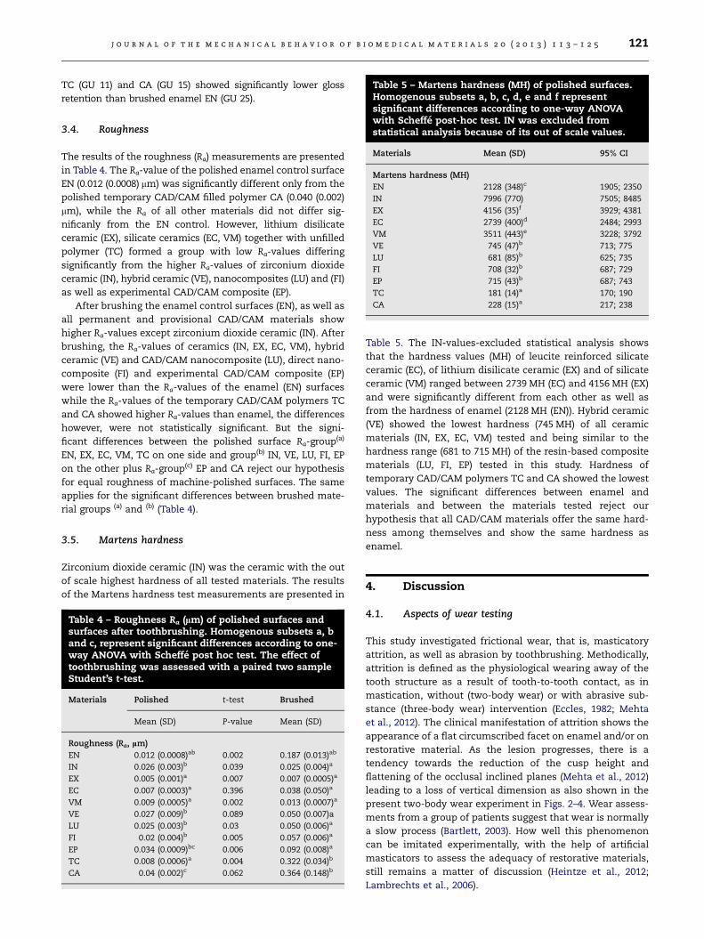
Table 5 – Martens hardness (MH) of polished surfaces.Homogenous subsets a, b, c, d, e and f representsignificant differences according to one-way ANOVAwith Scheffe post-hoc test. IN was excluded fromstatistical analysis because of its out of scale values.
Materials Mean (SD) 95% CI
Martens hardness (MH)
EN 2128 (348)c 1905; 2350
IN 7996 (770) 7505; 8485
EX 4156 (35)f 3929; 4381
EC 2739 (400)d 2484; 2993
VM 3511 (443)e 3228; 3792
VE 745 (47)b 713; 775
LU 681 (85)b 625; 735
FI 708 (32)b 687; 729
EP 715 (43)b 687; 743
TC 181 (14)a 170; 190
CA 228 (15)a 217; 238
j o u r n a l o f t h e m e c h a n i c a l b e h a v i o r o f b i o m e d i c a l m a t e r i a l s 2 0 ( 2 0 1 3 ) 1 1 3 – 1 2 5 121
TC (GU 11) and CA (GU 15) showed significantly lower gloss
retention than brushed enamel EN (GU 25).
3.4. Roughness
The results of the roughness (Ra) measurements are presented
in Table 4. The Ra-value of the polished enamel control surface
EN (0.012 (0.0008) mm) was significantly different only from the
polished temporary CAD/CAM filled polymer CA (0.040 (0.002)
mm), while the Ra of all other materials did not differ sig-
nificanly from the EN control. However, lithium disilicate
ceramic (EX), silicate ceramics (EC, VM) together with unfilled
polymer (TC) formed a group with low Ra-values differing
significantly from the higher Ra-values of zirconium dioxide
ceramic (IN), hybrid ceramic (VE), nanocomposites (LU) and (FI)
as well as experimental CAD/CAM composite (EP).
After brushing the enamel control surfaces (EN), as well as
all permanent and provisional CAD/CAM materials show
higher Ra-values except zirconium dioxide ceramic (IN). After
brushing, the Ra-values of ceramics (IN, EX, EC, VM), hybrid
ceramic (VE) and CAD/CAM nanocomposite (LU), direct nano-
composite (FI) and experimental CAD/CAM composite (EP)
were lower than the Ra-values of the enamel (EN) surfaces
while the Ra-values of the temporary CAD/CAM polymers TC
and CA showed higher Ra-values than enamel, the differences
however, were not statistically significant. But the signi-
ficant differences between the polished surface Ra-group(a)
EN, EX, EC, VM, TC on one side and group(b) IN, VE, LU, FI, EP
on the other plus Ra-group(c) EP and CA reject our hypothesis
for equal roughness of machine-polished surfaces. The same
applies for the significant differences between brushed mate-
rial groups (a) and (b) (Table 4).
3.5. Martens hardness
Zirconium dioxide ceramic (IN) was the ceramic with the out
of scale highest hardness of all tested materials. The results
of the Martens hardness test measurements are presented in
Table 4 – Roughness Ra (lm) of polished surfaces andsurfaces after toothbrushing. Homogenous subsets a, band c, represent significant differences according to one-way ANOVA with Scheffe post hoc test. The effect oftoothbrushing was assessed with a paired two sampleStudent’s t-test.
Materials Polished t-test Brushed
Mean (SD) P-value Mean (SD)
Roughness (Ra, lm)
EN 0.012 (0.0008)ab 0.002 0.187 (0.013)ab
IN 0.026 (0.003)b 0.039 0.025 (0.004)a
EX 0.005 (0.001)a 0.007 0.007 (0.0005)a
EC 0.007 (0.0003)a 0.396 0.038 (0.050)a
VM 0.009 (0.0005)a 0.002 0.013 (0.0007)a
VE 0.027 (0.009)b 0.089 0.050 (0.007)a
LU 0.025 (0.003)b 0.03 0.050 (0.006)a
FI 0.02 (0.004)b 0.005 0.057 (0.006)a
EP 0.034 (0.0009)bc 0.006 0.092 (0.008)a
TC 0.008 (0.0006)a 0.004 0.322 (0.034)b
CA 0.04 (0.002)c 0.062 0.364 (0.148)b
Table 5. The IN-values-excluded statistical analysis shows
that the hardness values (MH) of leucite reinforced silicate
ceramic (EC), of lithium disilicate ceramic (EX) and of silicate
ceramic (VM) ranged between 2739 MH (EC) and 4156 MH (EX)
and were significantly different from each other as well as
from the hardness of enamel (2128 MH (EN)). Hybrid ceramic
(VE) showed the lowest hardness (745 MH) of all ceramic
materials (IN, EX, EC, VM) tested and being similar to the
hardness range (681 to 715 MH) of the resin-based composite
materials (LU, FI, EP) tested in this study. Hardness of
temporary CAD/CAM polymers TC and CA showed the lowest
values. The significant differences between enamel and
materials and between the materials tested reject our
hypothesis that all CAD/CAM materials offer the same hard-
ness among themselves and show the same hardness as
enamel.
4. Discussion
4.1. Aspects of wear testing
This study investigated frictional wear, that is, masticatory
attrition, as well as abrasion by toothbrushing. Methodically,
attrition is defined as the physiological wearing away of the
tooth structure as a result of tooth-to-tooth contact, as in
mastication, without (two-body wear) or with abrasive sub-
stance (three-body wear) intervention (Eccles, 1982; Mehta
et al., 2012). The clinical manifestation of attrition shows the
appearance of a flat circumscribed facet on enamel and/or on
restorative material. As the lesion progresses, there is a
tendency towards the reduction of the cusp height and
flattening of the occlusal inclined planes (Mehta et al., 2012)
leading to a loss of vertical dimension as also shown in the
present two-body wear experiment in Figs. 2–4. Wear assess-
ments from a group of patients suggest that wear is normally
a slow process (Bartlett, 2003). How well this phenomenon
can be imitated experimentally, with the help of artificial
masticators to assess the adequacy of restorative materials,
still remains a matter of discussion (Heintze et al., 2012;
Lambrechts et al., 2006).
j o u r n a l o f t h e m e c h a n i c a l b e h a v i o r o f b i o m e d i c a l m a t e r i a l s 2 0 ( 2 0 1 3 ) 1 1 3 – 1 2 5122
Lambrechts et al. (2006) characterised the Zurich computer-
controlled masticator as a three-body artificial wear machine.
However, in the present study, the Zurich wear system did not
include toothbrushing with abrasive slurry in addition to
mastication but the effects of toothbrushing were assessed
separately on additional specimens. Consequently, masticatory
action implied two bodies only, the polished material specimen
and the non-standardised natural enamel as the antagonistic
stylus (Krejci et al., 1999). Wear measurements and SEM
evaluation were restricted to the occlusal contact area (Krejci
et al., 1999). This experimental arrangement is in accordance
with two-body wear as defined by the ISO/TS wear norm
14569-2 ISO/TS (2001). As control for the material specimens,
again human enamel served as the reference for permanent
and for temporary restorative materials. If we accept the
estimate of normal vertical loss of enamel from physiological
wear to be approximately 20–38 mm per annum (Lambrechts
et al., 1989), the mean total wear of 96.8 mm found for enamel
in the present study after 1.2 million chewing impacts, would
meet the lower end of the estimate of Lambrechts et al. (1989)
with 19.4 mm total enamel wear per annum, given that the
equivalence of our laboratory test to a 5-year clinical service
period is valid (Krejci and Lutz, 1990) or at least weakly related
(Heintze et al., 2012). Anyway, it establishes the reference for
the wear rates of the restorative materials and should allow
comparative evaluation and ranking, particularly of the new
and experimental CAD/CAM materials such as IN, VE, LU and
EP, under the conditions of the present study.
4.2. Two-body wear of zirconium dioxide ceramics
The zero m-wear for monolithic zirconium dioxide ceramics
(IN) and minimal a-wear (25.5 mm) of its enamel antagonists
both impressed (Fig. 1; Table 2), even though these properties
have already been reported for zirconium dioxide ceramics by
recent laboratory studies (Jung et al., 2010; Preis et al., 2012;
Rosentritt et al., 2012; Stawarczyk et al., 2013). The contact
area of IN looked slightly polished at the end of the test,
while, other than the rougher contact areas of the control
facets of a-enamel against m-enamel (Fig. 2a), its enamel
antagonist showed a still denser, very smooth and flat sur-
face on the wear facet (Fig. 2b). If monolithic translucent
zirconium dioxide ceramic is considered to be used for CAD/
CAM generated non-veneered crowns in patients with nat-
ural teeth as antagonists, it should be kept in mind that our
in vitro mastication test started with a highly machine-
polished specimen of IN with very low roughness (Ra¼0.026 mm;
Table 4), exhibiting a regular dense fine particle structure and the
highest hardness of all materials tested in the present study
(Table 5); whereas the enamel controls started the test with
natural unpolished m-enamel surfaces. Machine polishing
results in a significantly higher surface gloss than manual
polishing with tools for intraoral polishing (Heintze et al.,
2006). Abrasive treatment of zirconium dioxide ceramics raises
structural aspects but polishing may enhance the strength of
zirconium dioxide ceramics (Vagkopoulou et al., 2009). Air ab-
raded zirconium dioxide ceramic specimens polished extraorally
by a dental technician with a goat hairbrush and diamond paste
yielded similar low two-body wear of enamel antagonists as in
the present study (Stawarczyk et al., 2013).
In the clinical situation however, after cementation, con-
tact areas mostly need to be adjusted manually by corrective
grinding with rotating diamond-coated instruments creating
rough surfaces. Whether rough zirconium dioxide ceramic
surfaces can be manually polished in the mouth to a degree
which does not forward excessive wear of the antagonist, will
have to be proved, before monolithic zirconium dioxide
ceramic crowns can be considered as single tooth restora-
tions opposing natural teeth. One case report of upper and
lower full-arch fixed detachable implant-retained restora-
tions, manufactured from monolithic zirconium dioxide
ceramic, at least shows that no fractures and no wear
occurred on the full zirconium dioxide ceramic teeth func-
tioning against each other after two years of clinical service
(Rojas-Vizcaya, 2011), despite concerns regarding the struc-
tural stability of zirconium dioxide ceramics when exposed to
the oral environment (Koutayas et al., 2009).
4.3. Two-body wear of silicate and hybrid ceramics
In the present study, wear of material specimens (m-wear)
tended to increase the lower the hardness of the material. On
the other hand, antagonists showed lower enamel wear the
lower the hardness of the material specimens, except zirco-
nium dioxide ceramic IN. After zirconium dioxide ceramic IN,
lithium disilicate EX was the hardest and the acrylic poly-
mers TC and CA were the softest materials (Fig. 1; Tables 2
and 5). Glass ceramics EX and EC showed circular cracking
patterns parallel to the inside fringe of the facet, VM exposed
its filler structure whereat the fillers were flattened at the
facet’s surface (Fig. 3a). The facets of the enamel antagonists
show typical sliding patterns or exposure of local enamel
structures (Figs. 2c, d; 3a). As opposed to findings in other
laboratory studies (Jung et al., 2010; Preis et al., 2012;
Rosentritt et al., 2012) the wear of enamel antagonists by
feldspathic ceramic (VM) and glass ceramic (EC) was not
significantly different from that of zirconium dioxide cera-
mics (Table 2).
The hybrid ceramic VE (Bojemuller and Coldea, 2012;
Giordano, 1996, 2000, 2005; He and Swain, 2011) tends to
result in lower a-wear than other ceramics except zirconium
dioxide ceramic, which is still lower, and VE shows a-wear
like composites and acrylic polymers in the present study.
The m-wear of VE itself is similar to that of the other
ceramics, except zirconium dioxide ceramic, and also similar
to that of composites (Fig. 1; Table 2). Thus, wear perfor-
mance of VE combines the characteristics of ceramic and
composites and by trend appears similar to that of the CAD/
CAM block nanocomposite LU, while at the same time it is
not significantly different from that of enamel (Fig. 1; Table 2).
Its hardness is positioned significantly below that of enamel
and stays at the low end of the hardness of ceramics while
being not significantly higher than that of composites
(Table 5). In the SEM at 1 Kx the fringe of the contact area
shows a sharp line and the contact surface exhibits only
minimal pitting. This reaction of the hybrid structure to the
repetitive impact of the antagonist may be influenced by its
modulus of elasticity of 30 GPa (Bojemuller and Coldea, 2012),
which positions VE between resin-based composites (�15
GPa; 3M ESPE, 2011 and feldspathic ceramic (VM:�60 GPa;
j o u r n a l o f t h e m e c h a n i c a l b e h a v i o r o f b i o m e d i c a l m a t e r i a l s 2 0 ( 2 0 1 3 ) 1 1 3 – 1 2 5 123
Bojemuller and Coldea, 2012; Datzmann, 1996) as well as
between dentin (5–17 GPa) and enamel (60 GPa; Menig et al.,
2000). The modulus of elasticity of 30 GPa of VE opens up a
section of elastic properties for restorative materials, which
has so far not been accessible. Concerning thermo-cycling in
the present investigation up to 50 1C, the coefficient of
thermal expansion of hybrid ceramic (11.9–12.4�10�6 K�1)
stays on the side of ceramics (8.8�10�6 K�1, Datzmann, 1996)
and of the natural tooth (enamel 10�10�6 K�1; dentin
11.4�10�6 K�1; Toparli et al., 2000).
4.4. Two-body wear of resin-based and acrylic polymermaterials
The CAD/CAM block nanocomposite LU (3M ESPE, 2011) and
the direct light curing composite FI (Ferracane, 2011; Kramer
et al., 2009) do not differ significantly in any aspect from the
two-body wear in the present study (Fig. 1; Table 2). This may
confirm that the physical properties of resin-based composite
as a material for the fabrication of dental restorations,
particularly full posterior crowns, are not improved structu-
rally by block-fabrication for CAD/CAM use as with ceramics
(Tinschert et al., 2000) at least with respect to the degree
of conversion of direct and block composite (Kelly, 2011;
Vanoorbeek et al., 2010). However, close inspection of the
contact surfaces at 1K� magnification shows some micro-
pores and flaws as well as circular microcracking in the direct
composite material FI, which are not visible in the CAD/CAM
block composite LU to the same extent, apart from singular
very fine microcracks (Fig. 3c and d). The slight difference of
gloss between LU (44 GU) and FI (39 GU) may hint at a slightly
less dense surface quality of the direct composite compared
to the CAD/CAM block material. While the wear behaviour of
both nanocomposites poses no problem, as shown in the
present study (Fig. 3c and d), possibly the low modulus of the
elasticity of composites (10–15 GPa, 3M ESPE, 2011) under load
may contribute to the loosening of composite crowns after
some clinical service time, while the higher E-moduli of
ceramics do not (Kelly, 2011; Vanoorbeek et al., 2010). How-
ever, for overlay restorations, the elastic properties of a CAD/
CAM composite proved to be beneficial compared to the
performance of ceramics (Magne and Knezevic, 2009). Resin-
based composite CAD/CAM inlays performed as well as
porcelain CAD/CAM inlays after three years of clinical service
(Fasbinder et al., 2005).
The experimental CAD/CAM block composite EP
(Lendenmann and Wanner, 2011) shows promising wear
performance (Fig. 1; Table 2), whereas the acrylic polymer
materials TC (Wanner, 2010) and CA (Baltzer and Kaufmann-
Jinoian, 2007) exhibit a significantly higher m-wear than all
permanent restorative materials in the present study, con-
firming their temporary character.
4.5. Toothbrushing wear: roughness and gloss retention
Zirconium dioxide ceramic machine-polished specimen sur-
faces, as prepared for toothbrushing wear testing, yielded the
high gloss value of 128 GU, which we attribute to its high
refractive index and high whiteness (Vagkopoulou et al., 2009)
both increasing the remission of light (Table 3). Abrasive
toothbrushing even slightly increased the gloss of zirconium
dioxide ceramics to 133 GU, its roughness staying the same,
no doubt related to the very high hardness of zirconium
dioxide ceramics (Table 5). The gloss of polished enamel
(53 GU) as well as that of all other polished material speci-
mens lay between 50 and 57 GU, significantly lower than that
of zirconium dioxide ceramic (IN) while the roughness (Ra)
values of polished enamel and the polished specimens of all
other materials were not significantly different from that of
zirconium dioxide ceramic (IN) with the exception of the
acrylic temporary material CA showing significantly less
roughness.
The experimental toothbrushing wear in the present study
significantly reduced the gloss of enamel and of all material
specimens, except zirconium dioxide ceramic, by the high
abrasivity of the toothpaste slurry used and formed material
groups of similar gloss retention. The ceramics EX, EC and
VM formed a top group of high gloss retention between 51
and 54 GU with Martens hardness between 2739 MH and
4156 MH including enamel with 3228 MH (Table 5). Another
group of good gloss retention and lower hardness was formed
by the hybrid ceramic VE (41 GU) together with block nano-
composite LU (44 GU) and direct nanocomposite FI (39 GU).
The temporary acrylic polymer materials TC and CA with
initial excellent polishability and high gloss of 50 to 51 GU
proved to be very susceptible to abrasive toothbrushing,
reducing their gloss to 11 GU and 15 GU with corresponding
surface roughness of 0.32 to 0.36 mm. Roughness of this
magnitude is way above the critical threshold for microbial
plaque accumulation (Teughels et al., 2006) making regular
hygiene controls indispensable during the clinical service
time of temporary restorations.
5. Conclusions
Within the limitations of this in vitro study, it can be
concluded that: all permanent aesthetic CAD/CAM block
materials tested, whether ceramics, hybrid ceramics or
resin-based nanocomposites and even direct nanocompo-
sites, behave similarly or better with respect to two-body
and toothbrushing wear than natural enamel, which is not
true for temporary acrylic polymer CAD/CAM block materials.
Ceramics show the best gloss retention compared to hybrid
ceramics, composites and acrylic polymers.
Acknowledgements
The authors would like to thank the manufacturers of
restorative and temporary materials for providing the CAD/
CAM blocks. We are grateful to our senior technician Mr. Felix
Schmutz for operating the Zurich computer-controlled mas-
ticator. The study was supported in part by the Foundation
for the Advancement of Computer-Assisted Dentistry, Zurich,
Switzerland.

j o u r n a l o f t h e m e c h a n i c a l b e h a v i o r o f b i o m e d i c a l m a t e r i a l s 2 0 ( 2 0 1 3 ) 1 1 3 – 1 2 5124
r e f e r e n c e s
Baltzer, A., Kaufmann-Jinoian, V., 2007M. VITA CAD-Temp forinLab and CEREC 3D. International Journal of ComputerizedDentistry 10 (1), 99–103.
Bartlett, D.W., 2003. Retrospective long term monitoring of toothwear using study models. British Dental Journal 194, 211–213.
Bindl, A., Luthy, H., Mormann, W.H., 2003. Fracture load of CAD/CAM-generated slot inlay FPDs. International Journal ofProsthodontics 16, 653–660.
Bojemuller, E., Coldea, A., 2012. In: VITA ENAMIC Technical-ScientificDocumentation. VITA Zahnfabrik, Bad Sackingen, Germany.
Chen, H.Y., Hickel, R., Setcos, J.C., Kunzelmann, K.H., 1999. Effectsof surface finish and fatigue testing of CAD–CAM and pressed-ceramic crowns. Journal of Prosthetic Dentistry 82, 468–475.
Da Costa, J., Adams-Belusko, A., Riley, K., Ferracane, J.L., 2010. Theeffect of various dentifrices on surface roughness and gloss ofresin composites. Journal of Dentistry 38 (Suppl. 2), e123–e128(Epub 2010 Mar 1).
Datzmann, G., 1996. CEREC Vitablocs Mark II machinable ceramic.In: Mormann, W.H. (Es.), CAD/CIM in Aesthetic Dentistry;CEREC 10 Year Anniversary Symposium, Proceedings, Chicago,Quintessence, pp. 205–215.
Eccles, J., 1982. Tooth surface loss from abrasion, attrition anderosion. Dental Update 9, 373–381.
Esquivel-Upshaw, J.F., et al., 2012. Three years in vivo wear: core-ceramic, veneers, and enamel antagonists. Dental Materials28, 615–621.
Fasbinder, D.J., Dennison, J.B., Heys, D.R., Lampe, K., 2005. Theclinical performance of CAD/CAM generated composite inlays.Journal of the American Dental Association 136 (12), 1714–1723.
Ferracane, J.L., 2011. Resin composite—state of the art. DentalMaterials 27, 29–38.
Fischer, K., Buhler-Zemp, P., Volkel, T., 2011a. ScientificDocumentation IPS e.max CAD. Ivoclar Vivadent, Schaan.
Fischer, J., Roeske, S., Stawarczyk, B., Hammerle, C.H.F., 2011b.Investigations in the correlation between Martens hardnessand flexural strength of composite resin restorative materials.Dental Materials Journal 29 (2), 188–192.
Giordano, R.A., 1996. Infused Ceramic Network for FabricatingOdontoforms and Dental Restorations. European PatentApplication no. 95114658.8, 20.03.1996, EP 0 701 808 A2.
Giordano, R.A., 2000. Method for Fabricating Ceramic NetworkMaterial. United States Patent no. 6,159,417, Dec. 12, 2000.
Giordano, R.A., 2005. Method for Fabricating Endodontic andDirect Restorations having Infused Ceramic Network. UnitedStates Patent, US 6,881,488 B2, Apr. 19, 2005.
Gohring, T.N., Besek, M.J., Schmidlin, P.R., 2002. Attritional wearand abrasive surface alterations of composite resin materialsin vitro. Journal of Dentistry 30 (2/3), 119–127.
Gohring, T.N., Schonenberger, K.A., Lutz, F., 2003. Potential ofrestorative systems with simplified adhesives: quantitativeanalysis of wear and marginal adaptation in vitro. AmericanJournal of Dentistry 16 (4), 275–282.
He, Li-Hong, Swain, M., 2011. A novel polymer infiltrated ceramicdental material. Dental Materials 27 (6), 527–534.
Heintze, S.D., Faouzi, M., Rousson, V., Ozcan, M., 2012. Correlation ofwear in vivo and six laboratory wear methods. Dental Materials28, 961–973.
Heintze, S.D., Forjanic, M., Rousson, V., 2006. Surface roughnessand gloss of dental materials as a function of force andpolishing time in vitro. Dental Materials 22, 146–165.
ISO/TS, 14569-2, 2001. Dental Materials—Guidance on Testing ofWear—Part 2: Wear by Two/or Three Body Contact.
Jones, C.S., Billington, R.W., Pearson, G.J., 2004. The in vivoperception of roughness of restorations. British DentalJournal 196, 42–45.
Jung, Y.S., Lee, J.W., Choi, Y.J., Ahn, J.S., Shin, S.W., Huh, J.B., 2010.A study on the in-vitro wear of the natural tooth structure byopposing zirconia or dental porcelain. Journal of AdvancedProsthodontics 2, 111–115.
Kelly, J.R., 2011. Article analysis and evaluation: Computer-AidedDesigned/Computer-Assisted Manufactured (CAD/CAM) all-ceramic crowns appear to perform better than all-compositeresin crowns following the first 3 years of placement. Journalof Evidence-Based Dental Practice 11, 203–205.
Klimek, K., Hellwig, E., Ahrens, G., 1982. Fluoride taken up byplaque, by the underlying enamel and by clean enamel fromthree fluoride compounds in vitro. Caries Research 16, 156–161.
Koutayas, S.O., Vagkopoulou, T., Pelekanos, S., Koidis, P., Strub,J.R., 2009. Zirconia in dentistry; part 2. Evidence-based clinicalbreakthrough. European Journal of Esthetic Dentistry 4 (4),348–380.
Kramer, N., Reinelt, C., Richter, G., Petschelt, A., Frankenberger,R., 2009. Nanohybrid vs. fine particle composite in class IIcavities: clinical results and margin analysis after four years.Dental Materials 25, 750–759.
Krejci, I., Albert, P., Lutz, F., 1999. The influence of antagoniststandardization on wear. Journal of Dental Research 78 (2),713–719.
Krejci, I., Albertoni, M., Lutz, F., 1990a. An in-vitro test procedurefor evaluating dental restoration systems. 2. Toothbrush/toothpaste abrasion and chemical degradation. SchweizerMonatsschrift fur Zahnmedizin 100 (10), 1164–1168.
Krejci, I., Heinzmann, J.L., Lutz, F., 1990b. The wear on enamel,amalgam and their enamel antagonists in a computer-controlled mastication simulator. Schweizer Monatsschrift furZahnmedizin 100 (11), 1285–1291.
Krejci, I., Lutz, F., 1990. In-vitro test results of the evaluation ofdental restoration systems. Correlation with in-vivo results.Schweizer Monatsschrift fur Zahnmedizin 100 (12),1445–1449.
Krejci, I., Lutz., F., Zedler, C., 1992. Effect of contact area size onenamel and composite wear. Journal of Dental Research 71 (7),1413–1416.
Krejci, I., Reich, T., Lutz, F., Albertoni, M., 1990c. An in vitro testprocedure for evaluating dental restoration systems. 1.A computer-controlled mastication simulator. SchweizerMonatsschrift fur Zahnmedizin 100 (8), 953–960.
Kunzelmann, et al., 2007. All-Ceramics at a Glance. Introductionto Indications, Material Selection, Preparation and Insertion ofAll-Ceramic Restorations. Society for Dental Ceramics,Ettlingen (pp. 73–83).
Kurbad, A., Reichel, K., 2005. CAD/CAM-manufactured restorationsmade of lithium disilicate glass ceramics. International Journalof Computerized Dentistry 8, 337–348.
Lambrechts, P., Braeme, M., Vuylsteke-Wauters, M., Vanherle, G.,1989. Quantitative in vivo wear of human enamel. Journal ofDental Research 68, 1752–1754.
Lambrechts, P., Debels, E., Van Landuyt, K., Peumans, M., VanMeerbeek, B., 2006. How to simulate wear? Overview ofexisting methods. Dental Materials 22, 693–701.
Lee, J.H., Shin, S.J., Seo, D.G., Park, J.W., Lee, J.Y., 2010. Glossretention of nanocomposite resins after toothbrushing. DentalMaterials 26 (2), e132, http://dx.doi.org/10.1016/j.dental.2009.11.094.
Lendenmann, U., Wanner, M., 2011. Tetric EvoCeram ScientificDocumentation. Ivoclar Vivadent, Schaan.
Lutz, F., Krejci, I., 2000. Amalgam substitutes: a critical analysis.Journal of Esthetic Dentistry 12 (3), 146–159.
Lutz, F., Krejci, I., Barbakow, F., 1992. Chewing pressure vs. wear ofcomposites and opposing enamel cusps. Journal of DentalResearch 71 (8), 1525–1529.
Magne, P., Knezevic, A., 2009. Simulated fatigue resistance ofcomposite resin versus porcelain CAD/CAM overlay
j o u r n a l o f t h e m e c h a n i c a l b e h a v i o r o f b i o m e d i c a l m a t e r i a l s 2 0 ( 2 0 1 3 ) 1 1 3 – 1 2 5 125
restorations on endodontically treated molars. QuintessenceInternational 40 (2), 125–133.
Mehta, S.B., Banerji, S., Millar, B.J., Suarez-Feito, J.M., 2012.Current concepts on the management of tooth wear: part 1.Assessment, treatment planning and strategies for theprevention and the passive management of tooth wear.British Dental Journal 212 (1), 17–27.
Menig, R., Meyers, M.H., Meyers, M.A., Vecchio, K.S., 2000. Quasi-static and dynamic mechanical response of Hallotis rufescens(abalone) shells. Acta Materialia 48, 2383–2398.
Mormann, W.H., 2004. The origin of the CEREC method: apersonal review of the first 5 years. International Journal ofComputerized Dentistry 7, 11–24.
Preis, V., Behr, M., Handel, G., Schneider-Feyrer, S., Hahnel, S.,Rosentritt, M., 2012. Wear performance of dental ceramicsafter grinding and polishing treatments. Journal of theMechanical Behavior of Biomedical Materials 10, 13–22.
Ramfjord, S.P., Ash, M.M., 1983. Occlusion, 3rd ed. Saunders,Philadelphia.
Rojas-Vizcaya, F., 2011. Full zirconia fixed detachable implant-retained restorations manufactured from monolithic zirconia:clinical report after two years in service. Journal ofProsthodontics 20, 570–576.
Rosentritt, M., Preis, V., Behr, M., Hahnel, S., Handel, G., Kolbeck,C., 2012. Two-body wear of dental porcelain and sub-structure oxide ceramics. Clinical Oral Investigations 16 (3),935–943.
Shahdad, S.A., McCabe, J.F., Bull, S., Rusby, S., Wassell, R.W., 2007.Hardness measured with traditional Vickers and Martenshardness methods. Dental Materials 23 (9), 1079–1085.
Sirona, 2011. InCoris TZI, Translucent Zirconia Ceramic Blocks forinLab. Processing Instructions: Restoration Production forCrowns and Bridges. Bensheim, Sirona Dental Systems, Orderno. 6359801, D3487.
Stawarczyk, B., Egli, R., Roos, M., Ozcan, M., Hammerle, C.H.F.,2011. The impact of in vitro aging on the mechanical andoptical properties of indirect veneering composite resins.Journal of Prosthetic Dentistry 106 (6), 386–398.
Stawarczyk, B., Ozcan, M., Schmutz, F., Trottmann, A., Roos, M.,Hammerle, C.H.F., 2013. Two-body wear of monolithic,
veneered and glazed zirconia and their corresponding enamelantagonists. Acta Odontologica Scandinavica, 71, 102–112.
Suputtamongkol, K., Anusavice, K.J., Suchatlampong, C.,Sithiamnuai, P., Tulapornchai, C., 2008. Clinical performanceand wear characteristics of veneered lithia-disilicate-basedceramic crowns. Dental Materials 24, 667–673.
Teughels, W., Van Assche, N., Sliepen, I., Quirynen, M., 2006.Effect of material characteristics and or surface topography onbiofilm development. Clinical Oral Implants Research 17(Suppl. 2), 68–81.
Tinschert, J., Zwez, D., Marx, R., Anusavice, K.J., 2000. Structuralreliability of alumina-, feldspar-, leucite-, mica- and zirconia-based ceramics. Journal of Dentistry 28 (7), 529–535.
Toparli, M., Gokay, N., Aksoy, T., 2000. An investigation of tem-perature and stress distribution a restored maxillary secondpremolar tooth using a three-dimensional finite elementmethod. Journal of Oral Rehabilitation 27, 1077–1081.
Vagkopoulou, T., Koutayas, S.O., Koidis, P., Strub, J.R., 2009.Zirconia in dentistry; part 1. Discovering the nature of anupcoming bioceramic. European Journal of Esthetic Dentistry4 (2), 130–151.
Vanoorbeek, S., Vandamme, K., Lijnen, I., Naert, I., 2010.Computer-aided designed/computer-assisted manufacturedcomposite resin versus ceramic single-tooth restorations: a3-year clinical study. International Journal of Prosthodontics23 (3), 223–230.
Vollbrecht, J., 2007. Sirona CEREC blocs and inCoris. InternationalJournal of Computerized Dentistry 10, 127–134.
Wanner, M., 2010. In: Scientific Documentation Telio CAD. IvoclarVivadent, Schaan, Liechtenstein.
Wiedhahn, K., 2007. From blue to white: new high-strengthmaterial for CEREC—IPS e. max CAD LT. InternationalJournal of Computerized Dentistry 10, 79–91.
Wiegand, A., Schwerzmann, M., Sener, B., Magalhaes, A.C., Roos,M., Ziebolz, D., Imfeld, T., Attin, T., 2008. Impact of toothpasteslurry abrasivity and toothbrush filament stiffness onabrasion of eroded enamel—an in vitro study. ActaOdontologica Scandinavica 66 (4), 231–235.
3M ESPE, 2011. Lava Ultimate CAD/CAM Restorative TechnicalProduct Profile. St. Paul, 3M Espe Dental Products.