wbv15 6/27/03 11:09 pm page 229 replication patterns of … · replication patterns of ... one...
TRANSCRIPT

ReplicationPatterns ofSpecific Viruses
✷ REPLICATION OF POSITIVE-SENSE RNA VIRUSES
✷ REPLICATION OF POSITIVE-SENSE RNA VIRUSESWHOSE GENOMES ARE TRANSLATED AS THE FIRSTSTEP IN GENE EXPRESSION
✷ POSITIVE-SENSE RNA VIRUSES ENCODING ASINGLE LARGE OPEN READING FRAME
✷ POSITIVE-SENSE RNA VIRUSES ENCODING MORETHAN ONE TRANSLATIONAL READING FRAME
✷ REPLICATION OF PLANT VIRUSES WITH RNAGENOMES
✷ REPLICATION OF BACTERIOPHAGE WITH RNAGENOMES
✷ REPLICATION STRATEGIES OF RNA VIRUSESREQUIRING RNA-DIRECTED mRNATRANSCRIPTION AS THE FIRST STEP IN VIRAL GENEEXPRESSION
✷ REPLICATION OF NEGATIVE-SENSE RNA VIRUSESWITH A MONOPARTITE GENOME
✷ INFLUENZA VIRUSES — NEGATIVE-SENSE RNAVIRUSES WITH A MULTIPARTITE GENOME
✷ OTHER NEGATIVE-SENSE RNA VIRUSES WITHMULTIPARTITE GENOMES
✷ VIRUSES WITH DOUBLE-STRANDED RNA GENOMES
✷ SUBVIRAL PATHOGENS
✷ REPLICATION STRATEGIES OF SMALL ANDMEDIUM-SIZED DNA VIRUSES
✷ PAPOVAVIRUS REPLICATION
IV P A R T
WBV15 6/27/03 11:09 PM Page 229

✷ THE REPLICATION OF ADENOVIRUSES
✷ REPLICATION OF SOME SINGLE-STRANDED DNAVIRUSES
✷ REPLICATION OF SOME NUCLEAR-REPLICATINGEUKARYOTIC DNA VIRUSES WITH LARGE GENOMES
✷ HERPESVIRUS REPLICATION AND LATENCY
✷ BACULOVIRUS, AN INSECT VIRUS WITHIMPORTANT PRACTICAL USES IN MOLECULARBIOLOGY
✷ REPLICATION OF CYTOPLASMIC DNA VIRUSES ANDLARGE BACTERIOPHAGES
✷ POXVIRUSES — DNA VIRUSES THAT REPLICATE INTHE CYTOPLASM OF EUKARYOTIC CELLS
✷ REPLICATION OF “LARGE” DNA-CONTAININGBACTERIOPHAGES
✷ A GROUP OF ALGAL VIRUSES SHARES FEATURES OFITS GENOME STRUCTURE WITH POXVIRUSES ANDBACTERIOPHAGES
✷ RETROVIRUSES: CONVERTING RNA TO DNA
✷ RETROVIRUS FAMILIES AND THEIR STRATEGIES OFREPLICATION
✷ MECHANISMS OF RETROVIRUS TRANSFORMATION
✷ DESTRUCTION OF THE IMMUNE SYSTEM BY HIV
✷ CELLULAR GENETIC ELEMENTS RELATED TORETROVIRUSES
✷ HEPADNAVIRUSES: VARIATIONS ON THERETROVIRUS THEME
✷ VIRUSES AND THE FUTURE — PROMISES ANDPROBLEMS
✷ DISCUSSION AND STUDY QUESTIONS FOR PART IV
✷ ADDITIONAL READING FOR PART IV
WBV15 6/27/03 11:09 PM Page 230

Replication of Positive-sense RNA Viruses
✷ RNA viruses — general considerations✷ A general picture of RNA-directed RNA replication✷ REPLICATION OF POSITIVE-SENSE RNA VIRUSES WHOSE
GENOMES ARE TRANSLATED AS THE FIRST STEP IN GENE EXPRESSION
✷ POSITIVE-SENSE RNA VIRUSES ENCODING A SINGLE LARGEOPEN READING FRAME
✷ Picornavirus replication✷ Flavivirus replication✷ POSITIVE-SENSE RNA VIRUSES ENCODING MORE THAN ONE
TRANSLATIONAL READING FRAME✷ Two viral mRNAs are produced in different amounts during
togavirus infections✷ A somewhat more complex scenario of multiple translational
reading frames and subgenomic mRNA expression: coronavirusreplication
✷ REPLICATION OF PLANT VIRUSES WITH RNA GENOMES✷ Viruses with one genome segment✷ Viruses with two genome segments✷ Viruses with three genome segments✷ REPLICATION OF BACTERIOPHAGE WITH RNA GENOMES✷ Regulated translation of bacteriophage mRNA✷ QUESTIONS FOR CHAPTER 15
RNA viruses — general considerations
By definition, RNA viruses use RNA as genetic material and thus, must use some relatively subtlestrategies to replicate in a cell since the cell uses DNA. Ultimately, to express its genetic informa-tion, any virus must be able to present genetic information to the cell as translatable mRNA, but theway this happens with RNA viruses will depend on the type of virus and the nature of the encapsi-dated RNA.
15C H A P T E R
WBV15 6/27/03 11:09 PM Page 231

According to Watson-Crick base-pairing rules, once the sequence of one strand of either RNAor DNA is known, the sequence of its complementary strand can be inferred. The complementarystrand serves as a template for synthesis of the strand of RNA or DNA in question. While the sequence of a strand of RNA is in a sense equivalent to its complement, the actual “sense” of the information encoded in the virion RNA is important for understanding how the virus replicates.As noted in Chapter 1, viral mRNA is the obligate first step in the generation of viral protein; therefore, an RNA virus must be able to generate something that looks to the cell like mRNA before its genome can be replicated.
The ways that viruses, especially RNA viruses, express their genomes as mRNA, of necessity, arelimited and form an important basis of classification. The use of this criteria in the Baltimore clas-sification of viruses was outlined in Chapter 5. The fundamental basis of this classification for RNAviruses is whether the viral genome can be directly utilized as mRNA or whether it must first betranscribed into mRNA. This classification breaks RNA viruses that do not utilize a DNA inter-mediate (an important exception) into two basic groups: the viruses containing mRNA as theirgenomes and those that do not. This second group, which comprises the viruses encapsidating an RNA genome that is complementary (antisense) to mRNA and the viruses that encapsidate adouble-stranded (ds) RNA genome, requires the action of a specific viral-encoded transcriptase.Such viral transcriptases are contained in the virion as a structural protein, and utilize the virion genomic RNA as a template for transcription.
The basic strategy for the initiation of infection by these two groups of viruses, members ofwhich are described in some detail in this and the following chapter, is outlined in Fig. 15.1a.
This classification ignores a very significant complication: It makes no accommodation for thefact that a very important group of viruses with genomes that can serve as mRNA use DNA as theintermediate in their replication. These are the retroviruses. These viruses and their relatives use avery complex pattern of viral-encoded and cellular functions in their replication, and are describedonly after a full survey of the “simpler” RNA and DNA viruses is presented.
A general picture of RNA-directed RNA replicationWith the exception of retroviruses and some unusual viruses related to viroids, single-stranded (ss)RNA virus genome replication requires two stages; these are shown in Fig. 15.1b. First, the inputstrand must be transcribed (using Watson-Crick base-pairing rules) into a strand of complemen-tary sequence and opposite polarity. Replication occurs as a “fuzzy,” multibranched structure.This complex, dynamic structure contains molecules of viral transcriptase (replicase), a number ofpartially synthesized product RNA strands (“nascent” strands), and the genome-sense templatestrand. The whole ribonucleoprotein (RNP) complex is termed the type 1 replicative interme-diate or RI-1. The single-stranded products generated from RI-1 are antisense to the genomicRNA.
This complementary strand RNA serves as a template for the formation of more genomic-senseRNA strands. This second replicative intermediate (RI-2) is essentially the same in structure as RI-1 except that the template strand is of opposite sense to genomic RNA and the nascent product RNA molecules are of genome sense.
Remember:Virion RNA is the template in RI-1.RI-1 produces template RNA of opposite sense to virion RNA.RNA that is complementary to virion RNA is the template in RI-2.RI-2 is the intermediate for expression of RNA of the same sense as the virion.
One further general feature of the replication of RNA viruses is worth noting. The error fre-quency (i.e., the frequency of incorporating an incorrect base) of RNA-directed RNA replicationis quite high compared to that for dsDNA replication. Thus, typically DNA-directed DNA repli-
232 BASIC VIROLOGY
WBV15 6/27/03 3:57 PM Page 232

cation leads to incorporation of one mismatched base per 107 to 109 base pairs, while RNA-directed RNA synthesis typically results in one error per 105 bases. Indeed, the error rate in thereplication of some RNA genomes can be as high as one error per 104 nucleotides.
Part of the reason for this error rate for RNA is that there is no truly double-stranded intermedi-ate; therefore, there is no template for error correction or “proofreading” of the newly synthesizedstrand as there is in DNA replication. A second reason is that RNA polymerases using RNA tem-plates seem to have an inherently higher error frequency than those utilizing DNA as a template.
For these reasons, infection of cells with many RNA viruses is characterized by the generation ofa large number of progeny virions bearing a few or a large number of genetic differences from theirparents. This high rate of mutation can have a significant role in viral pathogenesis and evolution;
CHAPTER 15 REPLICATION OF POSITIVE-SENSE RNA VIRUSES 233
(+)-senseRNA
dsRNA(-)- sense RNA
Virion transcriptase Virion transcriptase
mRNA mRNA
Translation Translation Translation
Viral protein Viral protein Viral protein
5'
5'
5'
5'
5'
5'
5'
5'
5'
5'
5'
5'
3'
3'
3'
3'
Virion-genomic RNA
Replicase
Replicase
New genomic-sense RNA
Opposite sense to genomic RNA
Genomic-sense RNA
(a)
(b)
RI-1
RI-2
Fig. 15.1 Some general features of viruses containing RNA
genomes that use RNA-directed RNA transcription in their
replication. a. The general relationship between viruses
containing a genome that can be translated as the first step in the
expression of viral genes versus those viruses that first must carry
out transcription of their genome into mRNA utilizing a virion-
associated transcriptase. b. The basic rules for RNA-directed
RNA replication. As with DNA-directed RNA and DNA
synthesis, the new (nascent) strand is synthesized 5¢ to 3¢antiparallel to the template, and the Watson-Crick base-pairing
rules are the same, with U substituting for T. However, the very
high thermal stability of dsRNA leads to complications. The
major complication is that newly synthesized RNA must be
denatured and removed from the template strand to avoid its
“collapsing” into a double-stranded form. Formation of such
dsRNA is an effective inducer of interferon (see Chapter 8), and it
appears to be refractory to serving as a template when free in the
cytoplasm. A second complication is that in order to generate an
ssRNA molecule of the same coding sense as the virion genome,
two replicative intermediates (RI’s) must be generated. These
intermediates are dynamic structures of ribonucleoprotein
containing a full-length template strand, and a number of newly
synthesized product RNA molecules growing from virion-
encoded replicase that is traversing the template strand. RI-1
generates RNA complementary to the virion genomic RNA. This
serves as a template for new virion genome RNA in RI-2.
WBV15 6/27/03 3:57 PM Page 233

further, it provides the mechanistic basis for the generation of defective virus particles described inChapter 14. Indeed, many RNA viruses are so genetically plastic that the term quasi-speciesswarm is applied to virus stocks generated from a single infectious event, as any particular isolatewill be, potentially at least, genetically significantly different from the parental virus.
REPLICATION OF POSITIVE-SENSE RNA VIRUSESWHOSE GENOMES ARE TRANSLATED AS THE FIRST STEP IN GENE EXPRESSION
The first step in the infectious cycle of this group of positive-sense RNA viruses (also called positive(+) strand viruses) leading to expression of viral proteins is translation of viral protein. If the virion(genomic) RNA is incubated with ribosomes, transfer RNA (tRNA), amino acids, ATP, GTP, andthe other components of an in vitro protein synthesis system, protein will be synthesized.
Further, if virion RNA is transfected into the cell in the absence of any other viral protein, infec-tion will proceed and new virus will be produced. This can occur in the laboratory provided thereare proper precautions to protect the viral RNA, which is chemically labile.
Positive-sense RNA viruses (other than retroviruses) do not require a transcription step prior toexpression of viral protein. This means that the nucleus of a eukaryotic cell is either somewhat orcompletely superfluous to the infection process. All the replication steps can take place more or lessefficiently in a cell from which the nucleus is removed.
For instance, removal of the nucleus can be accomplished in poliovirus infections by use of adrug, cytochalasin B, which breaks down the actin-fiber cytoskeleton that anchors the nucleus inside the cell. Cells treated with this drug can be subjected to mild centrifugal force, causing thenucleus to “pop” out of the cell. Such enucleated cells can be infected with poliovirus and new virus synthesized at levels equivalent to those produced in normal nucleated cells.
A very large number of positive-sense RNA viruses can infect bacteria, animals, and especiallyplants, and the patterns of their replication bear strong similarities. The replication patterns of thepositive-sense RNA important to human health can be outlined by consideration of just a few, ifthe replication of retroviruses is considered separately.
A basic distinction between groups of positive-sense RNA viruses involves whether the viralgenome contains a single open translational reading frame (ORF) as defined in Chapter 13, ormultiple ones. This difference correlates with the complexity of mRNA species expressed duringinfection.
POSITIVE-SENSE RNA VIRUSES ENCODING A SINGLELARGE OPEN READING FRAME
Picornavirus replicationPicornaviruses are genetically simple and have been the subject of extensive experimental investi-gation owing to the number of diseases they cause. Their name is based on a pseudoclassical use ofLatin mixed with modern terminology: pico (“small”)-RNA-virus.
The replication of poliovirus (the best-characterized picornavirus, and perhaps, best-characterized animal virus) provides a basic model for RNA virus replication. Studies on polioviruswere initiated because of the drive to develop a useful vaccine against paralytic poliomyelitis. Thesestudies successfully culminated in the late 1950s and early 1960s. Protocols developed for replicat-ing the virus in cultured cells formed the basis for successful vaccine development and production.
234 BASIC VIROLOGY
WBV15 6/27/03 3:57 PM Page 234

At the same time, the relative ease of maintaining the virus and replicating it in culture led to itsearly exploitation for molecular biological studies. It is still a favored model.
Other closely related picornaviruses include rhinoviruses and hepatitis A virus. These replicatein a generally similar manner, as do a number of positive-sense RNA–containing bacterial andplant viruses. Indeed, close genetic relationships among many of these viruses are well established.
The poliovirus genetic map and expression of poliovirus proteins
A schematic of the icosahedral poliovirus virion is shown in Fig. 15.2. In accordance with its classi-fication as a positive-sense RNA virus, the poliovirus genomic RNA isolated from purified virionsis mRNA sense and acts as a viral mRNA upon infection. Full characterization and sequence analy-sis established that the genome is 7741 bases long with a very long (743-base) leader sequence be-tween the 5¢ end of the mRNA and the (ninth!) AUG, which initiates the beginning of an ORFextending to a translation termination signal near the 3¢ end. There is a short untranslated trailerfollowing the 7000 base ORF, and this is followed by a polyA tract. The polyA tail of the poliovirusmRNA is actually part of the viral genome; therefore, it is not added posttranscriptionally as withcellular mRNA (see Chapter 13). A simple genetic map of the viral genome is shown in Fig. 15.2.
While poliovirus RNA is mRNA and can be translated into protein in an in vitro translation sys-tem, it has two properties quite different from cellular mRNA. First, poliovirus virion RNA has aprotein VPg at its 5¢ end instead of the methylated cap structure found in cellular mRNA. The VPgprotein is encoded by the virus. The viral mRNA also has a very long leader that can assume a com-plex structure by virtue of intramolecular base pairing in solution. The structure of this leader sequence, especially near the beginning of the translational reading frame (the internal ribosomeentry site [IRES]), mediates association of the viral genome with ribosomes. With poliovirusRNA, the normal Kozak rules for the selection of the AUG codon to initiate translation in anmRNA (see Chapter 13) do not apply. Indeed, the AUG triplet that begins the large poliovirusORF is preceded by eight other AUG triplets within the leader that are not utilized to initiate translation.
Upon successful initiation of infection, viral genomic mRNA is translated into a single largeprotein that is the precursor to all viral proteins. This precursor protein is also shown in Fig. 15.2; itcontains all the poliovirus proteins that are expressed during infection. Thus, all the viral proteinssuch as those shown in Fig. 12.1 are derived from it.
The smaller proteins are cleaved from the precursor polyprotein by means of two proteases (2A and 3C) that comprise part of this large viral protein. As briefly outlined in Chapter 6, manyviruses utilize proteolytic cleavage of large precursor proteins via virus-encoded proteases duringthe replication process, and such proteases are important potential targets for antiviral chemother-apy (see Chapter 8). Indeed, the development of protease inhibitors is having a very encouragingeffect on attempts to treat AIDS.
The steps in processing are complex, and have yet to be fully worked out in complete detail.Both viral proteases utilize a cysteine residue as part of their active sites; thus, they are termed C-proteases. They exhibit a very high specificity, and although both cleave the precursor peptide atsites between specific amino acids (Tyr-Gly for protease 2A and Gln-Gly for protease 3C), neithercleaves all available sites and protease 2A does not cleave nonviral peptides with any efficiency at all.Clearly, secondary structure and other features of the substrate protein are important in determin-ing cleavage sites.
The first two cleavages take place intramolecularly, that is, within the protein in which the pro-teases are covalently linked. These cleavages result in the formation of three large precursor pro-teins, P1, P2, and P3. Protein P1 contains the capsid proteins, VP1, VP3, and VP0, as well as a shortleader protein (L) of unknown function. The P2 and P3 proteins are precursors for a number of
CHAPTER 15 REPLICATION OF POSITIVE-SENSE RNA VIRUSES 235
WBV15 6/27/03 3:57 PM Page 235

236 BASIC VIROLOGY
VP1
VP2
VP3
VP4
VPg743 nt Single ORF (~7000 nt)
AAA
VP0/VP3
VP2VP4
VP1
VP1
VP1
VP0 VP3
VP3
N CP-2
(2A)
2A
2A
2B/C 3A/B 3C/D
2B 2C 3A VPg
N CP-3
(VPg)
(VPg) (3C)
3C 3D
N C
P-1
(VP3) (VP1)(VP0)(leader)
Translate
Autocleavage
3C protein
(3D) (2A protein)
Capsid
NP-1 precapsid P-2 P-3
C
RNA core(VPg)
Pentamer
Capsid
Poliovirus
Fig. 15.2 Poliovirus, a typical picornavirus. The 30nm diameter icosahedral capsid comprises 60 identical subunits — each a pentamer of subunits (often
called protomers) containing a single copy of VP1, VP2, VP3, and VP4. The map of the approximately 7700 nucleotide (nt) single-stranded RNA genome
that serves as mRNA in the initial stages of replication is also shown. Unlike cellular mRNA, poliovirus genomic RNA has a viral protein (VPg) at its 5¢ end
instead of a methylated nucleotide cap structure. The RNA has a ca. 740nt sequence at the 5¢ end that encodes no protein, but assumes a complex secondary
structure to aid ribosome entry and initiation of the single translational reading frame. The single precursor protein synthesized from the virion RNA is
cleaved by internal proteases (2A and 3C) initially into three precursor proteins, P1, P2, and P3. Protein P1 is then proteolytically cleaved in a number of steps
into the proteins that assemble into the precapsid, VP0, VP1, and VP3. Proteins P2 and P3 are processed into replicase, VPg, and a number of proteins that
modify the host cell, ultimately leading to cell lysis. With three exceptions, all proteolytic steps are accomplished by protease 3C, either by itself or in
association with protein 3D. Protease 2A carries out the first cleavage of the precursor protein into P1 and P2 as an intramolecular event. It also mediates
cleavage of the protease 3CD precursor into protease 3C and protein 3D. It is not known how the third cleavage that does not utilize protease 3C occurs. This
is the maturation of the capsomers by the cleavage of VP0 into VP2 and VP4.
WBV15 6/27/03 3:57 PM Page 236
nonstructural proteins, including the viral replicase enzyme and proteins and enzymes that alterstructure of the infected cell. Protein P3 also contains the VPg protein. The general steps in deriva-tion of mature viral proteins from the precursor protein are shown in the genetic map of Fig. 15.2.
The later stages in processing of the precursor proteins involve mainly protease 3C, althoughprotease 2A cleaves the 3CD precursor of protease and replicase into variants then termed 3C¢ and3D¢. It is not known whether these have any role in replication, and they are not seen in infectionswith all strains of the virus. While protein 3D is not a protease (it is the replicase protein), it aids incleavage of the VP0-VP3 precursor into VP0 and VP3. The 3CD precursor itself, however can alsoact as a protease, and may have a specific role in some of the early cleavage events.
Since the poliovirus ORF is translated as a single, very large protein, poliovirus technically hasonly one “gene.” This is not strictly true, however, since different portions of the ORF contain in-formation for different types of protein or enzyme activities. Further, different steps in processingof the precursor proteins are favored at different times in the replication cycle; therefore, the patternof poliovirus proteins seen varies with time following infection, as shown earlier in Fig. 12.1.
The demonstration of precursor–product relationships between viral proteins can be tricky andexperimentally difficult, but the procedure’s theory is simple and based on analysis of proteins encoded by the virus, consideration of the virus’s genetic capacity to encode proteins, and a gener-al understanding of the translation process itself. The separation and enumeration of viral proteinsbased on their migration rates in denaturing gels, which is a function of protein size, are outlined in Chapter 12, and estimates of protein coding capacity based on genome size are described inChapter 14.
For poliovirus, many years of analysis can be summarized as follows: The total molecular size ofthe proteins encoded by the virus cannot exceed approximately 2300 amino acids (7000/3). De-spite this, the total size of viral proteins estimated by adding radioactive amino acids to an infectedcell and then performing size fractionation on the resulting radiolabeled material is significantlygreater. Further, it is known that poliovirus efficiently inhibits cellular protein synthesis, so mostproteins detected by the addition of radioactive precursor amino acids to infected cells (also termeda pulse of radioactive material) are, indeed, viral.
This conundrum can be resolved by using a technique called a pulse-chase experiment, and byusing amino acid analogues, which inhibit protease processing of the precursor proteins. In pulse-chase experiments, radioactive amino acids are added for a short time. This is the “pulse.” Then alarge excess of nonradioactive amino acids is added to dilute the label. This is the “chase.”
Only the largest viral proteins isolated from a poliovirus-infected cell exposed only to the ra-dioactive pulse for short periods (followed by isolation of the infected cell) had radioactivity. Thisfinding suggests that these proteins are the first viral products synthesized. If the pulse period is fol-lowed by chase periods of various lengths, radioactivity is eventually seen in the smaller viral pro-teins. Such a result is fully consistent with a kinetic precursor-product relationship between large(precursor) proteins and smaller mature (product) viral proteins.
The relationship between precursor and product was confirmed by adding translation in-hibitors at specific times following a pulse of radioactive amino acids. This step resulted in the lossof label incorporated into large proteins, but did not affect the appearance of label in the smallerproteins derived from the precursor proteins already labeled during the pulse. Finally, addition ofamino acid analogues that inhibited proteolysis of the precursor protein contributed a further con-firmation of the process.
The poliovirus replication cycle
As shown in Fig. 15.3, everything tends to “happen at once” during the poliovirus replication cycle.Viral entry involves attachment of the virions by association with the cellular receptor. As describedin Chapter 6 (see Fig. 6.2), this leads to the formation of a coated pit into which the capsid is
CHAPTER 15 REPLICATION OF POSITIVE-SENSE RNA VIRUSES 237
WBV15 6/27/03 3:57 PM Page 237

238 BASIC VIROLOGY
Virion
ATP
ADP
H+
Cytoplasm
VPg AAA
TranslateProteolyticprocessing
(+)-Sense genomic RNA
Membrane
mRNA(VPg cleaved)
Viral proteinsinduce lysis
P1
[VP0-VP1-VP3]
Procapsid
Release
(+)-Sense RNA
Viralreplicase
(3D)
RI-1 (-) RI-2
Receptor mediatedendocytosis
Virion assemblyPackage VPg-RNA
Nucleus
Viral RNA replicationin virus-modifiedendoplasmic reticulum
VPg
Fig. 15.3 The poliovirus replication cycle. The schematic representation is broken into discrete steps. Viral entry is by receptor-mediated endocytosis during
which the virion proteins are sequentially removed, releasing virion-associated positive-sense RNA. This RNA is translated into a large polyprotein. Viral
replicase released from the precursor protein then mediates generation of RI-1 and RI-2 to generate more mRNA that, unlike the original genomic RNA,
has the VPg protein cleaved off. As infection proceeds, the replication complexes become associated with cellular membrane structures into replication
compartments. Newly synthesized positive-sense RNA is also translated and the process repeats many times until sufficient capsid protein precursors are
formed to allow assembly of the procapsid. Procapsids associate with newly synthesized positive-sense RNA still containing VPg at its 5¢ end, and entry of viral
genomes results in capsid maturation. As the process continues, virions accumulate in the cytoplasm until viral proteins induce cell lysis and virus release
occurs. The entire process can take place in the absence of a nucleus.
WBV15 6/27/03 3:57 PM Page 238

engulfed and transported into the infected cell’s cytoplasm. The acidic environment of the endo-cytotic vesicle leads to a change in conformation of capsid proteins, leading to loss of VP4 and insertion of the RNA genome into the cytoplasm through the vesicle’s membrane. Viral RNA istranslated into protein, portions of which are involved in replication of the viral genome by gener-ation of RI-1 and RI-2. The protein VPg is a primer for this replication. Poliovirus replicase, pro-tein 3Dpol, catalyzes the generation of both negative- and positive-sense products. It has recentlybeen demonstrated that cis-acting sequence elements that control replication are present in the po-liovirus genome. Secondary structure features at the 5¢ end as well as within coding regions appearto be required for efficient RNA replication. Other poliovirus proteins are also involved, as well asone or more host proteins, since much of the viral genome’s replication takes place in membrane-associated compartments generated by these proteins within the infected cell’s cytoplasm. Genera-tion of new mRNA sense (positive) strands of poliovirus RNA leads to further translation, furtherreplication, and finally, capsid assembly and cell lysis.
Details of the poliovirus capsid’s morphogenesis were worked out several decades ago. Whilethere is still some controversy concerning the timing of certain steps in the assembly process (espe-cially the timing of the association of virion RNA with the procapsids), poliovirus assembly servesas a model for such processes in all icosahedral RNA viruses (see Chapter 6). Proteolytic cleavage ofprecursor proteins plays an important role in the final steps of maturation of the capsid. This cleav-age does not involve the action of either protease 2A or 3C. Rather, it appears to be an intramolec-ular event mediated by the capsid proteins themselves as they assemble and assume their matureconformation. The molecular sizes of the poliovirus capsid proteins are given in Table 11.1.
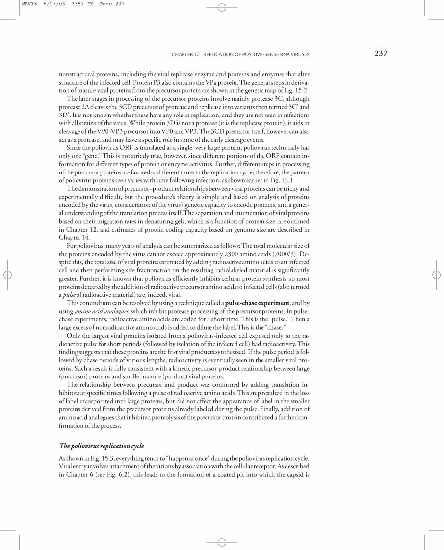
The most generally accepted scheme is shown in Fig. 15.4. In viral morphogenesis, P1 protein iscleaved from the precursor protein by protease 2A segment. Five copies of this protein aggregateand the protein are further cleaved by protease 3C into VP0, VP1, and VP3, which forms one of the60 capsid protomers. Five of these protomers assemble to form the 14s pentamer. Finally, twelve ofthese 14s pentamers assemble to form an empty capsid (procapsid).
This procapsid is less dense than the mature virion, so its proteins can be separated readily bycentrifugation. Analysis of the procapsid proteins demonstrates equimolar quantities of VP0,VP1, and VP3. Following formation of the procapsid, viral RNA associates with the particle and afinal cleavage of VP0 into VP2 and VP4 occurs to generate the mature virion. After virions are as-sembled, the cell lyses and virus is released.
Picornavirus cytopathology and disease
The most obvious cytopathology of poliovirus replication is cell lysis. But prior to this, the virusspecifically inhibits host cell protein synthesis. Inhibition of host cell protein synthesis involvesproteolytic digestion of the translation initiation factor eIF-4G so that ribosomes can no longerrecognize capped mRNA (see Chapter 13). Such modification leads to the translation of only un-capped poliovirus mRNA because its IRES allows it to assemble the translation complex with thevirus-modified ribosomes. Note that this rather elegant method of shutoff will not work with mosttypes of viruses because they express and utilize capped mRNA!
There are three related types, or serotypes, of poliovirus. They differ in the particular antigenicproperties of viral structural proteins. Most poliovirus infections in unprotected human popula-tions result in no or only mild symptoms, but one serotype (type 3) is strongly associated with thedisease’s paralytic form. Infection with this serotype does not invariably lead to a paralytic episode,but the probability of such an episode is much higher than with the others. All serotypes are dis-tributed throughout the regions where poliovirus is endemic in a population, although some pre-dominate in some locations.
Poliovirus is spread by fecal contamination of food or water supplies. Receptors for the virus arefound in the intestine’s epithelium, and infection results in local destruction of some tissue in the
CHAPTER 15 REPLICATION OF POSITIVE-SENSE RNA VIRUSES 239
WBV15 6/27/03 3:57 PM Page 239
intestine, which can result in diarrhea. Unfortunately, motor neurons also have receptors for po-liovirus, and if the virus gets into the bloodstream, it can replicate in and destroy such neurons,leading to paralysis. This result is of no value to the virus since the virus initiating neuronal infec-tion cannot be spread to other individuals and is eventually cleared; thus, the paralytic phase of thedisease is a “dead end” for the virus. The virus stimulates an immune response and the individual recovers and is resistant or immune to later infection.
Vaccination against poliovirus infections is accomplished effectively with both inactivated andattenuated live-virus vaccines, as described in Chapter 8. Since the only reservoir of poliovirus ishumans, immunity through vaccination against the virus is an effective way of preventing disease.
240 BASIC VIROLOGY
VP1
VP0
VP3
VP1VP3VP0
N CP2
N CP3
N C
P1
Nascent cleavage by 2A protease segment
Cleavage by 3C or 3CD protease segment
NP1 precapsid P2 P3
C
Protomer (5s)
Pentamer (14s)
Maturation
Virion (155s)
Procapsid (73s)
RNA encapsidation
Provirion (155s)
Cleavage of VP0 into VP2 + VP4Procapsid assembly
x 5
x 12
VP2
Fig. 15.4 The steps in the assembly of the poliovirus virion. Precursor proteins associate to form 5s protomers, which then assemble to form pentamers.
Twelve of these assemble to form the procapsid into which virion RNA is incorporated. Final cleavage of VP0 into VP2 and VP4 takes place to form the
mature capsid that has a diameter of 28 to 30nm.
WBV15 6/27/03 3:57 PM Page 240

Currently, a major effort is underway to completely eradicate the disease from the environment (seeChapter 22).
A number of other picornaviruses cause disease; many are spread by fecal contamination and in-clude hepatitis A virus, echoviruses, and coxsackievirus. Like poliovirus, these viruses occasionallyinvade nervous tissue. Coxsackievirus generally causes asymptomatic infections or mild lesions in oral and intestinal mucosa, but can cause encephalitis. Echoviruses are associated with enteric infections also, but certain echovirus serotypes cause infant nonbacterial meningitis, and some epidemic outbreaks with high mortality rates in infants have been reported.
Another widespread group of picornaviruses are the rhinoviruses, one of the two major groupsof viruses causing common head colds. Unlike the other picornaviruses detailed here, rhinovirusesare transmitted as aerosols. Because of the large number (~ 100) of distinct serotypes of rhinovirusit is improbable that an infection will generate immunity that prevents subsequent colds. There areno known neurological complications arising from rhinovirus infections.
Flavivirus replicationThe success and widespread distribution of picornaviruses and their relatives demonstrate that thereplication strategy found in translation of a single large ORF is a very effective one. If more evidence were needed on this score, the plethora of mosquito-borne flaviviruses should settle thematter completely!
Flaviviruses are enveloped, icosahedral, positive-sense RNA viruses. They appear to be related topicornaviruses, but clearly have distinct features, notably an envelope. Because mosquitoes andmost other arthropods are sensitive to weather extremes, it is not surprising that arboviral diseasesoccur throughout the year in the tropics and subtropics, but occur only sporadically, and in thesummer, in temperate zones.
Many flaviviruses demonstrate tropism for neural tissue, and flaviviruses are the causative agentsof yellow fever, dengue fever, and many types of encephalitis. In the United States, mosquito-borneSt. Louis encephalitis virus leads to periodic epidemics in the summer, especially during summersmarked by heavy rains and flooding, such as the summer of 1997 in northeastern states. An emerg-ing problem is the establishment of West Nile virus in regions of the eastern United States. In thelate summer of 1999, confirmed human cases of encephalitis were documented in New York. Al-though it is not known how this virus arrived, the strains that are present seem to be related to virusfound in the Middle East. During the summer of 2002, the virus spread was documented all theway to California. As of this writing, West Nile virus is now known to be present in virtually all ofthe contiguous 48 states.
An abbreviated outline of the yellow fever virus replication cycle can be inferred from the gen-etic and structural map shown in Fig. 15.5. The yellow fever genome is over 10,000 bases long, andunlike poliovirus, it is (i) capped at the 5¢ end and (ii) not polyadenylated at the 3¢ end. Like po-liovirus, the large ORF is translated into a single precursor protein that is cleaved by integral pro-teases into individual proteins. Some of these cleavage steps are shown in Fig. 15.5. The structuralprotein precursor includes an integral membrane protein (M) and an envelope glycoprotein. Thesemembrane-associated proteins are translated by membrane-bound polyribosomes, and the processof insertion into the cell’s membrane follows the basic outline described for togaviruses later. TheM protein contains a “signal” sequence at its N-terminal that facilitates the insertion of the nascentpeptide chain into the endoplasmic reticulum. This signal is cleaved from the PreM protein withinthe lumen of the endoplasmic reticulum — probably by the action of cellular enzymes. The NS(nonstructural) proteins encode the replicase enzymes and do not form part of the virion. Despitethis, it is interesting that antibodies directed against the precursor, NS1, protect animals against infection.
CHAPTER 15 REPLICATION OF POSITIVE-SENSE RNA VIRUSES 241
WBV15 6/27/03 3:57 PM Page 241

POSITIVE-SENSE RNA VIRUSES ENCODING MORE THANONE TRANSLATIONAL READING FRAME
A positive-sense RNA virus that must regulate gene expression while infecting a eukaryotic hostfaces a fundamental problem: The eukaryotic ribosome cannot initiate translation of an ORF fol-lowing translation of one upstream of it. While a positive-sense RNA virus genome could (andsome do) contain more than one ORF, these ORFs cannot be independently translated at differentrates during infection without some means to overcome this fundamental mechanistic limitation.
One way to overcome the problem is for a virus to encapsidate more than one mRNA (in otherwords, for the virus to contain a segmented genome). This approach is utilized by a number of positive-sense RNA viruses infecting plants, but has not been described for animal viruses. Thisfinding is somewhat surprising since there are numerous negative-sense RNA viruses with seg-mented genomes that are successful animal and human pathogens. The list contains influenzaviruses, hantaviruses, and arenaviruses.
Despite the disinclination of positive-sense RNA viruses that infect animal cells to encapsidatesegmented genomes, another strategy for regulating mRNA expression is utilized successfully. Thisstrategy involves the encoding of a cryptic (hidden) ORF in the genomic RNA, which can be trans-lated from a viral mRNA generated by a transcription step during the replication cycle. With thisstrategy, viral gene expression from the full-length positive-sense mRNA contained in the virion re-sults in translation of a 5¢ ORF, and this protein (an enzyme) is involved in generation of a second,smaller mRNA by transcription.
The second mRNA (which is not found in the virion), in turn, is translated into a distinct viralprotein. Such a scheme allows the nonstructural proteins encoded by the virus — the enzymes re-
242 BASIC VIROLOGY
Envelope
Lipid bilayer
Nucleocapsid
Capsid
pre M E
NS1 NS2 NS3 NS4 NS5
NS2A NS2B NS4A NS4B
118 nt Single ORF (10,233 nt) 51 nt
5' 7-mG
TranslationProteolytic cleavage
Translated on rough ER
Yellow fever virus(flavivirus)
Fig. 15.5 The yellow fever virus (a flavivirus) and its genome.
This flavivirus has a replication cycle very similar in broad outline
to that detailed for poliovirus. Unlike poliovirus, flaviviruses
encode a single envelope glycoprotein, and its approximately
10,000 nucleotide (nt) genome is capped, although not
polyadenylated. Also in contrast to poliovirus, the yellow fever
virus precursor polyprotein is cleaved into a large number of
products as it is being translated, so the very large precursor
proteins of poliovirus replication are not seen. The enveloped
capsid is larger than that of poliovirus, with a diameter of 40 to
50nm. (ER, endoplasmic reticulum.)
WBV15 6/27/03 3:57 PM Page 242

quired for replication — to be expressed in lesser amounts or at different times in the infection cyclethan the proteins ending up in the mature virion. Clearly, this approach is effective as witnessed bythe number of important pathogens that utilize it.
Two viral mRNAs are produced in different amounts during togavirus infectionsTogaviruses are enveloped RNA viruses that display a complex pattern of gene expression duringreplication. Sindbis virus is a well-studied example. This arthropod-borne virus causes only verymild diseases in (rare) humans, but its size and relative ease of manipulation make it a useful labo-ratory model for the group as a whole.
Sindbis virus has a capsid structure similar to picornaviruses and flaviviruses, and like flavivir-uses, the capsid is enveloped. The viral genome contains two translational ORFs. Initially, only thefirst frame is translated into viral replication enzymes. These enzymes both replicate the virionRNA and generate a second mRNA that encodes viral structural proteins.
The viral genome
Sindbis virus and its 11,700 base genome is shown in Fig. 15.6. The virion genomic RNA (termed49s RNA for its sedimentation rate in rate zonal centrifugation — see Chapter 11) has a capped 5¢end and a polyadenylated 3¢ end. Both capping and polyadenylation appear to be carried out byviral replication enzymes, possibly in a manner somewhat analogous to that seen for the negative-sense vesicular stomatitis virus (VSV), which is discussed in Chapter 16.
The Sindbis virus genome contains two ORFs. The 5¢ ORF encodes a replication protein pre-cursor that is processed by proteases to generate four different replicase polypeptides. The 3¢ ORFencodes capsid protein and envelope glycoproteins.
CHAPTER 15 REPLICATION OF POSITIVE-SENSE RNA VIRUSES 243
ORF-1 (~7000 nt) ORF-2 (~4000 nt)
5' Cap AAA
Replicase genes Structural genes
Suppressiblestop codon
(pol)
Sindbis virus
Lipid bilayer
C protein
Spike protein
Fig. 15.6 Sindbis virus — a typical togavirus. The virion (60–70
nm in diameter) and genetic map are shown. The Sindbis genome
contains two translational reading frames; only the upstream (5¢)one can be translated from the approximately 11,000 nucleotide
(nt) capped and polyadenylated 49s (positive) virion-associated
genomic RNA. This upstream translational frame encodes
nonstructural proteins via expression of two precursor proteins.
The larger, which contains the polymerase precursor, is translated
by suppression of an internal stop codon in the reading frame.
WBV15 6/27/03 3:57 PM Page 243

The virus replication cycle
Virus entry Viral entry is via receptor-mediated endocytosis as shown in Fig. 15.7a. The entirevirion, including envelope, is taken up in the endocytotic vesicle. Acidification of this vesicle leadsto modification of the viral membrane glycoprotein. This allows the viral membrane to fuse withthe vesicle, and causes the capsid to disrupt so that viral genomic mRNA is released into the cytoplasm.
Early gene expression As shown in Fig. 15.7b, only the 5¢ ORF can be translated from intact viralmRNA, because the eukaryotic ribosome falls off the viral mRNA when it encounters the firsttranslation stop signal (either UAA, UAG, or UGA — see Chapter 13). With Sindbis virus, this sit-uation is complicated by the fact that this first ORF in the genomic RNA contains a stop signalabout three-fourths of the way downstream of the initiation codon. This termination codon can berecognized to generate a shorter precursor to the nonstructural proteins, but it can also be sup-pressed. (In genetics, the term suppression refers to the cell periodically ignoring a translation stopsignal either because of an altered tRNA or a ribosomal response to secondary structure in themRNA encoding it.) With Sindbis virus infection, the suppression is ribosomal, and results inabout 25% of the nonstructural precursor protein containing the remaining information shown inORF-1 in the genetic map. As discussed in Chapter 20, suppression of an internal stop codon alsohas a role in the generation of retrovirus protein.
In Sindbis virus infection, translation of infectious viral RNA generates replication enzymesthat are derived by autoproteolytic cleavage (i.e., self-cleavage) of the replicase precursor protein.This can be considered an “early” phase of gene expression; however, things happen fast in the in-fected cell and this may only last for a few minutes.
Viral genome replication and generation of 26s mRNA The replication enzymes expressed from genomic 49s positive-sense mRNA associated with genomic RNA to generate 49s negative-senseRNA through RI-1 is shown in Fig. 15.8a. The next step in the process is critical to regulated expression of the two virus-encoded precursor proteins. With Sindbis, the negative-sense RNAcomplementary to genomic positive-sense RNA is the template for two different positive-sensemRNAs. Both are capped and polyadenylated. The first is more 49s positive-sense virion RNA.The second is 26s positive-sense RNA. The shorter 26s mRNA is generated by replicase beginningtranscription of negative-sense RNA in the middle and generating a “truncated” or subgenomicmRNA. The region on the negative-sense strand where the transcriptase binds is roughly analogousto a promoter, but its sequence does not exhibit the features of promoters found in DNA genomes.
Generation of structural proteins The short 26s mRNA contains only the second ORF containedin the full-length genomic RNA. This ORF was hidden or inaccessible to translation of the full-length virion mRNA. With the 26s mRNA, however, cellular ribosomes can translate the ORFinto precursors of capsid and envelope proteins. Expression of structural proteins, thus, requires atleast partial genome replication and is generally termed late gene expression, although it occurs verysoon after infection. Translation of the 5¢ region of late 26s mRNA generates capsid protein that iscleaved from the growing peptide chain by proteolytic cleavage. This cleavage generates a new N-terminal region of the peptide. The new N-terminal region of the peptide contains a stretch ofaliphatic amino acids, and the hydrophobic nature of this “signal” sequence results in the growingpeptide chain inserting itself into the endoplasmic reticulum in a manner analogous to synthesis ofany cellular membrane protein. This process is shown in Fig. 15.8b.
Following initial insertion of the membrane proteins’ precursor, the various mature proteins areformed by cleavage of the growing chain within the lumen of the endoplasmic reticulum. This mat-urational cleavage is carried out by cellular proteins.
244 BASIC VIROLOGY
WBV15 6/27/03 3:57 PM Page 244

CHAPTER 15 REPLICATION OF POSITIVE-SENSE RNA VIRUSES 245
ORF-1 (~7000 nt) ORF-2 (~4000 nt)
5' Cap AAA
Suppresssiblestop codon
N C
NSP1(540aa)
NSP2(807aa)
Protease,helicase
NSP3(549aa)
NSP4(610aa)
Pol
Translation ofnon-structural genes
49s genomic RNA (positive strand)
Cleavages
Release of Sindbisvirus genome
Fusion of viral membranewith endocytotic vesiclemembrane
(reduced molar amount of Pol expressed in relation to other replication proteins)
N C
Sindbis virion
ATP
ADP
H+
Cytoplasm
Receptor mediatedendocytosis
Coated pit
Coated vesicle
(a)
(b)
Fig. 15.7 The early stages of Sindbis virus infection. a. The first
step is receptor-mediated endocytosis, leading to fusion of the
viral membrane with that of the endocytotic vesicle, which leads
to release of the Sindbis virus genome (mRNA) into the infected
cell’s cytoplasm. As outlined in Chapter 6, internalization of
the enveloped virion within an endocytotic vesicle is followed
by acidification and covalent changes in membrane proteins.
This results in fusion of the viral membrane with that of the
endocytotic vesicle and release of the viral genome. b. Translation
of the virion RNA results in expression of the precursors to the
nonstructural replicase and other viral proteins encoded in the 5¢translational reading frame. These proteins mediate replicase,
capping, and protease functions.
WBV15 6/27/03 3:57 PM Page 245
Posttranslational processing, such as glycosylation of membrane-associated components of thelate structural protein, takes place in the Golgi apparatus, and viral envelope protein migrates to thecell surface. Meanwhile, capsid formation takes place in the cytoplasm, genomes are added, and the virion is formed by budding through the cell surface, as described in Chapter 6.
Togavirus cytopathology and disease
The replication process of togaviruses is a step more complex than that seen with picornaviruses,and the cell needs to maintain its structure to allow continual budding of new virus. Accordingly,there is less profound shutoff of host cell function until a long time after infection.
A major cytopathic change is alteration of the cell surface. This can lead to fusion with neigh-boring cells so that virus can spread without ever leaving the first infected cells. This alteration tothe cell surface also involves antigenic alteration of the cell. Such types of cytopathology are foundwith many enveloped RNA viruses, whether they are positive or negative sense.
246 BASIC VIROLOGY
5'
5'
5'
5'
5'
5'
5'
5'
5'
5'
5'5'
5'
5'3'
3'
Replicase enzymesencoded by ORF-1
RI-1
RI-2
ORF-1 (~7000 nt) ORF-2 (~4000 nt)
ORF-2
ORF-2ORF-1
5' Cap
5' Cap
5' Cap
AAA
AAA
AAA
Suppressiblestop codon
:
49s (-)
49s (+)
49s (+)
26s (+)
Interiorreplicasestart
Replicase
(a)
Fig. 15.8 a. The replication of Sindbis virus genome, and
generation of the subgenomic 26s mRNA. This mRNA is
expressed by an internal start site for viral replicase, and is
translated into structural proteins since it encodes only the open
reading frame (ORF) that was cryptic in the 49s positive-sense
virion RNA. b. The synthesis of Sindbis virus structural proteins.
Structural proteins are translated as a single precursor. When the
N-terminal capsid protein is cleaved from the precursor, a signal
sequence consisting of a stretch of aliphatic amino acids associates
with the endoplasmic reticulum. This association allows the
membrane protein portion of the precursor to insert into the
lumen of the endoplasmic reticulum. As the protein continues to
be inserted into the lumen, it is cleaved into smaller product
proteins by cellular enzymes. Cellular enzymes also carry out
glycosylation.
WBV15 6/27/03 3:57 PM Page 246

The togaviruses are an extremely successful group of viruses, and like the flaviviruses, many aretransmitted by arthropods. As noted in Chapter 5, it is for this reason that these two groups of positive-sense RNA viruses are termed arboviruses (arthropod-borne viruses). While this termi-nology is convenient for some purposes, it does not recognize significant differences in the replica-tion strategies of these two groups of viruses. Further, numerous other types of viruses are spread byarthropod vectors, and some togaviruses and flaviviruses are not transmitted by such vectors. Astriking example is rubella (German measles) virus.
Many togaviruses cause sporadic outbreaks of mosquito-borne encephalitis because they have apropensity for replication in cells making up the brain’s protective lining. Although such diseasecan be severe, many forms have a favorable prognosis with proper medical care, as neurons are notthe primary targets of infection.
The only known host for rubella virus is humans. The virus causes generally mild and oftenasymptomatic diseases in children and adults, although a mild rash may be evident. Despite thegenerally benign course of infection, it is remarkable that rubella is associated with a diverse groupof clinical diseases, including rubella arthritis and neurological complications.
Periodic local epidemics are characteristic of rubella virus infections, and although the virus in-duces an effective immune response, the endemic nature of the virus ensures that once a large-
CHAPTER 15 REPLICATION OF POSITIVE-SENSE RNA VIRUSES 247
Capsid 62K protein 6K
6K
E1
E1
Endoplasmic reticulum
Golgi apparatus
Glycosylation
Viral envelope proteinstransported in vesicles
Plasma membrane
Capsid proteins
49s(+)
New capsids
Capsidformation
Proteolytic cleavage
5' 7mG AAAA 3'
Lumen
Mature Sindbis virion released
EE
E
E
E
EE E E
EE
E
MM
MM
M
MM M
M
M
MM
M
NSignal sequence
62K
26s (+)-sense mRNA(b)
Fig. 15.8 Continued
WBV15 6/27/03 3:57 PM Page 247
enough pool of susceptible individuals arises, sporadic regional epidemics occur. The major prob-lem with these periodic occurrences is the very fact that the disease is often so mild as to be asymp-tomatic in adults of childbearing age. While the symptoms are very mild for adults and children,this is not the case for fetal infections. Infection of the mother in the first trimester of pregnancyoften leads to miscarriage, and a fetus who survives is almost inevitably severely developmentallyimpaired. Infection of the mother later in pregnancy has a more benign outcome.
The tragedy of rubella infections is that although there are effective vaccines, the disease is oftenso mild that an individual can be infected and can spread the virus without knowing it. For this reason, women of childbearing age who are in contact with young children or other adults at risk of infection should be vaccinated.
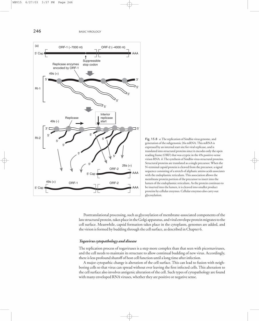
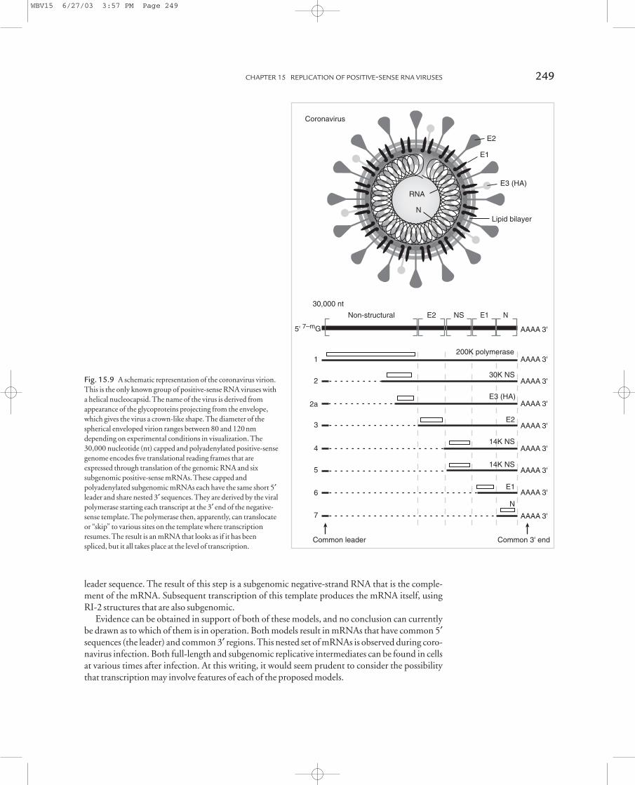
A somewhat more complex scenario of multiple translational readingframes and subgenomic mRNA expression: coronavirus replicationEven more complex scenarios exist for expression and regulation of gene function in infections bypositive-sense RNA viruses. The replication strategy of the coronaviruses is a good example of suchcomplexity. The structure of coronaviruses is shown in Fig. 15.9, and is unusual for a positive-senseRNA virus.
The nucleocapsid is helical within a roughly spherical membrane envelope, and the envelopeglycoproteins project as distinct “spikes” from this envelope. These glycoprotein spikes from thelipid bilayer appear as a distinctive crown-like structure in the electron microscope, hence, thename corona (crown)-viruses.
The 30kb coronavirus genome encodes at least five separate translational reading frames, and is the template for the synthesis of at least six subgenomic mRNAs. Each subgenomic mRNA contains a short, identical leader segment at the 5¢ end that is encoded within the 5¢ end of the genomic RNA. All subgenomic mRNAs have the same 3¢ end, and thus are a nested set of tran-scripts. Only the 5¢ translational reading frame is recognized in each, and the others are cryptic.These features are also shown in Fig. 15.9.
Coronavirus replication
Coronavirus replication involves the generation and translation of genomic and subgenomic viralmRNAs as shown in Fig. 15.10. Virus entry is by receptor-mediated fusion of the virion with theplasma membrane followed by release of genomic RNA. This RNA (one of the largest mRNAscharacterized) is translated into a replication protein that, interestingly, is encoded in an ORF encompassing 70% of the virus’s coding capacity. The reason why coronavirus replication proteinsare encoded by such a large gene is not yet known.
The mature replication proteins derived from the first translation product are used to produceall subsequent mRNA species. There are two competing models that have been presented for coro-navirus transcription (Fig. 15.10): leader-primed transcription and discontinuous transcriptionduring negative-strand synthesis.
Leader-primed transcription proposes that the replication proteins first produce a full-lengthnegative strand copy of the genome, using a standard RI-1 structure. From this template is thentranscribed multiple copies of the extreme 3¢ end, called the leader region. These leader transcriptsthen function to prime synthesis of subgenomic mRNAs, initiated at homologous regions in be-tween each of the genes (intergenic sequences).
Discontinuous transcription during negative-strand synthesis proposes that the replicationproteins transcribe negative-strand copies of the genome, using RI-1 structures. Some of theseproducts are subgenomic. These subgenomic species are produced when the replicase complex inthe RI-1 pauses at the intergenic regions and then jumps to the end of the genome, copying the
248 BASIC VIROLOGY
WBV15 6/27/03 3:57 PM Page 248
leader sequence. The result of this step is a subgenomic negative-strand RNA that is the comple-ment of the mRNA. Subsequent transcription of this template produces the mRNA itself, usingRI-2 structures that are also subgenomic.
Evidence can be obtained in support of both of these models, and no conclusion can currentlybe drawn as to which of them is in operation. Both models result in mRNAs that have common 5¢sequences (the leader) and common 3¢ regions. This nested set of mRNAs is observed during coro-navirus infection. Both full-length and subgenomic replicative intermediates can be found in cellsat various times after infection. At this writing, it would seem prudent to consider the possibilitythat transcription may involve features of each of the proposed models.
CHAPTER 15 REPLICATION OF POSITIVE-SENSE RNA VIRUSES 249
Non-structural E2 NS E1 N
30,000 nt
5' 7–mG AAAA 3'
Common leader Common 3' end
E2
E1
E3 (HA)
Lipid bilayerN
RNA
AAAA 3'200K polymerase
1
AAAA 3'30K NS
2
AAAA 3' E3 (HA)
2a
AAAA 3'E2
3
AAAA 3'14K NS
4
AAAA 3'14K NS
5
AAAA 3'E1
6
AAAA 3'
N
7
Coronavirus
Fig. 15.9 A schematic representation of the coronavirus virion.
This is the only known group of positive-sense RNA viruses with
a helical nucleocapsid. The name of the virus is derived from
appearance of the glycoproteins projecting from the envelope,
which gives the virus a crown-like shape. The diameter of the
spherical enveloped virion ranges between 80 and 120nm
depending on experimental conditions in visualization. The
30,000 nucleotide (nt) capped and polyadenylated positive-sense
genome encodes five translational reading frames that are
expressed through translation of the genomic RNA and six
subgenomic positive-sense mRNAs. These capped and
polyadenylated subgenomic mRNAs each have the same short 5¢leader and share nested 3¢ sequences. They are derived by the viral
polymerase starting each transcript at the 3¢ end of the negative-
sense template. The polymerase then, apparently, can translocate
or “skip” to various sites on the template where transcription
resumes. The result is an mRNA that looks as if it has been
spliced, but it all takes place at the level of transcription.
WBV15 6/27/03 3:57 PM Page 249

The specific mechanism of the transcriptase jumping in each model is not known, but it mayfunction to ensure that the virus only has one sequence of RNA needing to be capped (the 5¢ leadersequence). The addition of the polyA tracts onto the individual mRNAs also only requires therecognition of one sequence on the positive-sense template by viral replicase since all mRNAs havethe same 3¢ end.
250 BASIC VIROLOGY
Translate
Polymerase
(RI-1)
(-)
(RI-2)
mRNA (1-7) Translate
Rough endoplasmicreticulum
Golgi apparatus
Exocytotic vesicle
E1, E2, E3
3'
3'
3'
5'
5'
5'
CapLeader
Leader
Leader
(-)
(-)
(-)
Discontinuity
Pol skips down template orreinitiates with leader attached Interior start
pol
Release by fusionand lysis
Fusion
Coronavirus
Fig. 15.10 The replication cycle of a coronavirus. Replication is entirely cytoplasmic. Infection is initiated by receptor-mediated membrane fusion to release
the genomic mRNA. This RNA is translated into the very large (>200kd) polymerase/capping enzyme. The interaction between full-length virion positive-
sense RNA and replicase generates the templates for the mRNAs. Two models are proposed for the synthesis of subgenomic mRNA: leader-primed synthesis
and discontinuous negative-strand synthesis. The two models are shown in the figure and are described in the text. The result of both models is the synthesis of
a nested set of mRNAs that contain the same 5¢ leader sequence and overlapping 3¢ ends. Translation of the various subgenomic mRNAs leads to synthesis of
the various structural and nonstructural proteins encoded by interior translational reading frames. The mature virions assemble and become enveloped by
budding into intracytoplasmic vesicles; these exocytotic vesicles then migrate to the cell surface where virus is released. At later times, cell lysis occurs.
WBV15 6/27/03 3:57 PM Page 250

Cytopathology and disease caused by coronaviruses
The coronaviruses, along with the rhinoviruses, cause mild and localized respiratory tract infec-tions (head colds). The mildness of colds results from a number of both viral and cellular factors.
First, the viruses causing the common cold have a very defined tissue tropism for nasopharynxepithelium. Spread of the virus is limited by ill-defined localized immune factors of the host. Theability of a cold virus infection to remain localized at the site of initial infection is a great advantageto the virus. Local irritation leads to sneezing, coughing, and runny nose, all important for viralspread. Mildness and localization of the infection tend to limit the immune response, which is another distinct advantage. A mild infection results in short-lived immunity, and this, along withthe fact that a large number of serotypes exist as a result of the high error frequency of the genomereplication process, mean that colds are a common and constant affliction.
In the late winter and spring of 2003 a new illness broke out, focused in China and Singapore.Severe acute respiratory syndrome (SARS) proved to be more than the common cold, having a case fatality rate of 10–20%. As of this writing, the etiologic agent of SARS has been identified as acoronavirus, named SARS-CoV.
REPLICATION OF PLANT VIRUSES WITH RNA GENOMES
A large number of plant viruses contain RNA genomes, and many of the early discoveries in virol-ogy were accomplished with plant viruses. The discovery of viruses as specific infectious particles atthe end of the nineteenth century focused on work to elucidate the cause of tobacco mosaic disease,culminating in the first description of the tobacco mosaic virus (TMV). This virus took center stagefor a number of important early events in biochemical virology, including the first crystallization ofa virus particle by W. M. Stanley at University of California, Berkeley; demonstration of the infec-tious nature of a positive-sense RNA genome by Gierer and Schramm; and in vitro assembly fromisolated protein and RNA of an infectious particle by F. Fraenkel-Conrat.
The majority of plant RNA viruses are nonenveloped and have single-stranded genomes. Theexceptions are two groups of plant viruses with negative-sense genomes (the plant rhabdovirusesand the Tospovirus genus of the bunyavirus family) and one group with dsRNA genomes (e.g.,wound tumor virus).
All of the positive-sense plant RNA viruses have genomes that can be translated entirely or inpart immediately after infection. Structure of the genome RNA is varied (Table 15.1). The 5¢ endmay be capped or may have a covalently linked genome protein similar to picornavirus VPg. The 3¢
CHAPTER 15 REPLICATION OF POSITIVE-SENSE RNA VIRUSES 251
Table 15.1 Genomic structure of some positive-sense RNA viruses infecting eukaryotes.
Virus No. of genome segments 5¢ end 3¢ end
Poliovirus 1 VPg PolyA (genome encoded)Yellow fever virus 1 Methylated cap NonpolyASindbis virus 1 (expresses subgenomic mRNA) Methylated cap PolyA (A)Coronavirus 1 (expresses nested subgenomic Common leader with methylated PolyA (A)
mRNA) capTobacco mosaic virus 1 Methylated cap tRNAhisPotato virus Y 1 VPg PolyATomato bushy stunt virus 1 Methylated cap NonpolyABarley yellow dwarf virus 1 VPg NonpolyATobacco rattle virus 2 Methylated cap NonpolyACowpea mosaic virus 2 VPg PolyABrome mosaic virus 3 Methylated cap tRNAtyr
WBV15 6/27/03 3:57 PM Page 251

end may be polyadenylated or not, or may be folded into a tRNA-like structure that can actually becharged with a specific amino acid. There appears to be no role in virus translation for this tRNA,but the fact that the cytoplasm of eukaryotic cells has an enzyme that functions to regenerate theCCA at the 3¢ end of tRNA molecules suggests that the tRNA structure may provide the viralgenome with a means of avoiding exonucleolytic degradation from the 3¢ end.
While expression of the positive-sense RNA genomes of plant viruses follows the same generalrules outlined for replication of corresponding animal viruses, there is an added complication. Anumber of plant virus RNA genomes are segmented. This segmentation means that individualmRNA-sized genomic fragments can be (theoretically, at least) independently replicated and trans-lated. Independent replication and translation allow the virus to maintain a replication cycle inwhich individual viral genes can be expressed at significantly different levels.
Use of this strategy in virus replication adds the complication that the packaging process is potentially very inefficient. This is certainly true for the packaging of influenza virus described inthe next chapter. Alternatively, the packaging process might be controlled in some way to ensurethat each viral particle gets its requisite number of genomic fragments. Despite this complication,segmented genomes are a viable strategy for RNA virus replication, and it is not clear why it is notused in the replication of any known positive-sense animal viruses.
With viruses of vascular plants, the limitations in the size of objects that can pass through the cellwall led to another adaptation. The plant viruses with segmented positive-sense RNA genomespackage each segment separately. Although this separate packaging means that each cell must be in-fected with multiple virions, plant viruses seem to thrive using this approach, probably for the fol-lowing reason: Plant viruses are often transmitted mechanically and then spread from cell to cell viathe plant’s circulation without involvement of a specific immune defense; therefore, high concen-trations of virus at the surface of the cell can be maintained.
Viruses with one genome segmentTMV has a helical capsid that encloses a single RNA genome segment of 6.4kb. Primary transla-tion of the genome produces the replicase complex consisting of the 126kd and 183kd replicationproteins. Two subgenomic mRNAs are transcribed from negative-sense RNA generated from RI-1. The translation of these two species yields the 17.5kd coat protein and a 30kd protein involved in movement of the virus within the infected plant.
Tomato bushy stunt virus has a single RNA genome of 4.8kb packaged into an icosahedral capsid. Translation of the capped genome results in production of the 125kd viral replicase. Two subgenomic mRNAs are transcribed from the full-length negative-sense strand generatedfrom RI-1. Translation of these two species leads to synthesis of the 41kd coat protein and twoother proteins thought to be required for cell-to-cell movement of the virus.
Viruses with two genome segmentsThe genome of cowpea mosaic virus consists of two separate strands of RNA packaged into separateicosahedral particles. Since both strands are required for infection, a cell must be infected togetherby each of the two particles. The larger of the two RNAs (5.9kb) is translated into a polyproteinthat is cleaved into a 24kd protease, the 4kdVPg, a 110kd replicase, and a 32kd processing pro-tein. The smaller (3.5kb) RNA encodes a polyprotein that is cleaved into the 42kd and 24kd coatproteins and a set of proteins required for cell-to-cell movement of the virus.
Viruses with three genome segmentsBrome grass mosaic virus has three separate RNA genome strands (3.2kb, 2.8kb, and 2.1kb) con-
252 BASIC VIROLOGY
WBV15 6/27/03 3:57 PM Page 252

tained in three separate icosahedral particles. Again, since all three genome segments are required forinfection, cells must receive one of each of the particles. Each of the capped genome segments istranslated into a protein. These products include the 94kd viral replicase, a 109kd capping enzyme, and a 32kd cell-to-cell movement protein. In addition, one of the RNAs is transcribedinto a subgenomic mRNA that encodes the 20kd viral coat protein.
REPLICATION OF BACTERIOPHAGES WITH RNA GENOMES
The great majority of well-characterized RNA bacteriophages have linear, single-stranded, positive-sense genomes enclosed within small, icosahedral capsids. These phages (grouped together as the Leviviridae) include the male bacteria-specific phage Qb, MS2, and R17, which attach to the bacteria’s F pili.
In broad outline, the replication process of these RNA-containing bacteriophages follows thatdescribed for eukaryotic viruses. Infection begins with a translation step, and replication of the viralgenome occurs through production of the RI-1 and RI-2 intermediates described in the precedingsection.
Regulated translation of bacteriophage mRNAThere is a major difference in the way protein synthesis occurs on bacterial ribosomes as comparedto eukaryotic ribosomes, and this leads to a significant difference in the way expression of viral-encoded protein is controlled. As discussed in Chapter 13, bacterial ribosomes can initiate transla-tion at start sites in the interior of bacterial mRNA. This means that a bacterial mRNA moleculewith several ORFs can be translated independently into one or all of the proteins. In an RNA bac-teriophage infection, protein synthesis programmed by the incoming genome is characterized bysynthesis of viral RNA replicase only. Later in infection, after genome replication begins, transitionto synthesis of capsid and other proteins begins.
This temporal regulation is governed by the secondary structure of the genome, and initiationof protein synthesis encoded by interior ORFs by ribosomal mechanisms. This can be seen in thephage Qb, which is diagrammed in Fig. 15.11. This virus encodes three distinct translational read-ing frames encoding genes for the A (maturational) protein, the coat protein, and replicase. The
CHAPTER 15 REPLICATION OF POSITIVE-SENSE RNA VIRUSES 253
A protein Coat Replicase
5' 3'
Suppressiblestop codon
Bacteriophage Qb
Fig. 15.11 The approximately 25nm diameter icosahedral
capsid of positive-sense RNA bacteriophage Qb. The positive-
sense RNA genome contains three separate open reading frames
(ORFs). These ORFs can be independently translated from the
full-length virion RNA because unlike the situation in eukaryotic
viruses, bacterial ribosomes can initiate translation at interior
start signals provided that the ribosome can interact with them.
With this bacteriophage, ribosome attachment and translation
require active transcription to allow the nascent positive-sense
RNA to be unfolded so that the translation start is accessible.
WBV15 6/27/03 3:57 PM Page 253

coat protein translational reading frame has a translation terminator that is misread (suppressed) asa tryptophan residue about 1% of the time, and when this happens, a larger capsid protein with additional amino acids is generated. Suppression of the termination is absolutely required forphage replication.
A portion of the replication cycle of Qb is shown in Fig. 15.12. Ribosomes can associate with thegenomic RNA, but this positive-sense genome is folded in such a way that the only start codonavailable for interaction with a ribosome is the one that begins translation of phage RNA replicase.All other start codons are involved in base-pairing interactions as a part of the secondary structure.For this reason, replicase is the only phage protein expressed at the start of infection.
Synthesis of new positive-sense genomes takes place through formation of RI-1 and RI-2. As
254 BASIC VIROLOGY
CAC CAC
CAC
CAC
CAC
GUG
5'
5'
5'
5'
5'
5'
3'
3'
3'
3'
3'3'
Replicase enzyme
Positive strand RNA
Negative strand RNA
Replicase enzyme
Nascent A protein
GUG open
AUG open
GUG blocked
30s Ribosome
Leader
Leader
GU
G
GU
G
AUG
(+)
(+)
Fig. 15.12 Coupled transcription-translation of bacteriophage
Qb RNA results in opening the blocked translational start site for
the A (maturational) and coat proteins. As the replicase enzyme
passes the region containing the translation start site on the
negative-sense template (which is a GUG for the A protein) the
nascent positive-sense mRNA can interact with a ribosome before
it has a chance to fold into a structure in which this initiator
codon is sterically blocked. Multiple ribosome entry results in
translation of a large number of copies of the maturational and
coat proteins being synthesized. High levels of coat protein
specifically inhibit translation of replicase from full-length
genomic RNA so that replicase is only synthesized at early times
in the replication cycle. For this reason, it is often termed an
“early” protein or gene product.
WBV15 6/27/03 3:57 PM Page 254

new positive-sense genomic RNA disassociates from the negative-sense template near the replicase,secondary structure has not yet formed. This results in the start codon for the A and coat proteinsbeing available to begin translation. The A protein uses a GUG instead of an AUG initiationcodon. Similarly, newly replicated positive-sense strands immediately interact with ribosomes toyield the capsid proteins necessary for the formation of new virus particles.
This simple mechanism ensures that the earliest protein expressed will be replicase. Further,since a relatively large amount of RI-2 will need to be present, synthesis of A and capsid proteins willonly occur when there are a large number of genomes waiting to be encapsidated. Multiple entry ofribosomes onto the nascent viral mRNA ensures that a large amount of structural protein will beavailable when necessary.
Finally, the phage controls the amount of replicase synthesized in infection so that progeny pos-itive-sense strand does not end up recycling too long. Such control is accomplished by the capsidprotein actually inhibiting synthesis of replicase from mature positive-sense RNA. Therefore, afterabout 20 minutes, increasing levels of capsid proteins shut off replicase synthesis.
CHAPTER 15 REPLICATION OF POSITIVE-SENSE RNA VIRUSES 255
QUESTIONS FOR CHAPTER 15
1 What are the steps in the attachment and entry of poliovirus in a susceptible host cell?
2 The Picornaviridae (e.g., poliovirus) have, as theirgenome, one molecule of single-stranded RNA. This genomic RNA functions in the cell as a monocistronicmRNA. However, picornavirus-infected cells contain 10or more viral proteins.
a What mechanism have these viruses evolved suchthat this monocistronic mRNA produces this largenumber of translation products?
b The poliovirus mRNA does not have a 5¢ methylatedcap that is present on host cell mRNA. How do hostcell ribosomes begin translation of this message?
3 Foot-and-mouth disease virus (FMDV) is a member ofthe family Picornaviridae. Based on your knowledge ofthe properties of members of this family, complete thefollowing table with respect to FMDV and each of thecharacteristics listed. State whether the characteristic ispresent or absent.
Characteristic Present or absent for FMDV
5¢ methylated cap
Subgenomic RNAs
3¢ polyadenylation
Single-stranded, positive-sense genome
Expression of genome as a polyprotein
4 The poliovirus genome is a single-stranded RNA ofabout 7500 nucleotides, with a covalently linked termi-nal protein, VPg, at the 5¢ end and a polyA sequence atthe 3¢ end. The polyA tail is not added after replicationbut is derived from the template during replication. VPg
is important for replication of this viral RNA, along withpoliovirus polymerase and certain host enzymes.
There are two models for the action of VPg:Model 1. VPg may act as a primer for RNA synthesis,
being used as VPg-pUOH.Continued
WBV15 6/27/03 3:57 PM Page 255

256 BASIC VIROLOGY
Model 2. VPg may act as an endonuclease, attaching it-self to the 5¢ end of a new RNA chain. In this model,RNA synthesis is primed after addition of U residuesto the 3¢ A at the end of the genome by a host enzyme,followed by a loop-back and self-priming mecha-nism.
Given these two models, imagine that you have an in vitrosystem to test the properties of poliovirus genome replica-tion. Your system contains viral genomic RNA as a tem-plate and all of the necessary proteins, except as indicatedbelow.
a Assume that model 1 is true. What would you ex-pect to see as the product of the reaction if VPg wasleft out of the mixture?b Assume that model 2 is true. What would you expect to see as a product of the reaction if endonucle-olytic activity of VPg was inhibited?
5 Draw the structure of the poliovirus RI-1 and RI-2.What are the similarities and differences of these twostructures?
6 Which of the following statements is (are) true in regards to the poliovirus genome?
a It lacks posttranscriptional addition of repeatingadenines.b It is approximately 1400 bases long.c It contains a VPg protein that is cleaved prior topackaging.d It has a single precursor protein that is cleaved bycellular cytoplasmic nucleases.
7 How are the structural proteins of Sindbis virus gener-ated during the infectious cycle?
WBV15 6/27/03 3:57 PM Page 256