water cycle bat algorithm and dictionary-based deformable
TRANSCRIPT

Research ArticleWater Cycle Bat Algorithm and Dictionary-Based DeformableModel for Lung Tumor Segmentation
Mamtha V. Shetty ,1 D. Jayadevappa ,1 and G. N. Veena 2
1JSS Academy of Technical Education, Bengaluru, VTU, India2Ramaiah Institute of Technology, Bengaluru, India
Correspondence should be addressed to D. Jayadevappa; [email protected]
Received 21 August 2021; Revised 29 September 2021; Accepted 2 November 2021; Published 22 November 2021
Academic Editor: Jyh-Cheng Chen
Copyright © 2021 Mamtha V. Shetty et al. This is an open access article distributed under the Creative Commons AttributionLicense, which permits unrestricted use, distribution, and reproduction in any medium, provided the original work isproperly cited.
Among the different types of cancers, lung cancer is one of the widespread diseases which causes the highest number of deathsevery year. The early detection of lung cancer is very essential for increasing the survival rate in patients. Although computedtomography (CT) is the preferred choice for lungs imaging, sometimes CT images may produce less tumor visibility regionsand unconstructive rates in tumor portions. Hence, the development of an efficient segmentation technique is necessary. Inthis paper, water cycle bat algorithm- (WCBA-) based deformable model approach is proposed for lung tumor segmentation.In the preprocessing stage, a median filter is used to remove the noise from the input image and to segment the lung loberegions, and Bayesian fuzzy clustering is applied. In the proposed method, deformable model is modified by the dictionary-based algorithm to segment the lung tumor accurately. In the dictionary-based algorithm, the update equation is modified bythe proposed WCBA and is designed by integrating water cycle algorithm (WCA) and bat algorithm (BA).
1. Introduction
Lung cancer is considered as the second most common kindof cancer for both male and females worldwide. As per theWorld Health Organization (WHO) statistics, 1.3 milliondeaths are happening because of lung cancer. Besides, it iscalculated in the United States (US) that every year, approx-imately 228,820 people are newly affected by lung cancer inwhich 112,520 are women and 116,300 are men. Also, nearly135,720 deaths are caused by lung cancer disease. The com-puter vision system has various tools, and these tools areused in different medical applications, especially for medicalimage analysis to diagnose various diseases [1, 2]. Computedtomography (CT) is a basic imaging modality which effec-tively helps for the detection of lung cancers. As per the sta-tistics, lung cancer is the fourth major cause of deathglobally. The initial process of lung cancer detection is man-ual detection of lung regions in CT images by specialists,which is a more challenging and tedious process for com-puter vision models. The number of deaths due to lung can-cer can be considerably decreased, when the lung CT
screening is effective. However, it is a challenging processfor radiologists to make effective and precise detection forlarge scale of CT images. Hence, automated segmentationtechniques are introduced to overcome these difficulties. Inaddition, end-to-end probabilistic detection model wasdeveloped based on deep three dimensional convolutionalneural networks (CNNs) to overcome uncertainty complex-ities [3]. Lung cancer is a malignant tumor, which is devel-oped due to the abnormal development of cells in lungregions. The early diagnosis of lung cancer can drasticallyreduce the death rate, and survival rate of patients can beimproved. Therefore, the early prediction is essential forenhancing the clinical situations of patients; thereby, it ismore crucial for developing an effective technique for earlyprediction of lung cancer. In fact, low dose CT is consideredas a secure and effective tool for preventive detection of highrisk populations [4]. Segmentation of CT images plays a verysignificant role in lung cancer detection [5]. However, thereare few issues such as same image densities in scanning pro-tocols and variations of pulmonary structures due to scan-ner. Most of the existing semiautomatic segmentation
HindawiInternational Journal of Biomedical ImagingVolume 2021, Article ID 3492099, 12 pageshttps://doi.org/10.1155/2021/3492099

techniques depends on human factors, thereby affecting thesegmentation accuracy. Recently, various lung segmentationtechniques have been developed using deep learning archi-tectures, which are presently applied for clinical applica-tions. Among these, U-Net architecture offers betterperformance in deep neural networks.
Generally, radiologists frequently use computer-aideddesign (CAD) system in order to offer secondary consider-ation for more precise detection. Moreover, this system ismore useful to enhance the efficiency of detection rate. Inthe literature, various approaches are available to performmedical image segmentation. But, they are unable to seg-ment lung nodules which are connected to the lung walls.Therefore, deep learning techniques are more effective forsuch applications. Deep learning models are capable of iden-tifying the significant features of medical images; thereby,major drawbacks of handcrafted features are resolved [6].Semiautomated segmentation methods are also availablefor the lung tumor segmentation and lung cancer classifica-tion, which includes marker-controlled watershed technique[7] and single click ensemble model [8]. But automatic seg-mentation and classification approaches are more effectiveand accurate as compared semiautomatic methods [9, 10].Furthermore, every image classification model needs a suit-able object segmentation approach. Several researches arefocused for enhancing effective classification of lung cancersusing multiscale Gaussian filter based on active contour andCNN method [11] and also convolutional transfer neuralnetwork with modified U-Net structures.
The main intention of this research is to design thedevelopment and performance evaluation of lung cancersegmentation algorithm using WCBA based deformablemodel approach. Lung tumor segmentation is a challengingissue due to in-homogeneities in the lung regions. This rea-son motivated us to develop a new methodology for theeffective segmentation of lung tumor using CT images.
2. Literature Review
The early detection of lung cancer is a challenging issuedue to complex structure of cancer cells, where most ofthe cancer cells are overlapped each other. For early diag-nosis and treatment are very important to reduce thedeath rate. Researchers have been working towards thedevelopment of system which can detect cancer in its earlystage and also tried to improve the accuracy of the systemby incorporating preprocessing, segmentation, featureextraction, and classification techniques. The significantcontributions of the existing research work and their lim-itations are presented below.
Hu et al. [12] developed a mask region-based CNNapproach for lung tumor segmentation. In this method,Region Proposal Network (RPN) was employed for detect-ing object cases. This approach achieved better classificationaccuracy, but still failed to reduce the segmentation time.Ozdemir et al. [13] designed a deep learning approach forlung cancer segmentation using CT images. Here, data aug-mentation process was performed for reducing the overfit-ting issues. This technique obtained effectual diagnostic
interventions. However, this model failed to visually evaluatelearned feature representations for better performance. Sha-keel et al. [14] designed improved profuse clustering tech-nique (IPCT) and deep learning scheme for lung cancerdetection. In addition, a weighted mean histogram equaliza-tion model was applied for removing the noises from theinput CT image. In this method, IPCT-based spectral superpixel clustering was employed for the segmentation process.This approach has minimum misclassification error,although computational difficulty is high. Suresh andMohan [15] presented CNN architecture for detecting lungcancer. In this technique, preprocessing was carried out toeliminate noise and desired nodule region is extracted. Inaddition, generative adversarial network (GAN) was appliedfor generating similar character images. This modelenhanced the prediction speed, even though it is failed toexplore optimal size of input patch in order to enhance theperformance.
Jiang et al. [16] modeled multiple resolution residuallyconnected network (MRRN) for lung tumor segmentationusing CT images. This method resulted better segmentationaccuracy independent of tumor location and size, but stillthis approach failed to reduce the computational complexity.Yu et al. [17] devised adaptive hierarchical heuristic mathe-matical model (AHHMM) for lung cancer detection usingCT images. Here, weighted mean histogram approach wasapplied for removing of noise from input and enhancingthe image quality. Then, K-means clustering was applied inorder to obtain segmented tumor regions. Finally, a deeplearning is introduced for predicting lung cancer. Singadkaret al. [18] developed deep deconvolutional residual network(DDRN) for lung nodule segmentation using CT images.Also, summation-based long skip connection from the net-work was applied for conserving spatial information. Thistechnique obtained more precise classification of nodulesin lung cancer. However, it is failed to enhance pulmonarynodule segmentation accuracy for other kinds of nodules.Jalali et al. [19] introduced deep neural network (DNN) forlung cancer segmentation. Here, morphological functionwas applied for extracting ground truth images. Addition-ally, bidirectional convolutional long short term memory(CLSTM) and ResNet-34 network, termed Res BCDU-Net,were introduced for effective training. The execution timeof this approach was less; however, this model failed to inte-grate deep learning-based methods for obtaining better seg-mentation results.
The mask region-based CNN approach was developedfor lung tumor segmentation using CT images. But thismodel was failed to provide accurate classification. The deeplearning architecture was devised for the classification oflung cancer. But, this technique was failed to integratedecline choice and patient referral during the training pro-cess for better performance. The CNN architecture was usedfor detecting lung cancer, but this approach failed to pro-duce high-quality realistic fake lung nodule samples toenhance detection performance in cancer images. TheDNN approach was applied for the lung cancer segmenta-tion process, but is also failed to perform the testing processfor obtaining effectual segmentation output.
2 International Journal of Biomedical Imaging

3. Proposed Methodology
The proposed WCBA-based deformable approach for lungcancer segmentation model mainly includes three stages;preprocessing, lung lobe segmentation, and lung cancer seg-mentation. In the first stage, preprocessing of the inputimage is carried out using median filter to remove the noise.In the second stage, the lung lobe regions are segmentedusing Bayesian fuzzy clustering [20]. Finally, segmentationof lung cancer is performed using deformable model whichwas modified by dictionary-based algorithm [21]. Here, theequation is updated by the proposed WCBA optimizationalgorithm. This algorithm is designed by combining BA[22] and WCA [23]. The block diagram of the WCBA-based deformable model for lung cancer segmentation isdepicted in Figure 1.
Let us assume a database H with r number of images,which is expressed as
H = T1, T2,⋯Tn,⋯Trf g, ð1Þ
where Tn denotes nth image in database, and r is a totalamount of image in dataset. Here, the input image Tn is sub-jected to preprocessing to remove noise. The expression forthe filtered image is as follows.
P u, vð Þ =median Q1ð Þ∈Χu,vf Q1ðf g, ð2Þ
where u and v are coordinates of pixel positions. The medianfilter output is represented as Z1:
3.1. Lung Lobe Segmentation Using Bayesian FuzzyClustering (BFC). The preprocessed output Z1 is taken asinput for lung lobe segmentation. The estimation quantityis effectively increased with less computational complexityin Bayesian fuzzy clustering (BFC) approach. Thus, theBFC technique is employed for segmenting lung loberegions. This model combines joint likelihood function forsegmenting lone regions. The core and edema tumor sec-tions are identified by preserving edge and texture. Here,the BFC technique segments the input by prototype cluster-ing process. Moreover, BFC uses membership function Fand even symmetric Dirichlet proposal q for finding clusterprototypes, which is specified as
q+w ~ Dirichlet k = 1oð Þ, ð3Þ
where q+w indicates regular symmetric Dirichlet proposal,and O refers total clusters in segmentation process. In addi-tion, BFC uses conditional distribution with membershipfunction F in order to estimate cluster prototype. The condi-tional distribution employed in BFC is given as
eΤ Χms , qw Yjð Þ = Τ Χm
s qwj ,Dð ÞeΤ qwj Yð Þ,
eΤ Χms , qw Yjð Þ = 〠
Ο
j=1τ Χm
s twj , q−Ywj
� �q−Yτ/2wj Dirichlet ts ϖjð Þ,
ð4Þ
where Y indicates membership function, and tw ismaximum-a posteriori (MAP) sample. Themembership func-tion utilized for estimating cluster prototypes is varied due toGaussian distribution properties, thus, Markov chain state ruleis used to get cluster prototype, which is specified as
T Xms , qw Yjð Þ = T Xm
s Dj , qwð ÞT qwð Þ: ð5Þ
The likelihood value of cluster prototype is identified, andcluster prototype with enhanced likelihood value is taken asfinal segmented output. Hence, BFC technique predictsO seg-ments for image slice Xm
s is specified as
P = Ps,m1 , Ps,m
2 ,⋯, Ps,mw ,⋯, Ps,m
of g, ð6Þ
where Ps,mw indicates wth image segment Xm
s : The output oflung lobe segmentation process is represented as Rx, and it isgiven to lung cancer segmentation.
3.2. Lung Cancer Segmentation Using Water Cycle BatAlgorithm- (WCBA-) Based Deformable Model. The seg-mented lung lobe regions Rs from preprocessed image areconsidered as input for lung cancer segmentation. Here, thedeformable model is developed for segmenting the lung can-cer. Generally, deformable model is a type of surfaces or curvesdefined in image, which can move under the pressure of inter-nal forces. In addition, the deformable model is a geometricpattern, and its shape varies depending on time. Thus, in thistechnique, deformable approach is introduced by the modifi-cation dictionary-based image segmentation. Meanwhile, indictionary-based image segmentation, the equation is updatedbased on the proposed WCBA optimization technique and isdesigned by integrating WCA and BA models.
Bat algorithm is devised by the inspiration of echoloca-tion feature of microbats. This model is very effective to cre-ate improved features for solving multiobjectiveoptimization problems. In addition, it has better capacityfor solving high nonlinear problems with complex restric-tions. On the other hand, WCA is motivated by nature,and it is devised by water cycle observation, river over sea,and stream flow in real-time situation. Moreover, WCAhas the ability to solve various engineering design and man-aged optimization problems. WCA is effective to offer qual-itative solutions and successful computational effectiveness.This method is used to address real-time optimization com-plexities with sufficient accuracy. In addition, it is effectivefor controlling dissimilar combinatorial optimization prob-lems and affords optimal solution with less computationaldifficulty. Thus, the BA is integrated with WCA in order toobtain effective performance for lung cancer segmentation.The algorithmic steps for the proposed method are asfollows.
Step 1. Initialization.Initially, populations of raindrops are randomly created,
which is expressed as
L L1, L2,:⋯ , Lk,⋯Lf
� �; 1 ≤ k ≤ f , ð7Þ
3International Journal of Biomedical Imaging

where f represents total number of raindrops population,and Lk specifies v
th population.
Step 2. Fitness function estimation.The fitness measure is estimated in order to predict the
optimum solution for effectual lung cancer segmentationprocess. The fitness function with the least value is consid-ered as the best solution, and it is estimated by equation(26), which is specified as minimization function.
Step 3. Calculate each raindrop value.After the computation of fitness value, the cost of all
raindrops is computed by below equation
By = f Ly1, Ly2,⋯Lyf var
� �; y = 1, 2,⋯f pop, ð8Þ
where By denotes the cost of raindrop, f pop is the amount ofraindrops, and f var is the number of design variables.
Step 4. Calculation of flow intensity for sea and rivers.
The flow intensity of river and sea is estimated in orderto allocate raindrops, which is calculated as
f H = roundBi
∑f mo=1Bi
����������Xf raindrops
!; i = 1, 2,⋯f m, ð9Þ
where f Hi is number of streams, which flow to particular seaor rivers.
Step 5. Stream flow to river.After the computation of flow intensity for river and sea,
then streams flow to rivers is estimated by
Lb+1stream = Lbstream + rand z Lbriver − Lbstream� �
: ð10Þ
Step 6. River flow to sea.Here, stream flow to river is estimated in which solution
update is performed by developed WSBA. In this stage, riv-ers flow to sea is estimated by
Lb+1river = Lbriver + rand z Lbsea − Lbriver� �
: ð11Þ
Input image Preprocessing
Medianfiltering
Bayesian fuzzyclustering
Lung lobe segmentation
Lung cancer segmentation
Deformablemodel
Water cyclebat algorithm
Water cycle algorithm Bat algorithm
Echo of sound wavereflected back tothe bat Sonar waves
emitted by thebat
Distance from the prey
Wavelength
Segmentedimage output
Figure 1: Block diagram of the proposed WCBA-based lung cancer segmentation model.
4 International Journal of Biomedical Imaging
Lb+1river = Lbriver 1 − rand zð Þ + rand zLbsea�: ð12Þ
Moreover, the standard position update equation of BAis
Lba = La−1b +Qab,
Lba = La−1b +Qa−1b + Lab − Lsð Þlb:
ð13Þ
Moreover, it is expressed in terms of WCA,
Lbriver = Lb−1river +Qb−1 + Lbbriver − Lsea� �
lb, ð14Þ
Lbriver = Lb−1river +Qb−1 + Lbriverlb − Lsealb, ð15Þ
Lsea =Lb−1river +Qb−1 + Lbriverlb − Lbriver
lb, ð16Þ
Lsea =Lb−1river +Qb−1 + Lbriver lb − 1ð Þ
lb: ð17Þ
Substituting equation (17) in (12),
Lb+1river = Lbriver 1 − rand zð Þ + rand zLb−1river +Qb−1 + Lbriver lb − 1ð Þ
lb
!,
ð18Þ
Lb+1river = Lbriver 1 − rand zð Þ + rand z lb − 1ð ÞLbriverlb
+ rand z Lb−1river +Qb−1� �
,
ð19Þ
Lb+1river = Lbriver1 − rand z + rand zlb − rand z
lb
� �+ rand z Lb−1river +Qb−1
� �,
ð20Þ
Lb+1river = Lbriver1 − rand z 2 − lbð Þ
lb
� �+ rand z Lb−1river +Qb−1
� �:
ð21ÞThus, the above expression defines the final updated
equation for the developed WSBA.Where rand specifies uniformly distributed random inte-
ger, which ranges between ½0, 1�, lb = lmin + ðlmax − lminÞk,lmin = 0, lmax = 100, and k = ½0, 1�, z ranges from 1 to 1, Qdenotes velocity, and Lbriver signifies the position of river atbth iteration.
Step 7. Replace river location.Here, river position is replaced with stream, and it
affords better solution. Moreover, if a river identifies the bestsolution than sea, then, location of river is replaced with sea.
Step 8. Evaporation circumstance.Evaporation is a most important factor, which avoids
rapid convergence, and evaporation circumstance, which is
given by
Lbsea −��� ���Lbsea Lbsea − Lbriver
��� ��� < Bmax ; b = 1, 2, 3,⋯f m − 1: ð22Þ
If above equation is satisfied, then, raining procedure isexecuted where Bmax is a small integer, which is near to zero.
Step 9. Raining process.Once the evaporation condition is satisfied, then, raining
process is done. The new position of newly generatedstreams is located by
Lnewstream = LB + rand UB‐LBð Þ, ð23Þ
where LB and UB indicate lower bound and upper bound.Moreover, computational performance and convergence
rate are improved by the following equation, and it is onlyutilized for stream, which is directly flow to sea.
Lnewstream = Lsea +ffiffiffiffiffiffiffiffiffiffiffiffiffiffiffiffiffiδ rand a
p1, f varð Þ, ð24Þ
where δ denotes a coefficient, and it shows the range ofsearching area near sea, rand a is randomly distributedinteger.
Step 10. Decrease value of user define parameter.The large amount of Bmax decreases a search, but mini-
mum value supports search intensity near a sea. The valueof Bmax is decreased by
Bb+1max = Bb
max −Bbmax
max iteration: ð25Þ
Step 11. Check the feasibility of solution.The optimal solution is estimated by fitness measure
using equation (26) and if a new solution is better than pre-vious solution then updates a previous value with new one.
3.3. Optimized Dictionary-Based Algorithm. The optimizeddictionary-based image segmentation process is as follows.
3.3.1. Region-Based Curve Evolution. Let us assume an imageJ with background and object, which is characterized by var-ious two intensities. The curve is initialized in image, and itis specified by zero level set of function ϕ, which directs topixel labeling as outside or inside. Moreover, mean labelintensities tout and tin are computed in which outsideincludes more backgrounds, whereas inside involves moreobjects. Meanwhile, the curve is introduced for segmentingthe image. Furthermore, curves are reduced, while pixelintensities are near to tout, and curves are enlarged, whenpixel intensities are near to tin. The threshold value amongenlargement and reduction is referred as ðtin + toutÞðtin +toutÞ/2, and this two-stage process is continued. The meanlabel intensities are recomputed in order to differentiateintensities of background and object, since curve is devel-oped, and at last curve segments out the object. The zero
5International Journal of Biomedical Imaging

level set evolution is specified by
∂φ∂t
= η∈ φð Þ tout − tinð Þ 2J − tout − tin + phð Þ½ �, ð26Þ
where p specified minimizing curve length, and h denotescurvature of level set curve and term weighted, which isexpressed as
p = ∇∇φ∇φj j
� �: ð27Þ
3.3.2. Texture Dictionary. Here, particular amount of TxTpatches is extracted from image and collects pixels intensi-ties in patch vector of t = T2length and cluster are patchesin dth clusters based on k-means approach and Euclideandistance for creating dictionary. All image patches are allo-cated to single dictionary component and all image pixelsrelated to t = T2 dictionary pixels in which the image patchesare overlapping. Moreover, allocation of particular imagepatch to certain dictionary component produces t pixelsfrom image patch relate to t pixels from dictionary part. Inaddition, binary relation among dictionary pixels and imagepixels is specified based on sparse binary matrix G with jφjrows and dt columns; here, jφj defines the whole amountof image pixel, and dt refers to the whole quantity of dictio-nary pixels. Here, matrix G captures texture details of imageby concurrently encoding two things, namely, spatial rela-tionship among patches and dictionary task of all imagepatches.
3.3.3. Label to Probability Transformation. Every patch fromimage J has equivalent label patch from Cin: All dictionaryunits have the amount of allocated image patches, and it per-mits to estimate dictionary component label. Moreover, dic-tionary unit label is estimated as pixel-wise average of labelpatch related to image patches, which are allocated to dictio-nary unit. Dictionary label is effectively estimated by order-
ing label image pixels in binary vector Cin and multiplyingwith G, and it is normalized. The resulting vector includespixel-wise frequency of dictionary unit belonging to theinside, which is expressed as
gin = diag GIð Þ−1Gcin: ð28Þ
The elements of gin is necessary to rearrange dependingon dictionary dimension for obtaining dictionary label.
The labels in gin and gout = 1 − gin are biased, because ofthe ratio of inside area jφinj and outside area jψout = jψj − jψminjj. Moreover, pixel normalization function works onevery element of gin, which is given by
~gin =1Sginψinj j ,
S =ginψinj j +
goutψoutj j :
ð29Þ
The next transformation includes the estimation ofpixel-wise image probabilities from dictionary labels, whichis carried out by averaging. Every dictionary label is locatedin image space at image patch location, which is allocated todictionary unit. In order to estimate pixel probability, tvalues are required to average, because of patch overlap.Here, the effective estimated is done by
Ein = diag GT1 �−1
GT~gin: ð30Þ
Moreover, Ein is different from Cin, because imagepatches from outside and inside is allocated to similar dictio-nary unit. The binary values from Cin is diffused dependingon the texture information encoded in G:
3.3.4. Multiple Labels. The layered image label with C1 to Cxlayers is generated for managing multiple layers. In addition,every layer is a binary indicator of label, and the layers are
Input: Rain drop population and initial parametersOutput: Optimal solutionBegin:Initialize a population from rain drop, sea and riverEstimate fitness functionEstimate a cost of each rain drop based on equation (8)Establish intensity of flow for rivers and sea based on equation (9)Stream flow to river is carried out by equation (10)River flow to sea is done by equation (21)Replace position of river and seaCheck evaporation conditionIf jLbsea − Lbriverj < Bmax ; b = 1, 2, 3,⋯f M − 1Perform training process using equation (23) and (24)End ifReplace user defined parameterCheck feasibility of solutionEnd.
Algorithm 1: Pseudocode of the proposed algorithm.
6 International Journal of Biomedical Imaging
sum to one in all pixels. The transformation is applied toevery layers; thereby, dictionary labels are g1 to gx:. Mean-while, normalization of area is carried out pixel-wise forevery layer,
~g1, ~g2,⋯, ~gΧð Þ = 1S
g1ψ1j j ,
g2ψ21j j ,⋯,
gΧψΧj j
� �,
S = 〠Χ
x=1
gΧ
ψΧj j :ð31Þ
Once area normalization is completed, then, transforma-tion is applied to every ~gΧ, thus, in X probability images E1to Ex , which is sum to one in every pixel.
3.3.5. Curve Evolution. The closed curve is denoted as zerolevel set of function ϕ, which defines label image Cin attains1, while ϕ is negative or else it is zero. The label image is con-verted to probability image Ein as in equation (30). Thecurve point at positions with large Ein should be move out-wards, whereas curve point with large Eout = 1 − Ein shouldmove inside, and curve must be convergence in band, whileEin = Eout. The curve evolution is specified as
∂ϕ∂t
=12− Εin + ph ∇φj j: ð32Þ
Every Χ labels with single level set function is repre-sented as ϕΧ, x = 1, :⋯ ,Χ for segmenting multiple labels.The pixel-wise transformation of probabilities for everylabels are expressed as
~e1,~e2,⋯~eΧð Þ =e1
e1 + max enð Þe2
e2 + max enð Þ ,⋯, eΧeΧ +max enð Þ
n ≠ 1 n ≠ 2 n ≠Χ
0@ 1A:
ð33Þ
Here, the level set evolution for a multilabel segmenta-tion is expressed by the following equation,
∂φx
∂t=12− ~Εx + ph ∇φxj j ; x = 1, 2,⋯,Χ: ð34Þ
Moreover, the curve update is performed by
φt+1 = φt + Δt∂φ∂t
: ð35Þ
Based on WCBA, curve update is equation (21) of equa-tion (35),
Lbriver =Lb+1river − rand z Lb−1river +Qb−1 � �
lb1 − rand z 2 − lbð Þ : ð36Þ
Substitute equation (36) in equation (35), the solution
becomes
φt+1 =φt+1 − rand z φt−1 +Qb−1 � �
lb1 − rand z 2 − lbð Þ + Δt
∂φ∂t
,
φt+1 =φt+1 lb
1 − rand z 2 − lbð Þ −rand zlb φt−1 +Qb−1 �1 − rand z 2 − lbð Þ + Δt
∂φ∂t
,
φt+1 −φt+1 lb
1 − rand z 2 − lbð Þ = Δt∂φ∂t
−rand zlb φt−1 +Qb−1 �1 − rand z 2 − lbð Þ ,
φt+1 1 −lb
1 − rand z 2 − lbð Þ� �
= Δt∂φ∂t
−rand zlb φt−1 +Qb−1 �1 − rand z 2 − lbð Þ ,
φt+1 1 − 2 rand z + rand zlb − lb1 − rand z 2 − lbð Þ
� �= Δt
∂φ∂t
−rand zlb φt−1 +Qb−1 �1 − rand z 2 − lbð Þ ,
φt+1 =1 − rand z 2 − lbð Þ
1 − rand z 2 − lbð Þ − lbΔt
∂φ∂t
−rand zlb φt−1 +Qb−1 �1 − rand z 2 − lbð Þ
" #:
ð37Þ
Therefore, the above expression is utilized for updatingthe curve; thereby, the proposed WCBA-based deformablemodel achieved better performance for lung cancer segmen-tation with less complexities.
4. Results and Discussion
The implementation of the proposed work is carried out inMatlab environment. The experiment is conducted usingLung Image Database Consortium image collection (LIDC-IDRI) [24]. This dataset was started by the Foundation forthe National Institutes of Health (FNIH). This datasetincludes three modalities; lung cancer screening CT scanswith annotated lesions, computed radiography, and digitalradiography. Moreover, eight medical imaging companiesand seven centers are incorporated to produce this databaseincluding 1018 cases.
4.1. Performance Metrics. The performance metrics whichare used to evaluate the proposed WCBA based deformablemodel approach is as follows.
4.1.1. Average Segmentation Accuracy. Segmentation accu-racy is utilized in order to predict the correctness of segmen-tation and is defined as
SegmentationAccuracy = A + BA + B + K + 1
, ð38Þ
where A, B, K , and I denote true positive, true negative, falsepositive, and false negative, respectively.
4.1.2. Average Jaccard Coefficient. Jaccard coefficient is usedto calculate the similarities of two samples. The Jaccard coef-ficient is expressed as
JaccardCoefficient =M ∩Nj j
Mj j + Nj j − M ∩Nj j , ð39Þ
where M and N are samples.
7International Journal of Biomedical Imaging

4.1.3. Average Dice Coefficient. This metric is employed tocompare segmentation of predicted output and target output.
DiceCoefficient =2 × Y ∩ Zð Þ
Y ∪ Zð Þ , ð40Þ
where Y is a target output and Z signifies predicted output.
4.2. Segmentation Results. The experiment is conducted onthree sample CT images. Figure 2 illustrates the segmenta-tion results obtained by the developed WCBA based deform-able model. Figure 2(a) represents three sample images of
CT, Figure 2(b) shows preprocessed images, Figure 2(c)depicts the lung lobes regions segmentation, andFigure 2(d) represents lung cancer segmentation. The per-formance evaluation and comparative analysis of the pro-posed method are discussed in the following sections.
4.3. Performance Evaluation. The performance of the pro-posed method is evaluated using three metrics, namely, aver-age segmentation accuracy, average dice coefficient, andaverage Jaccard coefficient for the cluster size of 7 with dif-ferent iteration values as depicted in Figure 3. For the clustersize of 7, the average segmentation accuracy of the proposed
(a)
(b)
(c)
(d)
Figure 2: Segmentation results of the proposed method: (a) input images, (b) preprocessed images, (c) lung lobes region segmentation, and(d) lung cancer segmentation.
8 International Journal of Biomedical Imaging

method is 0.8504, 0.8704, 0.8660, 0.8746, and 0.9205 for thecorresponding iterations of 20, 40, 60, 80, and 100 as shownin Figure 3(b). Similarly, the average dice coefficients are0.7605, 0.7281, 0.7862, 0.7909, and 100 as shown inFigure 3(b), and the average Jaccard coefficient is 0.7429,0.7304, 0.7844, 0.7913, and 0.8136 as shown in Figure 3(c).
4.4. Comparative Analysis. The comparative analysis of theproposed WCBA based deformable model is performed withthe existing techniques, namely, CNN [3], IPCT + NN [14],and dictionary-based segmentation [21]. Three evaluationmetrics, average segmentation accuracy, average Dice coeffi-cient, and average Jaccard coefficient, are used for the per-formance comparison with different cluster size as well asimage size as depicted in Figure 4. For cluster size 4 andimage size 256 × 256, the proposed method achieved an
average segmentation accuracy of 0.9245 and correspond-ingly the other methods achieved 0.8745, 0.8788, and0.8823 as shown in Figure 4(a).
Here, the proposed method obtained a better percentageimprovement of 5.40%, 4.94%, and 4.56% with respect to theexisting techniques. Likewise, in Figure 4(b), the average Dicecoefficient obtained by the proposed method is 08208 and forother techniques 0.7278, 0.7696, and 0.7863. Hence, the per-centage of improvement of the proposed method is 11.31%,6.23%, and 4.20%. In Figure 4(c), the average Jaccard coeffi-cient of the proposed method is 0.8155 and for the other tech-niques 0.7657, 0.7753, and 0.7953, respectively. In this case,the percentage of improvement is 6.10%, 4.92%, and 2.47%.
Similarly, the performance comparison o7f the proposedmethod with the existing techniques is shown in Figure 5 forthe cluster size 5 and image size 512 × 512. As depicted in
WCBA-based deformable modelwith iteration-20
30
0.10.20.30.4
0.50.60.70.80.9
1
4 5Number of clusters
6 7
WCBA-based deformable modelwith iteration-40WCBA-based deformable modelwith iteration-60WCBA-based deformable modelwith iteration-80WCBA-based deformable modelwith iteration-100
Aver
age s
egm
enta
tion
accu
racy
(a)
3 4 5Number of clusters
6 70
0.10.2
0.3
0.4
0.5
0.6
0.7
0.80.9
Aver
age d
ice c
oeffi
cien
t
(b)
3 4 5Number of clusters
6 70
0.1
0.20.3
0.40.5
0.6
0.7
0.80.9
Aver
age j
acca
rd co
effici
ent
(c)
Figure 3: Performance evaluation of the proposed method for different values of iterations: (a) average segmentation accuracy, (b) averageDice coefficient, and (c) average Jaccard coefficient.
9International Journal of Biomedical Imaging

Figure 5(a), the average segmentation accuracy achieved bythe proposed method is 0.9024, while other methodsachieved 0.8224, 0.8591, and 0.8766, respectively. Here, thepercentage improvement of the proposed model is 8.86%,4.79%, and 2.85% as against to the existing techniques. Fur-thermore, the average Dice coefficient obtained by the pro-posed method is 0.8261 and for other methods 0.7282,0.7832, and 0.7999, respectively, as shown in Figure 5(b).In this case, the improvement obtained by the proposedmethod is 11.84%, 5.20%, and 3.17%. Likewise, inFigure 5(c), the average Jaccard coefficient obtained by the
proposed method is 0.8228 and for other methods 0.7437,0.7702, and 0.7977, respectively. Here, the proposed methodattained the performance improvement of 9.61%, 6.39%, and3.05%, respectively. Table 1 summarizes the performance ofthe proposed method with the existing techniques in termsof average segmentation accuracy, Dice coefficient, and Jac-card coefficient for different cluster and image size.
Tables 2 and 3 illustrate the comparison of computa-tional time and memory utilization of the proposed methodand existing techniques. From Table 2, it is observed that theprocessing speed of the proposed method is high and hence
032 64
Image size
CNNIPCT+NNDictionary based segmentationProposed WCBA-baseddeformable model
128 256 512
0.10.20.30.40.50.60.70.80.9
1
Aver
age s
egm
enta
tion
accu
racy
(a)
32 64Image size
128 256 5120
0.10.2
0.3
0.4
0.5
0.6
0.70.8
0.9
Aver
age d
ice c
oeffi
cien
t
(b)
32 64Image size
128 256 5120
0.1
0.20.3
0.40.5
0.6
0.7
0.80.9
Aver
age j
acca
rd co
effici
ent
(c)
Figure 4: Comparative analysis of the proposed method for the cluster size 4: (a) average segmentation accuracy, (b) average Dicecoefficient, and (c) average Jaccard coefficient.
32 64Image size
128 256 5120
0.10.20.30.40.50.60.70.80.9
1
Aver
age s
egm
enta
tion
accu
racy
CNNIPCT+NNDictionary based segmentationProposed WCBA-baseddeformable model
(a)
32 64Image size
128 256 5120
0.10.2
0.3
0.4
0.5
0.6
0.70.8
0.9
Aver
age d
ice c
oeffi
cien
t
(b)
32 64Image size
128 256 5120
0.1
0.20.3
0.40.5
0.6
0.7
0.8
0.9
Aver
age j
acca
rd co
effici
ent
(c)
Figure 5: Comparative analysis of the proposed method for the cluster size 5: (a) average segmentation accuracy, (b) average Dicecoefficient, and (c) average Jaccard coefficient.
10 International Journal of Biomedical Imaging
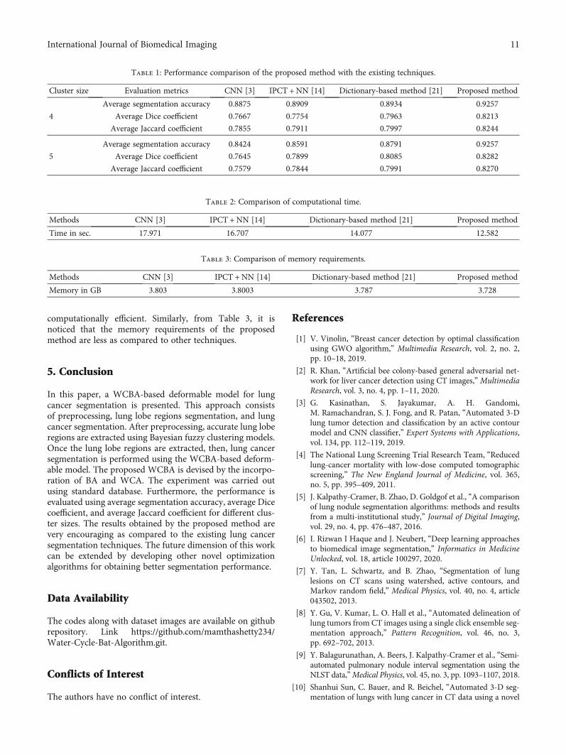
computationally efficient. Similarly, from Table 3, it isnoticed that the memory requirements of the proposedmethod are less as compared to other techniques.
5. Conclusion
In this paper, a WCBA-based deformable model for lungcancer segmentation is presented. This approach consistsof preprocessing, lung lobe regions segmentation, and lungcancer segmentation. After preprocessing, accurate lung loberegions are extracted using Bayesian fuzzy clustering models.Once the lung lobe regions are extracted, then, lung cancersegmentation is performed using the WCBA-based deform-able model. The proposed WCBA is devised by the incorpo-ration of BA and WCA. The experiment was carried outusing standard database. Furthermore, the performance isevaluated using average segmentation accuracy, average Dicecoefficient, and average Jaccard coefficient for different clus-ter sizes. The results obtained by the proposed method arevery encouraging as compared to the existing lung cancersegmentation techniques. The future dimension of this workcan be extended by developing other novel optimizationalgorithms for obtaining better segmentation performance.
Data Availability
The codes along with dataset images are available on githubrepository. Link https://github.com/mamthashetty234/Water-Cycle-Bat-Algorithm.git.
Conflicts of Interest
The authors have no conflict of interest.
References
[1] V. Vinolin, “Breast cancer detection by optimal classificationusing GWO algorithm,” Multimedia Research, vol. 2, no. 2,pp. 10–18, 2019.
[2] R. Khan, “Artificial bee colony-based general adversarial net-work for liver cancer detection using CT images,” MultimediaResearch, vol. 3, no. 4, pp. 1–11, 2020.
[3] G. Kasinathan, S. Jayakumar, A. H. Gandomi,M. Ramachandran, S. J. Fong, and R. Patan, “Automated 3-Dlung tumor detection and classification by an active contourmodel and CNN classifier,” Expert Systems with Applications,vol. 134, pp. 112–119, 2019.
[4] The National Lung Screening Trial Research Team, “Reducedlung-cancer mortality with low-dose computed tomographicscreening,” The New England Journal of Medicine, vol. 365,no. 5, pp. 395–409, 2011.
[5] J. Kalpathy-Cramer, B. Zhao, D. Goldgof et al., “A comparisonof lung nodule segmentation algorithms: methods and resultsfrom a multi-institutional study,” Journal of Digital Imaging,vol. 29, no. 4, pp. 476–487, 2016.
[6] I. Rizwan I Haque and J. Neubert, “Deep learning approachesto biomedical image segmentation,” Informatics in MedicineUnlocked, vol. 18, article 100297, 2020.
[7] Y. Tan, L. Schwartz, and B. Zhao, “Segmentation of lunglesions on CT scans using watershed, active contours, andMarkov random field,” Medical Physics, vol. 40, no. 4, article043502, 2013.
[8] Y. Gu, V. Kumar, L. O. Hall et al., “Automated delineation oflung tumors from CT images using a single click ensemble seg-mentation approach,” Pattern Recognition, vol. 46, no. 3,pp. 692–702, 2013.
[9] Y. Balagurunathan, A. Beers, J. Kalpathy-Cramer et al., “Semi-automated pulmonary nodule interval segmentation using theNLST data,”Medical Physics, vol. 45, no. 3, pp. 1093–1107, 2018.
[10] Shanhui Sun, C. Bauer, and R. Beichel, “Automated 3-D seg-mentation of lungs with lung cancer in CT data using a novel
Table 2: Comparison of computational time.
Methods CNN [3] IPCT +NN [14] Dictionary-based method [21] Proposed method
Time in sec. 17.971 16.707 14.077 12.582
Table 3: Comparison of memory requirements.
Methods CNN [3] IPCT +NN [14] Dictionary-based method [21] Proposed method
Memory in GB 3.803 3.8003 3.787 3.728
Table 1: Performance comparison of the proposed method with the existing techniques.
Cluster size Evaluation metrics CNN [3] IPCT +NN [14] Dictionary-based method [21] Proposed method
4
Average segmentation accuracy 0.8875 0.8909 0.8934 0.9257
Average Dice coefficient 0.7667 0.7754 0.7963 0.8213
Average Jaccard coefficient 0.7855 0.7911 0.7997 0.8244
5
Average segmentation accuracy 0.8424 0.8591 0.8791 0.9257
Average Dice coefficient 0.7645 0.7899 0.8085 0.8282
Average Jaccard coefficient 0.7579 0.7844 0.7991 0.8270
11International Journal of Biomedical Imaging

robust active shape model approach,” IEEE Transactions onMedical Imaging, vol. 31, no. 2, pp. 449–460, 2012.
[11] M. Liu, X. Jiang, Y. Liu, F. Zhao, and H. Zhou, “A semi-supervised convolutional transfer neural network for 3D pul-monary nodules detection,” Neurocomputing, vol. 391,pp. 199–209, 2020.
[12] Q. Hu, L. F. de Souza, G. B. Holanda et al., “An effectiveapproach for CT lung segmentation using mask region-basedconvolutional neural networks,” Artificial Intelligence in Med-icine, vol. 103, article 101792, 2020.
[13] O. Ozdemir, R. L. Russell, and A. A. Berlin, “A 3D probabilisticdeep learning system for detection and diagnosis of lung can-cer using low-dose CT scans,” IEEE Transactions on MedicalImaging, vol. 39, no. 5, pp. 1419–1429, 2020.
[14] P. M. Shakeel, M. A. Burhanuddin, and M. I. Desa, “Lung can-cer detection from CT image using improved profuse cluster-ing and deep learning instantaneously trained neuralnetworks,” Measurement, vol. 145, pp. 702–712, 2019.
[15] S. Suresh and S. Mohan, “ROI-based feature learning for effi-cient true positive prediction using convolutional neural net-work for lung cancer diagnosis,” Neural Computing andApplications, vol. 32, no. 20, pp. 15989–16009, 2020.
[16] J. Jiang, Y. C. Hu, C. J. Liu et al., “Multiple resolution residuallyconnected feature streams for automatic lung tumor segmen-tation from CT images,” IEEE Transactions on Medical Imag-ing, vol. 38, no. 1, pp. 134–144, 2019.
[17] H. Yu, Z. Zhou, and Q. Wang, “Deep learning assisted predictof lung cancer on computed tomography images using theadaptive hierarchical heuristic mathematical model,” IEEEAccess, vol. 8, pp. 86400–86410, 2020.
[18] G. Singadkar, A. Mahajan, M. Thakur, and S. Talbar, “Deepdeconvolutional residual network based automatic lung nod-ule segmentation,” Journal of Digital Imaging, vol. 33, no. 3,pp. 678–684, 2020.
[19] Y. Jalali, M. Fateh, M. Rezvani, V. Abolghasemi, and M. H.Anisi, “ResBCDU-Net: a deep learning framework for lungCT image segmentation,” Sensors, vol. 21, no. 1, p. 268, 2021.
[20] T. C. Glenn, A. Zare, and P. D. Gader, “Bayesian fuzzy cluster-ing,” IEEE Transactions on Fuzzy Systems, vol. 23, no. 5,pp. 1545–1561, 2015.
[21] A. B. Dahl and V. A. Dahl, “Dictionary based image segmenta-tion,” in Scandinavian Conference on Image Analysis, pp. 26–37, Copenhagen, Denmark, 2015.
[22] X. S. Yang, “A new metaheuristic bat-inspired algorithm,” inNature Inspired Cooperative Strategies for Optimization(NICSO 2010), vol. 284 of Studies in Computational Intelli-gence, Springer, Berlin, Heidelberg.
[23] H. Eskandar, A. Sadollah, A. Bahreininejad, and M. Hamdi,“Water cycle algorithm - a novel metaheuristic optimizationmethod for solving constrained engineering optimizationproblems,” Computers and Structures, vol. 110-111, pp. 151–166, 2012.
[24] “Lung Image Database Consortium Image Collection (LIDC-IDRI) dataset,” 2020, https://wiki.cancerimagingarchive.net/display/public/LIDC-IDRI.
12 International Journal of Biomedical Imaging