warp-based motion compensation for endoscopic …iphome.hhi.de/eisert/papers/eg2011_dl.pdfdavid c....
TRANSCRIPT

EUROGRAPHICS 2011 / N. Avis, S. Lefebvre Short Paper
Warp-based motion compensationfor endoscopic kymography
David C. Schneider1, Anna Hilsmann1 and Peter Eisert1,2
1Fraunhofer Heinrich Hertz Institute, Berlin, Germany2Humboldt Universität zu Berlin, Germany
AbstractEndoscopic videokymography is a method for visualizing the motion of the plica vocalis (vocal folds) for medicaldiagnosis. The diagnostic interpretability of a kymogram deteriorates if camera motion interferes with vocal foldmotion, which is hard to avoid in practice. We propose an algorithm for compensating strong camera motion forvideokymography. The approach is based on an image-based inverse warping scheme that can be stated as anoptimization problem. The algorithm is parallelizable and real-time capable on the CPU. We discuss advantagesof the image-based approach and address its use for approximate structure visualization of the endoscopic scene.
Categories and Subject Descriptors (according to ACM CCS): I.3.8 [Computer Graphics]: Application— I.4.8 [Im-age Processing and Computer Vision]: Scene Analysis—Motion
1. Introduction and related work
The opening and closing of the vocal folds (plica vocalis)at high frequencies is a major source of sound in humanspeech. Videokymography [SS95] is a technique for visual-izing the motion of the vocal folds for medical diagnosis:The vibrating folds are filmed with an endoscopic camerapointed into the larynx. The camera records at a very highframerate to capture vocal fold vibration. Alternatively, alow framerate and stroboscopic lighting at a frequency syn-chronized with the vibratory frequency of the vocal folds isused to obtain a temporal subsampling of the motion (seefigure 2 for example frames). The kymogram used for medi-cal diagnosis is essentially a time-slice image, i.e. an X-t-cutthrough the X-Y-t image cube of the endoscopic video (fig-ure 3). The quality and diagnostic interpretability of a ky-mogram deteriorates significantly if the camera moves rela-tive to the scene as this motion interferes with the vibratorymotion of the vocal fold in the kymogram. Scene-to-cameramotion caused by the patient or the operator of the endo-scope is hard to avoid in medical practice. In this paper, wepropose an approach to stabilizing the motion of endoscopicvideo for kymography.
This motion compensation problem is challenging anddifferent from motion compensation of handheld video (e. g.[LGJA09, LCCO09, GL08]) in several respects: Firstly, thecamera motion to be eliminated may be significantly larger
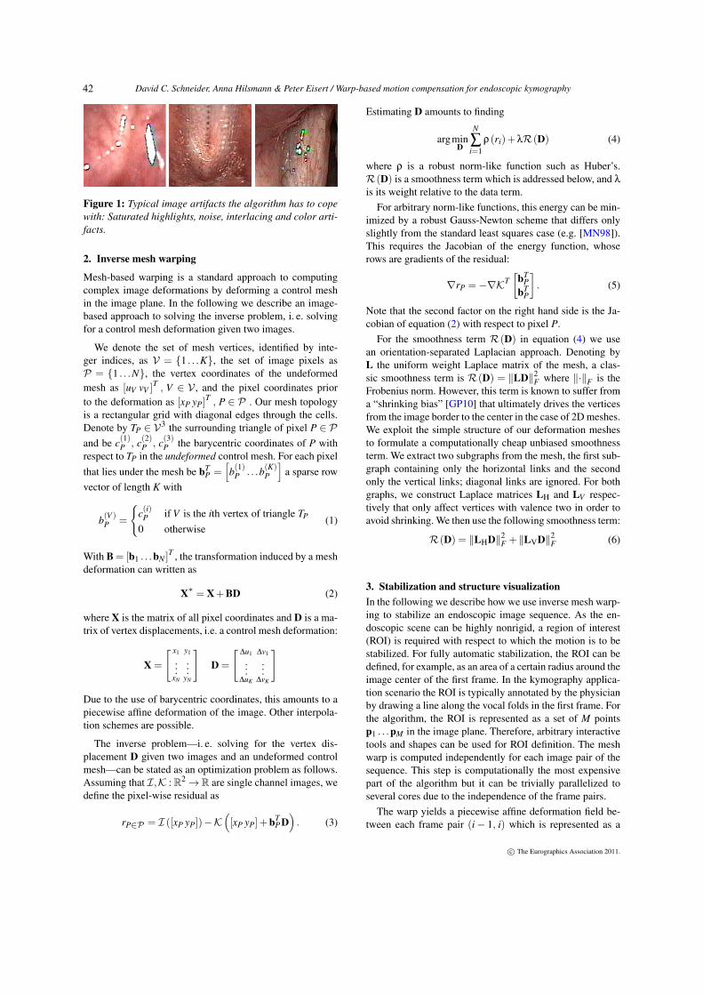
than a typical camera shake due to the short disctance be-tween camera and scene. Secondly, not only the camera andthe vocal folds move but the entire scene may be highlynonrigid, for example when the ariepiglottic fold and thecuneiform cartilage move when the patient takes breath.Therefore, a 3D camera estimation approach [LGJA09] isnot possible throughout the entire endoscopic sequence. Fi-nally, the image quality of the input material can be challeng-ing. Depending on the endoscopic system, the algorithm hasto cope with high noise levels, large areas of saturated high-lights, interlacing artifacts, depth of field blur, false colors,etc. (see figure 1).
We therefore propose an algorithm that deviates from thetypical feature-based approaches to motion compensation,but is neverthelss parallelizable and realtime capable evenon the CPU. Our method uses an image-based inverse meshwarping approach similar to [HSE10] that can be stated asan optimization problem and solved efficiently in a robustGauss-Newton framework (section 2). The inverse warpingyields a piecewise affine deformation field between two suc-cessive frames. Using the motion field, a rigid image trans-formation can be computed to compensate for the cameramotion. We discuss advantages of the image-based approachand show how the warp can be used to visualize the approx-imate 3D structure of the endoscopic scene from near-rigidparts of the sequence (section 3).
c© The Eurographics Association 2011.
David C. Schneider, Anna Hilsmann & Peter Eisert / Warp-based motion compensation for endoscopic kymography
Figure 1: Typical image artifacts the algorithm has to copewith: Saturated highlights, noise, interlacing and color arti-facts.
2. Inverse mesh warping
Mesh-based warping is a standard approach to computingcomplex image deformations by deforming a control meshin the image plane. In the following we describe an image-based approach to solving the inverse problem, i. e. solvingfor a control mesh deformation given two images.
We denote the set of mesh vertices, identified by inte-ger indices, as V = {1 . . .K}, the set of image pixels asP = {1 . . .N}, the vertex coordinates of the undeformedmesh as [uV vV ]
T , V ∈ V , and the pixel coordinates priorto the deformation as [xP yP]
T , P ∈ P . Our mesh topologyis a rectangular grid with diagonal edges through the cells.Denote by TP ∈ V3 the surrounding triangle of pixel P ∈ Pand be c(1)P , c(2)P , c(3)P the barycentric coordinates of P withrespect to TP in the undeformed control mesh. For each pixelthat lies under the mesh be bT
P =[b(1)P . . .b(K)
P
]a sparse row
vector of length K with
b(V )P =
{c(i)P if V is the ith vertex of triangle TP
0 otherwise(1)
With B = [b1 . . .bN ]T , the transformation induced by a mesh
deformation can written as
X∗ = X+BD (2)
where X is the matrix of all pixel coordinates and D is a ma-trix of vertex displacements, i.e. a control mesh deformation:
X =
[ x1 y1
......
xN yN
]D =
[∆u1 ∆v1
......
∆uK ∆vK
]
Due to the use of barycentric coordinates, this amounts to apiecewise affine deformation of the image. Other interpola-tion schemes are possible.
The inverse problem—i. e. solving for the vertex dis-placement D given two images and an undeformed controlmesh—can be stated as an optimization problem as follows.Assuming that I,K : R2→R are single channel images, wedefine the pixel-wise residual as
rP∈P = I ([xP yP])−K([xP yP]+bT
P D). (3)
Estimating D amounts to finding
argminD
N
∑i=1
ρ(ri)+λR(D) (4)
where ρ is a robust norm-like function such as Huber’s.R(D) is a smoothness term which is addressed below, and λ
is its weight relative to the data term.For arbitrary norm-like functions, this energy can be min-
imized by a robust Gauss-Newton scheme that differs onlyslightly from the standard least squares case (e.g. [MN98]).This requires the Jacobian of the energy function, whoserows are gradients of the residual:
∇rP =−∇KT[
bTP
bTP
]. (5)
Note that the second factor on the right hand side is the Ja-cobian of equation (2) with respect to pixel P.
For the smoothness term R(D) in equation (4) we usean orientation-separated Laplacian approach. Denoting byL the uniform weight Laplace matrix of the mesh, a clas-sic smoothness term is R(D) = ‖LD‖2
F where ‖·‖F is theFrobenius norm. However, this term is known to suffer froma “shrinking bias” [GP10] that ultimately drives the verticesfrom the image border to the center in the case of 2D meshes.We exploit the simple structure of our deformation meshesto formulate a computationally cheap unbiased smoothnessterm. We extract two subgraphs from the mesh, the first sub-graph containing only the horizontal links and the secondonly the vertical links; diagonal links are ignored. For bothgraphs, we construct Laplace matrices LH and LV respec-tively that only affect vertices with valence two in order toavoid shrinking. We then use the following smoothness term:
R(D) = ‖LHD‖2F +‖LVD‖2
F (6)
3. Stabilization and structure visualizationIn the following we describe how we use inverse mesh warp-ing to stabilize an endoscopic image sequence. As the en-doscopic scene can be highly nonrigid, a region of interest(ROI) is required with respect to which the motion is to bestabilized. For fully automatic stabilization, the ROI can bedefined, for example, as an area of a certain radius around theimage center of the first frame. In the kymography applica-tion scenario the ROI is typically annotated by the physicianby drawing a line along the vocal folds in the first frame. Forthe algorithm, the ROI is represented as a set of M pointsp1 . . .pM in the image plane. Therefore, arbitrary interactivetools and shapes can be used for ROI definition. The meshwarp is computed independently for each image pair of thesequence. This step is computationally the most expensivepart of the algorithm but it can be trivially parallelized toseveral cores due to the independence of the frame pairs.
The warp yields a piecewise affine deformation field be-tween each frame pair (i− 1, i) which is represented as a
c© The Eurographics Association 2011.
42

David C. Schneider, Anna Hilsmann & Peter Eisert / Warp-based motion compensation for endoscopic kymography
Figure 2: Frames from an endoscopic video sequence of the vocal folds (top), motion compensated frames (bottom).
matrix Di of vertex displacements. It can be efficiently eval-uated between the vertex locations using barycentric coor-dinates as in section 2 or a different interpolation scheme.Denote by Di (p) the displacement at p between frames(i− 1, i) . The tracked ROI point in frame i is given recur-
sively by p(i)m = p(i−1)
m +Di
(p(i−1)
m
). A stabilizing transfor-
mation T for the ith frame is given by
argminT
M
∑m=1
∥∥∥pm−T(
p(i)m
)∥∥∥2. (7)
For the kymography application, T is constrained to be arigid transformation and equation (7) is solved by an iterativedescent scheme. Additional to the stabilizing transformation,the vocal fold is centered and aligned with the Y axis beforekyogram computation.
The image-based approach to motion estimation has sev-eral key advantages for our application:
• We found it to be highly robust on images with high noiselevel and significant artifacts (see figure 1) if used with arobust error metric in equation (4). All results shown inthe paper were generated with the Huber function.
• For computing the transformation, no explicit handling ofoutliers by RANSAC or similar methods is required. Thisis an significant advantage over feature-based approaches.
• As a global optimization scheme, the appraoch benefitsfrom the “filling in” effect of the smoothness term thatpropagates information into image regions with little gra-dient information. It is therefore robust against unevendistribution of trackable image content.
• The choice of mesh granularity and weight of the regular-ization term allow for fine-grained control over the degreeof deformation the warp is allowed to follow. It can beadjusted to track camera motion and large-scale scene de-formation but to ignore the small-scale motion of the vo-cal folds which must not be “tracked away”. High qualitykymograms can be extracted even over large-scale defor-mations that occur when the patient takes breath.
The warp’s displacement fields can also be used to visual-ize the approximate threedimensional structure of the en-doscopic scene. Note that this part of the paper is workin progress. For structure computation, we manually iden-tify a part of the image sequence that displays relatively lit-tle large-scale nonrigid motion. We then use the frame-to-frame displacement fields to track the vertices of a densemesh in the image plane in the same way as the ROI pointsdescribed above. Note that this mesh is different from themeshes used for warp estimation, which are not temporallyconsistent over all frames in order to allow for parallel com-putation of the warps. From the vertex trajectories, affinestructure from motion is computed with the factorization al-gorithm of [TK92]. The endoscopic scenes generally violatethe rigidity assumption on which the factorization algorithmis based even when the “most rigid” part of a sequence ischosen. Consequently, structure results are not precise re-constructions but rather visualizations of the approximatethreedimensional shape of the scene. Examples are shownin figure 4.
4. Results and ConclusionThe algorithm processes about 25 frames per second on av-erage on a six core machine with parallel warp estimation.The GPU is currently not used as capable GPUs still arerarely available in medical computing environments. Fig-ure 2 shows frames from an endoscopic video sequence andtheir motion-compensated counterparts. Figure 3 shows ky-mograms computed from two sequences. For comparison,the kymograms were computed from the uncompensatedvideo sequence, from the sequence compensated with thewidely used “Deshaker” tool [Des] and from the sequencecompensated with our approach. While the Deshaker kymo-grams show less jitter than the uncompensated ones, large-scale camera motion is not properly corrected. Especially thefirst row in figure 3 shows that the kymograms compensatedwith our method convey significantly more information thanthe comparison. Regarding differences in the kymograms,
c© The Eurographics Association 2011.
43

David C. Schneider, Anna Hilsmann & Peter Eisert / Warp-based motion compensation for endoscopic kymography
Figure 3: Vocal fold kymograms from two endoscopic se-quences. Left column: no motion compensation. Center col-umn: Deshaker [Des] compensation. Right column: pro-posed method. At the wide openings in the first row the pa-tient takes breath during the recording.
also note that the scene moves relative to the scanline withrespect to which the kymogram is computed if the sequenceis not properly compensated. Figure 4 shows first results ofour structure visualization approach.
In conclusion, we proposed an image-based warp estima-tion approach to compensate camera motion in endoscopicvideo. Our method deals with large motion, largely non-rigid scenes as well as poor image quality and image arti-facts. Moreover, we showed first results on using our mo-tion data for visualizing the approximate 3D shape of theendoscopic scene. Identifying the parts of the sequence suit-able for structure computation and improving structure re-sults will be the principal topic of further research.
References[Des] http://www.guthspot.se/video/deshaker.htm. 3, 4
Figure 4: Structure computation results for two sequences.Artificial lighting was added to emphasize the 3D shape.
[GL08] GLEICHER M. L., LIU F.: Re-cinematography: Improv-ing the camera dynamics of casual video. ACM Trans. on Multi-media 5 (2008). 1
[GP10] GRADY L., POLIMENI J.: Discrete Calculus. Springer,2010. 2
[HSE10] HILSMANN A., SCHNEIDER D. C., EISERT P.: Real-istic cloth augmentation in single view video under occlusions.Comput. Graph. 34 (October 2010), 567–574. 1
[LCCO09] LEE K., CHUANG Y., CHEN B., OUHYOUNG M.:Video stabilization using robust feature trajectories. In Proc. Int.Conference on Computer Vision (Kyoto, japan, 2009), pp. 1397–1404. 1
[LGJA09] LIU F., GLEICHER M., JIN H., AGARWALA A.:Content-preserving warps for 3d video stabilization. ACM Trans.Graphics 28 (July 2009), 44:1–44:9. 1
[MN98] MCCULLAGH P., NELDER J.: Generalized Linear Mod-els. Chapman & Hall, 1998. 2
[SS95] SVEC J. G., SCHUTTE H. K.: Videokymography: High-speed line scanning of vocal fold vibration. Journal of Voice 10 /2 (1995), 201–205. 1
[TK92] TOMASI C., KANADE T.: Shape and motion from im-age streams under orthography: A factorization method. Interna-tional Journal of Computer Vision 9 (1992), 137–154. 3
c© The Eurographics Association 2011.
44