vulms.vu.edu.pk · web viewwater, ph and buffer solutions. 92 buffer solutions 93 citric acid...
TRANSCRIPT

BIO303
BIOCHEMISTRY-II

VIRTUAL UNIVERSITY OF PAKISTAN
Sr. No.
TitlePage
No.
1 BioEnergetics
2 The Standard Free-Energy
3 Eq constant and Standard free energy -I
4 Eq constant and Standard free energy -2
5 Chemical Logic and Common Biochemical Reactions-1

6 Chemical Logic and Common Biochemical Reactions -2
7Chemical Logic and Common Biochemical Reactions- II-check final-3
8 Phosphoryl Group Transfers and ATP-.pptx
9 Phosphoryl Group Transfers in thioesters.pptx
10 Bioenergetics -3-Hydrolysis of ATP- part 1
11 Bioenergetics -3-Hydrolysis of ATP- part2.pptx
12Bioenergetics -3-ATP energizes active transport and muscle contraction.pptx
13 Biological Oxidation -reduction reactions-2-4-18.pptx
14 Reduction potential -
15 Universal Electron Carriers
16 GLYCOLYSIS-1
17 Glycolysis- the pay off phase
18 Preparatory phase of Glycolysis Requires ATP.pptx
19 Preparatory phase of Glycolysis Requires ATP - cont-step 3
20 Preparatory phase of Glycolysis Requires ATP-3
21 Preparatory phase of Glycolysis -step 5
22 Pay off phase-Last steps of glycolysis-6-7
23 Last steps of glycolysis. -8-10

24 Energy generated during Glycolysis- part 1 and 2.pptx
25 Importance of Phosphorylated Intermediates.pptx
26 Fates of Pyruvate.pptx
27 Fates of Pyruvate -Lactic acid fermentation.pptx
28 Ethanol Fermentation-.pptx
29 Uses of Fermentation in Industry.pptx
30 Industrial Fermentation-steps of fermentation.pptx
31 Industrial Fermentation-cont-5
32 Lecture - 32 - batch fermentation.pptx
33 Continuous Fermentation-6.pptx
34 Gluconeogenesis
35 Gluconeogenesis -cont
36 Gluconeogenesis - 1st by pass pathway-complete
37 Gluconeogenesis - 2nd-3rd by pass pathways-
38 Gluconeogenesis - cont - 4
39 Gluconeogenesis - cont -5
40 Pentose Phosphate Pathway - PPP -1
41 Pentose Phosphate Pathway - PPP -2-.pptx

42 Pentose Phosphate Pathway - PPP -3
43 feeder pathways of glycolysis -1.
44 feeder pathways of glycolysis -2
45 feeder pathways of glycolysis-3 - galactosemia
46 Denaturation of DNA
47 Denaturation of DNA.. contd
48 Renaturation of DNA
49 RNA
50 Difference b/w DNA and RNA
51 Structure of RNA
52 2ndry structure of RNA
53 Function of mRNA
54 Transfer RNA
55 Transfer RNa..1
56 Transfer RNa..2
57 Ribosomal RNa
58 snRNA
59 Enzymes

60 IUB Classification of enzymes-
61 Lyases
62 Ligases
63 Ligand
64 Mechanism of enzyme action
65 Mechanism of enzyme action..cont -1
66 Mechanism of enzyme action..cont -2
67 Mechanism of enzyme action..cont -3
68 Cofactor, coenzyme and prosthetic groups
69 Cofactor, coenzyme and prosthetic groups-1
70 Reaction rate and order of reaction
71 Factors affecting enzyme activity
72 Factors affecting enzyme activity (cont)
73 Enzyme kinetics
74 Enzyme kinetics..cont I
75 Enzyme kinetics..cont II
76 Enzymes...Michaelis-Mention Eq.
77 Enzymes...Michaelis-Mention Eq.cont-1
78 Enzymes...Michaelis-Mention Eq.cont-2
79 Enzymes...Michaelis-Mention Eq.cont-3
80 Enzymes... Order of Reaction
81 Lineweaver-burk plot
82 Inhibition of enzyme activity
83 Competitive inhibition
84 Competitive inhibition.. contd
85 Non-competitive inhibition
86 Enzymes.. effect on lineweaver-burk plot
87 Water
88 Ionization of water
89 Buffer System.. working solutions
90 Weak acids and bases
91 Water, pH and Buffer Solutions.
92 Buffer Solutions
93 Citric acid cycle-1
94 Citric acid cycle-2
95 Citric acid cycle-3

96 PDH complex-Citric acid cycle - 4
97 PDH complex-2
98 Steps of TCA-8 enzymes-
99 TCA-step 1.
100 TCA-step2
101 TCA-step 4
102 TCA-step 5
103 TCA-ending.pptx
104 fatty acid metabolism-1
105 Anaplerotic reactions of TCA-
106 Glyoxalate cycle -1
107 Glyoxalate cycle -2
108 Glyoxalate cycle -3
109 Fatty acid-1.pptx
110 fatty acid metabolism-2
111 fatty acid metabolism-effect of hormones-
112 carnitine shuttle
113 fatty acid metabolism-fatty acid oxidation-3 stages

114 beta oxidation-complete
115 The four steps are repeated to yield acetyl CoA and ATP
116 "Oxidation of unsaturated fatty acids requires 2 additional steps "
117Complete Oxidation of odd-numbered fatty acids requires 3 additional steps
118 Coenzyme B12
119Transcription Factors Turn on the Synthesis of Proteins for Lipid Catabolism
120 Oxidation in peroxisomes -
121 Oxidation in peroxisomes -cont
122 Omega Oxidation
123 Oxidation of phytanic acid
124 Ketone bodies
125 Ketone bodies-continued


Topic#1
BIOENERGETICS
Welcome to the course biochemistry-II- In this course you will learn the principles of biochemistry and their application on health and disease. In the beginning we will discuss the concepts of bioenergetics.. As the terminology is self explanatory.. BIO and nergetics.. Meaning that we will be dealing with the concepts of energy transfer in biological systems.
Generally speaking, we know that the Living cells have to perform certain work to stay alive, to grow, and to reproduce.
They should have the ability to trap energy and use it into biological work is a fundamental property of all living organisms, but they use it in different ways.
Modern organisms use the chemical energy in fuels to synthesize complex, highly ordered macromolecules
For example: Plants use energy in such a manner that causes them to release oxygen as a waste product, while animals (which includes humans, of course) require oxygen as a component to survive.
BIOENERGETICS:
Bioenergetics is the quantitative study of the energy transductions, that is.. changes of one form of energy into another—that occur in living systems, and of the nature and function of the chemical processes underlying these transductions.

Bioenergetic processes:
Glycolysis is a biological process in which glucose is broken down into pyruvate thus generating two molecules of ATP (Adenosine triphosphate) per molecule of glucose
Gluconeogenesis is another process in which the cell synthesizes glucose from biomolecules such as proteins, amino acids and fats
Citric acid cycle generates molecules of ATP by oxidation of energy stored in food material
All animal and plant cells are powered by chemical energy stored in the chemical
Bonds of organic molecules. Animals convert the chemical energy of fuels into motion and heat and
In a few organisms such as fireflies chemical energy is converted into light…
Lets look at some Examples of major bioenergetic processes occurring in nature
ATP is the energy currency in biological systems
Citric acid cycle, that is the process of cellular respiration generates molecules of ATP by oxidation of energy stored in food material
These processes will be discussed in detail in coming lectures.
fireflies convert energy into light

Laws of Thermodynamics:
Biological Energy Transformations Obey the Laws of Thermodynamics.There are two fundamental laws of thermodynamics.
1. First law
The first law is the principle of the conservation of energy: it states that
“for any physical or chemical change, the total amount of energy in the universe remains constant; energy may change form or it may be transported from one region to another, but it cannot be created or destroyed.”
However, it looks that the direction of energy flow is from 1 direction to another.. To understand that we look at the 2nd law of thermodynamics for which should be familiar with the concept of entropy
Entropy:
The term “entropy,” was first used in 1851 by Rudolf Clausius, which literally means “a change within” a system.. and it can be described as disorder..
Entropy = disorder
Entropy is a quantitative expression for the randomness or disorder in a system. When the products of a reaction are less complex and more disordered than the reactants, the reaction is said to proceed with a gain in entropy.
Denoted by S
Delta S has a +ve sign when entropy increase and –ve when entropy decreases
units of entropy are joules/mole.Kelvin (J/mol.K) (joules/mole multiply by Kelvin).By convention, S has a positive sign when entropy increases and H, as noted above, has a negative sign when heat is released by the system to its surroundings.
Second Law of Thermodynamics:
With this idea about entropy, we come to the 2nd law ..which states that..

“the universe always tends towards increasing disorder and in all natural processes, the entropy of the universe increases… give example of hot cup.. Now the point is that entropy is not a measure of energy ..it only describes that how energy is distributed within a system.”
Enthalpy (H):
The term that describes the energy of a system is Enthalpy.Enthalpy is the heat content of the reacting system.
It reflects the number and kinds of chemical bonds in the reactants and products
When a chemical reaction releases heat, it is said to be exothermic and value of H is negative
When a chemical reaction takes up heat from surroundings, it is called as endothermic and have positive values of HWhen a chemical reaction releases heat, it is said to be exothermic; the heat content of the products is less than that of the reactants and H has a negative value.
On the other hand, Reacting systems that takes up heat from their surroundings are endothermic and have positive values of H. Enthalpy and entropy of a system are related and help us to understand Gibb’s free energy
Gibbs free energy (G):
Another terminology thermodynamic quantity that describe the energy changes occurring in a chemical reaction:
Gibbs free energy, G expresses the amount of energy capable of doing work during a reaction at constant temperature and pressure
When a reaction proceeds with the release of free energy the free-energy change, G, has a negative value and the reaction is said to be exergonic
In endergonic reactions, the system gains free energy and G is positive
The units of G and H are joules/mole or calories/mole
(1 cal is equal to 4.184 Joules)
Under the conditions existing in biological systems (including constant temperature and pressure), changes in free energy, enthalpy, and entropy are related to each other quantitatively by the equation

G: is the change in Gibbs free energy H: the change in enthalpyT: the absolute temperature S: change in entropyin which G is the change in Gibbs free energy of the reacting system, H is the change in enthalpy of the system, T is the absolute temperature, and S is the change in entropy of the system.
Either of these conditions, which are typical of favorable processes, tend to make G negative. In fact, G of a spontaneously reacting system is always negative.
Topic#2
The Standard Free-Energy
We already talked about Gibbs free energy.. Now we will show that why do we need to know about this terminology in biological systems…. We know that the living cells require some sources to provide Energy.. So that they can perform the functions of life..
will talk about Free Energy …that can be used by living cells…Cells Require Sources of Free Energy..
Cells are isothermal systems—they function at essentially constant temperature (and also function at constant pressure).
Heat flow is not a source of energy for cells, because heat can do work only as it passes to a zone or
object at a lower temperature. The energy that cells can and must use is free energy, is known as Gibbs freeenergy (G) in terms of thermodynamics.
.. This allows prediction of the direction of chemical reactions, their exact equilibrium position,
and the amount of work they can (in theory) perform at constant temperature and pressure. Both autotrophic
And Heterotrophic cells acquire it from absorbed solar radiation.

Both kinds of cells transform this free energy into
ATP and other energy-rich compounds that provide enrgy to carry out functions of life.
Cells require sources of Free Energy (G):
Significance of Free-Energy Changes (G):
There are some scientific facts that reveal the significance of Gibbs free energy..
Urdu.. Why is this so important for us.. Because of the reason that .. ..
Say all .. And we will discuss them while going through the present lesson
Actual free-Energy changes depend on Reactantand Product Concentrations
Allows prediction of the direction of chemical reactions, their exact equilibrium position and the amount of work done
The free-energy change for a reaction is independent of the pathway by which the reaction occurs
Free-energy changes are additive; the net chemical reaction that results from successive reactions sharing a common intermediate has an overall free-energy change that is the sum of the G values for the individual reactions
Standard free-energy G:
But before that we should know that what is standard free energy… Under standard conditions of temperature and pressure, meaning (298 K or 25C), when reactants and products are initially present at 1 M concentrations or, for gases, at partial pressures of 101.3 kilopascals (kPa), or 1 atm, the force driving the system toward equilibrium is defined as the standard free-energy change, G where knot is the symbol to depict that the reaction occurred under standard conditions 0-0-

The free energy of reaction at under standard conditions
When Temperature: 298K or 25C, when reactants are initially present at 1 M concentrations or, for gases, at partial pressures of 101.3 kilopascals (kPa), or 1 atm, the force driving the system toward equilibrium is defined as the standard free-energy change, G
G = H - T S
Key conventions in biochemical reactions
However, most biochemical reactions, however, occur in well-buffered aqueous solutions near pH 7; both the pH and the concentration of water (55.5 M) are taken as constant
Most biochemical reactions occur near pH 7 (buffered)
Standard biochemical state
(H+) is 10-7 M
concentration of water is 55.5 M
Both the pH and the concentration of water are essentially constant
Under these conditions, G is written as G‘ (with prime)
Concentration of water, H+ and Mg+ are not included in equations for simplicity
Molar concentration of water:
Suppose we have 1 L of water. (1000 ml). The density of water is 1 g / ml, therefore in 1000 ml of water must be 1000g. Well, if the water is pure, it remains to be seen how many moles of water we have. 1 mol of water is 18 g, so 1000 g will therefore be 55.555 moles. Now if molarity is defined as moles / L, then we 55.555 moles / L is therefore 55.555 M. Similarly, concentrations are taken to solutions, that is, is perfect that you have to aproach what concentration of OH- or H + ions are in the water (supposedly 10'-7 M) that leaves the water

Autoprotolysis. I don´t want to go around the bush but be careful what you use this "concentration" for because if you're in a water balance is not good that you consider water as a species, as its "concentration" is contained somehow in constant balanc
Density of pure water = 1000 g/L
Molar mass of water = 18 g/mol (2H + 1 Oxygen=2+16=18)
Molar concentration of water = d/m
= 1000 / 18.02
= 55.5 mol/L
Topic#3
Eq constant and Standard free energy -I
The Standard Free-Energy is directly related to the Equilibrium constant
What is Equilibrium constant (K)?:
Now we are going to talk about the eq- constant K .. And its relationship with free energy.. The Standard Free-Energy Change Is Directly Related to the Equilibrium Constant
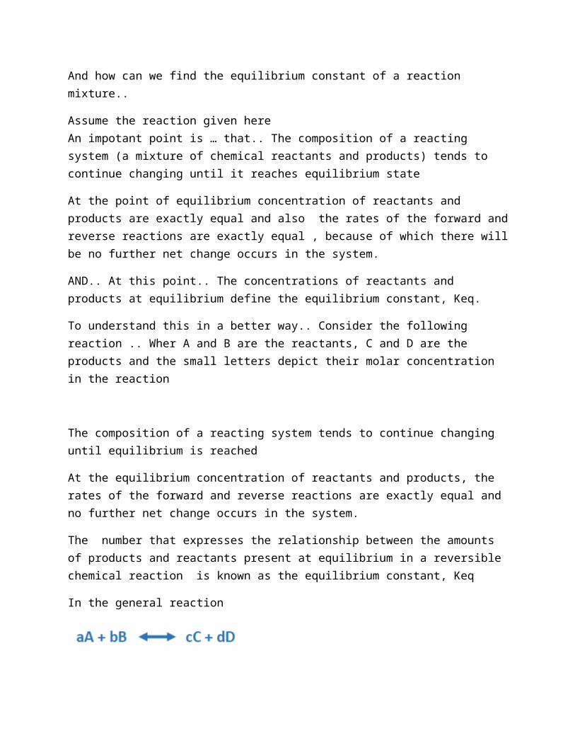
And how can we find the equilibrium constant of a reaction mixture..
Assume the reaction given hereAn impotant point is … that.. The composition of a reacting system (a mixture of chemical reactants and products) tends to continue changing until it reaches equilibrium state
At the point of equilibrium concentration of reactants and products are exactly equal and also the rates of the forward and reverse reactions are exactly equal , because of which there will be no further net change occurs in the system.
AND.. At this point.. The concentrations of reactants and products at equilibrium define the equilibrium constant, Keq.

To understand this in a better way.. Consider the following reaction .. Wher A and B are the reactants, C and D are the products and the small letters depict their molar concentration in the reaction
The composition of a reacting system tends to continue changing until equilibrium is reached
At the equilibrium concentration of reactants and products, the rates of the forward and reverse reactions are exactly equal and no further net change occurs in the system.
The number that expresses the relationship between the amounts of products and reactants present at equilibrium in a reversible chemical reaction is known as the equilibrium constant, Keq
In the general reaction
How to calculate Equilibrium constant?:
Lets talk how to get the equilibrium constant
Assume the reaction given here… Let’s try to understand it with an example.. We have a reacting system where A and B are the reactants, with molar concentration denoted by small a and b
Whereas C and D are the products of this particular reaction and their molar concentartions are denoted by small c and d respectively.
When the reaction is at equilibrium.. The eq. constant for this reaction is (the prioduct of conc. Of the products divided by the product of conc. Of the reactants
At this point, the equilibrium constant is the ratio of concentration of reactants to that of products and can be written as this equation..
When a reacting system is not at equilibrium, the
tendency to move toward equilibrium represents a
driving force, the magnitude of which can be expressed
as the free-energy change for the reaction, DG

The equilibrium constant is given by following equation where [A], [B], [C], and [D] are the molar concentrations of the reaction components at the point of equilibrium, equilibrium constant can be written as:
Standard free-energy change:
ADD EQUATION
The standard free-energy change of a chemical reaction is simply an alternative mathematical way of expressing its equilibrium constant.
G is characteristic for a given reaction and can be calculated from the equilibrium constant for the reaction:
Or we can say that delta G and eq constant are related b this equation .. Gibbs free energy (understandsrd conditions (rep by knot) = negative R (gas constant * temp _ln of eq constant
where
R is the gas constant (8.31 cal/Mol.K)
T is standard temperature (25 C or 298K)
K’eq is the equilibrium constant
And we should kn ow the value of delta G relevant to the value of eq constant

If the Keq is equal to 1.0
the standard free-energy change (GO) of that reaction is 0.0 (the natural logarithm (ln) of 1.0 is zero)…EQUILIBRIUM
If Keq of a reaction is greater than 1.0
GO is negative, forward direction is favored
If Keq is less than 1.0
GO is positive, reverse direction is favored
Topic#4
Eq constant and Standard free energy -2

The Standard Free-Energy is directly related to the Equilibrium constant.
G and G’ O are related:
The free energy change (DG) of a reaction determines its spontaneity. A reaction is spontaneous if DG is negative (if the free energy of products is less than that of reactants).
The actual free-energy change, G is a variable that depends on G’ and on the concentrations of reactants and products:
The concentration terms in this equation express the effects commonly called mass action, and the term [C]c[D]d/[A]a[B]b is called the mass-action ratio, Q.
Thus the equation can be written a follows:
Lets suppose that the reaction is under standard conditions..
But the conc. Of reactants and products is NEITHER equal to eachother… Nor the conc. Is 1M
non-standard concentrations
Actual conc. Of reactants and products but standard values of G’, R and T
Value of G at equilibrium (for spontaneous reaction)??
Standard Free-Energy Changes Are Additive:
Consider two sequential chemical reactions
Since the two reactions are sequential, we can write the overall reaction as
The ΔG'0 values of sequential reactions are additive.

Topic#5
Eq constant and Standard free energy -2
Chemical Logic and Common Biochemical Reactions
Most cells have the capacity to carry out thousands of specific, enzyme-catalyzed reactions:
for example, transformation of a simple nutrient such as glucose into amino acids, nucleotides, or lipids
extraction of energy from fuels by oxidation
polymerization of monomeric subunits into macromolecules
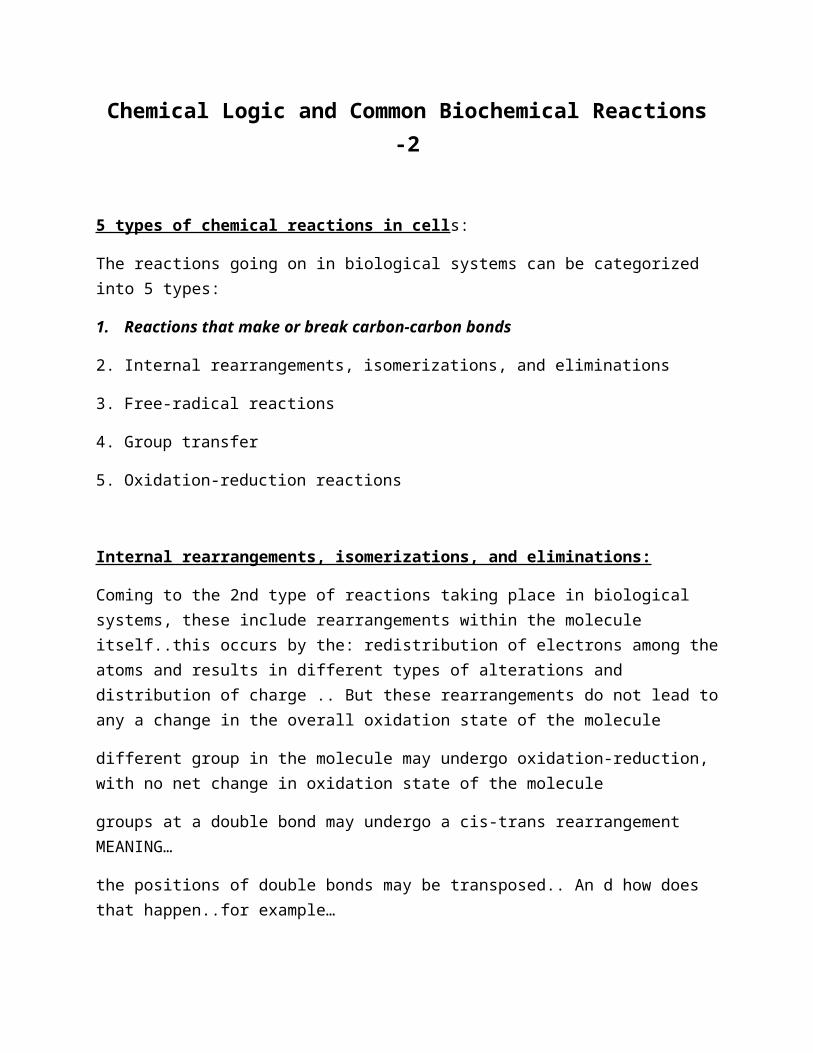
5 types of chemical reactions in cells:
The reactions going on in biological systems can be categorized into 5 types:
1. Reactions that make or break carbon-carbon bonds
2. Internal rearrangements, isomerizations, and eliminations
3. Free-radical reactions
4. Group transfer
5. Oxidation-reduction reactions
2 basic chemical principles:
And before we study the details of these chemical reactions we should revise 2 basic principles..1st principle is that..say on slide…
So what is a covalent bond.. Lets review the concept..A covalent bond, is a chemical bond that involves 2 atoms that share their electron pair. These electron pairs are known as shared pairs or bonding pairs, and the stable balance of attractive and repulsive forces between atoms, when they share electrons, is known as covalent bonding.

Atoms make covalent bond with other atoms in order to gain more stability, which is achieved by forming a full electron shell. By sharing their outer most (valence) electrons, atoms can fill up their outer electron shell and become more stable.
A covalent bond can be broken in two general ways
Homolytic cleavage
Heterolytic cleavage
A Covalent bond, also called a molecular bond, involves the sharing of electron pairs between atoms.
These electron pairs are known as shared pairs or bonding pairs, and the stable balance of attractive and repulsive forces between atoms, when they share electrons, is known as covalent bonding.
2 mechanisms for breaking a C-C or C-H bond:
Homolytic cleavage: each atom leaves the bond as a radical, carrying one unpaired electron
Heterolytic cleavage (more common): one atom retains both bonding electrons

Second principle:
Many biochemical reactions involve interactions between nucleophiles and electrophiles
Nucleophiles: functional groups rich in and capable of donating electrons
(Nucleophiles are Lewis bases)
nucleophiles: meaning nucleus loving”, or “positive-charge loving ..are the functional groups rich in electrons and can donate them to form a covalent bond
which is the exact definition of a Lewis base. In other words, nucleophiles are Lewis bases.

Electrophiles: electron-deficient functional groups that seek electrons
Electrophile means.. electron-loving”, or “negative-charge loving” these are the functional groups that are electron-deficient and seek electrons.. to form a covalent bond
Nucleophiles combine with Electrophiles and share electrons with them
There are some Common nucleophiles and electrophiles found in biochemical/biological reactions
Common nucleophiles and electrophiles in biochemical reactions:
There are some Common nucleophiles and electrophiles found in biological reactions
Examples of Nucleophiles include…. Name all of them and say.. As the picture shows, all these have –ve charge due to a pair of electrons that can be donated to make covalent bonds…And oxygen of hydroxide ion (Hydroxide is a diatomic anion with chemical formula OH−. It consists of an oxygen and hydrogen atom held together by a covalent bond, and carries a negative electric charge. It is an important but usually minor constituent of water. It functions as a base, a ligand, a nucleophile and a catalyst.)
Protonated imine group.. Imine group is a functional group in a chemical compound that contains a carbon–nitrogen double bond. In protonated imine group, R group is activated and donates electrons to the carbon atom that can be given to the nitrogen that has +ve charge..
Another example is when the phosphorus atom in phosphate group
Or when a hydrogen atom having positive charge accepts an elctron pair..such kind of hydrogen atom is called a Brønsted acid, or just “acid”.
the extent to which a species can accept a lone pair of electrons is called “electrophilicity”.
The nitrogen atom can be attached to a hydrogen (H) or an organic group (R). If this group is not a hydrogen atom, then the compound can sometimes be referred to as a Schiff base.
Phosphorus (15P) - being the fifteenth element - has fifteen electrons, five valence electrons and the following electron configuration:

1s22s2 2p63s2 3p3
one might expect it to form 3 single covalent bonds and be happy. But it doesn't, it forms five bonds, presumably using each of its five electrons. My first question is - why would it do this? It seems to me that this would result in the Phosphorus atom having ten electrons in its outer shell (four from the double bond with the oxygen and two each from the single bonds with the other three oxygens).
Apart from the oxygen with a double-bond, we have three oxygens forming single bonds. Does this not leave them one electron short?
Finally, why are the oxygens shown as carrying a negative charge?
phosphorus has stable oxidation states in compounds, ranging from −III to +V.
First of all, let me state the obvious: Phosphorus is awesome. After we got that out of the way we can focus on why.
Apart from this it is possible to formulate many different cations and anions, that are derived from the above molecular structures. Just to name a few, there are P3+,P5+,P7+,P9+
But now for the most important part, phosphorus has stable oxidation states in compounds, ranging from −III
to +V. Here are a few examples: P−IIIH3, P2−IIH4, [P−IH]n, P4±0, H3P+IO2, H4P2+IIO4, H3P+IIIO3, H2P2+IVO6, H3P+VO4
While dealing with these compounds it is usually completely unnecessary to describe bonding with hybrid orbitals.
In case of phosphane PH3
it would be wrong. Assuming sp3P one would expect (H−P−H)≈109 , while it is found to be ∠ ∘(H−P−H)=93.5 , which is almost the same angle as the porbitals are having towards each ∠ ∘
other.
In general your assumption is correct, that it is possible to form only three covalent bonds to reach a stable configuration. And that will most likely be the case when phosphorus forms compounds with more electropositive elements.
Now dealing with oxygen, means dealing with a much more electronegative element, i.e. En(O)≈3.4

, En(P)≈2.2. That also means that bonds are much more polarised towards the oxygen.
Analysing the phosphate anion PO43−
it is crucial to recognise its symmetry, which is tetrahedral Td. In this arrangement it is perfectly safe.
Very observant! What happens is that phosphorus is hybridized in the phosphate ion. Hybridization allows the central phosphate atom to form more bonds than you might expect from its ground state electron configuration.
Within the framework of hybridization, one may look at the bonds in the phosphate anion as having a great deal of ionic character.
The electronegativity of phosphorus is 2.19; the electronegativity of oxygen is 3.44. The difference is 1.25 - or rather sizable. As a result, electron density is largely withdrawn from the central phosphorus. Hence the negative charges you see on the three oxygen molecules in your picture.
Other depictions, which reject a large degree of d-orbital utilization in hybridization, might give a negative formal charge to all the oxygens and a positive formal charge to the central phosphorus by eschewing the P=O
double bond.
Apart from the oxygen with a double-bond, we have three oxygens forming single bonds. Does this not leave them one electron short?
Finally, why are the oxygens shown as carrying a negative charge?
You could draw double bonds for all the oxygens but there aren't that many valence electrons to be distributed. The structure shown above is a compromise between number of valence electrons and electronegativity considerations - we wouldn't want a surfeit of electrons around the central phosphorus since that's not as thermodynamically favorable as having the more electronegative oxygen support these electrons.
In addition, the sum of the formal charges on an atom must add up to the overall charge on the atom. Phosphate anion is triply negative charged; note how there are three oxygens bearing a single negative charge.
Electron pushing: The movement of a pair of electrons (a lone pair of electrons or a bond) from an electron rich site to an electron poor site.

Significance of Carbonyl group:
Carbonyl groups are particularly important in the chemical transformations of metabolic pathways…
The carbon of a carbonyl group has a partial positive charge due to the electron-withdrawing property of the carbonyl oxygen electrophilic carbon

Carbonyl group facilitate the formation of a carbanion’s on an adjoining carbon by delocalizing the carbanion’s negative charge
An imine group can serve a similar function
electrophile carbocation
Nucleophile carbanion

Topic#6
Chemical Logic and Common Biochemical Reactions -2
5 types of chemical reactions in cells:
The reactions going on in biological systems can be categorized into 5 types:
1. Reactions that make or break carbon-carbon bonds

2. Internal rearrangements, isomerizations, and eliminations
3. Free-radical reactions
4. Group transfer
5. Oxidation-reduction reactions
Internal rearrangements, isomerizations, and eliminations:
Coming to the 2nd type of reactions taking place in biological systems, these include rearrangements within the molecule itself..this occurs by the: redistribution of electrons among the atoms and results in different types of alterations and distribution of charge .. But these rearrangements do not lead to any a change in the overall oxidation state of the molecule
different group in the molecule may undergo oxidation-reduction, with no net change in oxidation state of the molecule
groups at a double bond may undergo a cis-trans rearrangement MEANING…
the positions of double bonds may be transposed.. An d how does that happen..for example…
Isomerization
Enediol intermediate:The enediol is also an intermediate for the epimerization of an aldose or ketose. The reactions are usually base catalyzed, but can also take place under acid or neutral conditions. A typical rearrangement reaction is that between the aldose glyceraldehyde and the ketose dihydroxyacetone in a chemical equilibrium.
Reaction is also an example of elimination as .. Is eliminated ..

Cis-trans rearrangement:
Cis–trans isomerism, also known as geometric isomerism or configurational isomerism. Cis is illustrated by... The presence of functional groups on the same side of the carbon chain while trans conveys that functional groups are on opposing sides of the carbon chain.
So while considering this kind of isomerism in living system, we can quote the example of proline amino acid. In general, Peptide bonds formed between amino acid residues are in trans-configuration… more than 99.95% have However, in case of proline, Peptide bonds formed are able to exist both as cis and trans isomers.. through peptidyl-prolyl (proline) isomerase enzyme
Prolyl isomerase ..this enzyme is (EC 5.2.1.8) found in both prokaryotes and eukaryotes and interconverts the cis and trans isomers of peptide bonds with the amino acid proline.
In general, Peptide bonds formed between amino acid residues with alpha carbons in trans-configuration… (more than 99.95% have trans configuration.)
For peptide bonds involving the imino nitrogen of proline, peptide bonds are able to exist both in cis and trans configuration.. (6% are in the cis configuration) through peptidyl-prolyl (proline) isomerase enzyme

Elimination:
A water molecule is lost.. And a double bond is formed. overall oxidation state is the loss of water from analcohol, resulting in the introduction of a CP C bond d b/w 2 adjacent carbon atoms
The loss of water from an alcohol, resulting in the introduction of a C=C bond b/w 2 adjacent carbon atoms
An elimination reaction with no changes in oxidation state

Free-radical reactions:
Coming to free radical reactions.. From where we get these free radicals.. Remember we talked about Homolytic cleavage of covalent bond in which electrons pair is equally distributed and radicals are generated.. So cells have got some free radicals and they use them in certain reactions like/..
There are certain biological reactions in which Vit.B12 is used a s cofactor by enzymes. Among them, methylmalonyl-CoA mutase (MCM) has been extensively studied. This enzyme catalyzes the reversible isomerization of L-methylmalonyl-CoA to succinyl-CoA using adenosylcobalamin (AdoCbl) as a cofactor participating in the generation of radicals that allow isomerization of the substrate.
Certain radical-initiated decarboxylation reaction
3) Some reduction reaction, such as that catalyzed by ribonucleotide reductase
4) Some rearrangement reactions, such as that catalyzed by DNA photolyase.. That is a DNA repair enzyme. It binds to UV-damaged DNA containing pyrimidine dimers and, breaks the cyclobutane ring joining the two pyrimidines of the dimer and helps to avoid possible mutations.
Vitamin B12 is an organometallic compound with important metabolic derivatives that act as cofactors of certain enzymes, which have been grouped into three subfamilies depending on their cofactors. Among them, methylmalonyl-CoA mutase (MCM) has been extensively studied. This enzyme catalyzes the reversible isomerization of L-methylmalonyl-CoA to succinyl-CoA using adenosylcobalamin (AdoCbl) as a cofactor participating in the generation of radicals that allow isomerization of the substrate. The crystal structure of MCM determined in Propionibacterium freudenreichii var. shermanii has helped to elucidate the role of this cofactor AdoCbl in the

reaction to specify the mechanism by which radicals are generated from the coenzyme and to clarify the interactions between the enzyme, coenzyme, and substrate.
Homolytic cleavage of covalent bond produce free radical
Isomerizations that make use of adenosylcobalamin (vitamin B12) for methylmalonyl-CoA mutase (MCM) reactions
Radical-initiated decarboxylation reaction
Some reduction reaction, such as that catalyzed by ribonucleotide reductase (converts ribonucleotides to deoxyribonucleotides)
Some rearrangement reactions, such as that catalyzed by DNA photolyase
Free-radical reactions:
Oxygen-independent coproporphysinogen III oxidase catalyzes decarboxylation
In bacteria this process occurs in an oxygen-independent fashion by the enzyme..coproporphyrinogen-III oxidase catalyzes the oxygen-independent conversion of coproporphyrinogen-III to protoporphyrinogen-IX.and the step includes decarboxylation through free radical mechanism.. As co2 is release along with an electron…The accptor fot the released electron is not known.


TOPIC#7
Chemical Logic and Common Biochemical Reactions- II-check final-3
Group Transfer Reactions:
In living cells, The transfer of acyl, glycosyl, and phosphoryl groups from one nucleophile to another is common
These include The transfer of certain acyl, glycosyl, and phosphoryl groups from one nucleophile carbonyl carbon of acyl group
to form a tetrahedral intermediate..and in nature, this takes place in Chymotrypsin reaction.. chymotrypsin is a proteolytic digestive enzyme of many organisms. breaks down proteins in the small intestine. It is secreted by the pancreas and converted into an active form by trypsin.
Glycosyl group transfer is enzyme catalyse reaction..occuring in … at C1 of sugar ring
In organic chemistry, the acryloyl group is form of enone with structure H₂C=CH–C–; it is the acyl group derived from acrylic acid. The preferred IUPAC name for the group is prop-2-enoyl, and it is also known as acrylyl or simply acryl
Acyl transfer: acyl group transfer generally involves the addition of a nucleophile to the carbonyl carbon of an acyl group to form a tetrahedral intermediate, eg. Chymotrypsin reaction
3) Transfer of phosphoryl group playes special roles in metabolic pathways
We can see that 3 o are single bonded while 4th ha 2 bonds with P giving a tetrahedral structure..
Nucleophilic substitution reactions requie Pi and PPI
- in several biochemical reactions where PI is required, inorganic orthophosphate (Pi): the ionized form of H3PO4 at neutral pH, a mixture of H2PO4- and HPO42-
- inorganic pyrophosphate (PPi): P2O74-
Nucleophilic substitution: attachment of a phosphoryl group to an poor leaving group (-OH): occur in hundreds of metabolic reactions
In chemistry, resonance or mesomerism is a way of describing delocalized electrons within certain molecules or polyatomic ions where the bonding cannot be expressed by one single Lewis structure.
Because oxygen is more electronegative than phosphorus, the sharing of electrons is unequal: the central phosphorus bears a partial positive charge and can therefore act as an electrophile
In many metabolic reactions, A phosphoryl group (-PO32-) can be transferred to an alcohol ( forming a phosphate ester), or to a carboxylic acid (mixed anhydride)

When a nucleophile attacks this Heletrophilic P in ATP, relatively stable pentacovalent structure forms as a reaction intermediate.
Reaction completes wiwhen ADP is released and so.. The trnsfer of
Kinases: enzymes that catalyze phosphoryl group transfer ..e g hexokinase transfers Phosphoryl group from ATP to glucose in glycolysis.
Thioalcohols (thiols): the oxygen atom of an alcohol is replaced with a sulfur atom
Oxidation-reduction reactions:
Oxidation
Addition of oxygen
loss of electrons
Loss of hydrogen
Reduction
Removal of oxygen
Gain of electrons
Gain of hydrogen

Oxidation-reduction reactions:
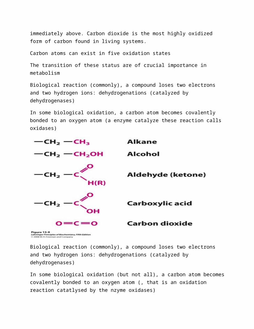
The oxidation levels of carbon in biomolecules. Each compound is formed by oxidation of the red carbon in the compound shown immediately above. Carbon dioxide is the most highly oxidized form of carbon found in living systems.
Carbon atoms can exist in five oxidation states
The transition of these status are of crucial importance in metabolism
Biological reaction (commonly), a compound loses two electrons and two hydrogen ions: dehydrogenations (catalyzed by dehydrogenases)
In some biological oxidation, a carbon atom becomes covalently bonded to an oxygen atom (a enzyme catalyze these reaction calls oxidases)
Biological reaction (commonly), a compound loses two electrons and two hydrogen ions: dehydrogenations (catalyzed by dehydrogenases)
In some biological oxidation (but not all), a carbon atom becomes covalently bonded to an oxygen atom (, that is an oxidation reaction catatlysed by the nzyme oxidases)
Oxygenases: if oxygen is derived directly from molecular oxygen (O2).

Vinyl group:
In chemistry, vinyl or ethenyl is the functional group with the formula −CH=CH2. It is the ethylene (IUPAC ethene) molecule (H₂C=CH₂) less one hydrogen atom.
Acyl group
It contains a double bonded oxygen atom and an alkyl group. In organic chemistry, the acyl group (IUPAC name: alkanoyl) is usually derived from a carboxylic acid. Acyl group is electron withdrawing due to the electronegativity of the oxygen atom.
The name is also used for any compound containing that group, namely R−CH=CH2 where R is any other group of atoms.

An oxoacid is an acid that contains oxygen. Specifically, it is a compound that contains hydrogen, oxygen, and at least one other element, with at least one hydrogen atom bond to oxygen that can dissociate to produce the H+ cation and the anion of the acid
In chemistry, alkyl refers to a group, a substituent, that is attached to other molecular fragments. For example, alkyl lithium reagents have the empirical formula Li(alkyl), where alkyl = methyl, ethyl, etc. A dialkyl ether is an ether with two alkyl groups, e.g., diethyl ether (O(C2H5)2).
Isopropyl group
Methyl group
In organic chemistry, an alkyl substituent is an alkane missing one hydrogen
organic compound characterized by
A hemiacetal or a hemiketal is a compound that results from the addition of an alcohol to an aldehyde or a ketone, respectively. The Greek word hèmi, meaning half, refers to the fact that a single alcohol has been added to the carbonyl group, in contrast to acetals or ketals, which are formed when a second alkoxy group has been added to the structure.[1]
Phosphoryl group
A phosphoryl group is the chemical entity PO32−. The term is usually used for the compounds in which the phosphoryl group is attached to other atoms, e.g. phosphoryl chloride, or in the description of catalytic mechanism.
Enediol

An alkene enol with a hydroxyl group attached to both carbon atoms of the carbon double bond. A reducing sugar can form an enediol.
Enone:
An enone, also called an α, β-unsaturated carbonyl, is a type of organic compound consisting of an alkene conjugated to a ketone. The simplest enone is methyl vinyl ketone or CH₂=CHCOCH₃.
Acryloyl group
In organic chemistry, the acryloyl group is form of enone with structure H₂C=CH–C(=O)–. It is the acyl group derived from acrylic acid. The preferred IUPAC name for the group is prop-2-enoyl, and it is also known as acrylyl or simply acryl.

TOPIC#8
Phosphoryl Group Transfers and ATP
Phosphoryl Group Transfers and ATP
Energy currency of living cells
ATP is used for Phosphoryl transfer reactions
Heterotrophic organisms obtain energy from ATP by breakage of covalent bond, and can undergo one of the two following reactions
A) ATP ADP+ Pi OR
B) ATP AMP+ 2 Pi
What we have studied so far has told us that ATP is the Energy currency in biological systems
And how does it do this? .. By undergoing for Phosphoryl Transfer Reactions
So let take a look at the special role of ATP that links catabolism and
Anabolism and produces energy for functions of life.
Heterotrophic organisms obtain energy from ATP and this involves the breakage of covalent bond, and can undergo one of the two following reactions
A) ATP ADP+ Pi OR
B) ATP AMP+ 2 Pi
Living cells use that energy to make ATP from ADP and Pi
Living cells obtain free energy in a chemical form by the catabolism of nutrient molecules .They use that energy to make ATP from ADP and Pi .
ATP donates some of its chemical energy to
Endergonic processes such as the synthesis of metabolic intermediates and macromolecules from smaller precursors

The transport of substances across membranes against concentration gradients
Mechanical motion (muscle contraction), etc.
Energy donation involves group transfer and hydrolysis of ATP/energy rich phosphate compounds
Energy donation does not involve only hydrolysis of ___ phosphoryl bond but also group transfer reactionsand cleaving of other energy rich phosphate compunds
The Free-Energy Change for ATP Hydrolysis Is Large and Negative
The hydrolytic cleavage of the terminal phosphoanhydride bond in ATP separates one of the three negatively charged phosphates and thus relieves some of the electrostatic repulsion in ATP
The terminal two phosphoanhydride bonds, are known as high energy bonds because their cleavage releases tremendous amount of free energy .Released Pi and ADP are highly soluble
The terminal two phosphoanhydride bonds, indicated in the red, are known as high energy bonds because when they are broken a tremendous amount of free energy is released.
Why large energy is released? This is depicted in this picture where we see
What we see in this picture is the hydrolytic cleavage of high energy bond.. That finally reduces the electrostatic repulsion among 4 negative charges on ATP

And the inorganic phosphate Pi is relaesed iis stabilized by the formation of several resonance forms not possible in ATP.. Now this mpolecule has got same degree of double bond character with all 4 oxygen atoms
3rdly ..Pi and ADP are highly soluble as compared to ATP that means that the products of reaction are in more stable condition as compared to the reactant

ADP2- is the other product of hydrolysis and it immediately ionizes, releasing H+ into a medium of very low [H+] ( ̴10-7 M)
the hydrolysis of ATP is highly exergonic (ΔG'0 = -30.5 kj/mol), the ATP is stable at pH 7
Rapid hydrolysis of ATP occurs only when catalyzed by an enzyme
Meaning… that in living cells the standard conditions of molar concentration cant be followed..
Wha we studied about standard conditions of temp, pressure, and Atmospheric pressure that temp: 300K/25C, Molar con: 1M and P=1atm pressure.
Actual free energy of hydrolysis (ΔG) of ATP in living cells is very different because
the cellular concentration of ATP, ADP, and Pi are not identical
ATP, ADP, and Pi concentration are much lower than the standard condition of 1M
Mg2+ in the cytosol binds to ATP and ADP

The free energy change for ATP hydrolysis is -30,5 kj/mol under standard conditions However,the actual free energy change (ΔG) of ATP hydrolysis in living cells is very different.
The cellular concentrations of ATP, ADP and Pi are not same and are much lower than the 1 M standard conditions.
Meaning… that in living cells the standard conditions of molar concentration cant be followed..
Wha we studied about standard conditions of temp, pressure, and Atmospheric pressure that temp: 300K/25C, Molar con: 1M and P=1atm pressure.
The cellular concentrations of ATP, ADP and Pi are not same and are much lower than the 1 M standard conditions.
In addition, Mg2+ in the cytosol binds to ATP and ADP and for most enzymatic reactions that involve ATP as phosphorly group donor, the true substrate is MgATP-2. The relevant ΔG'0 is therefore that for MgATP-2 hydrolysis.
In addition, Mg2+ in the cytosol binds to ATP and ADP and for most enzymatic reactions that involve ATP as phosphorly group donor, the true substrate is MgATP-2. The relevant ΔG'0 is therefore that for MgATP-2 hydrolysis.
For ATP as phosphoryl group donor, the true substrate is MgATP-2.
Phosphorylation potential: The actual free energy of hydrolysis of ATP under intracellular conditions is often called its phosphorylation potential, Gp.
Phosphorylated compounds
Here are few compounds tht are phosphorylated inn nature and play important roles in appropriate functioning of living cells
Phosphoenolpyruvate
1,3-bisphosphoglycerate
Phosphocreatine

ADP
ATP
AMP
PPi
Glucose 1-phosphate
Fructose 6-phosphate
Glucose 6-phosphate
Large Free Energies of Hydrolysis Phosphorylated Compounds and Thioesters
Orbital overlap between the O and C atoms allows resonance stabilization in oxygen esters; orbital overlap between S and C atoms is poorer and provides little resonance stabilization.
Other compounds that have large free energy of Hydrolysis
Phosphorylated compounds
Thioesters (Acetyl-CoA)
TOPIC#9
Phosphoryl Group Transfers in thioesters
Phosphoryl Group Transfers and ATP
Other Phosphorylated Compounds Have Large Free Energies of Hydrolysis
Orbital overlap between the O and C atoms allows resonance stabilization in oxygen esters; orbital overlap between S and C atoms is poorer and provides little resonance stabilization.
The products of both types of hydrolysis reaction have about the same free-energy content (G), but the thioester has a higher free-energy content than the oxygen ester.
Other compounds that have large free energy change
Phosphorylated compounds

Thioesters (Acetyl-CoA)
Phosphoenolpyruvate (PEP)
A phosphodiester bond occurs when exactly two of the hydroxyl groups in phosphoric acid react with hydroxyl groups on other molecules to form two ester bonds.[1]
Phosphodiester bonds are te basics of life on Earth,[fn 1] as they make up the backbone of the strands of nucleic acid. In DNA and RNA, the phosphodiester bond is the linkage between the 3' carbon atom of one sugar molecule and the 5' carbon atom of another, deoxyribose in DNA and ribose in RNA. Strong covalent bonds form between the phosphate group and two 5-carbon ring carbohydrates (pentoses) over two esterbonds.
The phosphate groups in the phosphodiester bond are negatively charged.
Phosphoenolpyruvate contains a phosphate ester bond that undergoes hydrolysis to yield to enol form of pyruvate.
The enol form of pyruvate can immediately tautomerize to the more stable keto form of pyruvate
Because phosphoenolpyruvate has only one form (enol) and the product, pyruvate, has two possible forms, the product is more stabilized relative to the reactant.
This is the greatest contributing factor to the high standard free energy change of hydrolysis of phosphoenolpyruvate (ΔG'0 = -61.9 kj/mol)
1,3-bisphosphoglycerate

1,3-bisphosphoglycerate contains an anhydride bond between the carboxyl group at C-1 and phosphoric acid.
Hydrolysis of this acyl phosphate is accompanied by a large, negative, standard free energy change (ΔG'0 = -49,3 kj/mol) .This large, negative ΔG'0 can, again, be explained in terms of the structure of reactants and products
Addition of water to anhyhride bond of 1,3-bisphosphoglycerate, one of the direct products, 3-phosphoglyceric acid, immediately leads to the lose a proton to give the carboxylate ion, 3-phosphoglycerate, which has two equally probable resonance forms
Removal of a direct product, 3-phospho-glyceric acid, and formation of resonance-stabilized ion favor the forward reaction.
Hydrolysis of 1,3-bisphosphoglycerate.
The direct product of hydrolysis is 3-phosphoglyceric acid, with an undissociated carboxylic acid. Its dissociation allows resonance structures that stabilize the product relative to the reactants.
Resonance stabilization of Pi further contributes to the negative free-energy change.
Phosphocreatine (Pcr)
In the phosphocreatine, the P-N bond can be hydrolyzed to generate free creatine and Pi.

The release of Pi and the resonance stabilization of creatine favor the forward reaction.
Pi is also resonance stabilized.
The standard free energy change of phosphocreatine is large and negative (ΔG'0 = -43 kj/mol).
Thioesters
Thioesters contain a sulfur atom in the position occupied
by an oxygen
Thioesters have large, negative standard free energy change
of hydrolysis.
Hydrolysis of acetyl-coenzyme A.. important in metabolism and has a large, negative, standard free energy of hydrolysis.
The acyl group in these compounds is activated for trans-acylation, condensation or oxidation-reduction reactions.Hydrolysis of the ester bond generates a carboxylic acid
ΔG'0 = -31,4 kj/mol for acetyl-CoA hydrolysis
Acetyl coenzyme A is one of many thioesters

Hydrolysis of acetyl-coenzyme A. Acetyl-CoA is a thioester with a large, negative, standard free energy of hydrolysis.

Free energy released by hydrolysis of THIOESTERS is is more than OXYGEN esters
Summary for hydrolysis reactions
For hydrolysis reactions with large, negative standard free energy changes, the products are more stable than the reactants for one or more of the following reasons:
1. The bond strain in reactants due to electrostatic repulsion is relieved by charge separation, as for ATP
2. The products are stabilized by ionization, as for ATP, acyl phosphates, thioesters
3. The products are stabilized by isomerization (tautomerization) as for phosphoenolpyruvate
4. The products are stabilized by resonance as for creatine released from phosphocreatine, carboxylate ion released from acyl phosphates and thioesters and phosphate released from anhydride or ester linkages
The term ‘high-energy phosphate bond’ was used by biochemists to describe P-O bond broken in hydrolysis reactions for a long time. But it is incorrect and misleading as it wrongly suggests that the bond itself contains the energy.
In fact, the breaking of all chemical bonds requires an input of energy. The free energy released by hydrolysis of phosphate compounds does not come from the specific bond that is broken
results from the products of the reaction having a lower free energy content than the reactants
The phosphate compounds found in living organisms can be arbitrarily divided into two groups based on their standard free energy changes of hydrolysis.
‘High-energy’ compounds have a ΔG'0 of hydrolysis more negative than -25 kj/mol
‘low-energy’ compounds have a less negative ΔG’0
ATP with a ΔG'0 of hydrolysis of -30 kj/mol is a high-energy compound
glucose 6-phosphate is a low-energy compound (ΔG'0 = -13,8 kj/mol)

TOPIC#10
Bioenergetics -3-Hydrolysis of ATP
ATP Provides Energy by Group Transfers,Not by Simple Hydrolysis
ATP serves as the universal energy currency in all living cells.ATP can carry energy from high-energy phosphate compounds produced by catabolism to compounds such as glucose, converting them into more reactive species.
ATP Provides Energy by Group Transfers,Not by Simple Hydrolysis
ATP hydrolysis in two steps.

The contribution of ATP to a reaction is often shown as a single step, but is almost always a two-step process.
Shown here is the reaction catalyzed by ATP-dependent glutamine synthetase.
A phosphoryl group is transferred from ATP to glutamate
the phosphoryl group is displaced by NH3 and released as Pi.
Hydrolysis accompanied biological processes
1. Noncovalent binding of ATP (or GTP), followed by its hydrolysis toADP (or GDP) and Pi, (to cycle some proteins between two conformations), in muscle contraction
2. In the movement of enzymes along DNA
3. In the movement of ribosomes along messenger RNA
4. The energy-dependent reactions catalyzed by helicases and some topoisomerasesGTP-binding proteins that act in signaling pathways to drive conformational changes

The phosphate compounds found in living organisms can be divided into two groups based on their standard free energy changes of hydrolysis.
‘High-energy’ compounds have a ΔG'0 of hydrolysis more negative than -25 kj/mol
‘low-energy’ compounds have a less negative ΔG’0
ATP with a ΔG'0 of hydrolysis of -30 kj/mol is a high-energy compound
glucose 6-phosphate is a low-energy compound (ΔG'0 = -13,8 kj/mol)
The term “high-energy phosphate bond,” used to describe the P-O bond broken in hydrolysis reactions, is incorrect and misleading as itwrongly suggests that the bond itself contains the energy.
the breaking of all chemical bonds requires an input of energyAs is evident from the additivity of free energy changes of sequential reactions, any phosphorylated compound can be synthesized by coupling the synthesis to the breakdown of another phosphorylated compound with a more negative standard free energy change of hydrolysis.
Notice that while the energy of overall reaction is represented as the algebraic sum of first two reactions, the overall reaction (third) does not involve Pi ; PEP donates a phosphoryl group directly to ADP.
On the basis of their standard free energy changes of hydrolysis, phosphorylated compounds as having a high or low phosphoryl group transfer potential.
The transfer of a phosphoryl group to a compound effectively puts free energy into that compound that can be used in subsequent metabolic transformations.
Much of catabolism is directed toward the synthesis of high-energy phosphate compounds, but their formation is not an end in itself; they are the means of activating a wide variety of compounds for further chemical transformation.

TOPIC#11
Bioenergetics -3-Hydrolysis of ATP- part2.
ATP Provides Energy by Group Transfers,Not by Simple Hydrolysis.. cont
Ranking of biological phosphate compounds by standard free energies of hydrolysis
flow of phosphoryl groups, represented by P, from high-energy phosphoryl group donors via ATP to acceptor molecules (such as glucose and glycerol) to form their low-energy phosphate derivatives.
This flow of phosphoryl groups is catalyzed by kinases, and proceeds with an overall loss of free energy under intracellular conditions.
Hydrolysis of low-energy phosphate compounds releases Pi, which has an even lower phosphoryl group transfer potential
(The location of each compound’s donor phosphoryl group along the scale approximately indicates the del G’o of hydrolysis.)

One more chemical feature of ATP is crucial to its role in metabolism: although in aqueous solution ATP is thermo-dynamically unstable and is therefore a good phosphoryl group donor, it is kinetically stable.
Because of high activation energies required for uncatalyzed reaction ATP does not spontaneously donate phosphoryl groups to water or to the other potential acceptors in the cell.
ATP hydrolysis occurs only when specific enzymes which lower the energy of activation are present
In aqueous solution ATP is thermodynamically unstable and is therefore a good phosphoryl group donor, it is kinetically stable.
Because of high activation energies required for uncatalyzed reaction ATP does not spontaneously donate phosphoryl groups to water or to the other potential acceptors in the cell.
ATP hydrolysis occurs only when specific enzymes which lower the energy of activation are present
thermodynamically unstable: when energy level of products is lower than that of reactants
kinetically stable is when reaction has high activation energy due to some strong bonds.
ATP Donates Phosphoryl, Pyrophosphoryl, and Adenylyl Groups
Nucleophilic displacement reactions of ATP.
Any of the three P atoms (, , or ) may serve as the electrophilic target for nucleophilic attack.
By the labeled nucleophile R-18O: The nucleophile may be an alcohol (ROH), a carboxyl group (RCOO-), or a phosphoanhydride (a nucleoside mono- or diphosphate, for example).
When the oxygen of the nucleophile attacks the position, the bridge oxygen of the product is labeled, indicating that the group transferred from ATP is a phosphoryl (2PO3-2 ), not a phosphate (OPO3-2 )

Each of the three phosphates of ATP is susceptible to nucleophilic attack and each position of attack yields a different type of product
Any of the three P atoms (, , or ) may serve as the electrophilic target for nucleophilic attack. In this case, by the labeled nucleophile R-18O: The nucleophile may be an alcohol (ROH), a carboxyl group (RCOO-), or a phosphoanhydride (a nucleoside mono- or diphosphate, for example).
When the oxygen of the nucleophile attacks the position, the bridge oxygen of the product is labeled, indicating that the group transferred from ATP is a phosphoryl (2PO3-2 ), not a phosphate (OPO3-2 )
Attack on the position displaces AMP and leads to the transfer of a pyrophosphoryl (not pyrophosphate) group to the nucleophile.
Attack on the a position displaces PPi and transfers the adenylyl group to the nucleophile.
Attack on the position displaces AMP and leads to the transfer of a pyrophosphoryl (not pyrophosphate) group to the nucleophile.
Attack on the a position displaces PPi and transfers the adenylyl group to the nucleophile.
5-phosphoribosyl-1-pyrophosphate is a key intermediate in nucleotide synthesis
Hydrolysis of a-b phosphoanhydride bond: ~46 kJ/mol

Hydrolysis of b-g phosphoanhydride bond: ~31 kJ/mol
PPi 2 Pi: -19.2 kJ/mol: total = -64.8 kJ/mol (Adenylation: produces Ppi hydrolysed to 2Pi by inorganic pyrophosphatase enzyme)
Activation of fatty acids
a) Energy yielding oxidation
b) Synthesis of more complex lipids (thiol esters)
The first step in the activation of a fatty acid— either for energy-yielding oxidation or for use in the synthesis of more complex lipids—is the formation of its thiol ester.
The direct condensation of a fatty acid with coenzyme A is endergonic, but the formation of fatty acyl–CoA is exergonic by stepwise removal of two phosphoryl groups from ATP.
First, adenylate (AMP) is transferred from ATP to the carboxyl group of the fatty acid, forming a mixed anhydride (fatty acyl adenylate) and liberating PPi.
The thiol group of coenzyme A then displaces the adenylyl group and forms a thioester with the fatty acid.
sum of these two reactions is energetically equivalent to the exergonic hydrolysis of ATP to AMP and Ppi
The formation of fatty acyl–CoA is endergonic but made energetically favorable (exergonic) by hydrolysis of the PPi by inorganic pyrophosphates.
Thus, in the activation of a fatty acid, both phosphoanhydride bonds of ATP are broken.
Luciferin
William McElroy and his colleagues at the Johns Hopkins University isolated the principal biochemical components: luciferin, a complex carboxylic acid, and luciferase, an enzyme.
The generation of a light flash requires activation of luciferin by an enzymatic reaction involving pyrophosphate cleavage of ATP to form luciferyl adenylate.
In the presence of molecular oxygen and luciferase, the luciferin undergoes a multistep oxidative decarboxylation to oxyluciferin.
This process is accompanied by emission of light.

Luciferin is regenerated from oxyluciferin in a subsequent series of reactions.
In the laboratory, pure firefly luciferin and luciferase are used to measure minute quantities of ATP by the intensity of the light flash produced. As little as a few picomoles (10-12 pico mol) of ATP can be measured in this way.
Next-gen pyrosequencing of DNA relies on flashes of light from the luciferin-luciferase reaction to detect the presence of ATP after addition of nucleotides to a growing strand of DNA
Assembly of Informational Macromolecules Requires Energy
Next heading under this topic.. The precursors for DNA and RNA synthesis are nucleoside triphosphates, and polymerization is accompanied by cleavage of the phosphoanhydride linkage between and phosphates, with the release of PPi (Fig. 13–20).
The moieties transferred to the growing polymer in these reactions are adenylate (AMP), guanylate (GMP), cytidylate (CMP), or uridylate (UMP) for RNA synthesis, and their deoxy analogs (with TMP in place of UMP) for DNA synthesis.
Adenylyl groups from ATP

required for the activation of amino acids for protein synthesis, several steps on the ribosome are also accompanied by GTP hydrolysis.
TOPIC#12
Bioenergetics -3-ATP energizes active transport and muscle contraction
ATP energizes active transport and muscle contraction
Transport processes are major consumers of energy
In brain and kidney, two third of total is consumed to pump sodium and potassium ions across plasma membrane (Na + K+ ATPase)
Na+ dependent phosphorylation of Na+ K+ ATPase changes protein conformation, K+ dependent dephosphorylation brings back original conformation.
Phosphoryl group transfer: to enzyme, not to substrate
ATP energizes active transport and muscle contraction
Direct hydrolysis of ATP (ATP ADP+Pi ) is the source of energy in the conformational changes that produce muscle contraction
ATP bind with head of myosin (not covalent) dissociate from actin ATP hydrolysis induces conformational change (still ADP and Pi bound tightly) Pi release conformational change of the head of myosin actin move

MUSCLE CONTRACTION

Transphosphorylations between nucleotides occur in all cell types
ATP is considered as the cell’s energy currency and donor of phosphoryl groups, all other nucleoside triphosphates (GTP, UTP, CTP) and all the deoxynucleoside triphosphates (dATP, dGTP, dTTP and dCTP) are energetically equivalent to ATP
The free energy changes associated with hydrolysis of their phosphoanhydride linkages are very nearly identical with those for ATP.
In preparation for their various biological roles, other nucleotides are generated as the nucleoside triphosphate (NTP) forms by phosphoryl group transfer to the corresponding nucleoside diphosphates (NDPs) and monophosphates (NMPs)

ATP is the primary high-energy phosphate compound produced by catabolism in the processes of glycolysis, oxidative phosphorylation. Several enzymes carry phosphoryl groups from ATP to the other nucleotides
Nucleoside diphosphate kinases, found in all cells, catalyzes the reaction
Although this reaction is fully reversible the relatively high ATP/ADP ratio in cells normally drives the reaction to the right, with the net formation of NTPs and dNTPs
Phosphoryl group transfers from ATP results in an accumulation of ADP. eg, when muscle is concracting vigorously ADP accumulates and interferes with ATP-dependent contraction.
During intense demand for ATP, the cell lowers the ADP concentration, and at the same time acquires ATP, by the action of adenylate kinase:
Ping-pong mechanism of nucleoside diphosphate kinase

Ping-pong mechanism of nucleoside diphosphate kinase: The enzyme binds its first substrate (ATP in our example), and a phosphoryl group is transferred to the side chain of a His residue. ADP departs, and another nucleoside (or deoxynucleoside) diphosphate replaces it, and this is converted to the corresponding triphosphate by transfer of the phosphoryl group from the phosphohistidine residue.
This reaction is fully reversible, so after the intense demand for ATP ends, the enzyme can recycle AMP by converting it to ADP which can then be phosphorylated to ATP in mitochondria
A similar enzyme guanylate kinase, converts GMP to GDP at the expense of ATP.
By these pathways energy conserved in the catabolic production of ATP is used to supply the cell with all required NTPs and dNTPs
Phosphocreatine (PCr) serves as a ready source of phosphoryl groups for the quick synthesis of ATP from ADP. The phosphocreatine concentration in skeletal muscle is considerably higher than those in the other tissues. The enzyme creatine kinase catalyzes the reversible reaction.
When a sudden demand for energy depletes ATP, the PCr reservoir is used to replenish ATP at a rate faster than ATP can be synthesized by catabolic pathways
When the demand for energy slackens ATP produced by catabolism is used to replenish the PCr reservoir by reversal of the creatine kinase reaction.
Inorganic polyphosphate is a donates phosphoryl group
Inorganic polyphosphate ((polyP)n): linear polymer of Pi … Present in simple organisms of other phyla like yeast
In yeast, polyP accumulated in vacuoles (= 200 mM)

Serve as a phosphagen (Reservoir of phosphoryl group).. Like PCr
The shortest polyP is PPi (n=2): can serve as the energy source for active transport of H+ in plant cells (across vacuolar membrane)
In bacteria, the enzyme polyphosphate kinase-1 (PPK-1) catalyzes the reversible reaction
polyphosphate kinase-2 (PPK-2), catalyzes the reversible synthesis of GTP (or ATP) from polyphosphate and GDP (or ADP):
TOPIC#13
Biological Oxidation -reduction reactions-2-4-18
Biological oxidation-reduction reactions
Central feature of metabolism
1) Phosphoryl group transfer
2) Electron transfer in oxidation-reduction
The flow of electrons in oxidation-reduction reactions is responsible for all work done by living organisms
Oxidation Reduction
Addition of oxygen Removal of oxygen
loss of electrons Gain of electrons
Loss of hydrogen Gain of hydrogen

Biological oxidation-reduction reactions
In nonphotosynthetic organism, source of electron: food
In photosythetic organism, source of electron: chemical compound excited by the absorption of light
Electrons move from various metabolic intermediates to specialized electron carriers in enzyme-catalyzed reaction energy release
1. The flow of electrons can do biological work
Electromotive force (emf): (measured in volts), is the voltage developed by any source of electrical energy such as a battery… In which two chemical species differ in their affinity for electrons, so the electrons flow spontaneously through the circuit, due to the difference in electron affinity
Living cells have an analogous biological “circuit”, with relatively reduced compound such as glucose (the source of electrons)
Glucose enzymatically oxidized release electron spontaneously flow through electron-carrier intermediates to another chemical species, such as O2 this electron flow is exergonic reaction because O2 has a higher affinity for electrons than do electron-carrier intermediates
The resulting emf provides energy to a variety of molecular energy transducers (enzymes and other proteins) that do biological work
The flow of electrons can do biological work
In the mitochondrion, membrane-bound enzymes couple electron flow a production of transmembrane pH difference and a transmembrane electrical potential which helps to accomplish osmotic and electrical work
The proton gradient thus formed has potential energy, sometimes called the proton-motive force by analogy with electromotive force.
Another enzyme, ATP synthase in the inner mitochondrial membrane, uses the proton-motive force to do chemical work: synthesis of ATP from ADP and Pi as protons flow spontaneously across the membrane.

Bacterial flagellar motion: use proton-motive force
Thus, the proton potential has potential energy: electron-motive force
Oxidation-Reductions Can Be Described as Half-Reactions
Half-Reactions:
A half reaction is either the oxidation or reduction reaction component of a redox reaction.
A half reaction is obtained by considering the change in oxidation states of individual substances involved in the redox reaction
Although oxidation and reduction occur together, we consider the two halves of an oxidation-reduction reaction separately.
For example, the oxidation of ferrous ion by cupric ion is written as follows,
Fe +2 + Cu +2 ↔ Fe +3 + Cu+
Same can be described as 2 half reactions in the following form:
1. Fe +2 ↔ Fe +3 + e-
2. Cu +2 + e- ↔ Cu +
Conjugate redox pair
The electron-donating molecule in an oxidation-reduction reaction is called the reducing agent or reductant
the electron-accepting molecule is the oxidizing agent or oxidant.

A given agent, such as an iron cation existing in the ferrous (Fe+2) or ferric (Fe+3) state functions as a conjugate reductant-oxidant pair (redox pair), just as an acid and corresponding base function as a conjugate acid-base pair.
Acid-base reactions can be written as a general equation:
proton donor ↔ H+ + proton acceptor
In redox reactions we can write a similar general equation:
electron donor ↔ e - + electron acceptor
In the reversible half-reaction (1) above, Fe+2 is the electron donor and Fe+3 is the electron acceptor; together, Fe+2 and Fe+3 constitute a conjugate redox pair.
The oxidation of reducing sugar by cupric acid
Aldehydes and ketones with anomeric carbon can act reducing sugars.. Having capability to reduce copper ions in medium.. The reducing sugar reduces the copper(II) ions in these test solutions to copper(I), which forms a brick red copper(I) oxide precipitate
Basis of qualitative carbohydrate test (Benedict’s test)
The reducing end of a disaccharide is the monosaccharide with a free anomeric carbon that is not involved in a glycosidic bond and is thus capable of converting to the open-chain form.
A nonreducing disaccharide has both anomeric carbons tied up in the glycosidic bond…
consider the oxidation of a reducing sugar by cupric ion:

This overall reaction can be expressed as two half reactions:
Because two electrons are removed from the aldehyde carbon, the second half-reaction (the one-electron reduction of cupric to cuprous ion) must be doubled to balance the overall equation.
The monosaccharides can be divided into two groups: the aldoses, which have an aldehyde group, and the ketoses, which have a ketone group. Ketoses must first tautomerize to aldoses before they can act asreducing sugars.. Having capability to reduce copper ions in medium.. The reducing sugar reduces the copper(II) ions in these test solutions to copper(I), which then forms a brick red copper(I) oxide precipitate has a free aldehyde group or a free ketone group
The reducing end of a disaccharide is the monosaccharide with a free anomeric carbon that is not involved in a glycosidic bond and is thus capable of converting to the open-chain form.
A nonreducing disaccharide has both anomeric carbons tied up in the glycosidic bond.The common dietary monosaccharides galactose, glucose and fructose are all reducing sugars.
Biological Oxidations Often Involve Dehydrogenation
In biological Systems oxidation (loss of electrons) is often synonymous with dehydrogenation and many enzymes that catalyze oxidation reactions are dehydrogenases.
The carbon in living cells exists in a range of oxidation status
Electronegativity: H<C<S<N<O
for attached C atoms, i.e. C-C bonds electrons shared, \count 0
for attached X atoms, i.e. C-X bonds (X more electronegative), \count -1 (per bond)

for attached H atoms, i.e. C-H bonds (H is less electronegative than C), \ count +1
OXIDATION STATE
It is calculated by counting all the electrons bonded to carbon and add them in following way:
for attached C atoms, i.e. C-C bonds electrons shared, count 0
for attached X atoms, i.e. C-X bonds (X more electronegative), count -1 (per bond)
for attached H atoms, i.e. C-H bonds (H is less electronegative than C), count +1
Add the total for atoms attached to the C in question, then switch the sign.
For the figure given
Different levels of oxidation of carbon compounds in the biosphere.
In order to estimate the level of oxidation of these compounds, focus on the red carbon atom and its bonding electrons.
When this carbon is bonded to the less electronegative H atom, both bonding electrons (red) are assigned to the carbon.
When carbon is bonded to another carbon, bonding electrons are shared equally, one of the two electrons is assigned to the red carbon. When the red carbon is bonded to the more electronegative O atom, the bonding electrons are assigned to the oxygen.
The number to the right of each compound is the number of electrons “owned” by the red carbon, a rough expression of the degree of oxidation of that compound. As the red carbon undergoes oxidation (loses electrons), the number gets smaller.

Oxidation states of C reducing sugar (an aldehyde or ketone) by carbon in various compounds; from fully reduced (methane to fully oxidized (carbon dioxide)
Electrons are transferred from one molecule to another in 4 ways
1. Directly as electrons: Fe2+/Fe3+ redox pair can transfer an electron to the Cu/Cu2+ redox pair
2. As hydrogen atoms: hydrogen atom consists of a proton (H+) and a single electron (e-)
AH2/A: conjugate redox pair can reduce another compound B

3. As a hydride ion (:H-) .. has two electrons
This reaction occurs in NAD-linked dehydrogenase
4. Through direct combination with oxygen, where Oxygen covalently incorporated into product

TOPIC#14
Reduction potential
Reduction potential measures affinity for electrons
Reduction potential:
Reduction potential (redox potential, oxidation/reduction potential), is a measure of the tendency of a chemical species to acquire electrons and thereby get reduced. Units: Volts (V), or millivolts (mV).. denoted by E.
Standard reduction potential:
The tendency for a chemical species to be reduced, at standard conditions. The more positive the potential is the more likely it will be reduced… denoted by EO.
They help to measure the …. Redox reaction.. In terms of volts (V), or millivolts (mV and like we said.. That .. measure of the tendency of a chemical species to acquire electrons .. Aquiring e is to undergo reduction…
And The standard reduction potential is the tendency for a chemical species to be reduced, and is measured in volts at standard conditions. The more positive the potential is the more likely it ...
concentrations of 1 mol/L and at 1 atm (101.3kp) and 25°C temp.
The cell potential, EcellEcell, is the measure of the potential difference between two half cells in an electrochemical cell.
he potential difference is caused by the ability of electrons to flow from one half cell to the other. Electrons are able to move between electrodes because the chemical reaction is a redox reaction. A redox reaction occurs when a certain substance is oxidized, while another is reduced. During oxidation, the substance loses one or more electrons, and thus becomes positively charged. Conversely, during reduction, the substance gains electrons and becomes negatively charged.
so
And how is that helpful for us? And how it can be measured ..

To measure the Reduction potential.. Electrochemists use half cell.. A half cell is one of the two electrodes in a cell or simple battery. separated from other. Half cell.. . two half cells make An electrochemical cell in which redox reaction can take place.
.. So that they work only in the presence of…? To initiate e flow and conduct electricity.. Placing a piece of reactant in an electrolyte solution makes a half cell.
A Half-cell
A half cell is one of the two electrodes in a galvanic cell or simple battery submerged in an aqueous solution of the same metal ions.
Until or unless they are separately kept in their solutions.. There would be no redox reaction.
. For example, in the Zn-Cu battery, the two half cells make an oxidizing-reducing couple. The electrode is connected to the other half cell, which contains an electrode of some other metal submerged in an aqueous solution of subsequent metal ions. An example of this would be a zinc electrode, dipped in zn solution. in which the zn atoms in the electrode lose two electrons and becomes Zn2+ (oxidized( .. The electrons lost by the zn atoms in the electrode are then transferred to the second half cell,.
.In order to balance the charge on both sides of the cell, the half cells are connected by a salt bridge..
. As the anode half cell becomes overwhelmed with zn2+ ions, the negative anion of the salt will enter the solution and stabilized the charge. Similarly, in the cathode half cell, as the solution becomes more negatively charged, cations from the salt bridge will stabilize the charge.
If no salt bridge were present, the solution in one half cell would accumulate negative charge and the solution in the other half cell would accumulate positive charge as the reaction proceeded, quickly preventing further reaction, and hence production of electricity
. Reduction potential of a half-cell depends on the activity of reduced and oxidized species which is approximated by their concentrations
On the anode, oxidation takes place. Zn Zn2+ + 2 e-Zn is the reducing agent, and Zn2+ the oxidizing agent.
On the cathode, reduction takes place.
Cu2+ + 2 e CuCu2+ is the oxidizing agent and Cu the reducing agent.

A standard Half-cell
Standard Hydrogen electrode..(SHE) is the primary reference electrode with platinum electrode dipped in solution with H ions under standard conditions
The electromotive force (emf) of this electrode is designated 0.00 V. (At pH 7, 25 OC)
Using observed emf (of test solution) and the known emf of the reference cell, the emf of the test cell with the redox pair can be calculated.
The cell that gains electrons has, by convention, the more positive reduction potential.
The electrode at which this half-reaction occurs (called a half-cell) is arbitrarily assigned an EO of 0.00 V.

But the fact is that only the difference of reduction potential can help us to calculate it.. That is why we need standard or a reference elctrode..The half consists of a metal electrode of a certain metal submerged in an aqueous solution of the same metal ions.
.. A half cell consists of an electrode and the species to be oxidized or reduced. A standard half cell conists of
Unless it is connected to another half cell via an electric conductor and salt bridge, no reaction will take place in a half cell.
Standard reduction potentials can be used to calculate free-energy change
Electrons tend to flow to the half-cell with the more positive E, and the strength of that tendency is proportional to Δ E, the difference in reduction potential
We can then predict the direction in which electrons will tend to flow when the two half-cells are connected through an external circuit
Electrons tend to flow to the half-cell with the more positive E the strength of flow depends on ΔE (difference in reduction potential)
The energy made available by this spontaneous electron flow (the free-energy change, Δ G, for the oxidation-reduction reaction) is proportional to Δ E:
Why are reduction potentials so useful to the biochemist?When E values have been determined for any two halfcells,relative to the standard hydrogen electrode, we
also know their reduction potentials relative to eachother. We can then predict the direction in which electronswill tend to flow when the two half-cells are connected
through an external circuit or when component of both half-cells are present in the same solution.
where n is the number of electrons transferred in the reaction
F is Faraday (unit of electricity), used in the study of electrochemical reactions and equal to the amount of electric charge that liberates one gram equivalent of any ion from an electrolytic solution
With this equation we can calculate the actual free-energy change for any oxidation-reduction reaction from the values of E ’o (reduction potentials) and the concentrations of reacting species

When acetaldehyde is reduced by the biological electron carrier NADH, ΔG = -35.3 kJ/mol.
Conjugate redox pair: An electron donor and its corresponding electron acceptor form; for example, Cu+ (donor) and Cu2+ (acceptor), or NADH (donor) and NAD+ (acceptor) form Conjugate redox pair.
Half cell: A half cell is one of the two electrodes in a galvanic cell or simple battery. For example, in the Zn-Cu battery, the two half cells make an oxidizing-reducing couple.
Reduction potential (also known as redox potential, oxidation/reduction potential, or Eh) measuresthe tendency of a chemical species to acquire electrons and thereby be reduced.

TOPIC#15
Universal Electron Carriers
So.. We had been talking about redox reactions.. And half cell in which redox reaction takes place.. And we mentioned several times that reduction is the addition of electrons. . What happens in biological system is.. That many of the enzymes that catalyze cellular oxidations reactions.. channel electrons from their substrates through some types of universal electron carriers. Some of these proteins are water-soluble, but others are peripheral or integral membrane proteins
During the catabolic processes these carriers are reduced, substrate is oxidized and this results in the conservation of free energy
Enrgy genration is not only by glucose and ATP, but there are some other Behind the scene contributors too.. These are electron carrier molecules.. Glucose
Cellular oxidation of glucose to carbon dioxide requires specialized electron carriers
The principles of oxidation-reduction energetics can apply to the many metabolic reactions that involve electron transfers
Cells does not convert to CO2 in a single, high energy releasing reaction but rather in a series of controlled reactions, some of which are oxidation for the ATP synthesis from ADP through NAD+ and FAD
A Few Types of Coenzymes and Proteins Serve as Universal Electron Carriers
1) The nucleotides NAD and NADP move readily from one enzyme to another
2) The flavin nucleotides FMN and FAD are usually very tightly bound to the enzymes, called flavoproteins, for which they serve as prosthetic groups
3) Lipid-soluble quinones: ubiquinone and plastoquinone act as electron carriers and proton donors in the non-aqueous environment of membranes

4) Iron-sulfur proteins and cytochromes, which have tightly bound prosthetic groups that undergo reversible oxidation and reduction, serve as electron carriers in many oxidation-reduction reactions
NAD, NADP, FMN, and FAD are water-soluble coenzymes and undergo reversible oxidation and reduction during many of the electron-transfer reactions of metabolism.
Enrgy genration is not only by glucose and ATP, but there are some other Behind the scene contributors too.. These are electron carrier molecules.. Glucose
. Some of these proteins are water-soluble, but others are peripheral or integral membrane proteins
The reduction of these carriers in catabolic processes results in the conservation of free energy released by substrate oxidation.
NAD, NADP, FMN, and FAD are water-soluble coenzymes and undergo reversible oxidation and reduction in many of the electron-transfer reactions of metabolism.
The nucleotides NAD and NAD move readily from one enzyme to another
The flavin nucleotides FMN and FAD are usually very tightly bound to the enzymes, called flavoproteins, for which they serve as prosthetic groups.
Lipid-soluble quinones: ubiquinone and plastoquinone act as electron carriers and proton donors in the non-aqueous environment of membranes.
Iron-sulfur proteins and cytochromes, which have tightly bound prosthetic groups that undergo reversible oxidation and reduction, also serve as electron carriers in many oxidation-reduction reactions.
NAD and NADP are soluble electron carriers that act with dehydrogenase enzymes
Activated electron carriers, involved in oxidation-reduction and coupling reactions
NAD+ (nicotinamide adenine dinucleotide)
NADP+ (nicotinamide adenine dinucleotide phosphate)
For NAD: NAD+: oxidized form
For NADP: NADP+: oxidized form
Joined by phosphoanhydride bond

Pyridine nucleotides
Nicotinamide moiety comes from NIACIN Vitamin
And it’s the flow 0f e in etc that is used to produce ATP ..
There are other important activated carriers involved in biosynthesis of molecules. These activated carriers are involved in oxidation-reduction reactions and involved in coupling reactions.
The most important of these carriers are NAD+ (nicotinamide adenine dinucleotide) and closely related molecule NADP+ (nicotinamide adenine dinucleotide phosphate).
A conjugate acid, within the Brønsted–Lowry acid–base theory, is a species formed by the reception of a proton (H+) by a base—in other words, it is a base with a hydrogen ion added to it. On the other hand, a conjugate base is merely what is left over after an acid has donated a proton during a chemical reaction
Mechanism. The reaction mechanism generally begins with the oxidative addition of an organic halide to the catalyst. ... The final step is reductive elimination of the two coupling fragments to regenerate the catalyst and give the organic product.

Both coenzymes undergo reversible reduction of the nicotinamide ring
As a substrate molecule undergoes oxidation (dehydrogenation), giving up two hydrogen atoms, the oxidized form of the nucleotide (NAD+ or NADP+) accepts a hydride ion (:H2, the equivalent of a proton and two electrons) and is reduced (to NADH or NADPH)
The second proton removed from the substrate is released to the aqueous solvent. The half-reactions for these nucleotide cofactors are:
NAD+ + 2e _ + 2H + + NADH NADH + H+
NADP + + 2e _ + 2H + + NADH NADPH + H +

Reduction of NAD or NADP converts the benzenoid ring of the nicotinamide moiety (with a fixed positive charge on the ring nitrogen) to the quinonoid form (with no charge on the nitrogen).
The reduced nucleotides absorb light at 340 nm; the oxidized forms do not (used for detection)
Plus sign with NAD and NADP does not indicate the net charge (both are negatively charged ) rather, it indicates that the nicotinamide ring is in its oxidized form, with a positive charge on the Nitrogen atom
“H” in NADH and NADPH, the denotes the added hydride ion
Note that the plus sign in the abbreviations NAD1 and NADP1 does not indicate the net charge on these molecules in fact, both are negatively charged); rather, it
indicates that the nicotinamide ring is in its oxidized form, with a positive charge on the nitrogen atom. In the abbreviations NADH and NADPH, the “H” denotes the
added hydride ion. To refer to these nucleotides without specifying their oxidation state, we use NAD and NADP.
Total concentration of NAD+ + NADH in most tissues: 10-5 M
Total concentration of NADP+ + NADPH in most tissues: 10-6 M
Concentration ratio: NAD+ > NADH favoring hydride transfer from a substrate to NAD+ to form NADH function in oxidation, occur in mitochondrial matrix
Concentration ratio: NADP+ < NADPH favoring hydride transfer from NADPH to a substrate function in reduction occur in cytosol
More than 200 enzymes are known to catalyze reactions in which NAD+ (or NADP+) accepts a hydride ion from a reduced substrate or NADPH (or NADH) donates a hydride ion to an oxidized substrate
NAD and NADP:

NAD and its phosphorylated analog NADP undergo reduction to NADH and NADPH, accepting a hydride ion from an oxidizable substrate.
The hydride ion is added to either the front (the A side) or the back (the B side) of the planar nicotinamide ring

Rossmann fold
Most dehydrogenases that use NAD or NADH contain a conserved protein binding domain called the Rossmann fold
Rossmann fold: consists of a six-stranded parallel beta sheet and four associated alpha helices
Nucleotide binding domain of lactate dehydrogenase with NAD is shown
NAD is bound to paired beta-alpha motifs of Rossmann fold

The association between dehydrogenases and cofactor is loose the coenzyme readily diffuse from one enzyme to another act as a water-soluble carrier of electron form one metabolite to another
In overall reaction, there is not net production or consumption of NAD+ or NADH no concentration change of NAD+ or NADH .. and NAD+ or NADH get recycled repeatedly
Dietary deficiency of Niacin causes Pellagra
Most coenzyme: derived from vitamins
NAD and NADP: derived from vitamin niacin (=nicotinic acid)
Human cannot synthesize sufficient quantities of niacin
Niacin derived from tryptophan
In the laboratory, nicotinic acid was first produced by oxidation of the natural product.. nicotine—thus the name
Maize: content low tryptophan
Niacin deficiency: affects all the NAD(P)-dependent dehydrogenase causes the serious human disease pellagra (=rough skin): characterized by the Three Ds (dermatitis, diarrhea and dementia) death
Too much alcohol drinking reduce niacin absorption from the intestine

Niacin deficiency: affects all the NAD(P)-dependent dehydrogenase causes the serious human disease pellagra (=rough skin): characterized by the Three Ds (dermatitis, diarrhea and dementia)
death
similar disease black tongue in dogs
Niacin is curative agent (scientists)
Too much alcohol drinking reduce niacin absorption from the intestine
Both nicotinic acid and nicotinamide cure pellagra, but nicotine (from cigarettes or elsewhere) has no curative activity
Niacin (nicotinic acid) and its derivative nicotinamide
The biosynthetic precursor of these compounds is tryptophan.
In the laboratory, nicotinic acid was first produced by oxidation of the natural product.. nicotine—thus the name.

Both nicotinic acid and nicotinamide cure pellagra, but nicotine (from cigarettes or elsewhere) has no curative activity
Flavoproteins
Flavoproteins: enzymes that catalyze oxidation-reduction reactions using coenzyme
Flavin mononucleotide (FMN)
Flavin adenine dinucleotide (FAD)
Flavin nucleotides derived from the vitamin riboflavin
Flavoproteins act in a quite different role, as light receptors
The fused ring structure of the flavin nucleotide (isoalloxazine ring)
Isoalloxazine ring undergoes reversible reduction, accepting either one or two electrons in the form of one or two hydrogen atoms (each atom an electron plus a proton) from a reduced substrate
When a fully oxidized flavin nucleotide accepts only one electron (one hydrogen atom), the semiquinone form of the isoalloxazine ring is produced
The fully reduced forms are abbreviated FADH2 and FMNH2
Flavoproteins derived from the vitamin riboflavin
Flavoproteins: enzymes that catalyze oxidation-reduction reactions using coenzyme
Flavin mononucleotide
(FMN) or flavin adenine dinucleotide (FAD)
Certain flavoproteins act in a quite different role, as light receptors.
The fused ring structure of the flavin nucleotide (isoalloxazine ring)
Isoalloxazine ring undergoes reversible reduction, accepting either one or two electrons in the form of one or two hydrogen atoms (each atom an electron plus a proton) from a reduced substrate.
The fully reduced forms are abbreviated FADH2 and FMNH2
When a fully oxidized flavin nucleotide accepts only one electron (one hydrogen atom), the semiquinone form of the isoalloxazine ring is produced, FADH2 and FMNH2

NAD+ --> NADH- Dehydrogenase always removes 2H+ (hydrogen protons) and 2e- from the metabolite. NAD can accept 2e- but only a single H+. Therefore, the second H+ is donated to the solution.FAD --> FADH2- When dehydrogenase removes 2H+ and 2e- from the glucose metabolite, FAD can accept 2e- AND 2H+. Therefore no extra hydrogen proton needs to be donated to solution
Oxidized and reduced forms of FAD and FMN
Some enzymes use flavin nucleotides for oxidation-reduction
Flavin nucleotides are tightly bound in flavoproteins
Fully reduced: 360 nm
Partially reduced: 450 nm
Fully oxidized: 370 and 440 nm
FMN consists of the structure above the dashed line on the FAD (oxidized form). The flavin nucleotides accept two hydrogen atoms (two electrons and two protons), both of which appear in the flavin ring system. When FAD or FMN accepts only one hydrogen atom, the semiquinone, a stable free radical, forms.

Enzymes that use FAD or FMN as coenzymes:
Cryptochromes
Cryptochromes: Family of flavoproteins, widely distributed in the eukaryotic phyla, that mediate the effects of blue light on plant development and the effects of light on mammalian circadian rhythms (oscillations in physiology and biochemistry, with a 24-hour period).
The cryptochromes are homologs of another family of flavoproteins, the photolyases. Found in both bacteria and eukaryotes, photolyases use the energy of absorbed light to repair chemical defects in DNA
mediate the effects of blue light on plant development and the effects of light on mammalian circadian rhythms (oscillations in physiology and biochemistry, with a 24-hour period)
Homologs of Photolyases
Photolyases: Use energy of light to repair defects in DNA

TOPIC#16
GLYCOLYSIS-1
GLUCOSE METABOLISM
Glucose is a simple sugar with the molecular formula C₆H₁₂O₆, which means that it is a molecule that is made of six carbon atoms, twelve hydrogen atoms, and six oxygen atoms. Glucose circulates in the blood of animals as blood sugar.
In animals and vascular plants, glucose has three major fates:
1. It may be stored (as a polysaccharide , i-e., glycogen or starch)
2. Oxidized to a three-carbon compound (pyruvate) via glycolysis to provide ATP and metabolic intermediates.
3. Oxidized via the pentose phosphate (phosphogluconate) pathway to yield ribose 5-phosphate for nucleic acid synthesis and NADPH for reductive biosynthetic processes.
Organisms that do not have access to glucose from other sources must make it
Photosynthetic organisms make glucose by first reducing atmospheric CO2 to trioses, then converting the trioses to glucose.
Non-photosynthetic cells make glucose from simpler three and four-carbon precursors by the process of gluconeogenesis, effectively reversing glycolysis in a pathway that uses many of the glycolytic enzymes.

GLYCOLYSIS
In glycolysis (from the Greek glykys, “sweet” or “sugar,” and lysis, “splitting”), a molecule of glucose C6H12O6is degraded in a series of enzyme-catalyzed reactions to yield two molecules of the three-carbon compound pyruvate CH3COCOO− + H+.
During glycolysis, the free energy released is used to form the high-energy compounds ATP (adenosine triphosphate) and NADH (reduced nicotinamide adenine dinucleotide).
The pathway of glycolysis was explained by Otto Warburg and Hans von Euler-Chelpin in yeast and in muscle by Gustav Embden and Otto Meyerhof in1930s.
The most common type of glycolysis is the Embden–Meyerhof–Parnas (EMP pathway), which was discovered by Embden and Meyerhof and J.K. Parnas.
Glycolysis occurs in most organisms in the cytosol of the cell.
It is an oxygen independent metabolic pathway, meaning that it does not use molecular oxygen (i.e. atmospheric oxygen) for any of its reactions.
The products of glycolysis, that is, pyruvate and NADH + H+) are sometimes disposed off using atmospheric oxygen.

Aerobic Glycolysis: When molecular oxygen is used in the disposal of the products of glycolysis the process is usually referred to as Aerobic Glycolysis.
Anaerobic Glycolysis: When no oxygen is used, the process is said to be Anaerobic.
2 phases of GLYCOLYSIS
The Preparatory Phase – in which ATP is consumed, hence also known as the investment phase
The Pay Off Phase – in which ATP is produced
Occurs in 10 steps, the first 5 of which constitute the preparatory phase.
The breakdown of the six-carbon glucose into two molecules of the three-carbon pyruvate
The Preparatory Phase
step 1: The first step in glycolysis is phosphorylation of glucose at the hydroxyl group on C-6 by a family of enzymes called hexokinases to form glucose-6-phosphate (G6P).
step 2: The D-glucose 6-phosphate thus formed is converted to D-fructose 6-phosphate,
step 3: which is again phosphorylated, this time at C-1, to yield D-fructose 1,6-bisphosphate
For both phosphorylations, ATP is the phosphoryl group donor.
step 4: Fructose 1,6-bisphosphate is split to yield two three-carbon molecules, dihydroxyacetone phosphate and glyceraldehyde 3-phosphate (step 4); this is the “lysis” step that gives the pathway its name.
step 5: The dihydroxyacetone phosphate is isomerized to a second molecule of glyceraldehyde 3-phosphate (step 5), ending the first phase of glycolysis.
To summarize:
In the preparatory phase of glycolysis the energy of ATP is invested, raising the free-energy content of the intermediates, and the carbon chains of all the metabolized hexoses are converted to a common product, glyceraldehyde 3-phosphate.
Note that during preparatory phase, 2 molecules of ATP are invested before the cleavage of glucose into two three-carbon pieces.

Preparatory phase of Glycolysis For each molecule of glucose that passes through the preparatory phase (a), two molecules of glyceraldehyde 3-phosphate are formed; both pass through the payoff phase.

TOPIC#17
Glycolysis- the pay off phase
GLYCOLYSIS-The Pay-off Phase
The Pay-off Phase
The energy gain comes in the payoff phase of glycolysis

step 6: Each molecule of glyceraldehyde 3-phosphate is oxidized and phosphorylated by inorganic phosphate (not by ATP) to form 1,3-bisphosphoglycerate
Energy is also conserved in the formation of two molecules of the electron carrier NADH per molecule of glucose.
steps 7 through 10: Much of this energy is conserved by the coupled phosphorylation of four molecules of ADP to ATP.
The net yield is two molecules of ATP per molecule of glucose, because two molecules of ATP were invested in the preparatory phase.
Pyruvate is the end product of the second phase of glycolysis.

For each glucose molecule, two ATP are consumed in the preparatory phase and four ATP are produced in the payoff phase,
This gives a net yield of two ATP per molecule of glucose converted to pyruvate.
each phosphoryl group, represented here as P, has two negative charges
Energy yield during Glycolysis
Two ATP are consumed in the preparatory phase and four ATP are produced in the payoff phase, giving a net yield of two ATP per molecule of glucose converted to pyruvate. Note that each phosphoryl group, represented as P, has two negative charges (2PO3-2 )
Much of this energy is conserved by the coupled phosphorylation of four molecules of ADP to ATP.
Energy is also conserved in the payoff phase in the formation of two molecules of the electron carrier NADH per molecule of glucose.
In the sequential reactions of glycolysis, three types of chemical transformations are noteworthy:
(1) degradation of the carbon skeleton of glucose to yield pyruvate
(2) phosphorylation of ADP to ATP by compounds with high phosphoryl group transfer potential, formed during glycolysis.
(3) transfer of a hydride ion to NAD+ forming NADH.
High Phosphoryl Transfer Potential Compounds. These compounds have a higher phosphoryl transfer potential than that of ATP and can be used to phosphorylate ADP to form ATP.

TOPIC#18
Lecture - 18 - Preparatory phase of Glycolysis Requires ATP
Preparatory phase of Glycolysis Requires ATP
Experiment by A. Harden and W. Young
Preparatory phase of glycolysis, two molecules of ATP are invested and the hexose chain is cleaved into two triose phosphates
In 1906, Arthur Harden and William Young tested their hypothesis that inhibitors of proteolytic enzymes would stabilize the glucose fermenting enzymes in yeast extract.
They added blood serum (known to contain inhibitors of proteolytic enzymes) to yeast extracts and observed the predicted stimulation of glucose metabolism.
However, in a control experiment with the boiling serum, they discovered that boiled serum was just as effective at stimulating glycolysis
Harden and Young soon discovered that glucose added to their yeast extract was converted to a hexose bisphosphate (the “Harden- Young ester,” eventually identified as fructose 1,6-bisphosphate.
Careful examination revealed that inorganic phosphate was responsible for the stimulation.
This was the beginning of investigations on the role of organic esters and anhydrides of phosphate in biochemistry, which has led to our current understanding of the central role of phosphoryl group transfer in biology.

Preparatory phase of Glycolysis Requires ATP1. Phosphorylation of Glucose
In the first step of glycolysis, glucose is activated for subsequent reactions by its phosphorylation at C-6 to yield glucose 6-phosphate, with ATP as the phosphoryl donor:
This reaction is irreversible under intracellular conditions, and catalyzed by hexokinase.
The acceptor in the case of hexokinase is a hexose, normally D-glucose, hexokinase also catalyzes the phosphorylation of other common hexoses, such as D-fructose and D-mannose, in some tissues.
Hexokinase requires Mg+2 for its activity, because the true substrate of the enzyme is not ATP-4 but the MgATP-2 complex
Hexokinase and other nine enzymes of are soluble cytosolic proteins
Note:
Isozymes
Two or more enzymes that catalyze the same reaction but are encoded by different genes are called isozymes
Kinase:
Kinases are a subclass of transferases...catalyse transfer of phosphate group from phosphate donating molecule to specific substrates
2. Conversion of Glucose 6-Phosphate to Fructose 6-Phosphate

The enzyme phosphohexose isomerase (phosphoglucose isomerase) catalyzes the reversible isomerization of glucose 6-phosphate, an aldose, to fructose 6- phosphate, a ketose.
The mechanism for this reaction involves an enediol intermediate
Enediol: An enediol is an alkene enol with a hydroxyl group attached to both carbon atoms of the carbon double bond.
3 steps for Conversion of Glucose 6-Phosphate to Fructose 6-Phosphate (phosphohexose isomerase reaction)
1. opening of ring
2. Isomerization of glucose to fructose through formation of enediol intermediate
3. Closing of fructose ring

Fig 14-5: The phosphohexose isomerase reaction.
The ring opening and closing reactions (steps 1 and 4) are catalyzed by an active-site His residue, by mechanisms omitted here for simplicity.
The proton (light red) initially at C-2 is made more easily abstractable by electron withdrawal by the adjacent carbonyl and nearby hydroxyl groups. After its transfer from C-2 to the active-site Glu residue (a weak acid), the proton is freely exchanged with the surrounding solution; that is, the proton abstracted from C-2 in step 2 is not necessarily the same one that is added to C-1 in step 3.

TOPIC#19
Preparatory phase of Glycolysis Requires ATP
Preparatory phase of Glycolysis Requires ATP - 2
3. Phosphorylation of Fructose 6-Phosphate to Fructose 1,6-Bisphosphate
Phosphofructokinase-1 (PFK-1) catalyzes the transfer of a phosphoryl group from ATP to fructose 6-phosphate to yield fructose 1,6-bisphosphate.
KEY CONVENTION:
Compounds that contain two phosphate or phosphoryl groups attached at different positions in the molecule are named bisphosphates (or bisphospho compounds); for example, fructose 1,6-bisphosphate and 1,3-bisphosphoglycerate. Compounds with two phosphates linked together as a pyrophosphoryl group are named diphosphates; for example, adenosine diphosphate (ADP). Similar rules apply for the naming of trisphosphates (such as inositol 1,4,5-trisphosphate;) and triphosphates (such as adenosine triphosphate, ATP)

The PFK-1 reaction is essentially irreversible under cellular conditions, and it is the first “committed” step in the glycolytic pathway
Glucose 6-phosphate and fructose 6-phosphate have other possible fates, but fructose 1,6-bisphosphate is targeted for glycolysis
Some bacteria and protists and perhaps all plants have a phosphofructokinase that uses pyrophosphate (PPi), not ATP, as the phosphoryl group donor in the synthesis of fructose 1,6-bisphosphate:
In metabolic pathways, enzymes catalyzing essentially irreversible reactions are potential sites of control
Phosphofructokinase-1 is subject to complex allosteric regulation
The PFK1 has got binding sites for both, ATP-Mg2+ and F6P binding sites

The activity of PFK-1 enzyme and glycolysis is inhibited whenever the cell has high levels of ATP
In this way, the cell can increase or decrease the rate of glycolysis in response to the cell's energy requirements
In Eukaryotes, fructose 2,6-bisphosphate (not to be confused with the PFK-1 reaction product, fructose 1,6-bisphosphate) is a potent allosteric activator of PFK-1
Ribulose 5-phosphate, an intermediate in the pentose phosphate pathway also activates phosphofructokinase indirectly.
Imine: An imine is a functional group or chemical compound containing a carbon–nitrogen double bond. The nitrogen atom can be attached to a hydrogen or an organic group.
Amine: an organic compound derived from ammonia by replacement of one or more hydrogen atoms by organic groups.
Schiff base intermediate:
They can be considered a sub-class of imines, being either secondary ketimines or secondary aldimines depending on their structure. (R-CH=NR' where R' ≠ H)

TOPIC#20
Preparatory phase of Glycolysis Requires ATP-3
Preparatory phase of Glycolysis Requires ATP - 2
In metabolic pathways, enzymes catalyzing essentially irreversible reactions are potential sites of control
Phosphofructokinase-1 is subject to complex allosteric regulation
The PFK1 has got binding sites for both, ATP-Mg2+ and F6P binding sites
The activity of PFK-1 enzyme and glycolysis is inhibited whenever the cell has high levels of ATP
In this way, the cell can increase or decrease the rate of glycolysis in response to the cell's energy requirements
In Eukaryotes, fructose 2,6-bisphosphate (not to be confused with the PFK-1 reaction product, fructose 1,6-bisphosphate) is a potent allosteric activator of PFK-1
Ribulose 5-phosphate, an intermediate in the pentose phosphate pathway also activates phosphofructokinase indirectly.
4. Cleavage of Fructose 1,6-Bisphosphate
The enzyme fructose 1,6-bisphosphate aldolase, often called simply aldolase, catalyzes a reversible aldol condensation (see Fig. 13–4..Lehninger Edition 6).
Fructose 1,6-bisphosphate is cleaved to yield two different triose phosphates, glyceraldehyde 3-phosphate, an aldose, and dihydroxyacetone phosphate, a ketose.

Two classes of aldolases
There are two classes of aldolases.
Class I, found in animals and plants, form the Schiff base intermediate
Class II, in fungi and bacteria, do not form the Schiff base intermediate
Imine: An imine is a functional group or chemical compound containing a carbon–nitrogen double bond. The nitrogen atom can be attached to a hydrogen or an organic group.
Schiff base intermediate:
They can be considered a sub-class of imines, being either secondary ketimines or secondary aldimines depending on their structure. (R-CH=NR' where R' ≠ H)
MECHANISM
FBPA, class I aldolase reaction. The reaction shown here is the reverse of an aldol condensation.
Cleavage between C-3 and C-4 depends on the presence of the carbonyl group at C-2, which is converted to an imine on the enzyme.
A and B represent amino acid residues that serve as general acid (A) or base (B).

Imine: An imine is a functional group or chemical compound containing a carbon–nitrogen double bond. The nitrogen atom can be attached to a hydrogen or an organic group.
Schiff base intermediate:
They can be considered a sub-class of imines, being either secondary ketimines or secondary aldimines depending on their structure. (R-CH=NR' where R' ≠ H)
Amine: an organic compound derived from ammonia by replacement of one or more hydrogen atoms by organic groups.
Secondary amine:
Secondary amines have two organic substituents (alkyl, aryl or both)
bound to the nitrogen together with one hydrogen.

Enamine:
An enamine is an unsaturated compound derived
by the condensation of an aldehyde or ketone with
a secondary amine.
