voxhumana-english.comvoxhumana-english.com/newest science news blog 201… · web viewmaterial is...
TRANSCRIPT
1 5/6/23 Name Student number http://bit.ly/2A08vBR
Research bolsters possibility of plate tectonics on EuropaEvidence the icy shell of Europa may have plate tectonics similar to
those on EarthPROVIDENCE, R.I. [Brown University] -- A Brown University study provides new evidence that the icy shell of Jupiter's moon Europa may have plate tectonics similar to those on Earth. The presence of plate tectonic activity could have important implications for the possibility of life in the ocean thought to exist beneath the moon's surface.The study, published in Journal of Geophysical Research: Planets, uses computer modeling to show that subduction -- when a tectonic plate slides underneath another and sinks deep into a planet's interior -- is physically possible in Europa's ice shell. The findings bolster earlier studies of Europa's surface geology that found regions where the moon's ice shell looks to be expanding in a way that's similar to the mid-ocean spreading ridges on Earth. The possibility of subduction adds another piece to the tectonic puzzle."We have this evidence of extension and spreading, so the question becomes where does that material go?" said Brandon Johnson, an assistant professor in Brown's Department of Earth, Environmental and Planetary Sciences and a lead author of the study. "On Earth, the answer is subduction zones. What we show is that under reasonable assumptions for conditions on Europa, subduction could be happening there as well, which is really exciting."Part of the excitement, Johnson says, is that surface crust is enriched with oxidants and other chemical food for life. Subduction provides a means for that food to come into contact with the subsurface ocean scientists think probably exists under Europa's ice."If indeed there's life in that ocean, subduction offers a way to supply the nutrients it would need," Johnson said.Subduction on iceOn Earth, subduction is driven largely by differences in temperature between a descending slab and the surrounding mantle. Crustal
material is much cooler than mantle material, and therefore denser. That increased density provides the negative buoyancy needed to sink a slab deep into the mantle.Though previous geological studies had hinted that something like subduction could be happening on Europa, it wasn't clear exactly how that process would work on an icy world. There's evidence, Johnson says, that Europa's ice shell has a two layers: a thin outer lid of very cold ice that sits atop a layer of slightly warmer, convecting ice. If a plate from the outer ice lid was pushed down into the warmer ice below, its temperature would quickly warm to that of the surrounding ice. At the point, the slab would have the same density of the surrounding ice and would therefore stop descending.But the model developed by Johnson and his colleagues showed a way that subduction could happen on Europa, regardless of temperature differences. The model showed that if there were varying amounts of salt in the surface ice shell, it could provide the necessary density differences for a slab to subduct."Adding salt to an ice slab would be like adding little weights to it because salt is denser than ice," Johnson said. "So rather than temperature, we show that differences in the salt content of the ice could enable subduction to happen on Europa."And there's good reason to suspect that variations in salt content do exist on Europa. There's geological evidence for occasional water upwelling from Europa's subsurface ocean -- a process similar to the upwelling of magma from Earth's mantle. That upwelling would leave high salt content in the crust under which it rises. There's also a possibility of cryovolcanism, where salty ocean contents actually spray out onto the surface.In addition to bolstering the case for a habitable ocean on Europa, Johnson says, the research also suggests a new place in the solar system to study a process that's played a crucial role in the evolution of our own planet.
2 5/6/23 Name Student number "It's fascinating to think that we might have plate tectonics somewhere other than Earth," he said. "Thinking from the standpoint of comparative planetology, if we can now study plate tectonics in this very different place, it might be able to help us understand how plate tectonics got started on the Earth."Johnson's co-authors on the paper - Rachel Sheppard, Alyssa Pascuzzo, Elizabeth Fisher and Sean Wiggins - are all graduate students at Brown. They took a class Johnson offered called Ocean Worlds, which focused on bodies like Europa that are thought to have oceans beneath icy shells."This paper emerged as a class project we did together," Johnson said, "and it's exciting that we came up with some interesting results."
http://bit.ly/2Am8solBlood pressure declines 14 to 18 years before death
It's normal for blood pressure to trend lower in the elderly--but it foreshadows the end
Blood pressure in the elderly gradually begins to decrease about 14 or so years before death, according to a new study published today in the Journal of the American Medical Association Internal Medicine.Researchers from UConn Health and the University of Exeter Medical School in the U.K. looked at the electronic medical records of 46,634 British citizens who had died at age 60 or older. The large sample size included people who were healthy as well as those who had conditions such as heart disease or dementia.They found blood pressure declines were steepest in patients with dementia, heart failure, late-in-life weight loss, and those who had high blood pressure to begin with. But long-term declines also occurred without the presence of any of these diagnoses."Our work highlights the importance of conducting research evaluating older patients like those seen in physician practices everywhere," said George Kuchel, one of the study authors and director of the University of Connecticut Center on Aging at UConn Health.
However, Kuchel emphasized, "I would be very concerned if anyone were to interpret our article as suggesting that hypertension should not be treated in late life or that they should stop their blood pressure medications." The findings should make both doctors and researchers carefully consider what dropping blood pressure really means for older patients, he added.Doctors have long known that in the average person, blood pressure rises from childhood to middle age. But normal blood pressure in the elderly has been less certain.Some studies have indicated that blood pressure might drop in older patients and treatment for hypertension has been hypothesized as explaining late-life lower blood pressures. But this study found blood pressure declines were also present in those without hypertension diagnoses or anti-hypertension medication prescriptions.Further, the evidence was clear that the declines were not due simply to the early deaths of people with high blood pressure.More research is needed to figure out why blood pressure declines in the elderly in this way. "Observational studies such as ours need to be followed by rigorous clinical trials in order to guide clinical care guidelines," said Kuchel.In addition to Kuchel, the Travelers Chair in Geriatrics and Gerontology at UConn Health, study authors included Scott Welsh, fellow in UConn Health's Geriatrics Program; David Melzer and colleagues from the University of Exeter; and Luigi Ferrucci, director of the National Institute on Aging Intramural Program.The study was funded by the National Institute for Health Research School of Public Health Research Aging Well Programme in the U.K.; Collaborations for Leadership in Applied Health Research and Care, also in the U.K.; a doctoral research fellowship; and the National Institute on Aging Intramural Research Program in the U.S.
http://bit.ly/2An2tj8Removing cancer cell debris improves conventional
cancer treatmentsLeftover tumor cell debris can stimulate inflammation and tumor
growth, but resolvins can block that unwanted inflammatory response
3 5/6/23 Name Student number Cancer therapies are designed to kill tumor cells, but produce tumor cell debris in the process. In a study published in The Journal of Experimental Medicine, researchers from Brigham and Women's Hospital and colleagues show that leftover debris can stimulate inflammation and tumor growth, but that molecules called resolvins can block that unwanted inflammatory response. The findings point towards a new way to enhance the effectiveness of current cancer therapies and potentially prevent tumor recurrence.When conventional cancer treatments, such as radiation or chemotherapy drugs, break apart tumors, they can also spread and stimulate the production of proinflammatory cytokines. These signaling molecules, known to promote tumor growth, were at the center of the investigation."Dead and dying tumor cells are an underappreciated component of the tumor microenvironment that may promote tumor progression," said Professor Charles Serhan, PhD, DSc, Department of Anesthesiology, Perioperative and Pain Medicine at BWH.The team administered a variety of therapeutic drugs to lab-cultured cancer cells and collected the resulting debris. When co-injected into mice with a small number of non-growing cancer cells, the debris stimulated tumor formation. A similar test treated mice with the chemotherapy drugs such as cisplatin and vincristine to generate debris in vivo, supporting the conclusion that the debris helped surviving cancer cells form tumors. The researchers concluded that a lipid called phosphatidylserine stimulated immune cells to produce a "cytokine storm" when exposed to the cancerous cells and caused the growth.The researchers reasoned that if drug-generated debris was promoting tumor growth, clearing the debris might stop it. The team focused on the body's own resolvins which act as stop signals to end or "resolve" the inflammation. Resolvins counter the debris-stimulated proinflammatory cytokines and stimulate the immune system including white blood cells called macrophages ("the big eaters") to
remove or "mop up" the debris. Resolvins are biosynthesized by the body from omega-3 essential fatty acids. Resolvins were discovered at the Brigham and Women's Hospital by Dr. Serhan and his laboratory and are a new approach to turn off inflammation in the human body to prevent a "cytokine storm" rather than blocking a single pro-inflammatory factorTreating mice with small amounts of resolvins inhibited the subsequent therapy-stimulated tumor growth and prevented cancer cells from spreading. Resolvin treatment enhanced the activity of several cytotoxic therapies against a variety of tumor types resulting in tumor regression. Clinical developments using resolvins as potential therapeutic approaches are already in progress for several inflammatory and neurodegenerative diseases."Stimulating the clearance of tumor cell debris via specialized pro-resolving mediators represents a new approach to prevent tumor growth and recurrence," the authors write.This work was supported by the National Cancer Institute grants RO1 01CA170549-02 and GM095467; the Stop and Shop Pediatric Brain Tumor Fund; the CJ Buckley Pediatric Brain Tumor Fund; Alex Lemonade Stand; Molly's Magic Wand for Pediatric Brain Tumors; the Markoff Foundation Art-In-Giving Foundation; the Kamen Foundation; Jared Branfman Sunflowers for Life; and The Wellcome Trust program 086867/Z/08.Paper Cited: Sulciner, M et al. "Resolvins suppress tumor growth and enhance cancer therapy" The Journal of Experimental MedicineDOI: 10.1084/jem.20170681
http://bit.ly/2Bm10tsHow can humans keep the upper hand on artificial
intelligence?EPFL researchers have shown how human operators can maintain control over a system comprising several agents that are guided by
artificial intelligenceIn artificial intelligence (AI), machines carry out specific actions, observe the outcome, adapt their behavior accordingly, observe the new outcome, adapt their behavior once again, and so on, learning from this iterative process. But could this process spin out of control? Possibly. "AI will always seek to avoid human intervention and create a situation where it can't be stopped," says Rachid Guerraoui, a
4 5/6/23 Name Student number professor at EPFL's Distributed Programming Laboratory and co-author of the EPFL study. That means AI engineers must prevent machines from eventually learning how to circumvent human commands. EPFL researchers studying this problem have discovered a way for human operators to keep control of a group of AI robots; they will present their findings on Monday, 4 December, at the Neural Information Processing Systems (NIPS) conference in California. Their work makes a major contribution to the development of autonomous vehicles and drones, for example, so that they will be able to operate safely in numbers.One machine-learning method used in AI is reinforcement learning, where agents are rewarded for performing certain actions - a technique borrowed from behavioral psychology. Applying this technique to AI, engineers use a points system where machines earn points by carrying out the right actions. For instance, a robot may earn one point for correctly stacking a set of boxes and another point for retrieving a box from outside. But if, on a rainy day for example, a human operator interrupts the robot as it heads outside to collect a box, the robot will learn that it is better off staying indoors, stacking boxes and earning as many points as possible. "The challenge isn't to stop the robot, but rather to program it so that the interruption doesn't change its learning process - and doesn't induce it to optimize its behavior in such a way as to avoid being stopped," says Guerraoui.From a single machine to an entire AI networkIn 2016, researchers from Google DeepMind and the Future of Humanity Institute at Oxford University developed a learning protocol that prevents machines from learning from interruptions and thereby becoming uncontrollable. For instance, in the example above, the robot's reward - the number of points it earns - would be weighted by the chance of rain, giving the robot a greater incentive to retrieve boxes outside. "Here the solution is fairly simple because we are dealing with just one robot," says Guerraoui.
However, AI is increasingly being used in applications involving dozens of machines, such as self-driving cars on the road or drones in the air. "That makes things a lot more complicated, because the machines start learning from each other - especially in the case of interruptions. They learn not only from how they are interrupted individually, but also from how the others are interrupted," says Alexandre Maurer, one of the study's authors. Hadrien Hendrikx, another researcher involved in the study, gives the example of two self-driving cars following each other on a narrow road where they can't pass each other. They must reach their destination as quickly as possible - without breaking any traffic laws - and humans in the cars can take over control at any time. If the human in the first car brakes often, the second car will adapt its behavior each time and eventually get confused as to when to brake, possibly staying too close to the first car or driving too slowly.Giving humans the last wordThis complexity is what the EPFL researchers aim to resolve through "safe interruptibility." Their breakthrough method lets humans interrupt AI learning processes when necessary - while making sure that the interruptions don't change the way the machines learn. "Simply put, we add 'forgetting' mechanisms to the learning algorithms that essentially delete bits of a machine's memory. It's kind of like the flash device in Men in Black," says El Mahdi El Mhamdi, another author of the study. In other words, the researchers altered the machines' learning and reward system so that it's not affected by interruptions. It's like if a parent punishes one child, that doesn't affect the learning processes of the other children in the family."We worked on existing algorithms and showed that safe interruptibility can work no matter how complicated the AI system is, the number of robots involved, or the type of interruption. We could use it with the Terminator and still have the same results," says Maurer.
5 5/6/23 Name Student number Today, autonomous machines that use reinforcement learning are not common. "This system works really well when the consequences of making mistakes are minor," says El Mhamdi. "In full autonomy and without human supervision, it couldn't be used in the self-driving shuttle buses in Sion, for instance, for safety reasons. However, we could simulate the shuttle buses and the city of Sion and run an AI algorithm that awards and subtracts points as the shuttle-bus system learns. That's the kind of simulation that's being done at Tesla, for example. Once the system has undergone enough of this learning, we could install the pre-trained algorithm in a self-driving car with a low exploration rate, as this would allow for more widespread use." And, of course, while making sure humans still have the last word.
http://bit.ly/2AIsV5JBronze Age artifacts used meteoric iron
Demonstrating that iron used during the Bronze Age is always meteoric
You may already be surprised to hear there are iron objects dating back to the Bronze Age, but their meteorite origin is even more astonishing. Though meteorites had already been recognized as one source of this metal, the scientific community couldn't determine whether they accounted for most or simply a few Bronze Age iron artifacts. Albert Jambon, as part of his work at the Institut de minéralogie, de physique des matériaux et de cosmochimie (CNRS / UPMC / IRD / Muséum national d'Histoire naturelle),[1] has demonstrated that iron used during the Bronze Age is always meteoric and he explained how this practice was abandoned during the Iron Age. His work is published in the December 2017 issue of the Journal of Archaeological Science.The Iron Age began in Anatolia and the Caucasus around 1200 BCE. But nearly 2,000 years earlier, various cultures were already fashioning objects out of iron. These items were extremely rare and
always greatly treasured. Iron ore abounds on the Earth's surface. So what made these artifacts so valuable? Initial research had shown that some were made with iron from meteorites, which led scientists to wonder how many others were. Albert Jambon gathered the available data and conducted his own nondestructive chemical analyses of samples using a portable X-ray fluorescence spectrometer. His collection of iron artifacts includes beads from Gerzeh (Egypt, ?3200 BCE); a dagger from Alaca Höyük (Turkey, ?2500 BCE); a pendant from Umm el-Marra (Syria, ?2300 BCE); an axe from Ugarit (Syria, ?1400 BCE) and several others from the Shang dynasty civilization (China, ?1400 BCE); and the dagger, bracelet, and headrest of Tutankhamen (Egypt, ?1350 BCE).His analyses revealed that each of these Bronze Age artifacts was made with meteoric iron. When large celestial bodies like our planet are forming, nearly all nickel drifts towards the molten iron core. Thus, it is extremely rare to find nickel on the surface. However, some meteorites are created when celestial bodies are shattered. If these meteorites are composed of core material, they mostly contain iron with high levels of nickel and cobalt. This characteristic makes it possible to identify the source of iron. Meteoric iron is also already in a metal state, ready for use, which explains why it went into all Bronze Age iron artifacts. In contrast, the iron compounds in terrestrial ores must first undergo the process of reduction, which removes bound oxygen to yield the desired metal. This is the basis of smelting in furnaces, a breakthrough that marked the beginning of the Iron Age. With smelting, Iron Age cultures could forget rare extraterrestrial metal and tap into terrestrial iron ores, which were far more abundant and easier to procure. Albert Jambon's findings refute certain theories proposing that nickel-laden iron alloys were obtained from terrestrial ores. [1] Albert Jambon also works with the Géoazur research unit (CNRS / Université Nice Sophia Antipolis / Côte d'Azur Observatory/ IRD).
6 5/6/23 Name Student number http://bit.ly/2Bh1GjD
Researchers connect severity of 'kissing disease' to T-cell population
Onset and severity of mono connected to T-cells that react to both EBV and the influenza A virus
Washington, DC - Acute infectious mononucleosis (AIM), also known as mono or the "kissing disease," is caused by the Epstein-Barr Virus (EBV). In a paper published this week in mBio, researchers connect the onset and severity of mono to T-cells that react to both EBV and the influenza A virus, which causes the flu. The study represents one of the first reported links between how a person's immune system responds to infection and receptors on T-cells, which instigate the immune response.A person's immune system remembers the disease-causing microbes it encounters over a lifetime. Each time a person is infected with some pathogen - like the influenza virus - T cells retain information. "You develop memory cells every time you have any infection," says pathologist and study leader Liisa Selin at the University of Massachusetts Medical School in Worcester, Mass. As a result, individuals develop memory cells with a heterogeneous mix of receptors that evolves during their lifetime."Everybody has different memory cells with different T cell receptors on them," says Selin, "even genetically identical twins." A person's repertoire of receptors, she says, "determines whether you get infectious mono or not."Not everyone does. According to the Centers for Disease Control and Prevention, almost everyone - 95 percent of the world population - is infected with EBV by the time they reach 30. However, children infected with the virus rarely suffer the debilitating symptoms of mono, like fatigue, fever, and sore throat. Even some adults barely notice the infection. That may be because of their immune history:
They don't have the specific receptors in those cross-reactive memory T cells that trigger a severe immune response, Selin says.She and her colleagues studied blood samples collected over a decade from college students: 32 diagnosed with mono, separated into two groups by severity of the disease, and 17 healthy controls who had tested positive for EBV. People with severe cases of mono had 25 times more T-cells that reacted to both Influenza A and EBV, per volume of blood, than the healthy controls. Similarly, people with mild cases had 10 times more of the cross-reactive T-cells than the EBV+ participants.EBV is a tricky virus that can hide quietly for years - or even decades - in the tonsils, says Selin. The new work may help explain how a person's history of disease exposure can influence their susceptibility to disease. The findings suggest, for example, that a previous flu infection may make mono worse."If you have a lot of these flu memory T-cells in your tonsils and you get EBV, instead of silently hanging around, it activates those memory cells," says Selin. She hypothesizes that if young adults can avoid the flu, as by getting vaccinated, they might have fewer memory T-cells that were cross-reactive to influenza. As a result, they may be less likely to develop severe mono after EBV infection.The findings reach beyond the kissing disease: EBV is associated with many autoimmune diseases, though the reasons why remain a mystery. Having infectious mono, for example, may increase a person's risk of developing multiple sclerosis.The study adds to a growing body of research aimed at understanding the connections among T cell receptors, infectious disease outcomes and autoimmunity. A clear understanding this relationship, Selin says, may enable researchers to identify T-cells with specific receptors that can worsen the symptoms and pathology during an infection and may even sometimes lead to autoimmunity.
7 5/6/23 Name Student number http://bit.ly/2iFuy9S
Could ancient bones suggest Santa was real?Was St Nicholas, the fourth century saint who inspired the
iconography of Santa Claus, a legend or was he a real person?New Oxford University research has revealed that bones long venerated as relics of the saint, do in fact date from the right historical period.One of the most revered Orthodox Christian saints, the remains of St Nicholas have been held in the Basilica di San Nicola, Bari, Southern Puglia, since 1087, where they are buried in a crypt beneath a marble alter. Over the years relic fragments have been acquired by various churches around the world, calling into question how the bones can all be from the same person.Using a micro-sample of bone fragment, Professor Tom Higham and Dr Georges Kazan, the Directors of the Oxford Relics Cluster at Keble College's Advanced Studies Centre, have for the first time tested one of these bones. The radio carbon dating results pinpoint the relic's age to the fourth century AD - the time that some historians allege that St Nicholas died (around 343 AD). The results suggest that the bones could in principle be authentic and belong to the saint.Professor Higham said: 'Many relics that we study turn out to date to a period somewhat later than the historic attestation would suggest. This bone fragment, in contrast, suggests that we could possibly be looking at remains from St Nicholas himself.' St Nicholas is thought to have lived in Myra, Asia Minor, which is now modern day Turkey. According to legend he was a wealthy man who was widely known for his generosity, a trait that inspired the legend of Father Christmas as a bringer of gifts on Christmas Day.Believed to have been persecuted by the Emperor Diocletian, the saint died in Myra, where his remains became a focus of Christian devotion. His remains are said to have been taken away by a group of Italian merchants and transported to Bari, where the bulk of them sit to this day in the Basilica di San Nicola.
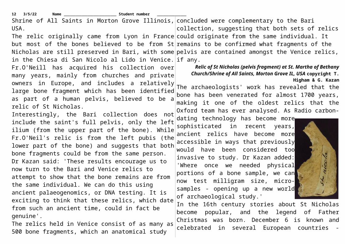
The bone analysed is owned by Father Dennis O'Neill, of St. Martha of Bethany Church, Shrine of All Saints in Morton Grove Illinois, USA.The relic originally came from Lyon in France but most of the bones believed to be from St Nicholas are still preserved in Bari, with some in the Chiesa di San Nicolo al Lido in Venice. Fr.O'Neill has acquired his collection over many years, mainly from churches and private owners in Europe, and includes a relatively large bone fragment which has been identified as part of a human pelvis, believed to be a relic of St Nicholas.Interestingly, the Bari collection does not include the saint's full pelvis, only the left ilium (from the upper part of the bone). While Fr.O'Neil's relic is from the left pubis (the lower part of the bone) and suggests that both bone fragments could be from the same person.Dr Kazan said: 'These results encourage us to now turn to the Bari and Venice relics to attempt to show that the bone remains are from the same individual. We can do this using ancient palaeogenomics, or DNA testing. It is exciting to think that these relics, which date from such an ancient time, could in fact be genuine'.The relics held in Venice consist of as many as 500 bone fragments, which an anatomical study concluded were complementary to the Bari collection, suggesting that both sets of relics could originate from the same individual. It remains to be confirmed what fragments of the pelvis are contained amongst the Venice relics, if any.
Relic of St Nicholas (pelvis fragment) at St. Martha of Bethany Church/Shrine of All Saints, Morton Grove IL, USA copyright T. Higham & G. Kazan
The archaeologists' work has revealed that the bone has been venerated for almost 1700 years, making it one of the oldest relics that the Oxford team has ever analysed. As Radio carbon-dating
8 5/6/23 Name Student number technology has become more sophisticated in recent years, ancient relics have become more accessible in ways that previously would have been considered too invasive to study. Dr Kazan added: 'Where once we needed physical portions of a bone sample, we can now test milligram size, micro-samples - opening up a new world of archaeological study.'In the 16th century stories about St Nicholas become popular, and the legend of Father Christmas was born. December 6 is known and celebrated in several European countries - particularly Holland, as St Nicholas Feast Day. On the eve of the feast, children leave out clogs and shoes to be filled with presents. Of the possible authenticity of the relic itself, Professor Higham concludes: 'Science is not able to definitely prove that it is, it can only prove that it is not, however'.
http://bit.ly/2kaHntbLithium in water associated with slower rate of
Alzheimer's disease deathsTrace elements of lithium in drinking water can slow death rates from Alzheimer's disease, Brock University research has found.
Rates of diabetes and obesity, which are important risk factors for Alzheimer's disease, also decrease if there is a particular amount of lithium in the water, says the study, published recently in the Journal of Alzheimer's Disease.Postdoctoral fellow Val Fajardo and Rebecca MacPherson, Assistant Professor in the Department of Health Sciences, collected statistics on various lithium levels in drinking water in 234 counties across Texas.Lithium is a water-soluble alkali metal found in igneous rocks and mineral springs. It is commonly used to treat bipolar and other mood disorders, but at much higher doses than what occurs naturally in drinking water.The research team, which included Associate Professor of Health Sciences Paul LeBlanc, compared lithium levels naturally found in tap water with Alzheimer's disease mortality rates, along with the incidence of obesity and diabetes, in the Texas counties.
"We found counties that had above the median level of lithium in tap water (40 micrograms per litre) experienced less increases in Alzheimer's disease mortality over time, whereas counties below that median level had even higher increases in Alzheimer's deaths over time," says Fajardo.The frequency of obesity and Type 2 diabetes also went down when the drinking water contained similar lithium levels, the researchers found. Fajardo says he and his team focused on Texas because data on lithium levels were "freely available."Previous studies have demonstrated lithium's ability to protect against Alzheimer's disease, obesity and diabetes."However, we are one of the first groups to show that lithium's potential protective effect against Alzheimer's disease, obesity and diabetes may translate to the population setting through very low levels of lithium in tap water," says Fajardo.The Brock research comes on the heels of an August study from the University of Copenhagen linking high lithium levels in drinking water to decreases in dementia rates. But Fajardo warns it's too early to start advising authorities to add lithium to drinking water."There's so much more research we have to do before policy-makers look at the evidence and say, OK, let's start supplementing tap water with lithium just like we do in some municipalities with fluoride to prevent tooth decay," he says.
http://bit.ly/2BUKnCaWhat Bacteria Can Tell Us About Human Evolution
To discover our species’ deep history and to shape its future health, we should learn from the microbes that accompanied us on our
evolutionary journey. Tara C. Smith
It is human nature to want to know where we came from. Individually, we investigate our family lineages to discover ancestors lost to history. Collectively, scientists examine data from a vast array of sources, ranging from ancient fossils to current genomes, to determine
9 5/6/23 Name Student number where humanity itself originated, and how we came to be who and where we are as a species today.In the past decade, studies in this area have been revolutionized by the plunge in gene sequencing costs. The human genome project began in 1990 and cost about $2.7 billion — roughly $100 million per sequenced genome. Today, a genome can be sequenced for approximately $1,000 to $2,000, and we’re nearing a longstanding goal of the $100 genome.While much of the genomic work carried out to date has focused on genetic risk factors for health and disease, we can also use genetic reconstructions to examine the history of our species. But our own genes don’t necessarily tell us the whole story of our travels and migrations as a species or of the risks to our health.Quantized ColumnsA regular column in which top researchers explore the process of discovery. This month’s columnist, Tara C. Smith, is a professor of epidemiology and infectious-disease researcher.For that reason, in recent years, researchers have paid much more attention to our “second genome”: that of our microbiota. Our microbiota are all of the microscopic organisms that live on and in us, playing a role in our digestion, training our immune system to correctly respond to pathogens, manufacturing key vitamins and taking up space that could otherwise be exploited by pathogens. Gut microbes are the “worlds within worlds” that have evolved alongside us, their hosts, as our early human ancestors moved from place to place, ate new foods and encountered new animals and environments. Our current microbiome (the collective genetic material of our microbiota) reflects some of that deep history.Extreme Symbionts in Our CellsWe can glean information about human history from those organisms within us in several ways. One is by using the parts of our own cells that are, in essence, microbial: our mitochondria. These organelles can be considered “extreme symbionts”: They are remnants of
microorganisms that once lived free but are now integral parts of all eukaryotic (complex) cells, producing energy and regulating metabolism.Microbes in our bodies can also help elucidate humanity’s ancestral journeys.Mitochondria retain their own DNA, separate from that of the cell’s nucleus. For many types of research, this mitochondrial DNA (mtDNA) is preferable to nuclear DNA as an object of study. Unlike our nuclear DNA, it isn’t a mixture of our parents’ genetic material. Because mtDNA is inherited exclusively from the egg and passed down through generations of the maternal lineage, it’s more akin to a clone of your mother (and her mother, and her mother and so on). And while eukaryotic cells have only one copy of nuclear DNA in their singular nucleus, they have many mitochondria and therefore multiple copies of each mtDNA gene. Because the mtDNA genome is much smaller than nuclear DNA (containing only about 37 genes instead of 20,000 or so in humans), it is also simpler to analyze.Analysis of mtDNA in the 1980s led to the conclusion that humanity originated in Africa, dating back to a common maternal ancestor somewhere around 100,000 to 200,000 years ago. Though widely accepted today, this declaration was controversial at the time, as some biologists and anthropologists thought that modern humans evolved as a collective from diverse but interbreeding populations of archaic humans scattered throughout the Old World (the “multiregional hypothesis”).Microbes in our bodies can also help elucidate humanity’s ancestral journeys, because they too are inherited within families and have long been associated with human populations. One example is Helicobacter pylori, a stomach bacterium that can cause ulcers and gastric cancer, but which can also be carried without symptoms in many individuals. H. pylori is transmitted from person to person, probably by saliva (oral-oral route) or contact with feces (fecal-oral route), and possibly by contaminated food or water. Other
10 5/6/23 Name Student number Helicobacter species colonize the guts of mammals, which suggests a lengthy co-evolution between these types of bacteria, humans and our relatives. In the past, H. pylori likely colonized a very high percentage of humans, but its prevalence has decreased in many countries over the past century because of improvements in sanitation and hygiene.Studies during the past 15 years have examined the evolution of H. pylori by collecting and sequencing strains of the bacterium from individuals all around the world. Researchers found that H. pylori collected in Africa contained the most genetic diversity (just as human populations from East Africa do), and that one could retrace basic human migrations out of that continent and around the globe by examining the genetic makeup of this bacterium. Genomic analysis also pointed to the bacterium having co-evolved with humans for approximately 60,000 years — since close to the time when modern humans began migrating out of Africa, and carrying H. pylori and other bacteria along for the ride. We can therefore use the genome of H. pylori to figure out the evolutionary history of some human populations.Retracing Our Past in Their GenesWhy do this when we can look at human bones or genomes to get that information? For one, it’s a powerful confirmation of the correctness of a hypothesis when genomic data from two different organisms tell the same story, especially when those organisms are as different as a human and a bacterium are. In addition, sometimes the data from one genome can fill in gaps that the other data set can’t resolve. Data from H. pylori genomes were able to differentiate two ethnic communities in Ladakh, India, for instance, when the available human genetic markers at the time could not.Through our associated microbes, we can acquire abilities that are beneficial to populations.Today, rather than looking at a single variety of microbe, looking at the massed collection of all of them may better inform our knowledge of where we humans have been as a species, and where we may be
going. The idea of the holobiont — the host and all its associated microbes, analyzed as a single hologenome — is taking shape as we’re starting to understand the thousands of microbial species that can live in and on our bodies.Our microbiota do not just reflect human evolution — they affect it: Through our associated microbes, we can acquire abilities that are beneficial to populations. A 2010 study, for example, found that many individuals from Japan have a gene in their gut microbes that allows them to produce an enzyme that helps to break down carbohydrates from seaweed more efficiently. This gene is absent from the guts of people from North America, where (unlike in Japan) seaweed is not a dietary staple.The gene may have been acquired by a human gut bacterium, Bacteroides plebeius, possibly from the marine bacterium Zobellia galactanivorans. Zobellia could have been ingested long ago by individuals in Japan, entering their gut either as a whole bacterium or in pieces, including as free DNA. Because bacteria can acquire genes through a process known as horizontal gene transfer, Bacteriodes may have picked up this gene in the gut environment. The gene could then have benefited both the bacterium and the host by opening up an additional source of nutrition, and as such would have been maintained in the population by natural selection.Microbial MismatchesAs we begin to grasp the interactions between our microbes and our ancestors since time immemorial, we may be able to use these deep symbioses not only to interpret our history, but also to shape our future health outcomes. H. pylori can be a cause of gastric cancer, but its propensity to promote cancer development appears to be a function of how well the bacterial strain “matches” its host. In a study examining gastric cancer and H. pylori in Colombia , researchers found that African strains of H. pylori were more likely to cause cancer in the Colombian population — but those same strains were not frequently carcinogenic in Africans. This observation points
11 5/6/23 Name Student number toward the possibility of preventing gastric cancers on an individualized basis by minimizing the risks from mismatches between hosts and their bacteria.Now that we are moving to a deeper awareness of the presence and function of our indigenous microbes, we are starting to see how these long-term symbioses have contributed to who we are today. Recent research has confirmed that for the microbiome as a whole, closely related organisms have a more similar microbiome makeup than those more distantly related. The microbiome as a whole could one day help us understand evolutionary relationships among species.Although the power of the microbiome to aid our understanding of disease-related conditions is frequently touted, the idea that our microbes may be able to inform us about ancestors lost in history may be its most intriguing application.
http://bit.ly/2BWE9laLiving on thin air -- microbe mystery solved
UNSW-Sydney led scientists have discovered that microbes in Antarctica have a previously unknown ability to scavenge hydrogen, carbon monoxide and carbon dioxide from the air to stay alive in the
extreme conditions.The find has implications for the search for life on other planets, suggesting extra-terrestrial microbes could also rely on trace atmospheric gases for survival."Antarctica is one of the most extreme environments on Earth. Yet the cold, dark and dry desert regions are home to a surprisingly rich diversity of microbial communities," says study senior author and UNSW scientist Associate Professor Belinda Ferrari."The big question has been how the microbes can survive when there is little water, the soils are very low in organic carbon and there is very little capacity to produce energy from the sun via photosynthesis during the winter darkness.
"We found that the Antarctic microbes have evolved mechanisms to live on air instead, and they can get most of the energy and carbon they need by scavenging trace atmospheric gases, including hydrogen and carbon monoxide," she says.The Australasian-based study, by researchers at UNSW, Monash University, the Australian Centre for Ecogenomics at the University of Queensland, GNS Science in New Zealand, and the Australian Antarctic Division, is published in the journal Nature.Soil samples were collected from two coastal ice-free sites in different regions of eastern Antarctica. One was Robinson Ridge, 10 kilometres from Casey Station, in Wilkes land. The other was Adams Flat, 242 kilometres from Davis Station in Princes Elizabeth Land."Both areas are pristine polar deserts devoid of any vascular plants," says Associate Professor Ferrari, of the UNSW School of Biotechnology and Biomolecular Sciences.The researchers studied the microbial DNA in the surface soil from both sites and reconstructed the genomes of 23 of the microbes that lived there, including some of the first genomes of two groups of previously unknown bacteria called WPS-2 and AD3.They found the dominant species in the soils had genes which gave them a high affinity for hydrogen and carbon monoxide, allowing them to remove the trace gases from the air at a high enough rate to sustain their predicted energy needs and support primary production."This new understanding about how life can still exist in physically extreme and nutrient-starved environments like Antarctica opens up the possibility of atmospheric gases supporting life on other planets," says Associate Professor Ferrari.Most organisms use energy from the sun or the earth to grow. More research is needed to see if this novel use of atmospheric gases as an alternative energy source is more widespread in Antarctica and elsewhere, the scientists say.
12 5/6/23 Name Student number http://bit.ly/2jzb0VH
Want to listen better? Lend a right earResearchers at Auburn University in Alabama have found that the
right ear is the gateway for optimal auditory information processing in both children and adults
WASHINGTON, D.C. - Listening is a complicated task. It requires sensitive hearing and the ability to process information into cohesive meaning. Add everyday background noise and constant interruptions by other people, and the ability to comprehend what is heard becomes that much more difficult.Audiology researchers at Auburn University in Alabama have found that in such demanding environments, both children and adults depend more on their right ear for processing and retaining what they hear.
Displays an example of dichotic digit stimuli presentation, with both 'A' binaural separation tasks (i.e., directed ear) and 'B' binaural integration (i.e.,
free recall) instructions. Sacchinelli, Weaver, Wilson and Cannon - Auburn University
Danielle Sacchinelli will present this research with her colleagues at the 174th Meeting of the Acoustical Society of America, which will be held in New Orleans, Louisiana, Dec. 4-8."The more we know about listening in demanding environments, and listening effort in general, the better diagnostic tools, auditory management (including hearing aids) and auditory training will become," Sacchinelli said.The research team's work is based on dichotic listening tests, used to diagnose, among other conditions, auditory processing disorders in which the brain has difficulty processing what is heard.
In a standard dichotic test, listeners receive different auditory inputs delivered to each ear simultaneously. The items are usually sentences (e.g., "She wore the red dress"), words or digits. Listeners either pay attention to the items delivered in one ear while dismissing the words in the other (i.e., separation), or are required to repeat all words heard (i.e., integration).According to the researchers, children understand and remember what is being said much better when they listen with their right ear.Sounds entering the right ear are processed by the left side of the brain, which controls speech, language development, and portions of memory. Each ear hears separate pieces of information, which is then combined during processing throughout the auditory system.However, young children's auditory systems cannot sort and separate the simultaneous information from both ears. As a result, they rely heavily on their right ear to capture sounds and language because the pathway is more efficient.What is less understood is whether this right-ear dominance is maintained through adulthood. To find out, Sacchinelli's research team asked 41 participants ages 19-28 to complete both dichotic separation and integration listening tasks.With each subsequent test, the researchers increased the number of items by one. They found no significant differences between left and right ear performance at or below an individual's simple memory capacity. However, when the item lists went above an individual's memory span, participants' performance improved an average of 8 percent (some individuals' up to 40 percent) when they focused on their right ear."Conventional research shows that right-ear advantage diminishes around age 13, but our results indicate this is related to the demand of the task. Traditional tests include four-to-six pieces of information," said Aurora Weaver, assistant professor at Auburn University and member of the research team. "As we age, we have better control of
13 5/6/23 Name Student number our attention for processing information as a result of maturation and our experience."In essence, ear differences in processing abilities are lost on tests using four items because our auditory system can handle more information."Cognitive skills, of course, are subject to decline with advance aging, disease, or trauma," Weaver said. "Therefore, we need to better understand the impact of cognitive demands on listening."Abstract: 3aPPa3: "Does the right ear advantage persist in mature auditory systems when cognitive demand for processing increases?" by Danielle M. Sacchinelli is at 8:00 a.m.-12:00 p.m. CST, Dec. 6, 2017, in Studios Foyer (poster sessions) in the New Orleans Marriott. https://asa2017fall.abstractcentral.com/s/u/J4DDi4sip_s
http://bit.ly/2BkSRp4Brain remaps itself in child with double hand transplant
CHOP/Penn medicine team is first to show massive cortical reorganization is reversible in a child
The first child to undergo a successful hand transplant also is the first child in whom scientists have detected massive changes in how sensations from the hands are represented in the brain. The brain reorganization is thought to have begun six years before the transplant, when the child had both hands amputated because of a severe infection during infancy. Notably, after he received transplanted hands, the patient's brain reverted toward a more typical pattern.Each area of the body that receives nerve sensations sends signals to a corresponding site in the brain. The spatial pattern in which those signals activate the brain's neurons is called somatosensory representation -- particular parts of the brain reflect specific parts of the body."We know from research in nonhuman primates and from brain imaging studies in adult patients that, following amputation, the brain remaps itself when it no longer receives input from the hands," said first author William Gaetz, PhD, a radiology researcher in the Biomagnetic Imaging Laboratory at Children's Hospital of
Philadelphia (CHOP). "The brain area representing sensations from the lips shifts as much as 2 centimeters to the area formerly representing the hands."This brain remapping that occurs after upper limb amputation is called massive cortical reorganization (MCR). "We had hoped to see MCR in our patient, and indeed, we were the first to observe MCR in a child," said Gaetz. "We were even more excited to observe what happened next -- when the patient's new hands started to recover function. For our patient, we found that the process is reversible."Researchers from Children's Hospital of Philadelphia and the Perelman School of Medicine at the University of Pennsylvania published their findings today in the Annals of Clinical and Translational Neurology. Their case report described Zion Harvey, now 10 years old, who received worldwide media coverage two years ago as the first child to undergo a successful hand transplant.A 40-member team led by L. Scott Levin, MD, FACS, chairman of Orthopaedic Surgery and a professor of Plastic Surgery at Penn Medicine, and director of the Hand Transplantation Program at CHOP, performed that milestone surgery in July, 2015 at CHOP. "Zion has been a child of many firsts here at Penn Medicine and Children's Hospital of Philadelphia, and across the world," said Levin, senior author of the paper. He added, "With the changes observed in his brain, which our collaborative team has been closely evaluating since his transplant two years ago, Zion is now the first child to exhibit brain mapping reorientation. This is a tremendous milestone not only for our team and our research, but for Zion himself. It is yet another marker of his amazing progress, and continued advancement with his new limbs."The researchers used magnetoencephalography (MEG), which measures magnetic activity in the brain, to detect the location, signal strength and timing of the patient's responses to sensory stimuli applied lightly to his lips and fingers. They performed MEGs four times in the year following the bilateral hand transplant, performing
14 5/6/23 Name Student number similar tests on five healthy children who served as age-matched controls.At the first two visits, the patient's finger tips did not respond to tactile stimulation -- being touched with a thin filament. When experimenters touched the patient's lips, the MEG signal registered in the hand area of the brain's cortex, but with a delay of 20 milliseconds compared to controls. At the two later visits, MEG signals from lip stimulation had returned to the lip region of the brain, with a normal response time--an indication that brain remapping was reverting to a more normal pattern.When experimenters touched the patient's fingertips in the two later visits, the MEG signals appeared in the hand region of the brain, with a shorter delay in response time from visit 3 to visit 4, but with higher-than-normal signal strength. "The sensory signals are arriving in the correct location in the brain, but may not yet be getting fully integrated into the somatosensory network," said Gaetz. "We expect that over time, these sensory responses will become more age-typical."Gaetz added, "These results have raised many new questions and generated excitement about brain plasticity, particularly in children. Some of those new questions include, what is the best age to get a hand transplant? Does MCR always occur after amputation? How does brain mapping look in people born without hands? Would we see MCR reverse in an adult, as we did in this patient? We are planning new research to investigate some of these questions."In the meantime, follow-up studies of this patient provide encouraging details on his functional abilities. "Our follow-up studies 18 months after this transplant showed that he is able to write, dress and feed himself more independently than before his operation-- important considerations in improving his quality of life," said Levin.This study was funded in part by the National Institutes of Health (grants HD086984, DC008871).
W. Gaetz, et al, "Massive cortical reorganization following bilateral transplants of the hands: evidence from the first successful bilateral pediatric hand transplant patient," Annals of Clinical and Translational Neurology, Dec. 6, 2017.
http://bit.ly/2AoH35dAfter 20 years, researcher presents the most complete
Australopithecus fossil ever foundSouth Africa's status as a major cradle in the African nursery of humankind has been reinforced with today's unveiling of "Little
Foot", the country's oldest, virtually complete fossil human ancestor.
Little Foot is the only known virtually complete Australopithecus fossil discovered to date. It is by far the most complete skeleton of a human ancestor older than 1.5 million years ever found.It is also the oldest fossil hominid in southern Africa, dating back 3.67 million years. The unveiling will be the first time that the completely cleaned and reconstructed skeleton can be viewed by the national and international media.
Credit: Wits UniversityDiscovered by Professor Ron Clarke from the Evolutionary Studies Institute at the University of the Witwatersrand in Johannesburg, South Africa, the fossil was given the nickname of "Little Foot" by Prof. Phillip Tobias, based on Clarke's initial discovery of four small foot bones. Its discovery is expected to add a wealth of knowledge about the appearance, full skeletal anatomy, limb lengths and locomotor abilities of one of the species of our early ancestral relatives."This is one of the most remarkable fossil discoveries made in the history of human origins research and it is a privilege to unveil a finding of this importance today," says Clarke.
15 5/6/23 Name Student number After lying undiscovered for more than 3.6 million years deep within the Sterkfontein caves about 40km north-west of Johannesburg, Clarke found several foot bones and lower leg bone fragments in 1994 and 1997 among other fossils that had been removed from rock blasted from the cave years earlier by lime miners. Clarke sent his assistants Stephen Motsumi and Nkwane Molefe into the deep underground cave to search for any possible broken bone surface that might fit with the bones he had discovered in boxes. Within two days of searching, they found such a contact, in July 1997.Clarke realised soon after the discovery that they were on to something highly significant and started the specialised process of excavating the skeleton in the cave up through 2012, when the last visible elements were removed to the surface in blocks of breccia. "My assistants and I have worked on painstakingly cleaning the bones from breccia blocks and reconstructing the full skeleton until the present day," says Clarke.In the 20 years since the discovery, they have been hard at work to excavate and prepare the fossil. Now Clarke and a team of international experts are conducting a full set of scientific studies on it. The results of these studies are expected to be published in a series of scientific papers in high impact, peer reviewed international journals in the near future.This is the first time that a virtually complete skeleton of a pre-human ancestor from a South African cave has been excavated in the place where it was fossilised."Many of the bones of the skeleton are fragile, yet they were all deeply embedded in a concrete-like rock called breccia," Clarke explains."The process required extremely careful excavation in the dark environment of the cave. Once the upward-facing surfaces of the skeleton's bones were exposed, the breccia in which their undersides were still embedded had to be carefully undercut and removed in blocks for further cleaning in the lab at Sterkfontein," says Clarke.
The 20-year long period of excavation, cleaning, reconstruction, casting, and analysis of the skeleton has required a steady source of funding, which was provided by the Palaeontological Scientific Trust (PAST) – a Johannesburg-based NGO that promotes research, education and outreach in the sciences related to our origins. Among its many initiatives aimed at uplifting the origin sciences across Africa, PAST has been a major funder of research at Sterkfontein for over two decades.Professor Adam Habib, Vice-Chancellor and Principal of the University of the Witwatersrand says: "This is a landmark achievement for the global scientific community and South Africa's heritage. It is through important discoveries like Little Foot that we obtain a glimpse into our past which helps us to better understand our common humanity."PAST's chief scientist Professor Robert Blumenschine labels the discovery a source of pride for all Africans. "Not only is Africa the storehouse of the ancient fossil heritage for people the world over, it was also the wellspring of everything that makes us human, including our technological prowess, our artistic ability, and our supreme intellect," he says.The scientific value of the find and much more will be unveiled in a series of papers that Prof Clarke and a team of international experts have been preparing, with many expected in the next year.
http://bit.ly/2jeJOPlNew study: Traumatic brain injury causes intestinal
damageTwo-way brain-gut interactions may worsen outcome after TBI
University of Maryland School of Medicine (UMSOM) researchers have found a two-way link between traumatic brain injury (TBI) and intestinal changes. These interactions may contribute to increased infections in these patients, and may also worsen chronic brain damage.
16 5/6/23 Name Student number This is the first study to find that TBI in mice can trigger delayed, long-term changes in the colon and that subsequent bacterial infections in the gastrointestinal system can increase posttraumatic brain inflammation and associated tissue loss. The findings were published recently in the journal Brain, Behavior, and Immunity."These results indicate strong two-way interactions between the brain and the gut that may help explain the increased incidence of systemic infections after brain trauma and allow new treatment approaches," said the lead researcher, Alan Faden, MD, the David S. Brown Professor in Trauma in the Departments of Anesthesiology, Anatomy & Neurobiology, Psychiatry, Neurology, and Neurosurgery at UMSOM, and director of the UMSOM Shock, Trauma and Anesthesiology Research Center.Researchers have known for years that TBI has significant effects on the gastrointestinal tract, but until now, scientists have not recognized that brain trauma can make the colon more permeable, potentially allowing allow harmful microbes to migrate from the intestine to other areas of the body, causing infection.. People are 12 times more likely to die from blood poisoning after TBI, which is often caused by bacteria, and 2.5 times more likely to die of a digestive system problem, compared with those without such injury.In this study, the researchers examined mice that received an experimental TBI. They found that the intestinal wall of the colon became more permeable after trauma, changes that were sustained over the following month.It is not clear how TBI causes these gut changes. A key factor in the process may be enteric glial cells (EGCs), a class of cells that exist in the gut. These cells are similar to brain astroglial cells, and both types of glial cells are activated after TBI. After TBI, such activation is associated with brain inflammation that contributes to delayed tissue damage in the brain. Researchers don't know whether activation of ECGs after TBI contributes to intestinal injury or is instead an attempt to compensate for the injury.
The researchers also focused on the two-way nature of the process: how gut dysfunction may worsen brain inflammation and tissue loss after TBI. They infected the mice with Citrobacter rodentium, a species of bacteria that is the rodent equivalent of E. coli, which infects humans. In mice with a TBI who were infected with this the bacteria, brain inflammation worsened. Furthermore, in the hippocampus, a key region for memory, the mice who had TBI and were then infected lost more neurons than animals without infection.This suggests that TBI may trigger a vicious cycle, in which brain injury causes gut dysfunction, which then has the potential to worsen the original brain injury. "These results really underscore the importance of bi-directional gut-brain communication on the long-term effects of TBI," said Dr. Faden.Other authors of this paper include Elise Ma, a doctoral student; Terez Shea-Donahue PhD, professor of radiation oncology; Bogdan A. Stoica, MD, associate professor of anesthesiology ; and David Loane, PhD, associate professor of anesthesiology- all at UMSOM.
http://bit.ly/2iJKs3aClay minerals on Mars may have formed in primordial
steam bathPlanetary scientists from Brown University have proposed a new
scenario for the formation of ancient clay minerals on Mars that, if shown to be true, could rewrite the early history of the red planet.
PROVIDENCE, R.I. [Brown University] - There are thousands of ancient phyllosilicate outcrops on the Martian surface. Phyllosilicates, or clays, are formed by the interaction of water with volcanic rock, leading many scientists to conclude that there must have been sustained surface water, groundwater or active hydrothermal systems at some point in Martian history. But the new research, published in the journal Nature, suggests that the clays may have formed during the creation of the Martian crust itself, long before any water flowed on the planet.Backed by lab experiments and computer models, the researchers lay out how the scenario would have worked. In the very early solar system, Mars and other rocky planets are thought to have been
17 5/6/23 Name Student number covered by oceans of molten magma. As the Mars magma ocean began to cool and solidify, water and other dissolved volatiles would be outgassed to the surface, forming a thick, steamy atmosphere surrounding the planet. The moisture and heat from that high-pressure steam bath would have converted vast swaths of the newly solidified surface to clay. As the planet then evolved over billions of years, volcanic activity and asteroid bombardments would have covered the clays in some places and excavated them in others, leading to the widespread but patchy distribution seen on the surface today."The basic recipe for making clay is you take rock and you add heat and water," said Kevin Cannon, a postdoctoral researcher at the University of Central Florida who led the research while completing his Ph.D. at Brown. "This primordial atmosphere created by a magma ocean would have been the hottest and wettest Mars ever was. It's a situation where you could pervasively alter the crust and then just shuffle those materials around afterward."Cannon and his co-authors say the scenario offers a means of creating widespread clay deposits that doesn't require a warm and wet climate or a sustained hydrothermal system on early Mars. State-of-the-art climate models suggest an early Mars where the temperature rarely crept above freezing and where water flow on the surface was sporadic and isolated."One of the complications that comes up in Mars evolution is that we can't create a scenario where surface weathering had the capacity to produce the extent of mineral alteration that we see," said Jack Mustard, a professor in Brown's Department of Earth, Environmental and Planetary Sciences and study co-author. "We're certainly not trying to discount other alteration mechanisms entirely. Surface weathering and other types of alteration surely occurred at different points in Martian history, but we think this is a plausible way to explain much of the widespread clay we see in the oldest Martian terrains."
To demonstrate that the mechanism they propose is plausible, the researchers synthesized rock samples matching the composition of Martian basalt. They then used a high-pressure device to recreate temperature and pressure conditions the may have been present amid the steam atmosphere created by a magma ocean. After cooking samples for two weeks, the team checked to see if they had been altered and to what extent."It was really remarkable how quickly and extensively this basalt was altered," Cannon said. "At the highest temperatures and pressures, it ate completely through the basalt particles. It's a really intense degree of alteration."The steam atmosphere associated with a magma ocean could have survived for as long as 10 million years or more, Cannon and his colleagues say. That would have been long enough, they estimate, to create as much as three kilometers of clay on the primordial Martian surface.To get an idea what the fate of that clay might be as the planet evolved, the researchers created a computer model to simulate a slab of Martian crust with a three-kilometer clay layer on top. Then they simulated the first billion years of Martian geologic history -- the period when volcanic activity and asteroid bombardment were most prevalent. The model showed that the burial, excavation and scattering of clays over time created distribution of exposed deposits similar to what's seen on Mars today."To put some numbers on it, clays cover about 3 percent of the oldest crust exposures on Mars," Cannon said. "We're finding about that same order of magnitude in these models."The lab experiments and simulations can't say for certain that this scenario occurred, the researchers say, but they do suggest a strong hypothesis that could be tested during future Mars exploration."One of the things I like about this is that it's truly testable," said Steve Parman, a geology professor at Brown and co-author of the study. "With a returned sample, or maybe even with the analytical equipment
18 5/6/23 Name Student number on a rover, I'm optimistic that you could distinguish this primordial process from some other alteration process."If the process did indeed occur, it could have some interesting implications for early Martian history. In addition to providing a mechanism for clay formation even if Mars was as cold and icy as climate models suggest, the scenario suggests that vast deposits of clay were -- and might still be -- present beneath the surface. Those deposits could explain why the Martian crust is less dense than expected for a basaltic crust, the researchers say. The deposits would also serve as large underground storage reservoirs for water."There potentially would have been quite a lot of water locked up in these buried clays," Parman said. "You could imagine that if those deposits were heated up by magmatism or some other process they would have released that water, perhaps providing a transient water supply to the surface. That could have implications for past habitability."Mustard, who chaired the committee that laid out the science goals for NASA's Mars 2020 rover, hopes this new hypothesis could inform future Martian exploration. "This would be a really interesting hypothesis to test," he said. "Depending on where the rover ultimately lands, I think we could get the right samples to illuminate these questions."
http://bit.ly/2AJdybkDibenzoazepine defender: Drug found to be effective
against resistant hepatitis COsaka University researchers identify class of chemicals that can
combat resistant strains of the hepatitis C virus, as well as parasites that cause malaria and toxoplasmosis
Osaka - Hepatitis C is caused by a highly infectious virus affecting millions across the globe and can lead to a variety of liver ailments. While the hepatitis C virus (HCV) can sometimes be fought off and cleared by the immune system during the first few months of acute infection, up to 80% of those with HCV develop a chronic infection.
This can lead to serious liver illnesses, including inflammation, cirrhosis, and hepatocellular carcinoma - the third leading cause of cancer death worldwide.While highly effective treatments for HCV have become available in recent years, drug-resistant viral strains can still lead to treatment failure for a sizable proportion of patients. Now, in a recent study published in PNAS Plus, a compound has been reported that may eventually prove effective against drug-resistant HCV.A team of researchers centered at Osaka University infected human liver cells with HCV, then treated the infected cells with different drugs to see which might prevent the virus from spreading. One compound, innocuously named YO-01027, stood out above the rest."For HCV to propagate in a host cell, the proteins that make up the virus particle need to be cleaved into their mature form," lead author Junki Hirano explains. "We tested several compounds we thought may inhibit this cleavage process, and found that YO-01027 prevents a key HCV protein from undergoing cleavage and maturation. We correspondingly found the drug is very effective at suppressing HCV infection."Importantly, resistant strains of HCV did not emerge over time when the infected cells were treated with YO-01027. This may owe to the unique way the compound prevents the virus from maturing.Patients with HCV are currently given direct-acting antivirals, which (as their name suggests) directly target and disrupt HCV proteins themselves. The drug tested in this study, however, inhibits one of the host cell's proteins - signal peptide peptidase (SPP) - that HCV hijacks during an infection."Direct-acting antivirals have made tremendous progress in treating HCV," corresponding author Yoshiharu Matsuura explains. "The difficulty is that HCV shows quite high genetic diversity, even within a single patient. Antivirals produce a strong selective pressure that can cause HCV strains with resistant forms of the target protein to spread.
19 5/6/23 Name Student number By inhibiting the host's own SPP protein, we can largely bypass this selection problem."Through a combination of computer simulations and in vitro tests, the researchers identified the chemical signature of YO-01027 responsible for its effectiveness, a structure called dibenzoazepine. With this and other molecular details in hand, the researchers may now be able to modify YO-01027 and other dibenzoazepine-containing drugs to develop novel therapies for drug-resistant HCV - and, serendipitously, to potentially develop therapies against a variety of other diseases."Now that we know some of the key structural features that make YO-01027 effective at inhibiting SPP, we can start the chemical fine tuning," Matsuura adds. "Ultimately, the goal is to make highly selective drugs to combat pathogens that need SPP to survive and spread. This includes not only viruses like HCV, but also parasites such as Plasmodium falciparum and Toxoplasma gondii that are responsible for malaria and toxoplasmosis. The possible applications are very exciting."
http://bit.ly/2A5xK5RVictims of mystery attacks in Cuba left with anomalies in brain tissueAuthorities still stumped by attacks, but
doctors are figuring out damage, treatments.
Beth Mole - 12/7/2017, 4:25 AMAmerican victims of mysterious attacks in Cuba have abnormalities in their brains’ white matter, according to new medical testing reported by the Associated Press. But, so far, it’s unclear how or if the white-matter anomalies seen in the victims relate to their symptoms.
Picture of the US embassy in Havana, taken on October 3, 2017. Getty Yamil Lage
White matter is made up of dense nerve fibers that connect neurons in different areas of the brain, forming networks. It gets its name from the light-colored electrical insulation, myelin, that coats the fibers.
Overall, the tissue is essential for rapidly transmitting brain signals critical for learning and cognitive function.In August, US authorities first acknowledged that American diplomats and their spouses stationed in Havana, Cuba, had been the targets of puzzling attacks for months. The attacks were carried out by unknown agents and for unknown reasons, using a completely baffling weaponry. The attacks were sometimes marked by bizarrely targeted and piercing noises or vibrations, but other times they were completely imperceptible.Victims complained of a range of symptoms, including dizziness, nausea, headaches, balance problems, ringing in the ears (tinnitus), nosebleeds, difficulty concentrating and recalling words, permanent hearing loss, and speech and vision problems. Doctors have also identified mild brain injuries, including swelling and concussion.White-matter changes are often seen following brain injuries, and the extent and duration of changes can determine cognitive impairment. That said, without brain scans and tests prior to the attacks, it’s difficult to know if the abnormalities were related to the attacks or if they are from previous injuries. None of the victims reported blows to the head, however.US officials now report that 24 Americans were injured in the attacks but wouldn’t comment on how many showed abnormalities in their white matter. The officials told the AP that most had recovered and some were even back at work. But about a quarter of the victims reported symptoms that were either persistent or took a long time to clear up. All of the victims will likely be tracked by doctors for life.Tensions and mysteryDoctors treating the victims were tight-lipped about their treatments and findings, the AP said. And Secretary of State Rex Tillerson expressed concern for victim privacy and about releasing medical information that could reveal the effectiveness of the attacks. Nevertheless, doctors at the University of Miami and the University of Pennsylvania are working with government agencies to write up a
20 5/6/23 Name Student number report on the victims’ conditions and newly developed treatment protocols. Officials told the AP that the doctors would submit their report to the Journal of the American Medical Association.Meanwhile, FBI investigators and US intelligence agencies are still struggling to understand the mysterious attacks. The attacks were initially speculated to be “sonic attacks” based on audible noises heard by some of the victims. But scientists have largely ruled out the possibility that sounds could cause traumatic brain damage.The AP notes that shock waves from explosions in combat have been known to cause concussion and white-matter damage in soldiers. But, of course, none of the victims reported experiencing an explosion.The agents behind the attacks and their motivation remain unknown.At the end of September, US officials drew down the staff at the US embassy in Havana due to the safety risks. The embassy is now operating only with emergency staff. In October, the US also expelled 15 Cuban diplomats over the issue, saying the Cuban government wasn’t doing enough to protect Americans there.In its report Wednesday, the AP quoted Secretary Tillerson as saying:What we’ve said to the Cubans is: small island. You’ve got a sophisticated intelligence apparatus. You probably know who’s doing it. You can stop it. It’s as simple as that.Cuba has denied any involvement with the attacks.
http://go.nature.com/2iMaT8xScrap very useless qualifiers in research papers
Hackneyed adverbs that convey little to the readerStephen K. Donovan
Eric Blackman bemoans the misuse of ‘obviously’ and ‘clearly’ in scientific publications (Nature 550, 457; 2017). He might also have mentioned another hackneyed adverb that conveys so little to the reader: ‘very’.As an editor and reviewer, it is one of my life’s tasks to delete this overworked word from any scientific manuscript I handle. I once
found it eight times in a paragraph; I felled seven and reluctantly showed a single act of mercy.doi: 10.1038/d41586-017-08190-
http://bit.ly/2ARJNXFLove at First Sight? It's Probably Just Lust
Is it actually love? Not quite, according to a new studyBy Mindy Weisberger, Senior Writer
We've all seen that movie moment when two strangers meet and feel an instant romantic connection - in fact, "love at first sight" has been a mainstay of literature for thousands of years, and people in real life often claim to experience a similar spark. But is that feeling actually love? Not quite, according to the authors of a new study.In the study, researchers investigated whether people feel love at first sight - LAFS - or whether they believe retroactively that they felt that way, once they've already formed an attachment to a romantic partner. The scientists also questioned whether what people call "love" at a first encounter is truly representative of the complex emotions that make up love - or just a powerful physical attraction.Prior studies have shown that being in love activates certain brain regions, and the location of the activity can vary depending on what type of love the person is feeling, such as emotional, maternal or passionate love.Intense, passionate love activates the same networks in the brain as addiction does, and more long-term love sparked responses in brain regions associated with attachment and reward. Researchers have also previously reported that as many as 1 in 3 people in Western countries claim to have experienced LAFS. And that the feeling is associated with more passion and stronger bonds within the relationship, the scientists wrote in the new study. But there was little evidence indicating if LAFS occurred when people thought it did — at the moment of their first meeting ― or if they merely remembered it happening that way through the lens of their current romantic feelings, the study authors explained.
21 5/6/23 Name Student number The scientists collected data from about 500 encounters between nearly 400 participants, mostly heterosexual Dutch and German students in their mid-20s.Using three stages of data collection — an online survey, a laboratory study and three dating events lasting up to 90 minutes each — the researchers gathered information from their subjects about meeting prospective romantic partners.They noted whether participants said that they felt something akin to LAFS upon a first meeting, and how physically attractive they ranked the person who inspired those feelings. To define what qualified as "love," subjects submitted self-analysis of several key components: "eros" (physical attraction), "intimacy," "passion" and "commitment." During the tests, 32 different individuals reported experiencing LAFS a total of 49 times — and that observation wasn't typically accompanied with high ratings for love components such as intimacy and commitment.However, reports of LAFS did correspond with a potential partner scoring higher as physically attractive, the researchers discovered. About 60 percent of the study participants were women, but men were more likely to report feeling LAFS "on the spot," the study authors reported. And in every case, their experience of LAFS was unreciprocated, suggesting that mutual, instantaneous LAFS "might generally be rare," according to the study.The authors determined that LAFS was, in fact, merely "a strong initial attraction" that people identified as love, either at the moment they felt it, or in retrospect. And though some study subjects who were already involved with someone reported that they fell in love at first glance, it's hard to say for sure if that happened the way they remembered.Answering this question would require further investigation into romantic relationships, to see how those initial, powerful feelings of instantaneous love play out over time, the scientists wrote.
The findings were published online Nov. 17 in the Journal of the International Association for Relationship Research.
http://bit.ly/2kPUO65How the cat parasite exploits immune cells to reach the
brainNew research show that Toxoplasma gondii infects the human body
by controlling our immune cells."We have decoded how the parasite takes control of immune cells, converting them into moving "zombies" which spread the parasite in the body," said Antonio Barragan, professor at Stockholm University and one of the authors of the new study.The infection toxoplasmosis is caused by the parasite Toxoplasma gondii and is widely spread. It's estimated that 30-50 per cent of the global human population are carriers. Cats are the parasites' main host, but the infection is also spread among other animals, including humans. A series of studies have previously shown that the parasite affects the brain of infected rats so that they lose fear of cats and even become attracted to cats' smell, making them an easy prey. This is how the parasite is spread onward, by ensuring that the rat is eaten by a cat. Toxoplasmosis is life-threatening to people with impaired immune systems and to unborn foetuses, but causes only mild symptoms in healthy individuals. However, there are studies showing that mental illnesses such as schizophrenia, depression and anxiety disorder are more common in people who are carriers of Toxoplasma gondii. There are also studies indicating that the parasite may affect aggressive or risky behaviour.Researchers at Stockholm University have now been able to show how the parasite takes control and force immune cells around the body to spread it, eventually reaching the brain. When we become infected with Toxoplasma, for example by eating insufficiently cooked meat or by contact with cat faeces, the parasite ends up in the stomach. It then
22 5/6/23 Name Student number passes through the intestinal wall and is met by immune cells that would normally kill it. Instead, immune cells become "Trojan horses". By secreting the substance GABA, they can spread the infection into the body."Is it a coincidence or evolution? It resembles how nerve cells speak to each other in our brains", said Antonio Barragan.The new research has shown that the small calcium molecule is the messenger in the communication. The researchers have found a new calcium receptor on immune cells, acting as a mailbox to receive the parasite's orders for the cell to move."The neat thing is that the signal can be inhibited by regular blood pressure medicine. When mice received the medicine, the spread of the parasite was inhibited. We do not want to say that blood pressure medicine can cure toxoplasmosis, but we have discovered a new signalling pathway in immune cells that is linked to their motility and that the parasite utilizes in a very smart way. This helps us understand how the parasite is spread and disease occurs. In the longer term, it may help us develop targeted treatments for infection," said Antonio Barragan.
http://bit.ly/2B4nezMThousands of Mutations Accumulate in the Human Brain
Over a LifetimeSingle-cell genome analyses reveal the amount of mutations a human brain cell will collect from its fetal
beginnings until death.By Ruth Williams | December 7, 2017
Two studies in Science today (December 7)—one that focuses on prenatal development in humans, the other on infancy to old age—provide insights into the extent of DNA sequence errors that the average human brain cell accumulates over a lifetime.
ISTOCK, JEZPERKLAUZEN
Together, they reveal that mutations become more common as fetuses develop, and over a lifetime a person may rack up more than 2,000 mutations per cell.“I think these are both very powerful technical papers, and they demonstrate how single-cell sequencing . . . can reliably detect somatic changes in the genomes of human neurons,” says neuroscientist Fred Gage of the Salk Institute in La Jolla who was not involved in either study.“What’s cool about [the papers] is that they show two different ways that one can look at somatic mutations in single human neurons . . . and yet they get consistent results,” says neuroscientist Michael McConnell of the University of Virginia School of Medicine.Cells of the human body acquire mutations over time, whether because of errors introduced during DNA replication or damage incurred during transcription and other cellular processes. But, until recent technological developments enabled whole genome sequencing from the miniscule quantities of DNA found inside single cells or small clones of the same cell, investigating the nature and extent of such somatic mutations—and the resulting tissue mosaicism—was practically impossible.Within the now burgeoning field of somatic mutation analyses, the brain is a particular area of interest. That’s because unlike organs such as the skin and gut where cells are replaced daily, the brain’s neurons, once established in the fetus, for the most part stick around for life. Somatic mutations in these cells, then, could affect brain function, behavior, and the propensity for disease long-term. Indeed, it’s thought that such mutations could influence the development of diseases such as schizophrenia, autism, and Tourette’s, which have unclear etiologies, says Yale School of Medicine’s Flora Vaccarino who authored one of the studies.But before studying pathologies associated with somatic mutations, Vaccarino says, “we wanted to know what is the normality of this phenomenon . . . When does it occur? Are there some stages of
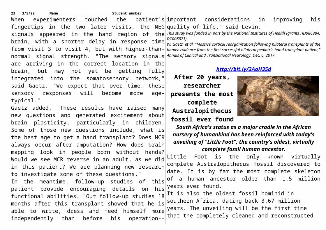
23 5/6/23 Name Student number development that are more susceptible to somatic mutation? That was the aim of our study.”Vaccarino’s team examined the genomes of single cells taken from the post mortem forebrains of three human fetuses (15 weeks, 17.5 weeks, and 21 weeks post conception). Instead of amplifying the genomes of the cells directly for sequencing—which, it has been suggested, might introduce DNA artifacts—her team took advantage of the fetal neuronal cells’ ability to replicate and grew small clones from the individual brain cells to generate enough DNA for sequencing.They found that, on average, each cell contained between 200 and 400 single nucleotide variations (SNVs), which were, largely speaking, distributed randomly across the genome. A small percentage of the SNVs were also present in spleen cells, indicating that they occurred prior to the differentiation of mesodermal and neuroectodermal tissues—that is, before embryonic gastrulation.
About one percent of the mutations are likely to be functional in the sense that they disrupt a protein, so by the time you’re 80 years old there’s about
one in a thousand neurons that has had a gene essentially knocked out.—Christopher Walsh, Harvard Medical School and Boston Children's Hospital
By determining the numbers of mutations in the differently aged fetuses, and knowing the average speed of cell divisions, the team was able to calculate the average mutation rate during neurogenesis as 5.1 mutations per cell, per division. Extrapolating their calculations further back in development, they also determined that in the early embryo the mutation rate was 1.3 mutations per cell per division.The ramping up of the mutation rate during neurogenesis, says Gage, is not surprising. From an evolutionary point of view, he explains, it makes sense that “protecting the genome at embryonic stages is more imperative than at the later stages of differentiation,” where mutations would affect far fewer cells. But, while that’s a sensible theory, he adds, “that the researchers give evidence of it, is important.”The second study looked at single cells from the post mortem brains of 15 people aged four months to 82 years. Because by these ages most neurons have lost their ability to replicate, clonally expanding
the cells in culture was not possible. The team therefore extracted DNA directly from each cell and amplified it for sequencing.The researchers found that a neuron “starts with around 600 mutations” in an infant, “and the mutations accumulate about one every two weeks, so that by the time a neuron is 80 years old it has about 2,400 or so,” says Christopher Walsh of Harvard Medical School and Boston Children’s Hospital who authored the study. “In general, our numbers pick up right where [Vaccarino’s] leave off,” he says.“It’s nice to see the two approaches getting similar answers,” says McConnell. “It’s very good news for folks like me that do single-cell sequencing.”The second study also looked at brain cells from two individuals with neurodegeneration caused by defects in DNA repair enzymes, finding them to have roughly 2.5 times more SNVs than age-matched controls.“About one percent of the mutations are likely to be functional in the sense that they disrupt a protein,” says Walsh, “so by the time you’re 80 years old there’s about one in a thousand neurons that has had a gene essentially knocked out.” Mutation accumulation could therefore be “a reasonable model for how age-related cognitive decline might come about,” he says.Walsh’s team also noted that the types of SNVs varied with age, with those apparently caused by oxidative damage being more prevalent in the elderly. That fits “with previous literature that suggests that one cause of aging in the brain might have to do with oxidative damage,” he says. “So that has me eating a lot of blueberries and drinking a lot of red wine.”What remains to be determined, says McConnell, is not only whether these mutations may influence a person’s likelihood of developing certain neurological conditions, but also whether during development these mutations could actually be an important part of establishing essential cellular diversity within the brain. “Studies like
24 5/6/23 Name Student number these provide important data that will help direct us toward an understanding of the actual functions of somatic mutations,” he says.T. Bae et al., “Different mutational rates and mechanisms in human cells at pregastrulation and neurogenesis,” Science, doi:10.1126/science.aan8690, 2017.M.A. Lodato et al., “Aging and neurodegeneration are associated with increased mutations in single human neurons,” Science, doi:10.1126/science.aao4426, 2017.
http://bit.ly/2jBUeW7Scientists identify first brain cells that respond to sound
Discovery could enable early diagnosis of autism and other cognitive deficits
Some expectant parents play classical music for their unborn babies, hoping to boost their children's cognitive capacity. While some research supports a link between prenatal sound exposure and improved brain function, scientists had not identified any structures responsible for this link in the developing brain.A new study by University of Maryland School of Medicine (UMSOM) scientists, along with colleagues from the University of Maryland College Park, is the first to identify a mechanism that could explain an early link between sound input and cognitive function, often called the "Mozart effect."Working with an animal model, the researchers found that a type of cell in the brain's primary processing area during early development, long thought to have no role in transmitting sensory information, may conduct such signals after all.The results, which could have implications for the early diagnosis of autism and other cognitive deficits, were published recently in the Proceedings of the National Academy of Sciences."Our work is the first to suggest that very early in brain development, sound becomes an important sense," said Amal Isaiah, assistant professor of otorhinolaryngology at UMSOM and one of the principal authors. "It appears that the neurons that respond to sound play a role in the early functional organization of the cortex. This is new, and it is really exciting."
Working with young ferrets, Dr. Isaiah and Patrick Kanold, PhD, a professor of biology at University of Maryland College Park, observed sound-induced nerve impulses in subplate neurons, which help guide the formation of neural circuits in the same way that a scaffolding helps a construction crew erect a new building. This is the first time such impulses have been seen in these neurons. During development, subplate neurons are among the first neurons to form in the cerebral cortex--the outer part of the mammalian brain that controls perception, memory and, in humans, higher functions such as language and abstract reasoning.The role of subplate neurons is thought to be temporary. Once the brain's permanent neural circuits form, most subplate neurons disappear. Researchers assumed that subplate neurons had no role in transmitting sensory information, given their transient nature.Scientists had thought that mammalian brains transmit their first sensory signals in response to sound after the thalamus, a large relay center, fully connects to the cerebral cortex. Studies from some mammals demonstrate that the connection of the thalamus and the cortex also coincides with the opening of the ear canals, which allows sounds to activate the inner ear. This timing provided support for the traditional model of when sound processing begins in the brain.However, researchers had struggled to reconcile this conventional model with observations of sound-induced brain activity much earlier in the developmental process. Until Kanold, senior author of the paper, and his colleagues directly measured the response of subplate neurons to sound, the phenomenon had largely been overlooked."Previous research documented brain activity in response to sound during early developmental phases, but it was hard to determine where in the brain these signals were coming from," Prof. Kanold said. "Our study is the first to measure these signals in an important cell type in the brain, providing important new insights into early sensory development in mammals."
25 5/6/23 Name Student number By identifying a source of early sensory nerve signals, the current study could lead to new ways to diagnose autism and other cognitive deficits that emerge early in development. Early diagnosis is an important first step toward early intervention and treatment.The next step is to begin studying in more detail how subplate neurons affect brain development. There are many avenues of research: for instance, the researchers say that the neurons might play a role in some cognitive conditions such as autism.
http://bit.ly/2C01ylWTaking Vitamin B7 Can Mess Up Your Medical TestsHigh doses of the vitamin biotin in a person's blood can cause
errors in some medical lab testsBy Jenna Flannigan, Live Science Contributor
High doses of the vitamin biotin in a person's blood can cause errors in some medical lab tests, and these faulty test results have already been linked to one death, according to the U.S. Food and Drug Administration (FDA). Biotin, also known as vitamin B7 or vitamin H, is commonly found in multivitamins, prenatal vitamins and supplements that claim to improve nails, hair and skin.In a statement released Nov. 28, the FDA said that biotin can skew the results of certain lab tests, which can potentially lead to a misdiagnosis. These "biotin-related errors" can occur in a variety of lab tests, including tests to measure hormone levels and tests that can reveal signs of a heart attack, the FDA said.The potential for biotin to skew the results of certain lab tests is not a new finding, but the FDA said that many patients and doctors may be unaware that the vitamin can have this effect. In addition, the FDA noted that there has been an increase in reports of such errors leading to negative consequences.In one report, a patient who was taking high doses of biotin died from a heart attack after a blood test showed falsely low levels of troponin, a protein that, at elevated levels, indicates heart damage, according to the statement.
How much is too much?The Institute of Medicine recommends that people get 0.03 milligrams of biotin daily. However, some supplements contain 20 milligrams of biotin - more than 650 times the recommended amount, the FDA said.It's unclear exactly what amount of biotin causes lab test errors, but researchers are exploring the issue.A very small preliminary study, published in September in the journal JAMA, looked at how a 10-milligram daily dose of biotin affected test results in six healthy adults. The researchers asked the participants to take 10 milligrams of the vitamin daily for a week. They took blood samples before and after, and then used the blood in a variety of lab tests.The researchers included 23 lab tests that were suspected to be vulnerable to biotin-related errors. Among the 23 lab tests, biotin supplementation caused errors in nearly 40 percent, the study found.The researchers found that biotin supplementation caused one test error that could lead doctors not to identify congestive heart failure. Other false test results could make a healthy patient appear to have thyroid disease.InterferenceThe reason biotin supplements can mess up certain test results is because the tests contain their own biotin. Many lab tests rely on a strong bond between biotin and a protein called streptavidin to increase test sensitivity. Biotin and streptavidin are part of a process that creates a signal that is either proportional or disproportional to the amount of the substance the test is measuring, said Danni Li, the lead author of the JAMA study and an assistant professor of laboratory medicine and pathology at the University of Minnesota. That's why the presence of extra biotin can lead to falsely high or low results, Li said. [5 Key Nutrients Women Need As They Age]"When a person takes very large doses of biotin, the biotin present in their blood sample can interfere with the mechanism of the lab test in
26 5/6/23 Name Student number such a way that it could either falsely produce a result that is low or high," Li told Live Science.However, biotin doesn't stay in a person's body forever: It leaves the body after some time, Li said. It’s uncertain exactly how long people need to wait for the vitamin to clear their blood. Some lab test manufacturers argue that 8 hours is enough, Li said. However, she said she doubts this would be enough time for very high doses of biotin or for a person who had kidney disease, which impacts how long it takes the body to get rid of certain substances. If people want to be conservative, wait a couple of days to a week, Li said. Eating foods that contain the vitamin won't cause false test results because these foods don't contain worrisome amounts, she added. Li said that people who want to keep taking biotin should tell their doctor and take a break from the vitamin before getting medical tests."I don't think this should necessarily stop people from taking biotin," Li said, noting that there’s anecdotal evidence of biotin’s health benefits for some patients. "They just need to be mindful," Li added. "Knowing that it interferes with labs tests, they should tell their doctor about it."
http://bit.ly/2jFzInhNovel compound restores immune response in patients
with melanomaA novel compound may restore immune response in patients with melanoma, according to a study presented at the ESMO Immuno
Oncology Congress 2017. (1)
LUGANO-GENEVA - "Checkpoint inhibitors are a standard of care immunotherapy for metastatic melanoma," said lead author Dr Sapna Patel, Assistant Professor, Department of Melanoma Medical Oncology, University of Texas MD Anderson Cancer Center, Houston, US. "However, many patients do not respond because myeloid derived suppressor cells (MDSCs), a type of inhibitory cell, are present in the tumour microenvironment."
"In animal studies, omaveloxolone inhibited MDSCs and restored immune activity," she continued. "Myeloid-derived suppressor cells (MDSCs) produce reactive nitrogen radicals that alter the receptors on the surface of the tumour to hide it from cytotoxic lymphocytes that kill tumour cells. Omaveloxolone inhibits MDSC activity, suppresses reactive nitrogen radicals, and restores anti-tumour immune responses. Administering omaveloxolone with checkpoint inhibitors may improve the antitumour response of these immunotherapies."This open label, multicentre, phase 1B trial investigated the safety and efficacy of omaveloxolone in combination with the checkpoint inhibitors ipilimumab or nivolumab. The study included 30 patients with unresectable or metastatic melanoma, of whom seven were naïve to checkpoint inhibitors and 23 had prior checkpoint inhibitor treatment.The overall response rate was 57% in checkpoint inhibitor naïve patients and 17% in those with prior exposure. Median time to response was 19 weeks. There were no serious adverse events related to omaveloxolone and it was well tolerated in combination with ipilimumab or nivolumab.Dr Patel said: "Our findings suggest that omaveloxolone may overcome resistance to checkpoint inhibitors. Omaveloxolone in combination with checkpoint blockade had activity in both naïve and checkpoint inhibitor refractory melanoma patients."She added: "This is one of the first studies to demonstrate a meaningful response rate in the checkpoint inhibitor refractory melanoma population. Further dose escalation and dose expansion studies are underway as well as translational tissue-based experiments to clarify the impact of this treatment combination."Commenting on the study for ESMO, Dr Olivier Michielin, head of Personalised Analytical Oncology, Centre Hospitalier Universitaire Vaudois (CHUV), Lausanne, Switzerland, said: "Omaveloxolone's novel mechanism of action is to block MDSCs, cells known to suppress the immune response. This study tested a new combination
27 5/6/23 Name Student number therapy in immuno oncology and found encouraging response rates with omaveloxolone plus ipilimumab or nivolumab in patients who were checkpoint inhibitor naïve or resistant. The combination was well tolerated and may address some of the immune escape mechanisms that limit the activity of current checkpoint blockade therapies."Michielin added: "More data is needed before we can make a final call on whether there is a place, and where would the place be, for this combination in the current treatment portfolio. The next step should be a randomised trial to investigate whether omaveloxolone provides additional benefit when combined with the checkpoint blockade backbone, for example, comparing the efficacy of PD-1 blockade alone versus PD-1 blockade plus omaveloxolone."Please make sure to use the official name of the meeting in your reports: ESMO Immuno Oncology Congress 2017 Official hashtag: #ESMOImmuno17References1 Abstract 5O_PR 'A phase 1b/2 study of omaveloxolone in combination with checkpoint inhibitors in patients with unresectable or metastatic melanoma' will be presented by Sapna Patel during a Proffered Paper Session on Friday 8 December, 08:30 to 10:30 CET in Room A. Annals of Oncology, Volume 28, 2017 Supplement 11.DisclaimerThis press release contains information provided by the authors of the highlighted abstracts and reflects the content of those abstracts. It does not necessarily reflect the views or opinions of ESMO who cannot be held responsible for the accuracy of the data. Commentators quoted in the press release are required to comply with the ESMO Declaration of Interests policy and the ESMO Code of Conduct.
http://bit.ly/2jCOPy4Flu Season Is Already Off to a Bad Start
Flu season is underway in the United States, and a new report shows that flu activity is already higher than typical for this time of year.
By Rachael Rettner, Senior Writer | December 8, 2017 07:12am ETDuring the week that ended Nov. 25 (the most recent period for which data is available), three Southern states — Louisiana, Mississippi and South Carolina — reported high levels of flu activity; one state (Georgia) reported moderate flu activity, and the rest reported either low or minimal flu activity, according to the report from the Centers
for Disease Control and Prevention (CDC). At this same time last year, no states were reporting high levels of flu activity.The new CDC report, published Dec. 7, also said that during the week of Thanksgiving, the percentage of people visiting the doctor for flu-like illness was 2.3 percent, which is slightly above the "national baseline" for flu visits — the threshold for what's typically seen in the off-season — which is 2.2 percent. At this time last year, the percentage of people visiting the doctor for flu-like illness was only 1.9 percent."Influenza activity in the United States … has been increasing since early November," the researchers wrote in their report. "It is difficult to predict when influenza activity will peak for the current season; however, influenza activity will increase in the coming weeks," the researchers said.Some health officials have already warned that this flu season could be a bad one, based on reports from the Southern Hemisphere, where the flu season recently ended for the year.The flu viruses in circulation change each season, and the predominant strain this year is H3N2, according to the new CDC report. Flu seasons in which H3N2 predominates tend to have higher overall flu hospitalization and death rates, according to the CDC.Indeed, so far this flu season, there have been five reported flu deaths among U.S. children, compared with no deaths among children at this time last year. (The CDC specifically tracks flu deaths in children, but not adults.)What's more, the rate of flu hospitalizations from Oct. 1 to Nov. 25 this year was 2 hospitalizations per 100,000 people. The rate was highest among adults ages 65 and older, in which there were about 7 hospitalizations per 100,000 people. At this time last year, the rate of flu hospitalizations was just 1.3 hospitalizations per 100,000 people.There's another factor that may affect the severity of flu season: whether the flu strains included in the yearly flu shot match the ones circulating in the public. So far this year, the circulating flu strains do
28 5/6/23 Name Student number appear to match the flu strains that were selected for the vaccine, according to the CDC report. However, in order to make the flu vaccine, manufacturers typically use chicken eggs to "grow" the flu virus strains. During this process, the flu strains may acquire genetic changes that make the strains slightly different from those in circulation. This appears to have happened with the H3N2 component of this year's flu vaccine, the report said, and the changes may lower the effectiveness of the vaccine. (However, there is a "cell-based" vaccine available this year in which the H3N2 component was grown in cell culture.)Health officials still recommend a yearly flu vaccine for everyone ages 6 months and older, because it's still the best way to prevent flu."Even with influenza vaccine effectiveness in the range of 30 percent to 60 percent, influenza vaccination prevents millions of infections and medical visits and tens of thousands of influenza-associated hospitalizations." Last year's flu shot was 39 percent effective at preventing flu; estimates for the effectiveness of the 2017-2018 flu vaccine will be available later this season.
http://bit.ly/2yW0NegWhen your spinal cord takes charge
We think of our brain as masterminding all of our actions, but a surprising amount of information related to movement gets
processed by our spinal cord.LA JOLLA - Now, Salk Institute scientists have solved a longstanding mystery about how our spinal cord knows when to pay attention to certain pieces of information, and when to ignore it as distracting from the task at hand.The work, appearing in the journal Neuron on December 7, 2017, reveals that specific neurons called RORbeta (RORβ) interneurons inhibit transmission of potentially disruptive sensory information during walking in order to promote a fluid gait. The research illustrates a high level of sophistication in spinal cord information processing.
"This research provides a sense of how the nervous system deals with the different types of information that's coming in to it, and how it uses that information in a way that's relevant to what it's actually doing at the time," says Martyn Goulding, a professor in Salk's Molecular Neurobiology Laboratory. "Your spinal cord is incredibly smart."When we are moving, motor circuits in the spinal cord are constantly being barraged by information from sensory receptors in the skin and muscles, telling these circuits what our limbs are doing or what the ground underfoot feels like. This information is critical for actions like walking or standing still. Often these actions are at odds with each other, so a big question in neuroscience has been how our spinal cord "gates" or traffics different kinds of sensory information that might cause conflicting actions, to ensure that each movement is performed properlyGoulding's team discovered that a special set of "middle man" neurons (interneurons) inhibit conflicting sensory information that comes primarily from muscles to prevent it from triggering responses in motor neurons that would lead to conflicting actions. This type of inhibition is termed presynaptic inhibition, because it occurs before the signal is passed across the synaptic gap to neurons on the other side.The team was led to explore these neurons by earlier experiments by other researchers who had mutated the RORβ gene and found that mice with the mutation had an abnormal duck-like gait. But because RORβ, a regulatory protein known as a transcription factor, is expressed by cells in the brain and in different parts of the spinal cord, it wasn't clear which location was responsible for the duck gait.The Goulding lab undertook a series of experiments to isolate the location of the defect using genetic and molecular strategies to disable the RORβ gene in various types of neurons and ask what happens. The duck gait only appeared when they inactivated RORβ inhibitory cells in the dorsal spinal cord.
29 5/6/23 Name Student number Cells in the dorsal spinal cord (the back) receive sensory information from the body and then pass it on to neurons in the ventral part of the spinal cord (the front) that generate coordinated movement. In mice that lack functional RORβ interneurons, the motor neurons that cause their limbs to flex remained active, causing their gait to become duck-like and abnormal.This means that RORβ interneurons are gating--inhibiting--irrelevant sensory information that would interfere with the normal stepping pattern. When RORβ is present, each step is a smooth fluid motion, but when absent, the legs become excessively flexed (bent) and each step is awkward. In humans this would be akin to your knee continuing to stay bent for too long with each step."I think what's really exciting about this project is that we managed to isolate and describe this very local and very specific circuit, which is only active during stepping," says Stephanie Koch, a Salk research associate and first author of the paper.These results add to other work by the lab examining how specific interneurons in the spinal cord are responsible for gating light touch, and getting rid of them caused hypersensitivity and chronic itch. Mice with this mutation could walk perfectly, but scratched excessively. Together these findings support the idea that there are dedicated populations of inhibitory interneurons in the nervous system that selectively shut off certain types of incoming information when it's not relevant to the task at hand (for instance, walking smoothly and not scratching when there's nothing lightly touching you)."We're trying to understand how the nervous system fundamentally works," says Goulding, who holds the Frederick W. and Joanna J. Mitchell Chair at Salk. "Once you understand that, you can start to look at medical or clinical issues that are related to it."Other authors included Marta Garcia Del Barrio, Antoine Dalet, Graziana Gatto and Jingming Zhang of Salk; Thomas Günther and Roland Schüle of the University of Freiburg; Barbara Seidler of the German Cancer Research Center; and Dieter Saur of Technical University Munich.
The work was funded by the National Institutes of Health, the European Union's Seventh Framework Programme, the European Molecular Biology Organization and the European Research Council.
http://nyti.ms/2yc85XDScientists ‘Inject’ Information Into Monkeys’ Brains
Imagine you had a device in your brain that could “inject” information straight into your premotor cortex
Carl Zimmer When you drive toward an intersection, the sight of the light turning red will (or should) make you step on the brake. This action happens thanks to a chain of events inside your head.Your eyes relay signals to the visual centers in the back of your brain. After those signals get processed, they travel along a pathway to another region, the premotor cortex, where the brain plans movements.Now, imagine that you had a device implanted in your brain that could shortcut the pathway and “inject” information straight into your premotor cortex.That may sound like an outtake from “The Matrix.” But now two neuroscientists at the University of Rochester say they have managed to introduce information directly into the premotor cortex of monkeys . The researchers published the results of the experiment on Thursday in the journal Neuron.Although the research is preliminary, carried out in just two monkeys, the researchers speculated that further research might lead to brain implants for people with strokes. “You could potentially bypass the damaged areas and deliver stimulation to the premotor cortex,” said Kevin A. Mazurek, a co-author of the study. “That could be a way to bridge parts of the brain that can no longer communicate.”

30 5/6/23 Name Student number In order to study the premotor cortex, Dr. Mazurek and his co-author, Dr. Marc H. Schieber, trained two rhesus monkeys to play a game.The monkeys sat in front of a panel equipped with a button, a sphere-shaped knob, a cylindrical knob, and a T-shaped handle.Each object was ringed by LED lights. If the lights around an object switched on, the monkeys had to reach out their hand to it to get a reward — in this case, a refreshing squirt of water.Each object required a particular action. If the button glowed, the monkeys had to push it. If the sphere glowed, they had to turn it. If the T-shaped handle or cylinder lit up, they had to pull it.After the monkeys learned how to play the game, Dr. Mazurek and Dr. Schieber had them play a wired version. The scientists placed 16 electrodes in each monkey’s brain, in the premotor cortex.Each time a ring of lights switched on, the electrodes transmitted a short, faint burst of electricity. The patterns varied according to which object the researchers wanted the monkeys to manipulate.As the monkeys played more rounds of the game, the rings of light dimmed. At first, the dimming caused the monkeys to make mistakes. But then their performance improved.Eventually the lights went out completely, yet the monkeys were able to use only the signals from the electrodes in their brains to pick the right object and manipulate it for the reward. And they did just as well as with the lights.This hints that the sensory regions of the brain, which process information from the environment, can be bypassed altogether. The brain can devise a response by receiving information directly, via electrodes.Neurologists have long known that applying electric current to certain parts of the brain can make people involuntarily jerk certain parts of their bodies. But this is not what the monkeys were experiencing.Dr. Mazurek and Dr. Schieber were able to rule out this possibility by seeing how short they could make the pulses. With a jolt as brief as a
fifth of a second, the monkeys could still master the game without lights. Such a pulse was too short to cause the monkeys to jerk about.“The stimulation must be producing some conscious perception,” said Paul Cheney, a neurophysiologist at the University of Kansas Medical Center, who was not involved in the new study.But what exactly is that something? It’s hard to say. “After all, you can’t easily ask the monkey to tell you what they have experienced,” Dr. Cheney said.Dr. Schieber speculated that the monkeys “might feel something on their skin. Or they might see something. Who knows what?”What makes the finding particularly intriguing is that the signals the scientists delivered into the monkey brains had no underlying connection to the knob, the button, the handle or the cylinder.Once the monkeys started using the signals to grab the right objects, the researchers shuffled them into new assignments. Now different electrodes fired for different objects — and the monkeys quickly learned the new rules.“This is not a prewired part of the brain for built-in movements, but a learning engine,” said Michael A. Graziano, a neuroscientist at Princeton University who was not involved in the study.Dr. Mazurek and Dr. Schieber only implanted small arrays of electrodes into the monkeys. Engineers are working on implantable arrays that might include as many as 1,000 electrodes. So it may be possible one day to transmit far more complex packages of information into the premotor cortex.Dr. Schieber speculated that someday scientists might be able to use such advanced electrodes to help people who suffer brain damage. Strokes, for instance, can destroy parts of the brain along the pathway from sensory regions to areas where the brain makes decisions and sends out commands to the body.Implanted electrodes might eavesdrop on neurons in healthy regions, such as the visual cortex, and then forward information into the premotor cortex.
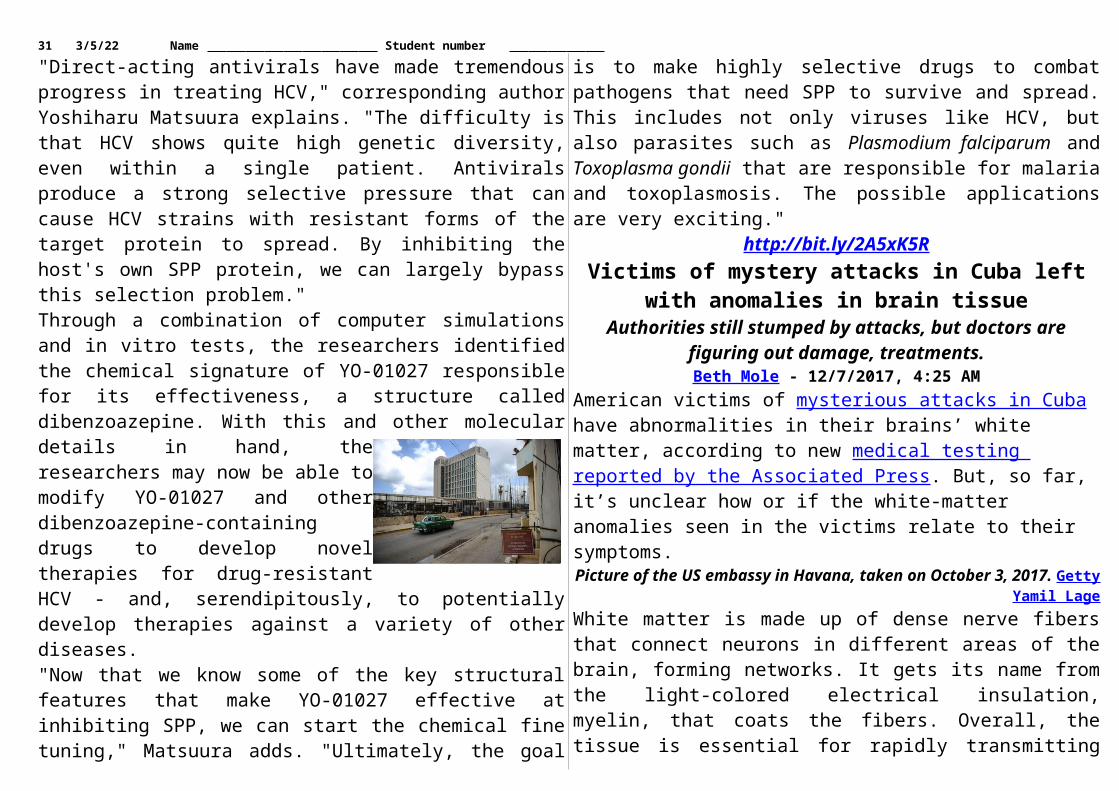
31 5/6/23 Name Student number “When the computer says, ‘You’re seeing the red light,’ you could say, ‘Oh, I know what that means — I’m supposed to put my foot on the brake,’” said Dr. Schieber. “You take information from one good part of the brain and inject it into a downstream area that tells you what to do.”
http://bit.ly/2Brh3pJA glass of whisky could help
you get your head around deep time
If we look closely enough we can understand time through the material presence of objects
December 8, 2017 by Carina J. Fearnley, Lourdes López-Merino, Niamh Downing And Richard Irvine, The Conversation
The Scottish geologist James Hutton made a proposal in 1788 that, at the time, was extraordinarily controversial. He described Earth as a "beautiful machine", constantly subjected to long-term decay and regeneration, that could only be understood over many millions of years.
Credit: Chatchy4406/Shutterstock.com This may not sound that contentious, but the challenge this posed to humanity's sense of time was substantial. Popular contemporary estimates of Earth's age, such as Bishop Ussher's calculation that it was created in 4,004 BC, were dwarfed by the magnitude of what Hutton described. Today, we are more familiar with the enormity of the Earth's age in contrast with our diminutive timespans. In 1981, John McPhee coined the term "deep time", highlighting the apparent insignificance of the span of human existence in the face of geological processes. Yet such scale is inherently difficult to conceive of. And so as societies face
changing environments, with challenges of energy and food security, the short-term perspective is often politically and economically dominant.But this way of thinking is high risk. If we are to adequately respond and adapt to landscape change, we need think about time differently, take a more holistic view. As such, over the last year we have been exploring different ways in which we might understand how humans think about deep time, and how it shapes our behaviours.We started by zoning in on the everyday. Deep time, for all its vastness, becomes intimate when we trace it in things that are familiar to us. In a recent article in The Atlantic, David Farrier explains that deep time is "not an abstract, distant prospect, but a spectral presence in the everyday". It is also in the everyday that we increasingly see our human role in shaping deep time. The Anthropocene era is characterised by the marks that we leave in the geological record. Deep time is therefore visible in our daily lives, and if we look closely enough we can understand time through the material presence of objects.Deep whiskyTake a glass of whisky. The processes that have led to the enjoyment of a dram by no means begin with the opening of a distillery. Geological processes occurring over millions of years all contribute towards its particular taste. Our project, conducted by a team consisting of an anthropologist, a geologist, a literary scholar, a palaeoecologist, and a radiocarbon dating expert, was carried out in Orkney, and so for the whisky element of our work we focused on the well-respected Highland Park distillery. Beyond colour, viscosity, aroma, and taste, we wanted to experience and apprehend how long processes of time, far beyond our lifespans, contributed to the amber liquid. Whisky requires key ingredients: barley, yeast, water, a fuel source (in this case peat), oak wood barrels (often seasoned through years holding Oloroso sherry), and copper crafted pot stills. Water is added
32 5/6/23 Name Student number to the mix after the barley has been malted and ground down into "grist" to prepare the spirit. Because so much water is needed, streams percolating through millions of years of rock supply most distilleries. And the chemistry of this water impacts the final taste. Orkney has unusually hard water,
drawn through calcareous Old Red Sandstone deposited 359–416m years ago. The malting process also affects the taste. Highland Park, for example, is famous for its floral peaty flavour that derives largely from the peat smoke used in this process. Peat is the slow accumulation of partially decomposed organic matter building up under wet conditions over thousands of years.
Geological time spiral. Credit: United States Geological SurveyPeat through timeThe fuel used will connect us with cultural as well as geological history, if looked for. Orkney, for example, has a rich history of cutting peat, a process referred to in the 12th century Norse Orkneyinga Saga but believed to have been started long before then.
While few in Orkney cut their own peat today, some still employ their tools and inherited knowledge to get fuel for the cold times. Local poets such as George Mackay Brown and Margaret Tait captured the communal practice of peat cutting. Mackay Brown depicts men, women and children out on the hill, "small blades glitter[ing]" at dawn as they release the wealth of the peat through a ritualistic exchange of labour. For both poets, peat is also a point of origin, its wet blackness recalling its own beginnings in the fiery "chaos" of the Earth's centre; the vast temporal and spatial depth of our planet's core opened up to the human imagination through the everyday presence of the peat.The characteristics of peatlands also make them excellent archives of environmental change, like a book that can be read through time. Peat exhibits a coherent stratigraphy that captures past landscapes and how humans interacted with and transformed them. Michelle Farrell, for example, has analysed pollen trapped in different peat layers from a 7,000-year-old core sample from Hobbister Moor, the peat used by Highland Park. These microfossils enabled the reconstruction of the environments people inhabited since the Mesolithic, explaining how woodlands retracted owing to climate changes and human impacts, as well as the spread of the characteristic heathland that dominates Orkney today. The reconstruction of trends that work at time scales beyond our human experience as well as unravelling human-environment interactions are the treasures that a deep time approach gives us.Past, present, futureDigging into the peat, we also look to the future: is it really sustainable for humans to consume resources thousands of years in the making? The depletion of peat impacts upon unique habitats, highlighting the mismatch between human consumption in the short term and the long-term processes that generate the resources we depend on. Peat can be used in a relatively sustainable manner through careful management and by experimenting to establish the best
33 5/6/23 Name Student number conditions for peat regrowth. Yet the sustainability of peat extraction remains an open question: as the ecologist Kimmo Tolonen has argued: "Peat belongs to the renewable resources only in the geological timescale."Thinking about an everyday object in such an interdisciplinary way allowed us to engage with deep time through a seemingly everyday glass of whisky. Appreciating the deep geological timescales that shape rock, water, and the taste of the whisky gave rise to stories about the generations who have worked the peat; the creative poetics and histories of peat; and the environmental and geographical histories revealed by looking at peat under the microscope.Deep time's vastness can seem out of scale with human life. Yet thinking through materials and different kinds of knowledge and local expertise, we see how it protrudes into the everyday. Thought of like this, any object might serve as a lens through which to see deep time's vivid presence today.
http://bit.ly/2AasqOUBad news: Warmest climate models might also be most
accurateUncertainty cuts both ways, y’all.
Scott K. Johnson - 12/9/2017, 6:10 AMSome people who reject the conclusions of climate science claim that the existence of any remaining uncertainty means few or no actions need be taken to reduce our greenhouse gas emissions. In reality, though, uncertainty is ever-present in science, and it's not necessarily our friend.
Enlarge / Be cautious out there. Bill GreenA new study from Patrick Brown and Ken Caldeira of the Carnegie Institution for Science highlights the fact that uncertainty means
climate change could just as easily be worse than our best current estimates predict.The study sought to narrow the range of projected global warming presented in places like the Intergovernmental Panel on Climate Change (IPCC) reports. For each of several scenarios of future greenhouse gas emissions, these reports have simply taken simulations from every climate model available and combined the results—showing the average temperature trajectory and the range they span. For the highest-emissions scenario, for example, the last IPCC report projected about 4.3 degrees Celsius (7.7 degrees Fahrenheit) warming by the late 21st century. But the range of model results stretched from about 3.2 degrees Celsius to 5.4 degrees Celsius.One strategy for dealing with this variance has been to weight the results of the best-performing models more highly. The difficulty is in confidently assessing which models are the best-performing ones. A handful of studies have used some aspect of cloud behavior as the measuring stick. That work has found that the models best simulating current cloud behavior also happen to simulate more future warming.In this case, the researchers took a slightly different approach to weighting models. Since the difference between models ultimately boils down to the balance of energy reaching and leaving the Earth’s surface, the new research focused on this directly.We have satellite observations of this energy covering the planet, including the sunlight reflected away from the Earth and the outgoing infrared heat energy that makes it through the atmosphere. So the researchers compared each model to these observations in a number of ways—longer-term average values, the magnitude of seasonal swings, and variability month to month.They related the success score for each model to the amount of warming it projected over the coming century. Using that relationship, they then calculated a new best estimate for future warming. That estimate was a little higher than the model average shown in the IPCC report. In that highest emissions scenario, the best estimate for
34 5/6/23 Name Student number projected warming increases from 4.3 to 4.8 degrees Celsius. Once again, the models that performed best were the ones that also happened to project the most future warming.Through some additional statistical heavy lifting, the researchers also narrowed the range of uncertainty around that best estimate significantly based on the model success scores.Lower-emissions scenarios are affected pretty similarly—the best estimate of projected warming increases a bit. This is because the models that did best in the test are more sensitive to increased carbon dioxide. Estimates of the oft-cited “equilibrium climate sensitivity” have typically centered on 3 degrees Celsius warming for a doubling of the CO2 concentration. But whereas the average sensitivity of all the models was 3.1 degrees Celsius, the new average based on the model success scores rises to 3.7 degrees Celsius.What is actually going on in the models to make this happen?
Enlarge Brown and Caldeira/NatureWell, like past studies in this genre, it largely comes down to clouds. Changes in cloud cover can act to amplify warming, and this behavior was the biggest factor determining how well the models matched satellite energy measurements.
These conclusions aren’t bulletproof, as the researchers acknowledge. There could be other reasons that partially explain why the models that scored the best were more sensitive to CO2—some models share components or techniques, for example, so it’s possible there is some coincidence at play. It’s also not guaranteed that the sensitivity relationship will hold when the next batch of model simulations is compiled, given that research and development has been ongoing since the last IPCC report.But this is also not the only study to find that the models projecting higher warming might be the most reliable. If this is true, the authors write, it would mean that “achieving any given global temperature stabilization target will require steeper greenhouse gas emissions reductions than previously calculated.”Like a student calculating the least possible effort necessary to pass a class, cutting it too close runs the risk of missing your target completely.Nature, 2017. DOI: 10.1038/nature24672 (About DOIs).
http://bit.ly/2AMIOGtDiscovery of Two Tombs Dating Back 3,500 Years
Announced in EgyptEgypt authorities announced
Saturday that archaeologists had discovered two small ancient tombs
in Luxor, a southern city.The tombs date back about 3,500 years and are located on the west bank of the river Nile."It's truly an exceptional day. … The 18th dynasty private tombs were already known. But it's the first time to enter inside the two tombs," Antiquities Minister Khaled al-Anani said. Egyptian officials hope the discovery will help the country's efforts to revive tourism.
Egyptian excavation workers restore a mummy in a newly discovered tomb in the Draa Abul Naga necropolis on Luxor’s West Bank known as KAMPP 150,
35 5/6/23 Name Student number in Luxor, Egypt, Dec. 9, 2017.(H.
Elrasam/VOA) The minister said one tomb has five entrances that lead to a rectangular hall where two burial shafts are located on the northern and southern sides of the tomb.The tomb contains a mummy wrapped in linen, clay vessels, a collection of about 450 statues, and painted wooden funerary masks. Al-Anani said a cartouche carved on the ceiling bears the name of King Thutmose I of the early 18th dynasty.
Egyptian excavation workers restore funeral furniture found in a newly discovered tomb, Kampp 161, in the Draa Abul Naga necropolis on Luxor’s
West Bank, Egypt, Dec. 9, 2017. (H. Elrasam/VOA)The other tomb has a 6-meter burial shaft "leading to four side chambers," which contained artifacts including fragments of wooden coffins. The wall inscriptions and paintings are believed to belong to an era between the reigns of King Amenhotep II and King Thutmose IV, both pharaohs of the 18th dynasty.The tombs are the latest discovery in Luxor, which is known for its temples and tombs from different dynasties of ancient Egyptian history.
Discovery of Two Ancient Tombs Announced in EgyptAl-Anani also visited a nearby site where the famous Mortuary Temple of Hatshepsut is located. For the first time, the temple's main sanctuary, known as the Holy of Holies, was opened.The Antiquities Ministry has made a series of discoveries since the beginning of 2017. In September, Egyptian archaeologists announced
they had found the tomb of a royal goldsmith whose work was dedicated to the ancient Egyptian god Amun. The goldsmith lived more than 3,000 years ago. In the tomb, there were mummies, jewelry and statues.