volume 48 number 32 28 august 2019 pages 11987–12350
TRANSCRIPT

Dalton Transactions
An international journal of inorganic chemistry
Dalton Transactions
An international journal of inorganic chemistryrsc.li/dalton
ISSN 1477-9226
PAPER Alessio Terenzi et al. Ruthenium–arene complexes bearing naphthyl-substituted 1,3-dioxoindan-2-carboxamides ligands for G-quadruplex DNA recognition
Volume 48 Number 32 28 August 2019 Pages 11987–12350

DaltonTransactions
PAPER
Cite this: Dalton Trans., 2019, 48,12040
Received 17th May 2019,Accepted 4th July 2019
DOI: 10.1039/c9dt02078k
rsc.li/dalton
Ruthenium–arene complexes bearing naphthyl-substituted 1,3-dioxoindan-2-carboxamidesligands for G-quadruplex DNA recognition†
Laura A. Hager,a Stephan Mokesch,a Claudia Kieler,b Silvia Alonso-de Castro,c
Dina Baier,b Alexander Roller,a Wolfgang Kandioller, a Bernhard K. Keppler,a
Walter Berger, b Luca Salassa c,d and Alessio Terenzi *a,c
Quadruplex nucleic acids – DNA/RNA secondary structures formed in guanine rich sequences – proved
to have key roles in the biology of cancers and, as such, in recent years they emerged as promising
targets for small molecules. Many reports demonstrated that metal complexes can effectively
stabilize quadruplex structures, promoting telomerase inhibition, downregulation of the expression of
cancer-related genes and ultimately cancer cell death. Although extensively explored as anticancer
agents, studies on the ability of ruthenium arene complexes to interact with quadruplex nucleic acids are
surprisingly almost unknown. Herein, we report on the synthesis and characterization of four novel Ru(II)
arene complexes with 1,3-dioxoindan-2-carboxamides ligands bearing pendant naphthyl-
groups designed to bind quadruplexes by both stacking and coordinating interactions. We show how
improvements on the hydrolytic stability of such complexes, by substituting the chlorido leaving ligand
with pyridine, have a dramatic impact on their interaction with quadruplexes and on their cytotoxicity
against ovarian cancer cells.
Introduction
In the last 15 years, there has been considerable interest innew DNA-targeting drugs able to recognize non-canonicalmotifs called G-quadruplexes (G4s).1 Such DNA structures areformed in G-rich sequences as a stacked arrangement ofguanine tetrads which are connected by loops of varyinglength. Even though they share the same core structure, G4sexhibit a polymorphic nature. This in turn leads to motifscharacterized by unique binding sites for the potentially selec-tive binding of small molecules. Furthermore, G4s are not ran-domly distributed within the human genome, but are over-represented in telomeres and in the promoter regions ofcancer-related genes, with key roles in their regulation.2 Forthese reasons they have become popular biological targets in
cancer research, and there might be the chance that G4s willbe the protagonists of a new DNA-based targeted therapy era.1
Currently, there are two fluoroquinolone-based G4-binderswhich entered clinical trials as G4-targeting anticancer drugcandidates.1
Complexes of different metals (e.g. Pt, Ni, Au) can effectivelyinteract with G4 structures.3–5 Many of these compounds havealso been tested against cancer cell lines both as potentialdrugs and/or probes. In contrast, reports on ruthenium-basedcompounds capable of binding (selectively) to G4s are onlystarting to emerge. This is somehow surprising consideringthat Ru complexes such as NAMI-A and IT-139 (formerlyNKP-1339) have been considered as promising metal-basedalternatives to clinically used Pt drugs.6–9 The ruthenium-based G4 binders reported so far are mostly substitutionallyinert polypyridyl complexes where one of the N,N-ligands hasan extended aromatic moiety which allows for partial stackingwith the G4 tetrads.10–19 Although extensively explored as anti-cancer agents,20–22 only one family of Ru–arene complexes hasbeen designed and investigated as G4-binding drugs to thebest of our knowledge. Liu and Mei developed Ru(II)–arenecomplexes bearing phenanthroimidazole ligands which provedto stabilize the quadruplex formed in c-Myc oncogene via agroove binding mechanism and to inhibit the proliferation,migration, and invasion of breast cancer cells.23–25 Overall, the
†Electronic supplementary information (ESI) available. CCDC 1870492–1870494.For ESI and crystallographic data in CIF or other electronic format see DOI:10.1039/c9dt02078k
aInstitute of Inorganic Chemistry, Faculty of Chemistry, University of Vienna,
Waehringer Str. 42, A-1090 Vienna, Austria. E-mail: [email protected] of Medicine I, Institute of Cancer Research and Comprehensive Cancer
Center, Medical University Vienna, Borschkegasse 8a, A-1090 Vienna, AustriacDonostia International Physics Center, Paseo Manuel de Lardizabal 4, Donostia,
20018, SpaindIkerbasque, Basque Foundation for Science, Bilbao, 48013, Spain
12040 | Dalton Trans., 2019, 48, 12040–12049 This journal is © The Royal Society of Chemistry 2019
Ope
n A
cces
s A
rtic
le. P
ublis
hed
on 0
4 Ju
ly 2
019.
Dow
nloa
ded
on 2
/24/
2022
9:5
0:58
AM
. T
his
artic
le is
lice
nsed
und
er a
Cre
ativ
e C
omm
ons
Attr
ibut
ion-
Non
Com
mer
cial
3.0
Unp
orte
d L
icen
ce.
View Article OnlineView Journal | View Issue

scarce use of Ru-based scaffolds for G4-binding can principallybe ascribed to the preferred octahedral or piano-stool geome-tries, which are not ideal for stacking on top of the guaninetetrads of the G4 structure.
In recent years, we developed different classes of metal-based compounds to selectively target G-quadruplexes, includ-ing Ni(II) salen-type derivatives,26 Pt(II) supramolecular coordi-nation complexes,27,28 and Ru(II) N-heterocyclic carbenes.29,30
The aim of this article is to explore a new strategy for thedesign of Ru–arene complexes possessing distinct G4 bindingabilities that can positively affect their anticancer activity. Thisapproach could pave the way for utilizing this promising classof compounds and their structural versatility for G-quadruplextargeting purposes.
In this work, we present the synthesis of four novel half-sandwich Ru(II) complexes attached to the 1,3-dioxoindan-2-carboxamide ligand scaffold bearing a pendant naphthyl-group. O,O-Dioxoindane-based ligands were selected amongour toolkit of ligands since they show important biologicalactivity via topoisomerase inhibition when coordinated to theorganometallic Ru(II) cym (cym = p-cymene) moiety.31 Herein,we modified the dioxoindane scaffold with alkyl linkers (ofdifferent length) attached to a pendant naphthyl group able toπ-stack on top of the G-quadruplex tetrads, thereby conferringG4 binding activity to the final compounds. Furthermore, weexplored the effect of substituting the chlorido leaving ligandwith pyridine, which has been shown to significantly increasehydrolytic stability.32–36 This approach was adopted consider-ing that the interplay between stability and dynamic behavior(ligand release via hydrolysis) is assumed to be a key step inthe activation of most organometallic Ru(II) complexes.6,9,20
We consequently investigated the ability of the resulting com-pounds to bind G4 motifs in vitro through spectroscopyand high resolution mass spectrometry and evaluated theiranti-proliferative activity against ovarian cancer cells.Overall, we have found that small improvements in the hydro-lytic stability of the Ru scaffold by pyridine coordination led todramatic enhancements in both G4 binding properties andcytotoxicity.
Results and discussionSynthesis and X-ray crystal structures
Ru complexes 1a–b and 2a–b were prepared via a 5-step syn-thetic route summarized in Fig. 1. Commercially available2-hydroxy-1,4-naphtoquinone (Lawsone) was converted to2-oxido-3-phenyl-iodonio-1,4-naphthoquinone which served asa precursor for both ligands.
The precursor, which undergoes a Wolff-type rearrange-ment to yield a (ring-contracted) ketocarbene intermediate,was then treated with the respective primary amine yieldingthe N-substituted 1,3-dioxoindan-2-carboxamides L1 and L2.Complexes were obtained by deprotonation of the respectiveligand and subsequent reaction with the dimeric rutheniumspecies bis[dichlorido(cymene)ruthenium(II)]. In order toreplace the leaving group by pyridine, the chlorido ligand wasabstracted by AgNO3, followed by addition of pyridine andNaClO4. As the formed Ru–N bond could be cleaved under pro-longed light irradiation, the last step of the reaction was per-formed under exclusion of light.
Complexes 1a and 2a were designed as multivalentG-quadruplex binders since they bear both a naphthyl moietyable to stack on top of the guanine tetrads as well as anexchangeable ligand directly coordinated to the rutheniumcenter, thereby providing a binding site for a guanine nitrogen(typically N7) after aquation of the metal center. Pyridinederivatives 1b and 2b were specifically designed to increasewater solubility and to evaluate the role of the N-donor aro-matic ligand on the G4 binding ability and the impact on thecytotoxicity of the complexes.
All the synthesized compounds were characterized by stan-dard spectroscopic and analytical methods confirming the for-mation and purity of the desired complexes (see for instanceFig. S1† for 1H NMR of L1, 1a and 1b). UV-Vis spectra inbuffered aqueous solutions of the precursors L1 and L2 and ofthe metal compounds 1a–b and 2a–b at different concen-trations are reported in Fig. S2.† Ligands L1 and L2 displayvery similar spectral features with a broad band centered at396 nm, a multi-peaked band in the range 290–320 nm and
Fig. 1 Synthetic route for the preparation of compounds 1a–b and 2a–b.
Dalton Transactions Paper
This journal is © The Royal Society of Chemistry 2019 Dalton Trans., 2019, 48, 12040–12049 | 12041
Ope
n A
cces
s A
rtic
le. P
ublis
hed
on 0
4 Ju
ly 2
019.
Dow
nloa
ded
on 2
/24/
2022
9:5
0:58
AM
. T
his
artic
le is
lice
nsed
und
er a
Cre
ativ
e C
omm
ons
Attr
ibut
ion-
Non
Com
mer
cial
3.0
Unp
orte
d L
icen
ce.
View Article Online
two intense peaks centered at 273 and 264 nm, respectively.Chlorido complexes 1a and 2a retain the broad band at about400 nm but lack the narrow multi-peaked bands at lower wave-lengths, exhibiting a single broad band at 277 nm. The sametrend is followed by the pyridine complexes 1b and 2b, which,besides the band at 400 nm, show an additional band with twomaxima at about 275 and 250 nm, respectively.
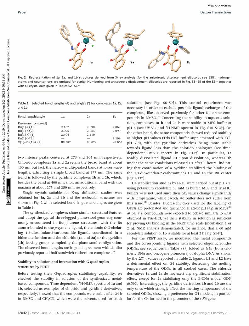
Single crystals suitable for X-ray diffraction studies wereobtained for 1a, 2a and 1b and the molecular structures areshown in Fig. 2 while selected bond lengths and angles are givenin Table 1.
The synthesized complexes share similar structural featuresand adopt the typical three-legged piano-stool geometry com-monly encountered in Ru(II) arene structures, with the Ruatom π-bonded to the p-cymene ligand, the anionic O,O-chelat-ing 1,3-dioxoindan-2-carboxamide ligands coordinated in abidentate fashion and the chlorido (1a and 2a) or the pyridine(1b) leaving groups completing the piano-stool configuration.The observed bond lengths are in good agreement with similarpreviously reported half-sandwich ruthenium complexes.31
Stability in solution and interaction with G-quadruplexstructures by FRET
Before testing their G-quadruplex stabilizing capability, wechecked the stability in solution of the synthesized metal-based compounds. Time dependent 1H-NMR spectra of 1a and1b, selected as examples of chlorido and pyridine derivatives,respectively, showed that the compounds were stable after 24 hin DMSO and CH3CN, which were the solvents used for stock
solutions (see Fig. S6–S9†). This control experiment wasnecessary in order to exclude possible ligand exchange of thecomplexes, like observed previously for other Ru–arene com-pounds in DMSO.37 Concerning the stability in aqueous solu-tion, complexes 1a–b and 2a–b were stable in MES buffer atpH 6 (see UV-Vis and 1H-NMR spectra in Fig. S10–S12†). Onthe other hand, the same compounds showed reduced stabilityat higher pH values (Tris-HCl buffer supplemented with KCl,pH 7.8), with the pyridine derivatives being more stabletowards ligand loss than the chlorido analogues (see time-dependent UV-Vis spectra in Fig. S13†). In particular, 1areadily dissociated ligand L1 upon dissolution, whereas 1bunder the same conditions released L1 after 3 hours, indicat-ing that coordination of a pyridine stabilized the binding ofthe 1,3-dioxoindan-2-carboxamides L1 and to the Ru center(Fig. S13†).
G4 stabilization studies by FRET were carried out at pH 7.2,using potassium cacodylate 60 mM as buffer. MES and Tris-HClbuffers were not used since their pKa values change significantlywith temperature, while cacodylate buffer does not suffer fromthis issue.38 Besides, fluorescent dyes used for the labeling ofODNs are protonated and quenched at acidic pH (e.g. in MES).39
At pH 7.2, compounds were expected to behave similarly to whatobserved in Tris-HCl, yet their stability in solution is sufficientfor proving G4 binding in the FRET time scale (incubation 1 or2 h). NMR analysis demonstrated, for instance, that a 60 mMcacodylate solution of 1b is stable for at least 2 h (Fig. S14†).
For the FRET assay, we incubated the metal compoundsand the corresponding ligands with selected oligonucleotides(ODNs, see sequences in Table S8†) folded as G4s (from telo-meric DNA and oncogene promoters) or duplex DNA. As shownby the ΔT1/2 values reported in Table 2, ligands L1 and L2 havea detrimental effect on G4 stability, decreasing the meltingtemperature of the ODNs in all studied cases. The chloridederivatives 1a and 2a do not exert any significant stabilizationeffect, except for 2a stabilizing only the B-DNA model ODNdsDNA. Interestingly, the pyridine derivatives 1b and 2b are theonly ones which strongly affect the melting temperature of theselected ODNs, showing a preference for G4 models, in particu-lar for the G4 formed in the promoter of the c-Kit gene.
Table 1 Selected bond lengths (Å) and angles (°) for complexes 1a, 2a,and 1b
Bond length/angle 1a 2a 1b
Ru–arene (centroid)Ru(1)–O(1) 2.107 2.098 2.069Ru(1)–O(2) 2.095 2.085 2.099Ru(1)–Cl(1) 2.404 2.410 —Ru(1)–N(2) — — 2.109O(1)–Ru(1)–O(2) 88.587 90.072 90.065
Fig. 2 Representation of 1a, 2a, and 1b structures derived from X-ray analysis (for the anisotropic displacement ellipsoids see ESI†); hydrogenatoms and counter ions are omitted for clarity. Numbering and anisotropic displacement ellipsoids are reported in Fig. S3–S5 of the ESI† togetherwith all crystal data given in Tables S2–S7.†
Paper Dalton Transactions
12042 | Dalton Trans., 2019, 48, 12040–12049 This journal is © The Royal Society of Chemistry 2019
Ope
n A
cces
s A
rtic
le. P
ublis
hed
on 0
4 Ju
ly 2
019.
Dow
nloa
ded
on 2
/24/
2022
9:5
0:58
AM
. T
his
artic
le is
lice
nsed
und
er a
Cre
ativ
e C
omm
ons
Attr
ibut
ion-
Non
Com
mer
cial
3.0
Unp
orte
d L
icen
ce.
View Article Online

Next, we performed a series of control experiments to betterrationalize this behavior and understand the effect of ligandhydrolysis on the G4-stablilization of 1b and 2b. We treatedthe ODNs with complex 3 used as control (Fig. 3A) and in com-bination with ligands L1 and L2 as they are the constituents ofcompounds 1b and 2b, respectively. Remarkably, incubation ofthe ODNs with these combinations did not result in such astrong effect as observed for 1b and 2b, demonstrating that allthe structural components of the complexes were needed toexert a distinct G4 stabilization (Table 2 and Fig. 3B).
Additionally, when we used a shorter incubation time (1 h),we observed an overall increase of the ODNs melting tempera-ture (Fig. 3C and Table S9†). In this case, besides 1b and 2b,also the chlorido complexes 1a and 2a exhibited G4 stabiliz-
ation, indicating that within this time window significantamounts of these compounds were still in their original form(i.e. not hydrolyzed). Furthermore, with the shorter incubationtime of 1 h, the selectivity toward G4 over duplex DNA of com-pounds 1b and 2b increased significantly. Even though theexact fraction of stable compounds interacting with theselected G4s is not known, the ΔT1/2 values obtained in thedescribed different conditions are strongly indicative of theimportance of the complexes stability for the G4 stabilization.
Interaction with 9-ethylguanine and G4s by mass spectrometry
Incubation experiments (2 h in H2O, 20% MeOH) of 1a and 1bwith 9-ethylguanine (9-EtG) followed by mass analyses demon-strated that both complexes are able to coordinate guanines atthe N7 position (Fig. S15 and S16†). Further time-dependentexperiments indicated a relatively fast exchange (within30 min) between the pyridine of complex 1b and the nucleo-base (Fig. S17†).
We then used ESI-TOF mass spectrometry to evaluate thebinding of 1a and 1b to two selected G-rich sequences, hTeloand c-Kit1 respectively. The two G4s contain stacks of threeG-quartets and retain monovalent potassium ions in theirfolded structure (see 3D models of hTelo and c-Kit1 in Fig. 4Aand B).40,41 Since non-volatile potassium salts are not compati-ble with their use in buffers for ESI mass spectrometry, weused ammonium acetate instead to ensure G4 formation, likepreviously reported.42
Mass spectra of the free G4s exhibited ions mainly in thecharge states of −5 and −4. In each charge state, there werethree peaks corresponding to the free oligodeoxynucleotide, oneNH4
+ ion adduct, and two NH4+ ions adduct (Fig. 4C and D).
Table 2 ΔT1/2 values of 0.2 µM ds-DNA and G4s upon interaction with4 µM binders. Uncertainty is ≤0.5 for the ΔT1/2 reported. Incubationtime: 2 h
ΔT (°C)
dsDNA hTelo c-Kit1 Bcl2 hTERT
L1 −3.5 −2.4 −7.6 −2.4 −4.0L2 −3.4 −2.1 −7.1 −2.3 −2.9
1a 4.0 0.9 −0.1 1.2 2.71b 10.2 11.0 20.9 5.1 9.53 + L1 6.3 −0.4 10.1 5.7 2.4
2a 10.4 3.0 1.8 3.7 2.62b 9.7 6.5 15.7 3.8 9.43 + L2 8.6 1.8 10.5 5.4 0.7
3 4.4 0.4 −0.3 −0.6 1.1
Fig. 3 (A) Structures of 1b and of its constituents L1 and 3. (B) FRET melting profiles of the c-Kit1 G4 (0.2 µM) upon interaction with the indicatedcompounds (4 µM). (C) FRET melting profiles at different incubation times.
Dalton Transactions Paper
This journal is © The Royal Society of Chemistry 2019 Dalton Trans., 2019, 48, 12040–12049 | 12043
Ope
n A
cces
s A
rtic
le. P
ublis
hed
on 0
4 Ju
ly 2
019.
Dow
nloa
ded
on 2
/24/
2022
9:5
0:58
AM
. T
his
artic
le is
lice
nsed
und
er a
Cre
ativ
e C
omm
ons
Attr
ibut
ion-
Non
Com
mer
cial
3.0
Unp
orte
d L
icen
ce.
View Article Online

After 1 h incubation of our compounds with the two G4s,we observed low-intensity peaks corresponding to 1 : 1 adducts(and their fragments) with 1a and 1b upon loss of chloride orpyridine, respectively (Fig. S18–S22†).
Furthermore, we focused our attention to the G4ammonium adduct distribution after incubation with 1a and1b. At first, we compared the relative intensity of the ESI-MSpeaks distribution of NH4
+ adducts for the 5-charge of hTelo−5
and c-Kit1−5 with the one of their complexes (or their frag-ments) with 1a and 1b (Fig. S23†). For both G-quadruplexes,an intensity decrease of the peak corresponding to theammonium free ODN occurred in the spectra of the adductswith 1a and 1b, indicating a lower tendency by the G4 torelease NH4
+ ions, hence a general stabilizing effect.43
Nevertheless, considering that the peaks corresponding to1a/1b-G4 adducts have low intensity and, especially in the caseof c-Kit1, different adduct fragments are present (seeFig. S20†), we also compared the ammonium distribution offree hTelo5− and c-Kit15−, before and after the interaction withour complexes. In this case, addition of 1a/1b to hTelo G4(Fig. 4C) led only to a minor shift in ammonium adduct distri-bution with the peak corresponding to hTelo5− with one andtwo associated ammonium becoming slightly less abundantcompared to the free G4. This could indicate a minor destabili-zation of the G4 structure with a small release of monovalent
ions after the interaction with 1a and 1b.43 On the other hand,the interaction with c-Kit1−5 confirmed the stabilization effect(Fig. 4D): upon addition of 1a/b, the relative abundance ofc-Kit15− with one or two coordinated ammonium ions clearlyincreased, indicating improved stability of the G4 structure. Inother words, c-Kit15−, once bound to the metal compounds,held NH4
+ ions between the G-tetrads more tightly than thefree G4 when introduced into the gas phase.43 The observedstabilizing effect was stronger for compound 1b than for 1a.
The hTelo sequence contains no free guanines besidesthose participating in the G4 fold (Fig. 4A),44 whereas c-Kit1features three non-stacked guanines (Fig. 4B), making thempotentially accessible for easier ruthenation. In both MSexperiments and FRET studies, c-Kit1 displays enhanced stabi-lization of the G4 fold by the pyridine derivative 1b as com-pared to hTelo, thus suggesting that the presence of unpairedguanines in the c-Kit1 DNA motif enables a dual bindingmode, allowing for N7 ruthenation besides the stacking actionof the naphthyl moiety.
Incubation experiments with 9-EtG and with the selectedG4 models clearly indicated that both compounds 1a and 1bcoordinate the N7 of guanines and simultaneously bind theG4s tetrad via stacking of the naphthyl group. The increasedstability of 1b toward ligand exchange is key for the success ofthis dual mode of binding in our experimental conditions.
Fig. 4 (A, B) 3D structures of hTelo (A) and (B) c-Kit1 G4s, generated with Chimera using PDB 2HY9 and 2O3M, respectively. Guanines belonging toG-tetrads are highlighted in orange and unpaired guanines are shown in green. (C, D) Negative ESI-TOF-MS enlargements of the distribution ofammonium adducts for the 5-charge state of free 10 μM hTelo (C) and (D) c-Kit1 sequences in the absence and presence of equimolar amount ofthe compounds 1a and 1b. The three peaks in each spectrum correspond to the free oligodeoxynucleotide (orange circles), one NH4
+ ion adduct,and two NH4
+ ions adduct (one and two green diamonds, respectively).
Paper Dalton Transactions
12044 | Dalton Trans., 2019, 48, 12040–12049 This journal is © The Royal Society of Chemistry 2019
Ope
n A
cces
s A
rtic
le. P
ublis
hed
on 0
4 Ju
ly 2
019.
Dow
nloa
ded
on 2
/24/
2022
9:5
0:58
AM
. T
his
artic
le is
lice
nsed
und
er a
Cre
ativ
e C
omm
ons
Attr
ibut
ion-
Non
Com
mer
cial
3.0
Unp
orte
d L
icen
ce.
View Article Online
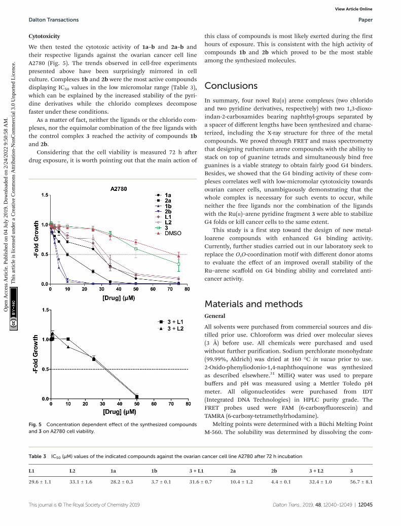
Cytotoxicity
We then tested the cytotoxic activity of 1a–b and 2a–b andtheir respective ligands against the ovarian cancer cell lineA2780 (Fig. 5). The trends observed in cell-free experimentspresented above have been surprisingly mirrored in cellculture. Complexes 1b and 2b were the most active compoundsdisplaying IC50 values in the low micromolar range (Table 3),which can be explained by the increased stability of the pyri-dine derivatives while the chlorido complexes decomposefaster under these conditions.
As a matter of fact, neither the ligands or the chlorido com-plexes, nor the equimolar combination of the free ligands withthe control complex 3 reached the activity of compounds 1band 2b.
Considering that the cell viability is measured 72 h afterdrug exposure, it is worth pointing out that the main action of
this class of compounds is most likely exerted during the firsthours of exposure. This is consistent with the high activity ofcompounds 1b and 2b which proved to be the most stableamong the synthesized molecules.
Conclusions
In summary, four novel Ru(II) arene complexes (two chloridoand two pyridine derivatives, respectively) with two 1,3-dioxo-indan-2-carboxamides bearing naphthyl-groups separated bya spacer of different lengths have been synthesized and charac-terized, including the X-ray structure for three of the metalcompounds. We proved through FRET and mass spectrometrythat designing ruthenium arene compounds with the ability tostack on top of guanine tetrads and simultaneously bind freeguanines is a viable strategy to obtain fairly good G4 binders.Besides, we showed that the G4 binding activity of these com-plexes correlates well with low-micromolar cytotoxicity towardsovarian cancer cells, unambiguously demonstrating that thewhole complex is necessary for such events to occur, whileneither the free ligands nor the combination of the ligandswith the Ru(II)–arene pyridine fragment 3 were able to stabilizeG4 folds or kill cancer cells to the same extent.
This study is a first step toward the design of new metal-loarene compounds with enhanced G4 binding activity.Currently, further studies carried out in our laboratory seek toreplace the O,O-coordination motif with different donor atomsto evaluate the effect of an improved overall stability of theRu–arene scaffold on G4 binding ability and correlated anti-cancer activity.
Materials and methodsGeneral
All solvents were purchased from commercial sources and dis-tilled prior use. Chloroform was dried over molecular sieves(3 Å) before use. All chemicals were purchased and usedwithout further purification. Sodium perchlorate monohydrate(99.99%, Aldrich) was dried at 160 °C in vacuo prior to use.2-Oxido-phenyliodonio-1,4-naphthoquinone was synthesizedas described elsewhere.31 MilliQ water was used to preparebuffers and pH was measured using a Mettler Toledo pHmeter. All oligonucleotides were purchased from IDT(Integrated DNA Technologies) in HPLC purity grade. TheFRET probes used were FAM (6-carboxyfluorescein) andTAMRA (6-carboxy-tetramethylrhodamine).
Melting points were determined with a Büchi Melting PointM-560. The solubility was determined by dissolving the com-
Fig. 5 Concentration dependent effect of the synthesized compoundsand 3 on A2780 cell viability.
Table 3 IC50 (µM) values of the indicated compounds against the ovarian cancer cell line A2780 after 72 h incubation
L1 L2 1a 1b 3 + L1 2a 2b 3 + L2 3
29.6 ± 1.1 33.1 ± 1.6 28.2 ± 0.3 3.7 ± 0.1 31.6 ± 0.7 10.4 ± 1.2 4.4 ± 0.1 32.4 ± 1.0 56.7 ± 8.1
Dalton Transactions Paper
This journal is © The Royal Society of Chemistry 2019 Dalton Trans., 2019, 48, 12040–12049 | 12045
Ope
n A
cces
s A
rtic
le. P
ublis
hed
on 0
4 Ju
ly 2
019.
Dow
nloa
ded
on 2
/24/
2022
9:5
0:58
AM
. T
his
artic
le is
lice
nsed
und
er a
Cre
ativ
e C
omm
ons
Attr
ibut
ion-
Non
Com
mer
cial
3.0
Unp
orte
d L
icen
ce.
View Article Online

pound in DMSO and subsequent dilution to a final concen-tration of 1% DMSO/MEM. The highest concentrated dilution,where no precipitation of the compound occurred, was thedetermined solubility. The NMR spectra were recorded at25 °C using a Bruker FT-NMR spectrometer Avance IIITM
500 MHz. NMR spectra were measured in deuterated dimethylsulfoxide (DMSO-d6) or chloroform (CDCl3) (see Fig. S24–S35†for the full spectra). CHN-elemental analyses were performedwith a Eurovector EA3000 Elemental Analyzer in the micro-analytical laboratory of the University of Vienna. Single crystalsof 1a, 2a, and 1b suitable for X-ray diffraction analysis weregrown by precipitation from DCM/Et2O at 4 °C. The X-rayintensity data were measured on Bruker D8 Venture andBruker X8 Apex2 diffractometer equipped with multilayermonochromators, Mo K/α INCOATEC micro focus sealed tubesand Kryoflex cooling systems. The structures were solved bydirect and patterson methods and refined by full-matrix least-squares techniques. Non-hydrogen atoms were refined with an-isotropic displacement parameters. Hydrogen atoms wereinserted at calculated positions and refined with riding model.The following software was used: Bruker SAINT v8.37A &V7.68A (Bruker AXS) using a narrow-frame algorithm for frameintegration, SADABS (by George M. Sheldrick) for absorptioncorrection, OLEX2 for structure solution,45 refinement, mole-cular diagrams and graphical user-interface, Shelxle for refine-ment and graphical user-interface,46 SHELXS-2015 for struc-ture solution, SHELXL-2015 for refinement,47 Platon for sym-metry check.48 Experimental data and CCDC-codes (availableonline at http://www.ccdc.cam.ac.uk/conts/retrieving.html) canbe found in Table S1.† Crystal data, data collection parameters,and structure refinement details are given in Tables S2–S7.†Crystal structures visualized in Fig. S3–S5.†
Synthesis
2-(Hydroxy(napththylmethylamino)methylene)-1H-indene-1,3(2H)-dione (L1). After 2-oxido-phenyliodonio-1,4-naphtho-quinone (350 mg, 931 µmol, 1 eq.) was dissolved in dry CHCl3(35 mL), 1-naphthylmethylamine (136 µL, 931 µmol, 1 eq.) wasadded under argon and the mixture was refluxed overnight(24 h). The solvent was removed, the residue was washed withdiethylether, and the obtained product dried in vacuo. (76%,233 mg, yellow powder): m.p. 192–193 °C; solubility 0.096 mgmL−1 ≡ 0.29 mM (MEM, 1% DMSO); 1H-NMR (500.10 MHz,DMSO-d6) δ: 5.04 (s, 2H), 7.45–7.64 (m, 9H), 7.86–7.91(m, 1H),7.96–8.00 (m, 1H), 8.17–8.22 (m, 1H), 9.18 (br s, 1H); 13C-NMR(125.75 MHz, DMSO-d6) δ 40.89 (C2′), 93.89 (C2), 120.86 (C4,C7), 123.23 (C7′), 125.22 (C4′), 125.56 (C5′), 126.03 (C9′), 126.57(C8′), 127.98 (C6′), 128.69 (C10′), 130.54 (C6′a), 133.13 (C5 andC6), 133.34 (C10′a), 137.72 (C3a, C7a), 165.77 (C1′), 190.60(Ca and C3) ppm.
m/z 328.14, mth: 328.10; elemental analysis calcd forC21H15NO3: C 76.58, H 4.59, N 4.25%; found: C 75.53, H 4.65,N 4.41%.
2-(Hydroxy(2-(1-naphthyl)ethylamino)methylene)-1H-indene-1,3(2H)-dione (L2). After 2-(1-naphthyl)ethylamine hydro-chloride (138 mg, 665 µmol, 1 eq.) and triethylamine (139 µL,
997 µmol, 1.5 eq.) were suspended in CHCl3 (25 mL) andstirred for 15 minutes under argon, 2-oxido-phenyliodonio-1,4-naphthoquinone (250 mg, 665 µmol, 1 eq.) was added andrefluxed overnight (20 h). The reaction mixture was washedwith water and dried over anhydrous sodium sulfate. The solu-tion was evaporated to dryness and the residue washed withdiethyl ether and dried in vacuo (78%, 179 mg, yellow powder):m.p. 175–176 °C; solubility (determined in aqueous cellculture medium MEM (Minimum Essential Medium Eagle,Sigma) with a final concentration of 1% DMSO 0.126 mg mL−1
≡ 0.37 mM (MEM, 1%DMSO); 1H-NMR (500.10 MHz,d6-DMSO) δ: 3.08–3.13 (m, 1H), 3.32–3.36 (m, 2H), 3.63–3.68(m, 2H), 7.39–7.46 (m, 2H), 7.51–7.60 (m, 6H), 7.80–7.82 (m,1H), 7.92–7.94 (m, 1H), 8.27–8.29 (m, 1H), 8.90 (br s, 1H);13C-NMR (125.75 MHz, d6-DMSO) δ 32.82 (C3′), 45.67 (C2′),120.37 (C4, C7), 123.91 (C8′), 125.67 (C5′), 125.74 (C6′), 126.16(C10′), 126.88 (C9′), 127.06 (C7′), 128.61 (C11′), 131.60 (C7′a),132.58 (C5, C6), 133.48 (C11′a), 134.95 (C4′), 138.12 (C3a, C7a),165.65 (C1′), 190.58 (C1, C3) ppm. m/z 342.16, mth: 342.11;elemental analysis calcd for C22H17NO3·0.15H2O: C 76.35, H5.04, N 4.05%; found: C 76.28, H 5.33, N 4.45%.
[(Chlorido)((1,3-dioxo-κO1-1H-inden-2(3H)-ylidene)(naphthyl-methylamino)methanolato-κO2)(p-cymene)ruthenium(II)] (1a).After stirring a solution of sodium methoxide (12.6 mg,234 µmol, 1.1 eq.) and L1 (70 mg, 213 µmol, 1 eq.) in MeOH/DCM (10 mL, 5 : 1) for an hour at r.t., bis[dichlorido(cym)ruthenium(II)] (58.7 mg, 96 µmol, 0.9 eq.) was added and themixture stirred for 22 h. The solvent was removed and residuedissolved in DCM, the resulting suspension was filteredand concentrated. The product was precipitated fromDCM/n-hexane and dried in vacuo (72%, 83 mg, brownpowder): m.p. >206 °C (decomp.); solubility 0.066 mg mL−1 ≡0.11 mM (MEM, 1%DMSO); 1H-NMR (500.10 MHz, CDCl3) δ:1.19–1.27 (m, 6H), 2.18 (s, 3H), 2.67–2.74 (m, 1H), 4.94–5.24(m, 2H), 5.24–5.30 (m, 2H), 5.42–5.53 (m, 2H), 7.40–7.62 (m,8H), 7.97–7.83 (m, 1H), 7.88–7.91 (m, 1H), 8.05–8.08 (m, 1H),9.00 (dd, 3J (H,H) = 6 Hz, 3J (H,H) = 6 Hz, 1H); 13C-NMR(125.75 MHz, CDCl3) δ 17.89 (cym-C10), 22.12 and 22.33 (cym-C8, -C9), 30.86 (cym-C7), 41.28 (C2′), 78.89 and 79.37 (cym-C3,-C5), 82.52 and 82.59 (cym-C2, -C6), 96.60 (cym-C4), 98.18 (C2),99.56 (cym-C1), 120.69 and 120.92 (C4, C7), 123.35 (C7′),125.07 (C4′), 125.48 (C5′), 126.02 (C9′), 126.65 (C8′), 128.19(C6′), 128.88 (C10′), 131.31 (C6′a), 132.13 and 132.32 (C5, C6),133.86 (C10′a), 136.41 (C3′), 137.78 (C3a, C7a), 165.92 (C1′),190.94 (C3), 192.51 (C1) ppm. m/z 564.33, mth: 564.11; elemen-tal analysis calcd for C31H28ClNO3Ru·0.25H2O: C 61.69, H4.76, N 2.32%; found: C 61.77, H 4.75, N 2.39%.
[(Chlorido)((1,3-dioxo-κO1-1H-inden-2(3H)-ylidene)(2-(1-naphthyl)ethylamino)methanolato-κO2)(p-cymene)ruthenium(II)] (2a).After stirring a solution of sodium methoxide (13.2 mg,240 µmol, 1.1 eq.) and L2 (75 mg, 219 µmol, 1 eq.) in MeOH/DCM (10 mL, 5 : 1), for an hour at r.t., bis[dichlorido(cym)ruthenium(II)] (60.2 mg, 98 µmol, 0.9 eq.) was added and themixture stirred for 22 h. The solvent was removed and residuedissolved in DCM, the resulting suspension was filtered andconcentrated. The product was precipitated from DCM/
Paper Dalton Transactions
12046 | Dalton Trans., 2019, 48, 12040–12049 This journal is © The Royal Society of Chemistry 2019
Ope
n A
cces
s A
rtic
le. P
ublis
hed
on 0
4 Ju
ly 2
019.
Dow
nloa
ded
on 2
/24/
2022
9:5
0:58
AM
. T
his
artic
le is
lice
nsed
und
er a
Cre
ativ
e C
omm
ons
Attr
ibut
ion-
Non
Com
mer
cial
3.0
Unp
orte
d L
icen
ce.
View Article Online

n-hexane and dried in vacuo. (75%, 90 mg, brown powder):m.p. >129 °C (decomp.); solubility 0.080 mg mL−1 ≡ 0.13 mM(MEM, 1%DMSO); 1H-NMR (500.10 MHz, CDCl3) δ: 1.28–1.32(m, 6H), 2.18 (s, 3H), 2.78–2.85 (m, 1H), 3.20–3.51 (m, 2H),3.71–3.97 (m, 2H), 5.06–5.09 (m, 1H), 5.15–5.23 (m, 2H),5.42–5.44 (m, 1H), 7.36–7.60 (m, 8H), 7.76–7.79 (m, 1H),7.88–7.91 (m, 1H), 8.13–8.15 (m, 1H), 8.70 (dd, 3J (H,H) = 6 Hz,3J (H,H) = 6 Hz, 1H); 13C-NMR (125.75 MHz, CDCl3) δ 17.98(cym-C10), 22.40 (cym-C8, -C9), 30.99 (cym-C7), 33.83 (C3′),39.97 (C2′), 78.79 and 79.38 (cym-C3, -C5), 82.06 and 82.61(cym-C2, -C6), 96.46 (cym-C4), 98.11 (C2), 99.54 (cym-C1),120.65 and 120.83 (C4, C7), 123.91 (C8′), 125.77 (C10′), 125.90(C5′), 126.17 (C9′), 127.25 (C7′), 127.35 (C6′), 129.05 (C11′),132.05 (C5, C6), 132.07 (C7′a), 132.21 (C5, C6), 134.06 (C11′a),135.37 (C4′), 136.39 and, 137.78 (C3a, C7a), 165.97 (C1′),190.64 (C3), 192.47 (C1) ppm. m/z 578.38, mth: 578.13; elemen-tal analysis calcd for C32H30ClNO3Ru·0.25H2O: C 62.23, H4.98, N 2.27%; found: C 61.99, H 5.04, N 2.38%.
[(κN-Pyridine)((1,3-dioxo-κO1-1H-inden-2(3H)-ylidene)(naphthyl-methylamino)methanolato-κO2)](p-cymene)ruthenium(II) per-chlorate (1b). After a solution of silver nitrate (159 mg,937 µmol, 1.4 eq.) and 1a (400 mg, 669 µmol, 1 eq.) in THF(2 mL) was stirred for 1 h 45 min, pyridine (65 µL, 803 µmol,1.2 eq.) was added and the reaction mixture subsequently pro-tected from light. After stirring for another 1 h 45 min, NaClO4
(122 mg, 870 µmol, 1.3 eq.) was added. The reaction mixturewas stirred 1 h 30 min before removal of the solvent. Theresidue was dissolved in DCM, filtered and concentrated. Theproduct was precipitated from DCM/n-hexane and driedin vacuo. (87%, 432 mg, yellow solid): m.p. >117 °C (decomp.);solubility 0.25 mg mL−1 ≡ 0.34 mM (MEM, 1%DMSO);1H-NMR (500.10 MHz, CDCl3) δ: 1.13–1.17 (m, 6H), 2.02 (s,3H), 2.51–2.59 (m, 1H), 5.14–5.17 (m, 2H), 5.55–5.69 (m, 4H),7.31–7.69 (m, 10H), 7.74–7.78 (m, 1H), 7.88–7.97 (m, 2H),8.08–8.12 (m, 1H), 8.28–8.31 (m, 2H), 8.72–8.75 (m, 1H), 9.13(dd, 3J (H,H) = 6 Hz, 3J (H,H) = 6 Hz, 1H); 13C-NMR(125.75 MHz, CDCl3) δ 17.59 (cym-C10), 22.04 and 22.31 (cym-C8, -C9), 30.82 (cym-C7), 41.64 (C2′), 81.67 and 81.72 (cym-C3,-C5), 83.72 and 83.84 (cym-C2, -C6), 97.50 (C2), 98.59 (cym-C4),103.11 (cym-C1), 121.25 and 121.31 (C4, C7), 122.84 (C7′),125.18 (C8′), 125.52 (C4′), 126.35 (C9′), 126.52 (pyr-C3, -C5),126.86 (C8′), 128.65 (C6′), 129.24 (C10′), 131.13 (C6′a), 133.13and 133.25 (C5, C6), 133.57 (C10′a), 135.31 (C3′), 137.11 (C3a,C7a), 139.30 (pyr-C4), 152.27 (pyr-C2, -C6), 165.62 (C1′), 190.73(C3), 192.54 (C1) ppm. m/z 643.36, mth: 643.15; elemental ana-lysis calcd for C36H33ClN2O7Ru·2.2H2O: C 55.31, H 4.82,N 3.58%; found: C 55.11, H 4.42, N 3.91%.
[(κN-Pyridine)((1,3-dioxo-κO1-1H-inden-2(3H)-ylidene)(2-(1-naphthyl)ethylamino)methanolato-κO2)](p-cymene)ruthenium(II)perchlorate (2b). After a solution of silver nitrate (19.8 mg,114 µmol, 1.4 eq.) and 2a (50.5 mg, 82 µmol, 1 eq.) in THF(2 mL) was stirred for 1 h 45 min, pyridine (7.9 µL, 98 µmol,1.2 eq.) was added and the reaction mixture subsequently pro-tected from light. After stirring for another 1 h 45 min, NaClO4
(13.4 mg, 106 µmol, 1.3 eq.) was added. The reaction mixturewas stirred 1 h 30 min before removal of the solvent. The
residue was dissolved in DCM, the resulting solution was fil-tered and concentrated. The product was precipitated fromDCM/n-hexane and dried in vacuo. (92%, 50 mg, greenpowder): m.p. >91 °C (decomp.); solubility 0.25 mg mL−1 ≡0.33 mM (MEM, 1%DMSO); 1H-NMR (500.10 MHz, CDCl3) δ:1.24–1.28 (m, 6H), 2.04 (s, 3H), 2.63–2.72 (m, 1H), 3.29–3.56(m, 2H), 3.85–4.21 (m, 2H), 5.32–5.52 (m, 4H), 7.36–7.68 (m,9H), 7.68–7.78 (m, 2H), 7.94–7.97 (m, 1H), 8.15–8.18 (m, 1H),8.43–8.46 (m, 2H), 8.65 (dd, 3J (H,H) = 7 Hz, 3J (H,H) = 7 Hz,1H), 8.77–8.80 (m, 1H); 13C-NMR (125.75 MHz, CDCl3) δ 17.56(cym-C10), 22.30 (cym-C8, -C9), 30.92 (cym-C7), 33.72 (C3′),39.90 (C2′), 81.64 and 82.02 (cym-C3, -C5), 83.09 and 83.53(cym-C2, -C6), 97.27 (C2), 98.40 (cym-C4), 103.04 (cym-C1),121.13 and 121.17 (C4, C7), 123.64 (C8′), 125.65 (C5′), 125.88(C6′), 126.09 (C10′), 126.57 (pyr-C3, -C5), 127.58 (C9′),127.63 (C7′), 129.21 (C11′), 131.88 (C6′a), 132.98 and 133.12(C5, C6), 134.09 (C10′a), 134.62 (C3a, C7a), 135.24 (C3′), 137.10(C3a, C7a), 139.32 (pyr-C4), 152.16 and 152.24 (pyr-C2, -C6),165.15 (C1′), 190.38 (C3), 192.41 (C1) ppm. m/z 657.40, mth:657.17.
UV-Vis
UV-Vis absorption spectra were recorded on a PerkinElmerLAMBDA 35 double beam spectrophotometer, equipped with aPeltier temperature controller. Measurements were carried outat 25 °C using 1 cm path-length quartz cuvettes. Compoundswere dissolved in acetonitrile and diluted in the respectiveworking buffer to the desired concentration, with the finalcontent of acetonitrile kept below 1%. Lambert–Beer extinctioncoefficients were determined in 2 mM MES buffer at pH = 6by adding compound stock in acetonitrile in smallincrements.
FRET
FRET experiments were performed in 96-well plates and runon an Applied Biosystems® 7500 Real-Time PCR cyclerequipped with a FAM filter (λexc = 492 nm; λem = 516 nm). Thelyophilized strands were first diluted in MilliQ water to obtain100 μM stock solutions. These were diluted to a concentrationof 400 nM in 60 mM potassium cacodylate buffer (pH 7.4) andthen annealed to form G4 structures by heating to 95 °C for5 min, followed by slowly cooling to room temperature over-night. Experiments were carried out in a 96-well plate with atotal volume of 30 μL. Final concentration of the oligonucleo-tides was 200 nM. All compounds were previously dissolved inDMSO or ACN to give 1 mM stock solutions. These werefurther diluted using 60 mM potassium cacodylate and addedto obtain the final concentration (with a total percentage ofDMSO or ACN ≤ 0.8%). Ramp temperature program was setwith a stepwise increase of 1 °C every 30 s starting from 25 °Cto reach 95 °C, and measurements were acquired after eachstep. To compare different sets of data, FAM emission datawere normalized (0 to 1).49 T1/2 is defined as the temperatureat which the normalized emission is 0.5. Measurements weremade in duplicate. Analysis and plotting of the data werecarried out using Origin 9.5 (OriginLab Corp.)
Dalton Transactions Paper
This journal is © The Royal Society of Chemistry 2019 Dalton Trans., 2019, 48, 12040–12049 | 12047
Ope
n A
cces
s A
rtic
le. P
ublis
hed
on 0
4 Ju
ly 2
019.
Dow
nloa
ded
on 2
/24/
2022
9:5
0:58
AM
. T
his
artic
le is
lice
nsed
und
er a
Cre
ativ
e C
omm
ons
Attr
ibut
ion-
Non
Com
mer
cial
3.0
Unp
orte
d L
icen
ce.
View Article Online
Mass
9-EtG assay. Stock solutions of the compounds were pre-pared in methanol. These were further diluted with MilliQwater and mixed with 9-EtG (also dissolved in water) at thedesired concentrations. The resulting mixtures were stirred fortwo hours and analysed by MS. Low-resolution experimentswere performed in the positive-ion mode. High-resolutionexperiments were performed in a maXis classic (BrukerDaltonik GmbH, Bremen, Germany) hybrid ESI-Qq/oa-TOF MSinstrument. Sample were diluted in ACN/MeOH 1% H2O andthe introduction was performed via direct infusion. The follow-ing parameters were used: flow rate 3 μl min−1; capillaryvoltage −4500 V; dry gas flow 4.0 L min−1 (nitrogen); dry temp-erature 180 °C; resolution: 20 000 FWHM; mass accuracy<5 ppm.
G4 assay. All ESI-TOF-MS experiments involving G4s wereconducted on a Bruker maXis impact mass spectrometer inthe negative mode with settings as previously reported: capillaryvoltage 13 500 V; nebulizer 1.0 bar; dry gas flow 4.0 L min−1
at 120 °C; end plate offset voltage 500 V.43 Data were analyzedwith the instrument software Bruker Daltonics DataAnalysis.100 µM stock solutions of oligonucleotides hTelo and c-Kit1were prepared in 100 mM ammonium acetate buffer andheated to 90 °C for 5 Min before being allowed to slowly coolto room temperature. Compounds 1a–b and 2a–b were pre-pared as 1 mM stock solutions in acetonitrile and diluted withMilliQ water to a concentration of 20 µM. An equimolarmixture of drug and G4 (10 µM, respectively) was injected con-taining 50 mM ammonium acetate. Experimental conditionswere adjusted to allow ammonium ions coordinated betweenG-quartets to be retained,50 while ammonium ions associatedto binding sites not specific for the G4 fold are mostly strippedduring ionization. Peak patterns were interpreted as previouslyreported.43 Charge states of the oligonucleotide with thehighest relative abundance (−5) were analyzed and interpreted.
Cell culture and viability assay. The ovarian cancer cell lineA2780 was purchased from Sigma-Aldrich and cultured inRPMI 1640 medium, supplemented with 2 mM glutamine and10% fetal bovine serum (FBS South America, Biowest, Nuaillé,France). A2780 cells cultures were incubated at 37 °C and 5%CO2 and regularly screened for Mycoplasma contamination(Mycoplasma Stain kit, Sigma, St Louis, Missouri, USA). Forthe viability assay, 2–3 × 10⁴ cells per mL were seeded in 96-wellplates and left to adhere overnight. Cells were treated with0–75 µM of the indicated compounds or their combinationsfor 72 h, followed by determination of cell viability by the3-(4,5-dimethylthiazol-2-yl)-2,5-diphenyltetrazolium bromide(MTT)-based vitality assay (EZ4U, Biomedica, Vienna, Austria)according to the manufacturer’s instructions. Concentrationsof the compounds leading to a reduction of cell number by50% as compared to the untreated control (IC50) were calcu-lated from whole dose–response curves generated by GraphPadPrism 5 software. Each data point in the response curves rep-resents the mean ± SD of three replicates of one representativeexperiment, which was performed at least three times.
Author contributions
A. T. conceived and directed the study in all its parts. L. A. H.,S. M. and W. K. performed the synthesis and the structuralcharacterization of the compounds. L. A. H., S. A. andA. T. carried out all the measurements in solution. A. R.resolved the crystal structures of the compounds. C. K.,D. B. and W. B. performed the in-cell studies. A. T., L. S.,W. B. and B. K. K. analyzed and interpreted the overall results.All the authors contributed to the final version of themanuscript.
Conflicts of interest
There are no conflicts to declare.
Acknowledgements
A. T. has received funding from the Mahlke-ObermannStiftung and the European Union’s Seventh FrameworkProgramme (grant agreement no. 609431) and from theEuropean Union’s Horizon 2020 Research and InnovationProgramme under Marie Sklodowska-Curie Actions grant no.746976.
References
1 S. Neidle, Nat. Rev. Chem., 2017, 1, 0041.2 R. Hänsel-Hertsch, M. Di Antonio and S. Balasubramanian,
Nat. Rev. Mol. Cell Biol., 2017, 18, 279–284.3 R. Vilar, in Metallo-Drugs: Development and Action of
Anticancer Agents, ed. A. Sigel, H. Sigel, E. Freisinger andR. K. O. Sigel, De Gruyter, Berlin, Boston, 2018,pp. 325–350.
4 Q. Cao, Y. Li, E. Freisinger, P. Z. Qin, R. K. O. Sigel andZ.-W. Mao, Inorg. Chem. Front., 2017, 4, 10–32.
5 S. N. Georgiades, N. H. Abd Karim, K. Suntharalingam andR. Vilar, Angew. Chem., Int. Ed., 2010, 49, 4020–4034.
6 S. M. Meier-Menches, C. Gerner, W. Berger, C. G. Hartingerand B. K. Keppler, Chem. Soc. Rev., 2018, 47, 909–928.
7 R. Trondl, P. Heffeter, C. R. Kowol, M. A. Jakupec,W. Berger and B. K. Keppler, Chem. Sci., 2014, 5, 2925–2932.
8 E. Alessio, Eur. J. Inorg. Chem., 2017, 2017, 1549–1560.9 L. Zeng, P. Gupta, Y. Chen, E. Wang, L. Ji, H. Chao and
Z.-S. Chen, Chem. Soc. Rev., 2017, 46, 5771–5804.10 E. Wachter, D. Moyá, S. Parkin and E. C. Glazer, Chem. –
Eur. J., 2016, 22, 550–559.11 L. Xu, X. Chen, J. Wu, J. Wang, L. Ji and H. Chao, Chem. –
Eur. J., 2015, 21, 4008–4020.12 E. Wachter, B. S. Howerton, E. C. Hall, S. Parkin and
E. C. Glazer, Chem. Commun., 2014, 50, 311–313.13 X. Chen, J.-H. Wu, Y.-W. Lai, R. Zhao, H. Chao and L.-N. Ji,
Dalton Trans., 2013, 42, 4386–4397.
Paper Dalton Transactions
12048 | Dalton Trans., 2019, 48, 12040–12049 This journal is © The Royal Society of Chemistry 2019
Ope
n A
cces
s A
rtic
le. P
ublis
hed
on 0
4 Ju
ly 2
019.
Dow
nloa
ded
on 2
/24/
2022
9:5
0:58
AM
. T
his
artic
le is
lice
nsed
und
er a
Cre
ativ
e C
omm
ons
Attr
ibut
ion-
Non
Com
mer
cial
3.0
Unp
orte
d L
icen
ce.
View Article Online

14 Q. Yu, Y. Liu, J. Zhang, F. Yang, D. Sun, D. Liu, Y. Zhou andJ. Liu, Metallomics, 2013, 5, 222–231.
15 K. McQuaid, H. Abell, S. P. Gurung, D. Allan, G. Winter,T. Sorensen, D. J. Cardin, J. A. Brazier, C. J. Cardin andJ. P. Hall, Angew. Chem., Int. Ed., 2019, 58, 9881–9885.
16 J. Weynand, A. Diman, M. Abraham, L. Marcélis, H. Jamet,A. Decottignies, J. Dejeu, E. Defrancq and B. Elias, Chem. –Eur. J., 2018, 24, 19216–19227.
17 S. A. Archer, A. Raza, F. Dröge, C. Robertson, A. J. Auty,D. Chekulaev, J. A. Weinstein, T. Keane, A. J. H. M. Meijer,J. W. Haycock, S. MacNeil and J. A. Thomas, Chem. Sci.,2019, 10, 3502–3513.
18 D. Bouzada, I. Salvadó, G. Barka, G. Rama, J. Martínez-Costas, R. Lorca, Á. Somoza, M. Melle-Franco,M. E. Vázquez and M. Vázquez López, Chem. Commun.,2018, 54, 658–661.
19 J. Rubio-Magnieto, S. Kajouj, F. Di Meo, M. Fossépré,P. Trouillas, P. Norman, M. Linares, C. Moucheron andM. Surin, Chem. – Eur. J., 2018, 24, 15577–15588.
20 G. Süss-Fink, Dalton Trans., 2010, 39, 1673–1688.21 Y. K. Yan, M. Melchart, A. Habtemariam and P. J. Sadler,
Chem. Commun., 2005, 4764–4776.22 B. Therrien, Coord. Chem. Rev., 2009, 253, 493–519.23 Q. Wu, K. Zheng, S. Liao, Y. Ding, Y. Li and W. Mei,
Organometallics, 2016, 35, 317–326.24 C. Fan, Q. Wu, T. Chen, Y. Zhang, W. Zheng, Q. Wang and
W. Mei, MedChemComm, 2014, 5, 597–602.25 D. Sun, R. Zhang, F. Yuan, D. Liu, Y. Zhou and J. Liu,
Dalton Trans., 2012, 41, 1734–1741.26 A. Terenzi, D. Lötsch, S. van Schoonhoven, A. Roller,
C. R. Kowol, W. Berger, B. K. Keppler and G. Barone, DaltonTrans., 2016, 3, 7758–7767.
27 O. Domarco, D. Lötsch, J. Schreiber, C. Dinhof, S. VanSchoonhoven, M. D. García, C. Peinador, B. K. Keppler,W. Berger and A. Terenzi, Dalton Trans., 2017, 46,329–332.
28 O. Domarco, C. Kieler, C. Pirker, C. Dinhof, B. Englinger,J. M. Reisecker, G. Timelthaler, M. D. García, C. Peinador,B. K. Keppler, W. Berger and A. Terenzi, Angew. Chem., Int.Ed., 2019, 131, 8091–8096.
29 W. Streciwilk, A. Terenzi, F. Lo Nardo, P. Prochnow,J. E. Bandow, B. K. Keppler and I. Ott, Eur. J. Inorg. Chem.,2018, 2018, 3104–3112.
30 W. Streciwilk, A. Terenzi, X. Cheng, L. Hager, Y. Dabiri,P. Prochnow, J. E. Bandow, S. Wölfl, B. K. Keppler andI. Ott, Eur. J. Med. Chem., 2018, 156, 148–161.
31 S. Mokesch, M. S. Novak, A. Roller, M. A. Jakupec,W. Kandioller and B. K. Keppler, Organometallics, 2015, 34,848–857.
32 S. Betanzos-Lara, L. Salassa, A. Habtemariam andP. J. Sadler, Chem. Commun., 2009, 6622–6624.
33 G. Ragazzon, I. Bratsos, E. Alessio, L. Salassa,A. Habtemariam, R. J. McQuitty, G. J. Clarkson andP. J. Sadler, Inorg. Chim. Acta, 2012, 393, 230–238.
34 E. Ruggiero, C. Garino, J. C. Mareque-Rivas, A. Habtemariamand L. Salassa, Chem. – Eur. J., 2016, 22, 2801–2811.
35 S. Betanzos-Lara, L. Salassa, A. Habtemariam, O. Novakova,A. M. Pizarro, G. J. Clarkson, B. Liskova, V. Brabec andP. J. Sadler, Organometallics, 2012, 31, 3466–3479.
36 A. Habtemariam, C. Garino, E. Ruggiero, S. Alonso-deCastro, J. Mareque-Rivas and L. Salassa, Molecules, 2015,20, 7276–7291.
37 M. Patra, T. Joshi, V. Pierroz, K. Ingram, M. Kaiser,S. Ferrari, B. Spingler, J. Keiser and G. Gasser, Chem. – Eur.J., 2013, 19, 14768–14772.
38 H. Fukada and K. Takahashi, Proteins: Struct., Funct.,Genet., 1998, 33, 159–166.
39 IDT, 5′ 6-FAM (Fluorescein), https://eu.idtdna.com/site/Catalog/Modifications/Product/1108.
40 D. Wei, G. N. Parkinson, A. P. Reszka and S. Neidle, NucleicAcids Res., 2012, 40, 4691–4700.
41 G. N. Parkinson, M. P. H. Lee and S. Neidle, Nature, 2002,417, 876–880.
42 F. Balthasart, J. Plavec and V. Gabelica, J. Am. Soc. MassSpectrom., 2013, 24, 1–8.
43 L. P. Bai, M. Hagihara, K. Nakatani and Z. H. Jiang, Sci.Rep., 2014, 4, 15–17.
44 J. Rodríguez, J. Mosquera, J. R. Couceiro, M. E. Vázquezand J. L. Mascareñas, Angew. Chem., Int. Ed., 2016, 55,15615–15618.
45 O. V. Dolomanov, L. J. Bourhis, R. J. Gildea, J. A. K. Howardand H. Puschmann, J. Appl. Crystallogr., 2009, 42, 339–341.
46 C. B. Hübschle, G. M. Sheldrick and B. Dittrich, J. Appl.Crystallogr., 2011, 44, 1281–1284.
47 G. M. Sheldrick, Acta Crystallogr., Sect. C: Struct. Chem.,2015, 71, 3–8.
48 A. L. Spek, Acta Crystallogr., Sect. D: Biol. Crystallogr., 2009,65, 148–155.
49 D. Renčiuk, J.-L. Mergny, A. Guédin, J. Zhou, L. Beaurepaireand A. Bourdoncle, Methods, 2012, 57, 122–128.
50 L.-P. Bai, J. Liu, L. Han, H.-M. Ho, R. Wang and Z.-H. Jiang,Anal. Bioanal. Chem., 2014, 406, 5455–5463.
Dalton Transactions Paper
This journal is © The Royal Society of Chemistry 2019 Dalton Trans., 2019, 48, 12040–12049 | 12049
Ope
n A
cces
s A
rtic
le. P
ublis
hed
on 0
4 Ju
ly 2
019.
Dow
nloa
ded
on 2
/24/
2022
9:5
0:58
AM
. T
his
artic
le is
lice
nsed
und
er a
Cre
ativ
e C
omm
ons
Attr
ibut
ion-
Non
Com
mer
cial
3.0
Unp
orte
d L
icen
ce.
View Article Online