vol. 17 | weekly issue 40 | 4 october 2012 - eurosurveillance
TRANSCRIPT

www.eurosurveillance.org
Vol. 17 | Weekly issue 40 | 4 October 2012
E u r o p e ’ s j o u r n a l o n i n f e c t i o u s d i s e a s e e p i d e m i o l o g y, p r e v e n t i o n a n d c o n t r o l
Rapid communications
The United Kingdom public health response to an imported laboratory confirmed case of a novel coronavirus in September 2012 2by RG Pebody, MA Chand, HL Thomas, HK Green, NL Boddington, C Carvalho, CS Brown, SR Anderson, C Rooney, E Crawley-Boevey, DJ Irwin, E Aarons, C Tong, W Newsholme, N Price, C Langrish, D Tucker, H Zhao, N Phin, J Crofts, A Bermingham, E Gilgunn-Jones, KE Brown, B Evans, M Catchpole, JM Watson
Severe respiratory illness caused by a novel coronavirus, in a patient transferred to the United Kingdom from the Middle East, September 2012 6by A Bermingham, MA Chand, CS Brown, E Aarons, C Tong, C Langrish, K Hoschler, K Brown, M Galiano, R Myers, RG Pebody, HK Green, NL Boddington, R Gopal, N Price, W Newsholme, C Drosten, RA Fouchier, M Zambon
Surveillance and outbreak reports
Epidemiological and microbiological investigation of a large outbreak of monophasic Salmonella Typhimurium 4,5,12:i:- in schools associated with imported beef in Poitiers, France, October 2010 11by ME Raguenaud, S Le Hello, S Salah, FX Weill, A Brisabois, G Delmas, P Germonneau
News
ESCAIDE 2012 – online registration open until 12 October 18by Eurosurveillance editorial team

2 www.eurosurveillance.org
Rapid communications
The United Kingdom public health response to an imported laboratory confirmed case of a novel coronavirus in September 2012
R G Pebody ([email protected])1, M A Chand1, H L Thomas1,2,3, H K Green1, N L Boddington1, C Carvalho1,3, C S Brown1,4, S R Anderson1, C Rooney1, E Crawley-Boevey1, D J Irwin1, E Aarons5, C Tong5, W Newsholme5, N Price5, C Langrish5, D Tucker5, H Zhao1, N Phin1, J Crofts1, A Bermingham1, E Gilgunn-Jones1, K E Brown1, B Evans1, M Catchpole1, J M Watson1
1. Health Protection Agency (HPA), London, United Kingdom2. Field Epidemiology Training Programme (FETP), Health Protection Agency, London, United Kingdom3. European Programme for Intervention Epidemiology Training (EPIET), European Centre for Disease Prevention and Control,
(ECDC), Stockholm, Sweden4. Centre for Clinical Infection and Diagnostics Research, King’s College London, London, England5. Guy’s and St Thomas’ NHS Foundation Trust and King’s Health Partners, London, United Kingdom
Citation style for this article: Pebody RG, Chand MA, Thomas HL, Green HK, Boddington NL, Carvalho C, Brown CS, Anderson SR, Rooney C, Crawley-Boevey E, Irwin DJ, Aarons E, Tong C, Newsholme W, Price N, Langrish C, Tucker D, Zhao H, Phin N, Crofts J, Bermingham A, Gilgunn-Jones E, Brown KE, Evans B, Catchpole M, Watson JM. The United Kingdom public health response to an imported laboratory confirmed case of a novel coronavirus in September 2012. Euro Surveill. 2012;17(40):pii=20292. Available online: http://www.eurosurveillance.org/ViewArticle.aspx?ArticleId=20292
Article submitted on 27 September 2012 / published on 4 October 2012
On 22 September 2012, a novel coronavirus, very closely related to that from a fatal case in Saudi Arabia three months previously, was detected in a previously well adult transferred to intensive care in London from Qatar with severe respiratory illness. Strict respiratory isolation was instituted. Ten days after last exposure, none of 64 close contacts had developed severe dis-ease, with 13 of 64 reporting mild respiratory symp-toms. The novel coronavirus was not detected in 10 of 10 symptomatic contacts tested.
The outbreak of Severe Acute Respiratory Syndrome (SARS) in 2003, which led to 8,422 cases and 916 deaths worldwide [1], highlighted the potential for newly emerging zoonotic coronaviruses to transmit from person to person, especially in healthcare set-tings, and to cause severe human illness.
On 22 September 2012, the Health Protection Agency (HPA) in London, United Kingdom (UK), confirmed infec-tion with a novel coronavirus in a patient in a London hospital who had been transferred from Qatar 11 days previously. This patient represents the second con-firmed case of severe acute respiratory illness caused by this novel coronavirus. The first case was identified in a Saudi Arabian national who died in June 2012 [2,3]. We describe the exposure history, the public health response and follow-up of close contacts of the case in London.
Case exposure history and laboratory investigationsThe case is a previously well 49 year-old male, who travelled to Saudi Arabia from 31 July to 18 August 2012, where he, and several of his travelling compan-ions, developed rhinorrhoea and fever (Figure 1). On 18 August he travelled to Qatar, where his respiratory
symptoms resolved three days later. While in Qatar, he spent time on a farm, where he keeps camels and sheep, although no direct contact with these animals was reported.
On 3 September, he reported a mild respiratory ill-ness. Six days later, he required hospitalisation due to development of bilateral pneumonia. His condition worsened and he subsequently required intubation and ventilation. On 12 September, he was transferred by air ambulance to an intensive care unit in London, where acute renal impairment was also detected. Due to further deterioration, he was transferred to another London hospital on 20 September [3].
Following the report on proMED on 20 September 2012 [2] of the detection of a novel coronavirus (until fur-ther taxonomic denomination herewith referred to as hCoV-EMC) in a Saudi Arabian patient who had died from severe respiratory illness and renal failure, and as no diagnosis had been established despite investi-gations for common causes of pneumonia and patho-gens endemic to the Middle East, the patient in London was investigated for novel coronavirus infection. On 21 September, a coronavirus was detected in respira-tory tract samples using a pan-coronavirus PCR assay, and on 22 September sequencing of the PCR amplicon showed a sequence very closely related to the hCoV-EMC detected in the earlier patient from Saudi Arabia [4]. The virus belongs to the genus beta-coronavirus, with closest relationship to bat coronaviruses [4].
Public health managementThe identification of a novel coronavirus of the same group as the SARS-CoV, with two clinically severe human cases including one fatality, led to a public health response being mounted to isolate the case,

3www.eurosurveillance.org
identify and test close contacts and to prevent onward transmission. Once the patient was found to have a novel coronavirus infection, he was isolated in a nega-tive-pressure single room, and full personal protective equipment (PPE), including gowns, gloves, eye protec-tion and high filtration masks were worn by staff and other contacts. Interim case and close contact defini-tions were developed [5].
A possible case was defined as any person with acute respiratory syndrome which includes fever (≥38º C) or history of fever and cough requiring hospitalisation or with suspicion of lower airway involvement (clinical or radiological evidence of consolidation) not explained by another infection or aetiology with history of either travel to or residence in Saudi Arabia or Qatar or close contact with a confirmed case in the ten days before onset of illness
A close contact was defined as the following persons
• Healthcare and social care workers: worker who pro-vided direct clinical or personal care or examination of a symptomatic confirmed case or within close vicinity of an aerosol generating procedure AND who was not wearing full personal protective equipment (PPE) at the time. Full PPE is defined as correctly fit-ted high filtration mask (FFP3), gown, gloves and eye protection.
• Household: any person who has had prolonged face-to-face contact with the confirmed case(s) any time during the illness after onset in a household setting.
• Other close contacts: any person who has had pro-longed face-to-face contact with a confirmed case while symptomatic in any other enclosed setting and not wearing a mask e.g. school, visitor to the hospi-tal to the bed side of a symptomatic confirmed case.
These definitions were used as the basis for identify-ing further possible cases and contacts. Guidelines were developed on the investigation and public health management of these cases and their close contacts.
Identification and follow-up of individuals who had close contact with the case at any time during his symptomatic period from entry into the UK up until implementation of full isolation on 21 September (including healthcare workers and family), was rapidly initiated by HPA staff and staff from the London hos-pitals’ Infection Control Teams. Close contacts were followed up for a period of 10 days since the date of last exposure to the index case. If contacts developed respiratory illness in this period, they were asked to self-isolate in their homes (or were isolated in hospital if requiring admission).
The hospital in Qatar was informed to allow them to ini-tiate appropriate follow-up for those who had been in contact with the patient.
HPA rapidly developed and published advice to health professionals, the public and travellers [5]. The case was immediately reported under the International Health Regulations to the World Health Organisation and through the European Union Early Warning and Response System (EWRS). Extensive laboratory work was undertaken to characterise the virus and develop new diagnostic tools [3].
Initial epidemiological investigation and preliminary findingsClose contacts of the case were followed up to deter-mine the transmissibility of this novel coronavirus. This included collection of information on clinical illness, virological swabbing of contacts they had
Figure 1Timeline of disease and travel history of novel coronavirus case, London, August-September 2012
AugustJuly September
Patient
Location
Time
Saudi Arabia Qatar b
Hospitalised
United Kingdom (London)
Air ambulanceRegular flight
Mildly ill a Clinically well Clinically well
Ventilation required
Mildly ill
ECMOrequired
Onset 1 (rhinorrhea, fever)
Onset 2 (cough, arthralgia)
31 1 2 3 4 5 6 7 8 9 10 11 12 13 14 15 16 17 18 19 20 21 22 23 24 25 26 27 28 29 30 31 1 2 3 4 5 6 7 8 9 10 11 12 13 14 15 16 17 18 19 20 21 22 23 24 25 26
ECMO: Extracorporeal Membrane Oxygenation.
a According to relatives of the patient.b Contact with farm animals during stay (camels, sheep).

4 www.eurosurveillance.org
respiratory symptoms and collection of paired sera from all contacts to determine if there was evidence of recent infection.
It is likely that the patient’s infection was acquired in Qatar as he was in Qatar for the 16 days prior to the onset of his most recent respiratory illness in September. The earlier mild upper respiratory tract infection, which began during his visit to Saudi Arabia, resolved two weeks before onset of the present illness.
By 4 October, tracing of contacts had identified 64 per-sons, among healthcare workers, family and friends, who were reported to have been in close contact with the confirmed case while he was symptomatic in the UK (Figure 2). Ten days after the date of last respective exposure, none of the close contacts had developed severe respiratory disease requiring hospital admis-sion. Interim results have identified thirteen close healthcare worker contacts with mild, self-limiting res-piratory symptoms. These contacts were self-isolated in their homes until asymptomatic. In addition, one hospitalised patient who had potential contact with the case and subsequently became unwell was iden-tified and subsequently tested negative using a pan-coronavirus assay [4]. The novel coronavirus has not
been detected in any of the ten symptomatic health-care worker contacts tested by 4 October 2012.
Four possible cases with a history of recent travel from Saudi Arabia or Qatar have also been identified and investigated in the UK since active case finding was commenced. Although the likelihood of novel coro-navirus infection in any of these was considered low, strict infection control measures were taken. For three of them, samples were available and the novel corona-virus was not detected. A fourth case, who died at the beginning of September, remains under investigation.
Public health implications We present a case of severe respiratory illness result-ing from a novel coronavirus acquired in the Middle East. The clinical picture is similar to that of a case previously described from Saudi Arabia and caused by a closely related virus. Although cases of SARS, for which the causative agent SARS-CoV is in the same group of coronaviruses, were reported with incubation periods beyond 10 days, 95% were reported to have an incubation period of less than 10 days [6]. In the light of this, the case of novel coronavirus that we report appears to have been acquired in Qatar based on the known time course of the patient’s infection and other
Figure 2Outcome of close contact follow-up ten days or more since last exposure to index case with a novel coronavirus infection, London, September 2012 (n=64)
Close contacts n=64
Family and friends n=8
Healthcare workers n=56
n=13 (23%) n=0 (0.0%)
Seasonal respiratory virus screen n=10
Novel coronavirus test n=10
Negative n=9
Rhinovirus positive n=1
Negative n=10
Type of contact
Developed respiratory symptoms
Laboratory investigation
Test results

5www.eurosurveillance.org
available information, unless the illness had an unu-sual biphasic nature or a very long incubation period. After 10 days of follow-up, there has been no confirmed evidence of ongoing person-to-person transmission resulting in severe disease or milder laboratory con-firmed infection among close contacts, despite exten-sive active contact tracing. Completion of case-contact investigation, including serological testing when avail-able, will determine whether mild or asymptomatic infection among close contacts has occurred. In addi-tion, serological investigation in the countries of origin of the two confirmed cases should be considered to look for evidence of possible previous infection in the general population. Studies in animals are also neces-sary to determine whether there is an animal reservoir for this infection and what it might be.
Early detection and investigation of cases of severe respiratory illness among travellers returning from countries where infection with novel coronavirus has been reported and their close contacts will support the further elucidation of the epidemiological characteris-tics of this novel virus. An outbreak of severe respira-tory illness of unknown aetiology was reported from the Middle East earlier in 2012 [7]. Work needs to be undertaken to determine if a novel coronavirus has been circulating more widely in the general population in the Middle East already for some time or if the virus was more recently introduced from an unknown animal reservoir.
References1. World Health Organization (WHO). WHO final summary
SARS, 15 August 2003: Summary table of SARS cases by country, 1 November 2002 - 7 August 2003. Geneva; WHO; 2003. Avaliable from: http://www.who.int/csr/sars/country/2003_08_15/en/index.html
2. ProMED-mail. Novel coronavirus - Saudi Arabia: human isolate. Archive Number: 20120920.1302733. September 20 September 2012. Available from: http://www.promedmail.org/?p=2400:1000
3. Corman VM, Eckerle I, Bleicker T, Zaki A, Landt O, Eschbach-Bludau M, et al. Detection of a novel human coronavirus by real-time reverse-transcription polymerase chain reaction. Euro Surveill. 2012;17(39):pii=20285. Available from: http://www.eurosurveillance.org/ViewArticle.aspx?ArticleId=20285
4. Bermingham A, Chand MA, Brown CS, Aarons E, Tong C, Langrish C, et al. Severe respiratory illness caused by a novel coronavirus, in a patient transferred to the United Kingdom from the Middle East, September 2012. Euro Surveill. 2012;17(40):pii=20290. Available from: http://www.eurosurveillance.org/ViewArticle.aspx?ArticleId=20290
5. Health protection Agency (HPA). Algorithm for investigation and management of possible cases of severe acute respiratory illness associated with a novel coronavirus. London; HPA; 2012. Available from: http://www.hpa.org.uk/webw/HPAweb&Page&HPAwebAutoListName/Page/1317136202637
6. Lessler J, Reich NG, Brookmeyer R, Perl TM, Nelson KE, Cummings DA. Incubation periods of acute respiratory viral infections: a systematic review. Lancet Infect Dis. 2009; 9(5):291-300.
7. European Centre for Disease Prevention and Control (ECDC). Communicable Disease Threats Report (Week 18, 29 April-5 May 2012). Stockholm: ECDC; 2012. Available from: http://ecdc.europa.eu/en/publications/Publications/CDTR%20online%20version%204%20May%202012.pdf

6 www.eurosurveillance.org
Rapid communications
Severe respiratory illness caused by a novel coronavirus, in a patient transferred to the United Kingdom from the Middle East, September 2012
A Bermingham1, M A Chand ([email protected])1, C S Brown1,2, E Aarons3, C Tong3, C Langrish3, K Hoschler1, K Brown1, M Galiano1, R Myers1, R G Pebody1, H K Green1, N L Boddington1, R Gopal1, N Price3, W Newsholme3, C Drosten4, R A Fouchier5, M Zambon1
1. Health Protection Agency (HPA), London, United Kingdom2. Centre for Clinical Infection and Diagnostics Research, King’s College London, London, England3. Guy’s and St Thomas’ NHS Foundation Trust and King’s Health Partners, London, United Kingdom4. Institute of Virology, University of Bonn Medical Centre, Bonn, Germany5. Department of Virology, Erasmus Medical Centre, Rotterdam, the Netherlands
Citation style for this article: Bermingham A, Chand MA, Brown CS, Aarons E, Tong C, Langrish C, Hoschler K, Brown K, Galiano M, Myers R, Pebody RG, Green HK, Boddington NL, Gopal R, Price N, Newsholme W, Drosten C, Fouchier RA, Zambon M. Severe respiratory illness caused by a novel coronavirus, in a patient transferred to the United Kingdom from the Middle East, September 2012. Euro Surveill. 2012;17(40):pii=20290. Available online: http://www.eurosurveillance.org/ViewArticle.aspx?ArticleId=20290
Article submitted on 27 September 2012 / published on 4 October 2012
Coronaviruses have the potential to cause severe transmissible human disease, as demonstrated by the severe acute respiratory syndrome (SARS) outbreak of 2003. We describe here the clinical and virological fea-tures of a novel coronavirus infection causing severe respiratory illness in a patient transferred to London, United Kingdom, from the Gulf region of the Middle East.
IntroductionCoronaviruses are recognised causes of mild respira-tory tract infections in humans, first identified in the 1960s [1]. These large RNA viruses affect a wide range of animals including domestic and companion animals and bats [2]. Limited surveillance data show that bats host the greatest diversity of coronaviruses, varying by region and species [3], suggesting that they may be the natural reservoir.
The severe acute respiratory syndrome (SARS) out-break of 2003 – affecting over 8,000 people across three continents with a case fatality ratio of about 10% [4] – indicates the potential of an animal coronavirus to jump species and transmit from person to person caus-ing severe illness. This experience has raised aware-ness of the potential threat from zoonotic coronaviral infections and the need to adopt strict infection con-trol measures when such cases are found, especially in healthcare settings. We describe here the clinical fea-tures and diagnostic detection of a novel coronavirus infection in a severely ill adult transferred to London, United Kingdom, from the Gulf region of the Middle East for medical care.
Case historyOn 14 September 2012, the United Kingdom Health Protection Agency (HPA) Imported Fever Service was notified of a case of unexplained severe respiratory
illness in a London intensive care unit. The patient had recently transferred from Qatar and had a history of travel to Saudi Arabia.
He was a previously well 49 year-old man who devel-oped a mild undiagnosed respiratory illness while visiting Saudi Arabia during August 2012, which fully resolved. He subsequently presented to a physician in Qatar on 3 September, with cough, myalgia and arthralgia, and was prescribed oral antibiotics. Five days later, he was admitted to a Qatari hospital with fever (38.4 °C) and hypoxia, with oxygen saturation of 91% on room air. A chest X-ray showed bilateral lower zone consolidation. He was treated with ceftri-axone, azithromycin and oseltamivir. After 48 hours, he required intubation and ventilation and was trans-ferred by air ambulance to London. During transfer, he was clinically unstable, requiring manual ventilation.
On admission to intensive care in London, he remained severely hypoxic, achieving an arterial PaO2 of 6.5 kPA (normal range: 11–13 kPA) on 100% oxygen with opti-mised pressure ventilation, and required low-dose norepinephrine to maintain blood pressure. His white blood cell count was 9.1 x 10⁹/L (normal range: 4–11 x 109/L), C-reactive protein 350 mg/L (normal range: 0–10 mg/L) and creatinine 353 μmol/L (normal range: 53–97 μmol/L), with normal liver function and coagulation. He was treated with corticosteroids and broad-spec-trum antibiotics, initially meropenem, clarithromycin and teicoplanin. Colistin and liposomal amphotericin B were subsequently added.
His condition deteriorated between 11 and 20 September, with progressive hypoxia. His C-reactive protein level peaked at 440 mg/L and procalcitonin at 68 ng/ml (normal level: <0.5 ng/ml). His renal func-tion worsened and haemofiltration was initiated on 14

7www.eurosurveillance.org
September. He was transferred to a specialist intensive care unit and on 20 September (day 17 of illness), extra-corporeal membrane oxygenation (ECMO) was started. As of 2 October, he remains stable but fully dependent on ECMO after 13 days (day 30 of illness).
Diagnostic approach Microbiological diagnostics in Qatar and London were used to look initially for common viral and bacterial causes of severe respiratory illness and subsequently for pathogens endemic in the Middle East (Table 1). By mid-September, the syndrome was considered most compatible with viral pneumonia. Upper and lower res-piratory tract samples were sent to the HPA Respiratory Virus Unit for extended influenza testing; all were neg-ative. On 20 September, a ProMED report described
a novel human coronavirus recovered from an adult male Saudi Arabian who died in June 2012 following acute respiratory illness, pneumonia and renal failure [5]. The Erasmus Medical Center (the Netherlands) had sequenced the virus and identified it as a previously undescribed coronavirus, related to known bat corona-viruses. Given that the patient described in our report had travelled to Saudi Arabia, HPA, in consultation with local clinicians, decided to investigate samples from the patient for the presence of the novel coronavirus.
Detection of a novel coronavirusWe used real-time PCR on upper (nose and throat swabs) and lower respiratory tract samples (sputum and tracheal aspirates) to test for a range of coronavi-ruses: OC43, 229E, NL63 and SARS-CoV. We also used
Source SampleDate of investigation (September 2012)
9 10 11 12 13 14 15 16 17 18 19 20 21 22 23 24 25
Qatar Broncho-alveolar lavage
London: ICU
Combined nose and throat swab
Local bacterial/viral testinga
Imported fever panel (blood/serum/urine/throat swab)b
Sputum
Nose swab
Throat swab
Tracheal aspirate
London: specialist ICU
Broncho-alveolar lavagec
Cerebrospinal fluid
Blood (EDTA/serum)
Stool
EDTA: ethylenediaminetetraacetic acid; ICU: intensive care unit; PCR: polymerase chain reaction.
Red = coronavirus detected (pan-coronavirus assay and real-time PCR assay for UpE and ORF1b (specific for novel coronavirus)Green = no pathogens detected, including testing by pan-coronavirus assayBlue = negative for all pathogens (not tested by pan-coronavirus assay)
a Included multiple blood and sputum cultures; urinalysis; atypical pneumonia screen; blood-borne virus screen; Epstein–Barr virus, cytomegalovirus, and varicella zoster virus; respiratory virus screen; mycobacterial respiratory screen; and tracheostomy site culture.
b Included dengue virus; West Nile virus; chikungunya virus; hantavirus; Sindbis virus; Rift Valley fever virus; sandfly viruses; Rickettsiae; Coxiella burnettii; Burkholderia mallei and B. pseudomallei.
c Negative for respiratory bacterial culture and mycobacterial stain and respiratory Influenza A/B, parainfluenza 1-4, RSV A/B, human metapneumovirus, enterovirus, rhinovirus, adenovirus, human bocavirus, and the human coronaviruses (NL63, 229E, OC43, HKU1).
Table 1Microbiological investigations performed on London patient with novel coronavirus infection, September 2012

8 www.eurosurveillance.org
a block-based pan-coronavirus PCR with degenerate primers targeted to the conserved RNA-dependent RNA polymerase (RdRp Pol) gene that detects all coronavi-ruses known to infect humans and a range of animal coronaviruses [6]. The pan-coronavirus assay yielded a band of the correct size in lower respiratory tract sam-ples, but the assays for OC43, 229E, NL63 and SARS-coronaviruses were negative. Sanger sequencing of the pan-coronavirus PCR product (a 251 base pair frag-ment encompassing nucleotides 104–354 of the NSP12 gene) yielded a sequence that on BLAST analysis gave genetic identity of 81% to bat coronavirus/133/2005 (GenBank accession number DQ648794.1) and 75% identity to porcine haemagglutinating encephalomy-elitis virus strain VW572 (GenBank accession number DQ011855.1) The sequence identified is available on the HPA website [7]. In response to this identification, a new set of real-time RT PCR assays were developed [8]. The results of these assays tested on novel corona-virus tissue culture material and clinical samples from this confirmed case are shown in Table 2.
On the basis of the sequence obtained, a maximum likelihood tree (Figure) showed that the virus belongs to the genus Betacoronavirus, with closest relation-ships to bat coronaviruses HKU4 and HKU5. Viruses that share more than 90% sequence identity in the conserved replicase domain are considered to belong to the same species by the International Committee on Taxonomy of Viruses (ICTV). Our sequence compari-sons suggested that the virus nucleic acid fragment identified is derived from a novel coronavirus that is distinct from all coronaviruses described to date.
A total of 13 close contacts of the index case were iden-tified who had developed mild self-limiting respira-tory illnesses since exposure to the case [8]. Ten of these have had nose and throat swabs tested by pan-coronavirus assay and the novel coronavirus was not detected.
DiscussionAscribing viral taxonomy on the basis of a small seg-ment of sequence representing less than 1% of a viral genome is highly presumptive. However, the replicase genes are extremely conserved within coronaviruses, and the gene targeted by the pan-coronavirus assay is highly correlated with taxonomic classification based on the whole genome [9], confirming the choice of assay and the validity of the phylogeny (Figure). Final allocation of taxonomy and nearest neighbour related-ness will require more extensive sequence obtained either through genomic analysis of virus isolates cul-tured from the available clinical material, or more extensive partial genome sequence derived directly from clinical material if virus isolation is not possible.
While most coronaviral infections of humans cause mild illness, zoonotic transmission of animal coronaviruses such as SARS-CoV can cause severe illness and death. Preliminary data sharing (Ron Fouchier, personal com-munication, 23 September 2012) indicates 99.5% iden-tity over the region of the replicase compared with the virus isolated from the patient in Saudi Arabia and described in ProMED. This is confirmed by the publica-tion of the whole genome sequence (GenBank acces-sion number JX869059.1). On the basis of the clinical and virological features, we believe that the fragment
Sample/isolateE Gene ORF 1b Gene
Rotorgene (Ct) ABI Taqman (Ct) Rotorgene (Ct) ABI Taqman (Ct)
Novel coronavirus isolated in the Netherlands (patient from Saudi Arabia) reported to ProMED
Cultured virus (approximate titre 106/ml) 18.9 17.5 22.7 21.9
Samples from confirmed case in London
Combined nose and throat swab 13/9/ 2012
30.5 28.8 35.6 35.4
Sputum 17/12/2012
28.3 26.6 32.8 31.7
Deep tracheal aspirate19/12/2012
26.2 24.9 31.4 30.0
Ct: cycle threshold; PCR: polymerase chain reaction.Results of specific real-time PCR assays [10] directed towards the upstream E gene (UpE) and the ORF 1b region of the new coronavirus tested
against cultured virus from the patient who died in Saudi Arabia, and clinical material from the confirmed case of novel coronavirus in London.
Table 2Real-time PCR results of coronavirus samples, September 2012

9www.eurosurveillance.org
of coronaviral sequence we have recovered represents a novel human coronavirus causing a severe respira-tory illness.
The rapid development of sensitive and specific molecular diagnostics for new organisms is facilitated by sharing information and data between laborato-ries with different capabilities or reagents. The initial molecular approaches used in this case were part of a broad screening approach based on experience gained during the response to SARS. The development of spe-cific diagnostics for the novel coronavirus will improve sensitivity and enable rapid exclusion or identification of potential clinical cases.
The origin for this novel virus is unknown. Epidemiological human and animal investigations in the region of origin are required to distinguish between an animal reservoir that either directly or indirectly transmits the virus occasionally to humans, and a pre-viously unrecognised endemic infection of humans that causes severe outcomes in a few of those infected. Distinguishing between these possibilities will require wider application of more specific and sensitive molec-ular assays for coronaviruses, and greater awareness of the possible presence of coronaviruses in human acute severe respiratory illness. Extensive serological testing of potentially exposed human populations and contacts will be a key indicator of the extent of disease due to novel coronaviruses.
Figure Phylogenetic relationships of partial sequences from the polymerase gene (nsp12) of the coronavirus sequence obtained at the Health Protection Agency, together with representative coronaviruses from different groups
The sequence obtained at the Health Protection Agency has been tentatively named as London1_novel CoV 2012. The phylogenetic tree was constructed with fastTree software, using the maximum-likelihood method with general time-reversible model of nucleotide substitution. Bootstrap values were obtained with 1,000 replicates. Coronavirus groups are shown on the right hand side of the tree, with 1, 2 and 3 corresponding to Alpha, Beta and Gammacoronaviruses respectively.

10 www.eurosurveillance.org
References1. Tyrrell DA, Bynoe ML. Cultivation of a novel type of common-
cold virus in organ cultures. Br Med J. 1965;1(5448):1467-70. 2. Shi Z, Hu Z. A review of studies on animal reservoirs of the
SARS coronavirus. Virus Res. 2008;133(1):74-87. 3. Anderson LJ, Tong S. Update on SARS research and other
possibly zoonotic coronaviruses. Int J Antimicrob Agents. 2010;36 Suppl 1:S21-5.
4. World Health Organization (WHO). Summary table of SARS cases by country, 1 November 2002 - 7 August 2003. Geneva: WHO; 15 Aug 2003. Available from: http://www.who.int/csr/sars/country/2003_08_15/en/index.html
5. ProMED mail. Novel coronavirus - Saudi Arabia: human isolate. Archive Number: 20120920.1302733. Available from: http://www.promedmail.org/?p=2400:1000
6. Bermingham A, Heinen P, Iturriza-Gómara M, Gray J, Appleton H, Zambon MC. Laboratory diagnosis of SARS. Philos Trans R Soc Lond B Biol Sci. 2004;359(1447):1083-9.
7. Health Protection Agency (HPA). Partial genetic sequence information for scientists about the novel coronavirus 2012. London: HPA. [Accessed 2 Oct 2012]. Available from: http://www.hpa.org.uk/Topics/InfectiousDiseases/InfectionsAZ/NovelCoronavirus2012/respPartialgeneticsequenceofnovelcoronavirus/
8. Pebody RG, Chand MA, Thomas HL, Green HK, Boddington NL, Carvalho C, et al. The United Kingdom public health response to an imported laboratory confirmed case of a novel coronavirus in September 2012. Euro Surveill. 2012;17(40):pii=20292. Available from: http://www.eurosurveillance.org/ViewArticle.aspx?ArticleId=20292
9. Drexler JF, Gloza-Rausch F, Glende J, Corman VM, Muth D, Goettsche M, et al. Genomic characterization of severe acute respiratory syndrome-related coronavirus in European bats and classification of coronaviruses based on partial RNA-dependent RNA polymerase gene sequences J Virol. 2010;84(21):11336-49.
10. Corman VM, Eckerle I, Bleicker T, Zaki A, Landt O, Eschbach-Bludau M, et al. Detection of a novel human coronavirus by real-time reverse-transcription polymerase chain reaction. Euro Surveill. 2012;17(39):pii=20285. Available online: http://www.eurosurveillance.org/ViewArticle.aspx?ArticleId=20285

11www.eurosurveillance.org
Surveillance and outbreak reports
Epidemiological and microbiological investigation of a large outbreak of monophasic Salmonella Typhimurium 4,5,12:i:- in schools associated with imported beef in Poitiers, France, October 2010
M E Raguenaud ([email protected])1, S Le Hello2, S Salah3, F X Weill2, A Brisabois4, G Delmas5, P Germonneau1
1. French Institute for public health surveillance, Poitiers regional office, France2. Institut Pasteur, National Reference Centre for Salmonella , Paris, France3. Direction générale de l’alimentation (DGAL), Paris, France 4. Laboratory for Food Safety (ANSES), Paris, France5. French Institute for public health surveillance (InVS), Department of infectious diseases, France
Citation style for this article: Raguenaud ME, Le Hello S, Salah S, Weill FX, Brisabois A, Delmas G, Germonneau P. Epidemiological and microbiological investigation of a large outbreak of monophasic Salmonella Typhimurium 4,5,12:i:- in schools associated with imported beef in Poitiers, France, October 2010. Euro Surveill. 2012;17(40):pii=20289. Available online: http://www.eurosurveillance.org/ViewArticle.aspx?ArticleId=20289
Article submitted on 16 March 2012 /published on 4 October 2012
An outbreak due to the emerging monophasic Salmonella Typhimurium 4,5,12:i:- occurred in four schools in Poitiers in October 2010. Food trace-back investigation led to the identification of beef burgers as the cause of the outbreak and their subsequent withdrawal. The Institute for Public Health Surveillance conducted a retrospective epidemiological investiga-tion to assess the extent of the outbreak and describe cases. Self-administered questionnaires were com-pleted by students and personnel attending each of the four schools affected. Clinical cases were defined as anyone having eaten at the school when the beef burgers were served and reporting diarrhoea or fever with at least one digestive symptom (nausea, vomiting or abdominal pain), within five days after the incrimi-nated school meal or with unknown date of onset within a 15-day period after the incriminated school meal. Of 1,559 persons exposed, 554 clinical cases were identified corresponding to an overall attack rate of 35.5%. Of 554 clinical cases, a total of 286 (53%) sought medical care and 31 (6%) were hospitalised for more than 24 hours. This multi-school outbreak is one of the biggest food-borne outbreaks of monophasic Salmonella Typhimurium 4,5,12:i:- described in France. Prompt notification of cases and rapid identification and withdrawal of the incriminated batch of beef burg-ers was crucial to limit the extension of this outbreak.
IntroductionIn France, during the period 2006–2008, Salmonella was the cause of 23% of dispersed food-borne out-breaks [1]. Monophasic Salmonella Typhimurium 4,5,12:i:-, an emerging variant of serotype Typhimurium, is responsible for an increasing number of food-borne outbreaks in Europe [2-4]. The prevalence of this sero-type among human salmonellosis cases has increased considerably since the mid-1990s. In the first decade of
the 2000s, this serotype represented one of the most common serotypes among human cases in many coun-tries around the world [5-8]. In France, the incidence of serotype 4,5,12:i:- increased in the past decade and has become the third most common serotype identi-fied in humans since 2008 [9]. In 2010 and 2011, the subspecies enterica serotype 4,5,12:i:- was identified in two nationwide outbreaks in France linked to dried pork sausage [2,10]. Data on the severity of clinical ill-ness for those infected with serotype 4,5,12:i:- are still limited and reported hospitalisation rates vary among studies [4,11].
In October 2010, the regional health agency, the local Food Control Unit and the regional office of the Institute for Public Health Surveillance investigated a food-borne outbreak involving about 50 known cases attending four schools of Poitiers. The first eight cases were notified during school holidays. Monophasic Salmonella Typhimurium 4,5,12:i:- was isolated from stool samples of these first cases. Investigations by the Food Control Unit quickly identified frozen beef burgers produced in another European Union (EU) Member State as the cause of the outbreak. The meat originated from a single batch that was served for lunch in the first two schools reporting cases. Two days after notification of the first cases and without awaiting microbiological results on the products, the national food safety authorities (DGAL) asked the dis-tributor to stop all deliveries to its clients (schools and restaurants). Four days later, DGAL initiated the with-drawal of all frozen beef burgers batches (22 batches in total) with the same production date as the incrimi-nated batch.
As the Salmonella serotype identified is an emerg-ing one in France and in Europe, the regional health

12 www.eurosurveillance.org
authorities and the Institute for Public Health Surveillance decided to conduct an in-depth analysis of the outbreak and to measure its extent. This arti-cle describes the epidemiological and microbiological investigations undertaken to estimate the total num-ber of cases involved in the outbreak of monophasic Salmonella Typhimurium 4,5,12:i:- in the schools of Poitiers and to describe their characteristics.
Methods
Epidemiological investigationTwelve days after the initial alert, the regional office of the Institute for Public Health Surveillance conducted a retrospective epidemiological investigation in the four schools of Poitiers where the incriminated batch of fro-zen beef burgers had been served and where at least one case of gastrointestinal disease was reported.
We obtained the weekly menus of food served at the schools directly from the kitchen supervisors in order to know at which day(s) the incriminated beef was served and to have confirmation that there was only one single type of meal served per day.
A self-administered questionnaire was distributed to all students and personnel of the four schools. The questionnaire focused on consumption of a school meal on the day(s) the incriminated beef was served, age and sex, timing of illness, clinical symptoms and treatment. As the questionnaire was sent nearly two weeks after the date the incriminated beef was served, we asked persons interviewed about the date they ate lunch at school instead of asking them about specific food items.
A clinical case was defined as a person having eaten the school meal on the day the incriminated beef was served and reporting either: (i) diarrhoea within five days after school meal, or (ii) fever with at least one digestive symptom (nausea, vomiting or abdominal
pain) within five days after school meal, or (iii) diarrhoea of unknown date of onset but within 15 days after the incriminated school meal, or (iv) fever with at least one digestive symptom and with unknown date of symp-toms within 15 days after the school meal. Confirmed cases were clinical cases with a positive stool culture for monophasic Salmonella Typhimurium 4,5,12:i:- as determined by the French National Reference Centre for Salmonella (NRC).
In order to calculate the participation rate, the number of meals delivered on the day(s) the contaminated beef was served was used as a proxy value for the number of individuals exposed to the incriminated meal. Only individuals who reported having eaten the school lunch on the day the incriminated beef was served were con-sidered to be exposed. We assume that all those who ate at the school consumed the beef as only one type of meal is served for lunch on a given day.
Attack rates for disease and their 95% confidence intervals (CI) were calculated by age, sex, and school. Statistical analyses were conducted in STATA 10 (StataCorp, Tx).
This investigation was conducted with the authorisa-tion of the French regulatory authority (Commission nationale de l’informatique et des libertés, request number 34.11.94 related to outbreak investigation).
Microbiological investigationsThe Food Control Unit collected frozen beef burger from one school and cooked beef burger from a sample meal from a second school. Concentration of bacterial inocu-lum was measured by the Laboratory for Food Safety (ANSES).
Twenty-five human Salmonella isolates collected between 25 and 31 October 2010 by the laborato-ries of Poitiers and the two food isolates were sent to the NRC in Paris for serotyping, subtyping and
School
Persons in the school (including personnel)
n
Questionnaires received and participation rate in schools
n (%)
Participants exposed to meala
n
Meals distributed
n
Participation rate of persons exposed to meal
%
A 560 515 (92) 268 250 >100b
B 922 838 (91) 712 752 95
C 687 524 (76) 449 578 78
D 554 254 (46) 130 226 58
Total 2,723 2,131 (78) 1,559 1,806 86
a Persons who ate lunch on 22 October at Schools B, C, D and on 19 October at School A.b More participants than meals served.
Table 1Study participation by school, Salmonella outbreak, Poitiers, France, October 2010

13www.eurosurveillance.org
antimicrobiological susceptibility analysis. Subtyping of the isolates was carried out by standardised XbaI-pulsed-field gel electrophoresis (PFGE) and by multi-locus variable-number tandem repeat analysis (MLVA), as previously described [2,12]. The MLVA profiles were expressed according to the nomenclature published by Larsson et al [13]. The antimicrobial susceptibility test-ing to 32 antimicrobials was performed by disk diffu-sion on Mueller-Hinton agar according to the guidelines of the Antibiogram Committee of the French Society for Microbiology. Human isolates were compared with iso-lates from food using the same typing methods.
Results
Epidemiological resultsFrom the 2,723 questionnaires distributed, 2,139 ques-tionnaires were received (78.5%), from which eight were excluded from analysis because school informa-tion was missing.
A total of 1,559 participants declared having eaten at school on the days the incriminated beef burgers were served. Based on number of meals distributed on the day(s) the incriminated beef burgers were served (n=1,806), this represented a study participation rate of 86% across all schools for persons exposed (Table 1).
Three schools (Schools A, B and C) were Junior high schools (with 4 different levels) and one school (School D) was a Senior high school (3 higher levels). Median age of adolescents (<20 years-old) attending the school was higher in School D (median age of 16 years, range 14–19) than in the other schools (median age of 12–13 years, range 10–16).
All schools were mixed (Table 2). The sex-ratio of study participants was 1.0. School D had a majority of male students (sex-ratio: 2.3) (Table 2).
Exposed adults (≥20 years-old) represented 11of 265 (4%) of all exposed in School A, 33 of 704 (5%) in School B, and seven of 440 (2%) in School C (Table 2), and were mainly personnel of the school. Adults in School D represented 14 of 130 exposed (11%) and included both personnel and students.
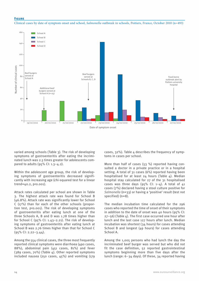
The epidemic curve of clinical cases per school is shown in the Figure. Only cases who reported the date of onset of their illness are included (n=493). The peak of the outbreak in School A occurred two days after the incriminated lunch was first served. At this school, the incriminated beef burgers were served on 19 October, and 25 additional beef burgers were also served on the following day. The peak of the outbreak in Schools B and D occurred one day after the incriminated lunch was served, and in School C, two days after. In the three later schools, the incriminated lunch was served on 22 October.
Among the 1,559 persons exposed, 554 were identified as clinical cases: 493 presented symptoms within five days after the lunch and 61 presented symptoms with an unknown time of onset within two weeks after the lunch. The global attack rate was 35.5% (554/1,559).
Attack rates were similar among persons of female and male sex (Table 3) (proportion test; p=0.46).
Attack rate was significantly higher (proportion test, p<0.001) among adolescents (<20 years-old) (534/1,474, 36%) compared to adults (10/65, 15%) although rates
School A B C D
Sex-ratio (male/female)a 1.0 1.0 0.9 2.3
Adults (≥20 years-old)b
Number of exposed 11 33 7 14
Number of clinical cases 0 3 3 4
Adolescents (<20 years-old)b
Number of exposed 254 671 433 116
Number of clinical cases 93 326 69 46
Median age in years (interquartile range)b
Adults (≥20 years-old) 43 (38–53) 40 (34–51) 41 (28–45) 39.5 (29–51)
Adolescents (<20 years-old) 13 (11–13) 12 (11–13) 12 (11–13) 16 (15–17)
a As 21 questionnaires missed information on sex, 1,538 questionnaires were used,b For cases whose age was known. As 20 questionnaires missed information on age, 1,539 questionnaires were used.
Table 2Characteristics of the exposed population, Salmonella outbreak, Poitiers, France, October 2010 (n=1,559)
14 www.eurosurveillance.org
varied among schools (Table 3). The risk of developing symptoms of gastroenteritis after eating the incrimi-nated lunch was 2.3 times greater for adolescents com-pared to adults (95% CI: 1.3–4.2).
Within the adolescent age group, the risk of develop-ing symptoms of gastroenteritis decreased signifi-cantly with increasing age (chi-squared test for a linear trend=40.2, p<0.001).
Attack rates calculated per school are shown in Table 3. The highest attack rate was found for School B (46.8%). Attack rate was significantly lower for School C (17%) than for each of the other schools (propor-tion test, p<0.001). The risk of developing symptoms of gastroenteritis after eating lunch at one of the three Schools A, B and D was 1.78 times higher than for School C (95% CI: 1.43–2.21). The risk of develop-ing symptoms of gastroenteritis after eating lunch at School B was 2.76 times higher than that for School C (95% CI: 2.22–3.44).
Among the 554 clinical cases, the three most frequently reported clinical symptoms were diarrhoea (490 cases, 88%), abdominal pain (451 cases, 81%) and fever (389 cases, 70%) (Table 4). Other reported symptoms included nausea (250 cases, 45%) and vomiting (179
cases, 32%). Table 4 describes the frequency of symp-toms in cases per school.
More than half of cases (53 %) reported having con-sulted a doctor in a private practice or in a hospital setting. A total of 31 cases (6%) reported having been hospitalised for at least 24 hours (Table 4). Median hospital stay calculated for 27 of the 31 hospitalised cases was three days (95% CI: 1–4). A total of 41 cases (7%) declared having a stool culture positive for Salmonella (n=33) or having a ‘positive’ result (test not specified) (n=8).
The median incubation time calculated for the 296 cases who reported the time of onset of their symptoms in addition to the date of onset was 40 hours (95% CI: 27–56) (Table 4). The first case occurred one hour after lunch and the last case 127 hours after lunch. Median incubation was shortest (34 hours) for cases attending School B and longest (49 hours) for cases attending School A.
Among the 1,005 persons who had lunch the day the incriminated beef burger was served but who did not fit the case definition, 52 reported gastrointestinal symptoms beginning more than five days after the lunch (range: 6–34 days). Of these, 34 reported having
FigureClinical cases by date of symptom onset and school, Salmonella outbreak in schools, Poitiers, France, October 2010 (n=493)
0
20
40
60
80
100
120
140
160
19/10/2010 20/10/2010 21/10/2010 22/10/2010 23/10/2010 24/10/2010 25/10/2010 26/10/2010 27/10/2010
Num
ber o
f clin
ical
cas
es
Date of symptom onset
School C
School B
School D
School A
Food-borne outbreak alert by Poitiers university
hospital
Beef burgers served at
Schools B, C, D
Additional beef burgers served at School A (n=25)
Beef burgers served at School A

15www.eurosurveillance.org
diarrhoea only and 18 reported fever with at least one digestive symptom.
Microbiological resultsThe two food isolates from frozen and from cooked beef burger sampled in the canteen of Schools A and B were monophasic Salmonella Typhimurium 4,5,12:i:- with resistance to ampicillin, streptomycin, sulphona-mides and tetracycline (R(resistant)-type ASSuT), which is common to this strain [9]. Concentration of bacterial inoculum was 270 to 18,000 (colony-forming unit) CFU/g for the 23 enumerations carried out.
The NRC confirmed the presence of monophasic Salmonella Typhimurium 4,5,12:i:- (R-type ASSuT) in the 25 human isolates. Among these, 21 belonged to students who satisfied the case definition used in the study, three belonged to students who were ill but who did not satisfy the case definition used in the study (one with fever only and two with time of onset at nine and 10 days after the incriminated lunch) and one belonged to a student who did not participate in the questionnaire survey.
Food and human isolates exhibited a unique PFGE profile (XTYM-151) and a unique MLVA profile (3-14-6-NA-211).
Discussion and conclusionA local school outbreak of monophasic Salmonella Typhimurium 4,5,12:i:- caused by imported food was identified through the national disease notification
system for food-borne illness. Investigation by the Food Control Unit quickly identified the cause of the outbreak as beef burgers and all batches of the same date than the incriminated batch were rapidly withdrawn. The impact of the consumption of the incriminated beef burgers was unknown at the time of outbreak notification as it occurred during school vacation. The retrospective epidemiological investiga-tion conducted in the four affected schools of Poitiers enabled us to identify 554 clinical cases among the 1,559 study participants who ate at school on the days the incriminated beef burgers were served. Twenty-one cases who participated in the study were biologically confirmed by the NRC by serotyping.
This multi-school outbreak is one of the biggest food-borne outbreaks due to monophasic Salmonella Typhimurium 4,5,12:i:- described in France. A previ-ously described large outbreak of Salmonella enterica serotype 4,[5],12:i:- in France involved 337 identified cases and occurred nationwide between 31 October and 18 December 2011 [10]. Another nationwide epi-demic involving the same Salmonella strain occurred in France between 1 August and 9 October 2011 with 682 cases reported [10]. In these two outbreaks, the inves-tigations indicated dried pork sausage as being the most likely source of the outbreaks [10]. The Poitiers school outbreak is the first large scale outbreak described in France of monophasic Salmonella enterica variants involving beef.
The overall attack rate of the Poitiers outbreak was higher than the average attack rate (7%) observed in Salmonella food-borne outbreaks that occurred in school canteens in 2006–2008 in France [1]. In the same period, no large-scale Salmonella outbreak was described in a school setting. The largest food out-break described in schools in France occurred in 2008 and involved 1,137 identified cases with a global attack rate of 50% and was of viral origin [14].
Although the Poitiers outbreak occurred two months before the annual winter peak of viral gastroenteritis, we cannot exclude that some of the cases identified in the survey were due to other gastrointestinal disease. The high attack rate observed in this outbreak could be explained by a particularly high initial concentration of bacterial inoculum. A meta-analysis by Teunis et al. [15] on food-borne outbreak data showed median infective dose (ID50) values ranging between 30 and 50 CFU/g for Salmonella.
The survey revealed an attack rate twice as high among adolescents as among adults. Moreover, the attack rate decreased with increasing age among ado-lescents. There are no obvious explanations for this finding other than the possibility that the younger stu-dents were served lunch at a different time than the others and thereby ate beef with a potentially different cooking time.
Cases / exposed
Attack rate % (95% confidence interval)
Age groupa
Adolescents (< 20 years-old) 534 / 1,474 36.2 (33,8–38,7)
Adults (≥20 years-old) 10 / 65 15.4 (7,6–26,5)
Sexa
Female 261 / 752 34.7 (31,3–38,2)
Male 287 / 786 36.5 (33,1–40,0)
School
Ab 95 / 268 35.4 (29,7–41,5)
Bb 333 / 712 46.8 (43,0–50,5)
Cb 76 / 449 17.0 (13,6–20,7)
Dc 50 / 130 38.5 (30,1–47,4)
All schools 554 / 1,559 35.5 (33,1–38,0)
a For persons with available information.b Junior high school (median age of students: 16 years).c Senior high school (median age of students: 12–13 years).
Table 3Attack rate per age group, sex and school, Salmonella outbreak, Poitiers, France, October 2010 (n=554)

16 www.eurosurveillance.org
The outbreak showed signs of severity with about half of the cases who sought medical care in a private prac-tice or an emergency service, of which 31 of 554 (6%) were hospitalised for more than 24 hours.
Incubation for Salmonella infection is known to range from six to 72 hours with longer incubations (up to 16 days) documented [16]. Two students with a positive stool culture and an incubation of nine and 10 days could be secondary cases contaminated by person-to-person spread. We observed that median incubation was shortest in the school with the highest attack rate (School B). Such negative correlation between attack rate and incubation has been documented in previ-ous outbreaks and retrospective analysis of human outbreaks [17]. This highlights the possibility that the infective dose was greater in beef burgers served in School B than in other schools, in particular School C. Moreover, hospitalisation rate used as a proxy meas-ure for disease severity in this context was 2.6 times greater among cases attending School B than among those attending School C. However, the possibility that frozen beef burgers were contaminated at differ-ent concentrations is unlikely because beef burgers supplied to schools were all from the same batch. One hypothesis for different attack rates observed between schools relates to different cooking practices.
Although the number of cases identified by the inves-tigation is probably close to the real number of cases of salmonellosis due to ingestion of contaminated beef burgers, the number could be underestimated because of non exhaustive study participation (response rate
78%), because of our assumption that all those who ate at the school consumed the beef, and because of errors in reporting disease onset for persons with clinical symptoms. Inversely, important local media attention before the conduct of the investigation could have induced a high participation rate of the ill student group and an over declaration of symptoms due to psy-chogenic-like effect. The two-week delay between the first cases and the questionnaire survey (due to school vacation) could have led to recall bias and errors, espe-cially in date reporting. The risk of error for exposure was minimised by asking students about the date they ate lunch at school instead of asking them the specific food items they had eaten.
At the country’s departmental level, two other local-ised outbreaks of monophasic Salmonella Typhimurium 4,5,12:i:- occurred in October 2010, one in a retirement institution and another in a recreation centre associated with the consumption of beef burgers from the incrimi-nated batch (18 cases, unpublished data). Interviewing at each of the eighteen other institutions in the depart-ment that received frozen beef burgers with the same production date as the incriminated batch revealed that none was aware of cases of diarrhoea within their institution (as of 9 November 2010). Although distribu-tion of potentially contaminated frozen beef burgers was widespread in France, no increase in food-borne outbreaks was detected through disease notification surveillance and no local increase in the serotype 4,5,12:i:- was detected by the NRC (as of 9 November 2010).
School An(%)a
School Bn(%)a
School Cn(%)a
School Dn(%)a
All schoolsn(%)a
Median incubation time in hours,IQR (number of cases)b
49,43–69 (n=41)
34, 25–5 (n=204)
46, 35–68 (n=22)
39, 30–70 (n=29)
40, 27–56(n=296)
Clinical symptoms 95 (100) 333 (100) 76 (100) 50 (100) 554 (100)
Nausea 56 (41) 164 (49) 30 (39) 17 (34) 250 (45)
Vomiting 34 (36) 119 (36) 12 (16) 14 (28) 179 (32)
Abdominal pain 74 (78) 285 (86) 52 (68) 40 (80) 451 (81)
Fever 72 (78) 254 (76) 40 (53) 23 (46) 389 (70)
Diarrhoea 82 (86) 295 (89) 65 (86) 48 (96) 490 (88)
Other symptoms 16 (17) 45 (14) 14 (18) 1 (2) 76 (14)
Treatment 95 (100) 333 (100) 76 (100) 50 (100) 554 (100)
Doctor consultation 48 (51) 201 (62) 18 (76) 19 (62) 286 (53)
Hospitalisation for at least 24 hours 6 (6) 22 (7) 2 (3) 1 (2) 31 (6)
Stool culture 16 (17) 51 (17) 7 (9) 4 (8) 78 (15)
IQR: interquartile range.a Unless otherwise specified.b For cases who reported the time of onset of their symptoms in addition to the date.
Table 4Characteristics of clinical cases with symptoms of gastroenteritis, Salmonella outbreak, Poitiers, France, October 2010 (n=554)

17www.eurosurveillance.org
The international dimension of this outbreak in France is demonstrated by the fact that the beef was produced in an establishment in another EU Member State. The Rapid Alert System for Food and Feed (RASSF) was used to inform authorities of the manufacturer coun-try and other EU countries likely to receive the prod-ucts. Only a French distributor received all the batches from this Member State manufacturer and sold them. Furthermore, no food-borne outbreaks caused by the monophasic Salmonella Typhimurium 4,5,12:i:- were reported in Europe at the time of the outbreak in Poitiers.
Monophasic Salmonella Typhimurium 4,5,12:i:- was associated with a severe outbreak, the largest Salmonella food-borne outbreak described in a school setting in France in recent years. Quick identification and withdrawal of incriminated food batch and respect of safe cooking practices for beef burgers were likely crucial to limit extension of outbreak. Informing other European countries was necessary as the incriminated beef was an imported food product.
References1. Delmas G, Jourdan da Silva N, Pihier N, Weill F-X, Vaillant V, de
Valk H. Les toxi-infections alimentaires collectives en France entre 2006 et 2008. [Collective food-borne infections in France between 2006 and 2008]. Bull Epidémiol Hebd. 2010;31-32:344-8. French.
2. Bone A, Noel H, Le Hello S, Pihier N, Danan C, Raguenaud ME, et al. Nationwide outbreak of Salmonella enterica serotype 4,12:i:- infections in France, linked to dried pork sausage, March-May 2010. Euro Surveill.2010;15(24):pii=19592. Available from: http://www.eurosurveillance.org/ViewArticle.aspx?ArticleId=19592
3. Echeita MA, Aladueña A, Cruchaga S, Usera MA. Emergence and spread of an atypical Salmonella enterica subsp. enterica serotype 4,5,12:i:− strain in Spain. J. Clin Microbiol. 1999;37(10):3425.
4. Mossong J, Marques P, Ragimbeau C, Huberty-Krau P, Losch S, Meyer G, et al. Outbreaks of monophasic Salmonella enterica serovar 4,[5],12:i:− in Luxembourg, 2006. Euro Surveill. 2007;12(6): pii=719. Available from: http://www.eurosurveillance.org/ViewArticle.aspx?ArticleId=719
5. Switt AI, Soyer Y, Warnick LD, Wiedmann M. Emergence, distribution, and molecular and phenotypic characteristics of Salmonella enterica serotype 4,5,12:i:-. Foodborne Pathog Dis. 2009;6(4):407-15.
6. Centers for Disease Control and Prevention (CDC). Investigation of Outbreak of Human Infections Caused by Salmonella I 4,[5],12:i:-. Atlanta: CDC. 29 Oct 2007. Available from: http://www.cdc.gov/salmonella/4512eyeminus.html
7. Amavisit P, Boonyawiwat W, Bangtrakulmont A. Characterization of Salmonella enterica serovar Typhimurium and monophasic Salmonella serovar 1,4,[5],12:i:- isolates in Thailand. J Clin Microbiol. 2005;43(6):2736-40.
8. Tavechio AT, Ghilardi AC, Fernandes SA. “Multiplex PCR” identification of the atypical and monophasic Salmonella enterica subsp. enterica serotype 1,4,[5],12:i:- in São Paulo State, Brazil: frequency and antibiotic resistance patterns. Rev. Inst Med Trop Sao Paulo. 2004;46(2):115-7.
9. Hopkins KL, Kirchner M, Guerra B, Granier SA, Lucarelli C, Porrero MC, et al. Multiresistant Salmonella enterica serovar 4,[5],12:i:- in Europe: a new pandemic strain?. Euro Surveill. 2010;15(22):pii=19580. Available from: http://www.eurosurveillance.org/ViewArticle.aspx?ArticleId=19580
10. Gossner CM, van Cauterem D, Le Hello S, Weill FX, Terrien E, Tessier S, et al. Nationwide outbreak of Salmonella enterica serotype 4,[5],12:i- infection association with consumption of dried pork sausage, France, November to December 2011. Euro Surveill. 2012;17(5):pii=20071. Available from: http://www.eurosurveillance.org/ViewArticle.aspx?ArticleId=20071
11. Agasan A, Kornblum J, Williams G, Pratt CC, Fleckenstein P, Wong M, et al. Profile of Salmonella enterica susp, enterica (subspecies I) Serotype 4,5,12 :i- Stains causing food-borne infections in New York City. J Clin Microbiol. 2002;40(6):1924-9.
12. Weill FX, Guesnier F, Guibert V, Timinouni M, Demartin M, Polomack L, et al. Multidrug resistance in Salmonella enterica serotype Typhimurium from humans in France (1993 to 2003). J Clin Microbiol. 2006;44(3):700-8.
13. Larsson JT, Torpdahl M, Petersen RF, Sorensen G, Lindstedt BA, Nielsen EM. Development of a new nomenclature for Salmonella Typhimurium multilocus variable number of tandem repeats analysis (MLVA). Euro Surveill. 2009;14(15):pii: 19174. Available from: http://www.eurosurveillance.org/ViewArticle.aspx?ArticleId=19174
14. Guinard A, Pouey J, Schwoebel V. Investigation d’une toxi-infection alimentaire collective alimentaire en milieu scolaire en Haute-Garonne et dans le Tarn [Investigation of a collective foodborne outbreak in Haute-Garonne and Tarn schools]..Saint-Maurice: Institut de veille sanitaire;Apr 2009. p. 33. [Accessed 27 Sep 2012]. French. Available from: http://www.invs.sante.fr/publications/2009/tiac_haute_garonne_tarn/tiac_haute_garonne_tarn.pdf
15. Teunis PF, Kasuga F, Fazil A, Ogden ID, Rotariu O, Strachan NJ. Dose-response modeling of Salmonella using outbreak data. Int J Food Microbiol. 2010;144(2):243-9.
16. Heymann DL, Control of communicable diseases manual. 19th ed, Washington DC: American public health association, 2008. p. 537.
17. Glynn JR, Bradley DJ. The relationship between infecting dose and severity of disease in reported outbreaks of Salmonella infections. Epidemiol Infect. 1992; 109(3):371-88.

18 www.eurosurveillance.org
News
ESCAIDE 2012 – online registration open until 12 October
Eurosurveillance editorial team ([email protected])1
1. European Centre for Disease Prevention and Control (ECDC), Stockholm, Sweden
Citation style for this article: Eurosurveillance editorial team. ESCAIDE 2012 – online registration open until 12 October . Euro Surveill. 2012;17(40):pii=20286. Available online: http://www.eurosurveillance.org/ViewArticle.aspx?ArticleId=20286
Article published on 4 october 2012
ESCAIDE 2012, the sixth European Scientific Conference on Applied Infectious Disease Epidemiology, is now less than three weeks away. The conference, spon-sored by the European Centre for Disease Prevention and Control (ECDC), is taking place in Edinburgh, United Kingdom, between 24 and 26 October.
This year’s conference programme includes planned keynote sessions on the following topics:
•zoonoses: the detection and management of emerging infections at the human/animal interface;
•vulnerability in 21st century public health; •public health microbiology and infectious disease
epidemiology: hand-in-hand in the field; •vaccination: effectiveness, safety and implementation
strategies for current and future vaccines.
For more details on the conference please consult the conference programme: http://ecdc.europa.eu/en/ESCAIDE/programme/Pages/programme.aspx
The deadline for online registration for ESCAIDE 2012 is Friday 12 October. After this date, registration on-site will also be available.
ESCAIDE 2012 has been accredited by the European Accreditation Council for Continuing Medical Education (EACCME). Participants can receive up to 16 Continuing Medical Education (CME) credits for attending ESCAIDE 2012.