viveca wallin bengtsson periodontitis, carotid ...1371535/fulltext01.pdf · to assess if carotid...
TRANSCRIPT

DO
CT
OR
AL
DIS
SE
RTA
TIO
N IN
OD
ON
TO
LOG
Y
VIV
EC
A W
AL
LIN
BE
NG
TS
SO
N
MA
LM
Ö U
NIV
ER
SIT
Y 2
01
9P
ER
IOD
ON
TIT
IS, C
AR
OT
ID C
ALC
IFIC
AT
ION
S A
ND
FU
TU
RE
CA
RD
IOV
AS
CU
LA
R D
ISE
AS
ES
IN O
LD
ER
IND
IVID
UA
LS
VIVECA WALLIN BENGTSSON PERIODONTITIS, CAROTID CALCIFICATIONS AND FUTURE CARDIOVASCULAR DISEASES IN OLDER INDIVIDUALS


© Copyright Viveca Wallin Bengtsson 2019
Coverpage Marie Jönsson
ISBN 978-91-7877-020-5 (Malmö) (print)
ISBN 978-91-7877-021-2 (Malmö) (pdf)
ISBN 978-91-87973-41-3 (Kristianstad)
Holmbergs, Malmö 2019


To Andreas, Alexander, Ludwig and Victoria


CONTENTS
ABBREVIATIONS ............................................................ 9
LIST OF PAPERS ........................................................... 12
ABSTRACT .................................................................. 14Background .........................................................................14Aims ...................................................................................14Methods ..............................................................................15Results .................................................................................15Conclusion ..........................................................................16
POPULÄRVETENSKAPLIG SAMMANFATTNING .................. 18
INTRODUCTION .......................................................... 20Oral health and older individuals ...........................................20Periodontitis and older individuals ..........................................21Periodontitis, systemic diseases and older individuals ................22Periodontitis, cardiovascular diseases (CVDs), mortality and older individuals ............................................................23Carotid calcifications, atherosclerotic lesions on panoramic radiographs .........................................................25
AIMS ......................................................................... 27
MATERIAL AND METHODS ............................................ 28Ethical considerations ...........................................................31Statistical analysis ................................................................32
RESULTS ..................................................................... 34Study I ................................................................................34Study II ................................................................................36Study III ...............................................................................37Study IV ..............................................................................42

DISCUSSION ............................................................... 45
CONCLUSIONS ........................................................... 53
FUTURE RESEARCH ....................................................... 55
CLINICAL IMPLICATIONS ............................................... 56
ACKNOWLEDGEMENTS ............................................... 57
REFERENCES ............................................................... 60
PAPERS I - IV ............................................................... 71

9
ACS- Acute coronary syndromes
AMI- Acute myocardial infarction
BMI- Body mass index
BL- Bone loss
BOP- Bleeding on probing
CC- Carotid calcification
CAL- Clinical attachment level
CEJ- Cementoenamel junction
CI- Confidence Interval
C3- Cervical vertebra 3
C4- Cervical vertebra 4
CVDs- Cardiovascular diseases
DS- Doppler Sonography
9

10
FDI- World Dental Federation
HR- Hazard ratio
ICC- Intraclass correlation
ICD- International classification of diseases
JB- Johan Sanmartin Berglund
MI- Myocardial infarction
OR- Odds ratio
OO- Old old age cohort 78-96 years
PASW- Predictive Analytics Soft Ware
PC- Personal computer
PD- Probing depth
PMX- Panoramic radiograph
REP- Rigmor Persson
SD- Standard deviation
SNAC- Swedish National Study of Aging and Care
SPP- Statistical Package for the Social Sciences
SWEAH- The Swedish National Graduate School for Competitive
Science on Ageing and Health
TIA- Transient ischemic contact
10

11
UN- United Nations
WHO- World Health Organization
WMA- World Medical Association
YO- Young old age cohort 60-72 years
11

12
This thesis is based on the following four papers, which will be re-
ferred to in the text by the Roman numerals I-IV as listed below.
I. Bengtsson VW, Persson GR, Renvert S. Assessment of ca-
rotid calcifications on panoramic radiographs in relation to
other used methods and relationship to periodontitis and
stroke: a literature review. Acta Odontol. Scand.
2014;72(6):401-12.
II. Bengtsson VW, Persson GR, Berglund J, Renvert S. A
cross-sectional study of the associations between periodon-
titis and carotid arterial calcifications in an elderly popula-
tion. Acta Odontol. Scand. 2016;74(2):115-20.
III. Bengtsson VW, Persson GR, Berglund J, Renvert S. Carotid
calcifications on panoramic radiographs are associated
with future stroke or ischemic heart diseases: a long-term
follow-up study. Clin Oral Investig. 2019;23(3):1171-
1179.
IV. Periodontitis related to cardiovascular events and mortali-
ty, a long-time longitudinal study. In manuscript.
Reprints of articles I and II were made with kind permission of the
publishers. Article III is distributed under the terms of the Creative
Commons Attribution 4.0 International License.
12

An
over
view
of
the
stud
ies
incl
uded
St
udy
A
im
Des
ign
Sam
ple
Dat
a C
olle
ctio
n M
ain
find
ings
I T
o ev
alua
te t
he u
se a
nd v
alue
of
pano
ram
ic
radi
ogra
phs
in a
sses
sing
car
otid
cal
cifi
cati
ons
in r
elat
ion
to o
ther
use
d m
etho
ds (
gold
sta
nd-
ards
). A
sec
ond
aim
was
to
asse
ss t
he li
tera
-tu
re o
n ca
roti
d ca
lcif
icat
ions
def
ined
fro
m
pano
ram
ic r
adio
grap
hs a
nd c
oncu
rren
t di
ag-
nosi
s of
str
oke
and
peri
odon
titi
s
Rev
iew
30
art
i-cl
es
Lit
erat
ure
sear
ch s
cree
ning
fo
r pu
blic
atio
ns u
sing
se
arch
ter
ms
such
as
pano
-ra
mic
rad
iogr
aph,
car
otid
ca
lcif
icat
ion,
str
oke
and
peri
odon
titi
s
The
sen
siti
vity
for
fin
ding
s of
car
otid
cal
cifi
cati
ons
on p
anor
amic
rad
iogr
aphs
com
pare
d to
Dop
pler
so
nogr
aphy
var
ied
betw
een
31.1
-100
%. T
he s
peci
-fi
city
for
fin
ding
s of
car
otid
cal
cifi
cati
ons
on p
ano-
ram
ic r
adio
grap
hs c
ompa
red
to D
oppl
er s
onog
ra-
phy
vari
ed b
etw
een
21.4
-87.
5%. I
ndiv
idua
ls w
ith
caro
tid
calc
ific
atio
n (C
C)
find
ings
fro
m p
anor
amic
ra
diog
raph
(PM
X)
had
mor
e pe
riod
onti
tis
and
risk
fo
r st
roke
II
To
eval
uate
if p
erio
dont
itis
is a
ssoc
iate
d w
ith
the
pres
ence
of
caro
tid
arte
rial
cal
cifi
cati
ons
diag
nose
d on
pan
oram
ic r
adio
grap
hs in
an
elde
rly
popu
lati
on
Cro
ss-
sect
iona
l n=
499
(313
w
omen
)
Clin
ical
and
rad
iogr
aphi
c ex
amin
atio
n In
divi
dual
s w
ith
peri
odon
titi
s ha
d a
high
er p
reva
-le
nce
of c
arot
id c
alci
fica
tion
s (χ
2= 4
.05,
OR
: 1.5
, 95
% C
I: 1
.0-2
.3, p
<0.0
5)
III
To
asse
ss if
car
otid
cal
cifi
cati
ons
dete
cted
on
pano
ram
ic r
adio
grap
hs a
re a
ssoc
iate
d w
ith
futu
re e
vent
s of
str
oke,
and
/or
isch
emic
hea
rt
dise
ases
ove
r 10
–13
year
s in
indi
vidu
als
be-
twee
n 60
and
96
year
s
Lon
gitu
dina
l n=
726
(350
w
omen
)
Clin
ical
, rad
iogr
aphi
c ex
-am
inat
ion
and
med
ical
his
-to
ry
A s
igni
fica
nt a
ssoc
iati
on b
etw
een
caro
tid
calc
ific
a-ti
ons,
and
fut
ure
even
ts o
f st
roke
and
/or
isch
emic
he
art
dise
ases
wer
e fo
und
(χ2=
9.1,
OR
: 1.6
, 95%
C
I: 1
.2-2
.2, p
<0.0
02).
In
the
youn
ger
age
grou
p, t
his
asso
ciat
ion
was
mor
e pr
onou
nced
(χ2
=12.
4,
OR
:2.4
, 95%
CI:
1.5
- 4.
0, p
<0.0
00)
IV
To
asse
ss i
f in
divi
dual
s ≥
60 y
ears
of
age
wit
h pe
riod
onti
tis
are
mor
e lik
ely
to d
evel
op s
trok
e or
isch
emic
hea
rt d
isea
ses
or a
re a
t hi
gher
ris
k of
dea
th o
ver
a pe
riod
of
17 y
ears
Lon
gitu
dina
l n=
858
(459
w
omen
)
Rad
iogr
aphi
c ex
amin
atio
n an
d m
edic
al h
isto
ry
Indi
vidu
als
wit
h pe
riod
onti
tis
had
an in
crea
sed
risk
fo
r is
chem
ic h
eart
dis
ease
s in
all
indi
vidu
als
(HR
:1.5
, CI:
1.1
-2.1
, p=0
.017
), in
wom
en (
HR
:2.1
, C
I:1.
3-3.
4, p
=0.0
02)
and
in O
O (
HR
:1.7
, CI:
1.0-
2.6,
p=0
.033
) dur
ing
the
17-y
ear
follo
w-u
p. I
ndi-
vidu
als
wit
h pe
riod
onti
tis
had
an in
crea
sed
risk
for
al
l cau
se m
orta
lity
in a
ll in
divi
dual
s (H
R:1
.4,
CI:
1.2-
1.8,
p=0
.002
), in
men
(H
R:1
.5, C
I:1.
1-1.
9,
p=0.
006)
, and
in Y
O (
HR
:2.2
, CI:
1.5-
3.2,
p=0
.000
).
13

15
Periodontitis is a chronic inflammatory disease with a microbiolog-
ical etiology affecting the supporting tissues of the tooth. The dis-
ease affects approximately 50% of the adult population. The prev-
alence of periodontitis increases with age. The complex bacterial
infection, as well as an exaggerated host inflammatory reaction,
may trigger subclinical atherosclerosis.
The overall aim of the present thesis was to study the associations
between periodontitis, cardiovascular diseases and mortality. The
specific aims were: I) to evaluate the use and value of panoramic
radiographs in assessing carotid calcifications in relation to other
used methods (gold standards) and to assess the literature on carot-
id calcifications defined from panoramic radiographs and concur-
rent diagnosis of stroke and periodontitis, II) to evaluate if perio-
dontitis is associated with the presence of carotid arterial calcifica-
tions diagnosed on panoramic radiographs in an elderly popula-
tion, III) to assess if carotid calcifications detected on panoramic
radiographs are associated with future events of stroke, and/or is-
chemic heart diseases over 10–13 years in individuals between 60
and 96 years, IV) to assess if individuals ≥ 60 years of age with per-
iodontitis are more likely to develop stroke or ischemic heart dis-
eases or are at higher risk of death over a period of 17 years.
14

16
A literature review based on peer-reviewed studies was performed
evaluating the use of panoramic radiographs in assessing carotid
calcifications compared to other methods. In study II, III, IV older
individuals, 60 years and older participating in the Swedish Na-
tional Study of Aging and Care (SNAC) were included in the stud-
ies. A dental hygienist performed a dental clinical and radiographic
examination. Probing depths (PD) and bleeding on probing (BOP)
was registered. From radiographic panoramic images, the distances
between the alveolar bone level and the cement enamel junction
(CEJ) were measured. In study II, a diagnosis of periodontitis was
declared, using a composite definition; if a distance between the al-
veolar bone level and the CEJ ≥5 mm on panoramic radiographs at
>10% of sites and PD ≥5 mm at one or more teeth and with BOP
>20% of teeth. In study IV, an indicator of a history of periodontal
disease was declared if a distance between the alveolar bone level
and the CEJ ≥5 mm on panoramic radiographs at ≥30% of sites.
Evidence of a radiopaque nodular mass in the intervertebral space
at or below the vertebrae C3-C4 was identified as carotid calcifica-
tion. In addition, a medical research team performed the medical
examinations, and a medical doctor (JB) reviewed all medical rec-
ords for information about events of stroke and ischemic heart dis-
eases. Stroke and ischemic heart diseases were registered according
to the ICD 10 codes: ICD 60-69 for stroke and ICD: 20-25 for is-
chemic heart diseases. Study I was a review of the literature, in
study II, a cross-sectional study design was employed. In studies III
and IV, a longitudinal prospective study design was used.
On the use of panoramic radiographs in assessing carotid calcifica-
tions in relation to other used methods, the sensitivity and specifici-
ty varied between studies published. Furthermore, only a small
number of studies were found concerning carotid calcifications and
stroke. These studies were primarily retrospective. Four studies
were found on the association between periodontitis and carotid
calcification.
15

17
Study II identified that older individuals with periodontitis had a
significantly higher prevalence of carotid calcifications than indi-
viduals who did not have a diagnosis of periodontitis. In study III,
a significant association was found between carotid calcifications
on panoramic radiographs and 13- year incidence of stroke using a
logistic regression analysis adjusted for confounders (BMI, diabetes
type 2, hypertension) in the 60-72 years. A statistically significant
crude association between radiographic evidence of carotid calcifi-
cations and incidence of ischemic heart diseases was found in indi-
viduals between 60-72 years. Such an association was, however,
not identified among individuals older than 72 years.
In study IV, Cox regression analysis was used, adjusted for con-
founders (age group, BMI >30, diabetes type 2, gender, hyperten-
sion, history of AMI, history of stroke, periodontitis, smoking) and
with a definition of periodontitis as having a distance between the
alveolar bone level and the CEJ ≥5 mm in panoramic radiographs
at ≥ 30% of sites. Periodontitis increased the risk for ischemic heart
diseases in all individuals, in women and in the 78-96 years age
group (OO). Associations between periodontitis, and mortality
were found in all individuals, in men and in the 60-72 years age
group (YO) in the long term follow-up.
1.
Study I identified that there are studies which have assessed the
value of panoramic radiographs in relation to other used methods
(gold standards). The sensitivity and the specificity varied, with the
specificity being more often higher. Few studies have considered
the relationship between radiographic evidence of carotid calcifica-
tions and stroke. Four studies identified a relationship between a
diagnosis of periodontitis and carotid calcifications on panoramic
radiographs.
16

18
2.
Study II identified a significant association between periodontitis
and carotid calcification in individuals 60-96 years.
3.
Study III identified that signs of carotid calcifications assessed from
panoramic radiographs from the 60-96-year-old individuals were
consistent with an incident of stroke and/or ischemic heart diseases
over 13 years follow-up.
4.
Study IV identified that periodontitis was associated with future
ischemic heart diseases in all individuals, in women and in the 78-
96 years age group. Periodontitis was associated with mortality in
all indviduals, in men and in the 60-72 years age group.
17

19
Munnen är en del av kroppen. Processer och sjukdomar som på-
verkar munhålan kan även påverka övriga delar av kroppen. Tand-
lossning är en kronisk inflammatorisk sjukdom som startar med
bakterier. Bakterieprodukter och inflammatoriska produkter orsa-
kar en destruktion av tandens stödjevävnader (parodontit). Om
sjukdomen, genom behandling, inte kan stoppas leder det till att
tanden lossnar, därför används ibland ordet tandlossning i allmänt
tal. Parodontit drabbar en stor del av befolkningen och ökar med
stigande ålder. De bakterier, bakterieprodukter och inflammato-
riska produkter som är involverade vid parodontit har påträffats i
blodkärlen. Det finns många andra sjukdomar som också är in-
flammationsdrivna så som hjärtkärlsjukdomar där ateroskleros an-
ses vara den huvudsakliga orsaken.
Det övergripande målet med denna avhandling var att studera
samband mellan parodontit och hjärtkärlsjukdomar respektive död
över tid. Mer specifika mål var att i en övergripande allmän littera-
turstudie studera användning och värde av panoramaröntgen för
att identifiera karotisförkalkningar. I samma litteraturstudie stude-
rades karotisförkalkningar i relation till stroke och parodontit.
Med hjälp av röntgenundersökning studerades karotisförkalkning-
ar liksom bennivån runt tänderna. Kliniskt undersöktes förekoms-
ten av tandköttsfickor och blödning vid ficksondering. Förhållan-
det mellan karotisförkalkning och parodontit studerades. Vidare
studerades om karotisförkalkningar, som identifierats på röntgen,
18

20
kunde sättas i samband med insjuknande i stroke och/eller hjärt-
sjukdomar över en uppföljningstid på 13 års. Ytterligare ett mål
var att undersöka om individer med parodontit hade större sanno-
likhet att utveckla stroke, hjärtsjukdom eller död, under en upp-
följningstid på 17 år. Individer 60 år och äldre boende i Karlskrona
kommun rekryterades inom projektet "Swedish National Study of
Aging and Care" (SNAC). Två tandhygienister utförde kliniska och
röntgenologiska undersökningar. En läkare granskade medicinska
journaler för att identifiera eventuella stroke eller hjärtsjukdomar.
Träffsäkerheten med panoramaröntgen jämfört med andra meto-
der varierade kraftig mellan analyserade studier. Ett fåtal studier
påträffades avseende samband mellan karotisförkalkning på pano-
ramaröntgen och stroke liksom karotisförkalkning och parodontit.
I flertalet studier som rörde stroke hade man granskat samband
mellan karotisförkalkning och en historia av stroke men inte fram-
tida insjuknande i stroke.
Hos individer 60-72 år upptäcktes ett starkt samband mellan karo-
tisförkalkning på panoramaröntgen och insjuknande i stroke
och/eller hjärtsjukdomar. Individer med parodontit visade sig ha en
ökad risk att dö under den 17 åriga uppföljningstiden jämfört med
individer utan parodontit. Parodontit visade sig ha ett samband
med ökad risk för hjärtsjukdomar.
Slutsatsen av denna avhandling är att;
Äldre individer med parodontit har ökad risk att dö och har oftare
karotisförkalkning, jämfört med de som inte har parodontit.
Karotisförkalkning på panoramaröntgen har ett samband med in-
sjuknande i hjärtkärlsjukdomar. Individer med parodontit utvecklar oftare hjärtsjukdomar.
19

21
Oral health is part of the overall health, well-being and quality of
life (Glick et al. 2017). For older individuals, oral health is vital to
avoid discomfort and pain, tooth loss, and being able to chew food
properly to avoid malnutrition (Eke et al. 2016). WHO defines
oral health as “a state of being free from chronic mouth and facial
pain, oral and throat cancer, oral infection and sores, periodontal
(gum) disease, tooth decay, tooth loss, and other diseases and dis-
orders that limit an individual’s capacity in biting, chewing, smil-
ing, speaking, and psychosocial “well-being" (Fisher et al. 2018). A
similar definition by the World Dental Federation (FDI) has been
declared, "oral health is multi-faceted and includes the ability to
speak, smile, smell, taste, touch, chew, swallow and convey a range
of emotions through facial expressions with confidence and with-
out pain, discomfort and disease of the craniofacial complex"
(Glick et al. 2017).
Life expectancy worldwide has increased, and most individuals can
expect to live beyond 60 years. The proportion of the world’s pop-
ulation >60 years is predicted to change from 12% to 22% be-
tween 2015 and 2050 (WHO 2018). The demographic shift con-
cerns particularly older adults of the developed countries in North
America, Western Europe and northeast Asia (Lamster 2016). The
definition of the older adult has been debated, but in 2017, the
United Nations (UN) agreed to use 60 years as a definition (United
Nations Ageing 2017).
20

22
In older individuals the presence of complete dentures and partial
dentures has steadily decreased over 40 years and the older popula-
tion has more remaining teeth at older age (Norderyd et al. 2015,
Tonetti et al. 2017, Schwendicke et al. 2018, Wahlin et al. 2018).
Wahlin et al. (2018) demonstrated that the number of teeth signifi-
cantly increased in a population between 20 to 80 years of age, es-
pecially in individuals with severe periodontitis. A decrease in the
number of caries affected teeth has also been demonstrated, and
this trend is expected to continue (Jordan et al. 2019). Accordingly,
the number of filled and decayed teeth has increased in older indi-
viduals and will be expected to continue to increase (Norderyd et
al. 2015, Jordan et al. 2019).
More remaining teeth and better oral health is considered a result
of progress in the prevention and treatment of caries and periodon-
titis (Tonetti et al. 2017). The growing aging population and the
increasing expectations of good oral health-related quality of life in
older individuals will, however, provide a considerable challenge to
the healthcare systems (Tonetti et al. 2017).
Periodontitis is a mixed bacterial infection, triggering inflammatory
destruction of the teeth supporting tissues (Hajishengallis et al.
2012). The transition from gingivitis, a reversible inflammation in
the gingiva, to periodontitis is caused by a disruption in the home-
ostasis between genetic (host immune system), environmental and
bacterial virulence factors (Harvey et al. 2017). Epidemiologic
studies performed in different parts of the world have shown that
colonization by certain species of periodontal bacteria, including
Aggregatibacter actinomycetemcomitans (A.a), Porphyromonas
gingivalis (P.g), Tannerella forsythia (T.f) and Campylobacter rec-
tus are detected in destructive periodontitis (Papapanou et al.
2014). If untreated, periodontitis may result in tooth loss and ac-
counts for one of the biggest reasons to lose teeth in adulthood
(Natto et al. 2014).
21

23
Until now, periodontitis has been classified as: 1) chronic perio-
dontitis, 2) aggressive periodontitis, and 3) periodontitis as a mani-
festation of systemic disease (Armitage, 2004). Recently a new
classification has been adopted. Periodontitis is grouped into one
single category and based on a multi-dimensional staging based on
the severity and complexity and assessment of progression of the
disease (Papapanou et al. 2018). Periodontitis is a chronic and cu-
mulative disease that affects more often older individuals
(Castrejón-Pérez et al. 2014). Data from The NHANES study in
US 2009 and 2010, demonstrated that 47% of the population, rep-
resenting 64.7 million adults 30 years and older in the US, had per-
iodontitis. The prevalence of periodontitis increased with increas-
ing age (Eke et al. 2012). The prevalence of severe periodontitis in
individuals 80 years and older was in a Swedish population of
50% (Holm-Pedersen et al. 2006). Another Swedish study identi-
fied periodontitis in 38% of men ≥ 81 years of age (Renvert et al.
2013). In a Norwegian population 67 years and older, 33% of the
subjects had periodontitis, and out of those, 12% had severe peri-
odontitis (Norderyd et al. 2012).
During the last 30 years, a new research field, periodontal medicine
has been launched. In this field, researchers have studied how peri-
odontal disease may influence the individual’s systemic health and
vice versa (Williams et al. 2000). Periodontitis has not only local
effects on the tooth-supporting tissues. Oral bacteria may also en-
ter the bloodstream through the ulcerated periodontal pockets and
cause infections in other organs (e.g. endocarditis, lung abscess and
pneumonia) (Lockhart et al. 2009, Zhang et al. 2013). Bacteria
from the periodontal lesions may cause inflammatory reactions
that could influence the progression of systemic diseases, such as
diabetes mellitus, atherosclerosis, adverse pregnancy outcomes,
rheumatoid arthritis and neurodegenerative diseases (Genco et al.
2010, Glick et al. 2014). Associations between periodontitis and
diabetes are well established (Chavarry et al. 2009, Borgnakke et
al. 2013, Sanz et al. 2018) and periodontitis is associated with im-
22

24
paired glycemic control in individuals with diabetes (Borgnakke et
al. 2013).
Development of atherosclerosis is a lifelong process, starting in ear-
ly childhood, with clinical manifestations many years after (Niini-
koski et al. 2012). Atherosclerosis is the main cause of CVDs
(Gasbarrino et al. 2016, Chapman et al. 2017) and CVDs are the
most common cause of death in the US (Xu et al. 2016). CVDs in-
clude all conditions associated with the heart and blood vessels,
such as coronary heart disease, stroke and heart failure (Waters et
al. 2013).
Periodontitis has been associated with an increased risk for CVDs
(Scannapieco et al. 2003, Kinane et al. 2008, Lockhart et al. 2012,
Cullinan et al. 2013, Dietrich et al. 2013). The etiopathogenic link
between periodontitis and CVDs is considered to be the results of
systemic inflammation (Aoyama et al. 2017). Through the ulcer-
ated periodontal pockets oral bacteria may enter the bloodstream
(Lockhart et al. 2009) resulting in systemic inflammatory and im-
munologic responses (Scannapieco et al. 2016) that could trigger
the initiation and progression of atherosclerosis (Ross et al. 1999,
Libby et al. 2009). Individuals with a diagnosis of acute coronary
syndrome (ACS) have demonstrated a significantly higher total oral
bacterial load and increased number of species associated with per-
iodontitis, such as Porphyromonas gingivalis, Tanerella forsythia
and Treponema denticola (Renvert et al. 2006). High-sensitivity
serum C-reactive protein levels, a mediator for inflammation, have
in several studies demonstrated associations to periodontitis
(Persson et al. 2005, Kumar et al. 2014). Data exist suggesting that
periodontitis is associated with subclinical atherosclerosis
(Desvarieux et al. 2003, Söder et al. 2005, Lockhart et al. 2012).
Having periodontitis increased the risk for stroke (Reyes et al.
2013, Schenkein et al. 2013, Tonetti et al. 2013). In a review of the
literature, periodontitis was reported to be associated with the oc-
23

25
currence of stroke (Lafon A et al. 2014) especially in men and in
younger subjects (Grau et al. 2004). Periodontitis has also been as-
sociated with myocardial heart infarction (Rydén et al. 2016).
Few prospective longitudinal studies on the association between
periodontitis and CVDs have been reported. In a 3-year follow-up
study, periodontitis was predictive of a future ACS event (Renvert
et al. 2010). Another 3-year follow-up study demonstrated that in-
dividuals with severe periodontitis and a diagnosis of coronary
vascular disease attained the combined endpoint (myocardial in-
farction, stroke/transient ischemic attack, cardiovascular death and
death caused by stroke) more often, although not significant, com-
pared to individuals without periodontitis (18.9% versus 14.2%)
(Reichert et al. 2016). Patients with periodontitis were reported to
have an increased risk of CVDs in a Danish nationwide cohort
study with a 15-year follow-up period (Hansen et al. 2016). In the
same study by Hansen et al. (2016) it was reported that periodonti-
tis increased the risk of all-cause mortality within 15 years. Indi-
viduals (30-40 years) with periodontitis and missing molars have
been reported to have an increased risk for death from diseases of
the circulatory system, over 16 years (Söder et al. 2007). Contra-
dictory to the results by Hansen et al. (2016) and Söder et al.
(2007) survival statistics from a 6-year longitudinal study failed to
show that periodontitis predicted mortality in older individuals
(Renvert et al. 2015). Missing teeth have been proposed as a surro-
gate marker for current or past periodontitis as it reflects an accu-
mulation of oral inflammation to which an individual has been ex-
perienced throughout life (Holmlund et al. 2012). In a Korean na-
tionwide cohort follow-up study of 7.6 years, tooth loss was asso-
ciated with an incident of myocardial infarction, heart failure or
ischemic stroke, especially in individuals with periodontitis (Lee et
al. 2019). A population-based survey over 13 years, demonstrated
that ≥ 5 missing teeth were significantly associated with an event of
coronary heart disease and acute myocardial infarction whereas no
association with stroke was observed (Liljestrand et al. 2015). In
another longitudinal study, with a median follow- up time 15.8
years, the number of teeth was significantly related to myocardial
24

26
infarction and heart failure but not to an event of stroke
(Holmlund et al. 2017). In the study by Liljestrand et al. (2015),
missing teeth ≥ 9 was associated with mortality.
Atherosclerotic disease is common in the area where the carotid ar-
tery bifurcates into the internal and external carotid arteries
(Cheng et al. 2006). Both invasive and non-invasive methods are
used to assess the presence and extent of arterial calcifications (Bos
et al. 2015). Intravascular ultrasound and intravascular optical co-
herence tomography are examples of invasive diagnostic methods,
and they may be the most predictable methods in identifying ele-
vated risks for stroke or other cardiovascular events (Denzel et al.
2004). Non-invasive diagnostic methods are Doppler sonography
(DS), computed tomography (CT) and magnetic resonance imaging
(MRI), all able to identify carotid calcifications (Huibers et al.
2015).
Panoramic radiography is a frequently performed radiographic di-
agnostic method in dentistry. Carotid calcifications are usually lo-
calized in the bifurcation area, posterior, inferior to the mandibular
angle, and adjacent to the space between the third or fourth cervi-
cal vertebrae (C3 and C4). When the area of the carotid bifurcates
is visible, panoramic radiography can also be utilized to identify
carotid artery calcifications (Friedlander et al. 1994, Cohen et al.
2002, Persson et al. 2002, Ravon et al. 2003, Tanaka et. al 2006,
Kumagai et al. 2007, Lee et al. 2014, Friedlander et al. 2015). In
3-15% of the adult population, these calcifications can be identi-
fied on panoramic radiographs (Friedlander et al. 1994, Carter et
al. 1997, Ohba et al. 2003, Johansson et al. 2011). Signs of carotid
calcifications are more frequently found in older individuals com-
pared to younger individuals (Bengtsson et al. 2016). A high level
of agreement in intra-examiner assessments of carotid calcification
signs on panoramic radiographs has been reported (Persson et al.
2002). Asymptomatic carotid stenosis of 50% is a risk marker for
25

27
future vascular events (Venketasubramanian et al. 2013, Brott et
al. 2011). Guidelines recommend secondary vascular prevention
and treatment of higher preventive intensity, for individuals with
50% carotid stenosis, even in the absence of vascular events (Ven-
ketasubramanian et al. 2013, Brott et al. 2011). Carotid stenosis of
50% or more can be detected in 75% of cases on panoramic radi-
ographs (Garoff et al. 2014).
26

28
I. To evaluate the use and value of panoramic radiographs in as-
sessing carotid calcifications in relation to other used methods
(gold standards). A second aim was to assess the literature on ca-
rotid calcifications defined from panoramic radiographs and con-
current diagnosis of stroke and periodontitis
II. To evaluate if periodontitis is associated with the presence of ca-
rotid arterial calcifications diagnosed on panoramic radiographs in
an elderly population
III. To assess if carotid calcifications detected on panoramic radio-
graphs are associated with future events of stroke, and/or ischemic
heart diseases over 10–13 years in individuals between 60 and 96
years
IV. To assess if individuals ≥ 60 years of age with periodontitis are
more likely to develop stroke or ischemic heart diseases or are at
higher risk of death over a period of 17 years
27

29
The first study I, included in this thesis is a review assessing carotid
calcifications on panoramic radiographs in relation to other used
methods and the relationship to periodontitis and stroke. Studies
II, III, IV are based on material from the Swedish National Study
on Aging and Care (SNAC) in Blekinge Sweden.
Study 1 was performed as a literature review based on a literature
search in PubMed (Medline) up to November 2012. The search
was based on the following search terms; (I) (panoramic radiog-
raphy and carotid calcification) OR (panoramic radiography and
carotid calcifications) OR (panoramic radiography and carotid ar-
tery atheroma); (II) (carotid calcifications and stroke and panoram-
ic radiography) OR (carotid calcification and dental) OR (carotid
calcification and stroke and panoramic radiography) OR (carotid
calcification and stroke panoramic radiography) and (III) (calcifica-
tions and periodontitis and panoramic radiography) OR (carotid
calcification and periodontitis and panoramic radiography) OR
(carotid calcification and periodontal disease and panoramic radi-
ography) OR (periodontitis and carotid calcification) OR (perio-
dontitis and carotid calcifications). Hard copy search of additional
references was also employed. Only literature published in English
was considered. The literature review included studies on the rela-
tionship between carotid calcifications identified on panoramic ra-
diographs and other methods of assessments and studies on the as-
sociation between carotid calcification and stroke and between ca-
28

30
rotid calcification and periodontitis. Case reports, review papers
and animal research were excluded.
Studies II, III and IV are based on data from the Swedish National
Study on Aging and Care (SNAC) in Blekinge. SNAC a national,
multidisciplinary project includes four participating areas: SNAC-
Blekinge, SNAC Kungsholmen, SNAC Nordanstig, and SNAC
Skåne (GÅS). In all four study areas, research centers conduct a
population study. In an older population, 60 years and over, data
are obtained through repeated medical examinations, tests, inter-
views, and questionnaires regarding health, illness, functional ca-
pability and social situation. In SNAC-Blekinge, an oral health ex-
amination is also incorporated. The study has a longitudinal design
and uses a common type of study design.
Study individuals were randomly selected from the Swedish popu-
lation database representing the ageing population (60–96 years) in
Karlskrona municipality (The Swedish Tax Agency electronic da-
tabase), Sweden. At baseline (2001), an equal number of randomly
selected individuals in age cohorts of 60, 66, 72 and 78 years were
included. In the age cohorts of 81, 84, 87, 90, 93 and 96 years, all
inhabitants were invited, representing the older population in
Karlskrona, Sweden. All individuals were invited by regular mail,
and the enrolment occurred between 2001-2003 (baseline). At
baseline, the response rate was 62% representing approximately
10% of the population ≥ 60 years. More individuals in the older
age cohorts declined participation.
During the enrolment, at baseline 2001-2003, two dental hygien-
ists performed the clinical examinations. The examinations were
performed in a dental chair at the research clinic. Probing depth
(PD) was measured with a periodontal probe (CP-12, Hu-Friedy
Inc. Chicago, IL) at four sites (mesial, mid-buccal, distal and mid-
29
31
lingual) per tooth. The deepest PD value of each tooth was used to
calculate the proportions of teeth with a probing depth ≥ 5 mm. Re-
liability measurements between randomly selected cases for double
assessments regarding the inter-observer agreement of PD values
between the two clinicians performing the examinations was 0.76
(Cronbach’s α) (95% CI:0.67-0.82; p<0.001). The proportion of
teeth with bleeding on probing (BOP) was registered as bleeding or
not per tooth and calculated on an individual level. In study II, per-
iodontitis was defined by the extent of bone loss, PD and BOP.
At baseline in 2001-2003, analogue panoramic radiographs (Or-
thopantomograph, OP 100 Instrumentarium Tuusula Finland)
were taken with a standard exposure of 75kV and 10 mA. An ex-
perienced independent examiner (REP) performed the radiographic
measurements blind to medical and dental information, age, gender
and survival status. A millimeter graded plastic ruler, a magnifica-
tion viewer (x 2) and a lightbox source, was used to measure the
distances between the alveolar bone level and cement enamel junc-
tion (CEJ). Alveolar bone loss was measured at the mesial and dis-
tal aspect of the existing teeth. The number of interproximal sites
with a distance ≥ 5 mm between the alveolar bone level and CEJ
was used. Reliability measurements were made for a second read-
ing of the panoramic radiographs. The intra-class correlation coef-
ficient (ICC) between the intra-observer measurements for the dis-
tance between CEJ and the apex was 0.93 (95% CI:0.91-0.96,
p<0.01). In study II, a diagnosis of periodontitis was made if a dis-
tance between the alveolar bone level and the CEJ ≥ 5 mm on pano-
ramic radiographs was detected at >10% of sites and PD ≥ 5 mm at
one or more teeth and with BOP >20% of teeth. In study IV, peri-
odontitis was defined if a distance between the alveolar bone level
and the CEJ ≥ 5 mm on panoramic radiographs was detected at ≥
30% of sites.
In study II and study III, evidence of a radiopaque nodular mass in
the intervertebral space at or below the vertebrae C3-C4 was iden-
tified as carotid calcification. Reliability measurements of carotid
30

32
calcifications were made through intra-examiner assessments from
100 randomly selected panoramic radiographs resulting in
Cronbach´s alpha of 0.91 with an interval between the two sets of
readings approximately one year.
Information about events of stroke (cerebrovascular diseases) and
ischemic heart diseases were collected from the electronic medical
database at the research center of the general hospital in Karlskro-
na, Sweden. A physician (JB) annually reviewed the medical rec-
ords for all individuals in the study. According to the world health
organization (WHO), diseases were classified into "the statistic
classification of diseases and related health problems, 10th revi-
sion" (ICD 10) (WHO 2019). These classifications are, for ischem-
ic heart diseases (including myocardial infarction) classified ICD
10: 20-25 and for cerebrovascular diseases (including stroke) ICD
10: 60-69.
The included studies comply with the ethical regulations for re-
search described by the World Medical Association (WMA), The
Declaration of Helsinki with amendments (WMA 2013). All the
four different ethical considerations were fulfilled: the information,
the consent, the confidentiality and the utility requirement. The
studies were based on voluntary participation. All study individuals
signed an informed consent to the registration of collected infor-
mation. All information was anonymized, coded and stored in or-
der to protect the integrity of the study individuals. The principal
investigator was the only one having access to the unique code key.
Permission for the studies has been requested and granted by the
Regional Research Ethics Committee at the Lund University (LU
dnr LU-604-00 and 744-00). All the authors had full access to the
data set.
31

33
Statistical computations were carried out using Statistical Package
for the Social Sciences (SPSS) Predictive Analytics Software (PASW)
(version 22.0, 23.0 and 25.0 SPSS Inc., Chicago, IL, USA) for
Windows.
Descriptive statistics were used to analyze the data. Independent t-
tests (equal variance not assumed) and non-parametric tests
(Mann-Whitney U-test) were performed to assess group differ-
ences. Pearson´s χ2 test and Mantel-Haenzel common odds ratio
were also used. Significance was declared at p<0.05. The SPSS
PASW version 22.0 for an Apple computer was used in the analy-
sis.
The SPSS PASW version 23.0 for Personal Computer (PC) comput-
er was used in the analysis. The data were analyzed using descrip-
tive and inferential statistics. The data were assumed not to follow
a normal distribution pattern, and therefore, both independent t-
test (equal variance not assumed) and nonparametric tests (Mann-
Whitney U test) was performed to assess group differences. Pearson
χ2 test and Mantel-Haenzel odds ratio were used to analyze di-
chotomous data. The data were also studied by binominal logistic
regression and adjusted for BMI, diabetes type 2, and hyperten-
sion. The Kaplan–Meier estimator (log rank Mantel–Cox) was
used to study events of stroke and ischemic heart diseases in study
individuals with or without radiographic evidence of carotid calci-
fications during the study period. Significance was set with α at
p<0.05.
The SPSS PASW version 25.0 for PC was used in the analysis. The
data were analyzed using descriptive and inferential statistics. Di-
chotomous data were analyzed using Pearson χ2 test, and Mantel–
32

34
Haenszel common odds ratio. Cox regression analysis, using the
enter method, was used to study adjusted associations (age group,
BMI, diabetes type 2, gender, hypertension, history of AMI, history
of stroke, periodontitis). Proportional hazards assumption was
evaluated graphically with “log-log” plots. Time was defined as
months from inclusion (dental examination) to either death, stroke,
ischemic heart diseases outcome or censoring due to emigration,
death or end of follow-up. Statistical significance was declared at
p<0.05.
33

35
Most of the studies were case series. Doppler sonography (the gold
standard) is often used to identify carotid calcifications and was
compared to panoramic radiographs in 12 out of the 16 studies.
The sensitivity for findings of carotid calcifications on panoramic
radiography compared to a finding by Doppler sonography varied
from 31.1-100% and the specificity varied from 21.4-87.5% on a
patient level. Two studies compared panoramic radiography to an-
terior-posterior projection radiography, and both were in total
concordance to panoramic radiographs (Friedlander 1995, Fried-
lander et al. 2001). One study compared panoramic radiographs
with computer tomography with a sensitivity of 22% and a speci-
ficity of 90% (carotid artery level) (Yoon et al. 2008). Digital sub-
traction angiography versus panoramic radiographs was studied in
one study with a sensitivity of 60% and a specificity of 48% (ca-
rotid artery level) (Damaskos et al. 2008).
Associations between findings of carotid calcifications on pano-
ramic radiographs and stroke were reported in 10 studies. Most of
the information was based on case series. In the majority of stud-
34

36
ies, the frequency of carotid calcifications on panoramic radio-
graphs differed but was mostly low. Three of the studies investigat-
ed individuals with a history of stroke. One of them showed that
all 14 individuals had carotid calcifications on panoramic radio-
graphs (Christou et al. 2010). In the other two studies, 7/19 (Fried-
lander et al. 1994), and 8/40 (Kumagai et al. 2007), individuals
with a history of stroke showed carotid calcifications on panoram-
ic radiographs. Four studies focused on individuals with carotid
calcifications on panoramic radiographs and if they had a history
of stroke. The figures varied considerably, 1/42 (Carter et al.
1997), 29/71 (Cohen et al. 2002), 5/29 (Ravon et al. 2003), 3/33
(Tanaka et al. 2006). In one study, no individuals with carotid cal-
cification on panoramic radiograph had a history of stroke (Lewis
et al. 1999). One study assessed a history of stroke in individuals
with carotid calcifications on panoramic radiographs both at base-
line and at 5 years follow-up. At baseline 5/46, individuals with
signs of carotid calcification had a history of stroke and 5 years
later, another five individuals had developed stroke (Friedlander et
al. 2007). In individuals 80 years old, one study showed that 2/106
individuals with carotid calcifications died from cardiovascular
diseases within 3.5 years (Tamura et al. 2005).
Relationships between carotid calcifications and a concurrent di-
agnosis of periodontitis were reported in four studies (Persson et al.
2002, Ravon et al. 2003, Beckström et al. 2007, Tiller at al. 2011).
The definition of periodontitis varied between these studies. One of
the studies focused on periodontitis risk (Tiller et al. 2011). When
periodontitis was defined as alveolar bone loss ≥ 4 mm between the
CEJ and the bone level at ≥ 30% of sites, an association was
shown with carotid calcification on panoramic radiographs
(Persson et al. 2002, Ravon et al. 2003) and a dose-response rela-
tionship between the size of the carotid calcification and the severi-
ty of periodontitis has been reported (Ravon et al. 2003). If perio-
dontitis was defined as >1 mm bone loss, 25.7% of individuals
with bilateral carotid calcifications had periodontitis compared to
35

37
10.4% in individuals without carotid calcifications on panoramic
radiographs (Beckström et al. 2007). Measuring periodontitis risk
and radiographic evidence of carotid calcification, no associations
were found when multivariate regression analysis was performed
(Tiller et al. 2011).
In the study, 714 individuals with ≥ 10 teeth as assessed from the
panoramic radiographs were included. The area of interest for
identifying carotid calcifications was not readable from 215 pano-
ramic radiographs (30.1%), leaving 499 individuals in the study
(313 women; 62.7%). When separating the individuals into a
young-old (YO) age cohort, 60-72 years, 293 (58.7%) individuals
were included and in the old-old (OO) age cohort 78-93 years, 206
(41.3%) were included. Individuals had on average 21.3 remaining
teeth (SD±5.1). Teeth with bleeding on probing (BOP) was on av-
erage 25.3% (SD±22.6%). A combined definition for periodontitis
was used (if a distance between the alveolar bone level and the CEJ
≥ 5 mm could be identified on the panoramic radiographs at >10%
of sites and PD ≥ 5 mm at one tooth or more and with bleeding on
probing at >20% of teeth). Using this definition of periodontitis 91
out of the 499 (18.4%) study individuals were diagnosed as perio-
dontitis patients. A diagnosis of periodontitis was more frequent in
men (Pearson χ2=12.9; p< 0.001) with an OR of 2.0 (95% CI: 1.4-
2.9, p <0.001). The prevalence of periodontitis was higher in the
OO age cohort with an OR of 1.8 (Pearson χ2 = 10.1, 95% CI:
1.3-2.6, p <0.001). No statistical differences were found in BOP
between the two age cohorts.
Radiographic signs of carotid calcification identified from pano-
ramic radiographs were found in 195/499 individuals (39.1%).
Men had more radiographic signs of carotid calcifications com-
pared to women (Pearson χ2 = 4.6, p <0.05), with an OR of 1.5
(95% CI:1.0-2.2, p <0.05). When comparing the age cohorts, the
OO had significantly more signs of carotid calcifications with an
OR of 1.7 (95% CI:1.1-2.4, p <0.01). In the younger age cohort,
the prevalence of carotid calcifications was significantly higher
36

38
among men (Pearson χ2 = 5.2, p <0.05) with a likelihood of 1.8
(95% CI:1.1- 2.9, p <0.05).
Statistical analysis demonstrated that individuals with periodontitis
had a higher prevalence of carotid calcification (Pearson χ2 = 4.05
p <0.05) and with an OR of 1.5 (95% CI: 1.0-2.3, p <0.05).
The study included 726 individuals with a readable panoramic ra-
diograph. The participants were divided into young-old (YO) 60-
72 years, including 350 study participants and old-old (OO) 78-96
years including 376 participants. During the follow-up period
(2001-2014) 66/350 (18.9%) in the YO and 285/376 (75.8%) in
the OO age group had died.
In all individuals, a significant association between signs of carotid
calcifications on panoramic radiographs at baseline and a future
event of stroke and/or ischemic heart diseases was identified (χ2 =
9.1, OR:1.6, 95% CI:1.2-2.2, p <0.002). Specifically, in the young-
er age group this was more pronounced (χ2 = 12.4, OR:2.4, 95%
CI:1.5-4.0, p <0.000). In the same group, a significant association
between carotid calcifications and an event of stroke was found (χ2
= 4.5, OR:2.3, 95% CI:0.9-6.2, p= 0.03).
At the baseline examination, a history of stroke and/or ischemic
heart diseases was reported in 91/726 (14.3%) individuals. Radio-
graphic signs of carotid calcifications were found in 45/91 (49.5%)
of these individuals. The gender distribution of such signs was
13/45 (28.9%) in women and 32/45 (71.1%) in men.
Data on the remaining 635 individuals who reported no previous
event of stroke and/or ischemic heart diseases at the baseline exam-
ination is described in a sub-analysis. Carotid calcification was
identified in 238/635 (37.5%) of the study individuals. Carotid
calcifications were more frequent in the OO age group 135/319
(42.3%) as compared to the YO age group 103/316 (32.6%) (χ2 =
6.4, OR:1.5, 95% CI:1.1-2.1, p = 0.01). Independent of age, radi-
37

39
ographic signs of carotid calcifications were more frequently identi-
fied in men (χ2 = 11.1, OR:1.8, 95% CI:1.3-2.5, p = 0.001). Dur-
ing 2001-2014, first events of stroke and/or ischemic heart diseases
were identified in 195/635 (30.7%) individuals. Men in the YO age
group had a higher OR for a stroke event than women (OR:3.1,
95% CI:1.0-8.3, p = 0.02). There was no gender difference for is-
chemic heart diseases (OR: 1.3, 95% CI:0.7-2.4, p = 0.4) in the
same YO group. Data analysis also failed to demonstrate gender
differences in the OO age group for the incidence of stroke
(OR:1.0, 95% CI:0.6-1.8, p=0.9) or for the incidence of ischemic
heart diseases (OR:1.4, 95% CI: 0.9-2.4, p=0.12).
A significant association was identified by logistic regression, crude
and adjusted (BMI, diabetes type 2, hypertension), in the YO be-
tween carotid calcifications on panoramic radiographs and stroke
and/or ischemic heart diseases crude (OR:2.4, 95% CI:1.5-4.0, p=
0.000), and adjusted (OR:1.9, 95% CI:1.1-3.5, p = 0.03) (Table I).
In the OO group, only a history of hypertension was significantly
associated with future stroke and/or ischemic heart diseases.
Table 1. Associations between 10-13 year incidence of stroke and/or ischemic
heart diseases and different independent variables. Logistic regression in 60-72
years without a history of stroke and/or ischemic heart diseases reported at
baseline
BMI= Body mass Index
* = p<0.0.5
Independent variables at baseline OR 95%CI p-value
BMI 1.0 1.0-1.1 0.14
Carotid calcifications 1.9 1.1-3.5 0.03*
Diabetes mellitus type 2 2.1 0.7-5.9 0.16
Hypertension 1.7 0.9-3.2 0.08
38

40
Data on causes of death was not available. Among those who died
during the course of the study, 47.0% had positive signs of radio-
graphic carotid calcifications compared to 31.2% in the material
that were survivors. For those with signs of carotid calcifications at
baseline, the OR of death before the endpoint was 2.0 (95% CI:
1.4-2.7, p <0.001).
Relationships between positive or negative signs of carotid calcifi-
cation and cumulative events of a first event of stroke and/or the
first event of ischemic heart diseases were illustrated by Kaplan-
Meier survival analysis (Figures 1 and 2). Study participants YO
with baseline radiographic carotid calcifications and no previous
history of an event had a mean cumulative stroke and/or ischemic
heart diseases survival time of 12.1 years compared to those with-
out carotid calcifications (mean cumulative survival time 13.0
years) (log rank Mantel–Cox χ2 = 10.7, p = 0.001). YO individuals
with baseline radiographic evidence of carotid calcifications and no
history of an ischemic event had a mean cumulative ischemic heart
diseases survival time 12.5 years compared to those without carot-
id calcifications (mean survival time was 13.2 years) (log rank
Mantel–Cox χ2 =9.5, p = 0.002).
39

41
Time to stroke and/or ischemic heart disease
Figure 1. Kaplan-Meier curves of 60-72 year old individuals with no history of stroke and/or ischemic heart diseases, comparing individuals with and without carotid calcification. 13 year cumulative stroke and/or ischemic heart diseases survival
40

42
Time to ischemic heart disease Figure 2. Kaplan-Meier curves of 60-72 year old individuals with no history of ischemic heart diseases, comparing individuals with and without carotid calci-fication. 13 year cumulative ischemic heart diseases survival.
41

43
Data were derived from 858 individuals (women 53.5%). The ages
at baseline varied between 60-93 years with a median age 72.0
years (SD: ±9.3). Individuals had, on average, 18.6 remaining teeth
(SD: ±7.5). Approximately half of the individuals 428/838 (51.1%)
reported that they did not or had never smoked. During the 17-
year follow-up 492/858 (57.3%) died, and 51/858 (5.9%) of the
individuals moved away from the Karlskrona area. Periodontitis
was defined as bone loss ≥ 5 mm at ≥ 30% of sites. At the baseline
examination, periodontitis was diagnosed in 212/858 (24.7%).
Periodontitis was more frequent among men (57.1%) (OR:1.8,
95% CI:1.3-2.4, p= 0.000). If dividing into a young-old (YO) age
cohort (60-72 years), 471/858 (54.9%) were included, and in an
old-old (OO) age cohort (78-96 years), 387/858 (45.1%) were in-
cluded.
The cumulative incidence of at the least one stroke during the 17-
year follow-up was 118/858 (13.8%), and for at the least one is-
chemic heart disease episode, the cumulative incidence was
203/858 (23.7%). Concerning gender, men and women almost
equally developed stroke 60/118 (50.8% men) and ischemic heart
diseases 102/203 (50.2% men). The incidence of stroke per year
was 24.86, which corresponds to 2898 strokes per 100 000 per-
sons and year. The same figures for ischemic heart diseases was
57.2 incidents per year and 6668 per 100 000 and year.
Cox regression analysis was used with periodontitis as an inde-
pendent variable. Incidence of death, the first event of a stroke or
ischemic heart diseases as the dependent variables were used, ad-
justed for independent variables: age group, BMI >30, diabetes
type 2, gender, hypertension, a history of acute myocardial infarc-
tion (AMI), a history of stroke and smoking. Periodontitis in-
creased the risk for ischemic heart diseases in all individuals
(HR:1.5, CI:1.1-2.1, p=0.017) (Figure 3). If having periodontitis
and being a woman (HR:2.1, CI:1.3-3.4, p=0.002) or having peri-
odontitis and being in the OO age group (HR:1.7, CI:1.0-2.6,
p=0.033) there was also an increased risk for ischemic heart diseas-
42

44
es. In men and in the YO age group no association between perio-
dontitis and ishemic heart diseases could be demonstrated (HR:1.1,
CI:0.7-1.7, p=0.749) respectively (HR:1.3, CI:0.8-2.2, p=0.229).
No association between periodontitis and stroke could be identi-
fied. Concerning death periodontitis increased the risk for all cause
mortality in all individuals (HR:1.4, CI:1.2-1.8, p=0.001) (Figure
4). If having periodontitis and being a man (HR:1.5, CI:1.1-1.9,
p=0.006) or in the YO age group (HR:2.2, CI:1.5-3.2, p=0.000),
the risk for death was also increased.
Figure 3. Cox regression curves: 17 year cumulative ischemic heart diseases survival of the total population, comparing individuals with and without peri-odontitis.
43

45
Figure 4. Cox regression curves: 17 year cumulative death survival of the total
study population, comparing individuals with and without periodontitis.
44

46
Periodontitis and cardiovascular diseases are both chronic diseases.
It is therefore not surprising that such conditions are more com-
mon in older individuals. In part, increasing longevity can be at-
tributed the extensive research resulting in better interceptive care.
Nevertheless, in most cases medical and dental clinicians can alle-
viate symptoms, prolonging the disease processes and consequences
of disease over an extended time. Changes in diagnostic criteria
over time, changes in therapies, changes in life-style, changes in so-
cio-economic factors, and other confounding factors that may af-
fect the clinical expression of both periodontitis and cardiovascular
diseases are challenging to control for using statistical modelling
alone. Until health sciences have identified a majority of the etio-
logical factors and effects of confounding factors, it may be diffi-
cult to go beyond studies considering associations between differ-
ent disease complexes.
Periodontitis driven by inflammation, like many other diseases, in-
cluding CVDs (Christodoulidis et al. 2014), has been shown to
trigger subclinical atherosclerosis through an inflammatory pro-
cess. Atherosclerosis is the main reason for CVDs (Cheng et al.
2006, Desvarieux et al. 2003). One type of atherosclerotic lesion is
the carotid calcification (van Gils et al. 2012), which most often is
identified in Doppler sonography (Denzel et al. 2004). The carotid
calcification can be identified on panoramic radiographs (Fried-
lander et al. 1994, Cohen et al. 2002, Persson et al. 2002, Ravon et
al. 2003, Tanaka et al. 2006, Kumagai et al. 2007, Lee et al. 2014,
45

47
Friedlander et al. 2015, Bengtsson Wallin et al. 2016, Bengtsson
Wallin et al. 2019) and more often in older individuals (Bengtsson
Wallin et al. 2016, Bengtsson Wallin et al. 2019). In study I, it
could be concluded, that there are only a few well-designed studies
in older individuals investigating carotid calcifications on pano-
ramic radiographs and associations with periodontitis and associa-
tions with a history of stroke. The specificity, when identifying ca-
rotid calcifications was in most studies high, meaning that the ab-
sence of radiographic evidence of carotid calcifications on pano-
ramic radiographs is consistent with negative findings using Dop-
pler sonography. To identify carotid calcifications against Doppler
sonography the sensitivity varied between the different studies.
There was a shortage of longitudinal or clinical follow-up studies,
specifically beyond assessments with Doppler sonography. It is,
therefore, no surprise that one significant meta-analysis 2012 iden-
tified that an association between periodontitis and atherosclerotic
heart disease exists independent of known confounders. No studies
have, however, identified a causative relationship (Lockhart et al.
2012).
In a cross-sectional study (Study II) an association between perio-
dontitis and carotid calcifications on panoramic radiographs was
shown. Similar results have been shown in other studies (Persson et
al. 2002, Ravon et al. 2003, Beckström et al. 2007). In two of the
studies (Persson et al. 2002, Ravon et al. 2003), the definition of
periodontitis was; alveolar bone loss > 4 mm between the ce-
mentoenamel junction (CEJ) and bone level at ≥ 30% of sites. In
our study a composite definition of periodontitis was used. Alt-
hough using different definitions of periodontitis, the results indi-
cate a statistical association between periodontitis and carotid cal-
cifications.
In study III, it was demonstrated that individuals with carotid cal-
cifications on panoramic radiographs at baseline, over 10-13 years
developed events of stroke and/or ischemic heart diseases more of-
ten than individuals without carotid calcifications and this was es-
pecially true in the younger age cohort (YO), 60-72 years. These
46

48
associations also remained when adjusted for confounders. Men
60-72 years, without previous events, had a higher OR of 3.1 to
develop stroke, compared to women. This association may be re-
lated to the finding of more carotid calcifications on panoramic ra-
diographs in men. There are several studies demonstrating associa-
tions between carotid calcifications on panoramic radiographs and
a history of stroke (Friedlander et al. 1994, Carter et al. 1997,
Lewis et al. 1999, Ravon et al. 2003, Kumagai et al. 2007, Chris-
tou et al. 2010). A few prospective studies have also demonstrated
an association between presence of carotid calcifications and stroke
(Cohen et al. 2002, Friedlander et al. 2007, Johansson et al. 2015).
Two studies with a follow-up of approximately 3 years studied
male veterans > 55 years, and found associations between carotid
calcifications on panoramic radiographs and an incidence of MI,
stroke, TIA, revascularization, and angina (Cohen et al. 2002,
Friedlander et al. 2007). Another recent study over 5 years demon-
strated that the risk of future vascular events was higher in indi-
viduals with carotid calcifications on panoramic radiographs com-
pared to individuals without such signs (Johansson et al. 2015).
This is in line with the results of our study with a follow-up of 10-
13 years. Our study is, as far as we know, the only long-term-study
with a follow-up time of 10 or more years in individuals 60 years
and older investigating associations between carotid calcifications
on panoramic radiographs and new events of medically diagnosed
stroke and/or ischemic heart diseases. In the YO age cohort with
carotid calcifications, the mean cumulative stroke and/or ischemic
heart disease survival time was approximately 1 year lower and
should be considered clinically significant. Among individuals with
carotid calcifications, more individuals died over time, with an OR
of 2.0 for reasons not known but most likely many died due to
CVD events.
According to the result in study IV, individuals 60 years and older
with periodontitis had a higher risk to die in the 17-year follow-up
period compared to individuals without periodontitis. Approxi-
mately half of the individuals died during the long-term follow-up
period, but we do not have access to the reasons for death. The
47

49
main reason for death in the US is coronary heart diseases, and the
second reason is stroke (Benjamin et al. 2018). Accordingly, it is
likely that some individuals in the study had CVD events that re-
sulted in a lethal outcome and therefore no cardiovascular event
was registered and likewise deaths due to CVDs could never be
clarified. In a registerbased study it was demonstrated that perio-
dontitis increased the risk for all cause mortality within 15 years
(Hansen et al. 2016). An association between periodontitis and
mortality in the 60-72 years (YO) was also shown in our study.
Söder et al. (2007) demonstrated that young individuals (30-40
years) with periodontitis and missing molars had an increased risk
for early death over 16 years. Some of the studies have used miss-
ing teeth as a proxy for periodontitis since if periodontitis is not
treated or arrested the consequence will be missing teeth and miss-
ing teeth due to periodontitis may in a way reflect an inflammatory
burden (Holmlund, & Lind, 2012). Lee et al. (2019) demonstrated
that edentulous individuals had the highest cardiovascular risk.
Periodontitis has been reported as one of the major causes for
tooth loss in adulthood (Natto et al. 2014). It is, however, difficult
to be sure of the reason for missing teeth unless it is clearly report-
ed in the dental record. Bone loss as used in the present study to
define periodontitis is, however, a very strong indicator of that the
patient have had periodontal inflammation and using periodontal
bone loss instead of missing teeth as a proxy of inflammatory bur-
den over time seems more relevant.
The second main finding in study IV was that periodontitis in-
creased the risk for ischemic heart diseases in the total population
over the 17-year. In women with periodontitis, the OR for the in-
cidence of ischemic heart disease was 2.1. This is in agreement with
data from Stramba et al. (2006) reporting that women after the
menopause, have a higher incidence of MI compared to age-
matched men. There are few other longitudinal studies investigat-
ing associations between periodontitis and CVDs. In a 3-year fol-
low-up study by Renvert et al. (2010), including individuals that
already had been diagnosed with ACS, an association between per-
iodontitis and recurrent ACS was shown (Renvert et al. 2010). In
48

50
study IV individuals were randomly selected from the population.
A register-based study with a 15-year follow-up identified that pa-
tients with periodontitis reported an increased risk of CVDs (Han-
sen et al. 2016). In this study, individuals >18 years were consecu-
tively included. A hospital diagnosis of chronic or acute periodonti-
tis, based on ICD codes was registered and also the subsequent in-
cidents of CVDs. Compared to our study the included ages differed
and the definition of periodontitis was more vague with no infor-
mation regarding the severity. In a Korean nationwide cohort fol-
low-up study of 7.6 years, a dose-dependent association between
missing teeth and incidence of myocardial infarction, heart failure
and ischemic stroke was found, especially in individuals with peri-
odontitis (Lee et al. 2019). The definition of periodontitis was in
the study by Lee et al. (2019) not mentioned and the study individ-
uals included were from 20 years with a history of a CVD event.
Those differences makes this study not comparable to our study.
In study IV the distance between the alveolar bone level and the
CEJ ≥ 5 mm at ≥ 30% of sites on panoramic radiographs was used
as the definition of periodontitis. This definition reflects the effects
of periodontitis. It is reported that progressive periodontitis among
older individuals includes bone loss, attachment loss and progres-
sive gingival recession rather than deep periodontal pockets (Die-
trich et al. 2006).
With the above mentioned definition 24.7% of the individuals
were diagnosed as having periodontitis. In other studies the preva-
lence of periodontitis varies from 33%-70% in older individuals,
all with different criteria for periodontitis and different ages
(Holm-Pedersen et al. 2006, Eke et al. 2012, Norderyd et al. 2012,
Renvert et al. 2013). The prevalence of periodontitis in this study
with an older population was low. Our definition of periodontitis
included a more severe stage of periodontitis as a bone loss of ≥ 5
mm from the CEJ to the alveolar bone level indicates a definitive
bone loss even accounting for the measurement error. A require-
ment in our definition was also that the bone loss should be pre-
sent at ≥30% of sites, which corresponds to a general disease dis-
49
51
tribution. Other reasons for the low prevalence of periodontitis can
be that the very sick and immobile individuals were not included in
the study as it was not possible to obtain a panoramic radiograph
at the research centre. While periodontitis is a cumulative disease,
more individuals develop periodontitis with age (Eke et al. 2016).
A prerequisite to having a diagnosis of periodontitis in the present
study was the presence of teeth. An inclusion criterion for being in-
cluded in the present study was presence of one tooth or more. In-
dividuals affected by periodontitis in younger ages are the most
sensitive individuals with a hyperactive responsive immune and in-
flammatory system (Fine et al. 2018). The most susceptible indi-
viduals with the rapid form of periodontitis progression are often
younger and have probably fewer teeth or are even edentulous with
older ages and therefore were not included in the study. A stronger
association between periodontitis and CVDs has been shown in
younger individuals compared to older individuals (Lee et al. 2019,
Schenkein et al. 2013). This could be one explanation for the lack
of association between periodontitis and incidence of stroke in the
present study, as it concerns older individuals.
A strength with the comprehensive SNAC study is that it has a
long-term follow-up of the individuals. It is valuable that both a
medical and a dental examination were performed with the same
time intervals. The clinical examinations were performed as a full
mouth examination and not as partial examinations of specific
teeth or areas. Partial mouth examinations have been shown to
underestimate the disease (Kingman et al. 2008). Most of the pub-
lished studies so far are cross-sectional studies. Several other stud-
ies have identified associations between periodontitis and CVDs
(Persson et al. 2003, Scannapieco et al. 2003, Renvert et al. 2006,
Kinane et al. 2008, Lockhart et al. 2012, Cullinan et al. 2013, Die-
trich et al. 2013). Using a long-term follow-up should increase the
possibility to explore true associations between diseases that may
be overlooked in cross-sectional studies. Not many studies have
this opportunity because they are time-consuming and expensive.
There are also limitations in the present studies. When studying as-
sociations in older individuals over a long time, many individuals
50

52
died, leaving fewer, possibly the healthier ones, until the end of the
study period. The most important limitation is, however, the lack
of information on the causes for death. The death register is held
by "The Swedish cause of death register” and was not available for
us to use due to restrictions. Knowing the reasons for death would
have made it possible to improve the accuracy of the associations
between periodontitis and death by CVDs. Both PD and BOP were
registered on a tooth level. The deepest pocket per tooth and pres-
ence of bleeding on probing was registered as BOP irrespective of
the number of surfaces. These measurements are broad, and the
BOP index and PD could have been different if it was based on site
level registrations. The diagnosis of periodontitis and the identifica-
tion of carotid calcifications was based on the baseline examina-
tion in 2001, accordingly the progression of periodontitis and ca-
rotid calcifications during the follow-up was not monitored.
Panoramic radiography is commonly used in dentistry. Such imag-
es, if well exposed, provide a wealth of information that can be
considered both for dental and medical diagnostics. One crucial
factor is that the radiation exposure to the individual from one
such panoramic overview is substantially less as from intra-oral full
mouth series (Ludlow et al. 2008). Signs of carotid calcification
can be detected assuming that the panoramic radiograph covers the
area of interest and that the image focus is correct. The radio-
graphic examination is sometimes hard for the aged individual
with a curvature of the neck, producing technical difficulties, and
sometimes the radiographic image will turn out not readable.
Sometimes, it is not possible to get an optimal image to detect a
possible carotid calcification and at the same time, focus on the
dentition. The consequence might be that two different images
must be obtained. For study purposes and the conditions of ethical
approval, only one image could be taken for presence of carotid
calcifications. This may explain why a rather large number of pan-
oramic radiographs could not be analyzed for the presence of ca-
rotid calcifications. In old-old individuals and more often in older
women, it may be very challenging and demanding to take a high-
quality panoramic radiograph. The panoramic radiographs in the
51

53
present work were taken by trained dental hygienists and not by
dental radiographic technicians. This may also, in part, explain
why approximately 30% of the images could not be evaluated
when studying associations between periodontitis and carotid calci-
fications, in study II. Worth considering is that in study II, one of
the inclusion criteria was 10 teeth or more which of course also af-
fected the lower number of readable panoramic radiographs. This
was not the case in study III, studying associations between carotid
calcifications and future events. The consequence of this was an
unwanted reduction in the number of observations.
52

54
Just a few well-designed studies in older individuals assessing
carotid calcifications on panoramic radiographs and associa-
tions to a history of stroke or associations to periodontitis ex-
isted
The sensitivity, when identifying carotid calcifications on pan-
oramic radiographs against Doppler sonography, varied a lot.
The specificity varied but tended, in most studies, to be high
In individuals 60-93 years, periodontitis was more common in
men than in women
Carotid calcifications identified from panoramic radiographs
were found in 37.5%-39.1% of individuals, more often in men
than in women and more often with increasing age
Periodontitis, diagnosed with a combined clinical and radio-
graphic definition, was associated with carotid calcifications
on panoramic radiographs
Signs of carotid calcifications identified on panoramic radio-
graphs were associated with future incidents of stroke and/or
ischemic heart diseases over 13 years follow-up
Individuals with periodontitis had an increased risk to die and
53

55
had an increased risk to develop ischemic heart diseases over
17 years follow-up
54

56
Relevant further research would include a long-term intervention
study, to analyse if medical interventions in patients with presence
of carotid calcifications on panoramic radiographs can reduce the
number of cardiovascular diseases. As cardiovascular diseases may
have severe consequences with disabilities accompanied by less
quality of life, and in the worst case mortality, prevention studies
are essential. Many visit their dentist/dental hygienist regularly.
Since an association between periodontitis and cardiovascular dis-
eases has been found, it would be valuable to study if well designed
preventive dental programs can influence the incidence of cardio-
vascular diseases in long term studies.
Studying different stages of periodontitis and its associations with
future cardiovascular events as well as analysing if progressive per-
iodontitis is related to the future incidence of cardiovascular dis-
eases could also give additional information on the relationship be-
tween periodontitis and cardiovascular diseases.
To better understand the associations between periodontitis and
cardiovascular diseases, studies further exploring the potential eti-
opathogenic mechanisms are needed.
55

57
When a panoramic radiograph is taken, the inter-vertebral space
between C3 and C4, should be assessed for carotid calcifications
especially if the patient has a diagnosis of periodontitis, is in older
age and male gender
If a carotid calcification on the panoramic radiograph is identified
the patient should be referred for further medical evaluation to
avoid an incident of stroke or ischemic heart disease
Improving periodontal health in older individuals could reduce
overall mortality
A collaboration between physicians and dental professionals would
be helpful to improve clinical management of periodontal and is-
chemic heart diseases
56

58
I would like to express my appreciation and gratefulness to all col-
leagues and friends who have helped and supported me throughout
my research.
I would like to thank:
Professor Stefan Renvert, my main supervisor and mentor, for your
support, and constructive feedback, helping me not making issues
too complicated. Thank you for helping me sort thoughts and help-
ing me grow in my professional ability. The intellectual journey has
been valuable. Thanks for giving me the opportunity to do the re-
search
Professor Rutger Persson, my co-supervisor and co-author for your
immediate guidance and huge engagement, fast thoughts and valu-
able criticism. Thanks for introducing me into and teaching me sta-
tistics. That was valuable
Professor Johan Sanmartin Berglund (JB), my co-supervisor and
co-author for your medical knowledge and valuable input in the
manuscript. Thank you for your statistical guidance
Associate Professor Rigmor Persson (REP), who assessed the radi-
ographs
57

59
Christina Karlsson, for collecting clinical dental data at the dental
examination, and your precursors Sara Henricsson, Ingrid Jonas-
son and Inger Kindblad, collecting clinical dental data at the base-
line
Ulrika Isaksson and Erik Resebo, for your support in collecting da-
ta, managing data files and whatsoever responsible for the logistics.
Johanna Renvert, for data management
Lisa Heitz Mayfield, for help with grammar review of the final the-
sis and for old friendship
Christina Stenervik, for all practical help and guidance, especially
in the very end of the doctoral time
Helena Nilsson, doctoral student colleague for all kinds of support
through the academic education
My colleagues, former colleagues and friends at the Dental Hy-
giene Program, Kristianstad University thank you for supporting
and encouraging me. I am happy to work together with all of you:
Ismail Abbas, Seida Ademovski Erovic, Anette Andersson, Pia An-
dersson, Sladjana Critén, Sara Henricsson, Pernilla Karlgren An-
dersson, Ann-Helene Ljunggren, Valentina Kristinsson, Randi
Madsen, Carina Mårtensson, Ingrid Rejnefelt, Susanna Sättlin,
Berit Vidén and Cecilia Widén.
Christel Lindahl, for supporting with all kinds of things, in the re-
search process. Thanks Christel for your caring
Marie Jönsson, for help with creating the front cover
Andreas, for your huge understanding and support especially with
practical things at home saving me time for research
Alexander, Ludwig and Victoria, my beloved children for just be-
ing in my life, and bringing me lot of happiness
58

60
Uno, Vivi-Ann and Hans for your caring and encouragement, al-
ways believing in me
The Ministry of Health and Social Affairs in Sweden and the par-
ticipating county councils, municipalities and university depart-
ments supported the Swedish National Study of Aging and Care
(SNAC)
The financial support from the Research foundation at Kristian-
stad University, is gratefully acknowledged
Last but not the least thanks to all the study participants, if it were
not for you, the studies and this thesis would not have been possi-
ble
The thesis and the included studies were accomplished within the
context of the "The Swedish National Graduate School for Com-
petitive Science on Aging and Health" (SWEAH) and funded by
the Swedish Research Council
59

61
Aoyama N, Suzuki JI, Kobayashi N, Hanatani T, Ashigaki N, Yoshida A, et
al. Periodontitis deteriorates peripheral arterial disease in Japanese popula-
tion via enhanced systemic inflammation. Heart Vessels. 2017;32(11):1314-
1319.
Armitage GC. Periodontal diagnoses and classification of periodontal diseases.
Periodontol 2000. 2004;34:9-21. Review.
Beckström BW, Horsley SH, Scheetz JP, Khan Z, Silveira AM, Clark SJ, et al.
Correlation between carotid area calcifications and periodontitis: a retro-
spective study of digital panoramic radiographic findings in pretreatment
cancer patients. Oral Surg Oral Med Oral Pathol Oral Radiol Endod.
2007;103(3):359-66.
Bengtsson VW, Persson GR, Berglund J, Renvert S. A cross-sectional study of
the associations between periodontitis and carotid arterial calcifications in
an elderly population. Acta Odontol Scand. 2016;74(2):115-20.
Bengtsson VW, Persson GR, Berglund J, Renvert S. Carotid calcifications in
panoramic radiographs are associated with future stroke or ischemic heart
diseases: a long-term follow-up study. Clin Oral Investig. 2019;23(3):1171-
1179.
Borgnakke WS, Ylostalo PV, Taylor GW, Genco RJ. Effect of periodontal dis-
ease on diabetes: systematic review of epidemiologic observational evi-
dence. J Periodontol. 2013; 84:S135-S152.
Bos D, Leening MJ, Kavousi M, Hofman A, Franco OH, van der Lugt A, et al.
Comparison of atherosclerotic calcification in major vessel beds on the risk
of all-cause and cause-specific mortality: the Rotterdam study. The Rotter-
dam Study. Circ Cardiovasc Imaging. 2015;8(12). pii: e003843.
Brott TG, Halperin JL, Abbara S, Bacharach JM, Barr JD, Bush RL; American
College of Cardiology Foundation/American Heart Association Task Force
on Practice Guidelines; American Stroke Association; American Association
of Neuroscience Nurses; American Association of Neurological Surgeons;
60

62
American College of Radiology; American Society of Neuroradiology;
Congress of Neurological Surgeons; Society of Atherosclerosis Imaging and
Prevention; Society for Cardiovascular Angiography and Interventions; So-
ciety of Interventional Radiology; Society of NeuroInterventional Surgery;
Society for Vascular Medicine; Society for Vascular Surgery. 2011
ASA/ACCF/AHA/AANN/AANS/ACR/ASNR/CNS/SAIP/SCAI/SIR/SNIS/SV
M/SVS guideline on the management of patients with extracranial carotid
and vertebral artery disease: executive summary: a report of the American
College of Cardiology Foundation/American Heart Association Task Force
on Practice Guidelines, and the American Stroke Association, American As-
sociation of Neuroscience Nurses,American Association of Neurological
Surgeons, American College of Radiology,American Society of Neuroradi-
ology, Congress of Neurological Surgeons, Society of Atherosclerosis Imag-
ing and Prevention, Society for Cardiovascular Angiography and Interven-
tions, Society of Interventional Radiology, Society of NeuroInterventional
Surgery, Society for Vascular Medicine, and Society for Vascular Surgery.
Vasc Med. 2011;16(1):35-77.
Carter LC, Haller AD, Nadarajah V, Calamel AD, Aguirre A. Use of pano-
ramic radiography among an ambulatory dental population to detect pa-
tients at risk of stroke. J Am Dent Assoc. 1997;128(7):977-84.
Castrejón-Pérez RC, Borges-Yáñez SA. Frailty from an Oral Health Point of
View. J Frailty Aging. 2014; 3(3):180-6.
Chapman AR, Adamson PD, Mills NL. Assessment and classification of pa-
tients with myocardial injury and infarction in clinical practice. Heart.
2017;(1):103:10-18.
Chavarry NG, Vettore MV, Sansone C, Sheiham A. The relationship between
diabetes mellitus and destructive periodontal disease: a meta-analysis. Oral
Health Prev Dent. 2009;7:107-127.
Cheng C, Tempel D, van Haperen R, van der Baan A, Grosveld F, Daemen
MJ, et al. Atherosclerotic lesion size and vulnerability are determined by
patterns of fluid shear stress. Circulation. 2006; 113:2744-53.
Christou P, Leemann B, Schimmel M, Kiliaridis S, Müller F. Carotid artery
calcification in ischemic stroke patients detected in standard dental pano-
ramic radiographs - a preliminary study. Adv Med Sci. 2010;55(1):26-31.
Christodoulidis G, Vittorio TJ, Fudim M, et al. Inflammation in coronary ar-
tery disease. Cardiol Rev. 2014;22:279-88.
Cohen S, Friedlander A, Jolly D, Date L. Carotid calcification on panoramic
radiographs: an important marker for vascular risk. Oral Surg Oral Med
Oral Pathol Oral Radiol Endod. 2002; 94:510-514.
Cullinan MP, Seymour GJ. Periodontal disease and systemic illness: will the
evidence ever be enough? Periodontol 2000. 2013;62: 271-286.
61

63
Damaskos S, Griniatsos J, Tsekouras N, Georgopoulos S, Klonaris C, Ba-
stounis E, et al. Reliability of panoramic radiograph for carotid atheroma
detection: a study in patients who fulfill the criteria for carotid endarterec-
tomy. Oral Surg Oral Med Oral Pathol Oral Radiol Endod.
2008;106(5):736-42.
Denzel C, Lell M, Maak M, Höckl M, Balzer K, Müller KM, et al. Carotid
artery calcium: accuracy of a calcium score by computed tomography-an in
vitro study with comparison to sonography and histology. Eur J Vasc
Endovasc Surg. 2004;28(2):214-20.
Desvarieux M, Demmer RT, Rundek T, Boden-Albala B, Jacobs DR Jr, Papa-
panou PN, et al. Oral Infections and Vascular Disease Epidemiology Study
(INVEST). Relationship between periodontal disease, tooth loss, and carot-
id artery plaque: The Oral Infections and Vascular Disease Epidemiology
Study (INVEST). Stroke. 2003; 34(9):2120-5.
Dietrich T, Kaye EK, Nunn ME, Van Dyke T, Garcia RI. Gingivitis suscepti-
bility and its relation to periodontitis in men. J Dent Res.
2006;85(12):1134-7.
Dietrich T, Sharma P, Walter C, Weston P, Beck J. The epidemiological. Evi-
dence behind the association between periodontitis and incident atheroscle-
rotic cardiovascular disease. J Clin Periodontol. 2013; 40(Suppl.14): S70-
S84.
Eke PI, Dye BA, Wei L, Thornton-Evans GO, Genco RJ; CDC Periodontal
Disease Surveillance workgroup: James Beck (University of North Carolina,
Chapel Hill, USA), Gordon Douglass (Past President, American Academy of
Periodontology), Roy Page (University of Washin.). Prevalence of periodon-
titis in adults in the United States: 2009 and 2010. J Dent Res. 2012;
91(10):914-20.
Eke PI, Wei L, Borgnakke WS, Thornton-Evans G, Zhang X, Lu H, et al. Per-
iodontitis prevalence in adults ≥ 65 years of age, in the USA. Periodontol.
2000. 2016; 72(1):76-95.
Fine DH, Patil AG, Loos BG. Classification and diagnosis of aggressive perio-
dontitis. J Periodontol. 2018;89 (Suppl 1):S103-S119.
Fisher J, Selikowitz HS, Mathur M, Varenne B. Strengthening oral health for
universal health coverage. Lancet. 2018; 15;392(10151):899-901.
Friedlander AH, Baker JD. Panoramic radiography; an aid in detecting pa-
tients at risk of cerebrovascular accident. Am Dent Assoc. 1994; 125:1598-
1603.
Friedlander AH. Identification of stroke-prone patients by panoramic and cer-
vical spine radiography. Dentomaxillofac Radiol. 1995;24(3):160-4.
62

64
Friedlander AH, Altman L. Carotid artery atheromas in postmenopausal
women. Their prevalence on panoramic radiographs and their relationship
to atherogenic risk factors. J Am Dent Assoc. 2001;132(8):1130-6.
Friedlander AH, Cohen SN. Panoramic radiographic atheromas portend ad-
verse vascular events. Oral Surg Oral Med Oral Pathol Oral Radiol Endod.
2007;103(6):830-5.
Friedlander AH, El Saden SM, Hazboun RC, Chang TI, WongWK, Garrett
NR. Detection of carotid artery calcification on the panoramic images of
post-menopausal females is significantly associated with severe abdominal
aortic calcification:a risk indicator of future adverse vascular events. Den-
tomaxillofac Radiol. 2015; 44(7):20150094.
Garoff M, Johansson E, Ahlqvist J, Jäghagen EL, Arnerlöv C, Wester P. Detec-
tion of calcifications in panoramic radiographs in patients with carotid ste-
noses ≥50%. Oral Surg Oral Med Oral Pathol Oral Radiol.
2014;117(3):385-91.
Gasbarrino K, Mantzoros C, Gorgui J, Veinot JP, Lai C, Daskaopoulou SS.
Circulating Chemerin Is Associated With Carotid Plaque Instability,
Whereas Resistin Is Related to Cerebrovascular Symptomatology. Arterio-
scler Thromb Vasc Biol. 2016; 36:1670-8.
Genco RJ, Williams RC. Periodontal disease and overall health: a clinician’s
guide. Yardley, PA: Professional Audience Communications, 2010: 319 p.
van Gils MJ, Vukadinovic D, van Dijk AC, Dippel DW, Niessen J, van der
Lugt A. Carotid atherosclerotic plaque progression and change in plaque
composition over time: a 5-year follow-up study using serial CT angi-
ography. AJNR Am J Neuro radiol. 2012; 33(7):1267-73.
Glick M. The oral-systemic health connection: a guide to patient care. Chica-
go, IL: Quintessence Publishing Co, Inc. 2014: ix, 301 pp.
Glick M, Williams DM, Kleinman DV, Vujicic M, Watt RG, Weyant RJ. A
new definition for oral health developed by the FDI World Dental Federa-
tion opens the door to a universal definition of oral health. J Public Health
Dent. 2017; 77(1):3-5.
Goodson JM, Haffajee AD, Socransky SS. The relationship between attach-
ment level loss and alveolar bone loss. J Clin Periodontol. 1984; 11(5): 348-
359.
Grau AJ, Becher H, Ziegler CM, Lichy C, Buggle F, Kaiser C, et al. Periodon-
tal disease as a risk factor for ischemic stroke. Stroke. 2004;35(2):496-501.
Hajishengallis G, Lamont RJ. Beyond the red complex and into more com-
plexity: the polymicrobial synergy and dysbiosis (PSD) model of periodon-
tal disease etiology. Mol Oral Microbiol. 2012;27(6):409-19.
63

65
Hansen GM, Egeberg A, Holmstrup P, Hansen PR. Relation of Periodontitis
to Risk of Cardiovascular and All-Cause Mortality (from a Danish Na-
tionwide Cohort Study). Am J Cardiol. 2016;15; 118(4):489-93.
Harvey JD. Periodontal Microbiology. Dent Clin North Am. 2017; 61(2):253-
269. Review.
Holm-Pedersen P, Russell SL, Avlund K, Viitanen M, Win blad B, Katz RV.
Periodontal disease in the oldest-old living in Kungsholmen, Sweden: find-
ings from the KEOHS project. J Clin Periodontol. 2006;33(6):376-84.
Holmlund A, Lind L. Number of teeth is related to atherosclerotic plaque in
the carotid arteries in an elderly population. J Periodontol. 2012;83(3):287-
91.
Holmlund A, Lampa E, Lind L. Oral health and cardiovascular disease risk in
a cohort of periodontitis patients. Atherosclerosis. 2017; 262:101-106.
Huibers A, de Borst GJ, Wan S, Kennedy F, Giannopoulos A, Moll FL. Non-
invasive carotid artery imaging to identify the vulnerable plaque: current
status and future goals. Eur J Vasc Endovasc Surg. 2015;50(5):563-572
Johansson E, Ahlqvist J, Garoff M, Levring Jäghagen E, Meimermondt A,
Wester P. Carotid calcifications on panoramic radiographs: a 5-year follow-
up study. Oral Surg Oral Med Oral Pathol Oral Radiol. 2015;120(4):513-
20.
Johansson EP, Ahlqvist J, Garoff M, Karp K, Jäghagen EL, Wester P. Ultra-
sound screening for asymptomatic carotid stenosis in subjects with calcifi-
cations in the area of the carotid arteries on panoramic radiographs: a
cross-sectional study. BMC Cardiovasc Disord. 2011;11:44.
Jordan RA, Krois J, Schiffner U, Micheelis W, Schwendicke F. Trends in caries
experience in the permanent dentition in Germany 1997-2014, and projec-
tion to 2030: Morbidity shifts in an aging society. Sci Rep. 2019; 2;
9(1):5534.
Kinane D, Bouchard P; Group E of European Workshop on Periodontology.
Periodontal diseases and health: Consensus Report of the Sixth European
Workshop on Periodontology. J Clin Periodontol. 2008;35(8 Suppl):333-7.
Kingman A, Susin C, Albandar JM. Effect of partial recording protocols on
severity estimates of periodontal disease. J Clin Periodontol.
2008;35(8):659-67.
Kumagai M, Yamagishi T, Fukui N, Chiba M. Carotid artery calcifications
seen on panoramic dental radiographs in the Asian population in Japan.
Dentomaxillofac Radiol. 2007;36:92-96.
Kumar KR, Ranganath V, Naik R, Banu S, Nichani AS. Assessment of high-
sensitivity C-reactive protein and lipid levels in healthy adults and patients
64

66
with coronary artery disease, with and without periodontitis—a cross-
sectional study. J Periodontal Res. 2014;49(6):836-44.
Lafon A, Pereira B, Dufour T, Rigouby V, Giroud M, Béjot Y, et al. Periodon-
tal disease and stroke: a meta-analysis of cohort studies. Eur J Neurol.
2014;21(9):1155-61, e66-7.
Lamster IB. Geriatric periodontology: how the need to care for the aging pop-
ulation can influence the future of the dental profession. Periodontol 2000.
2016;72(1):7-12. Review.
Lee HJ, Choi EK, Park JB, Han KD, Oh S. Tooth Loss Predicts Myocardial
Infarction, Heart Failure, Stroke, and Death. J Dent Res. 2019; 98(2):164-
170.
Lee JS, Kim OS, Chung HJ, Kim YJ, Kweon SS, Lee YH, et al. The correlation
of carotid artery calcification on panoramic radiographs and determination
of carotid artery atherosclerosis with ultrasonography. Oral Surg Oral Med
Oral Pathol Oral Radiol. 2014;118(6):739-745.
Lewis DA, Brooks SL. Cartoid artery calcification in a general dental popula-
tion: a retrospective study of panoramic radiographs. Gen Dent.
1999;47(1):98-103.
Libby P, Ridker PM, Hansson GK, Leducq.Transatlantic Network on A. In-
flammation in atherosclerosis: from pathophysiology to practice. J Am Coll
Cardiol. 2009; 54: 2129-2138.
Liljestrand JM, Havulinna AS, Paju S, Männistö S, Salomaa V, Pussinen PJ.
Missing Teeth Predict Incident Cardiovascular Events, Diabetes, and Death.
J Dent Res. 2015; 94(8):1055-62.
Lockhart PB, Brennan MT, Thornhill M, Michalowicz BS, Noll J, Bahrani-
Mougeot FK, et al. Poor oral hygiene as a risk factor for infective endocar-
ditis-related bacteremia. J Am Dent Assoc. 2009; 140: 1238-1244.
Lockhart PB, Bolger AF, Papapanou PN, Osinbowale O,Trevisan M, Levison
ME, et al. Disease Committee of the Council on Cardiovascular Disease in
the Young CoE, Prevention CoPVD, Council on Clinical C. Periodontal
disease and atherosclerotic vascular disease: does the evidence support an
independent association?: a scientific statement from the American Heart
Association. Circulation. 2012;125: 2520–2544.
Ludlow IB, Davies-Ludlow LE, White SC. Patient risk related to common den-
tal radiographic examinations: The impact of 2007 international commis-
sion on radiological protection recommendations regarding dose calcula-
tion. J Am Dent Assoc. 2008;139(9):1237-43.
Machtei EE, Hausmann E, Grossi SG, Dunford R, Genco RJ. The relationship
between radiographic and clinical changes in the periodontium. J Periodon-
tal Res. 1997; 32(8): 661-666.
65

67
Natto ZS, Aladmawy M, Alasqah M, Papas Al. Factors contributing to tooth
loss among the elderly: a cross sectional study. Singapore Dent J. 2014;
35:17-22.
Niinikoski H, Pahkala K, Ala-Korpela M, Viikari J, Rönnemaa T, Lagström
H, et al. Effect of repeated dietary counseling on serum lipoproteins from
infancy to adulthood. Pediatrics. 2012;129:e704-13.
Norderyd O, Henriksen BM, Jansson H. Periodontal disease in Norwegian
old-age pensioners. Gerodontology. 2012;29(1):4-8.
Norderyd O, Koch G, Papias A, Köhler AA, Helkimo AN, Brahm CO, et al.
Oral health of individuals aged 3-80 years in Jönköping, Sweden during 40
years (1973-2013). II.Review of clinical and radiographic findings. Swed
Dent J. 2015;39(2):69-86.
Ohba T, Takata Y, Ansai T, Morimoto Y, Tanaka T, Kito S, et al. Evaluation
of calcified carotid artery atheromas detected by panoramic radiograph
among 80-year-olds. Oral Surg Oral Med Oral Pathol Oral Radiol Endod.
2003; 96(5):647-650.
Papapanou P. Periodontal diseases: general concepts. In: Lamont RJ, Haj-
ishengallis GN, Jenkinson HF, editors. Oral microbiology and immunology.
Second edition. Washington, DC: ASM Press; 2014. p. 251-9, 261-271. 8.
Papapanou PN, Sanz M, Buduneli N, Dietrich T, Feres M, Fine DH, et al. Per-
iodontitis: Consensus report of workgroup 2 of the 2017 World Workshop
on the Classification of Periodontal and Peri-Implant Diseases and Condi-
tions. J Periodontol. 2018 Jun;89 Suppl 1:S173-S182. doi:
10.1002/JPER.17-0721.
Persson GR, Ohlsson O, Pettersson T, Renvert S. Chronic periodontitis, a sig-
nificant relationship with acute myocardial infarction. Eur Heart J
2003;24:2108-2115.
Persson GR, Pettersson T, Ohlsson O, Renvert S. High-sensitivity serum C-
reactive protein levels in subjects with or without myocardial infarction or
periodontitis. J Clin Periodontol. 2005;32(3):219-24.
Persson RE, Hollender LG, Powell VL, MacEntee M, Wyatt CC, Kiyak HA, et
al. Assessment of periodontal conditions and systemic disease in older sub-
jects. II. focus on cardiovascular diseases. J Clin Periodontol. 2002; 29:803-
810.
Persson RE, Tzannetou S, Feloutzis AG, Brägger U, Persson GR, Lang NP.
Comparison between panoramic and intra-oral radiographs for the assess-
ment of alveolar bone levels in a periodontal maintenance population. J
Clin Periodontol. 2003;30(9):833-9.
66
68
Ravon NA, Hollender LG, McDonald V, Persson GR. Signs of carotid calcifi-
cation from dental panoramic radiographs are in agreement with Doppler
sonography results. J Clin Periodontol. 2003; 30(12):1084-1090.
Reichert S, Schulz S, Benten AC, Lutze A, Seifert T, Schlitt M, et al. Periodon-
tal conditions and incidence of new cardiovascular events among patients
with coronary vascular disease. J Clin Periodontol. 2016: 43(11):918-925.
Renvert S, Pettersson T, Ohlsson O, Persson GR. Bacterial profile and bur-
denof periodontal infection in subjects with a diagnosis of acute coronary
syndrome. J Periodontol. 2006;77(7):1110-9.
Renvert S, Ohlsson O, Pettersson T, Persson GR. Periodontitis: a future risk of
acute coronary syndrome? A follow-up study over 3 years. J Periodontol.
2010;81(7):992-1000.
Renvert S, Persson RE, Persson GR. Tooth loss and periodontitis in older in-
dividuals: results from the Swedish National Study on Aging and Care. J
Periodontol. 2013;84(8):1134-44.
Renvert S, Wallin-Bengtsson V, Berglund J, Persson RG. Periodontitis in older
Swedish individuals fails to predict mortality. Clin Oral Investig.
2015;19(2):193-200.
Reyes L, Herrera D, Kozarov E, Rolda S, Progulske-Fox A. Periodontal bacte-
rial invasion and infection: contribution to atherosclerotic pathology. J Per-
iodontol. 2013; 84: S30- S50.
Ross R. Atherosclerosis – an inflammatory disease. N Engl J Med 1999: 340:
115-126.
Rydén L, Buhlin K, Ekstrand E, de Faire U, Gustafsson A, Holmer J, et al. Per-
iodontitis Increases the Risk of a First Myocardial Infarction: A Report
From the PAROKRANK Study. Circulation. 2016;9;133(6):576-83.
Sanz M, Ceriello A, Buysschaert M, Chapple I, Demmer RT, Graziani F, et al.
Scientific evidence on the links between periodontal diseases and diabetes:
Consensus report and guidelines of the joint workshop on periodontal dis-
eases and diabetes by the International Diabetes Federation and the Euro-
pean Federation of Periodontology. J Clin Periodontol. 2018;45(2):138-
149.
Scannapieco FA, Bush RB, Paju S. Associations between periodontal disease
and risk for atherosclerosis, cardiovascular disease, and stroke. A systemat-
ic review. Ann Periodontol. 2003; 8: 38-53.
Scannapieco FA, Cantos A.Oral inflammation and infection, and chronic med-
ical diseases: implications for the elderly. Periodontol. 2000. 2016;
72(1):153-75.
67

69
Schenkein HA, Loos BG. Inflammatory mechanisms linking periodontal dis-
eases to cardiovascular diseases. J of Clin Periodontol. 2013; 40 (Suppl 14),
51–69.
Schwendicke F, Krois J, Kocher T, Hoffmann T, Micheelis W, Jordan RA.
More teeth in more elderly: Periodontal treatment needs in Germany 1997-
2030. J Clin Periodontol. 2018;45(12):1400-1407.
Stramba-Badiale M, Fox KM, Priori SG, Collins P, Daly C, Graham I, et al.
Cardiovascular diseases in women: a statement from the policy conference
of the European Society of Cardiology. Eur Heart J. 2006;27(8):994-1005.
Sun HY, Jiang H, Du MQ, Wang X, Feng XP, Hu Y, et al. The Prevalence and
Associated Factors of Periodontal Disease among 35 to 44-year-old Chinese
Adults in the 4th NationalOral Health Survey. Chin J Dent Res.
2018;21(4):241-247.
Söder PO, Söder B, Nowak J, Jogestrand T. Early carotid atherosclerosis in
subjects with periodontal diseases. Stroke. 2005; 36(6):1195-200.
Söder B, Jin LJ, Klinge B, Söder P-O. Periodontitis and premature death: a 16-
year longitudinal study in a Swedish urban population. J Periodont Res.
2007; 42(4):361-6.
Tamura T, Inui M, Nakase M, Nakamura S, Okumura K, Tagawa T. Clinico-
statistical study of carotid calcification on panoramic radiographs. Oral
Dis. 2005;11(5):314-7.
Tanaka T, Morimoto Y, Ansai T, Okabe S, Yamada K, Taguchi A, et al. Can
the presence of carotid artery calcification on panoramic radiographs pre-
dict the risk of vascular diseases among 80-year-olds? Oral Surg Oral Med
Oral Pathol Oral Radiol Endod. 2006;101(6):777-83.
Tiller R, Bengel W, Rinke S, Ziebolz D. Association between carotid area cal-
cifications and periodontal risk: a cross sectional study of panoramic radio-
graphic findings. BMC Cardiovasc Disord. 2011:9;11:67.
Tonetti MS, Van Dyke TE; working group 1 of the joint EFP/AAP workshop.
Collaborators: Beck J, Bouchard P, Cutler C, D'Aiuto F, Dietrich T, Eke P,
et al. Periodontitis and atherosclerotic cardiovascular disease: consensus re-
port of the Joint EFP/AAP Workshop on Periodontitis and Systemic Diseas-
es. J Periodontol. 2013; 84(4 Suppl):24-9.
Tonetti MS, Bottenberg P, Conrads G, Eickholz P, Heasman P, Huysmans
MC, et al. Dental caries and periodontal diseases in the ageing population:
call to action to protect and enhance oral health and well-being as an es-
sential component of healthy ageing - Consensus report of group 4 of the
joint EFP/ORCA workshop on the boundaries between caries and perio-
dontal diseases. J Clin Periodontol. 2017;(44) (Suppl 18):S135-S144.
68

70
United Nations (2017) Ageing. Available at
https://www.un.org/en/sections/issues-depth/ageing/ (accessed June 1,
2019).
Venketasubramanian N, Hennerici MG. How to handle a rejection. Teaching
course presentation at the 21st European Stroke Conference, Lisboa, May
2012. Cerebrovasc Dis. 2013;35(3):209-12.
Wahlin Å, Papias A, Jansson H, Norderyd O. Secular trends over 40 years of
periodontal health and disease in individuals aged 20-80 years in Jönkö-
ping, Sweden: Repeated cross-sectional studies. J Clin Periodontol.
2018;45(9):1016-1024.
Waters AM, Trinh L, Chau T, Bourchier M, Moon L. Latest statistics on car-
diovascular disease in Australia. Clin Exp Pharmacol Physiol.
2013;40(6):347-56.
Williams RC, Offenbacher S. Periodontal medicine: the emergence of a new-
branch of periodontology. Periodontol 2000. 2000;23:9-12.
World Health Organization. World Oral Health Report (2018). Ageing and
health. Available at http://www.who.int/news-room/fact-
sheets/detail/ageing-and-health (accessed June 1, 2019).
World Health Organization. ICD classifications (2019). Available at
https://icd.who.int/browse10/2016/en (accessed June 11, 2019).
Xu J, Murphy SL, Kochanek KD, Arias E. Mortality in the United States,
2015. NCHS Data Brief. 2016; 267:1-8.
Yoon SJ, Yoon W, Kim OS, Lee JS, Kang BC. Diagnostic accuracy of pano-
ramicradiography in the detection of calcified carotid artery. Dentomaxillo-
fac Radiol. 2008;37(2):104-8.
Zhang W, Daly CG, Mitchell D, Curtis B. Incidence and magnitude of bacte-
raemia caused by flossing and by scaling and root planing. J Clin Periodon-
tol. 2013; 40:41-52.
69


71


Acta Odontologica Scandinavica. 2014; 72: 401–412
REVIEW ARTICLE
Assessment of carotid calcifications on panoramic radiographs inrelation to other used methods and relationship to periodontitis andstroke: a literature review
VIVECA WALLIN BENGTSSON1, G. RUTGER PERSSON1,2,3 & STEFAN RENVERT1,4,5
1Department of Oral Sciences, Kristianstad University, Kristianstad, Sweden, 2Departments of Periodontics and OralMedicine, University of Washington, Seattle WA, USA, 3Department of Periodontology University of Bern, Switzerland,4School of Dental Scences, Trinity Collage, Dublin, Ireland, and 5Blekinge Institute of Technology, Karlskrona, Sweden
AbstractObjectives. To assess the literature on carotid calcifications defined from panoramic radiographs (PMX) and concurrentdiagnosis of stroke and periodontitis. Materials and methods. A literature search screening for publications using searchterms such as PMX and carotid calcification, stroke and periodontitis was performed in November 2012. Results. A total of189 articles were retrieved, among which 30 were included in the review. The sensitivity for PMX findings of carotidcalcifications (CC) compared to a diagnosis by Doppler sonography varied between 31.1–100%. The specificity for PMXfindings of carotid calcifications compared to a diagnosis by Doppler sonography varied between 21.4–87.5%. Individuals withCC findings from PMX have more periodontitis and risk for stroke. Conclusions.There is a shortage of well-designed studiesin older dentate individuals assessing the associations between periodontitis and radiographic evidence of CC and in relation tostroke or other cardiovascular diseases. Statement of Clinical Relevance.Carotid calcifications are prevalent in patients withperiodontitis and such individuals may have an increased risk for stroke. The absence of signs of carotid calcification onpanoramic radiographs is indicative of no calcification of carotid arteries.
Key Words: panoramic radiograph, carotid calcification, periodontitis, review
Introduction
Atherosclerosis is a chronic inflammatory disease withamultifactorial nature, characterized by thickening andthe loss of elasticity of the arterial walls. Arterial cal-cification in major vessel beds outside the brain havebeen associated with vascular brain disease and linkedto future risk of dementia and stroke [1]. Atheroscle-rosis is a systemic vascular process and amajor cause ofcardiovascular and cerebrovascular disease. In the US,stroke is the third leading cause of death [2].Approximately 85% of all of stroke events are
ischemic in nature and are caused by atheroma inthe internal carotid artery [3]. Carotid artery sclerosisaccounts for 20–30% of ischemic stroke events. Datahave shown that neurologically asymptomatic indivi-duals with more than 50% stenosis of the internalcarotid artery have an elevated risk of developingstroke [4]. An accumulation of low-density lipopro-teins (LDL) occurs in the intimate layer of the
endothelium, culminating in the formation of ather-omatous plaque, with superimposition of calciumdeposits. Atheromas are composed of lipids andfibrous tissue, deposited on blood vessel cell walls[5]. Many factors pre-dispose to carotid atheroscle-rosis including: advancing age [6,7], male gender,hypertension [7], serum levels of low-density lipopro-tein (LDL) [8], smoking habits [9], diabetes mellitus[7,10] and heavy alcohol intake [11].Data also suggest that chronic infections attribute
to the pathogenesis of atherosclerosis [12]. Periodon-titis is a destructive inflammatory process of periodon-tal tissues initiated by primarily gram-negativebacteria [13]. Periodontitis has been associated withcoronary heart disease and stroke [14–22]. Periodon-titis has also been associated with endothelial dys-function and sub-clinical carotid atherosclerosisintima media thickness [18,19,21,23,24]. Thickeningof the intima media has been strongly associated withrisk factors for stroke and with prevalent stroke [25].
Correspondence: Stefan Renvert, Health Science, University of Kristianstad, SE 29188 Kristianstad, Sweden. Tel: +46 44204090. E-mail: [email protected]
(Received 14 June 2013; accepted 16 September 2013)
ISSN 0001-6357 print/ISSN 1502-3850 online � 2014 Informa HealthcareDOI: 10.3109/00016357.2013.847489

Data also suggest that evidence of carotid calcificationmay be related to periodontitis [20,26–28]. There iscurrently no scientific evidence explaining the asso-ciation between periodontitis and vascular diseases, i.e. stroke and acute coronary syndrome [29].Several methods are used to assess arterial calcifi-
cation. Plain radiographs, ultrasound-based carotidintimal-media-thickness assessment, brain magneticresonance imaging (MRI) and computer tomography(CT) are methods that are being used to assessvascular pathology including calcifications of bloodvessels associated with pathophysiology of vascularbrain disease. Studies have shown that, comparedwith ultrasound plaque imaging, quantification ofCT calcification provides additional information inregard to the ability to detect complex brain vascularconditions [1]. Angiography is considered the mostaccurate method to diagnose atherosclerotic diseases.While this procedure is invasive, complications mayoccur [30–32]. In other medical diagnostics, Dopplersonography (ultrasonography) methods have beenused as a surrogate gold standard while the resultsobtained with this non-invasive imagining method aresimilar to those obtained with angiography [33].Panoramic radiography is a frequently performed
diagnostic tool used in the dental practice. Athero-matous plaques, even in the case of partial calcifica-tions, resulting from the deposition of calcium salts,can be observed on panoramic radiographs. Calcifi-cations in the area of the carotid artery bifurcation canbe detected on panoramic radiographs in the range of2–5% of the population [34–41]. Such uni- or bilat-eral calcifications can present as one or more irregularradio-opacities eventually punctuated by vertical–lin-ear radiolucent areas. The calcification is usuallylocalized ~25 mm posterior and inferior to the man-dibular angle and adjacent to the space betweenvertebrae C3 and C4. Panoramic radiographs may,therefore, be suitable for the detection of such ather-omatous calcifications [20,34–36,41].The aim of the present review was to evaluate the
use and value of panoramic radiographs in assessingcarotid calcifications in relation to other used meth-ods (gold standards). A second aim was to assess theliterature on carotid calcifications defined from pan-oramic radiographs and concurrent diagnosis ofstroke and periodontitis.To identify studies included in the present review, a
literature search was conducted in November 2012.The following phrases were used for screening andselection of studies: (I) (panoramic radiography andcarotid calcification) OR (panoramic radiography andcarotid calcifications) OR (panoramic radiographyand carotid artery atheroma); (II) (carotid calcifica-tions and stroke and panoramic radiography) OR(carotid calcification and dental) OR (carotid calcifi-cation and stroke and panoramic radiography) OR(carotid calcifications and dental); and (III) (carotid
calcifications and periodontitis and panoramic radi-ography) OR (carotid calcification and periodontitisand panoramic radiography) OR (carotid calcifica-tions and periodontal disease and panoramic radiog-raphy) OR (carotid calcification and periodontaldisease and panoramic radiography) OR (periodon-titis and carotid calcification) OR (periodontitis andcarotid calcifications). Additional references werefound in the literature lists of selected papers.Papers not written in the English language, case
reports and animal research were excluded. Theabstracts of the publications obtained from thesearches were screened. Searching for articles com-paring panoramic radiography to other diagnosticmethods resulted in 116 potential publications,among which 16 reported on the relationship betweencarotid calcifications identified from panoramicradiographs and other methods of assessments.When searching for publications on the associationbetween carotid calcification and stroke, 65 potentialpublications were retrieved. From these publications10 studies reported on the associations betweencarotid calcifications and stroke. When searchingfor publications on the association between carotidcalcification and periodontitis, eight potential publi-cations were retrieved. After reading the full text ofthese publications, four were included in this review.A summery of findings on the ability to assess
carotid calcifications on panoramic radiographs incomparison to other standard procedures to diagnosecarotid calcifications is reported in Table I. Dopplersonography is often used as the ‘gold standard’ fordiagnosing carotid calcifications and was compared todiagnosis made on panoramic radiographs in 12 outof the 16 selected studies.Two studies compared carotid calcifications diag-
nosed on panoramic radiography to anterior posteriorprojection radiography [36,42]. According to these twostudies [36,42], carotid calcifications and anterior pos-terior radiography were in concordance in all cases.Yoon et al. [43] reported that 15% of carotid
arteries were interpreted as calcified on panoramicradiography vs 41% on computer tomography. Thisindicates that the sensitivity to assess carotid calcifi-cation on panoramic radiographs is rather low whencompared to computer tomography. Damaskos et al.[44] compared digital subtraction angiography toPMX on carotid level and showed a sensitivity of60% and a specificity of 48%.Sensitivity and specificity calculations, on a patient
level, were reported in nine of the 17 studies. Thus,the sensitivity for panoramic radiography findings ofcarotid calcifications compared to a diagnosis byDoppler sonography varied, on a patient level from31.1–100% [28,45–51,44]. A limitation for the iden-tification of calcifications on panoramic radiographs isthat the area of interest may not be included on theradiographic image or not detectable due to a low
402 V. W. Bengtsson et al.

Tab
leI.
Carotid
calcification
sdiagno
sedon
PM
Xco
mpa
redto
othe
rmetho
ds.
Referen
ce,year
Patient
selectionan
dgend
erM
etho
dsResults
Com
men
ts
Fried
land
er[36],
1995
134co
nsecutivecases‡6
5years
from
aclinic
intheUS
Men
=13
2(91%
)W
omen
=13
(9%)
Meanage=68
.2years
Caseseries
PMXwereevalua
tedforpresen
ceor
absenc
eof
CCby
one
clinicianan
dco
mpa
redto
anterior
posteriorcervical
spine
radiog
raph
sCCwas
define
das:rad
io-opa
cities
withinthesofttissue
sof
thene
ckatthelevelo
fthe
lower
marginof
thethirdcervical
verteb
ra
6pa
tien
tsha
dCC
onPM
X6/6in
agreem
entwithdiagno
sison
posteriorc
ervicalspine
radiog
raph
s(100
%)
Only6casesexam
ined
100%
agreem
ent
Fried
land
eran
dAltman
[42],20
0152
femalepa
tien
ts,w
ithahistoryof
amen
orrhea
>12
mon
thsagowere
recruitedfrom
567screen
edcases
treatedat
aclinic
intheUS
Wom
en=52
Meanage=70
years
Ran
ge=55
–90
years
Caseseries
PMXwereevalua
tedforpresen
ceor
absenc
eof
CCby
one
clinicianan
dco
mpa
redto
anterior
posteriorcervical
spine
radiog
raph
sCC
was
define
das:calcification
inthearea
ofthecarotid
bifurcationthen
unde
rwen
tradiog
raph
icexam
inationof
thean
terior
posteriorview
ofthecervical
spine
16/52pa
tien
ts(31%
)ha
dCC
onPM
X16
/16in
agreem
entwitha
diagno
sison
posteriorcervical
spineradiog
raph
s(100
%)
Onlyfemalepa
tFew
cases
100%
agreem
ent
Alm
oget
al.[53],
2002
778co
nsecutivepa
tien
ts,‡
55years
ataUniversityin
theUS
Caseseries
PMX
wereretrospe
ctivelyevalua
tedforpresen
ceor
absenc
eof
CC
byan
oral
radiolog
ist
Avascular
surgeo
ninterpretedDS
CC
was
define
das;he
terogene
ousradioop
acitiesin
avertico-lin
earorientationad
jacent
tothehy
oidbo
ne,
epiglottis
andthecervical
verteb
raeat
orab
oveor
below
intervertebral
spaceC3–
4on
PM
X
20pa
tien
tswithPM
Xfind
ings
ofCC
wereevalua
tedagainstDS
Ofthe
40side
swithdiagno
sedCC
onPM
X,
26/40ha
dcarotidartery
sten
osis
14/40didno
tha
vecarotidartery
sten
osis
onDS
Sub
ject
characteristics
unkn
own
Site-ba
sedan
alysis
onfew
subjects
was
performed
Ravon
etal.[28],
2003
83pa
tien
tswho
hadreceived
aDS
andpe
riod
ontalassessmen
twithin
36mon
thsat
aUniversityclinic
intheUS
Men
=40
Meanage=60
years
Wom
en=43
Meanage=64
years
Blin
dedcase
controlstud
yPM
Xwereevalua
tedforpresen
ceor
absenc
ean
dsize
ofCC
byape
riod
ontist
andan
oral
radiolog
ist
CC
was
define
das;aradiog
raph
icagreem
entof
radio
opacitiesin
thearea
ofC3–
C4
29/83pa
tien
tsha
dCC
onDSan
d32
/83pa
tha
dCC
onPM
XOdd
sratioof
agreem
entCC
onPM
Xan
dDS32
.4Predictivevaluepo
sitive:83
%Predictivevaluene
gative:74
%Sen
sitivity:80
%Spe
cificity:83
%
Highlevelof
accu
racy
betw
eenCC
onPMX
and
DS
Fried
land
eret
al.
[45],20
0515
48pa
tien
ts>50
yearsattend
ing
oral
surgeryin
theUS
Men
=14
87(96%
)W
omen
=61
(3.9%)
Ran
ge=50
–79
years
Caseseries
PM
Xwerean
alyzed
bytheau
thors
Aradiolog
istreview
edtheDSsono
gram
sCC
was
define
das:aradioop
aque
nodu
larmassad
jacent
tothecervical
verteb
raat
orbe
low
theintervertebral
disc
levelC3–
C4
Tho
sewithpresum
ptiveathe
roma’sun
derw
entDS
65pa
tien
tsha
dCC
onPM
XDSco
nfirm
edall65
cases
15/65pa
tien
tsha
dsten
osis>50
%
100%
agreem
entbe
tween
PM
Xan
dDS
Carotid calcification and periodontitis 403

Tab
leI.
(Contin
ued).
Referen
ce,year
Patient
selectionan
dgend
erM
etho
dsResults
Com
men
ts
Bayram
etal.[46],
2006
4106
patien
ts‡4
0yearsrand
omly
chosen
from
theco
mpu
ter
databa
seat
aUniversityclinic
inTurkey
Men
=16
78(41%
)W
omen
=24
28(59%
)
Caseseries
PMX
wereretrospe
ctivelyevalua
tedforpresen
ceor
absenc
eof
CC
byaradiolog
ist
CC
was
define
das:on
eor
moreradioop
aque
mass
adjacent
tothecervical
verteb
raeat
orbe
low
the
intervertebral
spacebe
tweenC3an
dC4on
PMX
88pa
tien
tsha
dCC
onPMX
23pa
tien
tswithCC
onPMX
consen
tedto
DS
DSco
nfirm
edin
8/23
patien
tsa
CC
find
ingon
PMX
Low
prevalen
ceof
CC
Few
patien
tsan
alyzed
with
DS
Meanagean
drang
eun
clear
Low
sensitivity
Mad
denet
al.[47],
2007
52pa
tien
ts‡1
8yearswho
recently
hadcarotidDSexam
inationat
aUniversityclinic
intheUS
Men
=32
(61.5%
)W
omen
=20
(38.5%
)M
eanage=66
.6years±11
.6years
Ran
ge=29
–88
years
Caseseries
PM
Xwereevalua
tedforthepresen
ceor
absenc
eof
CC
andinterpretedby
atraine
doral
radiolog
ist(C
ohen
k=0.73
betw
eenrepe
ated
PMX
assessmen
ts)
DSan
alysis
was
performed
byacardiologist
defining
sten
osis
bya5grad
edscale
CC
was
define
das:a
nyde
tectab
lecalcification
son
PM
X.
22/52ha
dCC
onPMX
14/52ha
dCC
onDS
PM
Xspecificity
=87
.5%
PMX
sensitivity=31
.1%
Negativepred
ictive
value=42
.5%
(95%
CI:
31–55
%)
Positivepred
ictive
value=78
.7%
(95%
CI:
61–90
%)
PM
Xis
notrelia
blein
iden
tifyingcarotidartery
calcification
orsten
osiswhe
nco
mpa
redto
DS
Rom
ano-
Sou
saet
al.[48],
2009
16pa
tien
tsfrom
thearch
ives
ofthe
HealthDep
artm
entin
Brazil
Caseseries
2oral
radiolog
ists
interpretedthePMX
images
The
DSim
ages
werean
alyzed
byamed
ical
ultrasou
ndspecialist
CC
was
define
das:calcification
sin
thecervical
region
The
radiog
raph
icfind
ings
ofcalcification
swereco
mpa
red
totheresultsof
colorDS
TruePositive=59
.4%
TrueNegative=28
.1%
False
Positive=9.4%
False
Negative=3.1%
Few
patien
tsevalua
ted
Selection
criteria
unclear
Fairagreem
entbe
tween
PM
Xan
dDS
Dam
asko
set
al.
[44],20
0840
patien
tswithco
nfirm
edCC
ata
Universityclinic
inGreece
Men
=31
(77.5%
)W
omen
=9(22.5%
)M
eanagemen
=69
years
Meanagewom
en=71
years
Caseseries
Digital
subtractionan
giog
raph
yco
nfirm
eddiagno
sisan
dthede
gree
ofsten
osis.T
woradiolog
istsevalua
tedthePMX
images
CC
was
define
das:radioop
aque
nodu
larhe
terogene
ous
radioop
acitiesin
avertico-lin
earorientation,
adjacent
tothecervical
verteb
raat,ab
oveor
below
theC3–
C4intervertebral
disc
levelor
atan
angleof
45�from
the
mid
anglean
dpresen
ton
PM
X
Inallcases,
histolog
yco
nfirm
edthepresen
ceof
calcium
depo
sition
s28
/40pa
tien
tsha
dCC
onPM
XPMX
sensitivity70
%on
apa
tien
tlevelan
d60
%on
carotidlevel
Spe
cificity
48%
(oncarotidlevel)
Predictivevaluepo
sitive:62
%Predictivevaluene
gative:46
%
Low
sensitivity
Low
specificity
Low
amou
ntsof
calcification
smay
explainthe
difficu
ltiesin
find
inglesion
sin
PM
X
Gou
veaet
al.[49],
2009
1000
patien
ts‡5
0yearsat
ade
ntal
scho
olin
Brazil
Caseseries
PM
Xwerereview
edretrospe
ctivelyan
devalua
tedfor
calcification
sin
thecarotidregion
CC
was
define
das:radioop
aque
alteration
sin
avertico-
linearorientationor
below
intervertebral
spaceC3–
4.
35pa
tien
tsha
dCC
onPM
X10
/35pa
tien
tsun
derw
entDS
DSde
tected
calcification
s7/
10pa
tien
tsTruepo
sitive:70
%
Meanage,
gend
er,ran
gean
dcase
selectionun
clear
Unc
lear
who
interpretedDS
Few
patien
tsinclud
ed
404 V. W. Bengtsson et al.

Tab
leI.
(Contin
ued).
Referen
ce,year
Patient
selectionan
dgend
erM
etho
dsResults
Com
men
ts
Yoo
net
al.[43],
2008
110pa
tien
tsat
aUniversityClin
icin
Korea
Men
=66
(60%
)W
omen
=44
(40%
)M
eanage=65
.2years
Ran
ge=50
–82
years
Caseseries
220carotidarteries
wereevalua
ted
CC
was
define
das:he
terogene
ousradio
opacitiesin
avertico-lin
earorientationad
jacent
tothe
hyoidbo
ne,ep
iglottis
andthecervical
verteb
raeeither
abov
eor
belowtheintervertebralspacebe
tweenC3an
dC4
CT
scan
swereinterpretedby
ane
uro-radiolog
ist
33/220
carotidarteries
hadCC
onPM
Xan
d90
/220
onCT
scan
sSen
sitivity
=22
%Spe
cificity
=90
%Positivepred
ictive
value:
61%
Negativepred
ictive
value:
63%
Unc
lear
patien
tselection
Low
sensitivity
Highspecificity
Christouet
al.[27],
2010
14isch
emic
stroke
patien
tsat
aUniversityho
spital
inSwitzerlan
dM
en=7(50%
)W
omen
=7(50%
)M
eanage=71
years±10
mon
ths
Caseseries
PM
Xweretakenan
devalua
tedforpresen
ceor
absenc
eof
CC
bytw
ode
ntists
CC
was
define
das:an
area
that
extend
ed1.5cm
inferior
and2.5cm
posteriorof
theco
rticalrim
ofthemid-point
ofthemdan
gleon
PMX
DSwas
takenfrom
allpa
tien
ts
Using
DS21
arteries
were
diagno
sedwithCC
15/21werediagno
sedon
PMX
Few
patien
tsUnc
lear
who
interpretedDS
Fairsensitivity
All14
stroke
patien
tsha
dCC
onPM
X
Hok
eet
al.[57],
2010
411pa
tien
tsrand
omly
selected
withpre-existing
cardiovascular
diseasein
Austria
Men
=27
1(66%
)W
omen
=14
0(34%
)M
eanage=69
years
Ran
ge=62
–76
years
Caseseries
PM
Xweretakenan
devalua
tedforthepresen
ceor
absenc
eof
CC
The
degree
ofcarotidsten
osis
was
measuredby
DS
226pa
tien
tsha
dCC
onPM
XNostatistically
sign
ificant
associationbe
tweenCC
detected
onPM
Xan
dthepresen
ceof
asign
ificant
carotidsten
osis(>
25%)
orthede
gree
ofcarotidsten
osison
DS(kap
paan
alysis)(k
=0.08
)
Patientswithpre-existing
cardiovascular
disease
Sen
sitivity
andspecificity
not
repo
rted
PM
Xfind
ings
onCC
have
noclinical
conseq
uenc
e
Ertas
andSisman
[50],20
1110
5pa
tien
ts>40
yearsUniversity
clinic
inTurkey
Men
=38
(36.2%
)W
omen
=67
(63.8%
)
Caseco
ntrolstud
yPM
Xwereevalua
tedforthepresen
ceor
absenc
eof
CC
stud
iedby
2oral
radiolog
ists
Patientswho
sePMX
show
edCC
wereinclud
edin
the
stud
ygrou
p70
/105
Patientswitho
utCC
35/105
wereinclud
edin
theco
ntrol
grou
pCC
was
define
das:a
sing
lediscrete
radioop
aque
nodu
lar
masswithinthesofttissue
sof
thene
cklocated1.5cm
inferior
and2.5cm
posteriorto
theco
rticalrim
ofthemid-
pointof
themdan
gle
Tho
sewithCC
onPM
Xwerereferred
toDS
70/105
patien
tsha
dCC
onPM
Xin
103/21
0arteries
64/105
patien
tsha
dCC
byDSan
din
104/21
0arteries
PMX
sensitivity=80
%PM
Xspecificity
=81
%
Allarteries
show
ingclinically
sign
ificant
sten
osis
inDS
werealso
diagno
sedby
PM
Xas
having
CC
Highlevelof
sensitivityan
dspecificity
Joha
nssonet
al.
[58],20
1111
7pa
tien
tswitho
utprevious
cerebrov
ascu
lareven
tat
aUniversityClin
ic,Swed
enAge
rang
e:18
–74
years
Caseseries
PM
Xwereinterpretedforcalcification
sin
thearea
ofthe
carotidarteries
bytw
ooral
radiolog
ists
DSexam
inations
werepe
rformed
byexpe
rien
cedvascular
sono
grap
hers
117pa
tien
tswitheviden
ceof
CC
onPM
Xan
don
anterior–po
sterior
projection
swereiden
tified
DSco
nfirm
edcalcified
athe
roscleroticlesion
in10
8/10
9of
thepa
tien
ts
Inform
ationon
gend
erno
trepo
rted
DScalcified
athe
rosclerotic
lesion
was
foun
din
99%
ofthepa
tien
tswith
Carotid calcification and periodontitis 405

degree of calcification [52]. Thus, it is critical that thearea of interest is properly projected.The specificity for panoramic radiography findings
of carotid calcifications compared to a diagnosis byDoppler sonography varied from 21.4–87.5%[28,47,50]. Thus, the specificity is higher than thesensitivity, indicating that a negative diagnostic find-ing on a routine panoramic radiograph should beconsidered as an indication of no carotid stenosis,assuming the area of interest is available for analysis.A positive finding should be viewed with scrutiny toprevent false interpretations. If warranted, furthermedical attention is needed. It should also be pointedout that not all calcifications imply significant stenosisand not all atherosclerotic lesions are calcified [53].The associations between carotid calcifications as
defined from panoramic radiographs and stroke arereported from 10 studies (Table II). In a majority ofthe studies, carotid calcifications were detected in a fewcases, limiting the power of the studies. For example, inthe study by Kumagai et al. [54] with 2374 individuals,only 95 of these individuals had evidence of carotidcalcification on PMX. Among these individuals, onlyeight of them had a history of stroke. Similarly, in thepublication by Lewis and Brooks [55] only 9/1156 indi-viduals had evidence of carotid calcification on PMX.None of these individuals had a history of stroke. Incontrast, in the publication by Persson et al. [20], 198/1064 patients had evidence of carotid calcification onPMX. The study population included study individuals‡60 years and the authors reported a high level ofassociation between carotid calcification on PMXand a history of stroke [20]. In contrast, only 15%were ‡55 years in the previously reported study [55]. Inseven of the studies selected fewer than 10 individualswith stroke were identified. In two studies [38,55] themean age of the patients was lower than age 50, limitingthe possibilities of detecting carotid calcification onPMX and that the patients should have a history ofstroke at that age. In the study by Christou et al. [27]based on a limited number of individuals with a historyof stroke (n = 14) all of them demonstrated evidence ofcarotid calcification on PMX. Based on a limitednumber of studies with sufficient number of studyindividuals at potential risk for stroke, radiographicevidence of carotid calcification may indicate indivi-duals at risk for stroke. Thus, studies to assess thepresence of carotid calcification on panoramic radio-graphs and how such findings are related to cerebro-vascular events should primarily be performed,including individuals at risk for such events. If studiesare performed in populations with a low likelihood ofcarotid calcification the diagnostic specificity of pan-oramic radiography to detect such calcifications wouldapproach 100% and with the reverse for test sensitivity.Studies on the association between carotid calcifi-
cations defined from panoramic radiographs and adiagnosis of periodontitis are presented in Table III.T
able
I.(C
ontin
ued).
Referen
ce,year
Patient
selectionan
dgend
erM
etho
dsResults
Com
men
ts
calcification
sof
thecarotid
arteries
Iman
imog
hadd
amet
al.[51],20
1296
0pa
tien
ts>40
yearsat
aUniversityClin
icin
Iran
Men
=43
6(45.4%
)W
omen
=52
4(54.6%
)M
eanage=55
.5±10
.6years
Caseseries
PMXwereevalua
tedby
anoralradiolog
istfor
thepresen
ceor
absenc
eof
CC
CC
was
define
das:on
eor
morehe
terogene
ousradio
opacitiesin
avertico-lin
earorientationad
jacent
tothecervical
verteb
rae
ator
below
theintervertebral
spaceC3–
C4
15/960
patien
tsha
dCC
onPMX
12/15pa
tien
tsha
dCC
iden
tified
byDS
Sen
sitivity:80
%
Large
numbe
rof
patien
tswerescreen
edbu
tfew
cases
stud
iesdu
eto
exclusion
criteria
CC
onPMX
confi
rmed
in80
%of
thecases
CC,Carotid
calcification
;DS,Dop
pler
sono
grap
hy;PMX,Pan
oram
icRad
iograp
h;TIA
,Transient
Isch
emic
Attack.
406 V. W. Bengtsson et al.

Tab
leII.Carotid
calcification
son
PM
Xan
dstroke.
Referen
ce,year
Patient
selectionan
dgend
erM
etho
dsResults
Com
men
ts
Fried
land
eret
al.
[35],19
9419
patien
ts,Veteran
sAdm
inistration,
US
Treatmen
tforacerebrov
ascu
lar
accide
ntM
enon
lyM
eanage=66
years
Ran
ge=51
–78
years
Caseseries
PM
Xwereevalua
tedforpresen
ceor
absenc
eof
CCby
one
clinician
CC
was
define
das
aradio-op
aque
nodu
larmassad
jacent
tothecervical
verteb
raat
orbe
low
theintervertebral
disc
levelC3–
C4
Med
ical
charts
werereview
ed
7/19
patien
tsha
dCC
onPMX
Sen
sitivity:36
.8In
patien
tswithCC
onPM
X:
5/7ha
dahistoryof
TIA
2/7ha
dahistoryof
stroke
Patientswithcerebrov
ascu
lar
accide
nts
Few
patien
tsOnlymalepa
tien
tsPM
Xevalua
tion
withlow
sensitivity
Carteret
al.[38
],19
97Con
secu
tive
patien
tsattend
inga
UniversityClin
ic,US
1175
patien
tsM
en=57
3(45.7%
)W
omen
=60
2(54.3%
)M
eanage=40
.1years
Caseseries
PMX
wereevalua
tedforthepresen
ceor
absenc
eof
CC
byan
oral
radiolog
ist
CC
was
define
das:radio-op
acitiesinferior
tothean
gleof
theman
dibleacrossfrom
thesupe
rior
border
ofthethyroid
cartila
geop
posite
theC3or
C4verteb
ralbo
dies
Allpa
tien
tswereinterviewed
abou
tthe
irmed
icalhistoryin
regardsto
stroke
42/117
5pa
tien
tsha
dCC
onPM
XIn
patien
tswithCC
onPM
X:
1/42
ofthepa
tien
tswithCC
onPM
Xha
da
historyof
TIA
:sor
priorcerebrov
ascu
lar
accide
nt
Evengend
erdistribu
tion
low
meanage
One
case
iden
tified
amon
gPM
XCC
positive
cases
Noda
taon
stroke
amon
gPMX
negative
patien
tsiden
tified
Coh
enet
al.[59
],20
02Ran
dom
sampleof
PMX
colle
cted
Veteran
sAdm
inistration,
US
1879
PMX
Onlymen
>55
yearsof
age
Lon
gitudina
lcase
series
(follow-up3.6years)
CC
iden
tified
onPM
XCC
was
iden
tified
as:an
area
ofinterest
that
extend
ed1.5cm
inferior
and2.5cm
posteriorto
theco
rtical
rim
ofthemid-point
oftheman
dibu
laran
gle,usingaview
ingbo
xCha
rtswerereview
edforpre-existing
vascular
even
ts
71/187
9pa
tien
tsha
dCC
onPM
XIn
patien
tswithCC
onPM
X:
29/71ha
dcerebrov
ascu
lareven
tsdu
ring
follo
w-up
Strok
e5/29
TIA
2/29
Onlymalepa
tien
tsUnc
lear
who
evalua
tedPM
X7%
(5)pa
tien
tswithCC
onPM
Xde
velope
dstroke
Mostpa
tien
tswithCC
had
multiplerisk
factors
Fried
land
eran
dCoh
en[60],
2007
1409
patien
tswithPM
X,Veteran
sAdm
inistration,
US
Meanage=66
years
Malepa
tien
ts
Lon
gitudina
lcase-series
CC
evalua
tedfrom
PMX
CC
was
define
das:an
area
ofinterest
extend
ing2.5cm
inferior
and2.5cm
posteriorto
theco
rticalrim
ofthemid-
pointof
theman
dibu
laran
gle
Med
ical
chartreview
forTIA
/strok
es
46/140
9pa
tien
tsha
dCC
onPM
XIn
patien
tswithCC
onPM
X:
5/46
patien
tsha
dahistoryof
stroke/T
IA.A
talatertimean
othe
r5pa
tien
tsha
dastroke/
TIA
foratotalof
10/46pa
tien
tsM
eantimeto
anad
verseeven
t2.9years
App
roximately20
%withCC
find
ingon
PM
Xha
veor
will
expe
rien
ceTIA
Kum
agai
etal.
[54],20
0723
74pa
tien
tsreceivingtreatm
entin
anoral
surgeryclinic,Japa
nM
en=91
5(38.5%
)W
omen
=14
59(61.4%
)Ran
ge=2–
92years
Caseseries
CC
evalua
tedfrom
PMX
bytw
oexam
iners
CC
was
define
das:aradioop
aque
nodu
larmass
inde
pend
entof
thehy
oidbo
ne,ad
jacent
tothecervical
verteb
raat
orbe
low
theC3–
C4intervertebral
disc
level
The
med
ical
historywas
review
ed
95/237
4pa
tien
tsha
dCC
onPM
X40
/237
4ha
dahistoryof
stroke
8/40
hadCC
andstroke
The
relation
ship
betw
eenCC
andhistoryof
stroke
(p<0.00
1)
The
reis
arelation
ship
betw
eenPM
Xeviden
ceof
CC
andahistoryof
stroke
Lew
isan
dBrook
s[40],
1999
1156
patien
tsad
mittedto
Emergenc
yUniversityClin
icwith
PM
X,US
Men
=55
4W
omen
=60
2M
eanage=32
years
Caserepo
rtPMX
wereevalua
tedforthepresen
ceor
absenc
eof
CC
byon
eau
thor
CC
was
define
das:v
isua
lizationof
cervical
spinepresen
ceof
radiog
raph
icartifactsan
dab
norm
alradioop
acityin
the
region
oftherigh
tan
dleftco
mmon
carotidartery
Med
ical
chartreview
9/11
56pa
tien
tsha
dCC
onPM
XNon
eof
thepa
tien
tsha
dahistoryof
stroke
Large
stud
ypo
pulation
,case
series,few
casesiden
tified
withCC.Non
eof
these
patien
tsha
dahistoryof
stroke
Carotid calcification and periodontitis 407

Tab
leII.(C
ontin
ued).
Referen
ce,year
Patient
selectionan
dgend
erM
etho
dsResults
Com
men
ts
Ravon
etal.[28
],20
0383
patien
tswithaDSassessmen
twithor
witho
utpo
sitive
results
wereiden
tified
from
clinical
reco
rdsat
aUniversityclinic,US
Blin
dedcase
controlstud
yPM
Xwereevalua
tedforpresen
ceor
absenc
ean
dsize
ofCC
byape
riod
ontist
andan
oral
radiolog
ist
CC
was
define
das;aradiog
raph
icagreem
entof
radio
opacitiesin
thearea
ofC3–
C4
5/29
patien
tswithCCha
dahistoryof
stroke
Non
eof
the54
patien
tswitho
utCC
hada
historyof
stroke
Absen
ceof
CC
onPMX
had
ahigh
likelihoo
dof
nohistory
ofstroke
Tam
uraet
al.
[61],20
0525
68pa
tien
tsat
anoral
surgery
Universityclinic,Japa
nM
en=12
21(47.5%
)W
omen
=13
47(52.5%
)M
eanage=62
.2years
Ran
ge=50
–70
years
Caseseries
PMXswereretrospe
ctivelyevalua
tedforthepresen
ceor
absenc
eof
CC
bythreeclinicians
CC
was
define
das:a
radio-op
aque
nodu
larmassad
jacent
tothecervicalverteb
raeat
orbe
lowtheintervertebralspace
C3an
dC4
Med
ical
reco
rdsan
dfollo
w-upinterviewswithpa
tien
tsha
ving
CC
106/25
68pa
tien
tsha
dCC
onPM
XIn
patien
tswithCC
onPM
X:
2pa
tien
tsdied
within3.5yearsfrom
cerebrov
ascu
lardisease
Large
numbe
rof
individu
als
stud
ied,
few
caseswithCC
andvery
few
repo
rted
with
cerebrov
ascu
lar
complications
Tan
akaet
al.
[62],20
0665
9pa
tien
ts80
-yearoldreside
nts,
Japa
nM
en=26
2(39.7%
)W
omen
=39
7(60.3%
)
Caseseries
PMXswereretrospe
ctivelyevalua
tedforthepresen
ceor
absenc
eof
CC
byon
eexpe
rtradiolog
ist
CC
was
define
das:aradioop
aque
nodu
larmassad
jacent
tothecervicalverteb
raeat
orbe
lowtheintervertebralspace
betw
eenC3an
dC4
Baselinean
d5-year
follo
w-upexam
ination
Secon
dexam
inationyears5:
occu
rren
ceof
thesamevascular
even
tswereco
llected
from
191of
theindividu
als
Cau
sesof
deathwereevalua
tedin
108individu
alswho
had
died
within5yearsafterba
selin
eexam
ination
33/657
individu
alsha
dCC
onPMX.
Inpa
tien
tswithCC,3/33
hadahistoryof
cerebrov
ascu
lardisease.
Inpa
tien
tswitho
utCC,10
/626
hadcerebrov
ascu
lardiseases
(p<0.05
)19
1individu
alsco
mpleted
aseco
ndexam
ination8/19
1ha
dCC
atba
selin
eNon
eof
the8individu
alswithCC
expe
rien
cedacerebrov
ascu
larincide
nce
during
follo
w-up,
whe
reas
10/183
witho
utCC
hadsuch
even
ts
Old
individu
alswerestud
ied
CC
onPMX
hadalow
pred
ictive
valuein
iden
tifying
thoseat
risk
for
cerebrov
ascu
lardisease.
Christouet
al.
[27],20
1014
patien
tswith
Isch
emic
stroke
University
HospitalSwitzerlan
dan
dwithDS
reco
rdings
Men
=7(50%
)W
omen
=7(50%
)M
eanage=71
Caseseries
PMX
wereevalua
tedforthepresen
ceor
absenc
eof
CC
bytw
ode
ntists
CC
was
define
das:an
area
that
extend
ed1.5cm
inferior
and2.5cm
posteriorof
theco
rticalrim
ofthemid-point
ofthemdan
gle
All14
stroke
casesha
dPM
Xeviden
ceof
CC
Sen
sitivity:10
0%15
/21CC
onindividu
alleft/right
side
sde
tected
onDSan
dalso
foun
don
PMX
Sen
sitivity:71
.4%
Few
individu
als
Highde
gree
ofPMX
detectionon
asubject-ba
sed
levelinthosewithahistoryof
stroke
CC,Carotid
calcification
s;PMX,Pan
oram
icRad
iograp
h;TIA
,Transient
Isch
emic
Attack.
408 V. W. Bengtsson et al.

Tab
leIII.
Carotid
calcification
son
pano
ramic
radiog
raph
san
dpe
riod
ontitis.
Referen
ce,year
Patient
selectionan
dgend
erDefi
nition
ofpe
riod
ontitis
Metho
dsResults
Com
men
ts
Beckström
etal.
[26],20
0720
1pre-treatm
entcanc
erpa
tien
tsM
en=63
(31.3%
)W
omen
=13
8(68.6%
)M
eanag
e=52
.1years
Ran
ge=11
–87
years
>1mm
bone
loss
onPM
XCCon
PM
Xwereevalua
tedby
anoral
radiolog
ist
CC
was
define
das:measuresof
soft
tissue
triang
lesan
terior
tothethird
andfourth
cervical
verteb
rae.
Carotid
region
sof
interest
werezoom
edAlveo
larbo
neloss
was
measuredon
DigitalPM
Xas
root
leng
thfrom
CEJ-
root
tip.
Distanc
efrom
CEJ-alveolar
crestminus
1was
divide
dby
theroot
leng
than
dmultipliedby
100(P
RI
inde
x)
23/47pa
tien
tsha
dun
ilateralCC
from
PM
Xan
dwith24
.2%
alveolar
bone
level.
24/47pa
tien
tsha
dbilateralCC
from
PM
Xan
dwith25
.7%
alveolar
bone
level
Patientswithno
CC
–10
.4%
alveolar
bone
leveland
sign
ificantly
differen
tfrom
thosewithun
ior
bilateralCC
Pre-treatmen
tcanc
erpa
tien
tsSignificant
correlationCC
-ABL
loss
Persson
etal.[20],
2002
1064
patien
tsfrom
Seattle,
USan
dVan
couv
er,Can
ada
Men
=48
5(45.6%
)W
omen
=57
9(54.4%
)M
eanage=67
.2±4.7years
Ran
ge=60
–75
years
Poc
ketde
pth‡5
mm
Clin
ical
attach
men
tlevel
‡4mm
Bon
eloss
onPM
X=the
distan
cebe
tweenthebo
nelevelan
dtheCEJ
‡4mm
CC
onPM
Xwas
evalua
tedby
two
exam
iners
CCwas
define
das:a
verticallyaligne
dradiop
aque
nodu
larstructure
appe
aringad
jacent
tothecervical
verteb
ralspaceC3–
C4
The
extent
ofalveolar
horizontalbo
nehe
ight
was
assessed
usingan
inde
xsystem
0–3
181/10
64pa
tien
tsha
dCC
onPM
XAlveo
larbo
neloss
in51
6/10
64pa
tien
tsOdd
sratioassociation
period
ontitisan
dCCwas
2.1(95%
CI:
1.3–
3.2,
p<0.00
1)
Significant
associationbe
tween
period
ontitisan
dCC
Ravon
etal.[28],
2003
83pa
tien
tswho
hadreceived
aDSan
dpe
riod
ontal
assessmen
twithin36
mon
ths
ataUniversityclinic
inthe
US
Men
=40
Meanage=60
years
Wom
en=43
Meanage=64
years
If‡3
0%of
theteethha
da
distan
cecemen
t–en
amel
junc
tion
tobo
nelevel‡
4mm
CC
onPMX
wereevalua
tedby
ape
riod
ontist
andan
oral
radiolog
ist
CC
was
define
das:aradiog
raph
icagreem
entof
radioop
acitiesin
the
area
ofC3–
C4
32/83pa
tien
tsha
dCC
onPM
X29
/32pa
tien
tsha
dCC
onDS
24/29of
theDSpo
sitive
subjects
hadpe
riod
ontitis
6/54
ofDSne
gative
subjects
had
period
ontitis
The
odds
ratiothat
patien
tswith
period
ontitisha
dCC
was
38.4
(95%
CI:
10.6–13
8.7,
p<0.00
1)
Ado
se–respon
serelation
ship
betw
eenthesize
ofCC
andthe
severity
ofpe
riod
ontitiswas
iden
tified
onPM
X
Tiller
etal.[56],
2011
824pa
tien
tsfrom
ade
ntal
practice
inGerman
yM
en=34
9(42%
)W
omen
=47
5(58%
)M
eanage=
48.32±
16.52years
Periodo
ntal
risk
acco
rding
to:distan
ceCEJ–bo
ne,in
relation
topa
tien
tage
includ
ingabo
neloss
factor
formissing
teeth
(low
,mod
erate,
high
)
CC
onPM
Xwereexam
ined
bya
calib
ratedinvestigator
Bon
eloss
level=dividing
the
maxim
umbo
neloss
byroot
leng
thBon
eloss/Age
Inde
x=Dividingthe
bone
loss
valueby
patien
tage
74/824
patien
tsha
dCC
onPM
XIn
patien
tswithlow
risk
ofpe
riod
ontitis9/28
2ha
dCC
Inpa
tien
tswithmod
erate
period
ontitisrisk
33/335
hadCC
Inpa
tien
tswithhigh
period
ontitis
risk
32/107
hadCC
Statistical
analysisfaile
dto
show
asign
ificant
association
betw
eenpe
riod
ontitisrisk
andCC
CC,Carotid
calcification
s;DS,Dop
pler
sono
grap
hy;PM
X,Pan
oram
icradiog
raph
y.
Carotid calcification and periodontitis 409

Four studies were retrieved. When periodontitis wasdefined as alveolar bone loss > 4 mm between thecement–enamel junction and bone level at ‡30% ofsites, a dose–response relationship between the size ofthe carotid calcification and the severity of periodon-titis on panoramic radiographs was reported [28].Using the same definition of periodontitis,Persson et al. [20] also reported an associationbetween carotid calcification on panoramic radio-graphs and periodontitis. In individuals with peri-odontitis, defined as > 1 mm bone loss on apanoramic radiograph, 25.7% of individuals withbilateral carotid calcifications had periodontitis com-pared to 10.4% among those without carotid calcifi-cations [26]. In contrast, Tiller et al. [56] failed toshow a significant periodontitis risk and radiographicevidence of carotid calcification on PMX when mul-tivariate regression analysis was performed. It shouldalso be recognized that the study by Tiller et al. [56]studied the risk of periodontitis and not a diagnosis ofperiodontitis.Although it requires training to detect calcifications
on panoramic radiographs, analysis of panoramicradiographs for carotid calcification made under opti-mal conditions with the help of view boxes andmagnifying lenses could be a useful tool in assessingindividuals at risk for stroke. The use of digital imagescould also enhance the ability to detect carotid calci-fications. A shortcoming with digital panoramicimages is the reduced dimensions resulting in thatsuch images often do not include the area of interestfor detection of carotid calcifications. On the otherhand, digital radiographs can be adjusted for contrast.Color enhancement through computer programs mayalso facilitate the detection of radio-opacities, suggest-ing calcified carotid arteries.
Conclusions
The study designs, selection and number of studyindividuals and the age of the included individualsdiffered among the studies, making it difficult to drawany definite conclusions. The results of the presentliterature review indicate, though, that carotid calci-fications are more frequently found in panoramicradiographs from older individuals and that periodon-titis as evidence of alveolar bone loss was more oftenidentified among individuals with radiographic evi-dence of carotid calcifications. The sensitivity of pan-oramic radiographs to identify carotid calcificationsagainst Doppler sonography is in most studies low.The absence of radiographic evidence of carotid cal-cification on PMX is consistent with negative findingsusing Doppler sonography and, thus, the specificity ishigh. A positive diagnostic finding on a routine pan-oramic radiograph suggests further medical attention.There is a shortage of well-designed studies in older
dentate individuals assessing the associations between
periodontitis and radiographic evidence of carotidcalcification on panoramic radiographs and with afurther relationship to a history of stroke or othercardiovascular diseases. The majority of the studiesavailable for review can be considered as case seriesand there are few, if any, published longitudinal dataor clinical follow-up studies beyond assessments withDoppler sonography and angiography.
Declaration of interest: The authors report noconflicts of interest. The authors alone are responsiblefor the content and writing of the paper.
References
[1] Bos D, Ikram MA, Elias-Smale SE, Krestin GP, Hofman A,Witteman JC, et al. Calcification in major vessel beds relates tovascular brain disease. Arterioscler Thromb Vasc Biol 2011;31:2331–7.
[2] American Heart Association. Heart disease and stroke statis-tics: 2008 update. American Heart Association. Circulation2008;117:e25–146.
[3] Hubar JS. Carotid artery calcification in the black population:a retrospective study on panoramic radiographs. DentomaxillofacRadiol 1999;28:348–50.
[4] Nadareishvili ZG, Rothwell PM, Beletsky V, Pagniello A,Norris JW. Long term risk of stroke and other vascular eventsin patients with asymptomatic carotid artery stenosis. ArchNeurol 2002;59:1162–6.
[5] Zhadanov VS, Sternby NH.Monitoring of atherosclerosis. IntJ Cardiol 2004;95:39–42.
[6] Madhuri V, Chandra S, Jabbar A. Age associated increase inintima media thickness in adults. Indian J Physiol Pharmacol2010;54:371–5.
[7] Schneer S, Bachar GN, Atar E, Koronowski R, Dicker D.Evaluation of Framingham and systematic coronary risk eval-uation scores by coronary computed tomographic angiogra-phy in asymptomatic adults. Am J Cardiol 2013;111:700–4.
[8] Shah S, Casas JP, Drenos F, Whittaker J, Deanfield J,Swerdlow DI, et al. Causal relevance of blood lipid fractionsin the development of carotid atherosclerosis Mendelian ran-domization analysis. Circ Cardiovasc Genet 2013;6:63–72.
[9] Giudice R, Izzo R, Manzi MV, Pagnano G, Santoro M,Rao MA, et al. Lifestyle-related risk factors, smoking statusand cardiovascular disease. High Blood Press Cardiovasc Prev2012;19:85–92.
[10] Reunanen A.Mortality in type 2 diabetes. Ann Clin Res 1983;15:26–8.
[11] Tousoulis D, Ntarladimas I, Antoniades C, Vasiliadou C,Tentolouris C, Papageorgiou N, et al. Acute effects of differ-ent alcoholic beverages on vascular endothelium, inflamma-tory markers and thrombosis fibrinolysis system. Clin Nutr2008;27:594–600.
[12] Lindsberg PJ, Grau AJ. Inflammation and infections as riskfactors for ischemic stroke. Stroke 2003;34:2518–32.
[13] Carrion J, Scisci E, Miles B, Sabino GJ, Zeituni AE, Gu Y, etal. Microbial carriage state of peripheral blood dendritic cells(DCs) in chronic periodontitis influences DC differentiation,atherogenic potential. J Immunol 2012;189:3178–87.
[14] Beck J, Garcia R, Heiss G, Vokonas PS, Offenbacher S.Periodontal disease and cardiovascular disease.J Periodontol 1996;67:1123–37.
[15] Destefano F, Anda RF, Kahn HS, Williamson DF,Russel CM. Dental disease and risk of coronary heart diseaseand mortality. BMJ 1993;306:688–91.
410 V. W. Bengtsson et al.

[16] Dörfer CE, Becher H, Ziegler CM, Kaiser C, Lutz R,Jörss D, et al. The association of gingivitis and periodontitiswith ischemic stroke. J Clin Periodontol 2004;31:396–401.
[17] Grau AJ, Buggle F, Ziegler C, Schwarz W, Meuser J,Tasman AJ, et al. Association between acute cerebrovascularischemia and chronic and recurrent infection. Stroke 1997;28:1724–9.
[18] Grau AJ, Becher H, Ziegler CM, Lichy C, Buggle F,Kaiser C, et al. Periodontal disease as a risk factor for ischemicstroke. Stroke 2004;35:496–501.
[19] Joshipura KJ, Hung HC, Rimm EB, Willett WC, Ascherio A.Periodontal disease, tooth loss, and incidence of ischemicstroke. Stroke 2003;34:47–52.
[20] Persson RE, Hollender LG, Powell VL, MacEntee M,Wyatt CC, Kiyak HA. Assessment of periodontal conditionsand systemic disease in older subjects. II. Focus on cardio-vascular diseases. J Clin Periodontol 2002;29:803–10.
[21] Kinane D, Bouchard P; Group E of European Workshop onPeriodontology. Periodontal diseases and health. ConsensusReport of the Sixth European Workshop on Periodontology.J Clin Periodontol 2008;35:333–7.
[22] Bouchard P, Boutouyrie P, Dáiuto F, Deanfield J,Deliargyris E, Fernandez- Avvilés F, et al. European workshopin periodontal health and cardiovascular disease-consensusreport. Euro Heart J 2010;12:13–22.
[23] Desvarieux M, Demmer RT, Rundek T, Boden-Albala B,Jacobs DR Jr, Papapanou PN, et al. Relationship betweenperiodontal disease, tooth loss, and carotid artery plaque: theOral Infections and Vascular Disease Epidemiology Study(INVEST). Stroke 2003;34:2120–5.
[24] Söder PO, Söder B, Nowak J, Jogestrand T. Early carotidatherosclerosis in subjects with periodontal diseases. Stroke2005;36:1195–2000.
[25] Ebrahim S, Papacosta O, Whincup P, Wannamethee G,WalkerM, Nicolaides AN, et al. Carotid plaque, intima mediathickness, cardiovascular risk factors, and prevalent cardio-vascular disease in men and women: the British RegionalHeart Study. Stroke 1999;30:841–50.
[26] Beckström BW, Horsley SH, Scheetz JP, Khan Z,Silveira AM, Clark SJ, et al. Correlation between carotidarea calcifications and periodontitis: a retrospective study ofdigital panoramic radiographic finding in pre-treatment can-cer patients. Oral Surg Oral Med Oral Pathol Oral RadiolEndod 2007;103:359–66.
[27] Christou P, Leemann B, Scimmel M, Kiliaridis S, Müller F.Carotid artery calcification in ischemic stroke patientsdetected in a standard dental panoramic radiographs a pre-liminary study. Adv Med Sci 2010;55:26–31.
[28] RavonNA,Hollender LG,McDonaldV, PerssonGR. Signs ofcarotid calcification from dental panoramic radiographs are inagreementwithDoppler sonography results. JClinPeriodontol2003;35:277–90.
[29] Lockhart PB, Bolger AF, Papapanou PN, Osinbowale O,Trevisan M, Levison ME, et al. American Heart AssociationRheumatic Fever, Endocarditis, and Kawasaki Disease Com-mittee of the Council on Cardiovascular Disease in theYoung, Council on Epidemiology and Prevention, Councilon Peripheral Vascular Disease, and Council on ClinicalCardiology. Periodontal disease and atherosclerotic vasculardisease: does the evidence support an independent associa-tion?: a scientific statement from the American Heart Asso-ciation. Circulation 2012;125:2520–44.
[30] Cinat M, Lane CT, Pham H, Lee A, Wilson SE, Gordon I.Helical CT angiography in the preoperative evaluation ofcarotid artery stenosis. J Vasc Surg 1998;28:290–300.
[31] Moneta GL, Edwards JM, Chitwood RW, Taylor LM Jr,Lee RW, Cummings CA, et al. Correlation of NorthAmerican Symptomatic Carotid Endarterectomy Trial(NASCET) angiographic definition of 70% to 99% internal
carotid artery stenosis with duplex scanning. J Vasc Surg 1993;197:152–7.
[32] Rossouw JE. Lipid-lowering interventions in angiographictrials. Am J Cardiol 1995;28:76 86C–92C.
[33] Huber P. Cerebral angiography. 2nd ed. New York: Thieme;1982. pp 290–3; 257–82.
[34] Friedlander AH, Lande A. Panoramic radiographic identifi-cation of carotid arterial plaques. Oral Surg Oral Med OralPathol 1981;52:102–4.
[35] Friedlander AH, Manesh F, Waster lain CG. Prevalence ofdetectable carotid artery calcifications on panoramic radio-graphs of recent stroke victims. Oral Surg Oral Med OralPathol 1994;77:669–73.
[36] Friedlander AH. Identification of stroke-prone patients bypanoramic and cervical spine radiography. DentomaxillofacRadiol 1995;24:160–4.
[37] Friedlander AH, Friedlander IK. Panoramic dental radiogra-phy: an aid in detecting individuals prone to stroke. Br Dent J1996;181:23–6.
[38] Carter LC, Haller AD, Nadarajah V, Calamel AD, Aguirre A.Use of panoramic radiography among an ambulatory dentalpopulation to detect patients at risk of stroke. J AmDent Assoc1997;128:977–84.
[39] Friedlander AH, Friedlander IK, Yueh R, Littner MR. Theprevalence of carotid atheromas seen on panoramic radio-graphs of patients with obstructive sleep apnea and theirrelation to risk factors for atherosclerosis. J Oral MaxillofacSurg 1999;57:516–21.
[40] Lewis DA, Brooks SL. Carotid artery calcification in a generaldental population: a retrospective study of panoramic radio-graphs. Gen Dent 1999;47:98–103.
[41] Almog DM, Tsimidis K, Moss ME, Gottlieb RH, Carter LC.Evaluation of training program for detection of carotid arterycalcifications on panoramic radiographs. Oral Surg Oral MedOral Pathol Oral Radiol Endod 2000;90:111–17.
[42] Friedlander AH, Altman L. Carotid artery atheromas inpostmenopausal women. Their prevalence on panoramicradiographs and their relationship to atherogenic risk factors.J Am Dent Assoc 2001;132:1130–6.
[43] Yoon SJ, Yoon W, Kim OS, Lee JS, Kang BC. Diagnosticaccuracy of panoramic radiography in the detection of calcifiedcarotid artery. Dentomaxillofac Radiol 2008;37:104–8.
[44] Damaskos S, Griniatsos J, Tsekouras N, Georgopoulos S,Klonaris C, Bastounis E, et al. Reliability of panoramicradiograph for carotid atheroma detection: a study in patientswho fulfill the criteria for carotid endarterectomy. OralSurg Oral Med Oral Pathol Oral Radiol Endod 2008;106:736–42.
[45] Friedlander AH, Garrett NR, Chin EE, Baker JD. Ultra-sonographic confirmation of carotid artery atheromas diag-nosed via panoramic radiography. J AmDent Assoc 2005;136:635–40.
[46] Bayram B, Uckan S, Acikgoz A, Müderrisoglu H, Aydinalp A.Digital panoramic radiography: a reliable method to diagnosecarotid artery atheromas? Dentomaxillofac Radiol 2006;35:266–70.
[47] Madden RP, Hodges JS, Salmen CW, Rindal DB, Tunio J,Michalowicz BS, et al. Utility of panoramic radiographsin detecting cervical calcified carotid atheroma. OralSurg Oral Med Oral Pathol Oral Radiol Endod 2007;103:543–8.
[48] Romano-Sousa CM, Krejci L, Medeiros FM, Graciosa--Filho RG, Martins MF, Guedes VN, et al. Diagnosticagreement between panoramic radiographs and colorDoppler images of carotid atheroma. J Appl Oral Sci 2009;17:45–8.
[49] Gouvea AF, Vargas PA, Jorge J, Lopes MA. Using panoramicradiographs to detect carotid artery calcifications: are they ahelpful diagnostic tool? Gen Dent 2009;57:480–4.
Carotid calcification and periodontitis 411

[50] Ertas ET, Sisman Y. Detection of incidental carotidartery calcifications during dental examinations: panoramicradiography as an important aid in dentistry. Oral SurgOral Med Oral Pathol Oral Radiol Endod 2011;112:e11–17.
[51] Imanimoghaddam M, Rah Rooh M, Mahmoudi Hashemi E,Javadzade Blouri A. Doppler sonography confirmation inpatients showing calcified carotid artery atheroma in pan-oramic radiography and evaluation of related risk factors.J Dent Res Dent Clin Dent Prospects 2012;6:6–11.
[52] Friedlander AH. Odd practice. Br Dent J 2009;207:464–5.[53] Almog DM, Horev T, Illig KA, Green RM, Carter LC.
Correlating carotid artery stenosis detected by panoramicradiography with clinically relevant carotid artery stenosisdetermined by duplex ultrasound. Oral Surg Oral MedOral Pathol Oral Radiol Endod 2002;94:768–73.
[54] Kumagai M, Yamagishi T, Fukui N, Chiba M. Carotid arterycalcification seen on panoramic dental radiographs in theAsianpopulation in Japan. Dentomaxillofac Radiol 2007;36:92–6.
[55] Lewis DA, Brooks SL. Carotid artery calcification in a generaldental population: a retrospective study of panoramic radio-graphs. Gen Dent 1999;47:98–103.
[56] Tiller R, Bengel W, Rinke S, Ziebolz D. Association betweencarotid area calcifications and periodontal risk: a cross sec-tional study of panoramic radiographic findings. BMC Car-diovasc Disord 2011;11:67.
[57] Hoke M, Schmidt B, Schillinger T, Kluger W, Wiesbauer F,Mlekusch W, et al. Evidence of carotid atherosclerosis inorthopantomograms and the risk for future cardiovascularevents. Vasa 2010;39:298–304.
[58] Johansson EP, Ahlqvist J, Garof MM, Karp K, Jäghagen EL,Wester P. Ultrasound screening for asymptomatic carotidstenosis in subjects with calcifications in the area of the carotidarteries on panoramic radiographs: a cross-sectional study.BMC Cardiovasc Disord 2011;11:44.
[59] Cohen SN, Friedlander AH, Jolly DA, Date L. Carotidcalcification on panoramic radiographs: an important markerfor vascular risk. Oral Surg Oral Med Oral Pathol Oral RadiolEndod 2002;94:510–14.
[60] Friedlander AH, Cohen SN. Panoramic radiographic ather-omas portend adverse vascular events. Oral Surg Oral MedOral Pathol Oral Radiol Endod 2007;103:830–5.
[61] Tamura T, Inui M, Nakase M, Nakamura S, Okumura K,Tagawa T. Clinico-statistical study of carotid calcification onpanoramic radiographs. Oral Dis 2005;11:314–17.
[62] Tanaka T, Morimoto Y, Ansai T, Okabe S, Yamada K,Taguchi A, et al. Can the presence of carotid arterycalcification on panoramic radiographs predict the risk ofvascular diseases among 80-year-olds? Oral Surg OralMed Oral Pathol Oral Radiol Endod 2006;101:777–83.
412 V. W. Bengtsson et al.


Acta Odontologica Scandinavica. 2016; 74: 115–
ORIGINAL ARTICLE
A cross-sectional study of the associations between periodontitis andcarotid arterial calcifications in an elderly population
VIVECA WALLIN BENGTSSON1, G. RUTGER PERSSON1,2,3, JOHAN BERGLUND4 &STEFAN RENVERT1,4,5
1University of Kristianstad, Kristianstad, Sweden, 2Department of Periodontics, University of Washington, Seattle, WA,USA, 3Department of Oral Medicine, University of Washington, Seattle, WA, USA, 4Institute of Technology,Karlskrona, Sweden, and 5Dublin Dental Hospital Trinity College, Dublin, Ireland
AbstractObjective.To evaluate if the presence of periodontitis is associated with carotid arterial calcifications diagnosed on panoramicradiographs in an elderly population. Materials and methods. Study individuals were randomly selected from the Swedishcivil registration database representing the aging population (60–96 years) in Karlskrona, Sweden. Bleeding on probing (BOP)and the deepest probing measurement at each tooth were registered. The proportions of teeth with a probing depth ‡5 mm andthe proportion of teeth with bleeding on probing were calculated. Analog panoramic radiographs were taken and theproportion of sites with a distance ‡5 mm between the alveolar bone level and the cement–enamel junction (CEJ) wereassessed. A diagnosis of periodontitis was declared if a distance between the alveolar bone level and the CEJ ‡5 mm could beidentified from the panoramic radiographs at >10% of sites, probing depth of ‡5 mm at one tooth or more and with BOP at>20% of teeth. Results. Readable radiographs were obtained from 499 individuals. Carotid calcification was identified in39.1%. Individuals were diagnosed with periodontitis in 18.4%. Data analysis demonstrated that individuals with periodontitishad a higher prevalence of carotid calcifications (Pearson x2 = 4.05 p < 0.05) and with a likelihood of 1.5 (95% CI = 1.0, 2.3,p < 0.05). Conclusions. Data analysis demonstrated a significant association between periodontitis and carotid calcification.
Key Words: Carotid arterial calcifications, panoramic radiographs, periodontitis
Introduction
Over the past decades, the lifespan inWestern societieshas steadily increased. The main cause of seriousillness or death has shifted from acute infections tochronic diseases [1,2]. In developed countries cardio-vascular diseases (CVD) comprise a variety of heartand vascular conditions and are responsible for ~30%of all cases of mortality [1]. Atherosclerosis is theprimary reason for myocardial infarction, stroke andthromboembolic events. Development of atheroscle-rosis is a lifelong process, starting in early childhood,with clinical manifestations many years after [3]. Ath-erosclerotic disease is common in the area where thecarotid artery bifurcates into the internal and externalcarotid arteries [4]. It is widely accepted that age,gender, high blood pressure, smoking, dyslipidaemiaand diabetes aremajor risk factors for the development
of cardiovascular disease [5]. Chronic inflammationmay also result in atherosclerosis and is considered arisk factor for cardiovascular disease [6,7].Over the last 10 years, epidemiological studies have
focused on potential associations between chronicoral infections and cardiovascular disease. In a sys-tematic review assessing the incidence of cardiovas-cular events a statistically significant increased risk foratherosclerotic cardiovascular disease was reported inindividuals with periodontitis [8]. Periodontitis is abacterially-induced, chronic inflammatory diseasethat destroys connective tissues and alveolar bone,resulting in tooth loss [9].Periodontitis may result in bacteraemia. The bac-
teria may invade the endothelial layer and furtherspread into deeper tissue and activate the host inflam-matory response by multiple mechanisms [10,11].Atheromas can grow due to macrophage-secreted
Correspondence: Viveca Wallin Bengtsson, DDS, Senior Periodontist, Health Science, University of Kristianstad, SE 29188 Kristianstad, Sweden.Tel: +46 44208594. Fax: +46 723093606. E-mail: [email protected]
(Received 14 January 2015; accepted 6 May 2015)
ISSN 0001-6357 print/ISSN 1502-3850 online � 2015 Informa HealthcareDOI: 10.3109/00016357.2015.1050603
120

growth factors mediating smooth muscle cell prolif-eration [7]. Data have shown that patients withperiodontitis have higher C-reactive protein values(hs-CRP) in serum indicating a generalized inflam-matory effect of the disease [12–14]. Periodontitis hasbeen associated with endothelial dysfunction and sub-clinical carotid atherosclerosis (intima media thick-ness) correlating with future cardiovascular diseaseevents including stroke [15–21] and it has also beendemonstrated that an infection with major periodon-tal pathogens is associated with a future stroke [22].Carotid artery calcification can be identified on
panoramic dental radiographs [23–27]. Data suggestthat periodontitis may be related to evidence ofcarotid calcification [28–32]. A recent literaturereview reporting on the associations between peri-odontitis and carotid calcification on panoramic den-tal radiographs concluded that periodontitis was moreoften identified among individuals with radiographicevidence of carotid calcification. This literaturereview also indicated that carotid calcification ismore frequently found in panoramic dental radio-graphs from older adults [33].The objective of the present study was to evaluate if
periodontitis is associated with the presence of carotidarterial calcifications diagnosed on panoramic radio-graphs in an elderly population.
Materials and methods
Individuals
The Regional Ethics Committee Lund, Sweden,approved the study (LU 605-00, LU 744-00). Allenrolled participants signed an informed consentform. Details of the study population have beenpresented elsewhere [34–36]. Study individualswere randomly selected from the Swedish civilregistration database representing the aging popula-tion (60–96 years) in Karlskrona community, Sweden.The enrolment of study individuals occurred between2001–2004. Study individuals were invited by mail.The study samples were selected randomly in agecohorts of 60, 66, 72 and 78 years. In the age cohortsof 81, 84, 87, 90, 93 and 96 years, all inhabitantswere included. Study individuals were divided intosub-groups in the age cohorts between 60–72 years(young-old) and individuals between ages 78–96 years(old-old).In the present report, dentate individuals with
>10 teeth and with panoramic radiographs allowingfor analysis of the area of interest for detection ofcarotid calcification were included.
Dental examinations
Two experienced dental hygienists performedroutine clinical dental examinations. Periodontal
probes (CP-12 probes, Hu-Friedy Inc. Chicago,IL) were used to measure probing depths (PD) atfour sites of all existing teeth including fully eruptedthird molars. Within each individual, the presence ofbleeding or not, and the PD value using the value atthe site of each tooth with the deepest PD was used tocalculate the proportions of teeth with a probingdepth ‡ 5 mm and the proportion of teeth withbleeding on probing.Analog panoramic radiographs (Orthopantomo-
graph OP 100, Instrumentarium, Tuusala, Finland)were taken with a standard exposure of 75 kV/10 mA. The extent of alveolar bone loss was mea-sured at the mesial and distal aspects of existingteeth (third molars not included). The number ofinter-proximal sites that could be assessed from thepanoramic radiographs was used to calculate theproportion of sites with a distance ‡5 mm betweenthe alveolar bone level and the cement–enameljunction (CEJ). A millimeter graded transparentplastic ruler, a 2� magnification viewer and a lightview box source provided the conditions by whichthe distances between the alveolar bone level andCEJ were measured. An independent experiencedexaminer (REP) masked to the information aboutmedical and dental information, gender, ageand survival status performed all the radiographicmeasurements.Reliability measurements were made for double
reading of X-rays. The ICC correlation coefficientbetween the two observer measurements for the dis-tance between the apex and CEJ was 0.93 (95%CI = 0.91–0.96, p < 0.01) and was based on a totalof 91 observations, with a mean difference of 0.94 mm(SD = ±1.3).The periodontal diagnosis was defined by the
extent of bone level loss, probing depth (PD) andbleeding on probing (BOP). Thus, a diagnosis ofperiodontitis was declared if a distance between thealveolar bone level and the CEJ ‡5 mm couldbe identified from the panoramic radiographs pre-sented at >10% of sites, PD of ‡5 mm at one toothor more and with bleeding on probing at >20% ofteeth.
Statistical methods
The data were analyzed using descriptive statistics.Independent t-tests (equal variance not assumed)and non-parametric tests (Mann–Whitney U-test)were performed to assess group differences. Thedata were also analyzed using Pearson’s x2 testand Mantel–Haenszel common odds ratio. Statisti-cal significance was declared at p < 0.05. The SPSSPASW 22.0 statistical software (SPSS Inc.,Chicago, IL) for an Apple computer was used inthe analysis.
V. W. Bengtsson et al.116

Results
The present study included 714 individuals with‡10 teeth as assessed from the panoramic radiographs.The area of interest for detecting carotid calcificationswasnot readable on215 radiographs (30.1%), leavingatotal of 499 individuals (313 women; 62.7%) in thestudy. Thus, the study included 293 (58.7%) in the agecohort between 60–72 years (young-old) and206 (41.3%) individuals between ages 78–93 (old-old).On average, study individuals had 21.3 remainingteeth (SD=±5.1).The average proportionof teethwithBOP was 25.3% (SD = ±22.6%). A positive findingsuggesting carotid calcification was identified in 195/499 individuals (39.1%). Using the composite defini-tionof periodontitis, 91out of the 499 individuals had adiagnosis of periodontitis (18.4%).
Differences in relation to age and gender
Results from the data analysis for the comparisonsbetween the young-old and old-old cohorts are pre-sented in Table I. Statistical analysis failed to dem-onstrate differences in BOP by age category. Theprevalence of periodontitis as defined above washigher in the older cohort (Pearson x2 = 10.1,p < 0.001) with a likelihood of 1.8 (95% CI = 1.3,2.6, p < 0.001). A diagnosis of periodontitis washigher among men (Pearson x2 = 12.9, p < 0.001)with a likelihood of 2.0 (95%CI = 1.4, 2.9, p < 0.001).Based on panoramic readings, the older cohort hadsignificantly more signs of carotid calcification with alikelihood of 1.7 (95% CI = 1.1, 2.4, p < 0.01). In theyounger age cohort, signs of carotid calcification weresignificantly higher among men (Pearson x2 = 5.2,p < 0.05) with a likelihood of 1.8 (95% CI = 1.1, 2.9,p < 0.05). Males had more evidence of carotid calci-fications than women (Pearson x2 = 4.6, p < 0.05),with a likelihood of 1.5 (95% CI = 1.0, 2.2, p < 0.05).
Relationship between periodontitis and the evidence ofcarotid calcification
Data analysis demonstrated that individuals with peri-odontitis had a higher prevalence of carotid calcifica-tions (Pearson x2 = 4.05 p < 0.05) and with alikelihood of 1.5 (95% CI = 1.0, 2.3, p < 0.05).
Discussion
The present study has shown that there was a signif-icant association between periodontitis and carotidarterial calcifications as measured on panoramicradiographs in this aging population (60–96). Theprevalence of periodontitis as well as signs of carotidcalcification was higher in the older cohort. Using thecomposite definition of periodontitis, 18.4% had adiagnosis of periodontitis and with higher likelihoodamong men. This observation is consistent with otherstudies demonstrating worse periodontal conditionsamong men [37,38]. In a recent publication byKassebaum et al. [39] severe periodontitis wasreported to be the sixth most prevalent condition inthe world and the prevalence of periodontitis wasreported to increase with age. Depending on thecriteria used to define periodontitis, the prevalenceof periodontitis in the US varies between 12–76%[38,40]. A past experience of periodontitis can beassessed by attachment loss and/or loss of alveolarbone, whereas assessment of present disease requiresadditional measurement such as BOP and PD. A highagreement between panoramic radiographs, periapi-cal radiographs and posterior bite-wing radiographswith regards to periodontal bone height measure-ments has been demonstrated [41,42].Data from ‘The National Health and Nutrition
Examination Survey (NHANES) 2009–2010’reported the prevalence of moderate and severe peri-odontitis in the adult American population to be
Table I. Comparisons between the young-old and the old-old age cohorts with >10 teeth.
Variable Young-Old (n = 433) Old-Old (n = 281) Significance
Remaining teeth, n 23.0 ± 4.4 18.7 ± 5.0 p < 0.001
BoP (%) 24.1 ± 21.4 27.1 ± 24.2 p = 0.10
PeriodontitisComposite definition (%)
Positive Negative Positive Negative c2 =10.1p < 0.001OR = 1.8, 95%CI = 1.3, 2.6,p < 0.001
16.2 83.8 26.0 74.0
Carotid calcification (%) Positive Negative NR* Positive Negative NR* c2 = 7.3p < 0.01OR = 1.7, 95%CI = 1.2, 2.4p < 0.01
23.1 44.6 32.3 33.7 39.5 26.7
* Non Readable (NR) radiographs were not included in the x2 analysis.
Periodontitis and carotid calcifications 117

38.5%. Periodontitis was based on clinical parametersinto (1) severe periodontitis, as the presence of two ormore interproximal sites with ‡6 mm attachment level(AL) and one or more interproximal site with ‡5 mmPD; (2) moderate periodontitis, as two or more inter-proximal sites with ‡4 mm AL or two or moreinterproximal sites with PD ‡5 mm; or (3) mildperiodontitis, defined as two or more interproximalsites with ‡3 mm AL and two or more interproximalsites with ‡4 mm PD or one site with ‡5 mm [40].The definition of periodontitis used in the presentstudy combined clinical parameters PD >5 mm at oneor more teeth, radiographic parameters as a distancebetween bone level and the CEJ ‡5 mm at >10% ofsites and with BOP at >20% of teeth. In spite of thesemore strict criteria for periodontitis, 18.4% of theindividuals were identified in the present study ashaving periodontitis. Severe periodontitis was foundin 8.5% among individuals ‡65 years in the NHANES(National Health and Nutritional Examination Sur-vey) study by Eke et al. [40]. Most likely, the diagnosisof periodontitis in our study is more comparable tothe severe form according to the definitions used byEke et al. [40].To be included, the study individuals had to have a
minimum of >10 teeth and in the dental examinationall teeth were measured at four sites including fullyerupted thirdmolars. The presence of bleeding or notandthePDvalueusingthevalueat thesiteofeachtoothwith the deepest PD were registered. Kingman et al.[43] showed that half-mouth protocol correctly iden-tified 60% of patients with attachment loss ‡3 mm,whereas full-mouth examination of the same sitesidentified 74%. According to Kingman et al. [43],our study with full mouth examination shouldcorrectly identify more patients with periodontitiscompared to a half-mouth examination.In the present study alveolar bone loss was assessed
from panoramic radiographs. It has been demon-strated that findings of carotid calcifications on pan-oramic radiographs are in agreement withultrasonography assessments [28]. Associationsbetween periodontitis and the evidence of carotidcalcifications on panoramic radiographs (PMX)have previously been reported using both small popu-lations [28,30] and larger populations [25,44]. Thepresent study included 499 individuals and the resultsare in agreement to what has previously been reportedfrom four studies. Ravon et al. [28] demonstrated adose–response relationship between absence andpresence and the size of carotid calcification andseverity of periodontitis. In a recent review the authorsconcluded that periodontitis was more often identi-fied among individuals with radiographic evidence ofcarotid calcifications on panoramic radiographs and isconsistent with the result in the present study [33].Carotid artery calcification in the area of the carotid
artery bifurcation can be detected on panoramic
radiographs in the range of 2–5% of the population[33]. In the present study, a positive finding suggest-ing carotid calcification was identified in 195/499individuals (39.1%). The frequency of carotidcalcification is high in the present study. One expla-nation could be the high age of the individuals.Previous studies and literature reviews have shownthat carotid calcifications are more frequent in olderindividuals [44,45]. In the present study, carotidcalcifications were frequently detected among menin the younger age cohort. This could, to somedegree, explain why life expectancy is shorter formen. There is a general agreement that men in theWestern world have a shorter life expectancy thanwomen and that men on average are younger thanwomen at stroke onset [46].In our study we never met the frailest individuals as
they were unable to come to the research clinic and,accordingly, could not undergo dental examination orpanoramic radiographic examination. This is of coursea limitation of the present study, but is also inevitablewhen working with a group of elderly individuals. Theresults reported in the present study, however, reflectthe situation among elderly individuals healthy enoughto be able to attend a dental examination.Only 10 individuals (2%) reported a current
smoking habit, therefore data were not correctedfor smoking habits.In summary the results of the present study suggest
that if a panoramic radiograph is taken the clinicianshould assess the inter-vertebral space betweenC3 and C4, especially if the patient has a diagnosisof periodontitis, is of older age and male gender. Incases where carotid calcifications are found thepatient should be referred to medical evaluation.
Acknowledgements
We acknowledge the contribution of Rigmor Persson(REP), University of Washington, in assessing theradiographs. We also want to acknowledge IngridJonasson, Blekinge Institute of Technology, for clin-ical data collection and Johanna Renvert, Universityof Kristianstad, for data management.
Declaration of interest: The authors report noconflicts of interest. The authors alone are responsiblefor the content and writing of the paper.
References
[1] Minino AM, Murphy SL, Xu J, Kochanek KD. Deaths: Finaldata for 2008. Natl Vital Stat Rep 2011;59:1–126.
[2] Go AS, Mozaffarian D, Roger VL, Benjamin EJ, Berry JD,Borden WB, et al. Heart disease and stroke statistics –
2013 update: A report from the American Heart Association.Circulation 2013;127:e6–e245.
V. W. Bengtsson et al.118

[3] Niinikoski H, Pahkala K, Ala-Korpela M, Viikari J,Rönnemaa T, Lagström H, et al. Effect of repeated dietarycounseling on serum lipoproteins from infancy to adulthood.Pediatrics 2012;129:e704–13.
[4] Cheng C, Tempel D, van Haperen R, van der Baan A,Grosveld F, Daemen MJ, et al. Atherosclerotic lesion sizeand vulnerability are determined by patterns of fluid shearstress. Circulation 2006;113:2744–53.
[5] Kannel WB. Sixty years of preventive cardiology:A Framingham perspective. Clin Cardiol 2011;34:342–3.
[6] Ross R. Atherosclerosis is an inflammatory disease. Am HeartJ 1999;138:419–20.
[7] Reyes L, Herrera D, Kozarov E, Roldán S, Progulske-Fox AJ.Periodontal bacterial invasion and infection: Contribution toatherosclerotic pathology. Clin Periodontol 2013;40:30–50.
[8] Tonetti MS, Van Dyke TE. Periodontitis and atheroscleroticcardiovascular disease: Consensus report of the joint EFP/AAP workshop on periodontitis and systemic diseases. Inproceedings of the Working group 1 of the joint EFP/AAPworkshop. J Clin Periodontol 2013;40:S24–9.
[9] Laine ML, Crielaard W, Loos BG. Genetic susceptibility toperiodontitis. Periodontol 2000;58:37–68.
[10] Roberts GJ. Dentists are innocent! “Everyday” bacteraemia isthe real culprit: A review and assessment of the evidence thatdental surgical procedures are a principal cause of bacterialendocarditis in children. Pediatr Cardiol 1999;20:317–25.
[11] Lockhart PB, Brennan MT, Thornhill M, Michalowicz BS,Noll J, Bahrani-Mougeot FK, et al. Poor oral hygiene as a riskfactor for infective endocarditis-related bacteremia. J AmDent Assoc 2009;140:1238–44.
[12] Loos BG, Craandijk J, Hoek FJ, Wertheim-van Dillen PM,van der Velden U. Elevation of systemic markers related tocardiovascular diseases in the peripheral blood of periodontitispatients. J Periodontol 2000;71:1528–34.
[13] Noack B, Genco RJ, Trevisan M, Grossi S, Zambon JJ,De Nardin E. Periodontal infections contribute to elevatedsystemic C-reactive protein level. J Periodontol 2001;72:1221–7.
[14] Kanaparthy A, Kanaparthy R, Niranjan N. Evaluation ofserum C-reactive protein levels in subjects with aggressiveand chronic periodontitis and comparison with healthy con-trols. Dent Res J 2012;9:261–5.
[15] Desvarieux M, Demmer R, Rundek T, Boden-Albala B,David JR, Papapanou PN, et al. Relationship between peri-odontal disease, tooth loss, and carotid artery plaque. The oralinfections and vascular disease epidemiol Study (INVEST).Stroke 2003;34:2120–5.
[16] Joshipura JK, Hung HH, Rimm EB, Wilett WC, Ascherio A.Periodontal disease, tooth loss, and incidence of ischemicstroke. Stroke 2003;34:47–52.
[17] Grau AJ, Becher H, Ziegler M, Lichy C, Buggle F,Kaiser C, et al. Periodontal disease as a risk factor for ischemicstroke. Stroke 2004;35:496–501.
[18] Kinane D, Bouchard P. Periodontal disease and health: Con-sensus Report of the Sixth European Workshop on Periodon-tology. J Clin Periodontol 2008;35:333–7.
[19] Jimenez M, Krall EA, Garcia RI, Vokonas PS, Dietrich T.Periodontitis and incidence of cerebrovascular disease in men.Ann Neurol 2009;66:505–12.
[20] KimHD, Sim SJ, Moon JY, Hong YC, HanDH. Associationsbetween periodontitis and haemorrhagic stroke among Kor-eans: A case control study. J Periodontol 2010;81:658–65.
[21] Pradeep AR, Hadge P, Arjun Raju P, Shetty SR, Shareef K,Guruprasad CN. Periodontitis as a risk factor for cerebrovas-cular accident: A case control study in the Indian population.J Periodontal Res 2010;45:223–8.
[22] Pussinen PJ, Jauhiainen M, Vilkuna-Rautiainen T,Sundvall J, Vesanen M, Mattila K, et al. Periodontitis
decreases the antiatherogenic potency of high density lipopro-tein. J Lipid Res 2004;45:139–47.
[23] Friedlander AH, Baker JD. Panoramic radiography; an aid indetecting patients at risk of cerebrovascular accident. AmDent Assoc 1994;125:1598–603.
[24] Cohen S, Friedlander A, Jolly D, Date L. Carotid calcificationon panoramic radiographs: An important marker for vascularrisk. Oral Surg Oral Med Oral Pathol Oral Radiol Endo 2002;94:510–14.
[25] Persson RE, Hollender LG, Powell VL, MacEntee M,Wyatt CC, Kiyak HA. Assessment of periodontal conditionsand systemic disease in older subjects II focus on cardiovas-cular diseases. J Clin Periodontol 2002;29:803–10.
[26] Kumagai M, Yamagishi T, Fukui N, Chiba M. Carotid arterycalcifications seen on panoramic dental radiographs in theAsian population in Japan. Dentomaxillofac Radiol 2007;36:92–6.
[27] Tanaka T, Morimoto Y, Ansai T, Okabe S, Yamada K,Taguchi A, et al. Can the presence of carotid artery calcifi-cation on panoramic radiographs predict the risk of vasculardiseases among 80-year olds? Oral Surg Oral Med Oral PatholOral Radiol Endod 2006;101:777–83.
[28] Ravon NA, Hollender LG, McDonald V, Persson GR. Signsof carotid calcification from dental panoramic radiographs arein agreement with Doppler sonography results. J Clin Period-ontol 2003;35:277–90.
[29] Persson RE, Kiyak AH, Wyatt CC, MacEntee M,Persson GR. Smoking a weak predictor of periodontitis inolder adults. J ClinPeriodontol 2005;32:512–17.
[30] Beckström BW, Horsley SH, Scheetz JP, Khan Z,Silveira AM, Clark SJ, et al. Correlation between carotidarea calcifications and periodontitis: A retrospective studyof digital panoramic radiographic finding in pre-treatmentcancer patients. Surg Oral Med Oral Pathol Oral RadiolEndod 2007;103:359–66.
[31] Christou P, Leemann B, Scimmel M, Kiliaridis S, Müller F.Carotid artery calcification in ischemic stroke patientsdetected in a standard dental panoramic radiographs a pre-liminary study. Adv Med Sci 2010;55:26–31.
[32] Friedlander AH, Sung EC, Chung EM, Garrett NR. Radio-graphic quantification of chronic dental infection and itsrelationship to the atherosclerotic process in the carotid arter-ies. Oral Surg Oral Med Oral Pathol Oral Radiol Endod 2010;109:615–21.
[33] Wallin Bengtsson V, Persson RG, Renvert S. Assessment ofcarotid calcifications on panoramic radiographs in relation toother used methods and relationship to periodontitis andstroke: A literature review. Acta Odontol Scand 2014;72:401–12.
[34] Fagerström C, Palmqvist R, Carlsson J, Hellström Y. Mal-nutrition and cognitive impairment among people 60 years ofage and above living in regular housing and in special housingin Sweden: A population-based cohort study. Int J Nurs 2011;48:863–71.
[35] Persson GR, Berglund J, Persson RE, Renvert S. Prediction ofhip and hand fractures in older persons with or without adiagnosis of periodontitis. Bone 2011;48:552–6.
[36] Lindström V, Andersson K, Lintrup M, Holst G, Berglund J.Prevalence of sleep problems and pain among the elderly inSweden. J Nutr Health and Aging 2012;16:180–3.
[37] Hugoson A, Laurell L, Lundgren D. Frequency distributionof individuals aged 20–70 years according to severity ofperiodontal disease experience in 1973 and 1983. J ClinPeriodontol 1992;19:227–32.
[38] Albandar JM. Periodontal diseases in North America. Period-ontol 2000 29:31–69.
[39] Kassebaum NJ, Bernabé E, Dahiya M, Bhandari B,Murray CJL, Marcenes W. Global burden of severe
Periodontitis and carotid calcifications 119

periodontitis in 1990–2010: A systematic review and meta-regression. J Dent Res 2014;93:1045–53.
[40] Eke PI, Dye BA, Wei L, Thornton-Evans GO, Genco RJ.Prevalence of periodontitis in adults in the United States:2009 and 2010. J Dent Res 2012;91:914–20.
[41] Åkesson L, Rohlin M, Håkansson J, Håkansson H,Näsström K. Comparison between panoramic and posteriorbitewing radiography in the diagnosis of periodontal bone loss.J Dent 1989;17:266–71.
[42] Persson RE, Tzannetou S, Feloutzis AG, Brägger U,Persson GR, Lang NP. Comparison between panoramicand intra-oral radiographs for the assessment of alveolarbone levels in a periodontal maintenance population. J ClinPeriodontol 2003;30:833–9.
[43] Kingman A, Susin C, Albandar JM. Effect of partial recordingprotocols on severity estimates of periodontal disease. J ClinPeriodontol 2008;35:659–67.
[44] Tiller R, Bengel W, Rinke S, Ziebolz D. Association betweencarotid area calcifications and periodontal risk: A crosssectional study of panoramic radiographic findings. BMCCardiovasc Disord 2011;11:67.
[45] Kumagai M, Yamagishi T, Fukui N, Chiba M. Long-termcigarette smoking increases the prevalence of carotid arterycalcification seen on panoramic dental radiographs in malepatients. Tohoku J Exp Med 2007;212:21–5.
[46] Luker JA, Bernhardt J, Grimmer-Somers KA. Age and genderas predictors of allied health quality stroke care. J MultidiscipHealthc 2011;4:239–45.
V. W. Bengtsson et al.120


ORIGINAL ARTICLE
Carotid calcifications in panoramic radiographs are associatedwith future stroke or ischemic heart diseases: a long-termfollow-up study
Viveca Wallin Bengtsson1& G. Rutger Persson1,2,3
& Johan Berglund4& Stefan Renvert1,4,5
Received: 16 April 2018 /Accepted: 19 June 2018 /Published online: 2 July 2018# The Author(s) 2018
AbstractObjective To assess if carotid calcifications detected in panoramic radiographs are associated with future events of stroke, and/orischemic heart diseases over 10–13 years in individuals between 60 and 96 years.Materials and methods Baseline (2001–2004) panoramic radiographs were assessed for evidence of carotid calcifications fromindividuals with no previous history of stroke and/or ischemic heart diseases. A radiopaque nodular mass adjacent to the cervicalvertebrae, at or below the intervertebral space C3–C4, was interpreted as carotid calcification. Annual medical records weresearched for ICD 10 codes through 2014.Results Signs of carotid calcificationwas demonstrated in 238/635 (37.5%) of the study individuals. Signs of carotid calcificationwas associatedwith future stroke and/or ischemic heart diseases (χ2 = 9.1, OR 1.6, 95%CI 1.2, 2.2, p < 0.002). In individuals 60–72 years, a significant association between radiographic signs of carotid calcification and stroke and/or ischemic heart diseases(χ2 = 12.4, OR 2.4, 95% CI 1.5, 4.0, p < 0.000) (adjusted for high blood pressure, diabetes type 2, BMI; OR 1.9, 95% CI 1.1, 3.5,p = 0.03). Individuals (60–72 years) with radiographic evidence of carotid calcifications had a mean cumulative stroke and/orischemic heart diseases survival time of 12.1 years compared to those without such evidence (13.0 years) (log rank Mantel-Coxχ2 = 10.7, p = 0.001).Conclusions Evidence of carotid calcifications in panoramic radiographs is associated with an event of stroke and/or ischemicheart diseases in 60–96-year-old individuals.Clinical relevance Radiographic evidence of carotid calcifications is associated with stroke and/or ischemic heart diseases.Patients with signs of carotid calcifications should therefore be referred for medical examination.
Keywords Carotid calcification . Panoramic radiographs . Stroke . Ischemic heart diseases
Introduction
Cardiovascular and/or cerebro-vascular diseases have beenassociated with atherosclerosis which is a consequence of
progressive chronic inflammatory conditions [1]. Such dis-eases cause approximately 30% of human mortality [2].
Data suggest that over a 5-year period, plaque in the arterialvessels changes with decreasing fibrous and lipid tissue
* Viveca Wallin [email protected]
G. Rutger [email protected]
Johan [email protected]
Stefan [email protected]
1 University of Kristianstad, Elmetorpsvägen 15, 29188 Kristianstad, Sweden
2 Department of Periodontics, University of Washington, Seattle, WA,USA
3 Department of Oral Medicine, University of Washington,Seattle, WA, USA
4 Institute of Technology, Karlskrona, Sweden5 Dublin Dental Hospital Trinity College, Dublin, Ireland
Clinical Oral Investigations (2019) 23:1171–1179https://doi.org/10.1007/s00784-018-2533-8

composition, and with increasing calcifications through a het-erogeneous but slow process [3]. Over time, an increase ofcarotid intima-media thickness, aortic calcifications, and ca-rotid plaques has been identified as predictors of stroke [4–6].
Both invasive and noninvasive methods are currentlyused to assess the presence and extent of arterial calcifica-tions [7]. Intravascular ultrasound and optical coherencetomography are examples of invasive diagnostic methodsthat may be the most predictable method in identifying anelevated risk for a stroke/cardiovascular event [8]. Dopplersonography (DS), computed tomography (CT), magneticresonance imaging (MRI), and digital subtraction angiogra-phy are examples of noninvasive diagnostic methods toidentify carotid calcifications [9, 10].
In dental practice, panoramic radiography is a frequentlyperformed radiographic diagnostic method that can also beutilized to identify carotid artery calcifications when thearea of the carotid artery bifurcates is visible [11–18].Calcifications in the area of the carotid bifurcation are usu-ally localized posterior and inferior to the mandibular angleadjacent to the space between the third or fourth cervicalvertebrae (C3 and C4). Data suggest that calcification iden-tified in panoramic radiographs can be identified in 3–15%of the adult population [11, 19–21]. A high level of accuracybetween signs of carotid calcifications diagnosed in pano-ramic radiographs and DS findings has been reported [18].In a recent literature review, we concluded that signs ofcarotid calcifications are more frequently found in pano-ramic dental radiographs from older individuals [22].
Few studies have assessed the relationship between radio-graphic signs of carotid calcifications and future medicalevents, or studies especially assessing if signs of carotid cal-cifications are associated with future stroke or ischemic heartdisease events [12, 13, 23].
The aim of the present study was to assess if carotid calci-fications detected in panoramic radiographs are associatedwith future events of stroke, and/or ischemic heart diseasesover 10–13 years in individuals between 60 and 96 years.
Material and methods
Description of study individuals
The Ethics Committee Lund, Sweden, approved the study (filenumbers 604/00 and 744/00). The study was conducted con-sistent with the principles of the Helsinki declarations. Allstudy participants signed informed consent. The authors hadaccess to the Swedish National Study of Ageingg and Care(SNAC) Blekinge database and take responsibility for the dataintegrity, and analysis. The data manager ensured the com-pleteness of the research dataset.
The enrollment of study individuals occurred between2001 and 2004 (baseline). The study participants were ran-domly selected from the Swedish population database fromthe metropolitan Karlskrona area (The Swedish Tax Agencyelectronic database). In order to represent the elderly popula-tion, individuals between ages 60 and 96 were enrolled. Arandomized selection was made from the Swedish civil regis-tration database in the age groups 60, 66, 72, and 78. In the agegroups of 81, 84, and 87 (and older), all individuals in thecommunity were invited. Study participants were invited byregular mail. The overall response rate was 62% representingapproximately 10% of the entire population ≥ 60 years of age.More individuals in the older age group declined participationcompared to individuals in the younger age groups. All studyparticipants were examined by medical and dental researchteams at the research center in Karlskrona.
Baseline inclusion criteria were as follows: age between 60and 96 years and living in the county of Karlskrona, Sweden,and with the completion of a comprehensive baselinemedical/dental examination. The study protocol required that, at base-line all study participants had a panoramic radiograph taken atthe research clinic. Only those individuals with a panoramicradiograph from which the area of interest (the bifurcationarea; intervertebral space at or below the C3 and C4 vertebrae)could be assessed. On an annual basis, the medical electronicdatabase through 2014 or until the study individual either waslost to follow-up or had died was used for the collection ofmedical events. The data collection was focused on informa-tion about events of stroke or ischemic heart diseases for therespective year. The medical records of the 726 enrolled studyparticipants were searched for conditions defined by theInternational Statistical Classification of Diseases andRelated Health Problems, 10th revision (ICD-10).
Examination of analogue panoramic radiographs
At baseline (2001–2004), analogue panoramic radiographsusing a standard exposure of 75 kV/10 mA were taken withan Orthopantomograph (OP 100, Instrumentarium, Tuusala,Finland). An example of a panoramic radiograph with theintervertebral space at or below the vertebrae C3–C4 visibleand suitable for the assessment of the presence, or absence of aradiopaque nodular mass is presented (Fig. 1). An indepen-dent, experienced examiner [24] from the Department ofOral Medicine, School of Dentistry, University ofWashington, Seattle, WA, USA, performed the radio-graphic measurements, with no information on medicalconditions, age, or gender of the study participants.Reliability measurements in assessing evidence of carot-id calcifications were performed. Reliability assessmentsof carotid calcifications were made from two differentoccasions through intraexaminer assessments of carotidcalcification signs from 100 randomly selected
1172 Clin Oral Invest (2019) 23:1171–1179

panoramic radiographs resulting in a Cronbach’s alphaof 0.91. The interval between the two sets of readingswas approximately 1 year.
Medical examinations
Events of stroke (cerebrovascular diseases) or ischemic heartdiseases (cardiovascular diseases) were registered accordingto the ICD 10 codes: ICD 60–69 for stroke and ICD 20–25 forischemic heart diseases. This information was collected fromthe electronic medical database at the research center of thegeneral hospital in Karlskrona, Sweden. Between 2001 and2014, a physician (JB) reviewed the medical records annuallyfor all individuals in the study.
Statistical methods
The data were analyzed using descriptive and inferential sta-tistics. Because the data were assumed not to follow a normaldistribution pattern, both independent t tests (equal variancenot assumed) and nonparametric tests (Mann–WhitneyU test)were also performed to assess group differences.Dichotomous data were analyzed using Pearson χ2 test,Mantel–Haenszel odds ratios. The data were also studied bybinominal logistic regression. The Kaplan–Meier estimator(log rank Mantel–Cox) was used to study events of strokeand ischemic heart diseases in study individuals with or with-out radiographic evidence of carotid calcifications during thestudy period. Statistical significance was set with α at p <0.05. The IBM SPSS version 23.0 statistical software package(SPSS Inc., Armonk, NY, USA) for Macintosh computer wasused in the analyses.
Results
Demographic and medical data in 60, 66, 72, and 78 years andolder study participants are presented (Table 1). The younger
age groups (60, 66, and 72 years) included 350 study partic-ipants. The older age groups (78, 81, 84, 87, 90, 93, and96 years) included 376 study participants. All study partici-pants were Caucasians. During the study period (2001–2014),66/350 (18.9%) in the younger age group and 285/376(75.8%) participants in the 78 age group had died.
The data on all study participants identified a significantassociation between carotid calcifications and a future eventof stroke and/or ischemic heart diseases (χ2 = 9.1, OR 1.6,95% CI 1.2, 2.2, p < 0.002). In the younger age group, thisassociation was more pronounced (χ2 = 12.4, OR 2.4, 95% CI1.5, 4.0, p < 0.000). In the younger age group, there was also asignificant association between carotid calcifications and anevent of stroke (χ2 = 4.5, OR 2.3, 95% CI 0.9, 6.2, p = 0.03).
At the baseline examination, 91 individuals (14.3%) of thetotal 726 individuals reported a history of stroke and/or ische-mic heart diseases. Radiographic signs of carotid calcifica-tions in panoramic radiographs were found in 45/91 (49.5%)of the individuals with a known previous history of stroke and/or ischemic heart disease. The gender distribution of suchsigns was 13/45 (28.9%) in women and 32/45 (71.1%) inmen. The mean age of these 91 individuals at the time ofexamination was 68 years among 60–72 years and the corre-sponding figure among 78–96 years was 81.8 years. Datareporting on future events of stroke or separately ischemicheart diseases or a combination of both on the remaining635 individuals that reported no previous event of strokeand/or ischemic heart diseases are reported in subanalysis.The subanalysis are described in Tables 2, 3, and 4. Medicalrecords between 2001 and 2014 identified events of strokeand/or ischemic heart diseases in 195/635 (30.7%) of thesestudy individuals (Table 1).
Men in the 60–72 years age group had a higher odds ratio(OR) for a stroke event than women (OR 3.1, 95% CI 1.0, 8.3,p = 0.02). Statistical analysis, however, failed to demonstrate agender difference for ischemic heart diseases (OR 1.3, 95% CI0.7, 2.4, p = 0.4). Data analysis also failed to demonstrategender differences in the 78 and older age group for the
Fig. 1 Panoramic radiographusing a standard exposure of75 kV/10 mA and taken with anOrthopantomograph (OP 100,Instrumentarium, Tuusala,Finland) with carotid calcificationon the right side
Clin Oral Invest (2019) 23:1171–1179 1173

incidence of stroke (OR 1.0, 95% CI 0.6, 1.8, p = 0.9) or forthe incidence of ischemic heart diseases (OR 1.4, 95% CI 0.9,2.4, p = 0.12).
A positive finding of carotid calcification was demonstrat-ed in 238/635 (37.5%) of the study individuals. Carotid calci-fications were more frequent in the older age group 135/319(42.3%) as compared to the 60–72 age group 103/316 (32.6%)(χ2 = 6.4, OR 1.5, 95% CI 1.1, 2.1, p = 0.01). Independent ofage, radiographic signs of carotid calcifications were morefrequently identified in men (χ2 = 11.1, OR 1.8, 95% CI 1.3,2.5, p = 0.001).
In Table 2, data on the associations between signs of carotidcalcifications in panoramic radiographs, gender, self-reportedhigh blood pressure at baseline, type 2 diabetes, body massindex (BMI), smoking reported at baseline, and future eventsof stroke and ischemic heart diseases are presented for indi-viduals without a previous history of the respectively event(stroke, ischemic heart diseases or stroke and/or ischemicheart diseases). The same information but in the 60–72 years
is presented in Table 3. Logistic regression analysis of theresults identified, in individuals 60–72 years, a significantassociation between carotid calcifications in panoramic radio-graphs and stroke and/or ischemic heart diseases, both crudeand adjusted for confounders (high blood pressure, type 2diabetes, BMI) (crude OR 2.4, 95% CI 1.4, 4.1, p = 0.002,and adjusted OR 1.9, 95%CI 1.1, 3.5, p = 0.03). A statisticallysignificant crude association between radiographic evidenceof carotid calcifications and a recorded diagnosis of ischemicheart diseases was also found in the younger age group (60–72) (crude OR 2.1, 95% CI 1.2, 3.7, p = 0.01). This associa-tion was, however, not statistically significant when adjustedfor confounders (high blood pressure, type 2 diabetes, BMI)(adjusted OR 1:7, 95% CI p = 0.07). When individuals 78–96 years were included in the analysis, only a history of highblood pressure was significantly associated with future strokeand/or ischemic heart diseases (Table 4).
We did not have data on cause of death available for anal-ysis. It should, however, be highlighted that when analyzing
Table 1 Summary of keycharacteristics for the studyparticipants at baseline,distributed in two study groups
Study individuals60–96 years
Study individuals60–72 years
Study individuals78–96 years
Number of study participants 726 350 376
Gender distributionidentifying % women
350/726 (48.4%) 230/350 (65.7%) 120/376 (40.0%)
BMI (mean and SD, atbaseline)
27.1 ± 4.1 27.5 ± 4.4 26.7 ± 3.7
Hypertension (self-reported atbaseline)
31.8% 30.5% 33.5%
Type 2 diabetes mellitus(self-reported at baseline
7.9% 6.9% 8.8%
An event of stroke and/or is-chemic heart diseases dur-ing the study period
34.4% 25.1% 43.1%
An event of stroke during thestudy period
13.1% 6.9% 18.9%
An event of ischemic heartdiseases during the studyperiod
23.4% 18.9% 27.7%
A first event of stroke and/orischemic heart diseasesduring the study period inindividuals with no historyof stroke and/or ischemicheart diseases (self-reportedat baseline)
195/635 (30.7%) 66/316 (20.9%) 129/319 (40.4%)
A first event of stroke duringthe study period inindividuals with no historyof stroke (self-reported atbaseline)
81/687 (11.8%) 17/331 (5.1%) 64/356 (18%)
A first event of ischemic heartdiseases during the studyperiod in individuals withno history of ischemic heartdiseases (self-reported atbaseline)
145/666 (21.8%) 59/331 (17.8%) 86/335 (25.7%)
BMI body mass index
1174 Clin Oral Invest (2019) 23:1171–1179

radiographic signs of carotid calcifications among those whohad died during the course of the study, 47.0% had positivesigns of carotid calcifications compared to 31.2% in the ma-terial that were survivors. The OR of death before the endpointof the study presenting with signs of carotid calcifications atbaseline was 2.0 (95% CI 1.4, 2.7, p < 0.001).
Kaplan–Meier survival analysis
Relationships between a positive/negative sign of carotid cal-cification and cumulative events of a first event of stroke and/or a first event of ischemic heart diseases were illustrated byKaplan–Meier survival analysis (Figs. 2 and 3). Study partic-ipants (60–72 years) with baseline radiographic evidence ofcarotid calcification and no previous history of an event had amean cumulative stroke and/or ischemic heart diseases
survival time of 12.1 years compared to those without suchevidence (mean cumulative survival time 13.0 years) (log rankMantel–Cox χ2 = 10.7, p = 0.001). Individuals (60–72 years)with baseline radiographic evidence of carotid calcifications,and no history of an ischemic event, had a mean cumulativeischemic heart diseases survival time 12.5 years compared tothose without such evidence (mean survival time was13.2 years) (log rank Mantel–Cox χ2 = 9.5, p = 0.002).
Discussion
The present long-term follow-up study demonstrated that inindividuals between 60 and 72 years, radiographic signs ofcarotid calcifications were found in panoramic radiographsand associated with future events of stroke and/or ischemic
Table 2 Associations between carotid calcifications, gender, high blood pressure, type 2 diabetes mellitus, BMI, smoking, and future events of strokeor ischemic heart diseases
An event of stroke and/or ischemicheart diseases (N = 635)
Stroke (N = 687) Ischemic heart diseases (N = 666)
Carotid calcifications χ2 = 7.0, OR 1.6, 95% CI 1.2–2.2,p < 0.008*
χ2 = 1.5, OR 1.3, 95% CI 0.8–2.1,p = 0.2
χ2 = 4.6, OR 1.5, 95% CI 1.0–2.2,p = 0.03*
Gender χ2 = 4.8, OR 1.5, 95% CI 1.0, 2.1,p = 0.03*
χ2 = 2.4, OR 1.4, 95% CI 0.9–2.3,p = 0.1
χ2 = 3.1, OR 1.4, 95% CI 1.0–2.1,p = 0.08
High blood pressure χ2 = 15.4, OR 2.0, 95% CI 1.4–2.9,p = 0.000*
χ2 = 2.7, OR 1.5, 95% CI 0.9–2.5,p = 0.1
χ2 = 10.7, OR 1.9, 95% CI 1.3–2.8,p = 0.001*
Type 2 diabetes mellitus χ2 = 2.7, OR 1.7, 95% CI 0.9–3.2,p = 0.1
χ2 = 0.3, OR 1.3, 95% CI 0.6–2.9,p = 0.6
χ2 = 1.6, OR 1.5, 95% CI 0.8–2.9,p = 0.2
BMI Correlation coefficient 0.06, p = 0.1 Correlation coefficient − 0.47, p ≤ 0.2 Correlation coefficient 0.1, p = 0.1
Smoking χ2 = 2.5, OR 0.6, 95% CI 0.4–1.1,p = 0.1
χ2 = 0.9, OR 0.7, 95% CI 0.3–1.5,p = 0.3
χ2 = 1.1, OR 0.7, 95% CI 0.4–1.3,p = 0.3
Self-reported at baseline: high blood pressure, Type 2 diabetes mellitus, smoking (yes = current and sometimes, no = never and former). BMI (body massindex) examined at baseline
Table 3 Associations between carotid calcifications, gender, high blood pressure, type 2 diabetes mellitus, BMI, smoking, and future events of strokeor ischemic heart diseases in individuals 60–72 years
An event of stroke and/or ischemicheart diseases (N = 316)
Stroke (N = 331) Ischemic heart diseases (N = 331)
Carotid calcifications χ2 = 9.6, OR 2.4, 95% CI 1.4–4.1,p < 0.002*
χ2 = 3.0, OR 2.3, 95% CI 0.9–6.2,p = 0.08
χ2 = 6.5, OR 2.1, 95% CI 1.2–3.7,p = 0.01*
Gender χ2 = 2.3, OR 1.5, 95% CI 0.9–2.7,p = 0.1
χ2 = 5.3, OR 3.1, 95% CI 1.1–8.3,p = 0.02*
χ2 = 0.8, OR 1.3, 95% CI 0.7–2.4,p < 0.4*
High blood pressure χ2 = 7.8 OR 2.2, 95% CI 1.3–4.0,p = 0.005*
χ2 = 1.4, OR 1.8, 95% CI 0.7–4.9,p = 0.2
χ2 = 8.7, OR 2.4, 95% CI 1.3–4.3,p = 0.003*
Type 2 diabetes mellitus χ2 = 6.4, OR 3.3, 95% CI 2.6–8.8,p = 0.01*
χ2 = 1.0, OR 2.2, 95% CI 0.5–10.3,p = 0.3
χ2 = 4.3, OR 2.7, 95% CI 1.0–7.0,p < 0.04*
BMI Correlation coefficient 0.16, p = 0.004* Correlation coefficient − 0.03, p = 0.5 Correlation coefficient 0.17, p = 0.002*
Smoking χ2 = 0.7, OR 0.7, 95% CI 0.3–1.6,p = 0.4
χ2 = 0.7, OR 1.6, 95% CI 0.5–5.2,p = 0.4
χ2 = 0.6, OR 0.7, 95% CI 0.3–1.6,p = 0.4
Self-reported at baseline: high blood pressure, type 2 diabetes mellitus, smoking (yes = current and sometimes, no = never and former). BMI (body massindex) examined at baseline
Clin Oral Invest (2019) 23:1171–1179 1175

heart diseases. The frequency of stroke and ischemic heartdiseases also increased with age. The associations betweencarotid calcifications stroke and/or ischemic heart diseasesor ischemic heart diseases alone were, however, not signif-icant in the old age group (78–96 years). It is possible thatseveral of the individuals in the older age group died fromevents of stroke and/or ischemic heart diseases. The numberof stroke and/or ischemic heart diseases may therefore beunderestimated especially in the older age group with thehighest mortality rate.
Characteristics of the study participants (i.e., age, gender,BMI, hypertension, type 2 diabetes mellitus) in the present
study (Table 1) suggested that many of the study participantspresented with conditions partly explanatory to future risks ofstroke and/or ischemic heart diseases. In the 60–72-year agegroup, a significant association between radiographic evi-dence of carotid calcifications and an event of stroke, and/orischemic heart diseases also remained when adjusted for con-founders (high blood pressure, type 2 diabetes, and BMI).
Several studies have previously demonstrated an associa-tion between carotid calcifications observed in panoramicradiographs and a history of stroke [11, 15, 18, 20, 25,26]. There are few prospective studies that have examinedif carotid calcifications detected in panoramic radiographs
Table 4 Associations between carotid calcifications, gender, high blood pressure, type 2 diabetes mellitus, BMI, smoking, and future events of strokeor ischemic heart diseases in individuals 78–96 years
An event of stroke and/or ischemicheart diseases (N = 319)
Stroke (N = 356) Ischemic heart diseases (N = 335)
Carotid calcifications χ2 = 0.1, OR 1.1, 95% CI 0.7–1.7,p = 0.7
χ2 = 0.008, OR 1.0, 95% CI 0.6–1.7,p = 0.9
χ2 = 0.2, OR 1.1, 95% CI 0.7–1.8,p = 0.7
Gender χ2 = 1.6, OR 1.3, 95% CI 0.8–2.1,p = 0.2
χ2 = 0.02, OR 1.0, 95% CI 0.6–1.8,p = 0.9
χ2 = 2.0, OR 1.4, 95% CI 0.9–2.4,p = 0.2
High blood pressure χ2 = 6.9, OR 1.9, 95% CI 1.2–3.1,p = 0.009*
χ2 = 1.1, OR 1.4, 95% CI 0.8–2.6,p = 0.9
χ2 = 2.8, OR, 95% CI 0.9–2.6,p < 0.1
Type 2 diabetes mellitus χ2 = 0.004, OR 1.0, 95% CI 0.4–2.2,p = 1.0
χ2 = 0.01, OR 0.9, 95% CI 0.6–1.7,p = 0.9
χ2 = 0.009, OR 1.0, 95% CI 0.4–2.3,p = 0.9
BMI Correlation coefficient 0.02, p = 0.8 Correlation coefficient − 0.73, p < 0.2 Correlation coefficient 0.06, p = 0.3
Smoking χ2 = 0.005, OR 0.9, 95% CI 0.4–2.3,p = 0.8
χ2 = 0.5, OR 0.6, 95% CI 0.2–2.2,p < 0.5
χ2 = 0.02, OR 0.9, 95% CI 0.3–2.6,p = 0.9
Self-reported at baseline: high blood pressure, type 2 diabetes mellitus, BMI, smoking (yes = current and sometimes no = never and former). BMI (bodymass index) examined at baseline
Fig. 2 Kaplan–Meier curves of316 individuals 60–72 years oldwith no history of stroke and/orischemic heart diseases,comparing individuals with andwithout carotid calcification.Cumulative stroke and/orischemic heart diseases survivalin relation to the number of yearsbefore an event of stroke and/orischemic heart diseases isestimating. X-axis: time to strokein years. Y-axis: first eventdescribing the percentage of thepopulation with stroke at a certaintime (year) from baseline
1176 Clin Oral Invest (2019) 23:1171–1179

are associated with future diagnostic events of stroke and/orischemic heart diseases [6, 13, 23]. To our best knowledge,the present study is the first study that has a long-time (>10 years) follow-up period of cases ≥ 60 years of age with/without radiographic signs of carotid calcifications investi-gating medically diagnosed future stroke and/or ischemicheart diseases.
In two reports with a follow-up period of approximately3 years, it was demonstrated that male veterans > 55 yearswith signs of carotid calcifications identified in panoramicradiographs had a higher OR of events of myocardial infarc-tion, stroke, TIA, revascularization, and angina than thosewithout such signs [13, 23]. Our data are also in accordancewith a recent study over 5 years demonstrating that individualswith carotid calcifications in panoramic radiographs had ahigher risk of future vascular events compared to controls [6].
The frequency of carotid calcifications in the presentstudy is high. This is in agreement with previous reportsand literature reviews demonstrating frequent findings ofradiographic signs of carotid calcifications in older indi-viduals [14, 15, 20, 21].
The present study identified a lower mean cumulativestroke and/or ischemic heart disease survival time, and a lowermean cumulative ischemic heart disease survival time, amongindividuals with a positive radiographic finding of carotid cal-cification. The survival time difference in comparison to thosewithout such evidence of carotid calcifications in panoramicradiography was approximately 1 year, which must be consid-ered clinically significant.
One shortcoming in the present study was that we wereunable to search the records for the reasons of death amongour study participants. The observation that the mortality ratefor individuals with radiographic evidence of carotid calcifi-cations was much higher than among individuals alive at thestudy endpoint is important. Information about cause of mor-tality could have contributed to a better understanding of therelationships between positive findings of carotid calcifica-tions in panoramic radiographs and future events of strokeand/or ischemic heart diseases. However, among the 351 in-dividuals that died during the course of the study, a diagnosisof stroke and/or ischemic heart diseases occurred in 155 indi-viduals. In the remaining 196 individuals, carotid calcificationwas detected in 46.4%.
To obtain panoramic radiographs suitable for the detectionof carotid calcifications, it is important that the patient isplaced in a correct position when the radiograph is taken.Otherwise, many cases may not be identified. Furthermore,atherosclerotic plaques can be either calcified or noncalcified[18], which is a limitation with a radiographic method of de-tection. Thus, the present study results are only based on ra-diographic findings suggesting the presence of calcifiedplaque. It is also possible that both positive and negative find-ings in panoramic radiographs may be misinterpreted by theexaminer resulting in either a false positive or negative score.There is a risk for misinterpreting images similar to carotidcalcifications representing, i.e., the hyoid bone, the epiglottis,calcified ligaments (stylomandibular and stylohoid),sialoliths, phleboliths, and calcified lymph nodes) [27–29].
Fig. 3 Kaplan–Meier curves of331 individuals 60–72 years oldwith no history of ischemic heartdiseases, comparing individualswith and without carotidcalcification. Cumulativeischemic heart diseases survivalin relation to the number of yearsbefore an event of ischemic heartdiseases is estimating. X-axis:time to ischemic heart diseases inyears. Y-axis: first eventdescribing the percentage of thepopulation with ischemic heartdiseases at a certain time (year)from baseline
Clin Oral Invest (2019) 23:1171–1179 1177

To minimize this risk, it is important that the examiner of theradiographs has good knowledge of the anatomical structuresand pathological conditions. In the present study, the examinerhad a high degree of agreement between repeated readings ofpanoramic radiographs.
In the present study, only radiographs exposed at base-line were analyzed. Therefore, any new development ofcarotid calcifications during the course of study prior to apossible event would not have been recorded resulting inan underestimation of the association between carotid cal-cifications and recorded events of stroke and/or other is-chemic heart diseases.
In dentistry, panoramic radiography is a common proce-dure performed to detect signs of oral diseases. Several studieshave identified that it is possible to detect signs of carotidcalcifications from such radiographs given that the region ofinterest is exposed. Radiographs, exposed for dental reasons,can be assessed for carotid calcifications. In order to evaluatepanoramic radiographs for possible signs of carotid calcifica-tion, the dentist should be familiar with the anatomy of theregion. It is also advisable to receive training by a radiologistto avoid potential misinterpretations. If a radiographic sign ofcarotid calcification is identified in a panoramic radiograph,the dentist should first consider other risk factors associatedwith stroke and ischemic heart diseases. It may also be advan-tageous to discuss the situation with a physician before a re-ferral for further medical evaluation. Dental students shouldbe given training in identifying radiographic signs in the areaof interest.
In conclusion, data from the present study demonstrat-ed that signs of carotid calcifications in panoramic radio-graphs are associated with future events of stroke and/orischemic heart diseases, or ischemic heart diseases alonein older individuals and especially among individuals60–72 years.
Acknowledgements The Ministry of Health and Social Affairs inSweden and the participating county councils, municipalities anduniversity departments supported the Swedish National study onAging and Care (SNAC) (www. snac.org). We are grateful to theparticipants, and the participating counties and municipalities. Wealso acknowledge the contribution by Research Associate ProfessorRigmor Persson (REP), University of Washington who assessed theradiographs. We also want to acknowledge Ms. Ingrid Jonasson,Blekinge Institute of Technology, Karlskrona, Sweden, whocollected the clinical dental data, and Ms. Johanna Renvert,University of Kristianstad, for data management. This study wasaccomplished within the context of the BSwedish National GraduateSchool for Competitive Science on Aging and Health^ (SWEAH)and funded by the Swedish Research Council.
Funding The Swedish National Study on Ageing and Care, SNAC(www.snac.org), is financially supported by the ministry of Health andSocial Affairs, Sweden, and the participating county councils,municipalities, and university departments.
Compliance with ethical standards
Conflict of interest The authors declare that they have no conflict ofinterest.
Ethical approval All procedures performed in the study, involving hu-man participants, were in accordance with the ethical standards of theregional research ethics committee andwith the 1964 Helsinki declarationand its later amendments.
Informed consent Informed consent was obtained from all individualparticipants included in the study.
Open Access This article is distributed under the terms of the CreativeCommons At t r ibut ion 4 .0 In te rna t ional License (h t tp : / /creativecommons.org/licenses/by/4.0/), which permits unrestricted use,distribution, and reproduction in any medium, provided you give appro-priate credit to the original author(s) and the source, provide a link to theCreative Commons license, and indicate if changes were made.
References
1. Cheng C, Tempel D, van Haperen R, van der Baan A, Grosveld F,Daemen MJ, Krams R, de Crom R (2006) Atherosclerotic lesionsize and vulnerability are determined by patters of fluid shear stress.Circulation 113:2744–2753
2. Minino AM, Murphy SL, Xu J, Kochanek KD (2011) Deaths: finaldata for 2008. Natl Vital Stat Rep 59:1–126
3. van Gils MJ, Vukadinovic D, van Dijk AC, Dippel DW, NiessenWJ, van der Lugt A (2012) Carotid atherosclerotic plaque progres-sion and change in plaque composition over time: a 5-year follow-up study using serial CT angiography. AJNR Am J Neuroradiol33(7):1267–1273
4. Esposito-Bauer L, Saam T, Ghodrati I, Pelisek J, Heider P, BauerM, Wolf P, Bockelbrink A, Feurer R, Sepp D,Winkler C, Zepper P,Boeckh-Behrens T, Riemenschneider M, Hemmer B, Poppert H(2013) MRI plaque imaging detects carotid plaques with a high riskfor future cerebrovascular events in asymptomatic patients. PLoSOne 24(8):7, 67927
5. Hollander M, Hak AE, Koudstaal PJ, Bots ML, Grobbee DE,Hofman A, Witteman JC, Breteler MM (2003) Comparison be-tween measures of atherosclerosis and risk of stroke: theRotterdam study. Stroke 34:2367–2373
6. Johansson E, Ahlqvist J, Garoff M, Levring Jäghagen E,Meimermondt A, Wester P (2015) Carotid calcifications on pano-ramic radiographs: a 5-year follow-up study. Oral Surg Oral MedOral Pathol Oral Radiol 120(4):513–520
7. Bos D, Leening MJ, Kavousi M, Hofman A, Franco OH, van derLugt A, Vernooij MW, Ikram MA (2015) Comparison of athero-sclerotic calcification in major vessel beds on the risk of all-causeand cause-specific mortality: the Rotterdam study. Circ CardiovascImaging 8(12):e003843
8. Denzel C, Lell M, Maak M, Höckl M, Balzer K, Müller KM,Fellner C, Fellner FA, Lang W (2004) Carotid artery calcium: ac-curacy of a calcium score by computed tomography-an in vitrostudy with comparison to sonography and histology. Eur J VascEndovasc Surg 28(2):214–220
9. Huibers A, de Borst GJ, Wan S, Kennedy F, Giannopoulos A, MollFL, Richards T (2015) Non-invasive carotid artery imaging to iden-tify the vulnerable plaque: current status and future goals. Eur JVasc Endovasc Surg 50(5):563–572
1178 Clin Oral Invest (2019) 23:1171–1179
10. Langlois Y, Roederer GO, Chan A, Phillips DJ, Beach KW, MartinD, Chikos PM, Strandness DE Jr.(1983) Evaluating carotid arterydisease. The concordance between pulsed Doppler/spectrum anal-ysis and angiography. Ultrasound Med Biol 9(1):51–63
11. Friedlander AH, Baker JD (1994) Panoramic radiography; an aid indetecting patients at risk of cerebrovascular accident. Am DentAssoc 125:1598–1603
12. Friedlander AH, El Saden SM, Hazboun RC, Chang TI, WongWK, Garrett NR (2015) Detection of carotid artery calcifica-tion on the panoramic images of post-menopausal females issignificantly associated with severe abdominal aortic calcifica-tion: a risk indicator of future adverse vascular events.Dentomaxillofac Radiol 44(7):20150094
13. Cohen S, Friedlander A, Jolly D, Date L (2002) Carotid calci-fication on panoramic radiographs: an important marker forvascular risk. Oral Surg Oral Med Oral Pathol Oral RadiolEndod 94:510–514
14. Persson RE, Hollender LG, Powell VL, MacEntee M, Wyatt CC,Kiyak HA, Persson GR (2002) Assessment of periodontal condi-tions and systemic disease in older subjects II focus on cardiovas-cular diseases. J Clin Periodontol 29:803–810
15. Kumagai M, Yamagishi T, Fukui N, Chiba M (2007) Carotid arterycalcifications seen on panoramic dental radiographs in the Asianpopulation in Japan. Dentomaxillofacial Radiology 36:92–96
16. Tanaka T, Morimoto Y, Ansai T, Okabe S, Yamada K, TaguchiA, Awano S, Kito S, Takata Y, Takehara T, Ohba T (2006) Canthe presence of carotid artery calcification on panoramic radio-graphs predict the risk of vascular diseases among 80-yearolds? Oral Surg Oral Med Oral Pathol Oral Radiol Endod101(6):777–783
17. Lee JS, Kim OS, Chung HJ, Kim YJ, Kweon SS, Lee YH, ShinMH, Yoon SJ (2014) The correlation of carotid artery calcificationon panoramic radiographs and determination of carotid artery ath-erosclerosis with ultrasonography. Oral Surg Oral Med Oral PatholOral Radiol 118(6):739–745
18. Ravon NA, Hollender LG, McDonald V, Persson GR (2003) Signsof carotid calcification from dental panoramic radiographs are inagreement with Doppler sonography results. J Clin Periodontol30(12):1084–1090
19. Johansson EP, Ahlqvist J, Garoff M, Karp K, Jäghagen EL, WesterP (2011, 2011) Ultrasound screening for asymptomatic carotid
stenosis in subjects with calcifications in the area of the carotidarteries on panoramic radiographs: a cross-sectional study. BMCCardiovasc Disord 11:44
20. Carter LC, Haller AD, Nadarajah V, Calamel AD, Aguirre A (1997,1997) Use of panoramic radiography among an ambulatory dentalpopulation to detect patients at risk of stroke. J Am Dent Assoc128(7):977–984
21. Ohba T, Takata Y, Ansai T, Morimoto Y, Tanaka T, Kito S, AwanoS, Akifusa S, Takehara T (2003) Evaluation of calcified carotidartery atheromas detected by panoramic radiograph among 80-year-olds. Oral Surg Oral Med Oral Pathol Oral Radiol Endod96(5):647–650
22. Bengtsson VW, Persson GR, Renvert S (2014) Assessment of ca-rotid calcifications on panoramic radiographs in relation to otherused methods and relationship to periodontitis and stroke: a litera-ture review. Acta OdontScand 72(6):401–412
23. Friedlander AH, Cohen SN (2007) Panoramic radiographic athero-mas portend adverse vascular events. Oral Surg Oral Med OralPathol Oral Radiol Endod 103(6):830–835
24. Persson RE, Hollender LG, Powell VL, MacEntee M, Wyatt CC,Kiyak HA, Persson GR (2002) Assessment of periodontal condi-tions and systemic disease in older subjects. II. Focus on cardiovas-cular diseases. J Clin Periodontol 29(9):803–810
25. Lewis DA, Brooks SL (1999) Cartoid artery calcification in a gen-eral dental population: a retrospective study of panoramic radio-graphs. Gen Dent 47(1):98–103
26. Christou P, Leemann B, Scimmel M, Kiliaridis S, Müller F (2010)Carotid artery calcification in ischemic stroke patients detected in astandard dental panoramic radiographs a preliminary study. AdvMed Sci 55:26–31
27. Almog DM, Tsimidis K,MossME, Gottilieb RH, Carter LC (2000)Evaluation of a training program for detection of carotid arterycalcifications on panoramic radiographs. Oral Surg Oral Med OralPathol Oral Radiol Endod 90:111–117
28. Carter LC (2000) Discrimination between calcified triticeous carti-lage and calcified carotid atheroma on panoramic radiography. OralSurg Oral Med Oral Pathol Oral Radiol Endod 90:108–110
29. Kamikawa RS, Fenyo-Pereira M, Fernandes A, Meurer MT (2006)Study of the localization of radiopacities similar to calcified carotidatheroma by means of panoramic radiography. Oral Surg Oral MedOral Pathol Oral Radiol Endod 101:374–378
Clin Oral Invest (2019) 23:1171–1179 1179


DR. VIVECA WALLIN BENGTSSON (Orcid ID : 0000-0002-8092-665X)
PROF. RUTGER PERSSON (Orcid ID : 0000-0002-3620-5978)
PROF. JOHAN SANMARTIN BERGLUNDH (Orcid ID : 0000-0003-4312-2246)
PROF. STEFAN RENVERT (Orcid ID : 0000-0003-0992-2362)
Article type : Original Article Clinical Periodontology
Periodontitis related to cardiovascular events and mortality, a
long-time longitudinal study
Viveca Wallin Bengtsson DDS, Senior Periodontist 1, G. Rutger Persson DDS, PhD,
Professor 1 2 3, Johan Sanmartin Berglund MD, PhD, Professor 4,5, Stefan Renvert 4,1,6,7 DDS,
PhD, Professor
1 University of Kristianstad, Kristianstad, Sweden
2 Department of Periodontics, University of Washington, Seattle, WA, USA
3 Departments of Periodontics and Oral Medicine, University of Washington, Seattle, WA,
USA
4 Department of Health, Blekinge Institute of Technology, Karlskrona, Sweden
5 Department of Clinical Sciences, Lund University, Lund, Sweden
6 Dublin Dental Hospital Trinity College, Dublin, Ireland
7 Faculty of Dentistry, The University of Hong Kong, Hong Kong SAR, China
Correspondence to Dr Viveca Wallin Bengtsson University of Kristianstad Elmetorpsvägen
15, 291 88 Kristianstad, Sweden. Phone: 0046 723093606.

Mail to: [email protected]
Running title: Periodontitis related to cardiovascular events and mortality, a long time
longitudinal study
Keyword: Periodontitis, cardiovascular events, mortality, longitudinal study
Acknowledgement The Ministry of Health and Social Affairs, Sweden, the participating
county councils, municipalities and university departments, the Swedish National study on
Aging and Care (SNAC) (www. snac.org) supported the study. The study was also supported
by the Research Foundation at Kristianstad University, Sweden. The sponsors had no role in
any aspect of the study design or reporting of findings.
We also acknowledge the contribution by Associate Professor Rigmor Persson (REP),
University of Washington who analyzed the radiographs. We also want to acknowledge Ms.
Ingrid Jonasson, Blekinge Institute of Technology, Karlskrona, Sweden, who collected the
clinical dental data, and Ms. Johanna Renvert, University of Kristianstad, for data
management. We are specifically grateful to the study participants. The study was
accomplished within the context of the Swedish National Graduate School for Competitive
Science on Aging and Health (SWEAH) and funded by the Swedish Research Council.

Conflict of interest: none.
Abstract
Aims: The present study aimed to assess if individuals ≥ 60 years of age with periodontitis
are more likely to develop stroke, or ischemic heart diseases, or at higher risk of death over a
period of 17 years.
Material and methods: At baseline, individuals ≥ 60 received a dental examination including
a panoramic radiograph. Periodontitis was defined as having ≥ 30% sites with ≥ 5 mm
distance from cemento-enamel junction to the marginal bone level. Medical records were
annually reviewed from 2001-2018. Findings from the medical records identifying an ICD10
code (stroke and ischemic heart diseases), or death was registered.
Results: Associations between periodontitis and incidence of ischemic heart disease was found
in this 17-years follow-up study in all individuals 60-93 years (HR:1.5, CI:1.1-2.1, p=0.017),
in women (HR:2.1, CI:1.3-3.4, p=0.002), and in individuals 78-96 years (HR:1.7, CI:1.0-2.6,
p=0.033). Periodontitis was associated with mortality in all individuals (HR:1.4, CI:1.2-1.8,
p=0.002), specifically in men (HR:1.5, CI:1.1-1.9, p=0.006) or in ages 60-72 years (HR:2.2,
CI:1.5-3.2, p=0.000). Periodontitis was more prevalent among men (OR: 1.8, CI:1.3-2.4,
p=0.000).
Conclusions: Individuals with periodontitis have an increased risk for future events of ischemic
heart diseases and to die.

Clinical relevance
Scientific rationale for the study: Cross sectionally associations between periodontitis and
cardiovascular diseases are well known. Understanding of the impact of periodontal disease, on
the incidence of cardiovascular diseases over time, among older adults, is limited.
Principal findings: This study demonstrated that in a group of older adults in Sweden,
periodontitis was a statistical risk indicator for ischemic heart diseases. Over the time studied,
periodontitis was significantly associated with mortality.
Practical implications: Improving periodontal health in older individuals may reduce overall
mortality and ischemic heart diseases. Both dental and medical professionals should be aware
of the associations, and in best case cooperate, as periodontitis increase the risk for ischemic
heart diseases and mortality.

Introduction
Periodontitis is a chronic disease with an infectious etiology, and with an inflammatory
response resulting in breakdown of soft and hard tissues around teeth (Bartold & van Dyke,
2013). Severe periodontitis has been identified as the sixth most prevalent disease in the world
(Kassebaum et al., 2014). In the US, the prevalence of periodontitis is reported to be 47%
(Eke, Dye, Wei, Thornton-Evans, & Genco, 2012). In older individuals, it is even more
prevalent (Eke et al., 2016).
Periodontal infections may cause bacteremia triggering host systemic inflammatory responses
and chronic inflammation and related to the pathogenesis of atherosclerosis (Aoyama et al.,
2017). Data suggest that periodontitis is associated with subclinical atherosclerosis
(Desvarieux et al., 2003; Lockhart et al., 2012; Söder, Söder, Nowak, & Jogestrand, 2005).
Atherosclerosis is considered the leading cause of cardiovascular diseases (CVDs)
(Desvarieux et al., 2003; Lockhart et al., 2012). CVDs are the most common causes of death
in the US (Roth et al., 2018). Periodontitis shares many risk indicators with CVDs, including
age, male gender, hypertension, diabetes, high body mass index (BMI). Having the same risk
indicators may constitute confounding factors for both periodontitis and CVDs (Lockhart et
al., 2012).
CVDs include all diseases associated with the heart and blood vessels, such as stroke,
coronary heart disease, and heart failure (Waters, Trinh, Chau, Bourchier, & Moon, 2013).
Overall, there is consistent and relatively stable epidemiologic evidence linking periodontitis
to CVDs and periodontitis is generally accepted as a CVD risk indicator. The majority of

studies assessing associations between periodontitis and CVDs are cross-sectional cohort or
case-control studies (Desvarieux et al., 2003; Lockhart et al., 2012, Tonetti, & Van Dyke,
2013). Few studies with a prospective longitudinal study design have been reported.
The present study aimed to assess if individuals ≥ 60 years of age with periodontitis are more
likely to develop stroke, or ischemic heart diseases, or at higher risk of death over a period of
17 years.
Material and Methods
Study individuals
The study individuals were selected by inclusion criteria from the Swedish National Study of
Aging and Care (SNAC). SNAC is a population based, prospective longitudinal study in
which SNAC-Blekinge is one participating research center. At the baseline in 2001-2003, an
equal number of study individuals in age cohorts of 60, 66, 72, and 78 were randomly selected
from the Swedish population database for Karlskrona city (The Swedish Tax Agency
electronic database) and were invited by regular mail. All individuals in the community at age
81, 84, 87, 90, 93 and 96 years were also invited to participate, representing the older
population in Karlskrona, Sweden. In total, 1402 individuals agreed to participate. All
participants signed informed consent. The principles of the Helsinki declarations were
followed. The Ethics Committee Lund, Sweden approved the study (LU 604‐00, LU 744‐00).
Baseline inclusion criteria were; (I) age between 60–96 years and living in the community of
Karlskrona, Sweden, (II) dentate, with one or more teeth. Exclusion criteria were; (I) non-
readable panoramic radiographs.

Medical and dental research teams at a research center in Karlskrona, Sweden examined the
study participants. The overall response rate was 62%, representing approximately 10% of the
entire population ≥ 60 years of age in the community.
Radiographic measurement
An analogue panoramic radiograph using a standard exposure of 75 kV/10 mA was obtained
using an Orthopantomograph (OP 100, Instrumentarium, Tuusala, Finland, film Kodak T-Mat
G/RA, intensifying screen Kodak Lanex Regular, film processor Durr XR 24). The
radiographic measurements were made from the panoramic radiographs exposed at baseline
between 2001 and 2003. Thus, 858 readable panoramic radiographs were identified with a
minimum of 1 tooth and were included in the study.
An independent, experienced examiner (REP) performed the radiographic measurements. The
examiner was masked to medical conditions, gender, age, and survival status of the study
individuals. Bone loss was measured, based on the number of readable interproximal sites, as
percent loss of bone from the enamel cement junction (CEJ) to the highest marginal bone
level on the mesial and distal surface of each tooth. Alveolar bone‐loss ≥ 5 mm distance from
CEJ to marginal bone level on ≥ 30% of sites was used as definition of periodontitis. Intra-
class coefficient (ICC) analysis between randomly selected cases for double assessments
regarding the reproducibility of the distance between CEJ to the apex was 0.93 (95% CI: 0.91-
0.96, p<0.01) between the first and second reading.

Medical examination
Cerebrovascular diseases (stroke) and ischemic heart diseases were registered from an
electronic medical database at the research center of the general hospital in Karlskrona, and in
accordance with the International Statistical Classification of Diseases and Related Health
Problems 10th revision ICD 10 codes (ICD-10): ICD I60-69 for stroke and ICD I20-25 for
ischemic heart diseases. A physician (JB) annually reviewed the medical database, including
all medical records between 2001-2018, assessing medical records of the participating
individuals in the 17 years period following the baseline examination. Any findings from the
medical records identifying an ICD10 code were identified as a positive finding. Death or the
first event of a stroke or ischemic heart disease was recorded as an event. Information on
hypertension, body mass index (BMI), diabetes type 2 or smoking was identified from self-
reported questionnaire on medical history at baseline, with focus on a history of stroke or a
history of acute myocardial infarction (AMI). Smoking included both current and former
smoking versus non-smoking included only those individuals with no current nor former
history of smoking.
Statistics
The Statistical Package for the Social Sciences (SPSS) Predictive Analytics Software (PASW)
25.0 statistical software package (SPSS Inc., Armonk, NY, USA) for Personal Computer (PC)
was used in the analyses. The data were analyzed using descriptive and inferential statistics.
Dichotomous data were analyzed using the Pearson χ2 test, and by Mantel–Haenszel common
odds ratio. Survival statistics with Cox regression analysis, method enter, was used to study
adjusted associations. Multivariable adjustment was done for age, body mass index (BMI)
>30, diagnosis of diabetes mellitus type 2, gender, hypertension, history of acute myocardial

infarction (AMI), history of stroke, periodontitis and smoking. Proportional hazards
assumption was evaluated graphically with “log-log” plots. Time was defined as months from
inclusion (dental examination) to either stroke, respectively ischemic heart diseases or death
outcome or censoring due to emigration, death or end of follow-up. Statistical significance
was declared at p<0.05.
Results
Demographic data
Data derived from 858 individuals (women 53.5%). During the 17 year follow-up period
492/858 (57.3%) died, and 51/858 (5.9%) moved away. The ages at baseline varied between
60-93 years with a median age 72.0 years (SD: ±9.3) years. On average, the individuals had
18.6 remaining teeth (SD: ±7.5). Approximately half of the individuals 428/838 (51.1%)
reported that they did not, or had never smoked. At the baseline examination, periodontitis
was declared in 212/858 (24.7%) (Table 1). Men had a higher prevalence of periodontitis than
women, 121/212 (57.1%) (OR: 1.8, CI:1.3-2.4, p=0.000). Dividing the individuals into a
young- old (YO) 60-72 years age group, and an old- old (OO) 78-96 years age group,
included 471/858 (54.9%) respectively 387/858 (45.1%) individuals.
Periodontitis and incidence of ischemic heart diseases or stroke during the follow-up
period
The cumulative incidence of ischemic heart diseases between 2001-2018, was 203/858 (23.7
%), with men 102/203 (50.2%) and women 101/203 (49.8%) (OR:1.2, CI:0.9-1.7, p=0.221).

The incidence of ischemic heart diseases was 57.2 incidences per year and 6668 per 100 000
and year. The cumulative incidence of stroke, was 118/858 (13.8%), almost equal men
respectively women developed stroke 60/118 (50.8%) respectively 58/118 (49.2%) (OR:1.2,
CI:0.8-1.8, p=0.308). Stroke incidence was 24.86 per year, which corresponds to 2898 strokes
per 100 000 persons and year.
Cox regression analysis with periodontitis as an independent variable and incidence of a first
event of stroke or ischemic heart diseases as the dependent variable and with adjustment for
the following variables: age group, BMI ≥ 30, diabetes type 2, gender, hypertension, history
of acute myocardial infarction, history of stroke, and smoking was used. Periodontitis
increased the risk for ischemic heart diseases in all individuals (HR:1.5, CI:1.1-2.1, p=0.017)
(Figure 1), in women (HR:2.1, CI:1.3-3.4, p=0.002) and in the OO group (HR:1.7, CI:1.0-2.6,
p=0.033) (Table 2). Concerning associations between periodontitis and stroke, no significant
association was identified in neither all individuals, women, men, YO nor OO (Table 3).
Associations to mortality during the follow up period
Data on the cause of death was not available, but during the 17-year follow up period 492/858
(57.3%) had died. Among those who had died, 160/492 (32.5%) had periodontitis whereas
among those who were alive 52/366 (14.2%) had periodontitis (OR:2.9, CI:2.1-41.1,
p=0.000). Cox regression analysis with periodontitis as the independent variable and mortality
as the dependent variable and with adjustment for the following variables: age group, BMI ≥
30, diabetes type 2, gender, hypertension, history of acute myocardial infarction (AMI),
history of stroke and smoking was used. Periodontitis increased the risk for all cause mortality
in all individuals (HR:1.4, CI:1.2-1.8, p=0.002)(Figure 2), in men (HR:1.5, CI:1.1-1.9,
p=0.006) and in the YO group (HR:2.2, CI:1.5-3.2, p=0.000). No associations in women or in
the OO individuals between periodontitis and mortality were identified (HR:1.4, CI: 1.0-1.9,
p=0.055) respectively (HR:1.2, CI:1.0-1.6, p=0.102) (Table 4).
Discussion
The present study identified that over the 17-year follow- up period, periodontitis increased the
risk of a future incidence of ischemic heart diseases in all individuals, in women and in the OO
age group. The incidence of CVDs increased with age, and the incidence for ischemic heart
diseases was higher in men. Another study has reported that after menopause, women have a
higher incidence of AMI compared to agematched men (Stramba et al. 2006). In the present
study, the incidence of a first ischemic heart disease was not significantly higher in women than
men. There are few other longitudinal studies concerning periodontitis and incidences of CVDs.
In a Danish national registerbased cohort study with a follow-up period of 15-years, patients
with periodontitis were reported to have an increased risk of CVDs (Hansen, Egeberg,
Holmstrup, & Hansen, 2016). In this study the individuals were consecutively included with a
hospital diagnosis of chronic or acute periodontitis based on ICD 10 codes. The classifications
of periodontitis was different from our classification which have a more definitive definition
with the severity and distribution specified. The age of the study population was >18 years, and
gender differences were never reported which make comparisons impossible. A Korean
nationwide cohort follow-up study of 7.6 years, showed a dose-dependent association with
tooth loss and incident myocardial infarction, heart failure and ischemic stroke, especially in
individuals with periodontitis (Lee, Choi, Park, Han, & Oh, 2019). The circumstances between

the study above and our study are not the same. The classification of periodontitis was in the
study by Lee et al (2019) not defined and the study individuals included were from 20 years,
mean age not mentioned. Also the included individuals had a history of a CVD event meaning
that they were at an increased risk for a subsequent CVD (Park et al. 2013).
Another main finding in this study was that individuals with periodontitis were at higher risk
to die during the 17- years follow up compared to individuals without a diagnosis of
periodontitis. Recently published data have shown that periodontitis increased the risk for all-
cause mortality within 15 years (Hansen, Egeberg, Holmstrup, & Hansen, 2016). In a 3 year
follow-up study, individuals with severe periodontitis and with a diagnosis of coronary
vascular disease more often, but not significant, developed the combined endpoint
(myocardial infarction, stroke/transient ischemic attack, cardio-vascular death and death
caused by stroke), compared to individuals without periodontitis (18.9% versus 14.2%)
(Reichert et al., 2016). As we didn´t know the reasons for death such a combined endpoint
was not possible to include. In our study it was also shown that in men and in the YO (60-72
years) there was an association between periodontitis and mortality. Young individuals (30-40
years) with periodontitis and missing molars have been reported to have an increased risk for
early death over 16 years (Söder, Jin, Klinge, & Söder, 2007). A consequence of an
inflammatory burden from periodontitis, may be missing teeth which could explain the
identified association between periodontitis, and CVDs as being stronger in younger
individuals (Lee et al. 2019). In another recent study, ≥ 9 missing teeth was also associated
with mortality (Liljestrand et al. 2015).
Deaths by CVDs have decreased during the latest 60 years, as a result of preventive care and
advances in medicine (GBD Risk factor Collaborators, 2018). Many older individuals are

using preventive medications that may delay or even prevent a CVD event to occur (van der
Vorst et al., 2019).
Recent data reported that the numbers of missing teeth were related to heart failure and
myocardial infarction, whereas the number of teeth missing was not significantly related to
stroke in a longitudinal study with a median follow up time of 15.8 years (Holmlund, Lampa,
& Lind, 2017). In another study the same tendency was reported, missing teeth (≥ 5 missing
teeth) was statistically associated with an event of coronary heart disease and acute
myocardial infarction, whereas tooth loss was not associated with stroke in a 13-year
prospective longitudinal study (Liljestrand et al., 2015). Unfortunately, associations between
periodontitis and CVDs were not reported making comparisons to the present studies
impossible.
In the literature, different classifications and parameters for periodontitis have been used.
Missing teeth have also been proposed as proxi for current or past periodontitis as it is
considered to reflect an accumulation of oral inflammation to which an individual has been
experienced throughout life (Holmlund, & Lind, 2012). Correctly, if untreated, periodontitis
may result in tooth loss and is one of the major reasons for tooth loss in adulthood (Natto,
Aladmawy, Alasqah, & Papas, 2014). In a recent study by Lee et al. (2019) edentulous
individuals demonstrated the highest cardiovascular risk. It is, however, difficult to to be sure
of the reason for missing teeth unless it is clearly reported in the dental records. On the other
hand, bone loss as used in the present study to define periodontal disease is a very strong
indicator of that the patient have had periodontal inflammation. Accordingly using

periodontal bone loss instead of missing teeth may be a more certain way to define
inflammatory burden over time.
The prevalence of periodontitis in the present study was rather low (24.7%). In the US, the
reported prevalence of periodontitis in individuals 65 years and older has been reported to be
70%, and with increasing prevalence by increasing age (Eke, Dye, Wei, Thornton-Evans, &
Genco, 2012). Possible reasons to these differences may be related to the use of different
classifications of periodontitis and also the difference between CAL measurements and
radiographic evidence of bone loss. Eke et al. (2012), when defining “total periodontitis” used
CAL measurements and included both mild, moderat and severe stages of periodontitis.
Among individuals defined as periodontits patients in the present study, individuals with bone
loss corresponding to mild periodontitis were not included . The definition of periodontitis
used in the present study is based on bone loss of ≥ 5 mm from the CEJ to the alveolar bone
level on ≥ 30% of sites. The use of the 5 mm level is chosen to account for possible
measurement errors. Individuals affected with periodontitis in older ages reflects a long
history of periodontitis.
The new accepted classification of periodontitis includes clinical variables (clinical
attachment level (CAL) and probing pocket depth) as well as bone loss in radiographs
(Papapanou et al., 2018). The bone loss reflects the accumulated progressive effect of
periodontitis over long time (Needleman et al. 2018; Reddy, Geurs, Jeffcoat, Proskin, &
Jeffcoat, 2000). Clinical parameters partly reflecting the inflammatory activity, could be more
transient giving the information of the periodontal status at that specific moment. Studies have
reported that mean proportion of bone loss increased with age but proportion of teeth with
periodontal pockets remained unmodified (Billings et al., 2018). In another study the alveolar
bone loss progressed with age but was limited after the age of 50 (Persson, Hollender, &
Persson, 1998). In older individuals gingival recession is the main reason for attachment loss
(Billings et al., 2018). Crestal bone height and CAL have shown good correlation (Machtei,
Hausmann, Grossi, Dunford, & Genco 1997). It has been proven that attachment loss precedes
radiographic evidence of crestal alveolar bone loss during periods of periodontal disease
activity (Goodson, Haffajee, & Socransky, 1984) whereas over time these differences seem to
level off (Machtei, Hausmann, Grossi, Dunford, & Genco, 1997). In our study the limit of
alveolar bone‐loss ≥ 5 mm distance from CEJ to marginal bone level reflects a definitive bone
loss and such a bone loss being present on ≥ 30% of sites corresponds to a general disease
distribution.
One limitation with the present study was that the most fragile and medically compromised
individuals were not able to participate. This may have affected the results possibly lowering
the associations between periodontitis and CVDs. The individuals in the preset study were 60
years or older. While the follow-up period was long obviously many died during the course of
the study. The causes of death were not known but the main reasons for death are still CVDs
(GBD Causes of Death Collaborators, 2018). The consequence of not knowing the reasons for
death makes it impossible to include mortality by CVDs in a combined endpoint among
others, which should have strengthen the associations between periodontitis and CVDs.
A strength with the study is that it is a long time follow-up and the CVD events are easy to
follow and control. Because associations between periodontitis and future CVDs have been

shown it would be interesting to study if well designed preventive dental programs can
influence the incidence of CVDs in long term studies.
In conclusion, a diagnosis of periodontitis in individuals 60-93 years was associated with an
increased risk for death over time. Further a diagnosis of periodontitis was associated with an
increased risk to develop ischemic heart diseases.
References
Aoyama, N., Suzuki, J.I., Kobayashi, N., Hanatani, T., Ashigaki, N., Yoshida, A., … Isobe,
M. (2017). Periodontitis deteriorates peripheral arterial disease in Japanese population via
enhanced systemic inflammation. Heart Vessels, 32, 1314-1319. doi:10.1007/s00380-017-
1003-6.
Bartold, P. M., & van Dyke, T. E. (2013). Periodontitis: a host-mediated disrupture of
microbial homeostasis unlearning learned concepts. Periodontology 2000, 62, 203-217. doi:
10.1111/j.1600-0757.2012.00450.x.
Billings, M., Holtfreter, B., Papapanou, P. N., Mitnik, G. L., Kocher, T., & Dye, B. A. (2018).
Age-dependent distribution of periodontitis in two countries: Findings from NHANES 2009
to 2014 and SHIP-TREND 2008 to 2012. Journal of Periodontology, 89( Suppl. 1), 140-158.
doi: 10.1002/JPER.17-0670.
van der Vorst, E. P. C., Peters, L. J. F., Müller, M., Gencer, S., Yan, Y., Weber, C., & Döring,
Y. (2019). G-Protein Coupled Receptor Targeting on Myeloid Cells in Atherosclerosis. Front
Pharmacology, 22( 10), 531. doi: 10.3389/fphar.2019.00531.
Desvarieux, M., Demmer, R. T., Rundek, T., Boden-Albala, B., Jacobs, D. R. Jr., Papapanou,
P. N., & Sacco, R.L. (2003). Oral Infections and Vascular Disease Epidemiology Study
(INVEST). Relationship between periodontal disease, tooth loss, and carotid artery plaque:
The Oral Infections and Vascular Disease Epidemiology Study (INVEST). Stroke; 34( 9),
2120-2125.

Eke, P. I., Dye, B. A., Wei, L., Thornton-Evans, G. O., & Genco, R. J. (2012). CDC
Periodontal Disease Surveillance workgroup: James Beck, Gordon Douglass Roy Page.
Prevalence of Periodontitis in Adults in the United States: 2009 and 2010. Journal of Dental
Research, 91, 914-920.
Eke, P. I., Wei, L., Borgnakke, W. S., Thornton-Evans, G., Zhang, X., Lu, H… Genco, R. J.
(2016). Periodontitis prevalence in adults, ≥ 65 years of age, in the USA. Periodontology
2000, 72, 76-95. doi: 10.1111/prd.12145.
GBD 2017 Causes of Death Collaborators. (2018). Global, regional, and national
age-sex-specific mortality for 282 causes of death in 195 countries and territories, 1980-2017:
a systematic analysis for the Global Burden of Disease Study 2017. Lancet, 10, 392( 10159),
1736-1788. doi: 10.1016/S0140-6736(18)32203-7.
GBD 2017 Risk Factor Collaborators. (2018). Global, regional, and national comparative
risk assessment of 84 behavioural, environmental and occupational, and metabolic
risks or clusters of risks for 195 countries and territories, 1990-2017: a systematic analysis for
the Global Burden of Disease Study 2017. Lancet, 10, 392( 10159), 1923-1994. doi:
10.1016/S0140-6736(18)32225-6.
Goodson, J. M., Haffajee, A. D., & Socransky, S. S. (1984).The relationship between
attachment level loss and alveolar bone loss. Journal of Clinal Periodontology, 11( 5), 348-
359.

Hansen, G. M., Egeberg, A., Holmstrup, P., & Hansen, P. R. (2016). Relation of Periodontitis
to Risk of Cardiovascular and All-Cause Mortality (from a Danish Nationwide Cohort
Study). American Journal of Cardiology, 15, 118( 4), 489-493.
doi:10.1016/j.amjcard.2016.05.036.
Holmlund, A., & Lind, L. (2012). Number of teeth is related to atherosclerotic plaque in the
carotid arteries in an elderly population. Journal of Periodontology, 83( 3), 287-291. doi:
10.1902/jop.2011.110100.
Holmlund, A., Lampa, E., & Lind, L. (2017). Oral health and cardiovascular disease risk in a
cohort of periodontitis patients. Atherosclerosis, 262, 101-106.
doi:10.1016/j.atherosclerosis.2017.05.009.
Kassebaum, N. J, Bernabé, E., Dahiya, M., Bhandari, B., Murray, C. J. L., & Marcenes W.
(2014). Global burden of severe periodontitis in 1990–2010: a systematic review and meta-
regression. Journal of Dental Research, 93, 1045-1053. doi: 10.1177/0022034514552491.
Lee, H. J., Choi, E. K., Park, J. B., Han, K. D., & Oh, S. (2019). Tooth Loss Predicts
Myocardial Infarction, Heart Failure, Stroke, and Death. Journal of Dental Research, 98( 2),
164-170. doi: 10.1177/0022034518814829.
Liljestrand, J. M., Havulinna, A. S., Paju, S., Männistö, S., Salomaa, V., & Pussinen, P. J.
(2015). Missing Teeth Predict Incident Cardiovascular Events, Diabetes, and Death. Jornal of
Dental Research, 94( 8), 1055-1062. doi: 10.1177/0022034515586352.

Lockhart, P. B., Bolger, A. F., Papapanou, P. N., Osinbowale, O., Trevisan, M., Levison, M.
E., & Baddour, L. M. (2012). American Heart Association Rheumatic Fever, Endocarditis,
and Kawasaki Disease Committee of the Council on Cardiovascular Disease in the Young,
Council on Epidemiology and Prevention, Council on Peripheral Vascular Disease, and
Council on Clinical Cardiology. Periodontal disease and atherosclerotic vascular disease: does
the evidence support an independent association? A scientific statement from the American
Heart Association. Circulation, 22, 125, 2520-2544. doi: 10.1161/CIR.0b013e31825719f3.
Machtei, E. E., Hausmann, E., Grossi, S. G., Dunford, R., & Genco, R.J. (1997). The
relationship between radiographic and clinical changes in the periodontium. Journal of
Periodontal Research, 32( 8), 661-666.
Natto. Z. S., Aladmawy, M., Alasqah, M., & Papas, A. (2014). Factors contributing to tooth
loss among the elderly: A cross sectional study. Singapore Dental Journal, 35, 17-22. doi:
10.1016/j.sdj.2014.11.002.
Needleman, I., Garcia, R., Gkranias, N., Kirkwood, K. L., Kocher, T., Iorio, A. D., … Petrie,
A. (2018). Mean annual attachment, bone level, and tooth loss: A systematic review. Journal
of Periodontology, 89( Suppl 1), 120-139. doi:10.1002/JPER.17-0062.
Papapanou, P. N., Sanz, M., Buduneli, N., Dietrich, T., Feres, M., Fine, D. H., … Tonetti, M.
S. (2018). Periodontitis: Consensus report of workgroup 2 of the 2017 World Workshop on
the Classification of Periodontal and Peri-Implant Diseases and Conditions. Journal of
Periodontology, 89( Suppl 1), 173-182. doi: 10.1002/JPER.17-0721.

Park, H. W., Yoon, C. H., Kang, S. H., Choi, D. J., Kim, H. S., Cho, M. C., … Kim, Y. J.
(2013). Early- and late-term clinical outcome and their predictors in patients with ST-segment
elevation myocardial infarction and non-ST segment elevation myocardial infarction.
International Journal of Cardiology, 169( 4), 254–261.
Persson, R. E., Hollender, L. G., Persson, G. R. (1998). Assessment of alveolar bone levels
from intraoral radiographs in subjects between ages 15 and 94 years seeking dental care.
Journal of Clinical Periodontology; 25( 8):647-54. PubMed PMID: 9722269.
Reddy, M. S., Geurs, N. C., Jeffcoat, R. L., Proskin, H., & Jeffcoat, M. K. (2000). Periodontal
disease progression. Journal of Periodontology, 71( 10), 1583-1590.
Reichert, S., Schulz, S., Benten, A. C., Lutze, A., Seifert, T., Schlitt, M., … Schlitt, A. (2016).
Periodontal conditions and incidence of new cardiovascular events among patients with
coronary vascular disease. Journal of Clinical Periodontology, 43( 11), 918-925. doi:
10.1111/jcpe.12611.
Roth, G. A., Johnson, C. O., Abate, K. H., Abd-Allah, F., Ahmed, M., Alam, K., … Murray,
C. J. L. (2018). Global Burden of Cardiovascular Diseases Collaboration, The Burden of
Cardiovascular Diseases Among US States, 1990-2016. JAMA Cardiology, 3( 5), 375-389.
doi:10.1001/jamacardio.2018.0385.

Stramba-Badiale, M., Fox, K. M., Priori, S. G., Collins, P., Daly, C., Graham, … Tendera, M.
(2006). Cardiovascular diseases in women: a statement from the policy conference of the
European Society of Cardiology. European Heart Journal; 27( 8):994-1005.
Söder, P. O., Söder, B., Nowak, J., & Jogestrand, T. (2005). Early carotid atherosclerosis in
subjects with periodontal diseases. Stroke, 36( 6), 1195–2000.
Söder, B., Jin, L. J., Klinge, B., & Söder, P-O. (2007). Periodontitis and premature death: a
16-year longitudinal study in a Swedish urban population. Journal of Periodontal Research,
42( 4), 361–366.
Tonetti, M. S., & Van Dyke, T. E. (2013). Working group 1 of the joint EFP/AAP workshop.
Periodontitis and atherosclerotic cardiovascular disease: consensus report of the Joint
EFP/AAP Workshop on Periodontitis and Systemic Diseases. Journal of Periodontology, 84
( 4 Suppl), 24-29. doi: 10.1902/jop.2013.1340019.
Waters, A. M., Trinh, L., Chau, T., Bourchier, M., & Moon, L. (2013). Latest statistics on
cardiovascular disease in Australia. Clinical and Experimental Pharmacology and
Physiology, 40( 6), 347-356. doi: 10.111/1440-1681.12079.

Table 1. Baseline 2001-2003, characteristics of the study individuals
≥ 1 remaining tooth, dental
examination, panoramic radiograph
n=858
Variables
Median age, years 72.0 (SD: ±9.3)
Gender (women) 53.5%
Remaining teeth 18.6 (SD: ±7.5).
Periodontitis 212858 (24.7%)
No current nor former
smoker 428/838 (49.9%)
BMI ≥ 30 192/853 (22.4%)
Hypertension 260/847 (30.3%)
History of stroke 35/853 (4.1%)
History of AMI 66/850 (7.7%)
Diabetes type 2 66/856 (7.7%)
AMI= Acute myocardial infarction, BMI=Body mass index
Periodontitis= Alveolar bone‐loss ≥ 5 mm distance from CEJ to marginal bone level on ≥30% of
sites. Selfreported at baseline: No current nor former smoker, hypertension, history of stroke,
history of AMI and diabetes type 2

Table 2. Associations between ischemic heart diseases and different independent variables by Cox
regression analysis
AMI= Acute myocardial infarction, BMI=Body mass index,
Periodontitis= Alveolar bone‐loss ≥ 5 mm distance from CEJ to marginal bone level on ≥ 30% of
sites
* = p<0.0.5, **=p<0.01
Independent variables at baseline
All individuals (n=858) HR CI p-value
Women (n=459) HR CI p-value
Men (n=399) HR CI p-value
YO (n=471) HR CI p-value
OO (n=387) HR CI p-value
Age category
HR:1.9 CI:1.4-2.5 p=0.000**
HR:2.5 CI:1.6-3.8 p=0.000**
HR:1.4 CI:0.9-2.2 p=0.098
BMI≥30
HR:1.2 CI:0.9-1.7 p= 0.246
HR:1.2 CI:0.8-2.0 p=0.417
HR:1.3 CI:0.8-2.2 p=0.304
HR:1.3 CI:0.8-2.0 p=0.284
HR:1.2 CI:0.7-2.1 p=0.479
Diabetes type 2
HR:1.5 CI:1.0-2.4 p=0.079
HR:1.5 CI:0.8-3.0 p=0.243
HR:1.4 CI:0.7-2.6 p=0.310
HR:1.6 CI:0.8-2.9 p=0.153
HR:1.4 CI:0.7-3.0 p=0.311
Gender
HR:1.2 CI:0.9-1.6 p=0.349
HR:1.4 CI:1.0-2.2 p=0.079
HR:0.9 CI:0.6-1.5 p=0.768
Hypertension
HR:1.5 CI:1.1-2.0 p=0.009**
HR:1.8 CI:1.2-2.8 p=0.006**
HR:1.2 CI:0.7-1.8 p=0.498
HR:1.5 CI:1.0-2.3 p=0.049*
HR:1.5 CI:1.0-2.3 p=0.079
History of AMI
HR:2.3 CI:1.5-3.5 p=0.000**
HR:2.5 CI:1.1-5.8 p=0.033*
HR:2.4 CI:1.4-4.1 p=0.001**
HR:2.5 CI:1.2-5.0 p=0.010*
HR:2.2 CI:1.2-4.0 p=0.006**
Periodontitis
HR:1.5 CI:1.1-2.1 p=0.017*
HR:2.1 CI:1.3-3.4 p=0.002**
HR:1.1 CI:0.7-1.7 p=0.749
HR:1.3 CI:0.8-2.2 p=0.229
HR:1.7 CI:1.0-2.6 p=0.033*
Smoking
HR:1.1 CI:0.8-1.5 p=0.634
HR:1.0 CI:0.7-1.6 p=0.900
HR:1.2 CI:0.8-1.8 p=0.475
HR:1.3 CI:0.9-2.0 p=0.181
HR:0.9 CI:0.5-1.4 p=0.579

Table 3 Associations between stroke and different independent variables by Cox regression analysis
AMI= Acute myocardial infarction, BMI=Body mass index,
Periodontitis= Alveolar bone‐loss ≥ 5 mm distance from CEJ to marginal bone level on ≥ 30% of
sites
* = p<0.0.5, **=p<0.01
Independent variabels at baseline
All individuals (n=858) HR CI p-value
Women (n=459) HR CI p-value
Men (n=399) HR CI p-value
YO (n=471) HR CI p-value
OO (n=387) HR CI p-value
Age cut
HR:3.4 CI:2.3-5.1 p=0.000**
HR:3.6 CI:2.0-6.4 p=0.000**
HR:3.2 CI:1.8-5.7 p=0.000**
BMI≥30
HR:0.7 CI:0.4-1.1 p=0.157
HR:0.7 CI:0.3-1.4 p=0.282
HR:0.8 CI:0.4-1.6 p=0.479
HR:1.0 CI:0.5-2.0 p=0.953
HR:0.5 CI:0.3-1.1 p=0.098
Diabetes type 2
HR:0.8 CI:0.4-1.7 p=0.613
HR:0.6 CI:0.2-2.2 p=0.474
HR:1.0 CI:0.4-2.7 p=0.924
HR:0.4 CI:0.1-1.8 p=0.245
HR:1.2 CI:0.5-2.8 p=0.704
Gender
HR:1.3 CI:0.9-1.9 p=0.188
HR:1.2 CI:0.6-2.2 p=0.587
HR:1.3 CI:0.8-2.2 p=0.294
Hypertension
HR:1.8 CI:1.2-2.6 p=0.006**
HR:2.1 CI:1.2-3.6 p=0.013*
HR:1.6 CI:0.9-2.8 p=0.118
HR:1.7 CI:0.9-3.1 p=0.128
HR:1.8 CI:1.1-3.1 p=0.019*
History of Stroke
HR:3.7 CI:2.0-6.9 p=0.000**
HR:2.7 CI:1.0-7.5 p=0.052
HR:4.6 CI:2.1-10.2 p=0.000**
HR:6.2 CI:2.7-14.4 p=0.000**
HR:2.3 CI:0.9-6.1 p=0.088
Periodontitis
HR:1.2 CI:0.8-1.8 p=0.442
HR:1.3 CI:0.7-2.4 p=0.485
HR:1.2 CI:0.6-2.1 p=0.627
HR:1.1 CI:0.5-2.3 p=0.865
HR:1.2 CI:0.7-2.0 p=0.535
Smoking
HR:1.0 CI:0.7-1.5 p=0.915
HR:0.8 CI:0.4-1.4 p=0.381
HR:1.2 CI:0.7-2.1 p=0.544
HR:1.2 CI:0.7-2.3 p=0.496
HR:0.9 CI:0.6-1.6 p=0.834

Table 4 Associations between mortality and different independent variables by Cox regression
analysis
AMI= Acute myocardial infarction, BMI=Body mass index,
Periodontitis= Alveolar bone‐loss ≥ 5 mm distance from CEJ to marginal bone level on ≥ 30% of
sites
* = p<0.0.5, **=p<0.01
Independent variabels at baseline
All individuals (n=858) HR CI p-value
Women (n=459) HR CI p-value
Men (n=399) HR CI p-value
YO (n=471) HR CI p-value
OO (n=387) HR CI p-value
Age cut
HR:5.7 CI:4.6-7.0 p=0.000**
HR:5.9 CI:4.3-7.9 p=0.000**
HR:5.6 CI:4.2-7.5 p=0.000**
BMI≥30
HR:1.0 CI:0.8-1.3 p= 0.863
HR:1.0 CI:0.8-1.5 p=0.675
HR:1.0 CI:0.7-1.3 p=0.808
HR:1.4 CI:0.9-2.0 p=0.117
HR:0.9 CI:0.7-1.2 p=0.422
Diabetes type 2
HR:1.1 CI:0.8-1.5 p=0.591
HR:1.2 CI:0.7-2.0 p=0.554
HR:1.1 CI:0.7-1.7 p=0.835
HR:1.0 CI:0.6-1.9 p=0.887
HR:1.1 CI:0.7-1.6 p=0.671
Gender
HR:1.3 CI:1.1-1.6 p=0.009**
HR:1.3 CI:0.9-1.9 p=0.112
HR:1.3 CI:1.0-1.7 p=0.025*
Hypertension
HR:1.2 CI:1.0-1.5 p=0.033*
HR:1.4 CI:1.1-1.9 p=0.012*
HR:1.1 CI:0.9-1.5 p=0.371
HR:1.2 CI:0.8-1.7 p=0.444
HR:1.3 CI:1.0-1.6 p=0.057
History of AMI
HR:1.1 CI:0.8-1.5 p=0.540
HR:1.4 CI:0.7-2.7 p=0.287
HR:1.0 CI:0.7-1.4 p=0.996
HR:1.3 CI:0.7-2.6 p=0.397
HR:1.0 CI:0.7-1.5 p=0.813
History of Stroke
HR:1.9 CI:1.3-2.9 p=0.002**
HR:1.5 CI:0.8-2.8 p=0.225
HR:2.3 CI:1.3-3.9 p=0.004**
HR:1.8 CI:0.9-3.5 p=0.088
HR:1.9 CI:1.1-3.2 p=0.013*
Periodontitis
HR:1.4 CI:1.2-1.8 p=0.002**
HR:1.4 CI:1.0-1.9 p=0.055
HR:1.5 CI:1.1-1.9 p=0.006**
HR:2.2 CI:1.5-3.2 p=0.000**
HR:1.2 CI:1.0-1.6 p=0.102
Smoking
HR:1.0 CI:0.8-1.2 p=0.977
HR:0.9 CI:0.6-1.2 p=0.333
HR:1.1 CI:0.9-1.5 p=0.321
HR:1.1 CI:0.8-1.5 p=0.624
HR:1.0 CI:0.8-1.2 p=0.810

.
Figure 1 Cox regression curves:17 year cumulative ischemic heart diseases survival of the total
population, comparing individuals with and without periodontitis
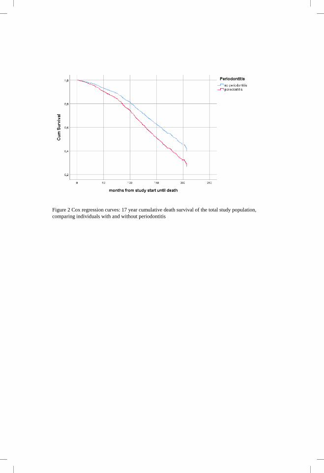
Figure 2 Cox regression curves: 17 year cumulative death survival of the total study population,
comparing individuals with and without periodontitis


MALMÖ UNIVERSITY
205 06 MALMÖ, SWEDEN
WWW.MAU.SE
isbn 978-91-7877-020-5 (print)
isbn 978-91-7877-021-2 (pdf)
isbn 978-91-87973-41-3 (Kristianstad)
VIV
EC
A W
AL
LIN
BE
NG
TS
SO
N
MA
LM
Ö U
NIV
ER
SIT
Y 2
01
9P
ER
IOD
ON
TIT
IS, C
AR
OT
ID C
ALC
IFIC
AT
ION
S A
ND
FU
TU
RE
CA
RD
IOV
AS
CU
LA
R D
ISE
AS
ES
IN O
LD
ER
IND
IVID
UA
LS