visual system development neural activity
TRANSCRIPT

Visua l S y s t e m Deve lopmen t andNeura l Activity
A. E. WIENCKEN-BARGER and V. A. CASAGRANDEVanderbilt University School of Medicine
IntroductionOverview of Visual System Development Axon Pathfinding: Wiring the LGN and Visual Cortex
IV. Conclusions
GLOSSARYamblyopia The impairment of vision without detectable organic lesion of the eye.
ectopic Positioned abnormally within the body.
neurotrophic factor A molecule, usually a protein, that willfacilitate the growth or repair of nerve cells.
ocular dominance column An area in the visual cortex thatreceives input predominantly from one eye.
transcription factor A protein required for recognition by RNApolymerases of specificstimulatory sequences in eukaryotic genes.
visuotopy The arrangement of cells and the connections betweenneural structures such that they maintain a topographic representa- tion of the visual field.
Neural structures are specified very early when the future nervous system is still a sheet of cells referred to as the neural plate and before this sheet folds to form the neural tube. These early steps involve evolutionarily conserved inductive signaling pathways that initiallyestablish regional identity. Subsequently, at the timewhen cells undergo their final cell division, the cellswithin different regions of the visual system becomecommitted to their specific fates or individual
Encyclopediaof the Human BrainVolume 4 791
ties. Evidence from studies in a variety of animalssuggests that molecular gradients, timing of axonarrival, and correlated spontaneous activity all help toshape early targeting decisions and to establish precise connections. Refinements of the system involve active growth and branching of axons and dendrites, forma-tion of synapses, and elimination of cells, axoncollaterals, and some synapses. Further refinementsof visual system development depend on activity andcompetition for limited supplies of neurotrophicfactors but do not necessarily require visual experi-ence. However, visual experience, especially duringcritical periods of active growth, can dramatically modify the final outcome.
INTRODUCTION
Mammalian development is an elegant process bywhich a single cell becomes a complete organismcontaining multiple organ systems that are highlyinterconnected. This is never more evident than duringneural development. A central issue in the study ofneurobiology concerns not only how nerve cellsbecome connected in the first place but also how thoseconnections become organized in such a way that thesensory world is represented. The topographical specificity of different parts of the mammalian visualsystem is dependent on highly ordered connections. Consider that the mammalian brain contains atleast 100 billion neurons and that each of these
Copyright2002, Science (USA).All rights reserved.

792 VISUAL SYSTEM DEVELOPMENT AND NEURAL ACTIVITY
neurons can make more than 1000specificconnectionswith other neurons. Specificity is particularly evident in the visual system, in which a topographic map ofvisual space is maintained throughout each level of visual processing. For example, in the macaque monkey, a modest estimate of the number of visualareas requires that at least 30 visuotopic maps connectcorrectly in the developing brain; in humans there may be even more visual areas. Each cell within one of thesemaps processes information from a specific zone of thevisual world, and that cell’s neighbor processes information from an adjacent zone and so forth.Neighboring cells connect with neighboring cells inother visual areas. This characteristic is called
One approach that may be useful in elucidating thecomplex wiring of the mammalian visual system is toassume that make connections through asequence of simpler evolutionarily conserved mechan-isms. In mammals, research suggests that the earlieststages in the development of the nervous system aresimilar across a range of diverse species and involvesimilar if not identical molecular pathways. The similarities in these early steps of neural developmentimply the existence of powerful constraints on the regulatory relationships between genes that controlearly phenotypes the characteristics and appear-ance of different parts of the nervous system). Goodexamples of these relationships come from geneticstudies of eye and head development in flies
mice, and humans. In mice and humans the paired gene and its in flies,the eyeless gene, play major roles in eye andfacial development. These genes control transcription factors that regulate a cascade of other genes impor-tant for eye and head formation. Whenfunction mutations are produced in the eyeless gene,flies develop with no eyes, very reduced eyes, ordefective eyes. Similar phenotypes are seen withgenetic mutations in the gene in miceand humans. Astoundingly, ectopic expression ofeither the fly eyeless gene or the pax 6 gene results inthe development of fully formed insect eyes on parts ofthe mutant fly’s body that do not normally have eyes,such as the leg or wing. These remarkable results notonly argue for a common evolutionary origin of eye development, but also reinforce the view that theearliest developmental programs are governed byhighly conserved rules.
Although it is not the purpose of this article to focuson these early steps in development, the previousexample has relevance. Later steps in neural develop-
ment, in general, and visual system development, in particular, also involve evolutionarily conserved me-chanisms. Since flies, mice, and humans differ inorganization, size, and complexity of the brain, it isobvious that there must be developmental differences. Nevertheless, dramatic differences in adult brainorganization can involve small changes in basic developmental mechanisms, such as changes in the number of cell divisions that occur before founderpopulations stop dividing, or changes in the number ofcells that are eliminated during the periods of celldeaththat occur as a part of normal development in all nervous systems. The role of other mechanisms such asneural activity may be more important to the devel-opment of connections in large, complex nervous systems, although the basic mechanisms that translatethat activity into cell growth or the formation ofsynaptic connections between nerve cells are alsoconserved across species.
In this article, we limit our consideration ofdevelopment to two interconnected and well-studiedvisual centers in the brain, the lateral geniculatenucleus (LGN) in the thalamus and the primary visualcortex or (Fig. 1). First, we provide an overview ofthe development of these visual areas in the largercontext of brain development and discuss how these structures take on their adult shape. Then, we showhow specialized laminar patterns and topographicconnections between these structures can developaccording to molecular cues. Finally, we explorepossible mechanisms for the establishment of specifi-city, including the role of neural activity in forming andmaintaining connections.
OVERVIEW OF VISUAL SYSTEM DEVELOPMENT
A. Early Neural Development
The brain begins as a simple plate of progenitor cellsthat eventually forms a tube that bends and balloonsout into three fluid-filled vesicles during the process ofdevelopment. Even before the neural plate forms a tube, however, communication takes place between cells that determines which progenitor or founder cellswill give rise to specific broad regions of the visualsystem, including regions that contain the retina, the LGN, and the visual cortex (Fig. 2). An enormousarray of transcription factors and extracellular mole-cular signals have been identified that are involved in

VISUAL SYSTEM DEVELOPMENT AND NEURAL ACTIVITY 793
Optic tract
Hypothalamus:regulation ofcircadian rhythms
Pretectum:reflex control ofpupil and lens
Superior colliculus: orienting movements of head and eyes
The visual pathways shown as a schematic viewed from the ventral surface of the human brain. The axons from ganglion cellslocated in the retina leave the eyes as the optic nerve. The ganglioncells located in the temporal retina (shown for the right eye) send axons to thesame side (ipsilateral) of the brain (arrow), whereas the axons in the nasal retina cross at the optic chiasm to the other hemisphere orcontralateral side of the brain. Once the axons have left the optic chiasm they bundle together as the optic tract. Groups of axons leave the optictract at various points to make synapses with groups of cells (nuclei) conserved with different visual functions, such as the hypothalamus (circadian rhythms), the lateral geniculate nucleus (LGN); (conscious vision), pretectum (pupillary and lens reflexes), and superior colliculus (orienting head and eyes). The superior colliculus is homologous with the optic tectum of nonmammalian vertebrates. Cells in the LGN sendaxons to the primary visual cortex or striate cortex, also called located at the back of the brain in the occipital lobe [reproduced with permission from Purves, D., et (Eds) (1997).Neuroscience. Sinauer, Sunderland, MA].
these early stages of regional specification. It has beenhypothesized that regional specification of the fore-brain, like the hindbrain, involves segmentation, in thiscase into a series of longitudinal and transversesegments defined by the expression of a number ofgenes;however, specificmolecules that define the exactboundaries of the developing LGN or primary visual cortex have not been identified.
As development progresses, cells divide close to theinner (ventricular) surface of the neural tube. Initially, all cells extend their processes across the full width ofthe neural tube, but later only a subset of cells, theradial glial cells, span the full width of the developingforebrain. Radial glia are used by other cells asmigratory railway guides to move from the ventricularzone to their final destinations, whether in thedeveloping LGN or visual cortex. Each neural pro-genitor cell, or stemcell, undergoes a certain number ofcell divisions in the young animal, until it divides nomore in the adult. This final mitotic division is termedthe birth date of the neuron. After the finalcell divisiona neuron differentiates or matures. Neurons in differ-
ent areas of the brain, such as the LGN and visualcortex, exhibit differences, reflecting their variable differentiation programs. Cells within different neuralstructures mature at different times in the develop-mental process and at different rates. Additionally, each neural structure may have its own gradient ofdevelopment, such that some cells in the nucleusmature before others. Finally, as neural structures mature many developing neurons are eliminatedthrough cell death. This natural cell death processnot only eliminates cells with incorrect connections butalso allows the production of transient cell populationsthat help to shape the structures and connections of thenervous system by providing temporary scaffoldingfor migrating neurons and guideposts for developingaxons. The development of the mammalian brain is ahighly dynamic process involving multiple overlap-ping gradients of progression. Cells in the brain experience multiple combinatorial genetic and activ-ity-dependent cues at different times in their develop-ment that ultimately produce the complex structure ofthe adult mammalian brain.

794 VISUAL SYSTEM DEVELOPMENT AND NEURALACTIVITY
A. Three-vesicle stage
Proencephalon
Mesencephalon
Rhombencephalon
Caudal neural tube
CervicalD.
flexure
B. Five-vesicle stage C.ILateralventricle
Third ventricle
Fourth ventricle (pons and cerebellum)
Spinal cord
Forebrain
Midbrain
Hind brain
Figure Successivestagesof developmentof the neural tube. The nervous system begins as a plate of tissue (not shown)that rollsup to form atube. (A) Three-vesicle stage. At early stages of development only three brain vesicles are present. (B) Five-vesicle stage. At later stages, two additional vesicles form, one in the area of the forebrain (la and lb) and the other in the hindbrain (3a and 3b). Before the neural tube forms,cells are prespecified such that the retina and lateral geniculate nucleus will form from part of the diencephalon the visual cortex from thetelencephalon (la), and the superior colliculus from the midbrain (2). (C) Micrograph showing a dorsal view of the neural tube at an early stage of development. The expansion of the future telencephalicvesicle is apparent (micrograph of the chick neural tube provided by G.(D)The positions of the cephalic, pontine, and cervical flexures [from Kandel, E. R., Schwartz,J. H., and T. M. (Eds.) (2000).Principlesof Neural Science, 4th ed. Reproduced McGraw-Hill Companies, with permission of the New York].
B. Development of LGN Structure
The adult LGN of many mammals, including humans, is laminated with compartmentalized input. EachLGN layer receives input from only one eye. Eachlayer also contains a topographic map of one-half ofthe visual world. The LGN layers are stacked on top ofeach other like a set of pancakes (Fig. 3). In primatesthere is layer specialization such that small, medium, and large cells are segregated into different layers andcarry different types of visual messages to primaryvisual cortex. During development, the future largeLGN cells are born and begin migrating from the ventricular surface toward their final destination before the future smaller LGN cells. The differing extracellular environment encountered by LGN cells
born at different times during development may change the genetic program of those cells, such thatthe different classes of cells adopt different morpho-logical and neurochemical identities during differen-tiation. In fact, in visual cortex there is evidencesupporting the view that important decisions aboutcell fate are made at the time progenitor cells divide inthe ventricular zone. It is likely that the same rules apply to cells throughout the developing neural tube.During development, LGN progenitor neurons des-tined to represent central vision versus peripheral vision divide and mature in a gradient such that cellsthat will carry information about the central visual world develop earlier than cells that will carryinformation from the visual periphery. Gradients inthe timing of cell birth and maturation of LGN

VISUAL SYSTEM DEVELOPMENT AND NEURAL ACTIVITY
A. Lateral geniculate nucleus B. Striate cortex
\
795
Figure The connections between the lateral geniculate nucleus (LGN) and striate cortex (primary visual cortex). The visual pathway isshown as a schematic viewed from the ventral surface of the human brain. This same view is also shown in Fig. 1.Although the LGN receivesinputs from both eyes, these are segregated in separate layers as shown in (A)The first two layers contain large cells (magnocellular layers), andthe remaining four layers (parvocellular cells)contain medium-sizedcells.Not shown are the smallcell (koniocellar) layers that lie between each of the main layers shown. (B) In many species, and most primates, the inputs from the two eyesremain segregated in ocular dominancecolumnsof layer IV, the primary target of LGN axons. Layer IV neurons send their axons to other cortical layers; it is at this stage that information from the two eyes converges onto individual neurons [reproduced with permission from Purves, D., et al., (Eds) (1997). Neuroscience. Sinauer,Sunderland, MA].
progenitors and within any structures connected to theLGN (such as the eye and the primary visual cortex) may be one of several simple mechanisms that ensurethat topographically correspondent regions connect properly.
The retina and the visual cortex, the main inputs tothe LGN, both innervate the LGN visuotopically.
There is evidence that axons leaving visual cortex andretina grow out very early but may not reach their targets at the same time. In monkeys, retinal ganglion cell axons of different classes grow to the LGN veryearly and terminate immediately within theate portions of the LGN, demonstrating that someretinal axons are molecularly specified to connect to

796 VISUAL SYSTEM DEVELOPMENTAND NEURAL ACTIVITY
specific LGN target regions. However, retinal gang-lion cell axons of the same class, arriving from the twoeyes initially invade overlapping regions of the LGN.The segregation of these axons into eye-specificlayersseems to depend on correlated waves of spontaneousactivity in the retina sinceblocking such activity blocks the normal segregation of retinal axons, at least in carnivores. Cortical axons, like retinal axons, ap-proach the LGN very early in development but, unlikeretinal axons, do not invade the nucleus immediately. A special early-born class of cortical cells, the cortical
cells, may be the first to send axons to thethalamus. These cells may pioneer the later-born layerVI cell axons that will form permanent connectionswith the LGN; however, the pioneer axons must still find their way to the LGN. Evidence suggests that theearliest axons use a variety of membrane-bound cuesto locate correct targets. When cortical axons doinvade the LGN, they immediately grow into
correct areas of the nucleus and do not appearto require a sorting period, although some smaller refinements may take place as these axons mature.
C. Development of Visual Cortical Structure
Multiple cortical areas, defined by morphology, phy-siology, and connections with other cortical andsubcortical nuclei, make up the neocortex, like apatchwork quilt. One patch of the quilt is the primary visual cortex, located in posterior or occipital cortex. The adult visual cortex contains a precise topographicmap and, like all of mammalian cortex, is laminated.The neocortex has six layers. Axons arriving from the LGN synapse primarily in layer IV of visual cortex. Inprimates, axons from LGN cells of different functionalclasses large versus small LGN cells) synapse in different divisions of cortical layer IV or in distinctcompartments located above layer IV. Additionally, LGN cells representing the left and right eye sendaxons to separate columns of cortex such that cells in aparticular column of cortex respond to a stimulus toone eye or the other preferentially (Fig. 3). Cells inlayer IV make connections with layers above andbelow layer IV, and this is where the fusion ofinformation from the two eyes occurs. Cortical cellsin layer VI send axons back to the LGN. Also, each ocular dominance column is divided into smallercolumns in which cells respond to one orientation ofa bar of light. Orientation columns are arrangedmainly in pinwheels, such that all orientations are roughly represented within each ocular dominance
domain and visuotopy is maintained across columns (Fig. 4). The aforementioned scenario is more simplis-tic than the biological situation but serves to empha-size some of the complexitiesof organization that mustbe achieved during development for the visual cortex to function properly.
As does the developing LGN, the developing cortex begins as a simple epithelium-a thin columnar sheetof cells.As development advances and cells divide, this sheet of cells becomes thicker. The radial glial cellsmaintain contact with both the ventricular and thesides of this sheet, whereas cortical neuroblasts begindividing at the ventricular zone. Soon, threetical subdivisions can be recognized. The ventricularzone is the most ventral, where excitatory cortical cellsare born from a resident stem cell pool. Interestingly,inhibitory cortical cells, like olfactory bulb cells, mayoriginate from a noncortical stem cell populationlocated in the ganglionic eminence the developingbasal ganglia). Above the ventricular zone is theintermediate zone, a highway for axons, wherecofugal and corticopetal axons are located. Above theintermediate zone is the preplate, a layer of early borncells that have migrated along radial glial fibers to lie
Figure Orientation columns in the visual cortex of the monkey.Comparison of optical image maps of orientation preference andocular dominance in monkey visual cortex. The thick black lines represent the borders between ocular dominance columns. The thin gray lines represent isoorientation contours, which converge atorientation pinwheel centers (arrow). Each left and right eye oculardominance column represents a common region of visual space. Anadjacent set of columns represents an adjacent region of visual space and so forth across the visual cortex [adapted with permission fromObermeyer, K., and Blasdel, G. G. (1993). Geometry of orientationand ocular dominance columns in monkey striatecortex. J. Neurosci.13,
VISUAL SYSTEM DEVELOPMENT AND NEURAL ACTIVITY 797
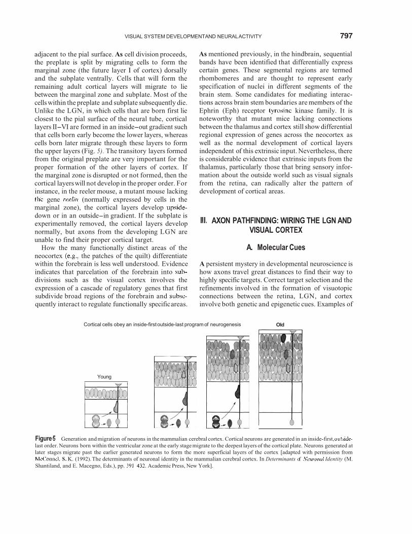
adjacent to the pial surface. As cell division proceeds, the preplate is split by migrating cells to form the marginal zone (the future layer I of cortex) dorsally and the subplate ventrally. Cells that will form the remaining adult cortical layers will migrate to liebetween the marginal zone and subplate. Most of thecells within the preplate and subplate subsequently die. Unlike the LGN, in which cells that are born first lieclosest to the pial surface of the neural tube, cortical layers II-VI are formed in an inside-out gradient suchthat cells born early become the lower layers, whereas cells born later migrate through these layers to formthe upper layers (Fig. 5). The transitory layers formed from the original preplate are very important for the proper formation of the other layers of cortex. Ifthe marginal zone is disrupted or not formed, then the
As mentioned previously, in the hindbrain, sequentialbands have been identified that differentially express certain genes. These segmental regions are termedrhombomeres and are thought to represent early specification of nuclei in different segments of thebrain stem. Some candidates for mediating interac-tions across brain stem boundaries are members of theEphrin (Eph) receptor kinase family. It isnoteworthy that mutant mice lacking connections between the thalamus and cortex still show differential regional expression of genes across the neocortex aswell as the normal development of cortical layersindependent of this extrinsic input. Nevertheless, there is considerable evidence that extrinsic inputs from the thalamus, particularly those that bring sensory infor-mation about the outside world such as visual signals
cortical layers will not develop in the proper order. Forinstance, in the reeler mouse, a mutant mouse lacking
gene (normally expressed by cells in themarginal zone), the cortical layers develop
from the retina, can radically alter the pattern ofdevelopment of cortical areas.
AXON PATHFINDING: WIRING THE LGN ANDVISUAL CORTEX
down or in an outside-in gradient. If the subplate isexperimentally removed, the cortical layers develop normally, but axons from the developing LGN areunable to find their proper cortical target.
How the many functionally distinct areas of theneocortex the patches of the quilt) differentiate
A. Molecular Cues
within the forebrain is less well understood. Evidence A persistent mystery in developmental neuroscience isindicates that parcelation of the forebrain into how axons travel great distances to find their way todivisions such as the visual cortex involves the highly specific targets. Correct target selection and theexpression of a cascade of regulatory genes that first refinements involved in the formation of visuotopicsubdivide broad regions of the forebrain and connections between the retina, LGN, and cortexquently interact to regulate functionally specificareas. involve both genetic and epigenetic cues. Examples of
Cortical cells obey an inside-first outside-last program
Young
of neurogenesis Old
Figure Generation and migration of neurons in the mammalian cerebral cortex. Cortical neurons are generated in an inside-first,last order. Neurons born within the ventricular zone at the early stage migrate to the deepest layers of the cortical plate. Neurons generated atlater stages migrate past the earlier generated neurons to form the more superficial layers of the cortex [adapted with permission from
S. K. (1992).The determinants of neuronal identity in the mammalian cerebral cortex. In Determinants of Identity (M.Shantiland, and E. Macegno, Eds.), pp. Academic Press, New York].

798 VISUAL SYSTEM DEVELOPMENT AND NEURAL ACTIVITY
genetic mechanisms include the presence of mem-brane-bound and secreted molecules, which are per-missive, instructive, or obstructive cues for axons withthe proper receptors. Axons can grow on varioussubstrates in the brain, including the extracellularmatrix, other neural cell membranes, and glial cell membranes (such as the radial glia on which LGN andcortical cells migrate). Developing axons have aspecialized structure, a growth cone, at their tip tohelp them navigate through the extracellular milieu(Fig. 6). This highly motile structure contains recep-tors capable of transmuting signals about the localenvironment. The growth cone guides the axon alongpathways toward the target region defined by acombination of positive or negative cues.
Once an axon reaches its target, it often will send outa simple T-shaped branch as shown for the LGN axongrowing into visual cortex in Fig. 7. Within the visualsystem, axons often reach their target region while thetarget cells are still migrating into position or just aftermigration is complete. Growth cones on the tip of axonbranches can detect gradients of membrane-boundmolecules to guide axons to their general addresses within a target structure. Axons respond differentially to these molecular gradients within the target based on their initial specification that likely occurred at thetime of final cell division and prior to axon outgrowth.
Figure 6 An example of a thalamocortical axon with a growthcone in neonatal mouse brain. The growth cone is a complex,dynamic structure at the tip of the growing axon that samples the local environment, guiding the axon to its target.
Figure Thalamocortical initially exhibit collateral T-shaped side branches when they begin to innervate the cortical plateas shown here from the neocortex of a mouse on the day of birth.
Retinal ganglion cell axons from different retinal locales will therefore respond differently to cues at thetarget based on their unique growth cone receptors. As mentioned previously, a good example of this process involves the specificity with which different retinal ganglion cell classes target regions of the LGN.Although many molecules have been identified thatmay guide retinal ganglion cell axons to the righttargets, evidence has converged on two membrane-bound ligands of the receptor kinases, ephrinA2 and ephrin A5. Both of these ligands are expressedin gradients in the optic tectum, another brain area innervated by retinal ganglion cells. Eph kinases aredistributed in gradients in the retina. These twomolecular gradients may regulate retinotectal andretinogeniculate topography because ephrins bind toEph kinases and activation of Eph kinases inhibits axon outgrowth. In particular, the levels of ephrin A2and A5 are higher in the posterior tectum than in theanterior tectum, and this could inhibit growth oftemporal retinal axons, which are rich in the appro-priate Eph kinases (Fig. 8). Moreover, retinal axons in mice with mutations in ephrin A2 and A5 exhibit a lossof visuotopy and innervate areas of the optic tectumthat they normally would avoid. Interestingly, mem-bers of this same family of ephrin molecules areimplicated as cues for establishing the proper laminar connections in visual cortex. Therefore, members ofthe ephrin family may be involved in early regional

VISUAL SYSTEM DEVELOPMENT AND NEURAL ACTIVITY
A P
Figure8 Eph kinases are distributed in gradients in the retina, andephrins are distributed in gradients in the optic tectum. These two molecular gradients may regulate retinotectal visuotopy because ephrins bind to eph kinases and activation of eph kinases inhibits axon outgrowth.
differentiation in the brain, as well as the establishmentof axonal connections later. This example illustrates how a single family of similar molecules can directseveral very different developmental programs, some-times simultaneously, in different neural regions andwith overlapping expression patterns, and it under-scores the high level of conservation of mechanisms inneural development.
B. The Role of Activity
As neurons mature, they begin to display electrical activity. Neural activity has been shown to play animportant role in the correct targeting of axonalconnections as well as the formation and maintenanceof synapses. The role of neural activity in shapingconnections is not restricted to the postnatal periodwhen animals begin to use their senses to explore their environment. Neurons are active well before birth,especially in animals such as primates that are born at arelativelymature developmental stage. Experiments incats and ferrets have demonstrated that prior tovisually driven activity (at a time when the eyelids arestill closed), correlated waves of spontaneous activityalready exist in the retina and brain. Neurons at thisstage are coupled by developmentally transient gapjunctions (at least in the retina and the visual cortex, but probably also within other areas of the visualsystem). The waves of spontaneous activity appear tobe important to the segregation of left and right eyeinputs within the developing LGN layers and to thesegregation of eye-specific inputs into ocular dom-inance columns in cortex. As mentioned earlier, blocking this activity in prenatal cats and postnatalferrets disrupts the binocular segregation process. Inthe examples cited previously, activity likely affects theconstruction of new axonal branches and synapsesandnot the retraction of inappropriate connections since
the manipulations are inigeniculocortical axons stillmorphology.
During the period of act
iated when retinal andhave a simple stick-like
vity-dependent modifica-tion of connections, many manipulations are made tothe visual system in an effort to define the mechanismsthat relate activity to the growth of neural processesand to synaptogenesis. The most famous of theseexperiments were done in the 1960s by David Hubel and Wiesel, who showed that preventing one eye of a kitten from seeingduring early life resulted in a number of dramatic changes in the connections andfunction of the visual system. In their experiments, they sutured the lid of one eye closed, but it is nowknown that the same effects are obtained as long asuseful pattern vision is prevented; the amount of lightreaching the retina is not as important. In theseexperiments, they found that kittens could no longersee well out of the deprived eye they developed amblyopia) if the deprivation continued past 3 monthsof age. These investigators also found thatcortical axons from the deprived eye occupied lessterritory in layer IV of visual cortex (the deprived ocular dominance column was smaller) and cells in the cortex now responded mainly to the normal eye. If thekittens were allowed to develop normally for the first 3months of age, monocular lid suture would no longeraffect ocular dominance columns, suggesting thatthere is a window of time or critical period duringwhich visual experience can have an impact on oculardominance column formation. If the eye was suturedand shortly afterwards opened again during the criticalperiod, then normal ocular dominance formationwould occur. The effects of monocular deprivation are not the consequences of disuse, as Hubel andWiesel clearly demonstrated, since depriving both eyesof useful pattern vision has much milder effects on thevisual system (Fig. 9).
These experiments, along with numerous experi-ments that followed, suggest that developinglocortical axons that receive input from the left andright eyescan compete with one another for territory incortex. The working hypothesis is that appropriatelevels of activity and the temporal correlation of thatactivity in potential pre- and postsynaptic partnersprovide axons access to appropriate levels of growthfactors neurotrophins). These neurotrophins, in turn, activate the cellular machinery that allows axonsto grow and develop synapses (Fig. 10).Axon growthis clearly constrained by many other factors in thetarget tissue discussed earlier, including factors thatdirectly repel or attract axons or substrates that

800 VISUAL SYSTEM DEVELOPMENT AND NEURAL ACTIVITY
Ocular Dominance Column Plasticity Normal Development Monocular Suture
MechanismsI
1Left
2 3 4 5 6 7Right
Numbersof cells in Layer drivenby the left (1) and right (7)eyes
1 2 3 4 5 6 7Left Right
Figure Changes within the LGN and visual cortex (area 17) seen followingmonocular lid suture in a macaque monkey. Axons from the leftand right eyessegregateinto six layers within the LGN early in development. LGN axons segregate into ocular dominancecolumns within layer IV of area 17. Axon segregation within the LGN and cortex takes place before birth, without visual experience. However, correlated spontaneous activity initially within each eye and subsequently via connections between segregated eye inputs in the LGN and cortex including corticogeniculate feedback may help axons segregate into ocular dominance territories. At birth, LGN axons are very immature. Neonatal lidclosure eliminates useful patterned activity within the sutured eye.As a result, deprived LGN axons grow less and their LGN cell bodies shrink,and nondeprived LGN axons expand more than normal. Since deprived LGN cell bodies within the monocular segments of the LGN do notshow these changes, it is likely that LGN axons innervated by the left (L) and right (R) eyes compete within cortex for limited quantities ofneurotrophic factors. Evidence suggests that the neurotrophins, brain-derived neurotrophic factor (BDNF) or neurotrophin (NT) areinvolved, as shown in the center. These neurotrophins bind to kinase receptor B (Trk-B), which can be located both andpostsynaptically. In this model activation of the cortical cell by axons would cause release of BDNF or which would act to promotegrowth and survival of the LGN axon. Neurotrophin release could also influence the dendritic growth of cells from which it was released via autocrine mechanisms [adapted with permission from Barker, R. A., and Barasi, S., (with Neal, M. J.) (1999). Neuroscience at a GlanceBlackwell Science, Oxford].
support growth. In addition, it is evident that patternvision per se is not essential for setting up oculardominancecolumns in the first place sincemonkeys areborn with well-developed ocular dominance columns. Nevertheless, basic cellular mechanisms that normallyhelp axons driven by the left and right eyes to segregateinto layers in the LGN and into columns in the cortex
are likely the same as those that allow for plasticchanges following manipulations of visually driven activity.
How does activity help some developing axons togrow processes or form or strengthen some synapses atthe expense of others? A working model for thisprocess was originally derived from the learning model
VISUAL SYSTEM DEVELOPMENT AND NEURAL ACTIVITY 801
Figure Selective access to growth factors can influence thesurvivalof neurons. Neurons (circles)compete for a limited supply ofgrowth factors (triangles). Neurons that do not get enough of thegrowth factor may die or have smaller axonal trees.
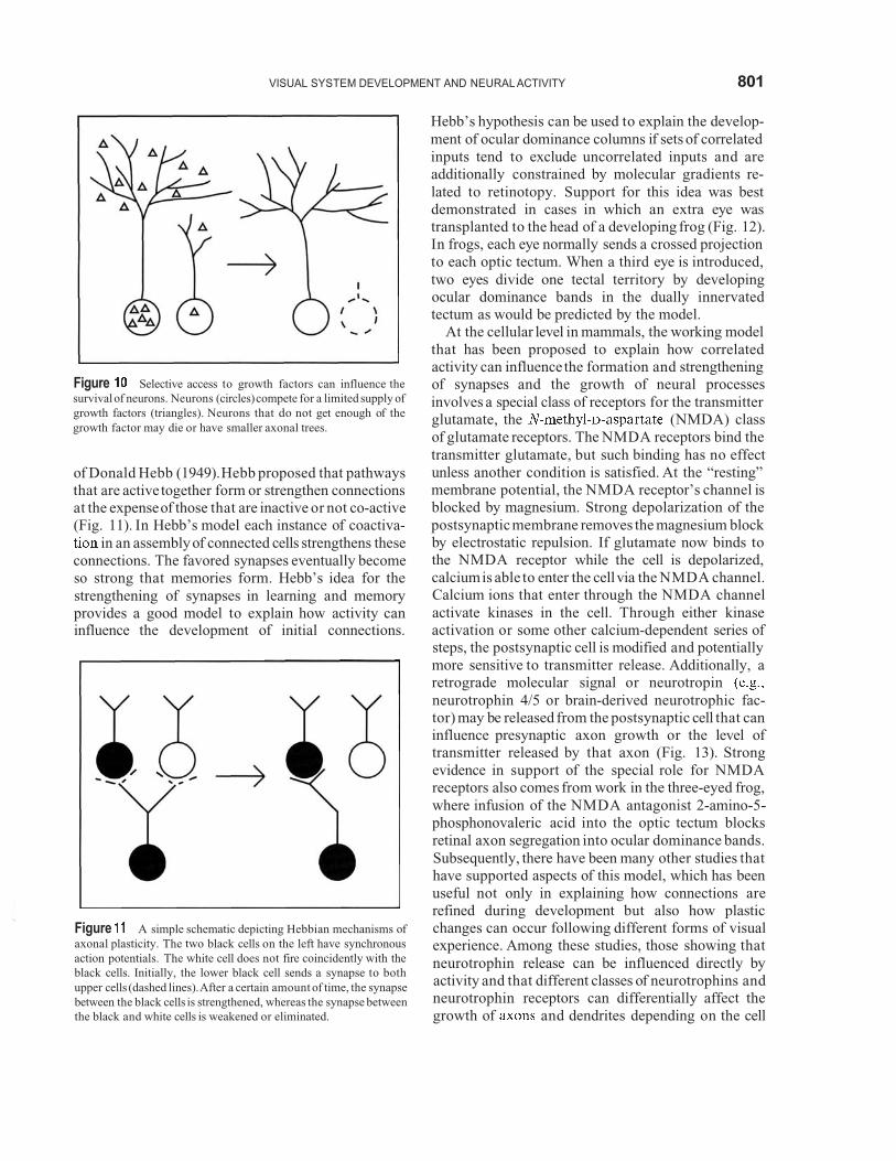
of Donald Hebb (1949).Hebb proposed that pathwaysthat are active together form or strengthen connections at the expenseof those that are inactive or not co-active(Fig. 11). In Hebb’s model each instance of coactiva-
in an assemblyof connected cells strengthens these connections. The favored synapses eventually become so strong that memories form. Hebb’s idea for the strengthening of synapses in learning and memoryprovides a good model to explain how activity caninfluence the development of initial connections.
Figure A simple schematic depicting Hebbian mechanisms ofaxonal plasticity. The two black cells on the left have synchronous action potentials. The white cell does not fire coincidently with the black cells. Initially, the lower black cell sends a synapse to bothupper cells (dashed lines). After a certain amount of time, the synapsebetween the black cells is strengthened, whereas the synapse between the black and white cells is weakened or eliminated.
Hebb’s hypothesis can be used to explain the develop-ment of ocular dominance columns if sets of correlatedinputs tend to exclude uncorrelated inputs and areadditionally constrained by molecular gradients re-lated to retinotopy. Support for this idea was best demonstrated in cases in which an extra eye wastransplanted to the head of a developing frog (Fig. 12).In frogs, each eye normally sends a crossed projection to each optic tectum. When a third eye is introduced,two eyes divide one tectal territory by developingocular dominance bands in the dually innervatedtectum as would be predicted by the model.
At the cellular level in mammals, the working model that has been proposed to explain how correlatedactivity can influencethe formation and strengtheningof synapses and the growth of neural processesinvolves a special class of receptors for the transmitter glutamate, the (NMDA) classof glutamate receptors. The NMDA receptors bind thetransmitter glutamate, but such binding has no effectunless another condition is satisfied. At the “resting”membrane potential, the NMDA receptor’s channel isblocked by magnesium. Strong depolarization of thepostsynaptic membrane removes the magnesium block by electrostatic repulsion. If glutamate now binds tothe NMDA receptor while the cell is depolarized,calcium is able to enter the cell via the NMDA channel.Calcium ions that enter through the NMDA channelactivate kinases in the cell. Through either kinase activation or some other calcium-dependent series ofsteps, the postsynaptic cell is modified and potentiallymore sensitive to transmitter release. Additionally, aretrograde molecular signal or neurotropinneurotrophin 4/5 or brain-derived neurotrophic fac-tor) may be released from the postsynaptic cell that caninfluence presynaptic axon growth or the level of transmitter released by that axon (Fig. 13). Strongevidence in support of the special role for NMDAreceptors also comes from work in the three-eyed frog, where infusion of the NMDA antagonist 2-amino-5-phosphonovaleric acid into the optic tectum blocks retinal axon segregation into ocular dominance bands. Subsequently, there have been many other studies thathave supported aspects of this model, which has beenuseful not only in explaining how connections arerefined during development but also how plastic changes can occur following different forms of visualexperience. Among these studies, those showing thatneurotrophin release can be influenced directly byactivity and that different classes of neurotrophins andneurotrophin receptors can differentially affect thegrowth of and dendrites depending on the cell

VISUAL SYSTEM DEVELOPMENT AND NEURALACTIVITY
Figure The three-eyed frog. (A)Three-eyed frogs have been studied by Martha Constantine-Paton and colleagues. (B) An autoradiographof the optic tectum showing the formation of stripes of inputs (black and white) from the normal and implanted eye. The inset shows anenlargement under dark-field illumination [adapted with permission from Constantine-Paton, M., and Law, M.I. (1978). Eye-specifictermination bands in tecta of three-eyed frogs Science 202, 639-641. Copyright 1998 American Association for the Advancementof Science].
type are especially relevant because they link activity with other basic cellular mechanisms involved in cellsurvival and selective outgrowth and targeting ofneural processes. In addition to neurotrophic factors and activity-dependent mechanisms, a variety of otherfactors can impact the development of visual systemconnections, including, but not limited to, levels of various hormones, a variety of neurotransmitters, andneuromodulators .
For the examplesgiven previously, it is important tokeep in mind that although ocular dominancecolumnsdo not require visual experience to form in the firstplace (at least in primates), there are other aspects ofvision that do require appropriate visual experience and are not present before birth even in primates.Stereoscopic depth perception, for example, appearsto require adequate coordinated activity from botheyes since it does not exist at birth and becomesdisrupted if input from the two eyes is not appro-priately correlated. Such a situation can arise in humans or other animals that are born and grow upwall-eyed or cross-eyed. In the latter case, stereoscopic depth perception is lost and binocular connections donot form. Only if the eyes are surgically aligned early in a child’s life will he or she develop normal stereoscopic
vision. Nevertheless, the same activity-driven mechan-isms described previously likely operate to allowappropriate binocular connections to form in visualcortex that, in turn, help us to appreciate our three-dimensional world.
CONCLUSIONS
During neural development, cells proliferate andinfluence each others’ fates through an elaborateinterplay of extrinsic and intrinsic signals. Cellsbecome committed to specific fates, such as laminar location in visual cortex, early in development before they begin to migrate. Molecular axon pathways areestablished through a variety of secreted factors andmembrane-bound molecules. Correct axon targetinginvolves gradients of chemoattractant and
molecules.Major waves of cell death take placeat this stage that help to sculpt connections. Unlike spontaneous neural activity, activity driven by visualexperience is not required for basic aspects of axontargeting, map formation, and segregation of into ocular territories within the LGN or visual cortex in precocial species such as primates. In all species,

803VISUAL SYSTEM DEVELOPMENT AND NEURAL ACTIVITY
NUCLEUS
Receptor Receptor
Neuriteelongation
receptor
Receptor
PKC Kinase
POSTSYNAPTICFigure 13 The NMDA receptor channel can open only during depolarization of the postsynaptic neuron from its normal resting level. Depolarization expels (not shown) from the NMDAchannel, allowing current to flowinto the postsynaptic cell. Since theNMDA channel is permeable to there is a significantentry into the cell that can trigger other events involving Through either kinase activation (protein kinase C) or a separate
mechanism (calmodulin kinase or other path-ways, neurotrophins can be released from the postsynaptic cell thatcan act in a paracrine fashion to influence growth of the presynaptic
or in an autocrine fashion to influence its own growthvia pathways shown in the diagram for the presynaptic Binding to neurotrophin Trk receptors permits Trk molecules tophosphorylate residues. Phosphorylation of specificosine residues creates binding sites for PI-3 and phospholipase C
and recruitment of these proteins into a complex, thusinitiating a signaling cascade that can lead to neurite elongation or,via the MAP kinase pathway, to transcription and ultimatelydifferentiation and growth [from Casagrande, V. A., and Wiencken,A. E. (2000). Developmental plasticity in the mammalian visual system. In The Mutable Brain. Dynamic and Plastic Features. (J. H.Kaas, Ed.), Copyright 2000 by Overseas Publishers Association Adapted with permission from Gordon and Breach Publishers].
however, visual system wiring can be profoundlyaffected by abnormal visual experience, especiallyduring the phase of rapid axonal and dendritic growth.
An elaborate interplay between genetic andnetic factors defines specific connections in the highly
complex system that is the mammalian nervoussystem. Molecular cues are important for guiding
to their general target region and neural activityrefines these connections. Even if genetic factorsinitially define axonal boundaries, it is not necessarilythe case that these boundaries are static. The bound-aries are partially plastic into adulthood and can bechanged even in adults as the result of insult or injury.Neural activity can affect the growth of neuralprocesses and synapses or strengthen existing synapses through Hebbian activity-dependent mechanisms and/or the limited availability of neurotrophins. Thesesame Hebbian mechanisms allow for more limitedplasticity in the wiring of the adult brain. Majorchanges in wiring only occur when processes aregrowing, but less dramatic changes are possible in theadult and likely involve the same cellular machinery that is used to establish and refine connectionsnormally. The mature visual system at all levels canrespond via expression of immediate early genes andregulation of a variety of transmitter andtide-related molecules to conditions of visual depriva-tion and damage. Despite the striking similarities thatare found between developmental and adult neuralplasticity, there are fundamental differences in thedeveloping and adult brain. Younger organismscharacteristically seek a variety of forms of visualstimulation and repeat visuomotor activities in waysmore sedentary adult animals do not. Adult animalsthat are forced to use their visual systems via trainingshow greater compensation for early damage thanadult animals that are not. Understanding the neural mechanisms that drive early visual self-stimulationand how these factors interact with thedependent mechanisms discussed to allow for com-pensation after visual system damage is one of thechallenges for future investigations.
See Also the Following Articles BRAIN DEVELOPMENT NEUROPLASTICITY, DEVELOP-MENTAL * SYNAPTOGENESIS * VISION: BRAIN MECHA- NISMS VISUAL CORTEX VISUAL DISORDERS
Suggested Reading
Casagrande, V. A., and Wiencken, A. E. (1996). Prenatal develop-ment of axon outgrowth and connectivity. Prog. Brain Res. 108,
Casagrande, V. A., and Wiencken, A. E. (2000). DevelopmentalPlasticity in the Mammalian Visual System. In The MutableBrain. Dynamic and Plastic Features (J. H. Kaas, Ed.), HarwoodAcademic, Reading, UK.
83-93.

804 VISUAL SYSTEM DEVELOPMENT AND NEURAL ACTIVITY
Castellani, V., Yue, Y., Gao, P. P., Zhou, R., and Bolz, J. (1998).Dual action of a ligand for Eph receptor kinases onspecificpopulations of during the development of corticalcircuits. J.Neurosci.
Chenn, A.,Braisted, J. E., S. K., and D. D. M.(1997). Development of the cerebral cortex mechanisms control-ling cell fate, laminar and areal patterning, and axonalconnectivity. In Molecular and Cellular Approaches to NeuralDevelopment (W. M. T. M. and S. L. Zipursky,Eds.), pp. Oxford Univ. Press, New York.
Cook, P. M., Prusky, G., and Ramoa, A. S. (1999). The role ofspontaneous retinal activity before eyeopening in the maturation of form and function in the retinogeniculate pathway of theferret. Vis. Neurosci. 16,491-501.
Florence, S. L., and Casagrande, V. A. (1990). Development ofgeniculocortical axon arbors in a primate. Visual Neurosci. 5,
Halder, G., Callaerts, P., and Gehring, W. J. (1995). Newperspectiveson eyeevolution. Opin. Genet. Dev. 5,602-609.
29 1-309.
D. H. (1988). Eye, Brain, and Vision. Sci. Am., New York.Irving, C., Flenniken, A., Allduc, G., and Wilkinson, D. G. (1996).
Cell-cell interactions and segmentation in the developingvertebrate. Biochem. Symp.
Kandler, K., and Katz, L. C. (1998). Coordination of neuronalactivity in developing visual cortex by gapbiochemical communication. Neurosci. 18, 1419-1427.
Katz, L. C., and Shatz, C. J. (1996). Synaptic activity and theconstruction of cortical circuits. Science 274, 1133-1138.
Rubenstein, J. L., Shimamura, K., Martinez, S., and Puelles, L.(1 998). Regionalization of the prosencephalic neural plate.Rev. Neurosci.
Shatz, C. J. (1990). Impulse activity and the patterning ofconnections during CNS development. Neuron 5,745-756.
Tessier-Lavigne, M., and Goodman, C. S. (1996). The molecularbiology of axon guidance. Science 274, 1123-1133.
Weliky, M., and Katz, L. C. (1999). Correlational structure ofspontaneous neuronal activity in the developing laterallate nucleus in Science 285, 599-604.