viruses not composed of cells. characteristics obligate intracellular parasites single type of...
TRANSCRIPT

Viruses
Not Composed of Cells

Characteristics
• Obligate intracellular parasites
• Single type of nucleic acid
• Protein coat
• Envelope
Two Forms
• Extracellular form
• Intracellular form

Host Range
• Can infect many hosts
• Determined by attachment to host cell– cell wall, flagella/fimbriae
• Receptor sites, or plasma membrane in animal cells

Structure
• Nucleic acid-core
• Either DNA or RNA, not both
• Follows central dogma of molecular biology– Genetic info flows from NA to protein
Structure
• Protein coat-capsid
• Envelope in some virions– May or may not have spikes-glycoproteins
Envelope
• Acquire membrane when budding or pass through membranes
• Advantages of lipid membrane
• Lose infectivity when envelope destroyed

Envelope
• Host’s phospholipids & viral proteins
• Disadvantage -damaged easily
• Without envelope- naked viruses– More resistant to chemicals/ disinfectants
Enzymes
• Required early in infection process
• Bacterial virus or bacteriophage
– lysozyme • Lysis of cell and release of virions

Enzymes
• Some have own NA polymerase
• RNA polymerase in some RNA virions
• Reverse transcriptase in retro viruses– RNA dependent DNA polymerase
• Neuramidases-release of virions
Morphology
• Helical
• Polyhedral- shape of icosohedron-20 sides
• Enveloped- usually spherical
Morphology
• Complex viruses– combination of helical and icosohedral– bacterial viruses-head and tail– poxviruses- several coats of protein
Growth of Bacteriophage
• Grow in suspensions of bacteria or in bacteria cultures on plate
• Plaque method for counting
Growth of Animal Viruses
• Living animals
• Embryonated eggs-influenza
• Cell cultures-continuous lines

CPE Cytopathic Effect
• Visible effect of viruses on cells
• Stop multiplication of cells
• Lysosomes release enzymes
• Inclusion bodies
• Syncytium-
• Interferons-

CPE
• Mark infected cells for destruction by immune system
• Transformation-abnormal cells

Multiplication of Bacteriophage
• Lytic cycle: produces virions
• T-even phages, virulent phages, on E. coli– ds DNA for over 100 genes-head– tail sheath-retracts– DNA moves from head into host–
Lytic Cycle
• Attachment stage (adsorption)– attachment site on virus with complementary
receptor on bacteria cell wall
– use fibers at end of tail as attachment sites– may attach to flagella or fimbriae

Penetration
• Injects DNA
• Tail releases enzyme lysozyme
• Tail core driven through cell wall
• Tail reaches cell membrane
• Capsid remains outside: uncoating

Biosynthesis
• In cytoplasm
• Host protein synthesis is stopped
• Uses host nucleotides and enzymes to synthesis copies of phage DNA

Biosynthesis
• Synthesis of phage capsid proteins
• Uses host ribosomes and amino acids for translation

Maturation
• Assemble into mature phages
• Head assembled and packed with DNA
• Phage tails assembled from plates, sheaths,
• Each head attached to tail
• Then fibers are attached
Release
• Lysis of PM, cell breaks open
• Virulent (lytic) phages
One Step Growth Curve
• One step growth curve
• Always present are mutant bacteria with altered receptors
Lysogenic Cycle
• Temperate phages do not always under go lytic cycle:
• Lysogeny-
• Phage NA incorporated into the host NA
• Lambda phage in E. coli– Integrates into bacterial chromosome– Prophage

Lysogenic Cycle
• Prophage replicated along with host DNA
• On rare event- can lead to popping out of phage DNA

Lysogenic Conversion
• Alteration of characteristics of bacteria
• Cells are immune to reinfection by same phage
• Not immune to infection by different phage

Phage Conversion
• Medical significance of conversion
• C. diphtheriae and C. botulinum
• Without prophage do not cause disease
• Strep with prophage can cause scarlet fever

Specialized Transduction
Mediated by lysogenic phage ( only temperate virions)–
DNA on either side of prophage can be picked up
Phage lambda picks up gene for galactose fermentation-gal from host
Carry this gene to new host which is gal negative

Multiplication of Animal Viruses
• Attachment– receptor sites on animal cells - proteins and
glycoproteins of PM
– sites are distributed all over surface of virus• Spikes or capsid

Penetration
• Trigger endocytosis-folding inward of PM-vesicle
• Enveloped viruses also can fuse with PM– Fusion protein facilitates this– Releases capsid via endocytosis
Uncoating-Removal of Capsid
• Varies with virus
• Separation of NA and protein coat– lysosomal enzymes inside vesicles
• Some enzymes in host cytoplasm

Biosynthesis of DNA viruses
• DNA viruses replicate DNA in nucleus of host
• Synthesize proteins in cytoplasm
• Early transcription- for enzymes & proteins needed for viral DNA replication
• Late transcription-capsid & structural proteins
Maturation
• Assembly of virus– Takes place in nucleus– Proteins transported via ER into nucleus
• Released from host cell– Budding – Lysis

Biosynthesis of RNA Viruses
• Multiply in host cell’s cytoplasm
• Picornavirus-polio, ss RNA,
• RNA is a sense strand or positive since it acts as mRNA
• Early translation-2 proteins– Inhibits host cell synthesis of RNA & protein– Produces RNA-dependent RNA polymerase

Biosynthesis of SS RNA Virus
Synthesizes another strand of RNA-antisense strand or negative strand– Serves as template for all + strands
• Late transcription and translation -proteins for capsids

Rhabdoviruses
• Rabies, bullet shaped
• Contains a single minus strand and RNA dependent RNA polymerase – makes + strands from minus strand
• + strand serves as mRNA for new viral RNA and for proteins
Maturation and Release
• Assemble capsids spontaneously
• Enveloped viruses– proteins in envelope encoded by viral genes – envelope wraps around capsid -budding
• Lipids and CH2O encoded by host cell
• Noneveloped viruses released via rupture
Retrovirus
• HIV
– Positive strand RNA virus
– Own RNA polymerase
– RNA dependent DNA polymerase
– Reverse transcriptase
– RNA to DNA
Provirus
• Viral DNA incorporated into host DNA –provirus
• Never comes out of host chromosome
• Protected from host’s immune system and antiviral drugs

Retrovirus Replication
• Provirus may remain in latent state replicating with host DNA OR
• Provirus may be expressed and produces new viruses

Consequences of Virus Infection
• Lytic infection: destruction of host cell– Acute infection-influenza
• Persistent infection : slow release of virions – Budding without lyzing cell
Consequences of Virus Infection
• Latent infections: delay between infection and lytic events
• Transformation: change in cell

Herpes Viruses
• Large, enveloped, latent
• Herpes type 1 and 2 – Cold sores, genital and neonatal herpes
– Varicella zoster- chickenpox, and shingles
• EBV-mononucleosis

Herpes Viruses
• CMV– Salivary gland virus-acute febrile illness, birth
defects
• Roseolovirus (6)– Infants with rash and fever
• HHV-7 rashes in infants
• HHV-8 Kaposi’s sarcoma

Transformation
• Normal cells become tumor cells– Benign and malignant
• Oncogene-cancer gene

Oncogenes
• Genes always turned on– Continuous cell division
• Activated to abnormal functioning by chemicals, radiation and viruses
• Loss of control of cell cycle– Result in formation of tumors

Oncogenic viruses
• 10- 20% of cancers known to be virus induced
• Oncogenic viruses incorporate into host DNA– Cells lack contact inhibition

DNA Oncogenic Viruses
• EBV- herpes virus, causes 2 human cancers– Burkitt’s lymphoma ( rare affecting children in Africa)– Nasopharyngeal cancer is worldwide
• 90% of population carry latent stage of EBV in lymphocytes
• Hepatitis B virus has casual role in liver cancer• Papilloma virus- can cause cervical & penile
cancer-vaccine
RNA Viruses
• Human T cell leukemia viruses

Prions
• Proteinaceous infectious particle – Lacks nucleic acid
• Degenerative changes in brain-large vacuoles– Dementia, wasting, & loss of motor control
Prions-CJD
• Modified forms of normal cellular proteins
• Cause disease by converting normal protein into abnormal forms
CJD
• Transmission– Ingestion of contaminated food
– Sporadic cases
– Contaminated surgical instruments• Neural electrodes or forceps etc.

Influenza
• 8 segments of RNA as genome: minus strands
• Protein capsid
• Envelope with projections (virus can change these so it survives each year)
Spikes
• H spikes -hemagglutinin (binds to host receptors)– Recognize cells and attach
• 100 N spikes -neuraminidase
– Release virus from infected cell
Influenza
• Spread by droplets-use regular mask
• HA bind to ciliated respiratory cells
• Envelope fuses with PM and enters cells
S & S
• Release of cytokines
• Incubation period- 1-3 days, spread day before symptoms
• Ill 7-10 days

Recovery
• Spontaneous
• Secondary infections
• Death

Strains of A Viruses
• Strains H1,H2, H3; N1 and N2
• Antigenic shift-responsible for outbreaks– Reassortment

Antigenic Shift (continued)
• Genetic reassortment
• 2 viral strains infect the same animal/human
• Swine can be infected by both human & avian strains-mixing vessel

Antigenic Shift (continued)
• New virions released from swine
• Must have a full complement of the RNA segments to be infective
• May occur every 10 years or more
Antigenic Drift
• Minor annual variations in genetic make-up of HA or NA
• RNA enzymes lack proofreading capability

H1N1 Virus• Originated in swine
• Virulence similar to seasonal influenza viruses
• Humans have little or no immunity
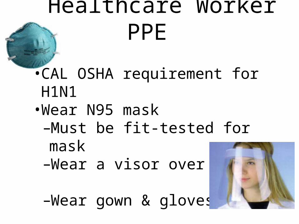
Healthcare Worker PPE
• CAL OSHA requirement for H1N1• Wear N95 mask
–Must be fit-tested for mask–Wear a visor over mask
–Wear gown & gloves
Vaccination• Get vaccinated yearly
• Seasonal vaccine
–2 types of strains of A: H1N1 & H3N2
–1 B strain
• H1N1 available
Antiviral Drugs for H1N1• Sensitive to neuraminidase inhibitors
–Tamiflu–Relenza
• Prevents virus from leaving cell
• Benefit if started within 48 hours of illness onset
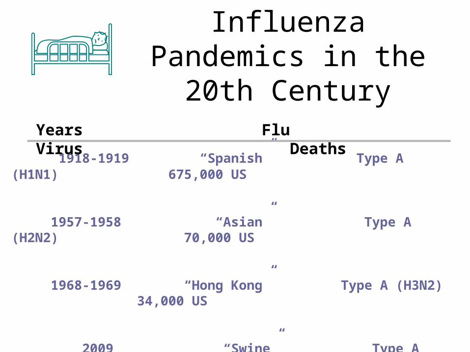
Influenza Pandemics in the
20th Century
1918-1919 “Spanish” Type A (H1N1) 675,000 US
1957-1958 “Asian” Type A (H2N2) 70,000 US
1968-1969 “Hong Kong” Type A (H3N2) 34,000 US
2009 “Swine” Type A (H1N1) 8000 US
Years Flu Virus Deaths

Pandemic Influenza
• Pandemic influenza virus– A new influenza A subtype can infect humans – Causes serious illness– Spreads easily from human-to-human
H5N1 is a likely candidate, but is not a pandemic virus yet
H1N1 is a pandemic virus
AIDS
• Final stage of a long infection with HIV– Attacks immune system
– T helper cells and macrophages

2 Types
• HIV 1-99% of all global cases
• HIV 2 discovered in West Africa– Reduced virulence – Causes milder disease
Structure of HIV
• 2 positive strands of RNA
• 2 identical strands of RNA enzyme- reverse transcriptase – Copies RNA into DNA
• Envelope with spikes termed gp120- glycoprotein of 120,000 mw

Pathogenicity
• Spikes allow attachment to CD4 receptors on host cells
• Coreceptors on T helper cells needed also for attachment
Fusion Protein
• Fuses CM with viral envelope– Nucleocapsid enters cell– Uncoated to release enzyme and RNA

Life Cycle
• RNA plus strands used for template only
• Viral DNA incorporates into host DNA Integrase enzyme (viral) joins viral DNA
with cellular DNA
• May be latent or cause disease

Protease
• Another enzyme in viral core
• Budding virus is not mature yet
• Proteins in core are in one long strand
• Must be cut by protease then virus is infectious

Mutations
• Rapid antigenic mutations
• Mutations at every position in genome many times each day

AIDS
• Progression to AIDS– based on T cell population
• Progression from HIV to AIDS is about 10 years
HIV Transmission• Contact with infected body fluids
• Blood-
• Semen -
• Heterosexual sex fastest growing risk factor
• Drug use and multiple partners
• Mother to baby, breast feeding
Treatment: Highly Active Anti Retrovirus Therapy
• Nucleoside analogs-AZT etc. inhibit reverse transcriptase
• Proteases- enzyme that cuts proteins into pieces reassembled into coat of new HIV particles
• Integrase inhibitors- enzyme that incorporates viral DNA into DNA of host
• Fusion inhibitors