virtual microscopy for hematology - webedcafe.com · h. elizabeth broome, m.d. approach to anemia...
TRANSCRIPT
H. Elizabeth Broome, M.D.
Approach to Anemia Diagnosis 1
Virtual Microscopy for Hematology
• H. Elizabeth Broome, M.D.Medical Director, Hematology Laboratories
• UC, San Diego Healthcare• April 24, 2014
Digital Images vs. Virtual Images
Digital images make virtual images easier to produce
H. Elizabeth Broome, M.D.
Approach to Anemia Diagnosis 2
Digital Imaging for Hematology-Outline
• Advantages and limitations of digital imaging
• Available digital imaging systems in US– Whole slide imaging such as Aperio
– Cellavision DM96 and DM1200
– Medica EasyCell
• FDA issues and validation
• Case Examples
Objectives
At the end of the talk, the participant should be able to:
• describe what digital imaging is currently available for clinical hematology
• compare the performance of digital imaging techniques to manual microscopy
• give specific examples where digital imaging techniques assisted in patient care
H. Elizabeth Broome, M.D.
Approach to Anemia Diagnosis 3
Conflict of Interest
• None
Virtual Microscopy; Why Now?
• Advances in technology mean:– Quality of digital microscopic image has come
close to the human eye
– Automated slide maker stainers mean better, more consistent blood smears
– Robotics create walk away slide imaging capabilities
– Computer analyses find the best areas of the slide, optimal focus, and “preclassification”
– Increased storage capacity and band width allow easy processing of large image files
H. Elizabeth Broome, M.D.
Approach to Anemia Diagnosis 4
Advantages
• Can view the same image across time and space for expert review, QC, competency testing and training– “Big Brother” is watching you perform your manual
differential!– Can we trash the slide “micro-locator” yet?
• Easier for viewers with limited expertise on the microscope
• Images do not degrade with time in storage• Can annotate or further analyze images
(computer-aided image analysis)
Disadvantages
• Images are not (yet) as good as with a high-quality microscope
• Optimal viewing requires high-quality equipment and technical expertise
• Does not show all of the smear; may miss cells on edges or platelet clumps in feathered edge
• Users may lose expertise with making smears and using microscope.
H. Elizabeth Broome, M.D.
Approach to Anemia Diagnosis 5
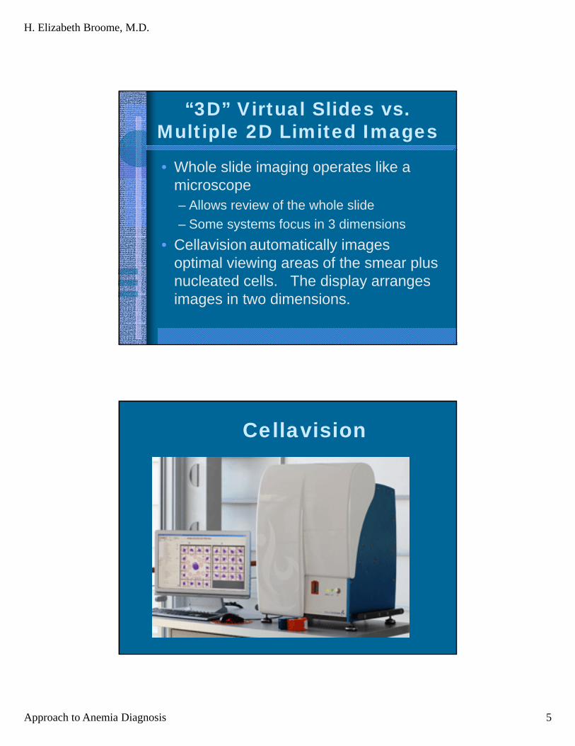
“3D” Virtual Slides vs. Multiple 2D Limited Images
• Whole slide imaging operates like a microscope– Allows review of the whole slide
– Some systems focus in 3 dimensions
• Cellavision automatically images optimal viewing areas of the smear plus nucleated cells. The display arranges images in two dimensions.
Cellavision

H. Elizabeth Broome, M.D.
Approach to Anemia Diagnosis 6
DI-60Integrates Cellavision with Sysmex Slide-
maker Stainer and Automated Line
Aperio
H. Elizabeth Broome, M.D.
Approach to Anemia Diagnosis 7
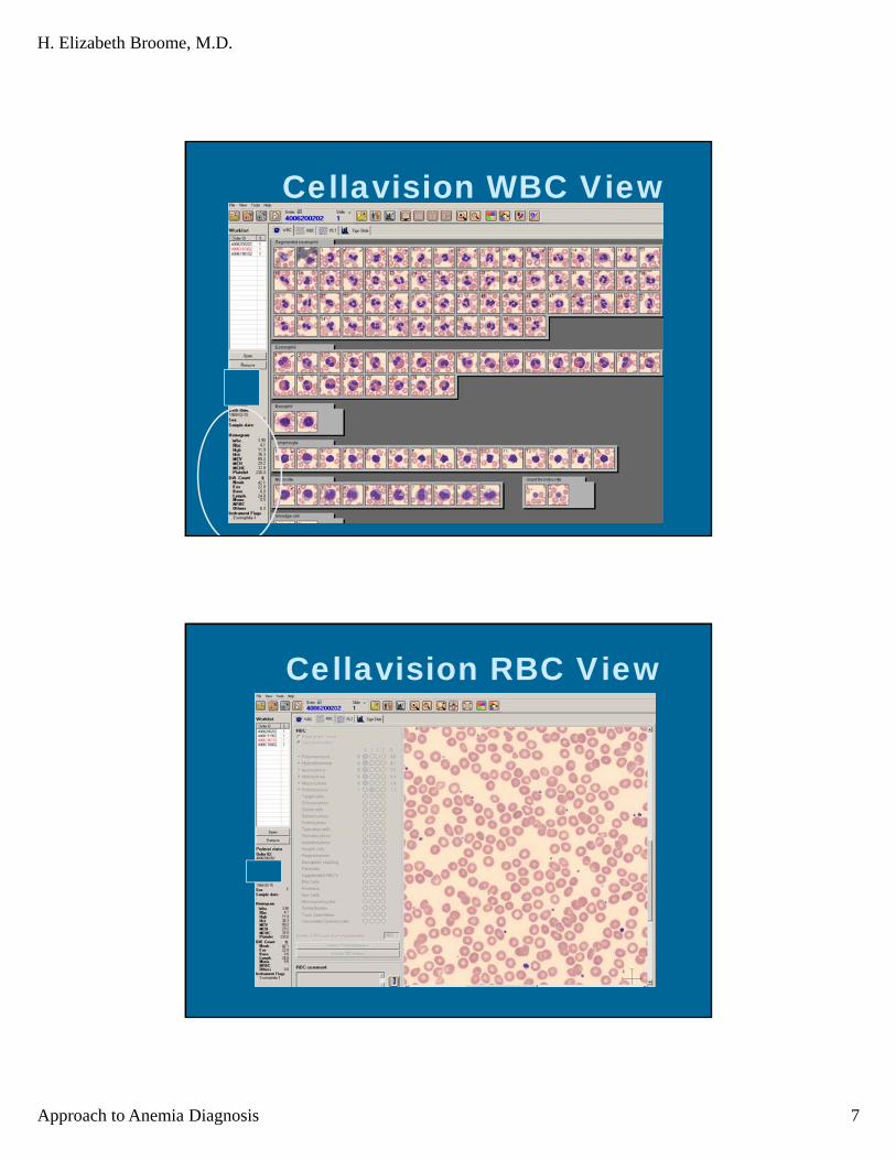
Cellavision WBC View
Cellavision RBC View
H. Elizabeth Broome, M.D.
Approach to Anemia Diagnosis 8
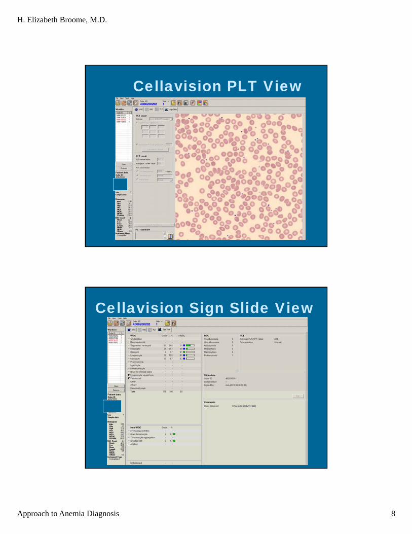
Cellavision PLT View
Cellavision Sign Slide View
H. Elizabeth Broome, M.D.
Approach to Anemia Diagnosis 9
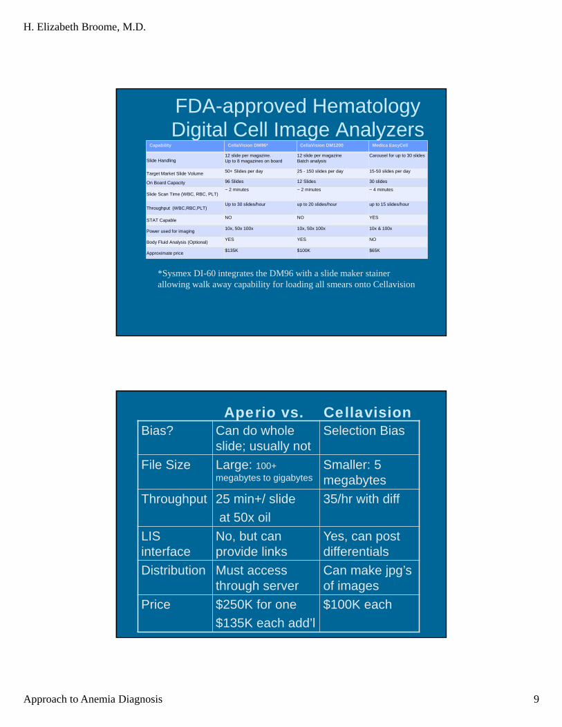
FDA-approved Hematology Digital Cell Image Analyzers
Capability CellaVision DM96* CellaVision DM1200 Medica EasyCell
Slide Handling 12 slide per magazine.Up to 8 magazines on board
12 slide per magazine Batch analysis
Carousel for up to 30 slides
Target Market Slide Volume 50+ Slides per day 25 - 150 slides per day 15-50 slides per day
On Board Capacity 96 Slides 12 Slides 30 slides
Slide Scan Time (WBC, RBC, PLT) ~ 2 minutes ~ 2 minutes ~ 4 minutes
Throughput (WBC,RBC,PLT)Up to 30 slides/hour up to 20 slides/hour up to 15 slides/hour
STAT CapableNO NO YES
Power used for imaging10x, 50x 100x 10x, 50x 100x 10x & 100x
Body Fluid Analysis (Optional)YES YES NO
Approximate price$135K $100K $65K
*Sysmex DI-60 integrates the DM96 with a slide maker stainer allowing walk away capability for loading all smears onto Cellavision
Aperio vs. CellavisionBias? Can do whole
slide; usually notSelection Bias
File Size Large: 100+ megabytes to gigabytes
Smaller: 5 megabytes
Throughput 25 min+/ slide
at 50x oil
35/hr with diff
LIS interface
No, but can provide links
Yes, can post differentials
Distribution Must access through server
Can make jpg’s of images
Price $250K for one
$135K each add’l
$100K each
H. Elizabeth Broome, M.D.
Approach to Anemia Diagnosis 10
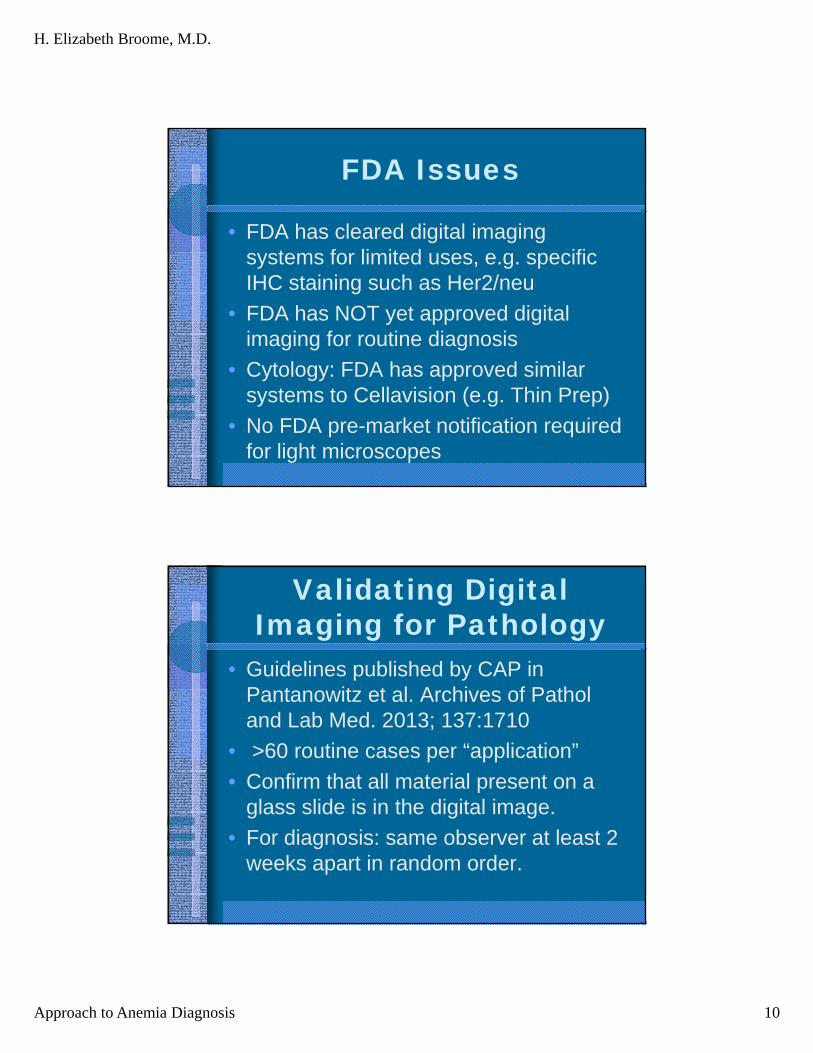
FDA Issues
• FDA has cleared digital imaging systems for limited uses, e.g. specific IHC staining such as Her2/neu
• FDA has NOT yet approved digital imaging for routine diagnosis
• Cytology: FDA has approved similar systems to Cellavision (e.g. Thin Prep)
• No FDA pre-market notification required for light microscopes
Validating Digital Imaging for Pathology
• Guidelines published by CAP in Pantanowitz et al. Archives of Pathol and Lab Med. 2013; 137:1710
• >60 routine cases per “application”
• Confirm that all material present on a glass slide is in the digital image.
• For diagnosis: same observer at least 2 weeks apart in random order.

H. Elizabeth Broome, M.D.
Approach to Anemia Diagnosis 11
Meta-analysis* of DigitalMicroscopsy for Diagnosis
Concordance 86%
Discordance 14%
Concordance and“minor” discordance
98%
*Pantanowitz et al. Arch Pathol Lab Med. 2013;137:171023 references for “standard” surgical pathology included. Concordance for each reference ranged from 72% to 98%.
Validation and QC of Cellavision
• Correlation with manual differential using 40+ cases with varied percentages
• Validate quality of images and pre-classification using different stains
• Validate platelet estimation; 40 samples
• Validate RBC morphology and abnormal WBC using truth tables
• Daily QC is just cell localization; >97%
H. Elizabeth Broome, M.D.
Approach to Anemia Diagnosis 12
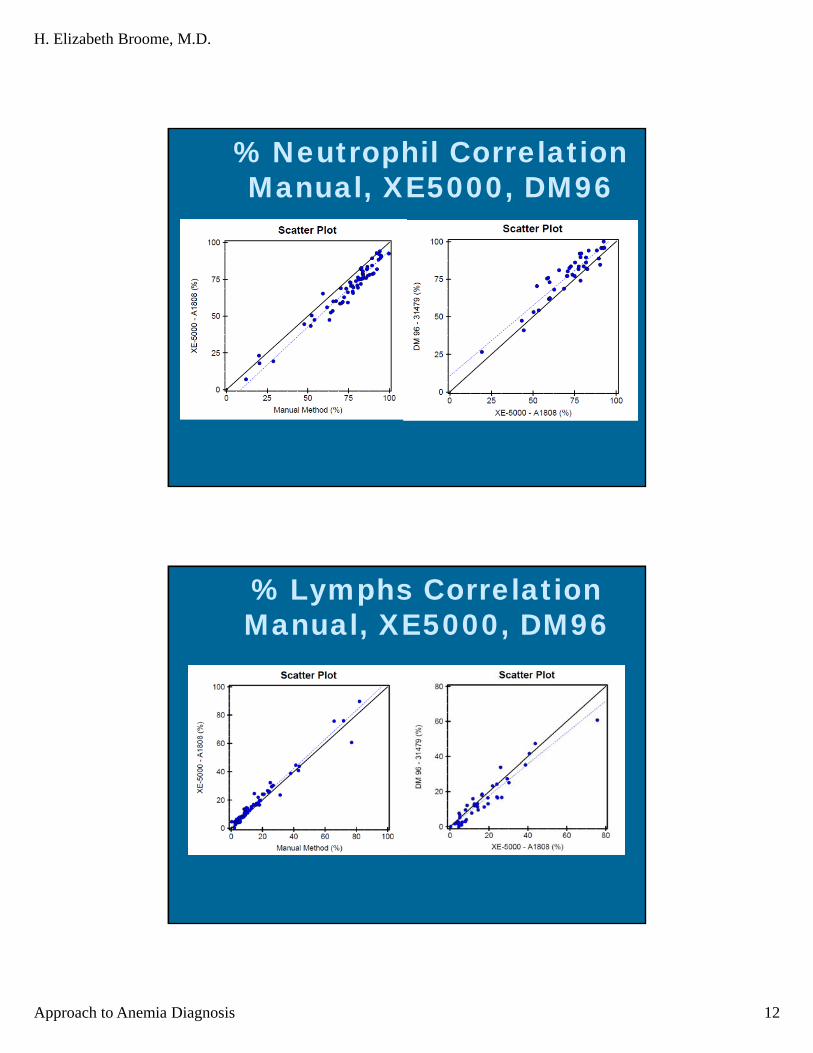
% Neutrophil Correlation Manual, XE5000, DM96
% Lymphs Correlation Manual, XE5000, DM96
H. Elizabeth Broome, M.D.
Approach to Anemia Diagnosis 13
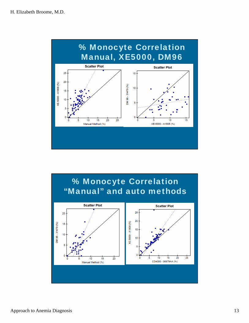
% Monocyte Correlation Manual, XE5000, DM96
% Monocyte Correlation “Manual” and auto methods

H. Elizabeth Broome, M.D.
Approach to Anemia Diagnosis 14
Briggs et al. Int. J. Lab. Hem. 2009, 31, 48
Kratz et al. AJCP2005 124:770
Correct Pre-classification by Cellavision
H. Elizabeth Broome, M.D.
Approach to Anemia Diagnosis 15
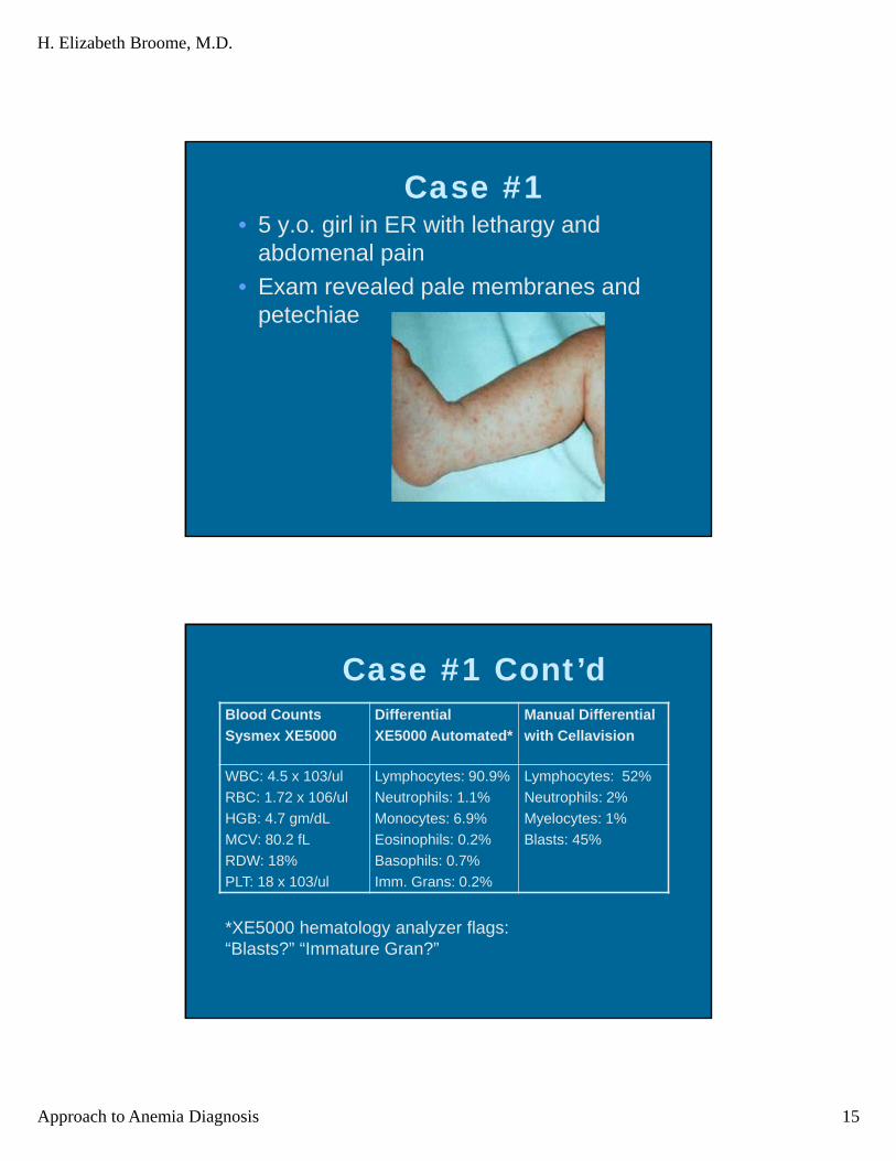
Case #1• 5 y.o. girl in ER with lethargy and
abdomenal pain
• Exam revealed pale membranes and petechiae
Case #1 Cont’dBlood Counts
Sysmex XE5000
Differential
XE5000 Automated*
Manual Differential
with Cellavision
WBC: 4.5 x 10³/ul
RBC: 1.72 x 10⁶/ul
HGB: 4.7 gm/dL
MCV: 80.2 fL
RDW: 18%
PLT: 18 x 103/ul
Lymphocytes: 90.9%
Neutrophils: 1.1%
Monocytes: 6.9%
Eosinophils: 0.2%
Basophils: 0.7%
Imm. Grans: 0.2%
Lymphocytes: 52%
Neutrophils: 2%
Myelocytes: 1%
Blasts: 45%
*XE5000 hematology analyzer flags: “Blasts?” “Immature Gran?”
H. Elizabeth Broome, M.D.
Approach to Anemia Diagnosis 16
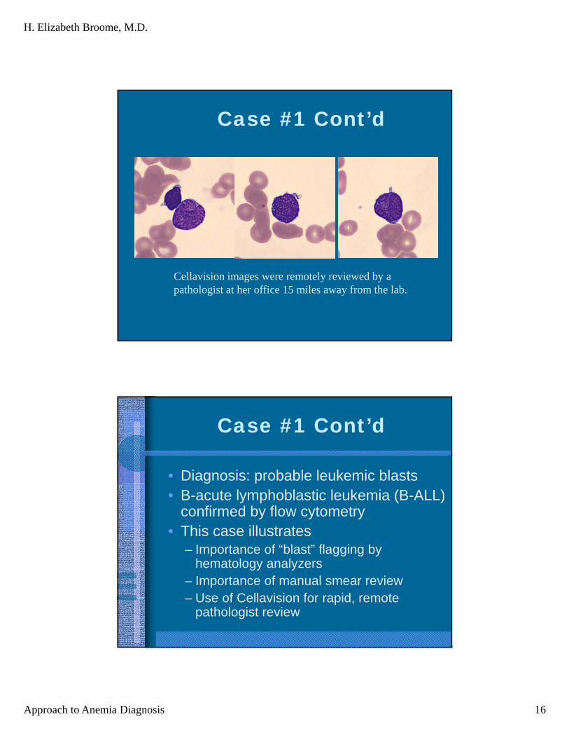
Case #1 Cont’d
Cellavision images were remotely reviewed by apathologist at her office 15 miles away from the lab.
Case #1 Cont’d
• Diagnosis: probable leukemic blasts • B-acute lymphoblastic leukemia (B-ALL)
confirmed by flow cytometry • This case illustrates
– Importance of “blast” flagging by hematology analyzers
– Importance of manual smear review– Use of Cellavision for rapid, remote
pathologist review
H. Elizabeth Broome, M.D.
Approach to Anemia Diagnosis 17
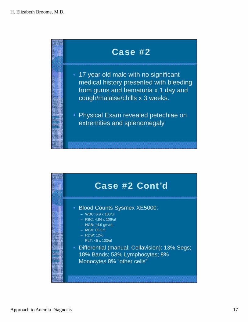
Case #2
• 17 year old male with no significant medical history presented with bleeding from gums and hematuria x 1 day and cough/malaise/chills x 3 weeks.
• Physical Exam revealed petechiae on extremities and splenomegaly
Case #2 Cont’d
• Blood Counts Sysmex XE5000:– WBC: 6.9 x 10³/ul
– RBC: 4.84 x 10⁶/ul
– HGB: 14.9 gm/dL
– MCV: 85.5 fL
– RDW: 12%
– PLT: <5 x 10³/ul
• Differential (manual; Cellavision): 13% Segs; 18% Bands; 53% Lymphocytes; 8% Monocytes 8% “other cells”

H. Elizabeth Broome, M.D.
Approach to Anemia Diagnosis 18
Case #2 cont’d“other cells”
Case #2 Cont’d
• “Others” are reactive lymphocytes• Diagnosis: Probable infectious
mononucleosis complicated by immune thrombocytopenia
• Positive “monospot” plus IgG and IgM anti-Epstein Barr Virus confirmed recent infection with Epstein Barr Virus
• Platelet count responded to treatment with corticosteroids and IVIg.
H. Elizabeth Broome, M.D.
Approach to Anemia Diagnosis 19
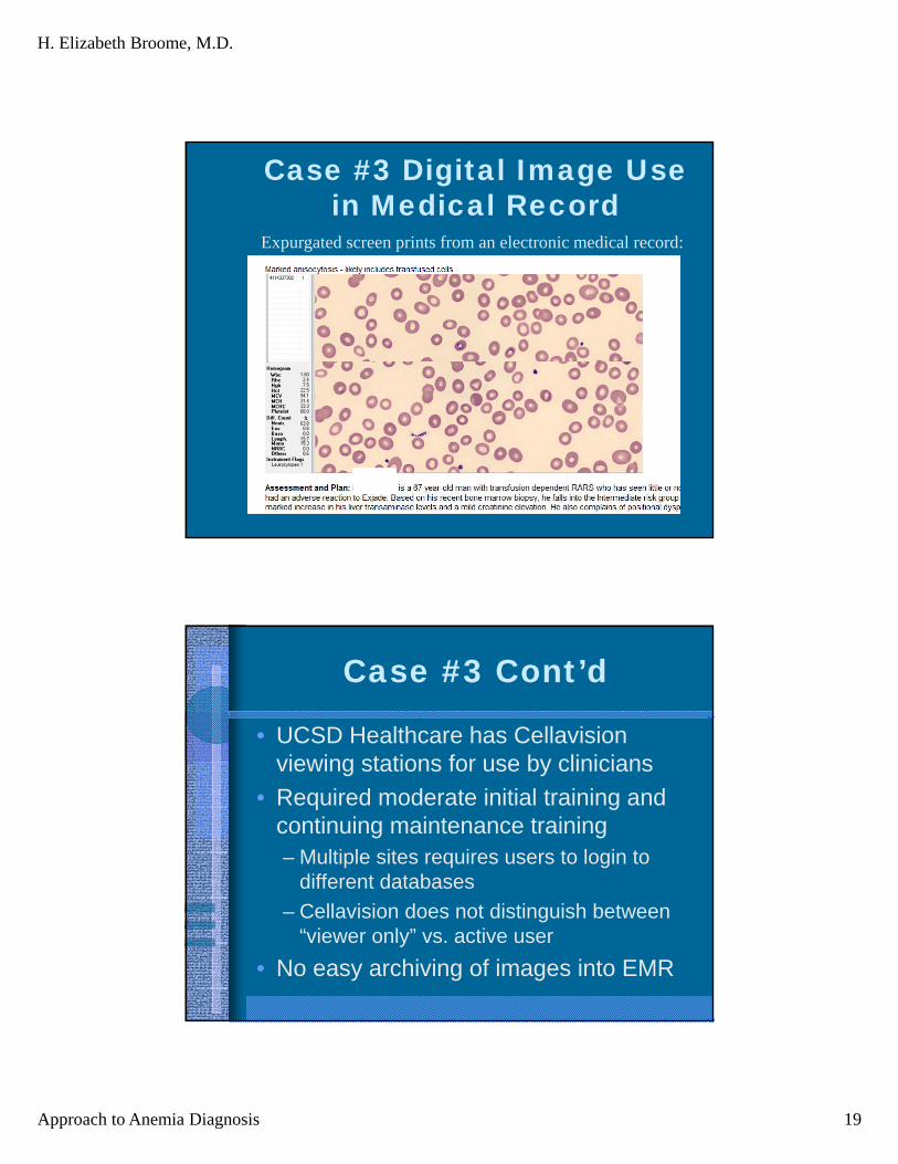
Case #3 Digital Image Usein Medical Record
Expurgated screen prints from an electronic medical record:
Case #3 Cont’d
• UCSD Healthcare has Cellavision viewing stations for use by clinicians
• Required moderate initial training and continuing maintenance training– Multiple sites requires users to login to
different databases
– Cellavision does not distinguish between “viewer only” vs. active user
• No easy archiving of images into EMR

H. Elizabeth Broome, M.D.
Approach to Anemia Diagnosis 20
Cellavision Advanced RBC (in development)
Spherocytes Highlighted
4/17/2014
H. Elizabeth Broome, M.D.
Approach to Anemia Diagnosis 21
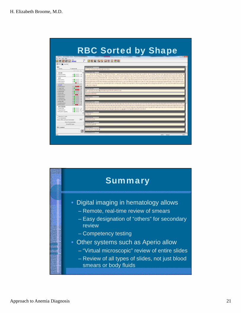
RBC Sorted by Shape
Summary
• Digital imaging in hematology allows– Remote, real-time review of smears
– Easy designation of “others” for secondary review
– Competency testing
• Other systems such as Aperio allow– “Virtual microscopic” review of entire slides
– Review of all types of slides, not just blood smears or body fluids
H. Elizabeth Broome, M.D.
Approach to Anemia Diagnosis 22
Future
• Improve RBC morphology functions
• Automated screening of blood smears and cytospins for abnormal cells– Blasts
– Increased band neutrophils
– Increased schistocytes or spherocytes
• Better integration of imaging with hematology automation