viktor's notes. operative techniques/300...2019/04/10 · brain biopsy op310 (2) 4. apply a...
TRANSCRIPT

BRAIN BIOPSY Op310 (1)
Brain Biopsy Last updated: April 10, 2019
RESOURCES ............................................................................................................................................ 1
INDICATIONS ............................................................................................................................................ 1 YIELD & ACCURACY ............................................................................................................................... 1
BIOPSY NEEDLES ..................................................................................................................................... 1 Nashold Biopsy Needle (Integra) ..................................................................................................... 1
PROCEDURE ............................................................................................................................................. 2 STEREOTACTIC ....................................................................................................................................... 2
Navigation scans .............................................................................................................................. 2 Registration ...................................................................................................................................... 2
Vertek® Passive Biopsy Kit with Vertek Probe .............................................................................. 2
Trajectory Guide Kit with Navigus Probe ........................................................................................ 2 Biopsy ............................................................................................................................................... 3
Tissue amount necessary .................................................................................................................. 3 Sampling area ................................................................................................................................... 3
Postoperatively ................................................................................................................................. 3
FORAMEN OVALE APPROACH ................................................................................................................. 3 Cannulation ...................................................................................................................................... 3
Gasserian ganglion procedures ......................................................................................................... 4 Biopsy ............................................................................................................................................... 4
Depth electrode ................................................................................................................................ 7
Postop ............................................................................................................................................... 7 PINEAL REGION TUMORS – ENDOSCOPIC APPROACH ............................................................................... 8
PINEAL REGION TUMORS – STEREOTACTIC NEEDLE ................................................................................ 8
COMPLICATIONS ...................................................................................................................................... 8 TISSUE PROCESSING TECHNIQUES, HISTOLOGICAL FINDINGS – see p. D34 >>
RESOURCES
See >>
Karl Storz NeuroEndoscopes and Instruments >>
INDICATIONS
1. Brain tumors - definitive tissue diagnosis necessary for treatment planning. see p. Onc1 >>
2. Differentiating residual tumor from radiation necrosis (coagulative necrosis and vasculopathic
changes).
3. Mass forms of infection (abscess, cerebritis, tuberculoma), incl. differentiation from CNS
lymphoma in AIDS patients.
4. Rare viral encephalitides (esp. rabies, Creutzfeldt-Jakob disease). see p. Inf9 >>
5. Vasculopathies (e.g. granulomatous angiitis).
N.B. do not biopsy vascular lesions!
Indications for open biopsies:
1) prominent blood vessels
2) hemorrhage within lesion
3) contemplated resection (during same procedure)
YIELD & ACCURACY
Diagnostic yield - percent of biopsies that obtain a histopathologic diagnosis (i.e. ability to obtain
diagnostic tissue).
82-99% in nonimmunocompromised (NIC) patients vs. 56-96%. in AIDS patients.
yield rate is higher for lesions that enhance with contrast on CT or MRI (in NIC patients - 99%
vs. 74%).
Diagnostic accuracy - proportion of biopsies that agree with the ‘‘gold standard’’ of surgical resection
or autopsy.
BIOPSY NEEDLES
NASHOLD BIOPSY NEEDLE (INTEGRA)
has a side window-type cutter at its tip - window is opened and closed by rotating the inner cannula
within the outer cannula, using the upper and lower hubs.
needle length – 249 mm
outer diameter – 2.0 mm
side cutting window – 10 mm
1. Set the depth stop on the outer cannula to the target length.
2. With the cutting window closed (see “Cutting Window Use”), insert the NBND.
3. At the target, open the cutting window.

BRAIN BIOPSY Op310 (2)
4. Apply a slight vacuum with a syringe attached to the Luer.
5. Draw a tissue sample into the open window.
6. Close the cutting window and shear off a tissue sample inside the inner cannula.
7. Withdraw the inner cannula (or entire assembly, if procedure is complete) containing the tissue
sample.
8. If desired, reposition the needle and repeat the procedure from step 1.
PROCEDURE
avoid MANNITOL if lesion is small – may shift target!!!
head secured in a 3-point Mayfield headholder (if using O-arm - radiolucent frame, including pins,
is ideal but not necessary)
carried out under general anesthesia (in adults, local anesthesia is possible).
STEREOTACTIC
NAVIGATION SCANS
Use CTA / MRA whenever available to plan trajectories away from vessels.
to improve tumor biopsy results:
1. MRS with maps (choline/creatine ratio) superimposed on MRI - where is necrosis, where is
tumor
2. pMRI (PWI) - will see if it is enhancing piece of necrosis (no perfusion) or real tumor
(perfusion↑)
REGISTRATION
Medtronic Stealth frameless navigation: registration → set target (definite) and skull entry
(preliminary) points.
May use alternative high accuracy registration – registration with O-arm (optional - may place one
bone fiducial to verify navigation accuracy after registration):
need to use Stealth “dogbone” to attach radiolucent Mayfield (with radiolucent pins) to table;
“dogbone” has two starbursts for attachment of two Vertek arms; “dogbone” uses different
separate screw to attach to table frame!!!
N.B. attach “dogbone” from outside (not from Mayfield inside) – easier to reach for Vertek
arm during the case.
O-arm is needed for registration only: attach passive cranial frame, spin O-arm and may remove O-
arm; remove passive cranial frame and use sterile one during surgery (leaving nonsterile one on
and covering with sterile camera drape also works but is tricky – need to deflect OR lights to avoid
reflections of light).
Entry points and trajectories:
o thalamus – slightly lateral to Kocher’s point.
Target
Note: probe tip on Stealth corresponds to needle tip; actual biopsy window is marked by lines
on probe on Stealth screen (when on trajectory views only)
VERTEK® PASSIVE BIOPSY KIT WITH VERTEK PROBE
Vertek® Probe (9733158) - has 70 mm shaft (vs. 50 mm in Navigus probe); may need to add to
instrument list on Cranial software module (as likely only Navigus Probe is there).
attach Mayfield leg to inside (facing patient feet) of Mayfield frame.
use “dogbone” attachment to Mayfield (has two starbursts for two Vertek arms – one nonsterile for
holding passive cranial frame, one sterile for instrument guide)
prep scalp, drape, place sterile passive cranial frame
attach sterile Vertek instrument holding arm to Mayfield (cut hole in drapes and assistant reaches
under drapes).
using Vertek Probe establish trajectory, then make incision:
a) use 15 blade on long handle to make stab through Vertek port
b) disassemble Vertek port and make 0.5 cm skin incision with Colorado Bovie tip (more
cosmetic than Navigus probe approach!!!)
twist hole is made with matchstick drill bit through Vertek arm port (don’t press on Vertek arm as
it will bend!); usually it opens dura as well (if not – use long spinal needle).
trajectory is verified using Vertek Probe and entry point is reset on Stealth.
press Stealth foot pedal – this locks trajectory (locking trajectory gives depth to target needed to set
depth gage on biopsy needle).
insert needle adapter to Vertek arm port and tighten.
perform biopsy >>
remove entire biopsy assembly.
optional (incision is 5 mm – unable to reach galea this way) - galea is approximated with one
inverted 2-0 Vicryl in interrupted fashion.
skin – Monocryl 3-0 / staples and Bacitracin ointment.
TRAJECTORY GUIDE KIT WITH NAVIGUS PROBE
Navigus® Probe (9733157)

BRAIN BIOPSY Op310 (3)
14 mm bur hole with perforator; ± remove (Kerrison or matchstick drill) bony shelf (esp. at
direction of biopsy)
biopsy assembly base with 3 cortical screws:
o angled (freedom 5° and 25°)
o straight (freedom 10° in all directions)
mount entire assembly: transparent holder (pops into socket), white cap
insert Navigus Probe: set new entry → align to trajectory → lock trajectory on Stealth (by pressing
Stealth pedal) – note distance to target on screen.
insert grey needle holder
open dura using monopolar cautery touching biopsy needle (becomes dirty – maybe to avoid this?)
and then spinal 14 G needle.
perform biopsy >>
remove biopsy needle (with closed window) and entire biopsy assembly.
place Gelfoam in bur hole, followed by cranial plate.
closure with bur hole cover
BIOPSY
set biopsy needle stopper (on special measurement board) to distance to target on screen.
N.B. maximum needle length is 180 mm (even if distance from skull entry point to target is <
180 mm, remember that Vertek Probe and needle adapter add 70 mm!)
insert both needles with closed biopsy window.
take core biopsy in four quadrants – rotating biopsy needle each time 90°; insert needle with closed
window → open window → apply suction (syringe half with saline is directly attached to needle
hub) → quickly close window → pull out needle → flush saline so tissue core moves on Telfa →
reinsert needle.
N.B. always flush needle on Telfa as sometimes there is more tissue
when reinserting inner needle, gently aspirate air (otherwise, air gets plunged into brain).
may repeat biopsy again (rotate 45°), thus, obtaining total of 8 tissue cores – send for frozen
pathology.
TISSUE AMOUNT NECESSARY
- few cells may be sufficient in diagnosis of metastatic carcinoma or lymphoma, whereas even
postmortem examination of entire brain may be inadequate to establish precise diagnosis in some
degenerative or metabolic diseases.
SAMPLING AREA
a) mass lesion / defined area on imaging studies
possibility of tissue heterogeneity! - multiple biopsies along needle trajectory through entire
tumor thickness (to the farthest tumor edge) may provide more complete picture of
pathological process.
N.B. sampling of only central area (e.g. complete tissue necrosis in tumors or
abscesses) may not yield diagnostic tissue!
b) diffuse pathology (no lesion on imaging) - NONELOQUENT AREAS of cerebrum.
N.B. random biopsies may be nondiagnostic
type of tissue sampled (gray or white brain matter, leptomeninges) is guided by suspected
diagnosis (tissue wedge consisting of cortex, overlying leptomeninges, and underlying
white matter provides most useful tissue sample).
tissue should be considered potentially infectious (precautions for Creutzfeldt-Jakob
disease should be taken).
POSTOPERATIVELY
immediate postoperative head CT – if normal, may go to the floor / gateway.
FORAMEN OVALE APPROACH
CANNULATION
patient supine under general anesthesia, tube facing down and taped well, under gel donut (Dr.
Holloway) or horseshoe headholder (Dr. Broaddus).
if doing glycerol injection, patient is on the stretcher – can be sit up during procedure.
guidance:
a) frameless navigation, e.g. Stryker mask; assemble 18 G 3.5 inch foramen ovale (vs. spinal)
needle with its plastic hub secured in Stryker tracking device; register-calibrate needle; for
details - see p. Op30 >>
b) fluoroscopy (C-arm) – regular OR table or stretcher are enough radiolucent for this purpose;
work under AP with beam aligned along trajectory (mouth corner to zygoma; see below) but
head rotated away from operated side – should clearly see foramen ovale (intersection of top of
petrous bone with clivus; confirmation – when moving patient’s head or AP fluoroscopy up or
down, foramen disappears as beam is no longer along foramen canal).
prep cheek with chlorhexidine or Betadine.
Härtel’s landmarks:
o needle entry point – 2-3 cm lateral to mouth corner (maybe also a little bit inferior – to
avoid lateral pterygoid plate in trajectory).
o surgeon inserts index finger in patient's mouth (to palpate and guide needle tip through
mucosa as it is advanced, making certain it does not breech mucosa); opposite hand is
used to advance needle; keep needle medial to coronoid process of mandible and
outside oral cavity i.e. deep to oral mucosa – needle slides on medial surface of
mandibular ramus (here needle trajectory becomes rather fixed, so if need to change
needle trajectory, withdraw needle back to this point).
o needle is aimed at inner aspect of ipsilateral pupil + at point 3.5 cm anterior to external
ear canal at level of zygoma (practically, it is inverse EVD target).
when patient shows evidence of pain, anesthetists gives brief general anesthesia.
needle is advanced through foramen ovale; slowly advanced until slight release is felt (penetration
through Gasserian ganglion into trigeminal cistern – remove stylet and check for CSF flow for
reassurance).
BRAIN BIOPSY Op310 (4)
— may feel mandible jerk when needle irritates CNV3
— may cause bradycardia – wait before advancing needle until heart rate recovers (have atropine
ready).
— change fluoroscopy to lateral to verify – needle tip has to be just posterior to clivus
verify good position (esp. if unable to get CSF):
a) inject 0.5 cm3 of saline – should go easily without resistance (but may mean that needle is
too far and already in the middle fossa).
b) insert foramen ovale electrode through needle – see if it slides along petrous surface
(means needle is in Meckel’s cave)
c) injecting Omnipaque – should fill Meckel’s cave under live fluoroscopy (notice volume
of Omnipaque needed – gives idea of cave volume).
GASSERIAN GANGLION PROCEDURES
- see p. CN5 >>
BIOPSY
A. 2 spinal needles:
1. One 18G size 3.5 in spinal needle (attach tracking device to track the tip if using
navigation) – cannulate just foramen
2. Second needle is 22G 5 inch spinal needle – wrap SteriStrip on shaft so tip protrudes
1.5* cm from first needle tip (*or whatever distance to tumor)
insert second needle back and forth several times (to get tissue cores in) then gently aspirate and
pass to cytologist for microscopy.
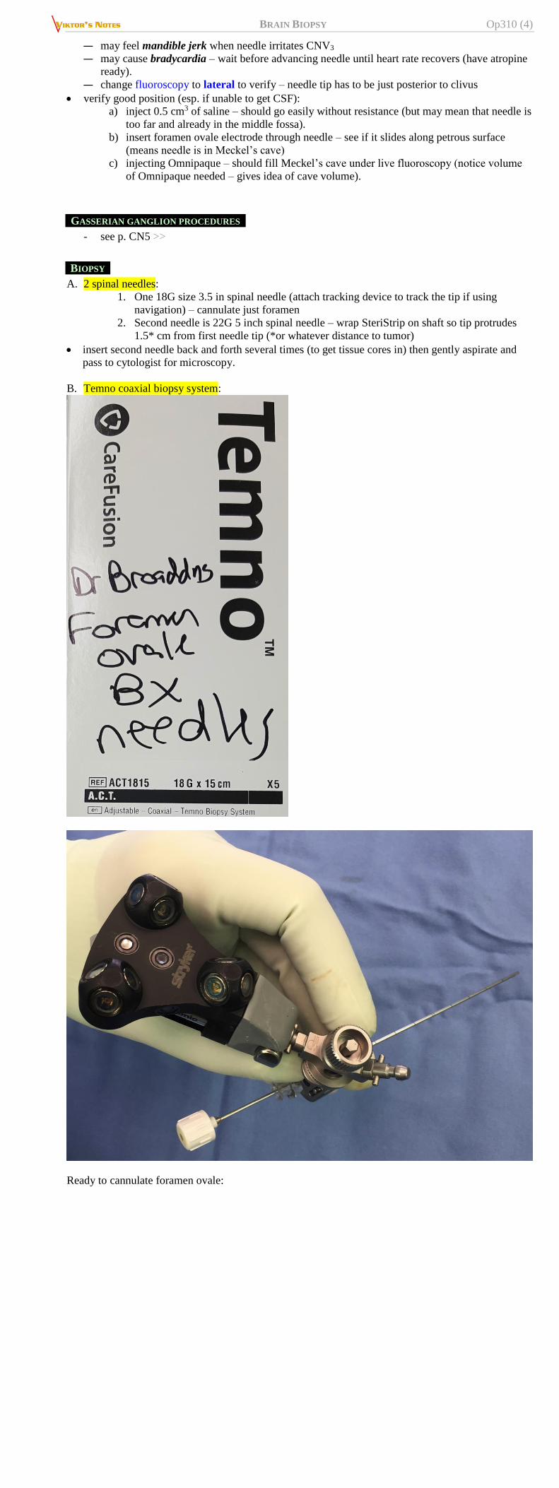
B. Temno coaxial biopsy system:
Ready to cannulate foramen ovale:

BRAIN BIOPSY Op310 (5)
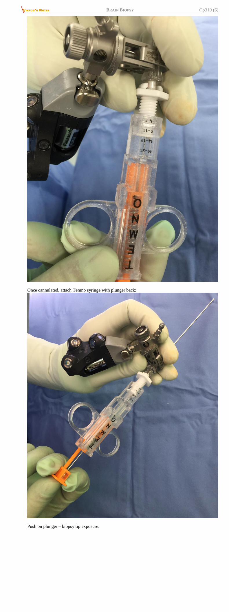
Temporary attach Temno syringe for biopsy window calibration prior to cannulation:
BRAIN BIOPSY Op310 (6)
Once cannulated, attach Temno syringe with plunger back:
Push on plunger – biopsy tip exposure:

BRAIN BIOPSY Op310 (7)
Keep squeezing plunger – will fire biopsy sheath over biopsy tip:
DEPTH ELECTRODE
- see p. E13 >>
POSTOP

BRAIN BIOPSY Op310 (8)
may skip postoperative head CT.
PINEAL REGION TUMORS – ENDOSCOPIC APPROACH
Do ETV first!
A. Use ETV bur hole and flexible endoscope to go through foramen of Monroe posteriorly
B. Use separate more frontal bur hole and rigid endoscope
PINEAL REGION TUMORS – STEREOTACTIC NEEDLE
- through parenchyma, avoiding ventricles:
a) frontal approach (but trajectory through thalamus)
b) parietal approach (but trajectory may go through sensory cortex)
COMPLICATIONS
Removal of nonregenerating brain tissue is accompanied by risk of permanent neurological deficit!
mortality < 0.2-1%.
major hemorrhage - risk 0-3% (0-12% in AIDS - reduced platelet count or function, vessel
fragility in primary CNS lymphoma).
*small hematoma at biopsy site is not unusual and is rarely clinically significant
edema exacerbation, infection, development of seizure focus, increased neurologic deficit.
Viktor’s Notes℠ for the Neurosurgery Resident
Please visit website at www.NeurosurgeryResident.net