view abstract - journal of scientific and innovative research
TRANSCRIPT
Mukul et. al. www. jsirjournal.com
Volume 1 Issue 3 2012 | JOURNAL OF SCIENTIFIC & INNOVATIVE RESEARCH
8
Volume 1 Issue 3 2012
Available online at: www. jsirjournal.com
JOURNAL OF SCIENTIFIC & INNOVATIVE RESEARCH
Role of metal and oxidative stress in mechanisms of Metal-induced cancer-A
review
Mukul Raizada*1
, Dinesh Singh1, Sandeep Kumar
1
1. Department of Chemistry, Aligarh Muslim University, Aligarh, Uttar Pradesh: 202002
[Email: [email protected]]
Abstract: Humans have been in contact with metals almost since the beginning of our existence.
In fact, one cannot even think on human evolution without considering the role played by metals
in evolution. Consequently, the presence of a variety of metals is necessary for the normal
functioning of cells and the survival of organisms. Metals play important roles in a wide variety
of biological processes of living systems. Metal ion transporters participate in maintaining the
required levels of the various metal ions in the cellular compartments. In order to avoid metal-
related toxic effects, cells and organisms have evolved sophisticated mechanisms for
sequestration and fine regulation of the available metal concentrations.
Keywords: Cancer; Oxidative stress, Metals, Iron.
Introduction: Metals play important
roles in a wide variety of biological
processes of living systems. Metal ion
transporters participate in maintaining the
required levels of the various metal ions in
the cellular compartments.1
Breakdown of metal-ion homeostasis can
lead to the metal binding to protein sites
different to those designed for that purpose
or replacement of other metals from their
natural binding.2The results have provided
evidence that toxic metals can interact with
DNA and proteins causing oxidative

Mukul et. al. www. jsirjournal.com
Volume 1 Issue 3 2012 | JOURNAL OF SCIENTIFIC & INNOVATIVE RESEARCH
9
deterioration of biological macromolecules.
Thus the process of breakdown of metal-ion
homeostasis has been involved in a plethora
of diseases.3-7
Metals are known to modulate
gene expression by interfering with signal
transduction pathways that play important
roles in cell growth and development.
Deregulation of cell growth and
differentiation is a typical characteristic of
the cancer phenotype. Actions of metals
interfere with deregulation of cell
proliferation by activating various
transcription factors, controlling cell cycle
progression and apoptosis.8
The generation of free radicals in living
systems is closely linked with the
participation of redox-active metals such as
iron, copper, chromium and cobalt, the
redox state of the cell is maintained within
strict physiological limits.9,10
Redox active
metals may undergo cycling reactions
participating in the transfer of electrons
between metals and substrates and therefore
may play an important role in the
maintenance of redox homeostasis, a
phenomenon tightly linked with metal
homeostasis.11
Disruption of metal
homeostasis may lead uncontrolled metal-
mediated formation of deleterious free
radicals participating in the modifications to
DNA bases, enhanced lipid peroxidation,
and altered calcium and sulphydryl
homeostasis.12, 13
Metal-induced oxidative stress and
cancer:
Many studies have focused on metal-
induced toxicity and carcinogenicity,
emphasising their role in the generation of
reactive oxygen and nitrogen species in
biological systems. Metal-mediated
formation of free radicals may cause various
modifications to DNA bases, enhanced lipid
peroxidation, and changes in calcium and
sulphydryl homeostasis.14-20
1. Copper:
Copper is a cofactor of many enzymes
involved in redox reactions, such as
cytochrome c oxidase, ascorbate oxidase, or
superoxide dismutase. It is used in
biological systems for electron transport.5 It
is readily absorbed from the diet across the
small intestine (∼2mg/day) and stored in the
liver. The major excretory route of copper
stored in liver is via the biliary pathway
(∼80%).21
Copper is bound to either serum
albumin or histidine and trafficked through

Mukul et. al. www. jsirjournal.com
Volume 1 Issue 3 2012 | JOURNAL OF SCIENTIFIC & INNOVATIVE RESEARCH
10
the bloodstream for delivery to tissues or
storage in the liver.22
Copper can induce oxidative stress by two
mechanisms. First, it can directly catalyze
the formation of ROS via a Fenton-like
reaction.23, 24
Second, exposure to elevated
levels of copper significantly decreases
glutathione levels.25
Because copper is an essential component of
several endogenous antioxidant enzymes,
and that free radicals have been proposed to
play a role in the process of carcinogenesis,
the effects of dietary copper levels on the
development of cancer have been
investigated.26
The weight of evidence from in vitro and in
vivo assays indicates that copper (as the
copper salts) is not genotoxic.27
However, in
vitro studies have shown that cancer cells in
a high copper environment find it easy to
proliferate into tumour.28,29
Therefore, it has
been proposed that copper-lowering drug
may stabilise advanced cancer. Brewer and
his group tested a drug known as
tetrathiomolybdate (TM), which binds up
dietary copper before it can be absorbed by
the body, to see if they could reduce the
spread of tumours in patients with different
types of metastatic cancer. In five of six
patients kept at 80% of normal copper levels
for more than 90 days, existing tumours did
not grow and new tumours did not form for
more than 1 year. This suggests the use of
TM either as the sole therapy for cancer or
in conjunction with other treatments, such as
surgery, chemotherapy, radiation therapy
and others. Similarly to iron, copper is a
well-known pro-oxidant and may participate
in metal-catalyzed peroxidation of lipids.30
2. Chromium:
Chromium, one of the most common
elements in the earth’s exists in several
oxidation states. 31
Chromium(III) is an
essential trace element. It occures naturaly
and plays an important role in regulating
blood levels of glucose. Chromium(VI) is
potentially toxic and carcinogenic at high
doses.32,33,34
All chromates, Cr(VI), can
actively enter the cells through channels for
the transfer of isoelectric and isostructural
anions, such as those for SO4 2−
and HPO4
2−.35
Certain extracellularly generated Cr(V)
and Cr(III) complexes also have high
permeabilities through the cell membrane.
Once inside the cell, chromates are able to
generate free radicals.36
Cr(V) can also be
reduced by cellular reductants (e.g.
Mukul et. al.
Volume 1 Issue 3 2012 | JOURNAL OF SCIENTIFIC & INNOVATIVE RESEARCH
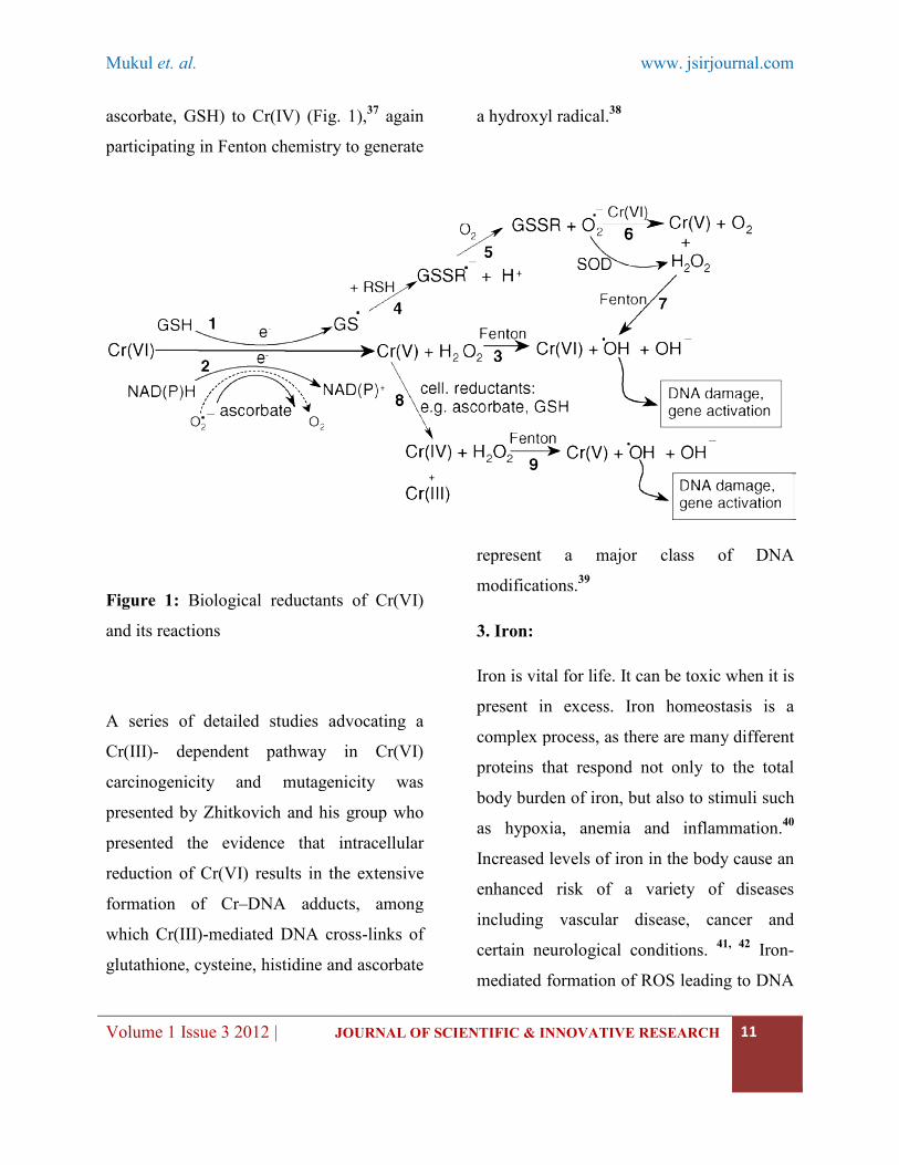
ascorbate, GSH) to Cr(IV) (Fig. 1),
participating in Fenton chemistry to generate
Figure 1: Biological reductants of Cr(VI)
and its reactions
A series of detailed studies advocating a
Cr(III)- dependent pathway in Cr(VI)
carcinogenicity and mutagenicity was
presented by Zhitkovich and his group who
presented the evidence that intracel
reduction of Cr(VI) results in the extensive
formation of Cr–DNA adducts, among
which Cr(III)-mediated DNA cross
glutathione, cysteine, histidine and ascorbate
www.
JOURNAL OF SCIENTIFIC & INNOVATIVE RESEARCH
ascorbate, GSH) to Cr(IV) (Fig. 1),37
again
participating in Fenton chemistry to generate
a hydroxyl radical.38
Biological reductants of Cr(VI)
A series of detailed studies advocating a
dependent pathway in Cr(VI)
carcinogenicity and mutagenicity was
presented by Zhitkovich and his group who
presented the evidence that intracellular
reduction of Cr(VI) results in the extensive
DNA adducts, among
mediated DNA cross-links of
glutathione, cysteine, histidine and ascorbate
represent a major class of DNA
modifications.39
3. Iron:
Iron is vital for life. It can be toxic when it is
present in excess. Iron homeostasis is a
complex process, as there are many different
proteins that respond not only to the total
body burden of iron, but also to stimuli such
as hypoxia, anemia and inflammation.
Increased levels of iron in the body cause an
enhanced risk of a variety of diseases
including vascular disease, cancer and
certain neurological conditions.
mediated formation of ROS leading to DNA
ww. jsirjournal.com
JOURNAL OF SCIENTIFIC & INNOVATIVE RESEARCH
11
represent a major class of DNA
It can be toxic when it is
present in excess. Iron homeostasis is a
complex process, as there are many different
proteins that respond not only to the total
body burden of iron, but also to stimuli such
as hypoxia, anemia and inflammation.40
Increased levels of iron in the body cause an
enhanced risk of a variety of diseases
including vascular disease, cancer and
certain neurological conditions. 41, 42
Iron-
mediated formation of ROS leading to DNA

Mukul et. al. www. jsirjournal.com
Volume 1 Issue 3 2012 | JOURNAL OF SCIENTIFIC & INNOVATIVE RESEARCH
12
and lipid damage appears to result from an
exaggeration of the normal function of iron,
which is to transport oxygen to tissues. Iron-
induced free radical damage to DNA
appears to be important for the development
of cancer and cancer cells are known
growrapidly in response to iron. 43
Many studies documented that mutations in
superoxide dismutase enzymes44
and iron-
uptake regulator may lead to excess levels of
superoxide anion radicals and iron overload.
Such a condition leads to the possibility of
redox active iron to participate in organic
and inorganic oxygen radical reactions, such
as stimulating lipid peroxidation and
catalyzing the formation of damaging
hydroxyl radicals with subsequent tissue
damage.45
Nelson and Babbs proposed that intestinal
exposure to ingested iron may be a principal
determinant of human colorectal cancer in
highly developed, meat-eating countries.46, 47
The bile acids (deoxycholic acid), the K
vitamins, iron(II) complexes and oxygen
interact to induce an oncogenic effect in the
colon by the generation of free radicals.48
The association between elevated body iron
stores and the development of hepatocellular
carcinoma in subjects with ironoverload
unrelated to genetic hemochromatosis along
with the experimental evidence of a co-
carcinogenic role of iron strongly support
the contention that iron is involved in the
development of hepatocellular carcinoma.49,
50 Occupational exposure to asbestos
containing about 30% (weight) of iron is
relate to an increased risk of asbestosis —
the second most important cause of lung
cancer after smoking.51
Intramuscular
injections of an iron–dextran complex,
frequently used for the treatment of anemia
in humans, caused spindle cell sarcoma or
pleomorphic sarcoma in rats at the site of
injection.52
Permanent modification of genetic material
resulting from free radical attacks represents
the initial step involved in mutagenesis,
carcinogenesis and ageing.53
In fact, as it has
been well documented, in various cancer
tissues free radical-mediated DNA damage
has occurred.54
4. Cobalt:
Various studies have investigated the
possibility that cobalt-mediated free radical
generation contributes to the toxicity of
cobalt. Hanna et al. performed EPR
spintrapping studies to detect the generation

Mukul et. al. www. jsirjournal.com
Volume 1 Issue 3 2012 | JOURNAL OF SCIENTIFIC & INNOVATIVE RESEARCH
13
of oxygen-free radicals from the reaction of
hydrogen peroxide with various Co
complexes under physiological conditions.55
Cobalt is known to be toxic to the heart and
suspected to be carcinogenic in animals
when given in large quantities. Exposure to
cobalt sulphate by inhalation resulted in
increased incidence of alveolar/bronchiolar
neoplasms and a spectrum of inflammatory,
fibrotic, and proliferative lesions in the
respiratory tracts of male and female rats
and mice.56
Injection of Co(II) into rats lead
to a pattern of oxidative DNA base damage
characteristic of hydroxyl radical attack via
the Fenton reaction.57
Trace amounts of
cobalt are needed in the diet because cobalt
is an integral metal of vitamin B12.58
5. Cadmium:
Cadmium is a heavy metal and the most
common oxidation number of cadmium is
+2. Food is the main source of cadmium for
the non-smoking population. Estimates of
dietary cadmium intake worldwide range
from 10–40g/day in nonpolluted areas to
several hundred micrograms in
cadmiumpolluted regions.59
Cadmium is a highly toxic metal. Cadmium
itself is unable to generate free radicals
directly, however, indirect generation of
various radicals involving the superoxide
radical, hydroxyl radical and nitric oxide has
been reported.60
Some experiments also
confirmed the generation of (non-radical)
hydrogen peroxide which itself in turn may
be a significant source of radicals via Fenton
chemistry.61
Cadmium is a potent human carcinogen and
occupational exposure to it has been
associated with cancers of the lung, the
prostate, pancreas and kidney. Cadmium
also used in the pathogenesis of human
pancreatic cancer and renal carcinoma.62
6. Arsenic:
Arsenic, known as a poison and has been
discovered to be a carcinogen in humans.
Many studies confirmed the generation of
free radicals during arsenic metabolism in
cells.63
In recent studies concerning the
mechanism of arsenite toxicity in the brain it
was reported that some of its effects have
been traced to the generation of the hydroxyl
radicals.64
Arsenic is a well-established
human carcinogen.65
Arsenic compounds
bind to SH groups and can inhibit various
enzymes, including glutathione reductase
Mukul et. al. www. jsirjournal.com
Volume 1 Issue 3 2012 | JOURNAL OF SCIENTIFIC & INNOVATIVE RESEARCH
14
radiation, to cause DNA mutations more
effectively.66
Arsenic is known to induce hypoxia
signalling pathways. For example in prostate
cancer cells treated with arsenite induced
HIF-1alpha expression in a concentration-
and time-dependent manner, whereas the
level of HIF-1beta remained unaffected.67
Arsenic is a well-documented carcinogen in
a number of studies.68
Chronic exposure to
inorganic arsenic from contaminated water
is responsible for various adverse health
effects such as developing tumours of the
lung, skin, liver, bladder and kidney. Skin
lesions, peripheral neuropathy and anemia
are hallmarks of chronic arsenic exposure.
Arsenic is also a potential risk factor for
atherosclerosis. While cardiovascular
disorders following oral exposure to arsenic
are well documented, there is some evidence
from epidemiological trials that also inhaled
inorganic arsenic can affect the
cardiovascular system.69
7. Nickel:
Nickel is a human carcinogen that can alter
gene expression by enhanced DNA
methylation and compaction, rather than via
mutagenic mechanisms.70
The nickel
compounds implicated as potential
carcinogens are insoluble dusts of nickel
subsulphides and nickel oxides, the vapor of
nickel carbonyl, and soluble aerosols of
nickel sulphate, nitrate, or chloride.71
Almost all cases of acute nickel toxicity
result from exposure to nickel carbonyl.
Patients with severe poisoning develop
intense pulmonary and gastrointestinal
toxicity. The lung is the primary target organ
for nickel toxicity in humans.72
Some other studies have shown that
workers’ inhalation of nickel refinery dust,
which contains nickel subsulphide, has
resulted in increased numbers of deaths from
nasal cavity cancers, and possibly cancer of
the larynx.73
8. Vanadium:
Vanadium is a transition metal element
which occurs in various oxidative states and
may participate in reactions involving
formation of free radicals.74
Vanadium in
plasma is rapidly reduced to vanadium by
both enzymatic (e.g. NADPH) and non-
enzymatic (ascorbic acid) plasmatic
antioxidants and is then transported and
bound to plasma proteins.75
The use of
vanadium compounds as inhibitors of

Mukul et. al. www. jsirjournal.com
Volume 1 Issue 3 2012 | JOURNAL OF SCIENTIFIC & INNOVATIVE RESEARCH
15
tyrosine phosphatases in studies of signal
transduction points to their potential to
induce oxidative stress.76
9. Zinc:
Zinc is a ubiquitous trace element found in
plants and animals. The adulthumanbody
contains approximately 1.5–2.5 g of zinc,
present in all organs, tissues, fluids and
secretions. The level of free intracellular
Zn(II) is as low as 0.5 nM, as estimated
from measurements of the zinc-specific 19F-
NMRsignal of a fluorinated metal chelating
probe.77
The observations performed in 1961 on
Iranian males have shown that zinc
deficiency may cause growth retardation and
hypogonadism in humans.78
Following
studies later showed that zinc was essential
for humans and that zinc deficiency was
prevalent in the Middle East.79
Zinc
deficiency has been associated with
increased levels of oxidative damage
including increased lipid, protein and DNA
oxidation.80
The zinc-supplemented group of
patients with sickle cell disease had
decreased incidences of infection in
comparison to the placebo group.81
After
zinc supplementation, antioxidant power
increased. In addition, plasma nitrite and
nitrate (NOx), lipid peroxidation products,
DNA oxidation products, and soluble
vascular cell adhesion molecule-1 (VCAM-
1) decreased compared to the placebo group.
Since oxidative stress and chronic
inflammation may play important causative
roles in many chronic diseases, including
atherosclerosis, cancers, neurological
disorders, and autoimmune diseases, more
thorough studies exploring the status of zinc
deficiency and supplementation are
necessary.
10. Lead:
Lead is one of the heavy metals. It is a
persistent metal and because of its unusual
physical–chemical properties it is used in
various industrial applications.82
Well
known is its use as a radiation shield. Lead
is a toxic metal to humans and animals and
its persistency causes prolonged occurrence
in the environment – in water, soil, dust and
in manufactured products containing lead.
Since young organisms bear the heaviest
burden of sensitivity to lead exposure, lead-
based paint covers represent a serious health
threat to children worldwide.83
Soil
containing lead also represents a serious

Mukul et. al. www. jsirjournal.com
Volume 1 Issue 3 2012 | JOURNAL OF SCIENTIFIC & INNOVATIVE RESEARCH
16
hazard for children. Gastrointestinal
absorption of lead is higher in children (40–
50%) than in adults (3–10%). Lead toxicity
is most commonly diagnosed through
elevated blood levels. Blood levels of
10g/dL (equivalent to 0.48mol/L) or higher
are considered toxic and result in
neurological disorders, cognitive
impairments, hypertension and other
disorders.84
Conclusions:
The current knowledge in the field of
metallo-biochemistry of oxidative stress
indicates that metal-induced and metal-
enhanced formation of free radicals and
other reactive species can be regarded as a
common factor in determining metal-
induced toxicity and carcinogenicity. The
above discussion provides an insight into the
role of metals capable of direct or indirect
generation of free radicals through various
mechanisms.
Acknowledgements:
I would like to thank the HOD of the
department of Chemistry.
References:
1. Rolfs, A., Hediger, M.A. Metal ion
transporters inmammals:structure,
function and pathological
implications. J. Physiol. (London)
1999; 518: 1–12.
2. Nelson, N. Metal ion transporters
and homeostasis. EMBO J. 1999; 18,
4361–4371.
3. Halliwell, B., Gutteridge, J.M.C.
Role of free radicals and catalytic
metal-ions in human disease—an
overview. Methods Enzymol. 1990;
186: 1–85.
4. Stohs, S., Bagchi, D. Oxidative
mechanisms in the toxicity of metal
ions. Free Radic. Biol. Med. 1995;
18: 321–336.
5. Valko, M.,Morris, H., Cronin,
M.T.D. Metals, toxicity and
oxidative stress. Curr. Med. Chem.
2005; 12: 1161–1208.
6. Matés, J.M. Effects of antioxidant
enzymes in the molecular control of
reactive oxygen species toxicology.
Toxicology 2000; 153: 83–104.
7. Matés, J.M., Pérez-Gómez, C.,
Nú˜nez de Castro, I. Antioxidant

Mukul et. al. www. jsirjournal.com
Volume 1 Issue 3 2012 | JOURNAL OF SCIENTIFIC & INNOVATIVE RESEARCH
17
enzymes and human diseases. Clin.
Biochem. 1999; 32: 595–603.
8. Evan, G.I., Vousden, K.H.
Proliferation, cell cycle and
apoptosis in cancer. Nature 2001;
411: 342–348.
9. Rahman, K. Studies on free radicals,
antioxidants, and co-factors. Clin.
Interv. Aging 2007; 2: 219–236.
10. Matés, J.M., Pérez-Gómez, C.,
Nú˜nez de Castro, I., Asenjo, M.,
Márquez, J. Glutamine and its
relationship with intracellular redox
status, oxidative stress and cell
proliferation/death. Int. J. Biochem.
Cell. Biol. 2002; 34: 439–458.
11. Lindeque, J.Z., Levanets, O., Louw,
R., van der Westhuizen, F.H. The
involvement of metallothioneins in
mitochondrial function and disease.
Curr. Protein Peptide Sci. 2010; 11:
292–309.
12. Gutteridge, J.M.C. Lipid
peroxidation and antioxidants as
biomarkers of tissue damage. Clin.
Chem. 1995; 41: 1819–1828.
13. Valko, M., Leibfritz, D., Moncol, J.,
Cronin, M.T.D., Mazur, M., Telser,
J. Free radicals and antioxidants in
normal physiological functions and
human disease. Int. J. Biochem. Cell
Biol. 2007; 39: 44–84.
14. M.Valko, H. Morris, M.T.D. Cronin,
Metals, toxicity and oxidative stress,
Curr. Med. Chem. 2005; 12: 1161–
1208.
15. S.S. Leonard, G.K. Harris, X.L. Shi,
Metal-induced oxidative stress and
signal transduction, Free Rad. Biol.
Med. 2004; 37: 1921–1942.
16. S.J. Stohs, D. Bagchi, Oxidative
mechanisms in the toxicity of metal-
ions, Free Rad. Biol. Med. 1995;
18:321–336.
17. I. Pekarkova, S. Parara,V. Holecek,
P. Stopka, L.Trefil, J. Racek, R.
Rokyta, Does exogenous melatonin
influence the free radicals
metabolism and pain sensation in
rat? Physiol. Res. 2001; 50: 595–
602.
18. M. Valko, D. Leibfritz, J. Moncol,
M.T.D. Cronin, J. Telser, Mutual
effect of free radicals, redox metals
and antioxidants, FEBS J. 2006.
19. F. Chen, M. Ding, V. Castranova,
X.L. Shi, Carcinogenic metals and
NF-kappa B activation, Mol. Cell.
Biochem. 2001; 222: 159–171.

Mukul et. al. www. jsirjournal.com
Volume 1 Issue 3 2012 | JOURNAL OF SCIENTIFIC & INNOVATIVE RESEARCH
18
20. B. Halliwell, J.M.C. Guteridge, Role
of free-radicals and catalytic metal-
ions in human-disease—an
overview, Meth. Enzymol. 1990;
186: 1–85.
21. Linder, M.C., Hazegh-Azam, M.
Copper biochemistry and molecular
biology. Am. J. Clin. Nutr. 1996; 63:
797S–811S.
22. Zhou, B., Gitschier, J. hCTR1: a
human gene for copper uptake
identified by complementation in
yeast. Proc. Natl. Acad. Sci. U.S.A.
1997; 94: 7481–7486.
23. Prousek, J. Fenton chemistry in
biology and medicine. Pure Appl.
Chem. 2007; 79: 2325–2338.
24. Liochev, S.I., Fridovich, I. The
Haber–Weiss cycle—70 years later:
an alternative view. Redox Rep.
2002; 7: 55–57.
25. Speisky, H., Gómez, M., Burgos-
Bravo, F., López-Alarcón, C.,
Jullian, C., Olea-Azar, C., Aliaga,
M.E. Generation of superoxide
radicals by copper-glutathione
complexes: redox-consequences
associated with their interaction with
reduced glutathione. Bioorg. Med.
Chem. 2009; 17: 1803–1810.
26. K.G. Daniel, R.H. Harbach,W.C.
Guida, Q.P. Dou, Copper storage
diseases: Menkes, Wilson’s, and
cancer, Front. Biosci. 2004; 9: 2652–
2662.
27. M. Olivares, F. Pizarro, H. Speisky,
B. Lonnerdal, R. Uauy, Copper in
infant nutrition: safety ofWorld
Health Organization provisional
guideline value for copper content of
drinkingwater, J. Pediatr.
Gastroenterol. Nutr. 1998; 26: 251–
257.
28. R.J. Coates, N.S. Weiss, J.R. Daling,
R.L. Rettmer, G.R. Warnick, Cancer
risk in relation to serum copper
levels, Cancer Res. 1989; 49: 4353–
4356.
29. T.J. Wu, C.T. Sempos, J.L.
Freudenheim, P. Muti, E. Smith,
Serum iron, copper and zinc
concentrations and risk of cancer
mortality in US adults, Ann.
Epidemiol. 2004; 14: 195–201.
30. G.J. Brewer, R.D. Dick, D.K.
Grover, V. LeClaire, M. Tseng, M.
Wicha, K. Pienta, B.G. Redman, T.
Jahan, V.K. Sondak,M.
Strawderman, G. LeCarpentier, S.D.
Merajver, Treatment of metastatic
Mukul et. al. www. jsirjournal.com
Volume 1 Issue 3 2012 | JOURNAL OF SCIENTIFIC & INNOVATIVE RESEARCH
19
cancer with tetrathiomolybdate, an
anticopper, antiangiogenic agent:
Phase I study, Clin. Cancer Res.
2000; 6: 1–10.
31. Cieslak-Golonka, M. Toxic and
mutagenic effects of chromium(VI).
A review. Polyhedron 1996; 15:
3667–3689.
32. A.D. Dayan, A.J. Paine, Mechanisms
of chromium toxicity,
carcinogenicity and allergenicity:
reviewof the literature from 1985 to
2000, Human Exp. Toxicol. 2001;
20: 439–451.
33. K.S. Kasprzak, Possible role of
oxidative damage in metalinduced
carcinogenesis, Cancer Invest. 1995;
13: 411–430.
34. M. Cieslak-Golonka, Toxic and
mutagenic effects of chromium(VI).
A review, Polyhedron 1996; 15:
3667–3689.
35. J. Singh, D.L. Carlisle, D.E.
Pritchard, S.R. Patierno, Chromium-
induced genotoxicity and apoptosis:
relationship to chromium
carcinogenesis (Review), Oncol.
Rep.1998; 5; 1307–1318.
36. K.J. Liu, X.L. Shi, In vivo reduction
of chromium(VI) and its related free
radical generation, Mol. Cell.
Biochem. 2001; 222: 41–47.
37. E. Cadenas, K.J.A. Davies,
Mitochondrial free radical
generation, oxidative stress, and
aging, Free Rad. Biol. Med. 2000;
29: 222–230.
38. C.Y. Li, R.M. Jackson, Reactive
species mechanisms of cellular
hypoxia-reoxygenation injury, Am.
J. Physiol.-Cell Physiol. 2002; 282:
C227–C241.
39. A. Zhitkovich, Importance of
chromium–DNAadducts in
mutagenicity and toxicity of
chromium(VI), Chem. Res. Toxicol.
2005; 18: 3–11 (and references
therein).
40. Lee, D.W., Andersen, J.K., Kaur, D.
Iron dysregulation and
neurodegeneration: the molecular
connection. Mol. Intervent. 2006a; 6:
89–97.
41. D. Berg, M. Gerlach, M.B.H.
Youdim, K.L. Double, L. Zecca, P.
Riederer, G. Becker, Brain iron
pathways and their relevance to
Parkinson’s disease, J. Neurochem.
2001; 79: 225–236.

Mukul et. al. www. jsirjournal.com
Volume 1 Issue 3 2012 | JOURNAL OF SCIENTIFIC & INNOVATIVE RESEARCH
20
42. C.W. Siah, D. Trinder, J.K. Olynyk,
Iron overload, Clin. Chim. Acta
2005; 358: 24–36.
43. H. Ullen, K. Augustsson, C.
Gustavsson, G. Steineck,
Supplementary iron intake and risk
of cancer: reversed causality? Cancer
Lett. 1997; 114: 215–216.
44. Deng, H.X., Hentati, A., Tainer, J.A.,
Iqbal, Z., Cayabyab, A., Hung,
W.Y., Getzoff, E.D., Hu, P.,
Herzfeldt, B., Roos, R.P.
Amyotrophic lateral sclerosis and
structural defects in Cu, Zn
superoxide dismutase. Science 1993;
261: 1047–1051.
45. Iolascon, A., De Falco, L.,
Beaumont, C. Molecular basis of
inherited microcytic anemia due to
defects in iron acquisition or heme
synthesis. Haematologica 2009; 94:
395–408.
46. C.F. Babbs, Free-radicals and the
etiology of colon cancer, Free Rad.
Biol. Med. 1990; 8: 191–200.
47. R.L. Nelson, Dietary iron and
colorectal-cancer risk, Free Rad.
Biol. Med. 1992; 12: 161–168.
48. M. Valko, H. Morris, M. Mazur, P.
Rapta, R.F. Bilton, Oxygen free
radical generating mechanisms in the
colon: do the semiquinones of
Vitamin K play a role in the
aetiology of colon cancer? Biochim.
Biophys. Acta 2001; 1527: 161–
166.
49. K.V. Kowdley, Iron,
hemochromatosis, and hepatocellular
carcinoma, Gastroenterology 2004;
127: S79–S86.
50. Y. Deugnier, B. Turlin, Iron and
hepatocellular carcinoma, J.
Gastroenterol. Hepatol. 2001; 16:
491–494.
51. L.T. Stayner, D.A. Dankovic, R.A.
Lemen, Occupational exposure to
chrysotile asbestos and cancer risk: a
review of the amphibole hypothesis,
Am. J. Public Health 1996; 86: 179–
186.
52. G. Bhasin, H. Kauser,M. Athar, Iron
augments stage-I and stage- II tumor
promotion in murine skin, Cancer
Lett. 2002; 183: 113–122.
53. Durackova, Z. Some current insights
into oxidative stress. Physiol. Res.
2010; 59: 459–469.
54. Marnett, L.J. Oxyradicals and DNA
damage. Carcinogenesis 2000; 21:
361–370.

Mukul et. al. www. jsirjournal.com
Volume 1 Issue 3 2012 | JOURNAL OF SCIENTIFIC & INNOVATIVE RESEARCH
21
55. P.M. Hanna, M.B. Kadiiska, R.P.
Mason, Oxygen-derived free radical
and active oxygen complex-
formation from cobalt(II) chelates in
vitro, Chem. Res. Toxicol. 1992; 5:
109– 115.
56. J.R. Bucher, J.R. Hailey,
J.R.Roycroft, J.K. Haseman, R.C.
Sills, S.L. Grumbein, P.W. Mellick,
B.J. Chou, Inhalation toxicity and
carcinogenicity studies of cobalt
sulfate, Toxicol. Sci. 1999; 49: 56–
67.
57. Z. Nackerdien, K.S. Kasprzak, G.
Rao, B. Halliwell, M. Dizdaroglu,
Nickel(II)-dependent and cobalt(II)-
dependent damage by hydrogen-
peroxide to theDNAbases in isolated
human chromatin, Cancer Res. 1991;
51: 5837–5842.
58. J.R. Roth, J.G. Lawrence, T.A.
Bobik, Cobalamin (coenzyme B-
12): synthesis and biological
significance, Ann. Rev.
Microbiol.1996; 50: 137–181.
59. Cuypers, A., Plusquin, M., Remans,
T., Jozefczak, M., Keunen, E.,
Gielen, H., Opdenakker, K., Nair,
A.R., Munters, E., Artois, T.J.,
Nawrot, T., Vangronsveld, J.,
Smeets, K. Cadmium stress: an
oxidative challenge. Biometals 2010;
23: 927–940.
60. A. Galan, L. Garcia-Bermejo, A.
Troyano, N.E. Vilaboa, C.
Fernandez, E. de Blas, P. Aller, The
role of intracellular oxidation in
death induction (apoptosis and
necrosis) in human promonocytic
cells treated with stress inducers
(cadmium, heat, X-rays), Eur. J.
Cell. Biol. 2001; 80: 312–320.
61. M. Watanabe, K. Henmi, K. Ogawa,
T. Suzuki, Cadmiumdependent
generation of reactive oxygen
species and mitochondrial DNA
breaks in photosynthetic and non-
photosynthetic strains of Euglena
gracilis, Comp. Biochem. Physiol.
CToxicol. Pharmacol. 2003; 134:
227–234.
62. M.Waisberg, P. Joseph, B. Hale, D.
Beyersmann, Molecular and cellular
mechanisms of cadmium
carcinogenesis, Toxicology 2003;
192: 95–117.
63. K.Yamanaka, F. Takabayashi, M.
Mizoi,Y. An, A. Hasegawa, S.
Okada, Oral exposure of
dimethylarsinic acid, a main

Mukul et. al. www. jsirjournal.com
Volume 1 Issue 3 2012 | JOURNAL OF SCIENTIFIC & INNOVATIVE RESEARCH
22
metabolite of inorganic arsenics, in
mice leads to an increase in 8-oxo-
2deoxyguanosine level, specifically
in the target organs for arsenic
carcinogenesis, Biochem. Biophys.
Res. Commun. 2001; 287: 66–70.
64. E. Garcia-Chavez, A. Santamaria, F.
Diaz-Barriga, P. Mandeville, B.I.
Juarez, M.E. Jimenez-Capdeville,
Arsenite-induced formation of
hydroxyl radical in the striatum of
awake rats, Brain Res. 2003; 976:
82–89.
65. P. Roy, A. Saha, Metabolism and
toxicity of arsenic: a human
carcinogen, Curr. Sci. 2002; 82: 38–
45.
66. M.P. Waalkes, J. Liu, J.M. Ward,
L.A. Diwan, Mechanisms underlying
arsenic carcinogenesis:
hypersensitivity of mice exposed to
inorganic arsenic during gestation,
Toxicology 2004; 198: 31–38.
67. Posey, T., Weng, T., Chen, Z.,
Chintagari, N.R., Wang, P., Jin, N.,
Stricker, H., Liu, L. Arsenic-induced
changes in the gene expression of
lung epithelial L2 cells: implications
in carcinogenesis. BMC Genomics
2008; 9: 115.
68. Waalkes, M.P., Liu, J., Ward, J.M.,
Diwan, L.A. Mechanisms underlying
arsenic carcinogenesis:
hypersensitivity of mice exposed to
inorganic arsenic during gestation.
Toxicology 2004; 198: 31–38.
69. Das, A.K., Sahu, R., Dua, T.K., Bag,
S., Gangopadhyay, M., Sinha, M.K.,
Dewanjee, S. Arsenic-induced
myocardial injury: protective role of
Corchorus olitorius leaves. Food
Chem. Toxicol. 2010; 48: 1210–
1217.
70. Y.W. Lee, C.B. Klein, B. Kargacin,
K. Salnikow, J. Kitahara, K. Dowjat,
A. Zhitkovich, N.T. Christie, M.
Costa, Carcinogenic nickel silences
gene-expression by chromatin
condensation and DNA methylation
— a new model for epigenetic
carcinogens, Mol. Cell. Biol. 1995;
15: 2547–2557.
71. D.G. Barceloux, Nickel, J. Toxicol.
Clin. Toxicol. 1999; 37: 239–258.
72. T.K. Grimsrud, S.R. Berge, T.
Haldorsen, A. Andersen, Can lung
cancer risk among nickel refinery
workers be explained by
occupational exposures other than

Mukul et. al. www. jsirjournal.com
Volume 1 Issue 3 2012 | JOURNAL OF SCIENTIFIC & INNOVATIVE RESEARCH
23
nickel? Epidemiology 2005; 16:
146–154.
73. V.J. Feron, J.H.E. Arts, C.F. Kuper,
P.J. Slootweg, R.A. Woutersen,
Health risks associated with inhaled
nasal toxicants, Crit. Rev. Toxicol.
2001; 31: 313–347.
74. D.C. Crans, J.J. Smee, E.
Gaidamauskas, L.Q. Yang, The
chemistry and biochemistry of
vanadium and the biological
activities exerted by vanadium
compounds, Chem. Rev. 2004; 104:
849–902.
75. S.I. Liochev, I. Fridovich, Vanadate-
stimulated oxidation of NAD(P)H in
the presence of biological-
membranes and other sources of
O2−, Arch. Biochem. Biophys.
1990; 279: 1–7.
76. L.V. Favreau, C.B. Pickett, The rat
quinone reductase antioxidant
response element — identification of
the nucleotidesequence required for
basal and inducible activity and
detection of antioxidant response
element-binding proteins in
hepatoma and non-hepatoma cell-
lines, J. Biol. Chem. 1995; 270:
24468–24474.
77. Benters, J., Flogel, U., Schafer, T.,
Leibfritz, D., Hechtenberg, S.,
Beyersmann, D. Study of the
interactions of cadmium and zinc
ions with cellular calcium
homoeostasis using F-19-NMR
spectroscopy. Biochem. J. 1997;
322: 793–799.
78. Prasad, A.S., Halsted, J.A., Nadimi,
M. Syndrome of iron deficiency
anemia, hepatosplenomegaly,
hypogonadism, dwarfism, and
geophagia. Am. J. Med. 1961; 31:
532–546.
79. Prasad, A.S., Miale, A., Farid, Z., et
al. Zinc metabolism in patients with
the syndrome of iron deficiency
anemia; hypogonadism and
dwarfism. J. Lab. Clin. Med. 1963;
61: 537–549.
80. Prasad, A.S. Zinc: role in immunity,
oxidative stress and chronic
inflammation. Curr. Opin. Clin.
Nutr. Metab. Care 2009; 12: 646–
652.
81. Bao, B., Prasad, A.S., Beck, F.W.,
Snell, D., Suneja, A., Sarkar, F.H.,
Doshi, N., Fitzgerald, J.T.,
Swerdlow, P. Zinc supplementation
decreases oxidative stress, incidence
Mukul et. al. www. jsirjournal.com
Volume 1 Issue 3 2012 | JOURNAL OF SCIENTIFIC & INNOVATIVE RESEARCH
24
of infection, and generation of
inflammatory cytokines in sickle cell
disease patients. Transl. Res. 2008;
152: 67–80.
82. Brannvall, M.L., Bindler, R.,
Renberg, I., Emteryd, O., Bartnicki,
J., Billstrom, K. The Medieval metal
industry was the cradle of modern
large scale atmospheric lead
pollution in northern Europe.
Environ. Sci. Technol. 1999; 33:
4391–4395.
83. Kumar, A., Clark, C.S. Lead
loadings in household dust in Delhi,
India. Indoor Air 2009; 19: 414–420.
84. Patrick, L. Lead toxicity, a review of
the literature. Part 1. Exposure,
evaluation, and treatment. Altern.
Med. Rev. 2006a; 11: 2–22.