video-assisted intercostal nerve cryoablation in managing intractable chest wall pain
TRANSCRIPT
BRIEF TECHNIQUE REPORTS
Video-assisted intercostal nerve cryoablation in managing intractablechest wall pain
Ian Hunt, FRCS, Donna Eaton, FRCS, Omar Maiwand, FRCS, and Vladimir Anikin, FRCS, Harefield,
Middlesex, United Kingdom
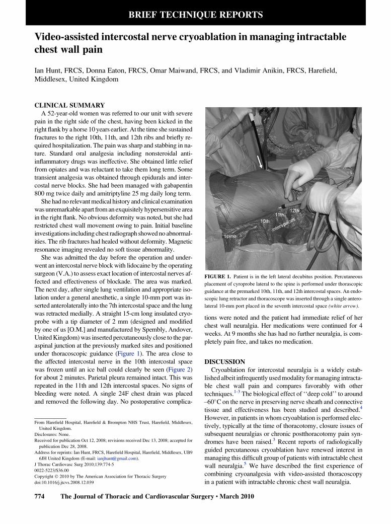
FIGURE 1. Patient is in the left lateral decubitus position. Percutaneous
placement of cyroprobe lateral to the spine is performed under thorascopic
guidance at the premarked 10th, 11th, and 12th intercostal spaces. An endo-
scopic lung retractor and thoracoscope was inserted through a single antero-
lateral 10-mm port placed in the seventh intercostal space (white arrow).
CLINICAL SUMMARYA 52-year-old women was referred to our unit with severe
pain in the right side of the chest, having been kicked in the
right flank by a horse 10 years earlier. At the time she sustained
fractures to the right 10th, 11th, and 12th ribs and briefly re-
quired hospitalization. The pain was sharp and stabbing in na-
ture. Standard oral analgesia including nonsteroidal anti-
inflammatory drugs was ineffective. She obtained little relief
from opiates and was reluctant to take them long term. Some
transient analgesia was obtained through epidurals and inter-
costal nerve blocks. She had been managed with gabapentin
800 mg twice daily and amitriptyline 25 mg daily long term.
She had no relevant medical history and clinical examination
was unremarkable apart from an exquisitely hypersensitive area
in the right flank. No obvious deformity was noted, but she had
restricted chest wall movement owing to pain. Initial baseline
investigations including chest radiograph showed no abnormal-
ities. The rib fractures had healed without deformity. Magnetic
resonance imaging revealed no soft tissue abnormality.
She was admitted the day before the operation and under-
went an intercostal nerve block with lidocaine by the operating
surgeon (V.A.) to assess exact location of intercostal nerves af-
fected and effectiveness of blockade. The area was marked.
The next day, after single lung ventilation and appropriate iso-
lation under a general anesthetic, a single 10-mm port was in-
serted anterolaterally into the 7th intercostal space and the lung
was retracted medially. A straight 15-cm long insulated cryo-
probe with a tip diameter of 2 mm (designed and modified
by one of us [O.M.] and manufactured by Spembly, Andover,
United Kingdom) was inserted percutaneously close to the par-
aspinal junction at the previously marked sites and positioned
under thoracoscopic guidance (Figure 1). The area close to
the affected intercostal nerve in the 10th intercostal space
was frozen until an ice ball could clearly be seen (Figure 2)
for about 2 minutes. Parietal pleura remained intact. This was
repeated in the 11th and 12th intercostal spaces. No signs of
bleeding were noted. A single 24F chest drain was placed
and removed the following day. No postoperative complica-
From Harefield Hospital, Harefield & Brompton NHS Trust, Harefield, Middlesex,
United Kingdom.
Disclosures: None.
Received for publication Oct 12, 2008; revisions received Dec 13, 2008; accepted for
publication Dec 28, 2008.
Address for reprints: Ian Hunt, FRCS, Harefield Hospital, Harefield, Middlesex, UB9
6JH United Kingdom (E-mail: [email protected]).
J Thorac Cardiovasc Surg 2010;139:774-5
0022-5223/$36.00
Copyright � 2010 by The American Association for Thoracic Surgery
doi:10.1016/j.jtcvs.2008.12.039
774 The Journal of Thoracic and Cardiovascular Surg
tions were noted and the patient had immediate relief of her
chest wall neuralgia. Her medications were continued for 4
weeks. At 9 months she has had no further neuralgia, is com-
pletely pain free, and takes no medication.
DISCUSSIONCryoablation for intercostal neuralgia is a widely estab-
lished albeit infrequently used modality for managing intracta-
ble chest wall pain and compares favorably with other
techniques.1-3 The biological effect of ‘‘deep cold’’ to around
�60�C on the nerve in preserving nerve sheath and connective
tissue and effectiveness has been studied and described.4
However, in patients in whom cryoablation is performed elec-
tively, typically at the time of thoracotomy, closure issues of
subsequent neuralgias or chronic postthoracotomy pain syn-
dromes have been raised.3 Recent reports of radiologically
guided percutaneous cryoablation have renewed interest in
managing this difficult group of patients with intractable chest
wall neuralgia.5 We have described the first experience of
combining cryoanalgesia with video-assisted thoracoscopy
in a patient with intractable chronic chest wall neuralgia.
ery c March 2010

FIGURE 2. Thoracoscopic image demonstrating formation of ice ball in
the affected intercostal space (white arrow) above the paravertebral sulcus
(obscured by overlying lung). A previous site of cyroprobe application in
the intercostal space below can be seen (black arrow). The black dashed
lines represent the lower margins of the 11th and 12th ribs.
Brief Technique Reports
The thoracoscopic technique uses readily available instru-
ments and allows for the safest approach, placement, and ap-
plication of the cyroprobe under direct vision. It avoids
repeat intercostal nerve blocks and epidurals and avoids
the potential for pneumothorax that is inherent from ‘‘blind’’
percutaneous approaches. We have used separate punctures
and direct placement of the cyroprobe into the intercostal
spaces affected rather than placing the probe through the
port so as to preserve pleura, ‘‘focus’’ the iceball’s effect,
and maintain accuracy of localization. It would not be unrea-
From the Department of Cardiovascular Surgery, Swiss Cardiovascular Center,
University Hospital Berne, Switzerland.
Disclosures: None.
Received for publication Nov 28, 2008; revisions received Dec 26, 2008; accepted for
publication Jan 19, 2009; available ahead of print Oct 9, 2009.
Address for reprints: Thierry Carrel, MD, Department of Cardiovascular Surgery,
University Hospital Berne, CH-3010 Berne, Switzerland (E-mail: thierry.carrel@
insel.ch).
J Thorac Cardiovasc Surg 2010;139:775-7
0022-5223/$36.00
Copyright � 2010 by The American Association for Thoracic Surgery
doi:10.1016/j.jtcvs.2009.01.011
The Journal of Thoracic and Ca
sonable to adapt the technique and attempt a transpleural ap-
plication of the probe through a single port, thereby avoiding
separate stab incisions over the intercostal spaces affected,
although for the reasons mentioned the cyroanlagesic effect
may be reduced. The position of the anterolateral 10-mm
port in the seventh intercostal space allows simple retraction
of the lung medially and excellent views of the paravertebral
area. The need for decortication in postthoracotomy patients
with intercostal neuralgia may add some difficulty to an oth-
erwise simple procedure, but additional port placements and
careful adhesiolysis does not preclude this approach.
In summary, this single-port minimally invasive tech-
nique allows multiple safe, precise, and direct applications
of the cyroprobe to the areas affected and eliminates the
need for further repetitive intercostal nerve blocks, epidu-
rals, and the need for long-term medication.
References1. Maiwand MO, Makey AR, Rees A. Cryoanalgesia after thoracotomy: improvement
of technique and review of 600 cases. J Thorac Cardiovasc Surg. 1986;92:291-5.
2. Pastor J, Morales P, Cases E, Cordero P, Piqueras A, Galan G, et al. Evaluation of
intercostal cryoanalgesia versus conventional analgesia in postthoracotomy pain.
Respiration. 1996;63:241-5.
3. Detterbeck FC. Efficacy of methods of intercostal nerve blockade for pain relief
after thoracotomy. Ann Thorac Surg. 2005;80:1550-9.
4. Moorjani N, Zhao F, Tian Y, Liang C, Kaluba J, Maiwand MO. Effects of cryoa-
nalgesia on post-thoracotomy pain and on the structure of intercostal nerves: a
human prospective randomized trial and a histological study. Eur J Cardiothorac
Surg. 2001;20:502-7.
5. Byas-Smith MG, Gulati A. Ultrasound-guided intercostal nerve cryoablation.
Anesth Analg. 2006;103:1033-5.
The Sorin Freedom SOLO stentless aortic valve: Technique ofimplantation and operative results in 109 patients
Thierry Aymard, MD, Friedrich Eckstein, MD, Lars Englberger, MD, Mario Stalder, MD,
Alexander Kadner, MD, and Thierry Carrel, MD, Berne, Switzerland
Aortic valve replacement with a biological prosthesis is
nowadays increasingly performed inasmuch as tissue valves
have improved regarding hemodynamic performance and
durability, although they leave younger patients (<60–65
years) at risk for reintervention.1 The first generation of
stentless valves usually required two suture lines at the annu-
lus level and above. The second generation includes adapta-
tion of the outside profile of the framework to simplify
technique of implantation. Whether this change in design
will crucially improve the long-term performance is cur-
rently unknown.
We summarize the technique of implantation and the early
performance of a consecutive series of 109 patients who re-
ceived a Sorin Freedom SOLO stenteless tissue valve (Sorin
Biomedica Spa, Saluggio, Italy).
rdiovascular Surgery c Volume 139, Number 3 775