verikinetm cynomolgus ifn beta elisa kit · precision & recovery: cynomolgus ifn beta was...
TRANSCRIPT

VeriKineTM Cynomolgus IFN Beta ELISA Kit
Catalog No. 46415
Assay Range: 5.47 – 350 pg/ml
Store all components at 2 - 8oC
Sold under license from Pestka Biomedical Laboratories, Inc. d/b/a PBL Assay Science. For research use only.
Not for diagnostic or clinical use in, or administration to, humans. Not for resale in original or any modified form, including inclusion in a kit, for any purpose.
Not for use in the preparation of any commercial product.© Copyright 2018 Pestka Biomedical Laboratories, Inc. All rights reserved.

INTRODUCTION
Interferons (IFNs) are a group of cytokines which exhibit pleiotropic activities that play major roles in both innate and adaptive immunity. Type I IFNs consist of multiple IFN Alpha (α) genes and at least one IFN Beta (β) gene in most vertebrates. IFN-β is used therapeutically to treat Multiple Sclerosis. Although well studied in the human and mouse, the role of IFN-Beta in Rhesus and Cynomolgus monkey responses is relatively limited.
IFN-β expression and secretion is primarily induced by signaling from pattern recognition receptors such as Toll-like (TLR) and RIG-I-like receptors (RLR). Overall, IFN-β is part of the first wave of cytokine response in cells. Pathogen infection can result in the activation of interferon regulatory factor 3 (IRF3) that functions in trans to activate IFN-β gene transcription.
Following expression and secretion, IFN-β binds to a trans-membrane heterodimeric receptor chain consisting of IFNAR1 & IFNAR2 on infected (autocrine) or neighboring cell (paracrine) surfaces. Receptor binding promotes a signal transduction cascade consisting of components of the JAK-STAT signaling pathway. This results in the expression of many genes including interferon regulatory factor 7 (IRF7) that upregulates the expression of many IFN-α subtype proteins. The IRF3/IRF7 signaling cascade is important for the initial and progressive responses to pathogens wherein hundreds of genes are regulated in a coordinated, temporal manner.
IFN-β is biologically unique when compared to other interferons. Studies have shown that IFN-β has overlapping and distinct gene expression patterns as compared to IFN-α. It appears that IFN-β binds to the Type 1 IFN receptor with higher affinity than other46415 Protocol Rev. 00
2

Type I IFNs and that it may also regulate receptor internalization in a different manner.
This kit has been developed to measure low/basal levels of Macaca fascicularis IFN-β in a variety of sample matrices including serum, plasma and tissue culture media. The basal levels of Type I IFNs, including IFN-β, are not fully understood. They are believed to be important for robust response to pathogens and may play additional roles in cellular homeostasis.
MATERIALS PROVIDED• Pre-coated microtiter plate• Plate sealers• Wash Solution Concentrate• Cyno IFN Beta Standard, 100,000 pg/ml• Standard Diluent• Sample Buffer• Antibody Concentrate• HRP Conjugate Concentrate• Assay Diluent• TMB Substrate Solution• Stop Solution
MATERIALS REQUIRED (NOT PROVIDED)• Microplate reader capable of reading an OD at a wavelength
of 450 nm• Plate shaker• Variable volume microtiter pipettes• Adjustable multichannel pipette (50-300 μl) • Reagent reservoirs• Wash bottle or plate washing system• Distilled or deionized water• Serological pipettes (1, 5, 10 or 25 ml)• Disposable pipette tips (polypropylene)
3

Specifications: This kit quantitates Cynomolgus monkey IFN beta in sera, plasma, tissue culture media and buffers by sandwich ELISA. Interferon binds to plates coated with antibody and detection is accomplished using a biotinylated detection antibody followed by streptavidin conjugated to horseradish peroxidase (HRP). The substrate is tetramethylbenzidine (TMB).
Speed: Incubation time, 3 hr
Specificity: Cynomolgus (Macaca fascicularis) IFN-β
Precision & Recovery: Cynomolgus IFN Beta was spiked into a single lot of normal cynomolgus serum at three different concentrations and analyzed.
Intra-Assay CV - 16 replicates of each concentration on a plateInter-Assay CV - 3-4 independent assays run by same operatorAverage Recovery - 9 independent assays
L M HConcentration (pg/ml) 6 40 200Intra-Assay CV 2.5% 1.6% 2.8%Inter-Assay CVOperator 1 (n=4) 5.4% 4.3% 2.3%
Inter-Assay CVOperator 2 (n=3) 4.4% 3.4% 5.8%
Average % Recovery 93.7% 97.1% 92.4%
Storage Conditions/Comments: For retention of full activity, all reagents should be kept at 2-8ºC in the dark when not in use.
4

Please note that the concentrations of the Antibody Concentrate and HRP Conjugate Concentrate differ from lot to lot as a result of calibrating each kit for optimal sensitivity. Please refer to the lot specific Certificate of Analysis (CoA) for their preparation.
CAUTION: Sample Buffer, Standard Diluent, Wash Solution Concentrate and Assay Diluent contain 0.1% Kathon CG/ICP as a preservative; all of these components should be handled with appropriate safety precautions and discarded properly. For further information, consult the material safety data sheet (SDS) for Kathon CG/ICP.
For laboratory use only. Not for use in human diagnostic or therapeutic procedures.
© 2018 Pestka Biomedical Laboratories, Inc. All rights reserved.
5
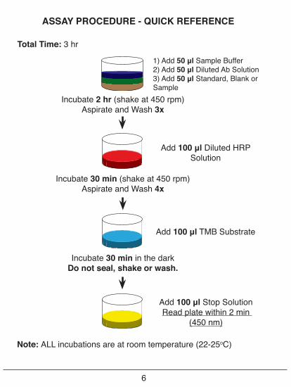
ASSAY PROCEDURE - QUICK REFERENCE
6
Total Time: 3 hr
Note: ALL incubations are at room temperature (22-25oC)
Incubate 2 hr (shake at 450 rpm)Aspirate and Wash 3x
Incubate 30 min (shake at 450 rpm)Aspirate and Wash 4x
Incubate 30 min in the darkDo not seal, shake or wash.
1) Add 50 μl Sample Buffer2) Add 50 μl Diluted Ab Solution3) Add 50 μl Standard, Blank or Sample
Add 100 μl Diluted HRP Solution
Add 100 μl TMB Substrate
Add 100 μl Stop SolutionRead plate within 2 min
(450 nm)

PREPARATION OF REAGENTS
Before starting the assay, the pre-coated plate, Wash Solution Concentrate, Standard Diluent, Sample Buffer, Assay Diluent, TMB Substrate, Stop Solution, applicable dilution matrices (e.g. tissue culture media, serum) and samples should be equilibrated to room temperature (RT), 22-25°C. The Cyno IFN-β Standard, Antibody Concentrate and HRP Conjugate Concentrate should be kept on ice (4°C) throughout the assay.
Wash Solution: Wash Solution Concentrate may contain crys-tals; place the bottle in a warm water bath and gently mix until completely dissolved. Prepare a 1:10 working wash solution (e.g. add 50 ml of Wash Solution Concentrate to 450 ml of distilled or deionized water). Mix thoroughly before use. Diluted Wash Solu-tion can be stored at RT (22-25°C) when not in use.
Antibody Solution: Dilute Antibody Concentrate with Assay Diluent. Refer to the lot specific Certificate of Analysis (CoA) for the correct amount of Antibody Concentrate to use. Prepare 15 minutes prior to use in step 1 and keep at RT (22-25°C).
HRP Solution: Dilute HRP Conjugate Concentrate with Assay Diluent. Refer to the lot specific Certificate of Analysis (CoA) forthe correct amount of HRP Conjugate Concentrate to use. Prepare 15 minutes prior to use in step 2 and keep at RT (22-25°C).
Cynomolgus IFN-β Solution: Using the Cyno IFN-β Standard, construct a standard curve in Standard Diluent or your Sample Matrix as shown in Figure 1. Examples of these matrices are IFN-β free cynomolgus serum, plasma or tissue culture medium containing 10% FBS. If Sample Matrix is not available, Standard Diluent may be used to prepare the standard curve.
7

Standard Curve Preparation:a) Prepare a 1:10 working stock of Cyno IFN-β standard by pi-
petting 10 μl of IFN standard into 90 μl of Standard Diluent or Sample Matrix. Mix thoroughly by gently pipetting up and down 10 times.
b) Label seven polypropylene tubes (S1-S7).c) Add indicated volumes of Sample Matrix or Standard Diluent
to the labeled tubes as indicated in Figure 1. d) Using polypropylene tips, add 17.5 μl of pre-diluted Cyno
IFN-β Standard to S7 and mix thoroughly by pipetting up and down 10 times to recover all material adhered to the inside of pipette tip.
e) Remove indicated amount from S7 and add to S6. Mix thor-oughly by pipetting up and down 5 times. Repeat to complete series to S1. Change tips between each serial dilution.
Sample Dilution Label S7 S6 S5 S4 S3 S2 S1 Blank
Dilution Matrix Vol. (μl) 482.5 250 250 250 250 250 250 250
IFN-β Conc. (pg/ml) 350 175 87.5 43.75 21.88 10.94 5.47 0
Sample Preparation: Prepare test samples of unknown IFN concentration. Measurements in duplicate are recommended.
8
Figure 1: 7-Point Standard Curve Prepared in Standard Diluent

ASSAY PROCEDURE
All incubations should be performed at RT (22-25°C), keeping the plate away from drafts and other temperature fluctuations. Set plate shaker speed to 450 rpm where indicated. Use plate sealers to cover the plate as directed. During all wash steps, remove contents of plate by inverting and shaking over a sink and blotting the plate on lint-free absorbent paper; tap the plate. Wash each well with a minimum of 300 μl of diluted Wash Solution for each wash step. Improper washing may result in increased background values and poor coefficient of variation (%CV) values. Refer to Preparation of Reagents for details on dilution of concentrated solutions. Any alteration of the described procedures can directly affect assay performance.
Figure 2: Example of a Typical Plate Setup
Sa
Sa
Sa
Sa
Sa
Sa
Sa
Sa
Sa
Sa
Sa
Sa
Sa
Sa
Sa
Sa
Sa
Sa
Sa
Sa
Sa
Sa
Sa
Sa
Sa
Sa
Sa
Sa
Sa
Sa
Sa
Sa
Sa
Sa
Sa
Sa
Sa
Sa
Sa
Sa
Sa
Sa
Sa
Sa
Sa
Sa
Sa
Sa
Sa
Sa
Sa
Sa
Sa
Sa
Sa
Sa
Sa
Sa
Sa
Sa
Sa
Sa
Sa
Sa
Sa
Sa
Sa
Sa
Sa
Sa
Sa
Sa
Sa
Sa
Sa
Sa
Sa
Sa
Sa
Sa
S4
S5
S6
S7
B
S1
S2
S3
S4
S5
S6
S7
B
S2
S3
E
F
G
H
C
D
A
B S1
3 4 5 6 7 8 9 10 11 121 2
1. Standards and Test Samples: Determine the number of microplate strips required to test the desired number of samples plus the appropriate number of wells needed to run blanks and standards. We recommend running the standard, blanks and samples in duplicate or triplicate (see Figure 2 for an example setup). A standard curve is required for each assay. Remove extra
9
B = BlankS1-S7 = Std CurveSa = Samples

microplate strips from the frame, seal in the foil bag provided and store at 2-8°C. Unused strips can be used in later assays.
Add 50 μl Sample Buffer to each well.Add 50 μl of diluted Antibody Solution (refer to Preparation of Reagents) to each well. Add 50 μl of Standard, Blank or Test Sample to each well.(For Blank, add Standard Diluent or relevant sample matrix.)(Total volume = 150 μl/well)
Cover with plate sealer and shake plate at 450 rpm at RT (22-25°C) for 2 hours.
After 2 hours, empty the contents of the plate and wash wells three times with 300 μl of diluted Wash Solution (refer to Preparation of Reagents).
2. HRP Solution: Add 100 μl of diluted HRP solution (refer to Preparation of Reagents) to each well. Cover with plate sealer and shake plate at 450 rpm at RT (22-25°C) for 30 minutes.
After 30 minutes, empty the contents of the plate and wash wells four times with 300 μl of diluted Wash Solution.
3. TMB Substrate: Add 100 μl of TMB Substrate Solution to each well. Incubate, in the dark, at RT (22-25°C) for 30 minutes. Do not use a plate sealer and do not shake during the incubation.
4. Stop Solution: After the 30 minute incubation of TMB, DO NOT EMPTY THE WELLS AND DO NOT WASH. Add 100 μl of Stop Solution to each well.
5. Read: Using a microplate reader, determine the absorbance at 450 nm within 2 minutes after the addition of the Stop Solution.
10

CALCULATION OF RESULTS
By plotting the optical densities (OD) using a 4-parameter logistic fit for the standard curve, the interferon titer in the samples can be determined. Based on user preference, blank ODs may be subtracted from the standard and sample ODs to eliminate background.
A shift in optical densities is typical between users and kit lots. The back fit concentration interpolated from the standard curve is a more accurate determination of the sample titer and performance of the kit. Variations from the typical curve provided can be a result of operator technique, altered incubation time, fluctuations in temperature or kit age.
Results of a typical standard curve using a 4-parameter logistic fit are provided for demonstration only and should not be used to obtain test results. A standard curve must be run for each set of samples assayed.
Figure 3: Typical Standard Curve in Standard Diluent
11
Cynomolgus IFN Beta (pg/ml)
1 10 100 10000
1
2
3
Graph#1
4-P Fit: y = (A - D)/( 1 + (x/C)^B ) + D: A B C D R^2Plot#1 (SampleDiluent: Concentration vs MeanV... 0.0582 1.08 693 9.2 1
__________Weighting: 1/MeanValue@SampleDiluent
Abso
rban
ce (4
50 n
m)
Cynomolgus IFN-β (pg/ml)

REFERENCES
1. Krause CD, Pestka S. Evolution of the Class 2 cytokines and receptors, and discovery of new friends and relatives. Pharmacol Ther. 2005 Jun;106(3):299-346.
2. Weinstock-Guttman B, Ramanathan M, Zivadinov R. Interferon-beta treatment for relapsing multiple sclerosis. Expert Opin Biol Ther. 2008 Sep;8(9):1435-47.
3. Lee MS, Kim YJ. Pattern-recognition receptor signaling initiated from extracellular, membrane, and cytoplasmic space. Mol Cells. 2007 Feb 28;23(1):1-10.
4. Heim MH. The Jak-STAT pathway: cytokine signaling from the receptor to the nucleus. J Recept Signal Transduct Res. 1999 Jan-Jul;19(1-4):75-120.
5. Taniguchi T, Takaoka A. The interferon-alpha/beta system in antiviral responses: a multimodal machinery of gene regulation by the IRF family of transcription factors. Curr Opin Immunol. 2002 Feb;14(1):111-6.
6. Lewerenz M, Mogensen KE, Uzé G. Shared receptor components but distinct complexes for alpha and beta interferons. J Mol Biol. 1998 Sep 25;282(3):585-99.
7. Jaks E, Gavutis M, Uzé G, Martal J, Piehler J. Differential receptor subunit affinities of type I interferons govern differential signal activation. J Mol Biol. 2007 Feb 16;366 (2):525- 39.
12

8. Marijanovic Z, Ragimbeau J, van der Heyden J, Uzé G, Pellegrini S. Comparable potency of IFNalpha2 and IFN beta on immediate JAK/ST A T activation but differential downregulation of IFNAR2. Biochem J. 2007 Oct 1;407(1):141-51.
9. Taniguchi T , T akaoka A. A weak signal for strong responses: interferon-alpha/beta revisited. Nat Rev Mol Cell Biol. 2001 May;2(5):378-86.
10. Aziz N, Nishanian P, Mitsuyasu R, Detels R, Fahey JL. Variables That Affect Assays for Plasma Cytokines and Soluble Activation Markers. Clin Diagn Lab Immunol. 1999 Jan: (6)1:89-95.
13

PLATE LAYOUTUse this plate layout as a record of standards and samples assayed.
14

NOTES
15

PBL Assay Science131 Ethel Road West, Suite 6, Piscataway, NJ 08854 USA
Tel: +1 732-777-9123, Fax: +1 732-777-9141Email: [email protected], Website: www.pblassaysci.com
© 2018 Pestka Biomedical Laboratories, Inc. All rights reserved.