ventilator-associated event (vae) - centers for disease ... · pdf fileventilator-associated...
TRANSCRIPT

Device-associated Module
VAE
January2018 10-1
Ventilator-Associated Event (VAE)
For use in adult locations only
Table of Contents:
Introduction 1
Settings 3
Definitions 3
Reporting Instructions 16
Figure 1 VAE Algorithm 19
Numerator Data 23
Denominator Data 23
Data Analyses 23
References 25
Appendix of Antimicrobial Agents 27
Frequently-Asked Questions 29
Introduction: Mechanical ventilation is an essential, life-saving therapy for patients with
critical illness and respiratory failure. Studies have estimated that more than 300,000 patients
receive mechanical ventilation in the United States each year [1-3]. These patients are at high
risk for complications and poor outcomes, including death [1-5]. Ventilator-associated
pneumonia (VAP), sepsis, Acute Respiratory Distress Syndrome (ARDS), pulmonary embolism,
barotrauma, and pulmonary edema are among the complications that can occur in patients
receiving mechanical ventilation; such complications can lead to longer duration of mechanical
ventilation, longer stays in the ICU and hospital, increased healthcare costs, and increased risk of
disability and death. Mortality in patients with acute lung injury on mechanical ventilation has
been estimated to range from 24% in persons 15-19 years of age to 60% for patients 85 years and
older [4].
Surveillance for ventilator-associated events in the National Healthcare Safety Network (NHSN)
prior to 2013 was limited to VAP. For the year 2012, VAP incidence for various types of
hospital units ranged from 0.0-4.4 per 1,000 ventilator days [6]. However, there is currently no
valid, reliable definition for VAP, and even the most widely-used VAP criteria and definitions
are neither sensitive nor specific [7-10].
A particular difficulty with many commonly-used VAP definitions, including the NHSN PNEU
definitions (revised in 2002), is that they require radiographic findings of pneumonia. Evidence
suggests that chest radiograph findings do not accurately identify VAP. The subjectivity and
variability inherent in chest radiograph technique, interpretation, and reporting make chest
imaging ill-suited for inclusion in a definition algorithm to be used for the potential purposes of

Device-associated Module
VAE
January2018 10-2
public reporting, inter-facility comparisons, and pay-for-reporting and pay-for-performance
programs. Another major difficulty with available VAP definitions is their reliance on specific
clinical signs or symptoms, which are subjective and may be poorly or inconsistently
documented in the medical record. The NHSN PNEU protocol includes multiple definition
pathways and special criteria for selected patient populations (for example, children,
immunocompromised patients), increasing its complexity.
The limitations of VAP surveillance definitions have implications for prevention. Valid and
reliable surveillance data are necessary for assessing the effectiveness of prevention strategies. It
is notable that some of the most effective measures for improving outcomes of patients on
mechanical ventilation do not specifically target pneumonia prevention [11-14].
In 2011, CDC convened a Working Group composed of members of several stakeholder
organizations to address the limitations of the NHSN PNEU definitions and propose a new
approach to surveillance for Ventilator-Associated Events (VAE) for NHSN [15]. The
organizations represented in the Working Group include: the Critical Care Societies
Collaborative (the American Association of Critical-Care Nurses, the American College of Chest
Physicians, the American Thoracic Society, and the Society for Critical Care Medicine); the
American Association for Respiratory Care; the Association of Professionals in Infection Control
and Epidemiology; the Council of State and Territorial Epidemiologists; the Healthcare Infection
Control Practices Advisory Committee’s Surveillance Working Group; the Infectious Diseases
Society of America; and the Society for Healthcare Epidemiology of America.
The VAE surveillance definition algorithm developed by the Working Group and implemented
in the NHSN in January 2013 is based on objective, streamlined, and potentially automatable
criteria that identify a broad range of conditions and complications occurring in mechanically-
ventilated adult patients [16]. Several modifications to the VAE definitions have been made
since January 2013. These modifications address issues raised by NHSN users and discussed
with the Working Group. There are three definition tiers within the VAE algorithm: 1)
Ventilator-Associated Condition (VAC); 2) Infection-related Ventilator-Associated
Complication (IVAC); and 3) Possible VAP (PVAP). Data indicate that streamlined, objective
algorithms to detect ventilator-associated complications (similar to the VAC tier of the VAE
algorithm) are easily implemented, can make use of electronic health record systems to automate
event detection, and identify events that are clinically important and associated with outcomes
such as ICU and hospital length of stay and mortality [16,17]. Research suggests that most VACs
are due to pneumonia, ARDS, atelectasis, and pulmonary edema [16]. These are significant
clinical conditions that may be preventable. VAE rates and event characteristics in 2014 in adult
inpatient locations reporting data to NHSN have been published [18].
NOTE: The VAE definition algorithm is for use in surveillance; it is not a clinical definition
algorithm and is not intended for use in the clinical management of patients. Examples provided
throughout this protocol and in the VAE “Frequently-Asked Questions” are for illustration
purposes only and are not intended to represent actual clinical scenarios.

Device-associated Module
VAE
January2018 10-3
Settings: Inpatient locations eligible to participate in VAE surveillance are those adult locations
in acute care hospitals, long term acute care hospitals, and inpatient rehabilitation facilities where
denominator data (ventilator and patient days) can be collected for patients. Such locations may
include critical/intensive care units (ICU), specialty care areas (SCA), step-down units and
wards. A complete listing of adult inpatient locations can be found in Chapter 15.
NOTE: Non-acute care locations in acute care facilities are not eligible to participate in VAE
surveillance.
NOTE: It is not required to monitor for VAEs after discharge if a patient is transferred to another
facility while still on mechanical ventilation. However, VAEs discovered within 2 calendar days
of discharge (where the day of discharge is day 1) should be reported to NHSN. No additional
ventilator days are reported.
Definitions:
VAE: VAEs are identified by using a combination of objective criteria: deterioration in
respiratory status after a period of stability or improvement on the ventilator, evidence of
infection or inflammation, and laboratory evidence of respiratory infection. The following pages
outline the criteria that must be used for meeting the VAE surveillance definitions (Figure 1). To
report VAEs, use the Ventilator-Associated Event form (CDC 57.112) and Instructions for
Completion.
NOTE: Patients must be mechanically ventilated for at least 4 calendar days to fulfill
VAE criteria (where the day of intubation and initiation of mechanical ventilation is day
1). The earliest date of event for VAE (the date of onset of worsening oxygenation) is day
3 of mechanical ventilation. Line lists of VAE data elements demonstrating scenarios that
meet and do not meet the VAE definitions are presented in “Frequently-Asked Questions
(FAQs)” number (no.) 2 at the end of this chapter.
NOTE: The baseline period of stability or improvement on the ventilator is defined as the
2 calendar days immediately preceding the first day of increased daily minimum PEEP or
FiO2, and must be characterized by ≥ 2 calendar days of stable or decreasing daily
minimum FiO2 or PEEP values (specifically the daily minimum PEEP or FiO2 on the
second day of the baseline period of stability or improvement must be equal to or less
than the daily minimum PEEP or FiO2 on the first day of the baseline period of stability
or improvement). The definitions of “daily minimum PEEP” and “daily minimum FiO2”
are included below. Note that the minimum daily PEEP or FiO2 used for VAE
surveillance is the lowest setting during a calendar day that was maintained for > 1 hour
(see daily minimum PEEP and FiO2 definitions for exception to 1 hour requirement).

Device-associated Module
VAE
January2018 10-4
For the purposes of VAE surveillance, PEEP values between 0 cmH2O and 5 cmH2O will
be considered equivalent. This means that patients with daily minimum PEEP values
from 0 to 5 cmH2O must then have an increase in the daily minimum PEEP to at least 8
cmH2O, sustained for at least 2 calendar days, to meet the VAC definition.
EXAMPLE: In the example below, the baseline period is defined by mechanical
ventilation (MV) days 1 through 4 (shaded in light gray), and the period of worsening
oxygenation by MV days 5 and 6 (shaded in darker gray), where the daily minimum
PEEP is ≥ 3 cmH2O greater than the daily minimum PEEP of the first day in the baseline
period. Note that there is no VAC on MV day 3, because PEEP values 0-5 cmH2O are
considered equivalent for the purposes of this surveillance.
EXAMPLE: In the example below, the baseline period is defined by mechanical
ventilation (MV) days 1 through 4 (shaded in light gray), and the period of worsening
oxygenation by MV days 5 and 6 (shaded in darker gray), where the daily minimum
PEEP is ≥ 3 cmH2O greater than the daily minimum PEEP of the first day in the baseline
period. In this example, note that MV days 1-4 are considered a baseline period even
though the daily minimum PEEP increases from 0 to 3 to 5 cmH2O during this time
period—because PEEP values from 0-5 cmH2O are considered equivalent for the
purposes of this surveillance.
MV Day Daily minimum PEEP (cmH2O)
Daily minimum FiO2 (oxygen concentration, %)
VAE
1 0 (5) 1.00 (100%) -
2 0 (5) 0.50 (50%) -
3 5 0.50 (50%) -
4 5 0.50 (50%) -
5 8 0.50 (50%) VAC
6 8 0.50 (50%) -
MV Day Daily minimum PEEP (cmH2O)
Daily minimum FiO2 (oxygen concentration, %)
VAE
1 0 (5) 1.00 (100%) -
2 0 (5) 0.50 (50%) -
3 3 (5) 0.50 (50%) -
4 5 0.50 (50%) -
5 8 0.50 (50%) VAC
6 8 0.50 (50%) -

Device-associated Module
VAE
January2018 10-5
EXAMPLE: In the example below, the baseline period is defined by mechanical
ventilation (MV) days 3 and 4 (shaded in light gray), and the period of worsening
oxygenation by MV days 5 and 6 (shaded in darker gray), where the daily minimum FiO2
is ≥ 0.20 (20 points) over the daily minimum FiO2 of the first day in the baseline period.
EXAMPLE: In the example below, there is no VAC, because the FiO2 on MV day 4 is
higher than the FiO2 on MV day 3 (and therefore not stable or decreasing) – even though
the FiO2 on MV days 3 and 4 meets the 20-point threshold when compared with the daily
minimum FiO2 on MV days 5 and 6.
NOTE: Patients on high frequency ventilation or extracorporeal life support are
EXCLUDED from VAE surveillance.
NOTE: Patients who are receiving a conventional mode of mechanical ventilation while
in the prone position and patients who are receiving a conventional mode of mechanical
ventilation while receiving nitric oxide therapy, helium-oxygen mixtures (heliox) or
epoprostenol therapy are INCLUDED in VAE surveillance.
NOTE: Patients on Airway Pressure Release Ventilation (APRV) or related modes (see
FAQ nos. 22 and 23), are INCLUDED, but the VAE period of stability or improvement
on the ventilator and the period of worsening oxygenation should be determined by
changes in FiO2 only, since changes in PEEP as indicated in this surveillance algorithm
may not be applicable to APRV. In addition, patients with VAE who are on APRV or
related modes of mechanical ventilation can optionally be indicated as such on the VAE
Form (CDC 57.112).
NOTE: VAEs are defined by a 14-day period, starting on the day of onset of worsening
oxygenation (the event date, day 1). A new VAE cannot be identified or reported until
this 14-day period has elapsed. See FAQ no. 4.
MV Day Daily minimum PEEP (cmH2O)
Daily minimum FiO2 (oxygen concentration, %)
VAE
1 8 1.00 (100%) -
2 6 0.50 (50%) -
3 5 0.40 (40%) -
4 5 0.40 (40%) -
5 6 0.70 (70%) VAC
6 6 0.70 (70%) -
MV Day Daily minimum PEEP (cmH2O)
Daily minimum FiO2 (oxygen concentration, %)
VAE
1 8 1.0 (100%)
2 6 0.50 (50%)
3 5 0.35 (35%)
4 5 0.40 (40%)
5 6 0.70 (70%) No event
6 6 0.70 (70%)
Device-associated Module
VAE
January2018 10-6
Date of event: The date of onset of worsening oxygenation. This is defined as the first calendar
day in which the daily minimum PEEP or FiO2 increases above the thresholds outlined in the
VAE definition algorithm (specifically day 1 of the required ≥ 2-day period of worsening
oxygenation following a ≥ 2-day period of stability or improvement on the ventilator).
EXAMPLE: A patient is intubated in the Emergency Room for severe community-
acquired pneumonia and admitted to the MICU (day 1). The patient stabilizes and
improves on days 2-5, with a daily minimum FiO2 of 0.35 (35%) on days 4 and 5. On day
6, the patient experiences respiratory deterioration, and requires a minimum FiO2 of 0.60
(60%) on days 6 and 7, meeting the criteria for a VAC. The date of the VAC event is day
6.
NOTE: The “date of event” is NOT the date on which all VAE criteria have been met. It
is the first day (of a ≥ 2-day period) on which either of the worsening oxygenation
thresholds (for PEEP or FiO2) is met.
VAE Window Period: This is the period of days around the event date (specifically the day of
onset of worsening oxygenation) within which other VAE criteria must be met. It is usually a 5-
day period and includes the 2 days before, the day of, and the 2 days after the VAE event date
(specifically the first day of worsening oxygenation, the day of VAE onset). There is an
exception, however, in which the VAE Window Period is only 3 or 4 days, as follows:
In cases where the VAE event date corresponds to MV day 3 or day 4, the window period
described above may only be a 3-day or a 4-day window, because it can NOT include any
days before the 3rd day of MV. For example, if the VAE event date is MV day 3, then the
window period includes only the day of VAE onset and the 2 days after VAE onset
(because the 2 days before VAE onset are before the 3rd day of MV).
Positive End-Expiratory Pressure (PEEP): “A technique used in respiratory therapy in which
airway pressure greater than atmospheric pressure is achieved at the end of exhalation by the
introduction of a mechanical impedance to exhalation” [19]. In patients on mechanical
ventilation, PEEP is one of the key parameters that can be adjusted depending on the patient’s
oxygenation needs, and is typically in the range of 0 to 15 cmH2O. A sustained increase (defined
later in this protocol) in the daily minimum PEEP of ≥ 3 cmH2O following a period of stability
or improvement on the ventilator is one of two criteria that can be used in meeting the VAC
definition. For the purposes of this surveillance, PEEP values from 0 to 5 cmH2O are considered
equivalent.
Fraction of inspired oxygen (FiO2): The fraction of oxygen in inspired gas. For example, the
FiO2 of ambient air is 0.21; the oxygen concentration of ambient air is 21%. In patients on
mechanical ventilation, the FiO2 is one of the key parameters that can be adjusted depending on
the patient’s oxygenation needs, and is typically in the range of 0.30 (oxygen concentration of
30%) to 1.0 (oxygen concentration of 100%). A sustained increase (defined later in this protocol)

Device-associated Module
VAE
January2018 10-7
in the daily minimum FiO2 of ≥ 0.20 (20%) following a period of stability or improvement on the
ventilator is the second of the two criteria that can be used in meeting the VAC definition.
Daily minimum PEEP: The lowest value of PEEP during a calendar day that is set on the
ventilator and maintained for > 1 hour. This requirement that the daily minimum PEEP be the
lowest setting maintained for > 1 hour will ensure that units monitoring and recording PEEP
settings hourly or more frequently than once per hour are able to apply the VAE surveillance
PEEP criterion in a standardized way. In the event that ventilator settings are monitored and
recorded less frequently than once per hour, the daily minimum PEEP is simply the lowest value
of PEEP set on the ventilator during the calendar day. In circumstances where there is no value
that is documented to have been maintained for > one hour (for example., the lowest value of
PEEP is set late in the calendar day, mechanical ventilation is discontinued early in the calendar
day, PEEP settings are changed very frequently throughout the calendar day) the daily minimum
PEEP should default to the lowest PEEP setting during the calendar day (regardless of how long
that setting was maintained). For example, a patient who is intubated and started on mechanical
ventilation at 11:30 pm on June 1, with a PEEP setting of 10 cmH2O from 11:30 pm to midnight,
would have a daily minimum PEEP of 10 cmH2O on June 1 for the purposes of VAE
surveillance.
NOTE: In units tracking PEEP settings every hour or more frequently than every hour,
there must be sufficient consecutive recordings of a specific PEEP setting to meet the
minimum required duration of > 1 hour. For example, in units tracking PEEP every 15
minutes, 5 consecutive recordings of PEEP at a certain level would be needed to meet the
required >1 hour minimum duration (for example., at 09:00, 09:15, 09:30, 09:45 and
10:00). In units tracking PEEP every 30 minutes, 3 consecutive recordings of PEEP at a
certain level would be needed to meet the required >1 hour minimum duration (for
example, at 09:00, 09:30, and 10:00). In units tracking PEEP every hour, 2 consecutive
recordings of PEEP at a certain level would be needed to meet the required >1 hour
minimum duration (for example, at 09:00 and 10:00).
EXAMPLE: The patient is intubated at 6 pm. PEEP is set at the following values through
the remainder of the calendar day:
Time 6 pm 7 pm 8 pm 9 pm 10 pm 11 pm
PEEP
(cmH2O)
10 8 5 5 8 8
In this example, the daily minimum PEEP for the purposes of VAE surveillance is 5
cmH2O. PEEP settings are being monitored and recorded every hour. There are two
consecutive hours where the PEEP setting is noted to be 5 cmH2O (8 pm and 9 pm), and
therefore required minimum duration of >1 hour is met.

Device-associated Module
VAE
January2018 10-8
EXAMPLE: The patient is intubated at 6 pm. PEEP is set at the following values through
the remainder of the calendar day:
Time 6 pm 7 pm 8 pm 9 pm 10 pm 11 pm
PEEP
(cmH2O)
8 8 5 8 5 8
In this example, the daily minimum PEEP for the purposes of VAE surveillance is 8
cmH2O. PEEP settings are being monitored and recorded every hour. Although the
lowest PEEP is 5 cmH2O, it is recorded at two non-consecutive time points only (8 pm,
then 10 pm), and so the required >1 hour minimum duration is not met. There are two
consecutive hours where the PEEP setting is noted to be 8 cmH2O (6 pm and 7 pm), and
therefore the required minimum duration of >1 hour is met to allow use of this setting as
the daily minimum value for VAE surveillance.
EXAMPLE: PEEP is set at the following values through the course of a calendar day:
Time 12 am 4 am 8 am 12 pm 4 pm 8 pm
PEEP
(cmH2O)
5 8 5 8 8 10
In this example, the daily minimum PEEP is 5 cmH2O. PEEP settings are being
monitored and recorded every 4 hours; therefore the lowest recorded PEEP setting for the
calendar day is the value used in VAE surveillance.
EXAMPLE: You are reviewing a patient’s ventilator settings on Wednesday morning to
determine the daily minimum PEEP values for Monday and Tuesday. The MICU
monitors and records PEEP settings for mechanically ventilated patients every 30
minutes. You see that the lowest PEEP setting on Monday (5 cmH2O) was recorded at
11:30 pm when the episode of mechanical ventilation was initiated for this patient. The
patient remained at this PEEP setting for an additional 30 minutes on Tuesday morning,
and was then maintained on PEEP 10 cmH2O for the rest of the day on Tuesday. What do
you record as the daily minimum PEEP for Monday and for Tuesday? In this example,
the only PEEP setting recorded on Monday was 5 cmH2O. Because there is no value on
Monday that has been maintained for > one hour, the lowest (and only) setting of 5
cmH20 is recorded as the daily minimum PEEP for that calendar day. On Tuesday, the
daily minimum PEEP should be recorded as 10 cmH2O, which is the lowest PEEP setting
maintained for > 1 hour on Tuesday.

Device-associated Module
VAE
January2018 10-9
Day Time PEEP (cmH2O)
Monday 23:30 5
Tuesday 00:00 5
Tuesday 00:30 5
Tuesday 01:00 10
Tuesday 01:30 10
Tuesday 02:00 through 23:30 10
Daily minimum FiO2: The lowest value of FiO2 during a calendar day that is set on the ventilator
and maintained for > 1 hour. This requirement that the daily minimum FiO2 be the lowest setting
maintained for > 1 hour will ensure that units monitoring and recording FiO2 settings hourly or
more frequently than once per hour are able to apply the VAE surveillance FiO2 criterion in a
standardized way. In the event that ventilator settings are monitored and recorded less frequently
than once per hour, the daily minimum FiO2 is simply the lowest value of FiO2 set on the
ventilator during the calendar day. Similarly, in circumstances where there is no value that has
been maintained for > 1 hour (for example , the lowest value of FiO2 is set late in the calendar
day, mechanical ventilation is discontinued early in the calendar day) the daily minimum FiO2 is
the lowest value of FiO2 set on the ventilator during the calendar day.
NOTE: In units tracking FiO2 settings every hour or more frequently than every hour,
there must be sufficient consecutive recordings of a specific FiO2 setting to meet the
minimum required duration of >1 hour. For example, in units tracking FiO2 every 15
minutes, 5 consecutive recordings of FiO2 at a certain level would be needed to meet the
required >1 hour minimum duration (for example, 09:00, 09:15, 09:30, 09:45 and 10:00).
In units tracking FiO2 every 30 minutes, 3 consecutive recordings of FiO2 at a certain
level would be needed to meet the required >1 hour minimum duration (for example,
09:00, 09:30, and 10:00). In units tracking FiO2 every hour, 2 consecutive recordings of
FiO2 at a certain level would be needed to meet the required >1 hour minimum duration
(for example, 09:00 and 10:00).
EXAMPLE: The patient is intubated at 6 pm. FiO2 is set at the following values through
the remainder of the calendar day:
Time 6 pm 7 pm 8 pm 9 pm 10 pm 11 pm
FiO2 1.0 0.8 0.5 0.5 0.8 0.8
In this example, the daily minimum FiO2 for the purposes of VAE surveillance is 0.5.
FiO2 settings are being monitored and recorded every hour. There are two consecutive
hours where the FiO2 setting is noted to be 0.5 (8 pm and 9 pm), and therefore required
minimum duration of >1 hour is met.

Device-associated Module
VAE
January2018 10-10
EXAMPLE: The patient is intubated at 6 pm. FiO2 is set at the following values through
the remainder of the calendar day:
Time 6 pm 7 pm 8 pm 9 pm 10 pm 11 pm
FiO2 0.8 0.8 0.5 0.8 0.5 0.8
In this example, the daily minimum FiO2 for the purposes of VAE surveillance is 0.8.
FiO2 settings are being monitored and recorded every hour. Although the lowest FiO2 is
0.5, it is recorded at two non-consecutive time points only (8 pm, and then 10 pm), and so
the required 1 hour minimum duration is not met. There are two consecutive hours where
the FiO2 setting is noted to be 0.8 (6 pm and 7 pm), and therefore the required minimum
duration of >1 hour is met to allow use of this setting as the daily minimum value for
VAE surveillance.
EXAMPLE: FiO2 is set at the following values through the course of a calendar day:
Time 2 pm 4 pm 6 pm 8 pm 10 pm 12 am
FiO2 1.0 0.60 0.40 0.50 0.55 0.60
In this example, the patient was intubated at 2 pm. The daily minimum FiO2 is 0.40. FiO2
settings are being monitored and recorded every 2 hours; therefore, the lowest recorded
FiO2 setting for the calendar day is the value used in VAE surveillance.
EXAMPLE: You are reviewing a patient’s ventilator settings on Friday morning to
determine the daily minimum FiO2 value for Thursday. The patient was intubated and
initiated on mechanical ventilation at 21:45 hours on Thursday. The ICU monitored and
recorded FiO2 settings for the patient every 15 minutes during the remainder of the day
on Thursday. Based on the information recorded in the table below, what should you
record as the daily minimum FiO2 for Thursday? In this example, since there is no setting
that is maintained for > 1 hour during the calendar day, the daily minimum FiO2 for
Thursday is 0.70 (70%). This is the lowest value of FiO2 set on the ventilator during the
calendar day.
Day Time FiO2 Thursday 21:45 Intubated; 1.0
22:00 1.0
22:15 0.90
22:30 0.90
22:45 0.70
23:00 0.80
23:15 0.85
23:30 0.85
23:45 0.85

Device-associated Module
VAE
January2018 10-11
Ventilator: a device used to support, assist or control respiration (inclusive of the weaning
period) through the application of positive pressure to the airway when delivered via an artificial
airway, specifically oral/nasal endotracheal or tracheostomy tube.
Note: Ventilation and lung expansion devices that deliver positive pressure to the airway (for
example: CPAP, Bipap, bi-level, IPPB and PEEP) via non-invasive means (for example: nasal
prongs, nasal mask, full face mask, total mask, etc.) are not considered ventilators unless positive
pressure is delivered via an artificial airway (oral/nasal endotracheal or tracheostomy tube).
Episode of mechanical ventilation: Defined as a period of days during which the patient was
mechanically ventilated for some portion of each consecutive day.
NOTE: A break in mechanical ventilation of at least one full calendar day, followed by
reintubation and/or reinitiation of mechanical ventilation during the same hospitalization,
defines a new episode of mechanical ventilation.
EXAMPLE: A patient is intubated and mechanical ventilation is initiated at 11 pm on
hospital day 1. The patient remains intubated and mechanically ventilated from hospital
days 2-10. The patient is extubated at 9 am on hospital day 11, and remains extubated on
hospital day 12. The patient is reintubated and mechanical ventilation is reinitiated on
hospital day 13. The patient remains intubated and mechanically ventilated from hospital
day 14-18. This patient has had two episodes of mechanical ventilation (days 1-11 and
days 13-18), separated by at least one full calendar day off of mechanical ventilation.
New antimicrobial agent: Defined as any agent listed in the Appendix that is initiated on or after
the third calendar day of mechanical ventilation AND in the VAE Window Period (specifically,
the period typically defined by the 2 calendar days before, the day of, and the 2 calendar days
after the onset date of the VAE). The agent is considered new for the purposes of this definition
if it was NOT given to the patient on either of the 2 days preceding the current start date.
EXAMPLE: A patient is intubated and mechanically ventilated on hospital day 1 in the
MSICU. Ceftriaxone and azithromycin are started on day 1 and administered daily. After
3 days of improving respiratory status, the patient’s oxygenation deteriorates on days 4
and 5, with a daily minimum PEEP that is 4 cmH2O higher than it was on days 2 and 3.
Criteria for the VAC definition are met; the date of the event is hospital day 4.
Ceftriaxone is discontinued and meropenem is begun on day 5. Azithromycin is
continued. In this case, meropenem is a new antimicrobial agent: 1) it was begun on day
5 of mechanical ventilation, and 2) within the VAE Window Period (on the day after
VAE onset), and 3) it was not given to the patient on either of the 2 days preceding the
current start date. By contrast, ceftriaxone and azithromycin would not be considered new
antimicrobial agents, since they were begun on day 1 of mechanical ventilation and
continued daily into the VAE Window Period.

Device-associated Module
VAE
January2018 10-12
The antimicrobial agent(s) must have been given by one of the routes of administration outlined
in Table 1, and therapy with one or more new antimicrobial agents must be continued for at least
4 calendar days (referred to as 4 “qualifying antimicrobial days” or “QADs”). For further
guidance on identification of new antimicrobial agents and on how to determine whether the
requirement for 4 QADs is met, refer to FAQs nos. 6-10 at the end of this chapter.
Table 1: Definitions of routes of administration
Route of Administrationa Definitionb
Intravenous An intravascular route that begins with a vein.
Intramuscular A route that begins within a muscle.
Digestive Tract A route that begins anywhere in the digestive tract extending
from the mouth through rectum.
Respiratory Tract A route that begins within the respiratory tract, including the
oropharynx and nasopharynx. aOther routes of administration are excluded (for example, antibiotic locks, intraperitoneal, intraventricular, irrigation,
topical). bDefinitions per SNOMED Reference Terminology
Qualifying Antimicrobial Day (QAD): A day on which the patient was administered an
antimicrobial agent that was determined to be “new” within the VAE Window Period. Four
consecutive QADs are needed to meet the IVAC antimicrobial criterion—starting within the
VAE Window Period. Days on which a new antimicrobial agent is administered count as QADs.
Days between administrations of a new antimicrobial agent also count as QADs as long as there
is a gap of no more than 1 calendar day between administrations. For example, if levofloxacin is
given on VAE Day 1, has not been given in the 2 preceding calendar days, and is given again on
VAE Days 3, 5 and 7, there are 7 QADs—because the days between levofloxacin doses also
count as QADs. By contrast, days between administrations of different antimicrobial agents do
NOT count as QADs; for example, if levofloxacin is given to the patient on VAE Days -2 and -1
only, no antimicrobials are given on VAE Day 1, and meropenem is given only on VAE Day 2
(remember there is no VAE Day 0), then there are not 4 consecutive QADs. VAE Days -2 and -1
count as 2 consecutive QADs, but VAE Day 1 cannot be counted as a QAD because it is a day
between different antimicrobial agents.
Purulent Respiratory Secretions: Defined as secretions from the lungs, bronchi, or trachea that
contain >25 neutrophils and <10 squamous epithelial cells per low power field [lpf, x100].
NOTE: Some clinical laboratories may use different results reporting formats for direct
examinations of respiratory secretions. Additional instructions for using the purulent
respiratory secretions criterion are provided in Table 2, below (see also FAQ no. 19).
Device-associated Module
VAE
January2018 10-13
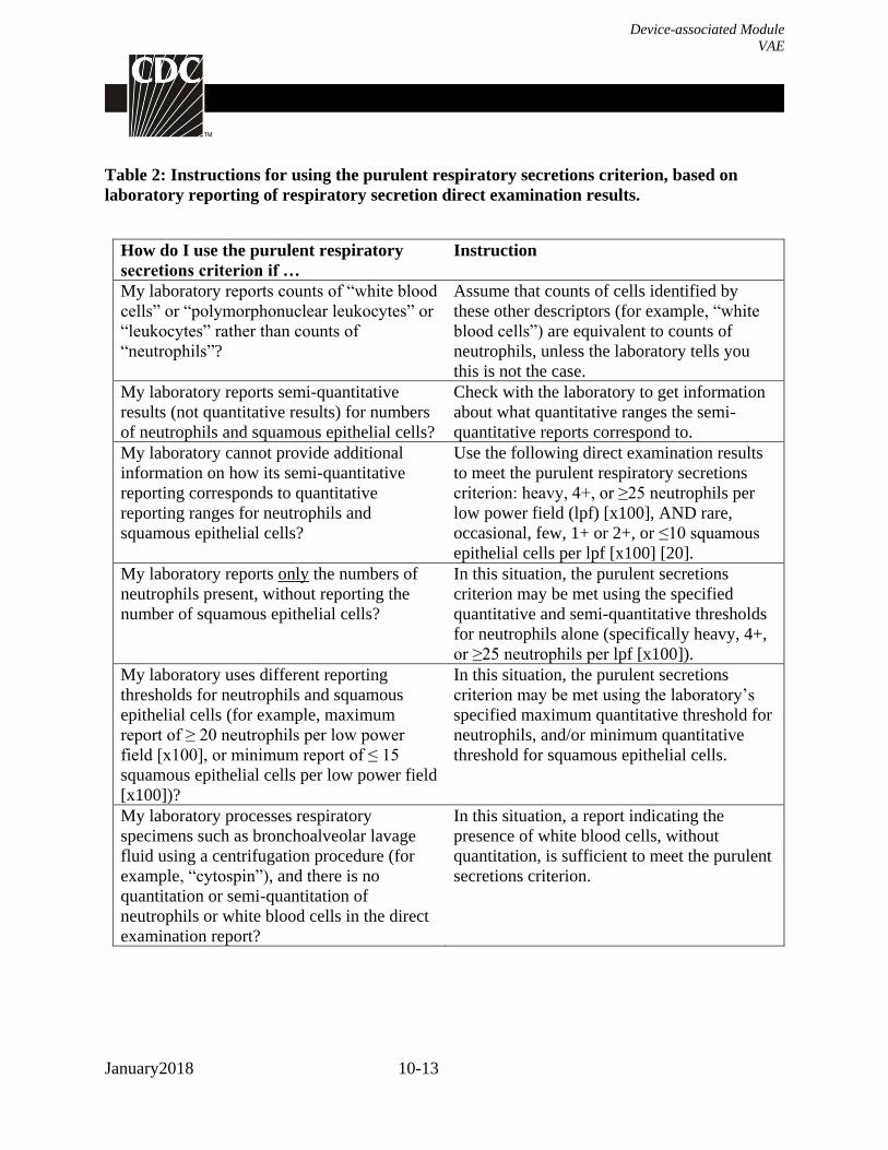
Table 2: Instructions for using the purulent respiratory secretions criterion, based on
laboratory reporting of respiratory secretion direct examination results.
How do I use the purulent respiratory
secretions criterion if …
Instruction
My laboratory reports counts of “white blood
cells” or “polymorphonuclear leukocytes” or
“leukocytes” rather than counts of
“neutrophils”?
Assume that counts of cells identified by
these other descriptors (for example, “white
blood cells”) are equivalent to counts of
neutrophils, unless the laboratory tells you
this is not the case.
My laboratory reports semi-quantitative
results (not quantitative results) for numbers
of neutrophils and squamous epithelial cells?
Check with the laboratory to get information
about what quantitative ranges the semi-
quantitative reports correspond to.
My laboratory cannot provide additional
information on how its semi-quantitative
reporting corresponds to quantitative
reporting ranges for neutrophils and
squamous epithelial cells?
Use the following direct examination results
to meet the purulent respiratory secretions
criterion: heavy, 4+, or ≥25 neutrophils per
low power field (lpf) [x100], AND rare,
occasional, few, 1+ or 2+, or ≤10 squamous
epithelial cells per lpf [x100] [20].
My laboratory reports only the numbers of
neutrophils present, without reporting the
number of squamous epithelial cells?
In this situation, the purulent secretions
criterion may be met using the specified
quantitative and semi-quantitative thresholds
for neutrophils alone (specifically heavy, 4+,
or ≥25 neutrophils per lpf [x100]).
My laboratory uses different reporting
thresholds for neutrophils and squamous
epithelial cells (for example, maximum
report of ≥ 20 neutrophils per low power
field [x100], or minimum report of ≤ 15
squamous epithelial cells per low power field
[x100])?
In this situation, the purulent secretions
criterion may be met using the laboratory’s
specified maximum quantitative threshold for
neutrophils, and/or minimum quantitative
threshold for squamous epithelial cells.
My laboratory processes respiratory
specimens such as bronchoalveolar lavage
fluid using a centrifugation procedure (for
example, “cytospin”), and there is no
quantitation or semi-quantitation of
neutrophils or white blood cells in the direct
examination report?
In this situation, a report indicating the
presence of white blood cells, without
quantitation, is sufficient to meet the purulent
secretions criterion.

Device-associated Module
VAE
January2018 10-14
Location of attribution: The inpatient location where the patient was assigned on the date of the
VAE, which is further defined as the date of onset of worsening oxygenation.
EXAMPLE: Patient is intubated and ventilated in the Operating Room on hospital day 1,
and then is admitted post-operatively to the SICU on hospital day 1, still on the
ventilator. On hospital day 3, the patient experiences the onset of worsening
oxygenation, manifested by an increase in the daily minimum FiO2 of ≥ 0.20 (20%). On
day 4 (also the 4th day of mechanical ventilation) the patient meets criteria for a VAC.
This is reported to NHSN as a VAC for the SICU.
EXCEPTION:
Transfer Rule: If a VAE develops on the day of transfer or the day following transfer
from one inpatient location to another in the same facility or to a new facility (where the
day of transfer is day 1), the event is attributed to the transferring location. This is called
the Transfer Rule, and examples are shown below:
EXAMPLE: Patient on a ventilator in the SICU who has had improving oxygenation for
3 days is transferred to the MICU, still on the ventilator. On the day of transfer, after the
patient has arrived in the MICU, the patient experiences an acute decompensation,
requiring an increase of 0.30 (30 points) in FiO2 that persists during the following
calendar day. VAC criteria are met on calendar day 2 in the MICU. Because the onset of
worsening oxygenation occurred on the day of transfer to the MICU, the VAC event is
attributed to the SICU.
EXAMPLE: Patient is extubated in the MICU and transferred to the medical stepdown
unit on hospital day 6. The next day, while in the stepdown unit (day 7), the patient
experiences worsening oxygenation and is reintubated and transferred back to the MICU.
Criteria for VAC are met the next day (day 8). In this case, the day prior to extubation
and the day of extubation (hospital days 5 and 6) count as the required 2-day period of
stability or improvement. The day of reintubation (day 7) and the following day (day 8)
count as the required 2-day period of worsening oxygenation. Because the onset of
worsening oxygenation occurred on the day following transfer out of the MICU, the
event is reported to NHSN as a VAC for the MICU.
EXAMPLE: Patient intubated and mechanically ventilated for 8 days in the MSICU of
Hospital A is transferred for further care on day 8 to the MSICU of Hospital B. The
patient was stable on the ventilator in Hospital A from days 3-8. On the day of transfer to
Hospital B (day 1 in Hospital B), the patient’s respiratory status deteriorates. The
patient’s respiratory status continues to worsen on the day after transfer (day 2 in
Hospital B), the patient meets criteria for VAC on hospital day 3. The date of the event is
day 2in Hospital B, the first day of the period of worsening oxygenation meeting VAE
PEEP or FiO2 thresholds. The infection preventionist (IP) from Hospital B calls the
Hospital A IP to report that this patient was admitted to Hospital B with a VAC. This

Device-associated Module
VAE
January2018 10-15
VAC should be reported to NHSN for and by Hospital A, and attributed to the Hospital A
MSICU. Date of event was day following transfer. No additional ventilator days are
reported by Hospital A.
REPORTING INSTRUCTIONS (additional guidance may be found in the FAQs at the end of
this chapter):
Conducting in-plan VAE surveillance means assessing patients for the presence of ALL
events included in the algorithm—from VAC to IVAC to PVAP. At this time, a unit
conducting in-plan VAE surveillance cannot decide, for example, that only surveillance
for VAC (and not for IVAC or PVAP) will be performed.
There is a hierarchy of definitions within VAE:
o If a patient meets criteria for VAC and IVAC, report as IVAC.
o If a patient meets criteria for VAC, IVAC and PVAP, report PVAP.
Do not upgrade an event using findings that occur outside the VAE Window period.
If the date of event (date of onset of worsening oxygenation) is on or after the date of
documentation of evidence of consent AND the patient is being supported for organ
donation purposes, the event should not be reported as a VAE.
Pathogens are not reported for VAC or IVAC events.
Secondary BSIs are not reported for VAC or IVAC events (see FAQ no.11).
Pathogens may be reported for PVAP events, provided they are isolated or identified
from appropriate specimen types according to the requirements of the algorithm and are
NOT on the list of excluded organisms and culture or non-culture based microbiologic
testing method results:
o Excluded organisms and culture or non-culture based microbiologic testing method
results that cannot be used to meet the PVAP definition are as follows: “Normal
respiratory flora,” “normal oral flora,” “mixed respiratory flora,” “mixed oral flora,”
“altered oral flora” or other similar results indicating isolation of commensal flora of
the oral cavity or upper respiratory tract; Candida species or yeast not otherwise
specified; coagulase-negative Staphylococcus species; and Enterococcus species,
when identified from sputum, endotracheal aspirates, bronchoalveolar lavage, or
protected specimen brushings specimens. These organisms can be reported as PVAP
pathogens if identified from lung tissue or pleural fluid specimens.
o Additionally, because organisms belonging to the following genera are typically
causes of community-associated respiratory infections and are rarely or are not
known to be causes of healthcare-associated infections, they are also excluded, and
cannot be used to meet the PVAP definition when isolated from any eligible
specimen type (to include lung and pleural fluid): Blastomyces, Histoplasma,
Coccidioides, Paracoccidioides, Cryptococcus and Pneumocystis.
There are three criteria that can be used to meet the PVAP definition (Figure 1):
o Criterion 1: Positive culture meeting specific quantitative or semi-quantitative
threshold (Table 3);

Device-associated Module
VAE
January2018 10-16
o Criterion 2: Purulent respiratory secretions AND identification of organisms
NOT meeting the quantitative or semi-quantitative thresholds specified in Table
3;
o Criterion 3: Organisms identified from pleural fluid specimen, positive lung
histopathology, lower respiratory specimen cytology findings suggestive of
infection and positive diagnostic test for Legionella species or selected
respiratory viruses.
See Table 3 for the required quantitative culture thresholds meeting the PVAP definition
(Criterion 1). Note that if your laboratory reports semi-quantitative culture results, you
should check with your laboratory to confirm that semi-quantitative results match the
quantitative thresholds noted in Table 3 (see also FAQ no. 24).
Table 3: Threshold values for cultured specimens used in the PVAP definition
Specimen collection/technique Values
Lung tissue ≥ 104 cfu/g tissue*
Bronchoscopically (B) obtained specimens
Bronchoalveolar lavage (B-BAL) ≥ 104 cfu/ml*
Protected BAL (B-PBAL) ≥ 104 cfu/ml*
Protected specimen brushing (B-PSB) ≥ 103 cfu/ml*
Nonbronchoscopically (NB) obtained (blind)
specimens
NB-BAL ≥104 cfu/ml*
NB-PSB ≥ 103 cfu/ml*
Endotracheal aspirate (ETA) ≥ 105 cfu/ml*
cfu = colony forming units, g = gram, ml = milliliter
*Or corresponding semi-quantitative result.
Secondary BSIs may be reported for PVAP events, provided that at least one organism
identified from the blood matches an organism isolated from an appropriate respiratory
tract specimen (including respiratory secretions, pleural fluid and lung tissue). The
respiratory tract specimen must have been collected on or after the 3rd day of mechanical
ventilation and within 2 calendar days before or after the day of onset of worsening
oxygenation to be considered as a criterion for meeting the PVAP definition. In addition,
the organisms identified from blood must have been collected during the 14-day event
period, where day 1 is the day of onset of worsening oxygenation (See FAQ no.13).
o In cases where PVAP is met with only the histopathology criterion and no culture or
non-culture based testing is performed on an eligible respiratory specimen, and there
is also a positive blood specimen a secondary BSI is not reported.
o In cases where a culture or non-culture based testing of respiratory secretions, pleural
fluid or lung tissue is performed and does not identify an organism that matches an
organism identified from blood, a secondary BSI is not reported.
o A matching organism is defined as one of the following:

Device-associated Module
VAE
January2018 10-17
1. If genus and species are identified in both specimens, they must be the same.
a. Example: A blood specimen resulted with Enterobacter cloacae and a
BAL specimen resulted with Enterobacter cloacae are matching
organisms.
b. Example: A blood specimen resulted with Enterobacter cloacae and a
BAL specimen resulted with Enterobacter aerogenes are NOT matching
organisms as the species are different.
2. If the organism is less definitively identified in one specimen than the other, the
lesser identified organism must be identified to at least the genus level and at that
level the organisms must be the same.
a. Example: A BAL resulted with Pseudomonas spp. and a blood specimen
resulted with Pseudomonas aeruginosa are considered a match at the
genus level and therefore the BSI can be reported as secondary BSI to
VAE
Exception: In cases where an organism is identified only as “yeast” or
“yeast not otherwise specified”, the organism can be considered a match to
other yeasts, when collected during the required timeframe, whether more
fully identified or not.
Example: A blood specimen reported as Candida albicans and a lung
tissue resulted with yeast not otherwise specified are considered to have
matching organisms. In this example the two organisms are considered
matching organisms because the organisms are complementary
(specifically Candida is a type of yeast). NOTE: This exception is limited
to yeast. It does not apply to identification of organisms as Gram positive
cocci, Gram negative rods, etc.
NOTE: Candida species or yeast not otherwise specified, coagulase-
negative Staphylococcus species, and Enterococcus species identified
from blood cannot be deemed secondary to a PVAP, unless the organism
was also identified from pleural fluid or lung tissue.

Device-associated Module
VAE
January2018 10-18
Figure 1: Ventilator-Associated Events (VAE) Surveillance Algorithm
Ventilator-Associated Condition (VAC)
After a period of stability or improvement on the ventilator, the patient has at least one of the following indicators of worsening oxygenation: 1) Increase in daily minimum* FiO2 of ≥ 0.20 (20 points) over the daily minimum FiO2 of the first day in the baseline period, sustained for ≥ 2
calendar days.2) Increase in daily minimum* PEEP values of ≥ 3 cmH2O over the daily minimum PEEP of the first day in the baseline period†, sustained for ≥ 2
calendar days.*Daily minimum defined by lowest value of FiO2 or PEEP during a calendar day that is maintained for > 1 hour.†Daily minimum PEEP values of 0-5 cmH2O are considered equivalent for the purposes of VAE surveillance.
On or after calendar day 3 of mechanical ventilation and within 2 calendar days before or after the onset of worsening oxygenation, the patient meets both of the following criteria:
1) Temperature > 38 °C or < 36°C, OR white blood cell count ≥ 12,000 cells/mm3 or ≤ 4,000 cells/mm3.AND2) A new antimicrobial agent(s) (see Appendix for eligible antimicrobial agents) is started, and is continued for ≥ 4 calendar days.
Infection-related Ventilator-Associated Complication (IVAC)
On or after calendar day 3 of mechanical ventilation and within 2 calendar days before or after the onset of worsening oxygenation, ONE of the following criteria is met (taking into account organism exclusions specified in the protocol):
1) Criterion 1: Positive culture of one of the following specimens, meeting quantitative or semi-quantitative thresholds as outlined inprotocol, without requirement for purulent respiratory secretions:
Endotracheal aspirate, ≥ 105 CFU/ml or corresponding semi-quantitative result
Bronchoalveolar lavage, ≥ 104 CFU/ml or corresponding semi-quantitative result
Lung tissue, ≥ 104 CFU/g or corresponding semi-quantitative result
Protected specimen brush, ≥ 103 CFU/ml or corresponding semi-quantitative result2) Criterion 2: Purulent respiratory secretions (defined as secretions from the lungs, bronchi, or trachea that contain >25 neutrophils and
<10 squamous epithelial cells per low power field [lpf, x100])† PLUS organism identified from one of the following specimens (to includequalitative culture, or quantitative/semi-quantitative culture without sufficient growth to meet criterion #1):
Sputum
Endotracheal aspirate
Bronchoalveolar lavage
Lung tissue
Protected specimen brush† If the laboratory reports semi-quantitative results, those results must correspond to the above quantitative thresholds. Seeadditional instructions for using the purulent respiratory secretions criterion in the VAE Protocol.
3) Criterion 3: One of the following positive tests:
Organism identified from pleural fluid (where specimen was obtained during thoracentesis or initial placement of chest tubeand NOT from an indwelling chest tube)
Lung histopathology, defined as: 1) abscess formation or foci of consolidation with intense neutrophil accumulation inbronchioles and alveoli; 2) evidence of lung parenchyma invasion by fungi (hyphae, pseudohyphae or yeast forms); 3) evidenceof infection with the viral pathogens listed below based on results of immunohistochemical assays, cytology, or microscopyperformed on lung tissue
Diagnostic test for Legionella species
Diagnostic test on respiratory secretions for influenza virus, respiratory syncytial virus, adenovirus, parainfluenza virus,rhinovirus, human metapneumovirus, coronavirus
*Excludes the following:
Possible Ventilator-Associated Pneumonia (PVAP)
Patient has a baseline period of stability or improvement on the ventilator, defined by ≥ 2 calendar days of stable or decreasing daily minimum* FiO2 or PEEP values. The baseline period is defined as the 2 calendar days immediately preceding the first day of increased daily minimum PEEP or FiO2. *Daily minimum defined by lowest value of FiO2 or PEEP during a calendar day that is maintained for > 1 hour.

Device-associated Module
VAE
January2018 10-19
Numerator Data: The Ventilator-Associated Event form (CDC 57.112) is used to collect and
report each VAE that is identified during the month selected for surveillance. The Instructions
for Completion of Ventilator-Associated Event Form includes brief instructions for collection
and entry of each data element on the form. The VAE form includes patient demographic
information and information on the start date and location of initiation of mechanical ventilation.
Additional data include the specific criteria met for identifying VAE, whether the patient
developed a secondary bloodstream infection, whether the patient died, and, where applicable,
the organisms detected and their antimicrobial susceptibilities.
Reporting Instruction: If no VAEs are identified during the month of surveillance, the “Report
No Events” box must be checked on the appropriate denominator summary screen, for example,
Denominators for Intensive Care Unit (ICU)/other locations (Not NICU or SCA), etc.
Denominator Data: Device days and patient days are used for denominators (see Chapter 16
Key Terms). Ventilator days, which are the numbers of patients managed with ventilatory
devices, are collected daily, at the same time each day, according to the chosen location using the
appropriate form (CDC 57.117 and 57.118). These daily counts are summed and only the total
for the month is entered into NHSN. Ventilator and patient days are collected for each of the
locations monitored. When denominator data are available from electronic sources (for example,
ventilator days from respiratory therapy), these sources may be used as long as the counts are not
substantially different (+/- 5%) from manually-collected counts, pre-validated for a minimum of
3 months.
NOTE: All ventilator days are counted, including ventilator days for patients on mechanical
ventilation for < 3 days, and patients on high frequency ventilation and other therapies excluded
from VAE surveillance. Patients with tracheostomies who are undergoing weaning from
mechanical ventilation using tracheostomy collar trials are included in ventilator day counts as
long as they spend some portion of the day on mechanical ventilation at a time that overlaps with
the daily time during which ventilator day counts are performed.
NOTE: In addition to the total number of patients on ventilators on each day of surveillance, the
number of patients on ventilators who are on the APRV mode of mechanical ventilation or
related modes (which is a subset of all patients on ventilators) can optionally be indicated on the
appropriate form (CDC 57.117 and 57.118). See FAQ nos. 22 and 23.
Collection of an additional denominator, episodes of mechanical ventilation (EMV), is optionally
available for VAE surveillance. The EMV denominator represents the sum of the number of
episodes of mechanical ventilation that occurred in that location during the month. A single
episode of mechanical ventilation for each patient is to be counted only once per month. Do
note, it is possible for a patient to have more than one episode of ventilation occur during a
month (for example, discontinuation of mechanical ventilation for greater than 1 calendar day
followed by re-initiation of mechanical ventilation). The EMV denominator is determined by
counting all, patients in the location who are on mechanical ventilation on the first day of the
month regardless of eligibility for inclusion in VAE surveillance. Then, on each subsequent day

Device-associated Module
VAE
January2018 10-20
of the month, count each additional patient that is started on mechanical ventilation. This would
include those that are admitted to the location already on mechanical ventilation, those that are
newly ventilated, and any previously ventilated patients who have new episodes of mechanical
ventilation occurring during the same month. The sum of the count for the first day and each
subsequent day of the month is entered in NHSN.
EXAMPLE: On January 1, there are 5 patients on mechanical ventilation in the MICU
(2 patients were started on mechanical ventilation on December 24, 2 patients on December 31,
and 1 patient on January 1). During the rest of the month, the following are noted: 1 patient is
started on mechanical ventilation on January 8; 2 patients are transferred to the MICU on
mechanical ventilation on January 15, and 1 patient who was previously ventilated (from January
1 through January 12) goes back on mechanical ventilation on January 20. No other patients are
on mechanical ventilation during the month of January. The number of EMV for January is nine.
This is calculated as follows: 5 patients (on mechanical ventilation on the first day of the month)
+ 4 patients who were either started on mechanical ventilation, transferred into the MICU on
mechanical ventilation, or re-initiated on mechanical ventilation after being off of the vent for at
least 1 calendar day = 9 EMV.
Data Analyses: The VAE rate per 1000 ventilator days is calculated by dividing the number of
VAEs by the number of ventilator days and multiplying the result by 1000 (ventilator days). The
rate per 100 episodes of mechanical ventilation is calculated by dividing the number of VAEs by
the number of episodes of mechanical ventilation and multiplying the result by 100 (episodes of
mechanical ventilation). Rates and SIRs that may be appropriate for use in public reporting,
inter-facility comparisons, and pay-for-reporting/pay-for-performance programs are the overall
VAE rate (where the numerator consists of all events meeting at least the VAC definition). Rates
and SIRs that may be appropriate for internal use within an individual unit or facility include the
“IVAC-plus” rate (where the numerator consists of all events meeting at least the IVAC
definition) rates of specific event types (e.g., events meeting only the VAC definition, events
meeting only the IVAC definition, events meeting only the PVAP definition). The Ventilator
Utilization Ratio is calculated by dividing the number of ventilator days by the number of patient
days. These calculations will be performed separately for the different types of ICUs, SCAs, and
other locations in the institution.
The Standardized Infection Ratio (SIR) is calculated by dividing the number of observed events
by the number predicted events. The number of predicted events, in the context of statistical
prediction, is calculated using VAE rates from a standard population during a baseline time
period as reported in the NHSN Report.
NOTE: The SIR should be calculated only if the number of predicted VAEs is ≥ 1.
SIR = Observed (O) VAEs / Predicted (O)VAEs
While the VAE SIR can be calculated for single locations, the measure also allows you to
summarize your data by multiple locations, adjusting for differences in the incidence of VAEs

Device-associated Module
VAE
January2018 10-21
among the location types. For example, you can calculate one VAE SIR adjusting for all locations
reported. Similarly, you can calculate one VAE SIR for all specialty care areas in your facility.
Descriptive analysis options of numerator and denominator data are available in the NHSN
application, such as line listings, frequency tables, and bar and pie charts. VAE rates and run
charts are also available. Guides on using NHSN analysis features are available from:
www.cdc.gov/nhsn/PS-Analysis-resources/reference-guides.html.

Device-associated Module
VAE
January2018 10-22
References
1) Behrendt CE. Acute respiratory failure in the United States: incidence and 31-day survival. Chest
2000;118:1100-5.
2) Kahn JM, Goss CH, Heagerty PJ, et al. Hospital volume and the outcomes of mechanical ventilation. N Engl J
Med 2006;355:41-50.
3) Wunsch H, Linde-Zwirble WT, Angus DC, Hartman ME, Milbrandt EB, Kahn JM. The epidemiology of
mechanical ventilation use in the United States. Crit Care Med 2010;38:1947-53.
4) Rubenfeld GD, Caldwell E, Peabody E, et al. Incidence and outcomes of acute lung injury. N Engl J Med
2005;353:1685-93.
5) Esteban A, Anzueto A, Frutos F, et al. Characteristics and outcomes in adult patients receiving mechanical
ventilation: a 28-day international study. JAMA 2002;287:345-55.
6) Dudeck MA, Weiner LM, Allen-Bridson K, et. al. National Healthcare Safety Network (NHSN) Report, Data
Summary for 2012, Device-associated Module. Am J Infect Control 2013;41:1148-66.
7) Klompas M. Does this patient have ventilator-associated pneumonia? JAMA 2007;297:1583-93.
8) Klompas M. Interobserver variability in ventilator-associated pneumonia surveillance. Am J Infect Control
2010;38:237-9.
9) Klompas M, Kulldorff M, Platt R. Risk of misleading ventilator-associated pneumonia rates with use of
standard clinical and microbiological criteria. Clin Infect Dis 2008;46:1443-6.
10) Zilberberg MD, Shorr AF. Ventilator-associated pneumonia: the clinical pulmonary infection score as a
surrogate for diagnostics and outcome. Clin Infect Dis 2010;51 Suppl 1:S131-5.
11) Girard T, Kress JP, Fuchs BD, et al. Efficacy and safety of a paired sedation and ventilator weaning protocol for
mechanically ventilated patients in intensive care (Awakening and Breathing Controlled trial): a randomised
controlled trial. Lancet 2008;371:126-34.
12) Strøm T, Martinussen T, Toft P. A protocol of no sedation for critically ill patients receiving mechanical
ventilation. Lancet 2010;375:475-80.
13) The Acute Respiratory Distress Syndrome Network. Ventilation with lower tidal volumes as compared with
traditional tidal volumes for acute lung injury and the acute respiratory distress syndrome. N Engl J Med
2000;342:1301-8.
14) Schweickert WD, Pohlman MC, Pohlman AS, et al. Early physical and occupational therapy in mechanically
ventilated, critically ill patients: a randomised controlled trial. Lancet 2009;373:1874-82.
15) Magill SS, Klompas M, Balk R, et al. Developing a new, national approach to surveillance for ventilator-
associated events. Critical Care Medicine 2013;41:2467-75.
16) Klompas M, Khan Y, Kleinman K, et al. Multicenter evaluation of a novel surveillance paradigm for
complications of mechanical ventilation. PLoS One 2011;6:e18062.
17) Klompas M, Magill S, Robicsek A, et al. Objective surveillance definitions for ventilator-associated pneumonia.
Crit Care Med;2012:3154-61.
18) Magill SS, Li Q, Gross C, et al. Incidence and characteristics of ventilator-associated events reported to the
National Healthcare Safety Network in 2014. Crit Care Med 2016; online ahead of print, available at:
https://www.ncbi.nlm.nih.gov/pmc/articles/PMC5113232/
19) Stedman’s medical dictionary. (28th ed). (2005). Philadelphia: Lippincott, Williams, & Wilkins.
20) Garcia, LS (Ed.). (2010). Clinical Microbiology Procedures Handbook. Herndon, VA: ASM Press, page
3.2.1.16
January2018 10-23
Device-associated Module
VAE
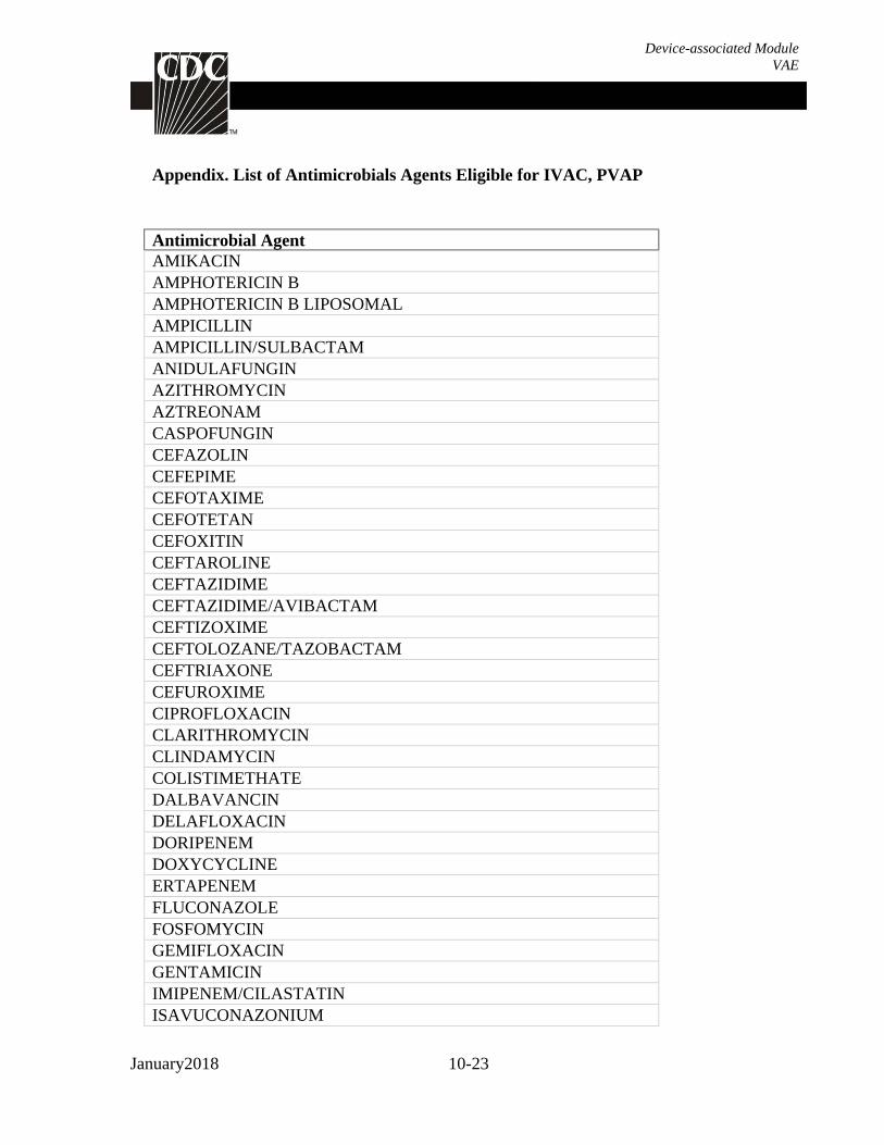
Appendix. List of Antimicrobials Agents Eligible for IVAC, PVAP
Antimicrobial Agent
AMIKACIN
AMPHOTERICIN B
AMPHOTERICIN B LIPOSOMAL
AMPICILLIN
AMPICILLIN/SULBACTAM
ANIDULAFUNGIN
AZITHROMYCIN
AZTREONAM
CASPOFUNGIN
CEFAZOLIN
CEFEPIME
CEFOTAXIME
CEFOTETAN
CEFOXITIN
CEFTAROLINE
CEFTAZIDIME
CEFTAZIDIME/AVIBACTAM
CEFTIZOXIME
CEFTOLOZANE/TAZOBACTAM
CEFTRIAXONE
CEFUROXIME
CIPROFLOXACIN
CLARITHROMYCIN
CLINDAMYCIN
COLISTIMETHATE
DALBAVANCIN
DELAFLOXACIN
DORIPENEM
DOXYCYCLINE
ERTAPENEM
FLUCONAZOLE
FOSFOMYCIN
GEMIFLOXACIN
GENTAMICIN
IMIPENEM/CILASTATIN
ISAVUCONAZONIUM

January2018 10-24
Device-associated Module
VAE
ITRACONAZOLE
LEVOFLOXACIN
LINEZOLID
MEROPENEM
METRONIDAZOLE
MICAFUNGIN
MINOCYCLINE
MOXIFLOXACIN
NAFCILLIN
ORITAVANCIN
OSELTAMIVIR
OXACILLIN
PENICILLIN G
PERAMIVIR
PIPERACILLIN
PIPERACILLIN/TAZOBACTAM
POLYMYXIN B
POSACONAZOLE
QUINUPRISTIN/DALFOPRISTIN
RIFAMPIN
SULFAMETHOXAZOLE/TRIMETHOPRIM
SULFISOXAZOLE
TEDIZOLID
TELAVANCIN
TELITHROMYCIN
TETRACYCLINE
TICARCILLIN/CLAVULANATE
TIGECYCLINE
TOBRAMYCIN
VANCOMYIN, intravenous only
VORICONAZOLE
ZANAMIVIR

January2018 10-25
Device-associated Module
VAE
VAE Frequently Asked Questions (FAQs)
1) When should I use VAE? Are there circumstances in which I should still use PNEU?
VAE surveillance is location based, and restricted to adult inpatient units only.
Pediatric and neonatal units are excluded from VAE surveillance (even in
circumstances where a pediatric unit may occasionally care for patients who are 18
years of age and older).
Locations mapped to mixed age CDC location codes are excluded from VAE
surveillance.
Ventilated patients who are 18 years of age and older and who are cared for in
pediatric units should be included in any in-plan PedVAP surveillance for that
location.
NOTE: It is NOT recommended to include in VAE surveillance young children
housed in adult ICU locations who are not thought to be physiologically similar to the
location’s adult patient population. Facilities may want to evaluate their location
mapping to be sure that locations are mapped appropriately to the correct CDC
location codes. In circumstances where the populations of adults and children cared
for in the same physical location is more mixed (for example, 50% adult patients and
50% pediatric patients), it is recommended that facilities weigh the possibility of
establishing a virtual pediatric location for the purposes of surveillance. More
information on virtual locations and location mapping can be found here:
www.cdc.gov/nhsn/PDFs/pscManual/15LocationsDescriptions_current.pdf
While on high frequency ventilation or extracorporeal life support, patients are
EXCLUDED from VAE surveillance.
NOTE: Patients who are receiving a conventional mode of mechanical ventilation
while in the prone position and patients who are receiving a conventional mode of
mechanical ventilation while receiving nitric oxide therapy, helium-oxygen mixtures
(heliox), or epoprostenol therapy are INCLUDED.
NOTE: Patients on Airway Pressure Release Ventilation (APRV) and related modes
of mechanical ventilation (see FAQ nos. 22 and 23) are INCLUDED; however,
during periods of time while the patient is on APRV, the VAE period of stability or
improvement on the ventilator and the period of worsening oxygenation should be
determined by changes in FiO2 only, since changes in PEEP as indicated in this
surveillance algorithm may not be applicable to APRV. In addition, patients with
VAE who are on APRV or a related mode of mechanical ventilation at the time of
VAE onset can be optionally indicated as such on the VAE Form (CDC 57.112).

January2018 10-26
Device-associated Module
VAE
In-plan surveillance for ventilator-associated PNEU may still be conducted for
pediatric patients ONLY (“PedVAP” surveillance).
The PNEU definitions are still available for those units seeking to conduct off-plan
PNEU/VAP surveillance for patients of any age and for assignment of a secondary
BSI.
2) I am having difficulty visualizing how to arrange the VAE data elements to facilitate easy
identification of events. Can you provide some additional guidance?
For units in which VAE surveillance will be conducted manually, we recommend that
you organize the necessary data elements in a table or spreadsheet to assist in
identifying VAEs. There are a number of different ways in which to organize the data
– you may consider limiting your spreadsheet to just include the daily minimum
PEEP and FiO2 values, and then, if a VAC event is identified, utilize other data
sources to gather information on the data elements included in the IVAC and PVAP
definitions. Alternatively, you may choose to include columns for all data elements
(from VAC through PVAP) in a single spreadsheet.
NOTE: For most patients under surveillance for VAE, the only data elements you will
need to record are the ventilator days, minimum daily PEEP, and minimum daily
FiO2. The maximum and minimum daily temperatures and white blood cell counts
only need to be recorded for those patients who are identified as having met criteria
for VAC. The antimicrobial criterion only needs to be assessed for those patients with
VAC and with an abnormal temperature or white blood cell count that meets the
criteria within the IVAC definition. Microbiology and related data elements included
as criteria in the PVAP definition only need to be assessed for those patients who
have met the IVAC definition.
NOTE: Keep in mind that the baseline period of stability or improvement on the
ventilator is defined as the 2 calendar days immediately preceding the first day of
increased daily minimum PEEP or FiO2, and must be characterized by ≥ 2 calendar
days of stable or decreasing daily minimum FiO2 or PEEP values (specifically the
daily minimum PEEP or FiO2 on the second day of the baseline period of stability or
improvement must be equal to or less than the daily minimum PEEP or FiO2 on the
first day of the baseline period of stability or improvement). Keep in mind, too, that
PEEP values of 0 to 5 cmH2O are considered equivalent for the purposes of VAE
surveillance. This means that any daily minimum value of 0 to 5 cmH2O will be
evaluated as if it were 5 cmH2O when determining whether a VAC has occurred or
not. Also, the daily minimum PEEP or FiO2 is defined as the lowest setting during a
calendar day that is maintained for > 1 hour.
EXAMPLE: In the table below, the data elements used to meet VAC, IVAC and
PVAP definition are organized in a fashion that facilitates identification of an event,
highlighted in the shaded region. In this example, MV days 3 and 4 constitute the
baseline period, with stable minimum PEEP of 5 cmH2O on each day. On MV days 5
and 6, the daily minimum PEEP is 8 cmH2O, which meets the VAC criterion for

January2018 10-27
Device-associated Module
VAE
worsening oxygenation. If we scan across the table, we can see that the IVAC
temperature/white blood cell count criterion is not met (there are no temperatures <
36°C or > 38°C, and no white blood cell counts ≤ 4,000 cells/mm3 or ≥ 12,000
cells/mm3) – so even though the patient was started on a new antimicrobial agent and
continued on that agent for 4 calendar days, IVAC is not met. Therefore, this event
would be reported as a VAC, with the date of event being MV day 5.
Patient MV Day PEEPmin FiO2min Tempmin Tempmax WBCmin WBCmax Abx Specimen Polys / Epis Organism VAE
1 1 10 1.0 37.1 37.6 4.3 4.3 None -- -- -- --
1 2 5 0.60 36.8 37.2 4.6 4.6 None -- -- -- --
1 3 5 0.40 37.0 37.9 5.4 5.4 None -- -- -- --
1 4 5 0.40 36.5 37.3 9.2 9.2 Yes -- -- -- --
1 5 8 0.50 36.3 36.9 8.4 8.4 Yes ETA ≥ 25 / ≤ 10 S.aureus VAC
1 6 8 0.40 37.2 37.5 8.5 8.8 Yes -- -- -- -
1 7 5 0.40 37.8 37.9 7.6 7.6 Yes -- -- -- -
MV = mechanical ventilation. PEEPmin = Daily minimum PEEP. FiO2min = Daily minimum FiO2. Tempmin = Daily minimum temperature. Tempmax = Daily maximum temperature. WBCmin = Daily minimum white blood cell count. WBCmax = Daily maximum white blood cell count. Abx = antimicrobial agents. Polys / epis = Polymorphonuclear leukocytes and squamous epithelial cells from respiratory specimen.
EXAMPLE: In the table below, by scanning across the data elements, you can see
that there are no periods in which there is a stable, 2-day baseline period followed by
a 2-day period where the PEEP or FiO2 are increased 3 cmH2O or 20 points over the
first day in the baseline period. On MV days 2 and 3, the PEEP values are 7 cmH2O
and 6 cmH2O respectively, and then increase to 9 cmH2O on MV days 4 and 5 – but
the difference between day 4 or day 5 and day 2 is only 2 cmH2O, rather than the
required 3 cmH2O. Also, the gradual increase in FiO2 from the time of initiation of
mechanical ventilation means that there are not two days on which the FiO2 is at least
20 points higher than on the 2 previous days. Therefore, although the temperature and
white blood cell counts exceed the required thresholds for IVAC on several
occasions, and the patient appears to have received a new antimicrobial agent for
several days in the setting of a positive blood culture, the VAC definition is not met,
and so no VAE is reported.

January2018 10-28
Device-associated Module
VAE
3) Is there a hierarchy of reporting for VAE? How do I know whether to report a VAC, an
IVAC or a PVAP?
Conducting in-plan VAE surveillance means assessing patients for the presence of
ALL events included in the algorithm—from VAC to IVAC to PVAP. At this time, a
unit participating in in-plan VAE surveillance cannot decide, for example, that only
surveillance for VAC (and not for IVAC or PVAP) will be performed.
There is a hierarchy of definitions within VAE:
o If a patient meets criteria for VAC and IVAC, report as IVAC.
o If a patient meets criteria for VAC, IVAC and PVAP, report PVAP.
4) How do I determine the duration of a VAE? Can a patient have more than one VAE
during a hospitalization?
Patients may have multiple VAEs during a single hospitalization. The event period is
defined by the 14-day period that starts on the date of onset of worsening
oxygenation. VAE criteria met during that 14-day period are attributed to the current
VAE.
EXAMPLE: Patient is intubated and mechanical ventilation is initiated in the MICU
(day 1). The patient is stable during the following 4 calendar days (days 2 through 5).
On days 6 and 7 the patient’s minimum daily FiO2 is increased more than 0.20 (20
points) over the first day in the baseline period, therefore meeting the VAC FiO2
threshold. The VAC episode is defined by the period encompassing days 6 through 19
(14 days, starting on day 1 of worsening oxygenation, which in this case is day 6). If
the patient were to experience a period of stability or improvement on the ventilator
on days 18 and 19, followed by another 2-day period of worsening on days 20 and 21,
a new VAE would be reported, since the second period of worsening oxygenation has
occurred more than 14 days after the start of the initial period of worsening
oxygenation.
Patient MV Day
PEEPmin FiO2min Tempmin Tempma
xWBCmin
WBCma
xAbx
Specimen
Polys / Epis
Organism
VAE
2 1 5 0.30 37.1 37.6 4.3 4.3 None -- -- -- --
2 2 7 0.30 36.8 37.2 4.6 4.6 None -- -- -- --
2 3 6 0.45 37.0 37.9 5.4 5.4 None -- -- -- --
2 4 9 0.45 36.5 37.3 9.2 9.2 None -- -- -- --
2 5 9 0.60 36.3 36.9 8.4 8.4 None ETA ≥ 25 / ≤ 10 S.aureus --
2 6 8 0.60 37.2 37.5 8.5 8.8 None -- -- -- --
2 7 6 0.75 37.8 37.9 7.6 7.6 None -- -- -- --
2 8 6 0.75 38.2 38.4 10.5 11.9 Yes Blood -- S. aureus --
2 9 5 0.80 38.5 38.9 12.7 12.7 Yes -- -- -- --
2 10 5 0.75 37.4 38.1 12.9 12.9 Yes -- -- -- --
2 11 5 0.70 37.2 37.9 9.4 9.4 Yes -- -- -- --
2 12 5 0.60 37.3 37.5 9.5 9.5 Yes -- -- -- --
2 13 7 0.60 37.2 37.8 8.2 8.2 Yes -- -- -- --
2 14 8 0.60 37.0 37.7 8.6 8.6 Yes -- -- -- --

January2018 10-29
Device-associated Module
VAE
5) Sometimes patients are intubated, extubated, and reintubated several times during a
single hospitalization. How do I define an episode of mechanical ventilation, and can a
VAE occur in a patient who has recently been extubated?
An episode of mechanical ventilation is defined as a period of days during which the
patient was mechanically ventilated for some portion of each consecutive day during
the period.
EXAMPLE: A patient is intubated and mechanically ventilated on hospital day 1. The
patient remains on mechanical ventilation from hospital day 2 through 12 noon on
hospital day 6. At noon on hospital day 6, the patient is extubated. The patient
remains extubated on hospital day 7, and is then reintubated on hospital day 8. In this
case, the first episode of mechanical ventilation is defined by days 1 through 6. Since
the patient was extubated on day 6 and remained extubated for a full calendar day on
day 7, the re-intubation of the patient on day 8 defines the start of a second episode of
mechanical ventilation. See figure, below.
Hosp Day No. 1 2 3 4 5 6 7 8 9 10
MV Episode 1 1 1 1 1 1 -- 2 2 2
MV Day No. 1 2 3 4 5 6—extubated
at noon -- 1--reintubated 2 3
EXAMPLE: A patient is intubated and mechanically ventilated on hospital day 1. The
patient remains on mechanical ventilation from hospital day 2 through hospital day 6
at 12 noon. At noon on hospital day 6, the patient is extubated. The patient is
reintubated at 9 pm on hospital day 7, and remains intubated and mechanically
ventilated till 2 pm on day 10. The patient is extubated at 2 pm on day 10 and remains
extubated until hospital discharge on day 15. In this case, there is only a single
episode of mechanical ventilation, defined by days 1 through 10, because the patient
was extubated on day 6 but reintubated the next calendar day (day 7). See figure,
below.
Hosp Day No. 1 2 3 4 5 6 7 8 9 10
MV Episode 1 1 1 1 1 1 1 1 1 1
MV Day No. 1 2 3 4 5 6—extubated at
noon 7—reintubated
at 9 pm 8 9
10—extubated at 2 pm
A VAE can occur in a patient who has been extubated and is then reintubated, subject to the
amount of time the patient was off the ventilator, as noted in the examples below.
1 full calendar day off mechanical ventilation, followed by reintubation, defines a new episode of mechanical ventilation.
Patient was reintubated on the calendar day following extubation (days 6-7). Because there is not 1 calendar day off mechanical ventilation, there is only 1 episode of mechanical ventilation.

January2018 10-30
Device-associated Module
VAE
EXAMPLE: A patient is intubated and mechanically ventilated on hospital day 1. The
patient remains on mechanical ventilation from hospital day 2 through 12 noon on
hospital day 6. At noon on hospital day 6, the patient is extubated. The patient remains
extubated on hospital day 7, and is then reintubated on hospital day 8. In this case,
because the patient has been extubated for 1 full calendar day (day 7), the “VAE clock”
starts over with reintubation on hospital day 8. To meet VAE during this second episode
of mechanical ventilation, the patient would have to have at least 2 days of stability or
improvement and at least 2 days of worsening oxygenation on the ventilator; therefore,
the earliest date on which the patient could meet VAE criteria would be hospital day 11
(stable or improving settings on days 8 and 9, increased ventilator settings on days 10 and
11). The VAE event date would be reported as day 10—the first day of worsening
oxygenation meeting VAE criteria. See figure, below.
EXAMPLE: A patient is intubated and mechanically ventilated on hospital day 1. The
patient remains on mechanical ventilation from hospital day 2 through 12 noon on
hospital day 6, when the patient is extubated. The patient is reintubated at 9 pm on
hospital day 7. In this case, there is no “new” episode of mechanical ventilation, since
there was not a full, ventilator-free calendar day. Therefore, the period of worsening
oxygenation may be determined to have started on day 7, the day of reintubation, as
long as PEEP or FiO2 criteria are met. PEEP and FiO2 data from hospital days 5 and 6
(through the time of extubation) may be used to determine whether a period of
stability and improvement occurred, and these data may be compared to PEEP and
FiO2 data obtained from the time of reintubation on day 7 and beyond to determine
whether at least 2 days of worsening oxygenation occurred. The earliest that the
patient could meet VAE criteria would be day 8 (assuming stable or improving
ventilator settings on days 5 and 6, and two days of worsening oxygenation meeting
criteria on days 7 and 8). The VAE event date would be reported as day 7—the first
day of worsening oxygenation meeting VAE criteria. See figure, below.
Hosp Day No. 1 2 3 4 5 6 7 8 9 10
MV Episode 1 1 1 1 1 1 1 1 1 1
MV Day No. 1 2 3 4 5 6—extubated
at noon 7—reintubated
at 9 pm 8 9 10
VAE Criterion Day 1 of
stability or improvement
Day 2 of stability or
improvement
Day 1 of worsening
oxygenation
Day 2 of worsening
oxygenation
A patient may also meet criteria for VAC while intubated, and then meet criteria for
IVAC (or PVAP) following extubation.
Hosp Day No. 1 2 3 4 5 6 7 8 9 10 11
MV Episode 1 1 1 1 1 1 -- 2 2 2 2
MV Day No. 1 2 3 4 5 6—extubated
at noon --
1--reintubated
2 3 4
VAE Criterion -- -- -- -- -- -- -- Day 1 of
stability or improvement
Day 2 of stability or
improvement
Day 1 of worsening
oxygenation
Day 2 of worsening
oxygenation

January2018 10-31
Device-associated Module
VAE
EXAMPLE: A patient is intubated and mechanically ventilated on hospital day 1. The
patient remains on mechanical ventilation till 11 am on hospital day 10, when the
patient is extubated. Criteria for VAC are met during the episode of mechanical
ventilation, based on 2 days of stability or improvement (MV days 5 and 6) followed
by 2 days of worsening oxygenation (MV days 7 and 8). The date of the event is MV
day 7, the day of onset of worsening oxygenation. Within the 2 days before and 2
days after the day of onset of worsening oxygenation, the patient has a temperature of
38.4°C, and a new antimicrobial agent is started (meropenem, on MV day 9—see
FAQ no. 6-10). The new antimicrobial agent is continued for at least 4 days (hospital
days 8 through 11). Therefore, even though the patient was extubated on hospital day
10 and remained extubated on hospital day 11 (the day on which all IVAC criteria
were fulfilled), the event should be reported as an IVAC. See figure, below.
Hosp Day No. 4 5 6 7 8 9 10 11
MV Day No. 4 5 6 7 8 9 Extubated at 11 am
--
VAE Criterion --
Day 1 of stability or
improvement
Day 2 of stability or
improvement
Day 1 of worsening
oxygenation
Day 2 of worsening
oxygenation
Temp 38.4°C
-- --
Antimicrobial agent
Ceftriaxone Ceftriaxone Ceftriaxone Ceftriaxone Meropenem Meropenem Meropenem Meropenem
6) What antimicrobial agents are included in the IVAC definition?
See the Appendix for a list of the antimicrobial agents eligible for consideration in the
IVAC definition (as well as the PVAP definition).
See Table 1 for eligible routes of administration.
7) How do I figure out if an antimicrobial agent is “new” for the IVAC definition?
A new antimicrobial agent is defined as any agent listed in the Appendix that is
initiated on or after 3 days of mechanical ventilation AND in the VAE Window
Period (defined by the two days before, the day of, and the two days after the onset
date of the VAE—as long as all of these days are on or after the 3rd day of mechanical
ventilation). The agent is considered new for the purposes of this definition if it was
NOT given to the patient on either of the 2 days preceding the current start date. The
agent must be administered via one of the routes listed in Table 1.
See the example in the figure below:
Patient has fulfilled all IVAC criteria, and IVAC should be reported. Date of the IVAC event is hospital day/MV day 7.

January2018 10-32
Device-associated Module
VAE
MV Day No. 4 5 6 7 8 9 10 11
VAE Criterion - - - Onset (day 1) of worsening oxygenation meeting VAE PEEP
or FiO2 thresholds
Day 2 of worsening oxygenation meeting VAE PEEP or FiO2
thresholds
- - -
EXAMPLE: A single dose of intravenous vancomycin is given to a patient on
the VAE onset date (which is the day of onset of worsening oxygenation
meeting VAE criteria, in this case MV day 7), and was not given to the patient
during the 2 previous days (MV days 5 and 6). Vancomycin is therefore
considered a new antimicrobial agent (see figure below).
MV Day No. 4 5 6 7 8 9 10
VAE Criterion --
Day 1 of stability or
improvement
Day 2 of stability or
improvement
Day 1 of worsening
oxygenation
Day 2 of worsening
oxygenation - -
Antimicrobial agent
None None None
Single dose of vancomycin ordered and administered
None None
Single dose of vancomycin ordered and administered
EXAMPLE: If meropenem is given to a patient on the VAE onset date (which
is the day of onset of worsening oxygenation meeting VAE criteria, in this
case MV day 7), and was not given to the patient during the 2 previous days
(MV days 5 and 6), then meropenem is considered a new antimicrobial agent
(see figure below). Note that the patient is also receiving ceftriaxone, and
receives doses during the 5-day period around the onset of worsening
oxygenation (first dose during the 5-day period was on MV day 5). However,
because ceftriaxone was given to the patient the day before the 5-day period
(on MV day 4), ceftriaxone does not count as a new antimicrobial agent for
the purposes of the IVAC definition.
MV Day No. 4 5 6 7 8 9 10
VAE Criterion --
Day 1 of stability or
improvement
Day 2 of stability or
improvement
Day 1 of worsening
oxygenation
Day 2 of worsening
oxygenation - -
Antimicrobial agent
Ceftriaxone Ceftriaxone Ceftriaxone Meropenem Meropenem Meropenem Meropenem
First dose of meropenem during the 5-day period around the onset of worsening oxygenation. Note that no meropenem was given in the 2 preceding days, and so
meropenem is a “new” antimicrobial agent for the purposes of the VAE definition.
Example of the 5-day period during which the first dose of a new antimicrobial agent must be given to meet requirements of IVAC definition
A single dose of vancomycin is ordered and administered to the patient within the period defined by the two days before, the day of, and the two days after the VAE onset date. Note that no vancomycin was given in the 2 preceding days, and so vancomycin is
a “new” antimicrobial agent for the purposes of the VAE definition.

January2018 10-33
Device-associated Module
VAE
8) I have figured out that a new antimicrobial agent was given to the patient. How do I
determine whether it was continued for 4 days?
Make sure you are using the Medication Administration Record. You need to know
which antimicrobial agents were actually administered to the patient. Antimicrobial
orders or dispensing information is not sufficient.
You do not need to know the dose or frequency of administration.
Four consecutive Qualifying Antimicrobial Days (QADs)—starting within the VAE
Window Period—are needed to meet the IVAC criterion. A QAD is a day on which
the patient was administered an antimicrobial agent that was determined to be “new”
within the VAE Window Period. Days between administrations of a new
antimicrobial agent also count as QADs as long as there is a gap of no more than 1
calendar day between administrations of the same antimicrobial agent. For example,
if levofloxacin is given on VAE Day 1, has not been given in the 2 preceding
calendar days, and is given again on VAE Days 3, 5 and 7, there are 7 QADs—
because the days between levofloxacin doses also count as QADs.
The requirement for 4 consecutive QADs can be met with 4 days of therapy with the
same antimicrobial (with a gap of no more than 1 calendar day between
administrations of that antimicrobial)—or it can be met with 4 days of therapy with
multiple antimicrobial agents, as long as each antimicrobial was started within the
VAE Window Period.
EXAMPLE: In the figure below, meropenem would meet the antimicrobial
criterion of the IVAC definition because at least one dose was given on 4
consecutive days.
MV Day No. 1 2 3 4 5 6 7
VAE Criterion -- Day 1 of Stability or
improvement
Day 2 of stability or
improvement
Day 1 of worsening
oxygenation
Day 2 of worsening
oxygenation
- -
Antimicrobial agent
Ceftriaxone Ceftriaxone Ceftriaxone Meropenem Meropenem Meropenem Meropenem
QAD No No No Yes Yes Yes Yes
EXAMPLE: In the figure below, the 3 drugs shown in bold lettering all
qualify as new antimicrobial agents, and therefore the antimicrobial criterion
of IVAC is met, since the patient is given 4 consecutive days of new
antimicrobial agents.
MV Day No. 1 2 3 4 5 6 7
VAE Criterion -- Day 1 of Stability or
improvement
Day 2 of stability or
improvement
Day 1 of worsening
oxygenation
Day 2 of worsening
oxygenation
- -
Antimicrobial agent
Ceftriaxone Ceftriaxone Ceftriaxone Meropenem Imipenem Piperacillin/ Tazobactam
Piperacillin/ Tazobactam
QAD No No No Yes Yes Yes Yes
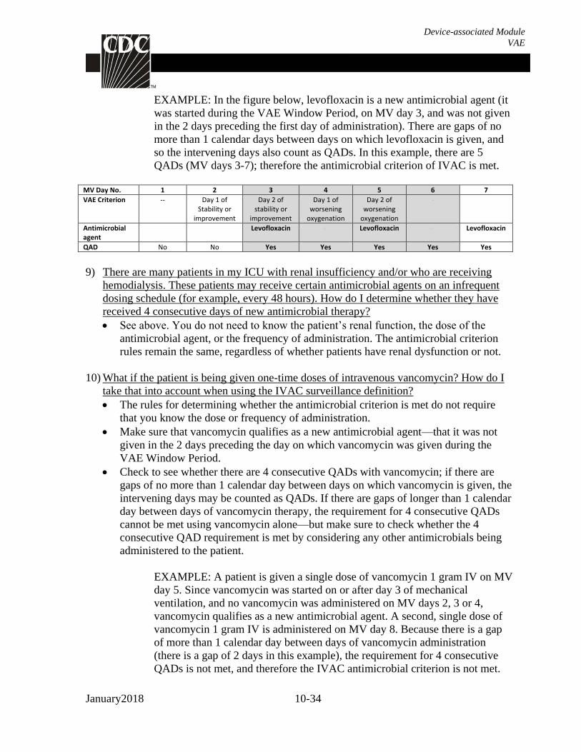
January2018 10-34
Device-associated Module
VAE
EXAMPLE: In the figure below, levofloxacin is a new antimicrobial agent (it
was started during the VAE Window Period, on MV day 3, and was not given
in the 2 days preceding the first day of administration). There are gaps of no
more than 1 calendar days between days on which levofloxacin is given, and
so the intervening days also count as QADs. In this example, there are 5
QADs (MV days 3-7); therefore the antimicrobial criterion of IVAC is met.
MV Day No. 1 2 3 4 5 6 7
VAE Criterion -- Day 1 of Stability or
improvement
Day 2 of stability or
improvement
Day 1 of worsening
oxygenation
Day 2 of worsening
oxygenation
- -
Antimicrobial agent
Levofloxacin Levofloxacin Levofloxacin
QAD No No Yes Yes Yes Yes Yes
9) There are many patients in my ICU with renal insufficiency and/or who are receiving
hemodialysis. These patients may receive certain antimicrobial agents on an infrequent
dosing schedule (for example, every 48 hours). How do I determine whether they have
received 4 consecutive days of new antimicrobial therapy?
See above. You do not need to know the patient’s renal function, the dose of the
antimicrobial agent, or the frequency of administration. The antimicrobial criterion
rules remain the same, regardless of whether patients have renal dysfunction or not.
10) What if the patient is being given one-time doses of intravenous vancomycin? How do I
take that into account when using the IVAC surveillance definition?
The rules for determining whether the antimicrobial criterion is met do not require
that you know the dose or frequency of administration.
Make sure that vancomycin qualifies as a new antimicrobial agent—that it was not
given in the 2 days preceding the day on which vancomycin was given during the
VAE Window Period.
Check to see whether there are 4 consecutive QADs with vancomycin; if there are
gaps of no more than 1 calendar day between days on which vancomycin is given, the
intervening days may be counted as QADs. If there are gaps of longer than 1 calendar
day between days of vancomycin therapy, the requirement for 4 consecutive QADs
cannot be met using vancomycin alone—but make sure to check whether the 4
consecutive QAD requirement is met by considering any other antimicrobials being
administered to the patient.
EXAMPLE: A patient is given a single dose of vancomycin 1 gram IV on MV
day 5. Since vancomycin was started on or after day 3 of mechanical
ventilation, and no vancomycin was administered on MV days 2, 3 or 4,
vancomycin qualifies as a new antimicrobial agent. A second, single dose of
vancomycin 1 gram IV is administered on MV day 8. Because there is a gap
of more than 1 calendar day between days of vancomycin administration
(there is a gap of 2 days in this example), the requirement for 4 consecutive
QADs is not met, and therefore the IVAC antimicrobial criterion is not met.
- - - -

January2018 10-35
Device-associated Module
VAE
MV Day No. 2 3 4 5 6 7 8 9
VAE Criterion -- -- Day 1 of Stability or
improvement
Day 2 of stability or
improvement
Day 1 of worsening
oxygenation
Day 2 of worsening
oxygenation
- -
Antimicrobial agent
None None None Vancomycin 1 gram IV x 1
dose
None None Vancomycin 1 gram IV x 1
dose
None
QAD No No No Yes No No Yes No
11) Can I report pathogens or secondary BSIs for VAC and IVAC?
Pathogens are NOT reported for VAC or IVAC events.
Secondary BSIs are NOT reported for VAC or IVAC events.
EXAMPLE: A patient hospitalized and mechanically-ventilated in the MICU
for 14 days develops worsening oxygenation following a 7-day period of
stability on the ventilator. VAC criteria are met on hospital day 15 (stable
ventilator settings on days 12 and 13, increased ventilator settings on days 14
and 15). The onset date is day 14. The white blood cell count is noted to be
15,500 cells/mm3 on day 14. Meropenem and intravenous vancomycin are
begun on day 15, administered through the patient’s right-sided central line,
which was inserted on ICU admission. The antibiotics continue to be
administered on day 18, meeting IVAC criteria. Endotracheal aspirate cultures
done on days 15 and 16 grow scant upper respiratory flora. A blood culture
collected on day 15 is positive for Klebsiella oxytoca. There are no other signs
or symptoms of infection. This patient should be reported as having an IVAC
and a central line-associated BSI if the BSI cannot be attributed as secondary
to another primary site of infection. The BSI cannot be reported as secondary
to the IVAC event.
12) Can I report pathogens for PVAP?
Pathogens may be reported for PVAP events, provided they are isolated or identified
from appropriate specimen types according to the requirements of the algorithm and
are NOT on the list of excluded organisms and culture results:
o Excluded organisms and culture results that cannot be used to meet the
PVAP definition are as follows: “Normal respiratory flora,” “normal oral
flora,” “mixed respiratory flora,” “mixed oral flora,” “altered oral flora” or
other similar results indicating isolation of commensal flora of the oral
cavity or upper respiratory tract; Candida species or yeast not otherwise
specified; coagulase-negative Staphylococcus species; and Enterococcus
species, when identified from sputum, endotracheal aspirates,
bronchoalveolar lavage, or protected specimen brushings. Only eligible
pathogens identified from eligible specimens with a collection date
occurring in the infection window period can be reported.
January2018 10-36
Device-associated Module
VAE
NOTE: When Candida species or yeast not otherwise specified, coagulase-negative
Staphylococcus species or Enterococcus species are identified from lung tissue or
pleural fluid, these organisms may be reported as PVAP pathogens.
Additionally, because organisms belonging to the following genera are usually causes
of community-associated respiratory infections and rarely or are not known to be
causes of healthcare-associated infections, they are also excluded, and cannot be used
to meet the PVAP definition when isolated from any eligible specimen type (to
include lung and pleural fluid): Blastomyces, Histoplasma, Coccidioides,
Paracoccidioides, Cryptococcus and Pneumocystis.
See Table 3 for the required quantitative culture thresholds associated with various
specimen types in the PVAP definition. Note that if your laboratory reports semi-
quantitative culture results, you should check with your laboratory to confirm that
semi-quantitative results match the quantitative thresholds noted in Table 3.
13) Can I report secondary BSIs for PVAP?
Secondary BSIs may be reported for PVAP events, provided that the organism
identified from blood specimen matches an organism identified from an appropriate
respiratory tract specimen (including respiratory secretions, pleural fluid and lung
tissue). The respiratory tract specimen must have been collected within 2 calendar
days before or after the day of onset of worsening oxygenation to be considered as a
criterion for meeting the PVAP definition. In addition, the positive blood specimen
must have been collected during the 14-day event period, where day 1 is the day of
onset of worsening oxygenation.
o In cases where PVAP is met with only the histopathology criterion and no
culture or non-culture based test is performed on an eligible respiratory
specimen, and there is also a positive blood specimen, a secondary BSI for
VAE is not reported.
o In cases where a culture or non-culture based test of respiratory secretions,
pleural fluid or lung tissue is performed and does not identify an organism that
matches an organism identified from blood, a secondary BSI for VAE is not
reported.
NOTE: Candida species or yeast not otherwise specified, coagulase-negative
Staphylococcus species, and Enterococcus species identified from blood cannot
be deemed secondary to a PVAP, unless the organism was also identified from
pleural fluid or lung tissue.
EXAMPLE: Patient hospitalized and mechanically ventilated in the MICU for 14
days develops worsening oxygenation following a 7-day period of stability on the
ventilator. VAC criteria are met on hospital day 15 (stable ventilator settings on
days 12 and 13, increased ventilator settings on days 14 and 15). The onset date is
day 14. The white blood cell count is noted to be 15,500 cells/mm3 on day 14.
Meropenem and vancomycin are begun on day 15, administered through the
January2018 10-37
Device-associated Module
VAE
patient’s right-sided central line (inserted on ICU admission). The antibiotics
continue to be administered on day 18, meeting IVAC criteria. Endotracheal
aspirate specimens collected on days 15 and 16 grow ≥ 105 CFU/ml Klebsiella
oxytoca. A blood culture collected on day 15 is positive for K. oxytoca. This
patient should be reported as having a PVAP with a secondary BSI due to K.
oxytoca.
EXAMPLE: Patient hospitalized and mechanically ventilated in the MICU for 14
days develops worsening oxygenation following a 7-day period of stability on the
ventilator. VAC criteria are met on hospital day 15 (stable ventilator settings on
days 12 and 13, increased ventilator settings on days 14 and 15). The onset date is
day 14. The white blood cell count is noted to be 15,500 cells/mm3 on day 14.
Meropenem and vancomycin are begun on day 15, administered through the
patient’s right-sided central line (inserted on ICU admission). The antibiotics
continue to be administered on day 18, meeting IVAC criteria. A thoracentesis is
performed on day 15 at the patient’s bedside using aseptic technique. Pleural fluid
is sent for culture and grows Candida albicans. A blood culture collected on day
16 is positive for C. albicans. This patient should be reported as having a PVAP
with a secondary BSI due to C. albicans.
EXAMPLE: Patient hospitalized and mechanically ventilated in the MICU for 14
days develops worsening oxygenation following a 7-day period of stability on the
ventilator. VAC criteria are met on hospital day 15 (stable ventilator settings on
days 12 and 13, increased ventilator settings on days 14 and 15). The onset date is
day 14. The white blood cell count is noted to be 15,500 cells/mm3 on day 14.
Meropenem and vancomycin are begun on day 15, administered through the
patient’s right-sided central line (inserted on ICU admission). The antibiotics
continue to be administered on day 18, meeting IVAC criteria. An endotracheal
aspirate collected on day 15 is a good quality specimen, with ≥ 25 neutrophils and
≤ 10 squamous epithelial cells per low power field, and grows Staphylococcus
aureus (qualitative result). A blood culture collected on day 24 is positive for S.
aureus and for coagulase-negative staphylococci (CoNS). This patient should be
reported as having a PVAP, with S. aureus reported as the pathogen. A secondary
BSI should also be reported for the PVAP, since the positive blood culture was
collected within the 14-day period of the VAE, and an organism isolated from
blood (S. aureus) matched an organism isolated from culture of the endotracheal
aspirate. The CoNS also isolated from the blood culture on day 24 is not reported
as a pathogen for the PVAP because it is an excluded organism.
14) Can I only report pathogens if they are isolated in cultures of appropriate specimens?
What about pathogens identified by non-culture-based diagnostic testing?
PVAP incorporates results of non-culture-based microbiological diagnostic testing.
For PVAP, pathogens that are grown in culture OR selected pathogens that are
identified as a result of other laboratory testing (for example, antigen testing, PCR,
immunohistochemistry, etc.) should be reported. Do not limit reporting to just those

January2018 10-38
Device-associated Module
VAE
organisms isolated in culture. For example, influenza A identified by polymerase
chain reaction (PCR) in a patient meeting PVAP criteria should be reported as a
pathogen for that event.
15) The “PVAP” criterion 3 includes “positive diagnostic tests” for Legionella species, and
selected viruses. What kinds of diagnostic tests can be used to meet the definition?
Diagnostic testing practices may vary from facility to facility and change over time as
better tests are developed. Listed here are some examples of diagnostic tests for
specific pathogens included in the PVAP definition. Positive results of these tests
may be used in meeting the PVAP definition. Your facility may use other testing
methods; positive results obtained using these methods may also be appropriate for
use in meeting the PVAP definition. If you have a question regarding a diagnostic test
method, check with your laboratory.
For Legionella species, positive results of any of the following, performed on the
appropriate specimen: urinary antigen, Legionella-specific respiratory culture, paired
serology (4-fold rise in titer between acute and convalescent specimens), direct
fluorescent antibody stain, immunohistochemistry stain, or nucleic acid detection
assays (such as PCR) performed on a respiratory specimen.
For respiratory viruses (influenza, respiratory syncytial virus [RSV], parainfluenza
viruses, human metapneumovirus, coronaviruses, rhinoviruses and adenovirus),
positive results for any of the following:
o Performed on an appropriate respiratory specimens – PCR or other viral
nucleic acid detection methods, antigen detection methods, including rapid
tests, viral cell culture, or
o Performed on appropriate pathologic specimens – immunohistochemical
assays, cytology, microscopy, or
o Performed on appropriately timed paired sera (acute and convalescent) –
serological assays demonstrating seroconversion or a significant rise in
antibody titer.
16) What about pneumonitis that occurs in a mechanically-ventilated patient and is
determined to be due to herpes simplex virus (HSV) or cytomegalovirus (CMV)? Can
these infections be reported as VAEs?
In most cases pneumonitis due to HSV and CMV represents reactivation of a latent
infection, and therefore would not be considered healthcare-associated, according to
the NHSN definition of a healthcare-associated infection.
17) Are there any culture results or microorganisms that CANNOT be used to meet the
PVAP definition?
The following pathogens and culture results may NOT be used to meet the definition
and may NOT be reported as causes of PVAP when they are identified from sputum,
endotracheal aspirates, bronchoalveolar lavages or protected specimen brushings:
o Culture results reported as “Normal respiratory flora,” “normal oral flora,”
“mixed respiratory flora,” “mixed oral flora,” “altered oral flora” or other

January2018 10-39
Device-associated Module
VAE
similar results indicating isolation of commensal flora of the oral cavity or
upper respiratory tract.
o Candida species or yeast not otherwise specified
o Coagulase-negative Staphylococcus species
o Enterococcus species
NOTE: These organisms are excluded because they are common upper respiratory
tract commensals, colonizers or contaminants, and are unusual causes of VAP. Their
exclusion from the surveillance definitions should NOT be used in clinical decision-
making regarding patient treatment. Providers must independently determine the
clinical significance of these organisms identified from respiratory specimens and the
need for treatment.
NOTE: When Candida species or yeast not otherwise specified, coagulase-negative
Staphylococcus species or Enterococcus species are identified from lung tissue or
pleural fluid, these organisms may be reported as PVAP pathogens.
Additionally, because organisms belonging to the following genera are typically
causes of community-associated respiratory infections and are rarely or are not
known to be causes of healthcare-associated infections, they are also excluded, and
cannot be used to meet the PVAP definition when isolated from any eligible
specimen type (to include lung and pleural fluid): Blastomyces, Histoplasma,
Coccidioides, Paracoccidioides, Cryptococcus and Pneumocystis.
When sputum, endotracheal aspirate, bronchoalveolar lavage or protected specimen
brushing culture or non-culture based testing results are mixed and contain one or
more of the excluded pathogens in addition to one or more non-excluded pathogens,
the culture may be used to meet the PVAP definition (depending on whether a
qualitative, semi-quantitative or quantitative culture was performed, and whether the
semi-quantitative or quantitative cfu/ml thresholds were met) BUT only the non-
excluded pathogen(s) should be reported.
EXAMPLE: Patient intubated and mechanically ventilated in the MSICU meets
IVAC criteria on day 8 of mechanical ventilation. On the day after the onset of
worsening oxygenation, an endotracheal aspirate is collected. The Gram stain shows
≥ 25 neutrophils and ≤ 10 squamous epithelial cells per low power field, and the
culture grows “heavy Staphylococcus aureus” and “heavy Candida albicans.” This
patient should be reported as having a PVAP (criterion1) due to Staphylococcus
aureus – as long as the semi-quantitative result “heavy” is equivalent to the
quantitative threshold of ≥ 105 cfu/ml for endotracheal aspirates. If the semi-
quantitative result is not equivalent to the quantitative threshold of ≥ 105 cfu/ml for
endotracheal aspirates, the patient should still be reported as PVAP (criterion 2).
Candida albicans from the endotracheal aspirate culture is not reported, because it is
an excluded result.

January2018 10-40
Device-associated Module
VAE
18) What about organisms identified from pleural fluid and lung tissue specimens? Can I
report any pathogen identified from a lung tissue, or from a pleural fluid specimen,
assuming the specimen was obtained during thoracentesis or at the time of chest tube
insertion?
Any pathogen identified from lung tissue, when that lung tissue was obtained during
an open lung biopsy, video-assisted thoracoscopic surgery, or via other transthoracic
or transbronchial biopsy approach, may be reported with the exception of the
excluded pathogens belonging to the following genera: Blastomyces, Histoplasma,
Coccidioides, Paracoccidioides, Cryptococcus and Pneumocystis.
Any pathogen identified from pleural fluid, when that fluid was obtained during
thoracentesis or at the time of initial chest tube insertion, may be reported with the
exception of the excluded pathogens belonging to the following genera: Blastomyces,
Histoplasma, Coccidioides, Paracoccidioides, Cryptococcus and Pneumocystis.
19) How are “purulent respiratory secretions” defined?
Purulent respiratory secretions used to meet Criterion #2 of the PVAP definition are
defined as:
o Secretions from the lungs, bronchi, or trachea with ≥ 25 neutrophils and ≤
10 squamous epithelial cells per low power field [lpf, x100].
o If the laboratory reports semi-quantitative results, you should check with
your laboratory to be certain that the semi-quantitative results match the
quantitative thresholds noted above.
If your laboratory is not able to provide additional information on how a semi-
quantitative reporting system corresponds to quantitative reporting ranges for
neutrophils and squamous epithelial cells, here is some guidance from the Clinical
Microbiology Procedures Handbook (3rd ed., 2010)*:
1+ = occasional or rare = <1 cell per low power field [lpf, x100]
2+ = few = 1-9 cells per low power field [lpf, x100]
3+ = moderate = 10-25 cells per low power field [lpf, x100]
4+ = heavy = >25 cells per low power field [lpf, x100]
*Reference: Garcia, LS (Ed.). (2010). Clinical Microbiology Procedures Handbook. Herndon, VA: ASM Press, page
3.2.1.16.
o With this range in mind, and in the absence of additional information from
your laboratory, “purulent respiratory secretions” are defined as secretions
that contain heavy, 4+ or ≥25 neutrophils per low power field [lpf, x100]
AND rare, occasional, few, 1+ or 2+, or ≤10 squamous epithelial cells per
low power field [lpf, x100].
If your laboratory uses a different reporting format for results of direct
examination of respiratory secretions, you may still be able to use the purulent
respiratory secretions in meeting the PVAP definition. See the instructions
available in the VAE Protocol, Table 2.

January2018 10-41
Device-associated Module
VAE
20) What is the definition of “positive lung histopathology” that can be used to meet the
PVAP definition?
If the lung tissue specimen was obtained via open lung biopsy, video-assisted
thoracoscopic surgery, or via other transthoracic or transbronchial biopsy approach, it
is eligible for consideration in meeting the PVAP definition (Criterion 3).
Histopathological findings that can be used to meet the PVAP definition include:
o Abscess formation or foci of consolidation with intense neutrophil
accumulation in bronchioles and alveoli;
o Evidence of lung parenchyma invasion by fungi (hyphae, pseudohyphae or
yeast forms);
o Evidence of infection with the viral pathogens listed in FAQ no. 14 based
on results of immunohistochemical assays, cytology, or microscopy
performed on lung tissue.
Additionally, lower respiratory specimen cytology findings suggestive of infection
are eligible for consideration in meeting the PVAP definition (Criterion 3).
21) I am still having trouble understanding the time frame that defines a VAE. Can you
explain what is meant by this statement that appears in the algorithm: “On or after
calendar day 3 of mechanical ventilation and within 2 calendar days before or after the
onset of worsening oxygenation”?
The intent of these criteria is to determine whether a VAC is due to an infectious
process (IVAC) and/or pneumonia (PVAP) by looking for corroborating
inflammatory and infectious signs at the time of VAC onset. The criterion, “on or
after calendar day 3” is intended to exclude inflammatory and infectious signs present
on the first two days of mechanical ventilation because they are more likely to be due
to pre-existing conditions than ventilator-acquired complications. The criterion,
“within 2 calendar days before or after the onset of worsening oxygenation,” is
intended to identify infectious and inflammatory signs that arise at the same time as
VAC and may therefore point to the cause of the VAC. +
The figures below illustrate the time frame that defines a VAE. The event date is the
first day of worsening oxygenation, defined by the PEEP and FiO2 thresholds
outlined in the algorithm. The event date defines the time frame within which all
other criteria must be met. In the examples below, the shaded area defines the VAE
Window Period in which IVAC criteria (temperature or white count abnormalities,
plus a new antimicrobial agent started and continued for at least 4 days) must be met,
and in which a PVAP criterion must be met.
NOTE: Keep in mind that VAE criteria must be met based on specimens collected or
antimicrobial agents started after day 2 of mechanical ventilation.
EXAMPLE 1: When the onset date of the VAE occurs early in the course of
mechanical ventilation (for example, day 3 or 4 of mechanical ventilation), the period
in which certain inflammatory and infectious criteria are sought for IVAC and PVAP
is shorter, because the first 2 days of mechanical ventilation are excluded from the
normal 5 day window surrounding the day of increased ventilator support.

January2018 10-42
Device-associated Module
VAE
MV Day No. 1 2 3 4 5 6 7
Worsening oxygenation -- Day 1 of Stability or
improvement
Day 2 of stability or
improvement
Day 1 of worsening
oxygenation
Day 2 of worsening
oxygenation
- -
Temperature abnormality or white blood cell count abnormality
- -
An abnormal temperature or white blood cell count, according to the algorithm parameters, must be documented within this
shaded period
-
Antimicrobial agent - -
New agent must be started on any day within this shaded period, and then continued for at least 4 days
-
Purulent respiratory secretions, positive culture, positive histopathology
- -
Specimen must be collected on any day within this shaded period
-
EXAMPLE 2: When the onset date of the VAE occurs later in the course of
mechanical ventilation, the period in which certain criteria must be met is a day
longer, because the patient has already been on mechanical ventilation for more than
3 days and therefore inflammatory and infectious signs arising anywhere in the full 5-
day window surrounding the day of increased ventilator settings can count towards
IVAC and PVAP.
MV Day No. 10 11 12 13 14 15 16
Worsening oxygenation -- Day 1 of Stability or
improvement
Day 2 of stability or
improvement
Day 1 of worsening
oxygenation
Day 2 of worsening
oxygenation
- -
Temperature abnormality or white blood cell count abnormality
- An abnormal temperature or white blood cell count, according to the algorithm
parameters, must be documented within this shaded period
-
Antimicrobial agent - New agent must be started on any day within this shaded period, and then continued for at least 4 days
-
Purulent respiratory secretions, positive culture, positive histopathology
- Specimen must be collected on any day within this shaded period
-
22) Providers in my ICU use different types of mechanical ventilation for different patients.
Can you explain the circumstances in which mechanically-ventilated patients are to be
excluded from VAE surveillance, and the circumstances in which mechanically-
ventilated patients should be included in VAE surveillance?
VAE surveillance is restricted to adult inpatient locations. Patients on mechanical
ventilation who are in adult inpatient locations in acute care and long-term acute care
hospitals and inpatient rehabilitation facilities are eligible for inclusion in VAE
surveillance.
Patients are excluded from VAE surveillance during periods of time when they are
receiving high frequency ventilation, or if they are receiving extracorporeal life
support (extracorporeal membrane oxygenation). Patients are included in surveillance if they are on a ventilator (as defined in the VAE
surveillance protocol), and are being mechanically ventilated through an endotracheal
or tracheostomy tube using a conventional mode of mechanical ventilation (such as
volume controlled, pressure controlled, or pressure support mechanical ventilation).

January2018 10-43
Device-associated Module
VAE
o Patients on conventional mechanical ventilation who are receiving nitric
oxide, helium-oxygen mixtures (heliox) or epoprostenol therapy are
included in surveillance. o Patients on conventional mechanical ventilation who are being ventilated
in the prone position are included in surveillance. Patients are also included in surveillance if they are on a ventilator (as defined in the
VAE surveillance protocol), and are being mechanically ventilated through an
endotracheal or tracheostomy tube using Airway Pressure Release Ventilation
(APRV) or related modes. Some terms that are used to indicate APRV or a related
mode of mechanical ventilation include (but may not be limited to): BiLevel, Bi Vent,
BiPhasic, PCV+, and DuoPAP. o For patients on APRV or related modes, the period of worsening
oxygenation following a period of stability or improvement on the
ventilator that is required for identification of a VAE will be defined by
the FiO2 criterion within the VAE surveillance definition algorithm. The
PEEP criterion may not be applicable in patients on APRV or related
modes of mechanical ventilation. If you have questions about mechanical ventilation, you should check with the
Respiratory Care or Respiratory Therapy and/or Critical Care departments in your
facility.
23) Do I need to indicate if a patient was on APRV at the time of VAE onset, and do I need
to indicate the number of patients on APRV in my ICU for each day of VAE
surveillance?
If the VAE occurred in a patient on Airway Pressure Release Ventilation (APRV) or a
related mode of mechanical ventilation (for example, BiLevel, Bi Vent, BiPhasic,
PCV+, DuoPAP) at the time of VAE onset, responding “Yes”.in the “APRV” field is
optional on the VAE Form (CDC 57.112). Otherwise, indicate “No.”
On the appropriate denominator form (CDC 57.117 or 57.118); in the column for
“Number of patients on a ventilator,” you will see that there are two sub-columns. In
the sub-column, “Total patients,” enter the total number of patients on a ventilator on
that day. It is optional to provide,the “Number on APRV,” in the sub-column . If
provided, enter the number for the subset of patients on a ventilator on that day who
are on the APRV mode of mechanical ventilation or related modes (for example,
BiLevel, Bi Vent, BiPhasic, PCV+, DuoPAP) at the time the count is performed. If
there are no patients on APRV or a related mode of mechanical ventilation, enter “0”
(zero).
24) My laboratory only performs semi-quantitative cultures of lower respiratory tract
specimens, and cannot provide me with additional guidance to help me know what semi-

January2018 10-44
Device-associated Module
VAE
quantitative culture result corresponds to the quantitative thresholds specified in
Criterion1 of the PVAP definition. Can you provide more information?
For the purposes of this surveillance, and in the absence of additional information
available from your laboratory, a semi-quantitative result of “moderate” or “heavy”
growth, or 2+, 3+ or 4+ growth, meets the PVAP definition (Criterion 1).