venous disease: understanding edema, its causes … · understanding edema, its causes and what we...
TRANSCRIPT
Chronic Venous Insufficiency
VENOUS DISEASE:
UNDERSTANDING EDEMA, ITS CAUSES AND
WHAT WE CAN DO AND WHERE WE NEED TO BE CAUTIOUS
Marlene Reid, DPM FACFAS, FASPS, FACFAOM

Lower Extremity Venous Disease
• By age 50, nearly 40% of men and 20% of women have
significant venous problems Venous Disease Foundation (2012)
• 600,000 DVT/yr in US with 50% to PE WebMD LLC (2013)
• Up to 1/3 of people with venous insufficiency can not
work outside of the home USF “Understanding Compression” 2001
• 30 million Americans affected by varicose veins or CVI,
only 1.9 million seek treatment annually Gloviczki P, et al,. JVS; May 2011
• “70% of us practice veins but only 20% of us had training“ CVC Vascular Surgeon, Chicago 2012

Chronic Venous Disease
• Etiology• Venous Obstruction (DVT)
• Valve Incompetence
• Post Thrombotic Syndrome
• Risk Factors• Heredity and age
• Prolonged Standing
• Heavy Lifting
• Multiple pregnancies
• Ligamentous Laxity
• Obesity
• Diabetes??
• PAD risk factors ??
• Inc. in circulatory fluid volume
• Dec. in skeletal muscular activity
• Manifestations
• Recurrent Edema
• Hyperpigmentation
• Venous Stasis Dermatitis
• Lipodermatosclerosis
• Varicose Veins
• Telangiectasias & reticular veins
• Corona Phlebectatica
• Venous Ulcerations
• Presentations• Superficial Venous Disease
• Deep Venous Disease
• Course• Progressive - Stages: I, II, III
• Preventable
• Manageable

Stages of Venous Disease
I. Varicose veins I. Heavy Foot
Syndrome
II. Ankle/ leg edema II. Intermittent edema
III. Stasis dermatitis III. Persistent edema and
IV. Lipodermatosclerosis skin changes
V. Venous stasis ulcer IV. Ulceration
I. Edema and Corona Phlebectatica
II. Dermatological changes
III. Ulceration
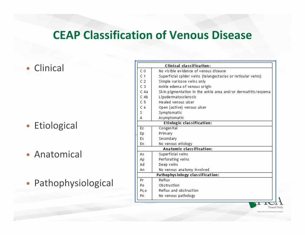
CEAP Classification of Venous Disease
• Clinical
• Etiological
• Anatomical
• Pathophysiological
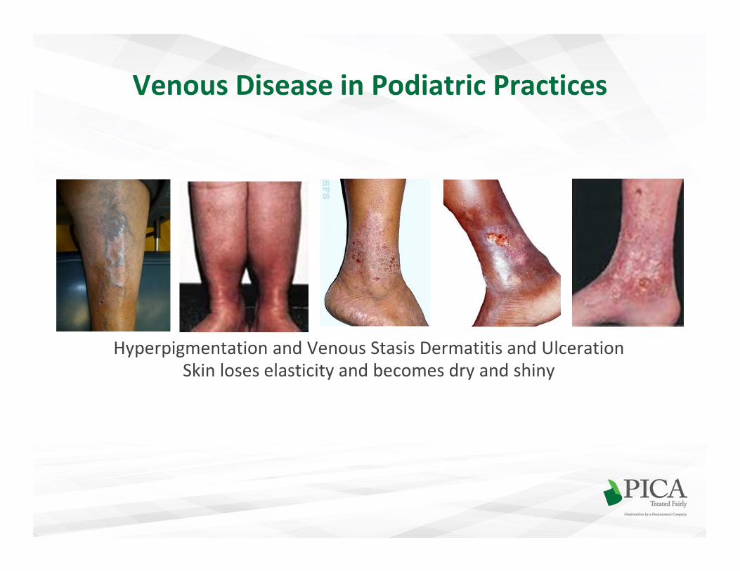
Venous Disease in Podiatric Practices
Hyperpigmentation and Venous Stasis Dermatitis and Ulceration
Skin loses elasticity and becomes dry and shiny

Lipodermatosclerosis (LDS)
• LDS literally means scarring of the skin and fat
• Chronic panniculitis with lipomembranous changes
• Found with long standing venous insufficiency
• Skin becomes brown, smooth, indurated and painful
• Proximal to the ankle, usually medial
• Tapering proximal to the ankles “inverted Champagne bottle”
• Skin is permanently and irreversibly damaged
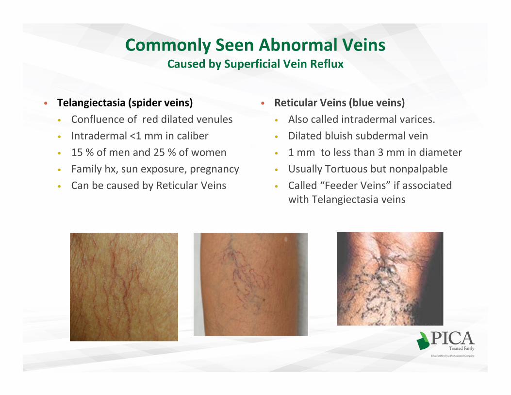
Commonly Seen Abnormal VeinsCaused by Superficial Vein Reflux
• Telangiectasia (spider veins)
• Confluence of red dilated venules
• Intradermal <1 mm in caliber
• 15 % of men and 25 % of women
• Family hx, sun exposure, pregnancy
• Can be caused by Reticular Veins
• Reticular Veins (blue veins)
• Also called intradermal varices.
• Dilated bluish subdermal vein
• 1 mm to less than 3 mm in diameter
• Usually Tortuous but nonpalpable
• Called “Feeder Veins” if associated
with Telangiectasia veins

Corona Phlebectatica (CP)
• CP is classically described as the presence of abnormally visible
cutaneous blood vessels at the ankle with four components:
"venous cups," blue and red telangiectases, and capillary "stasis
spots."
• Fine visible vessels too numerous to delineate
• Dilation and elongation of venules in the dermis of the ankle and
foot
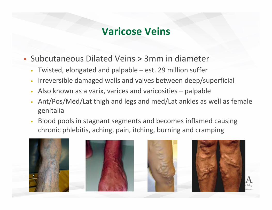
Varicose Veins
• Subcutaneous Dilated Veins > 3mm in diameter
• Twisted, elongated and palpable – est. 29 million suffer
• Irreversible damaged walls and valves between deep/superficial
• Also known as a varix, varices and varicosities – palpable
• Ant/Pos/Med/Lat thigh and legs and med/Lat ankles as well as female
genitalia
• Blood pools in stagnant segments and becomes inflamed causing
chronic phlebitis, aching, pain, itching, burning and cramping

Venous Ulcerations
• VLUs affect 1% of the adult population and may be
primary or secondary Johnson and Rodgers, Podiatry
Management August 2011
• Primary VLU are due to valvular defects and
hypertension in the superficial venous system
• 70-80% of venous ulcers have reflux in both superficial
and perforating veins Doughty, et al., Acute and
Chronic Wounds, Current Mgmt Concepts, 3rd edition,
Mosby Elsevier 2007.
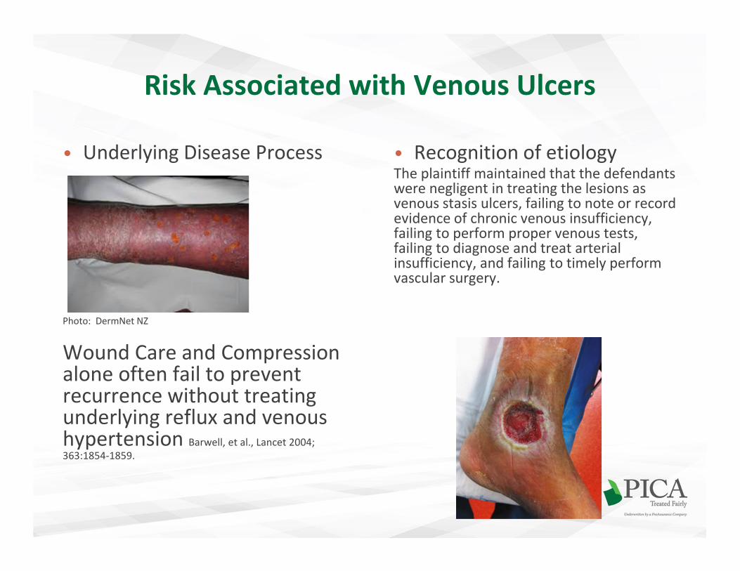
Risk Associated with Venous Ulcers
• Underlying Disease Process
Photo: DermNet NZ
Wound Care and Compression alone often fail to prevent recurrence without treating underlying reflux and venous hypertension Barwell, et al., Lancet 2004;
363:1854-1859.
• Recognition of etiologyThe plaintiff maintained that the defendants were negligent in treating the lesions as venous stasis ulcers, failing to note or record evidence of chronic venous insufficiency, failing to perform proper venous tests, failing to diagnose and treat arterial insufficiency, and failing to timely perform vascular surgery.
Venous Anatomy of the Lower Extremity
• Subcutaneous superficial system not paired with arteries
• Deep system within the muscular fasciae paired with
corresponding arteries
• Connecting or Perforating veins
• Dysfunction, mainly of the valves, may occur in each
system and in combination

Anatomy of a Vein
• Intima > valves
• Media• Smooth muscle
• Collagen
• Adventitia
• Larger superficial veins – thicker media more muscular
• Smaller superficial veins – thinner media, less strength
• Deep veins – more collagen, more strength and sheath, function with the muscular calf pump

Superficial Venous System
Small (Lesser) Saphenous Vein (SSV)
• Courses lateral to posterior
• Posterior to lateral malleolus
• Superficial to the deep fascia
• Midline to upper calf
• Between two heads of the
gastrocnemius
• Usually joins the Popliteal
• 1/3 time feeds into GSV via
communicating veins
Popliteal vein
Small saphenous vein

Superficial Venous System
Great Saphenous Vein (GSV)
• Anterior to medial malleolus
• Crosses posteriorly to lie medial
• Anteromedially AK
• Superficial to the deep fascia
• Passes thru foramen ovale
• Feeds common femoral vein at saphenofemoral junction
Saphenofemoral junction
Common Femoral Vein
• Other veins that feed into it around the junction:• the superficial inferior epigastric
• the superficial external pudendal
• the superficial circumflex iliac
• Above the knee• Anterior and Posterior Circumflex
• Below the knee• Anterior tributary
• Posterior Arch Vein
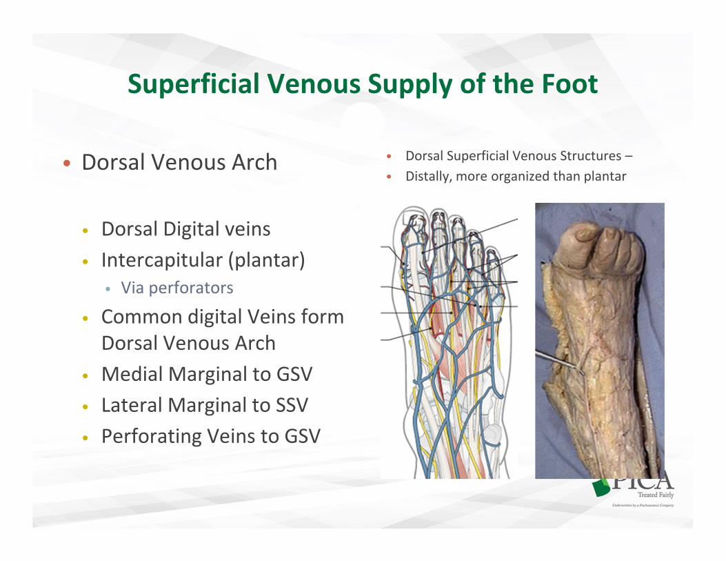
• Dorsal Venous Arch • Dorsal Superficial Venous Structures –
• Distally, more organized than plantar
• Dorsal Digital veins
• Intercapitular (plantar)
• Via perforators
• Common digital Veins form
Dorsal Venous Arch
• Medial Marginal to GSV
• Lateral Marginal to SSV
• Perforating Veins to GSV
Superficial Venous Supply of the Foot

Perforator Veins
• Direct from superficial to deep veins
• Bicuspid valves and unidirectional
• High variability with some consistent
groupings
• Mid thigh - Hunterian
• Distal thigh - Dodd
• Knee - Boyd
• Ankle and distal medial calf - Cockett
• Foot - multiple
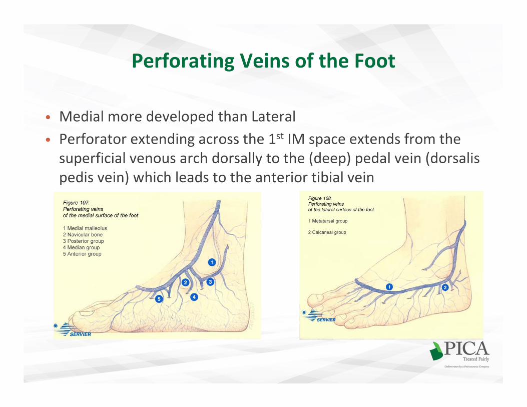
Perforating Veins of the Foot
• Medial more developed than Lateral
• Perforator extending across the 1st IM space extends from the
superficial venous arch dorsally to the (deep) pedal vein (dorsalis
pedis vein) which leads to the anterior tibial vein

Deep Venous System • Eventual return to the Right Atrium
• Valvular
• Intramuscular
• External Iliac Vein
• Common Femoral Vein
• Deep Femoral Vein • Short course with deep muscle tributaries
• Femoral Vein• Courses through the adductor canal
• Popliteal Vein
• Peroneal Vein • soleal and gastrocnemius intramuscular
venous plexi join mid calf from sinusoid venous network
• Anterior Tibial
• Posterior Tibial

• Deep Plantar Venous Arch • Dorsal Deep Veins • less prominent than Plantar
• “Plantar Venous Pump”
• Plantar digital veins ??
• Send intercapitular veins to superficial
dorsal venous arch
• Metatarsal plantar veins
• Deep plantar venous arch
• Medial/Lateral plantar veins
• Posterior Tibial Vein
• Dorsal digital veins are superficial
• Deep veins begin at digital clefts
• Pedal Vein (Dorsalis Pedis Vein)• Along with the 1st IM space perforator of the
superficial venous arch
• Anterior Tibial Vein
Deep Venous Supply of the Foot
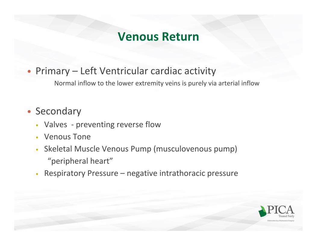
Venous Return
• Primary – Left Ventricular cardiac activityNormal inflow to the lower extremity veins is purely via arterial inflow
• Secondary
• Valves - preventing reverse flow
• Venous Tone
• Skeletal Muscle Venous Pump (musculovenous pump)
“peripheral heart”
• Respiratory Pressure – negative intrathoracic pressure
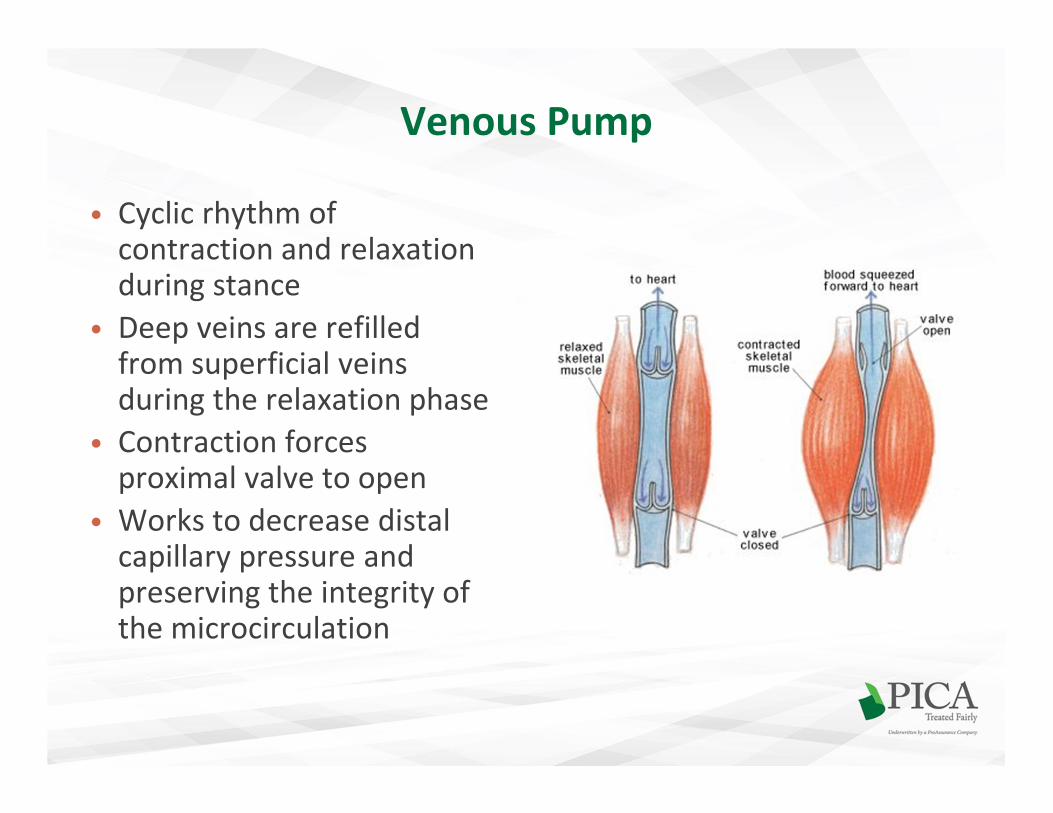
Venous Pump
• Cyclic rhythm of contraction and relaxation during stance
• Deep veins are refilled from superficial veins during the relaxation phase
• Contraction forces proximal valve to open
• Works to decrease distal capillary pressure and preserving the integrity of the microcirculation

• Valves of the Leg –approximate distribution
Deep Veins: More distally
• Popliteal vein 2 - 4
• Anterior tibial veins 9 -11
• Posterior tibial veins 9 -19
• Peroneal veins 7
Valves present at junctions
GSV/Femoral
SSV/Popliteal
Superficial Veins have fewer:
• GSV and SSV 7-9
• Valves near tributary terminals
• Flow is slower due to lack of muscular pump
• Valves in the LE outnumber the UE due to gravity
• Major Perforating veins have 1-3
• Absent in smaller perforating veins
• Perforators in the foot are absent of valves or valves lead to superficial veins
Venous Valves
Valvular Incompetence
Venous Insufficiency – Increased SVP
• Primary Cause of Chronic Venous Insufficiency (CVI)
• Venous Reflux Disease results
• Venous Outflow Obstruction
• Venous insufficiency also tends to be progressive
• Superficial venous reflux can cause spider veins, varicose veins, and lead to
edema, hyperpigmentation, and venous ulcers
• Perforator reflux can cause edema hyperpigmentation and ulcers
• Most patients with venous dysfunction have incompetent valves in both
superficial and perforator veins
• Symptoms: pain, swelling, leg heaviness and fatigue
• Many cases can be treated surgically
• Reflux extends to superficial vein trunk and increased superficial venous
pressure (SVP) leading to varicosities and ulcers
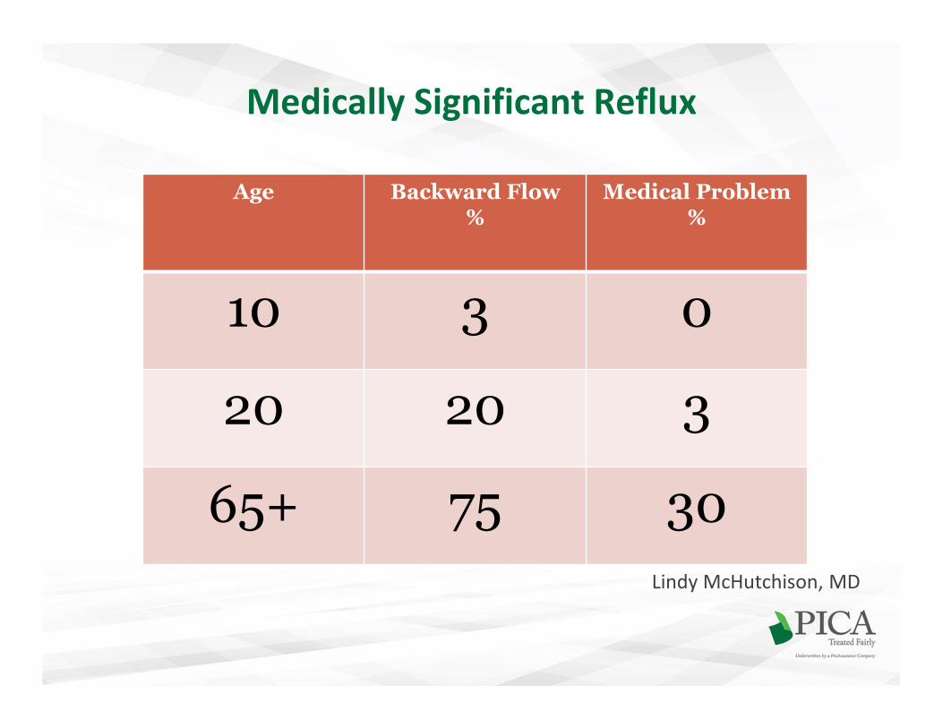
Medically Significant Reflux
Lindy McHutchison, MD
Age Backward Flow%
Medical Problem%
10 3 0
20 20 3
65+ 75 30
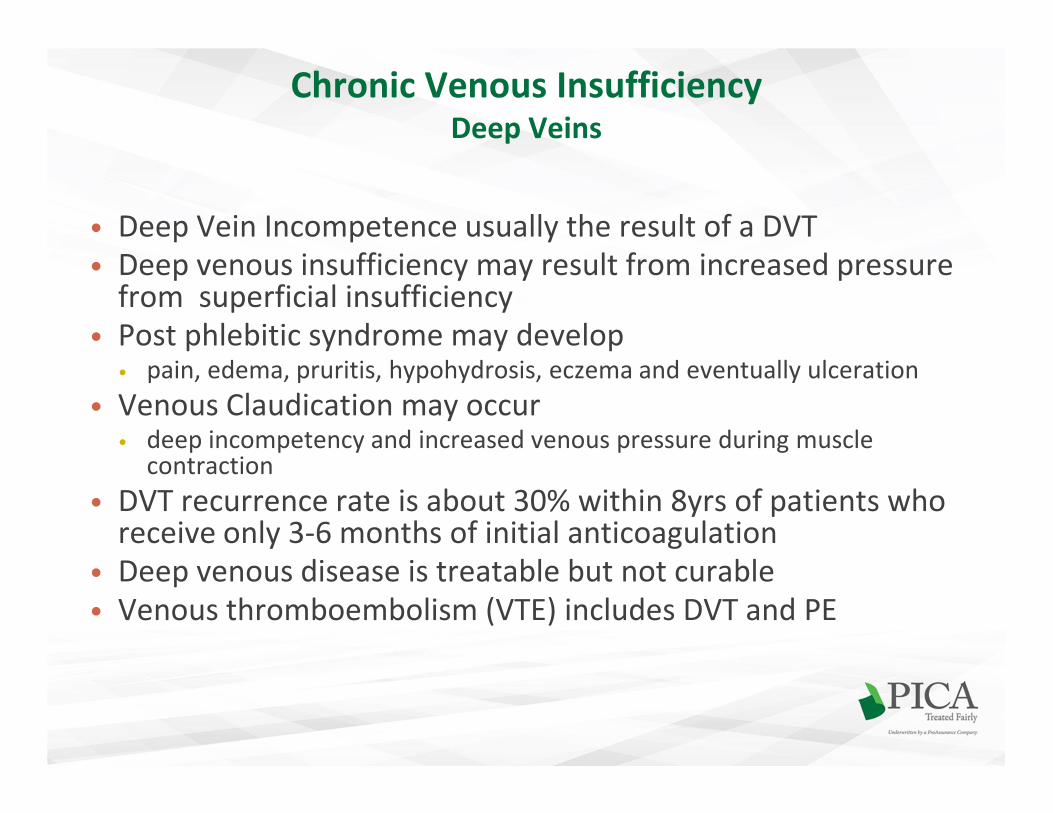
Chronic Venous InsufficiencyDeep Veins
• Deep Vein Incompetence usually the result of a DVT
• Deep venous insufficiency may result from increased pressure from superficial insufficiency
• Post phlebitic syndrome may develop • pain, edema, pruritis, hypohydrosis, eczema and eventually ulceration
• Venous Claudication may occur• deep incompetency and increased venous pressure during muscle
contraction
• DVT recurrence rate is about 30% within 8yrs of patients who receive only 3-6 months of initial anticoagulation
• Deep venous disease is treatable but not curable
• Venous thromboembolism (VTE) includes DVT and PE
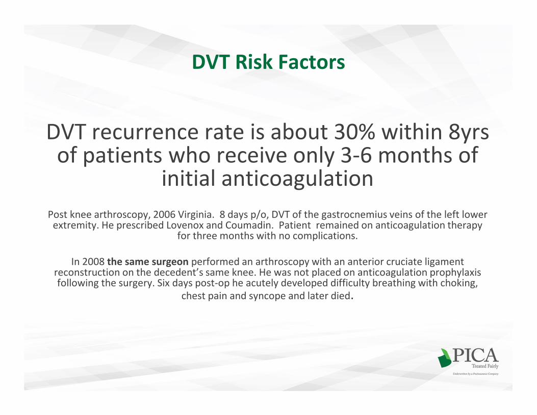
DVT Risk Factors
DVT recurrence rate is about 30% within 8yrs of patients who receive only 3-6 months of
initial anticoagulation
Post knee arthroscopy, 2006 Virginia. 8 days p/o, DVT of the gastrocnemius veins of the left lower extremity. He prescribed Lovenox and Coumadin. Patient remained on anticoagulation therapy
for three months with no complications.
In 2008 the same surgeon performed an arthroscopy with an anterior cruciate ligament reconstruction on the decedent’s same knee. He was not placed on anticoagulation prophylaxis following the surgery. Six days post-op he acutely developed difficulty breathing with choking,
chest pain and syncope and later died.
DVT
• Risk Factors
• Inheriting a blood-clotting disorder. ...
• Prolonged bed rest, such as during a long hospital stay, or paralysis. ...
• Injury or surgery. ...
• Pregnancy. ...
• Birth control pills (oral contraceptives) or hormone replacement therapy. ...
• Being overweight or obese. ...
• Smoking. ...
• Cancer

DVT Risk Factorswww.jeffdavislaw.com
• Paralysis
• Paresthesis or recent cast
immobilization of lower
extremities
• Major surgery requiring
regional or general
anesthetic in the past 12
weeks or was recently
bedridden for greater than
3 days
• Recent long-distance
travel
• Localized pain along
distribution of deep venous
system
• Swelling of entire leg
and/or calf greater than 3
centimeters
• Pitting edema
• Collateral superficial veins
• Previously documented
DVT or PE
• Active cancer
Thromboprophylaxis
• Provide prophylaxis or not??
• Risk Assessment vs. Bleeding
• Non-medical option:• In a study of the efficacy of intermittent pneumatic compression devices in multiple
postoperative patient groups versus no use of prophylaxis, Urbankova et al reported that the
incidence of DVT was reduced by 60%. However, the use of mechanical means of prophylaxis
alone is not effective in moderate or high-risk cases.
• Medical options:
• A large study performed in Europe, the Pulmonary Embolism
Prevention (PEP) study, found that the overall DVT rate was
decreased 30% with low-dose aspirin compared with placebo, and the
overall pulmonary thrombosis rate was decreased by 40%.
• Xarelto 10mg QD starting 6-10 hrs s/p Sx x ?? Days – x 12 days for
knee
Chronic Venous InsufficiencyOther Etiology and Pathogenesis
• Pathogenesis
• Defects in venous wall strength
• Valvular incompetence
• Venous hypertension
• Cellular problems
• 70% hereditary females > males – hormonal relaxation of
venous walls
• Inflammatory changes in the veins
• Inflammation causes ischemic and trophic injury to skin
Symptoms of CVI
• Pain, throbbing or cramping
• Burning or itching, especially around a vein
• Heaviness, aching or fatigue especially late in the day or after periods of dependency
• Night Cramps
• Restless Leg Syndrome
• Same symptoms found with pes valgus or PTTD
• DDx - can be found with periods of sitting or NWB
• Eczema and hair loss to lower legs• Course
• Progressive
• Preventable
• Manageable
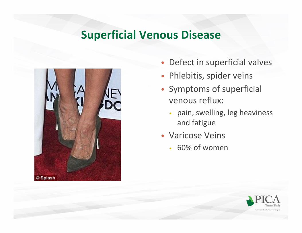
Superficial Venous Disease
• Defect in superficial valves
• Phlebitis, spider veins
• Symptoms of superficial
venous reflux:
• pain, swelling, leg heaviness
and fatigue
• Varicose Veins
• 60% of women

Varicose Veins
• Primary – SVD• Degeneration of venous wall
• Starts distally
• Inflammation
• Venous hypertension
• Collagen breakdown
• Intima remains intact
• Very common• 20-40% adult population
• Progresses slowly
• Venous Stasis SyndromeCaused by Primary or secondary
Post Phlebitic Syndrome only
one cause
• Secondary: • Deep Reflux
• Post Thrombolytic Syndrome
(Post Phlebitic Syndrome)
• Clot causes inflammation
• Obstruction > collaterals
• Damage to the intima
• Much less common• 20-50% of DVT > VV
• Most common with Iliofemoral clot
• Onset often 6 months post DVT
• Progress quicker – severe within 5 years

• Starling principle of microvascular
fluid exchange • Arterial End: Venous End:
• hydrostatic>osmotic osmotic>hydrostatic
• Exchange takes place at the capillary
bed
• Exchange takes place across the
capillary wall
• Capillaries are normally impermeable
to plasma proteins
• Capillaries are freely permeable to
water and low molecular- weight
solutes
• Normally, a relatively stable
interstitial fluid volume (IFV) is
maintained
• 10% fluid is left in the tissue and
picked up by the lymph system
Capillaries: Fluid exchange within tissueBetween the intra- and extravascular space

Hydrostatic Pressure
• Dependent on the gravitational forces in a standing position as well as the distance of the vein from the heart
• Superficial venous pressure (SVP)at the ankle is normally:
• 12mmHg when supine
• 104mmHg when in stance
• During gait, ambulatory venous pressure (AVP) is:
• 30mmHg
• With venous disease, pressure backs up and increases the venous capillary pressure (VCP)
• Increases in VCP causes increased transcapillary filtration which results in edema
• Venous reflux increases SVP and VCP increases and pressure in skin

Edema
• Clinical state characterized by an accumulation of fluid in
the interstitial or intracellular space
• Edema is the result of excess fluid in the interstitial space
and occurs when there is a breakdown in this pressure
gradient
• either high rate of transcapillary filtration into the tissue
• low lymphatic drainage rate
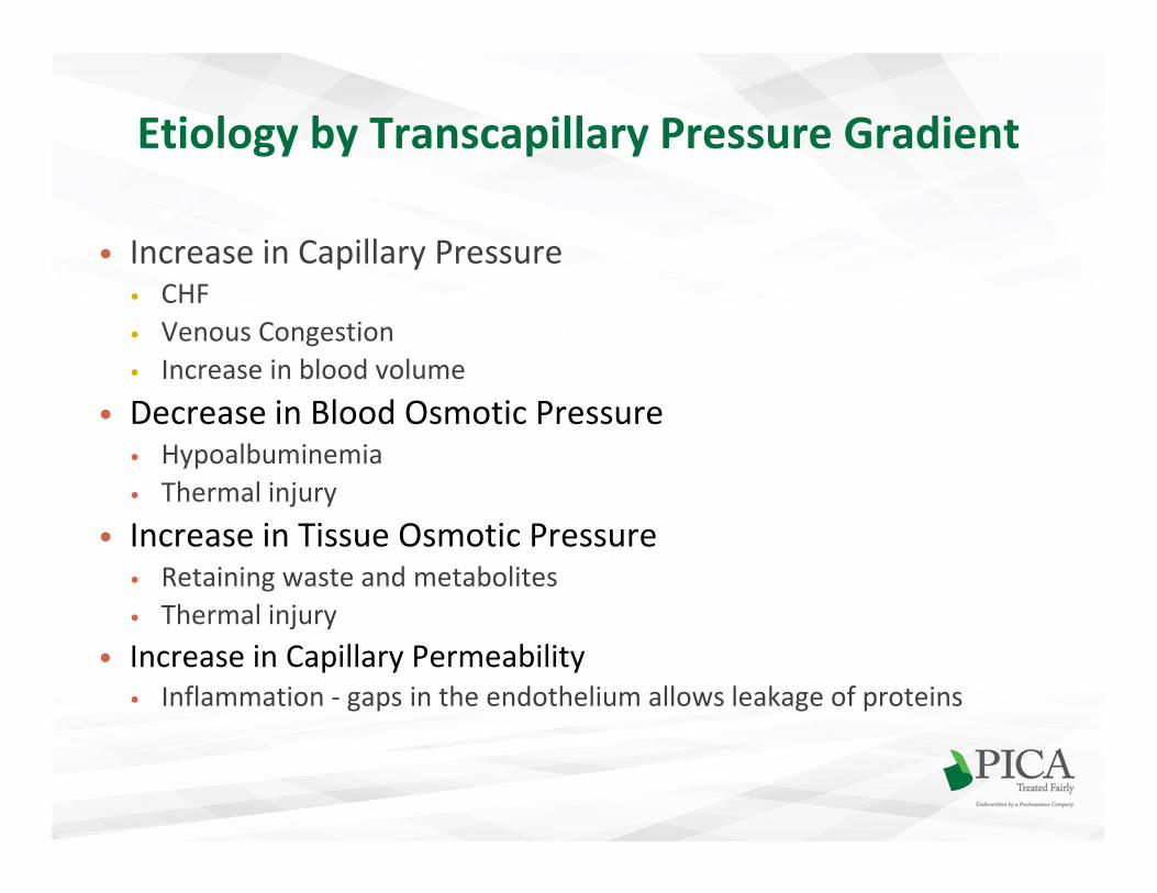
Etiology by Transcapillary Pressure Gradient
• Increase in Capillary Pressure• CHF
• Venous Congestion
• Increase in blood volume
• Decrease in Blood Osmotic Pressure• Hypoalbuminemia
• Thermal injury
• Increase in Tissue Osmotic Pressure• Retaining waste and metabolites
• Thermal injury
• Increase in Capillary Permeability
• Inflammation - gaps in the endothelium allows leakage of proteins

Edema by Classification
• Acute Edema
• DVT
• Trauma
• Cellulitis
• Septic Arthritis
• Allergic Reaction
• Chronic Edema
• Regional
• Varicose Veins
• Obstruction of venous return
• Lymphedema
• usually involves the foot –
venous edema does not
• Systemic
• CHF
• Nephrotic Syndrome
• Myxedema
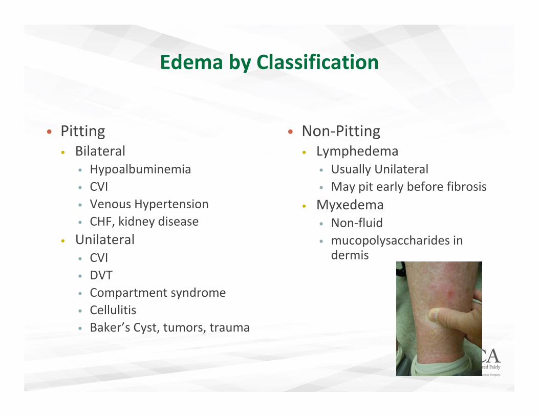
Edema by Classification
• Pitting
• Bilateral
• Hypoalbuminemia
• CVI
• Venous Hypertension
• CHF, kidney disease
• Unilateral
• CVI
• DVT
• Compartment syndrome
• Cellulitis
• Baker’s Cyst, tumors, trauma
• Non-Pitting
• Lymphedema
• Usually Unilateral
• May pit early before fibrosis
• Myxedema
• Non-fluid
• mucopolysaccharides in dermis
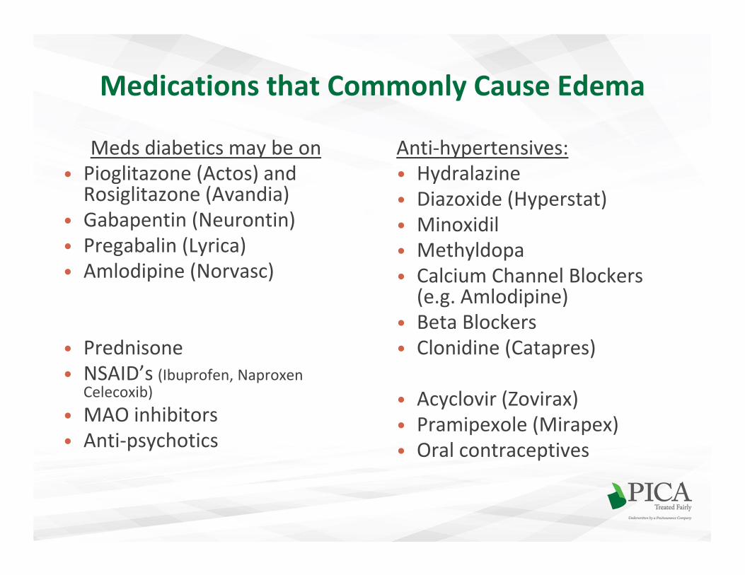
Medications that Commonly Cause Edema
Meds diabetics may be on
• Pioglitazone (Actos) and Rosiglitazone (Avandia)
• Gabapentin (Neurontin)
• Pregabalin (Lyrica)
• Amlodipine (Norvasc)
• Prednisone
• NSAID’s (Ibuprofen, Naproxen Celecoxib)
• MAO inhibitors
• Anti-psychotics
Anti-hypertensives:
• Hydralazine
• Diazoxide (Hyperstat)
• Minoxidil
• Methyldopa
• Calcium Channel Blockers (e.g. Amlodipine)
• Beta Blockers
• Clonidine (Catapres)
• Acyclovir (Zovirax)
• Pramipexole (Mirapex)
• Oral contraceptives
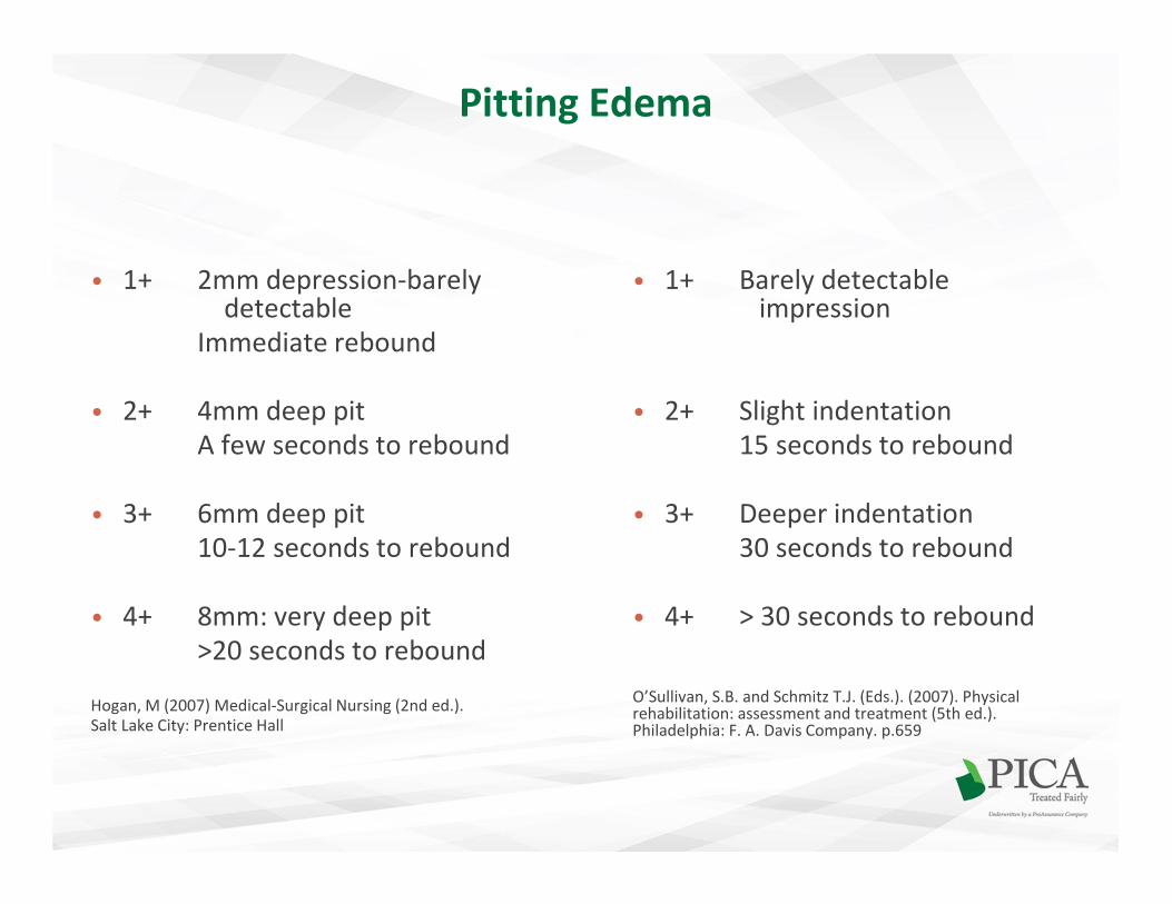
Pitting Edema
• 1+ 2mm depression-barely detectable
Immediate rebound
• 2+ 4mm deep pit
A few seconds to rebound
• 3+ 6mm deep pit
10-12 seconds to rebound
• 4+ 8mm: very deep pit
>20 seconds to rebound
Hogan, M (2007) Medical-Surgical Nursing (2nd ed.).
Salt Lake City: Prentice Hall
• 1+ Barely detectable impression
• 2+ Slight indentation
15 seconds to rebound
• 3+ Deeper indentation
30 seconds to rebound
• 4+ > 30 seconds to rebound
O’Sullivan, S.B. and Schmitz T.J. (Eds.). (2007). Physical rehabilitation: assessment and treatment (5th ed.). Philadelphia: F. A. Davis Company. p.659

Post Op Edema
Pre Op Considerations??
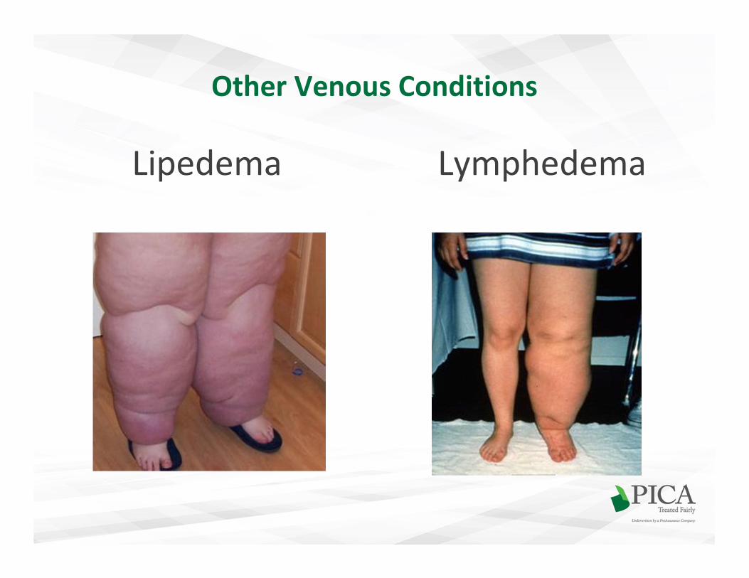
Other Venous Conditions
Lipedema Lymphedema
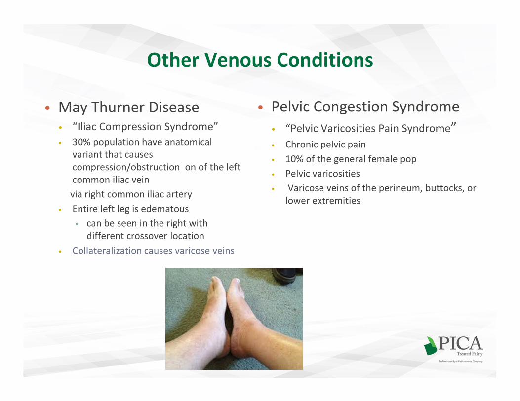
Other Venous Conditions
• May Thurner Disease• “Iliac Compression Syndrome”
• 30% population have anatomical
variant that causes
compression/obstruction on of the left
common iliac vein
via right common iliac artery
• Entire left leg is edematous
• can be seen in the right with
different crossover location
• Collateralization causes varicose veins
• Pelvic Congestion Syndrome
• “Pelvic Varicosities Pain Syndrome”• Chronic pelvic pain
• 10% of the general female pop
• Pelvic varicosities
• Varicose veins of the perineum, buttocks, or
lower extremities
Diagnosing CVI
• Duplex Ultrasound
• Provides visual image of vein and valve damage and possible venous obstruction
• Measures speed and blood flow
• Photoplethysmography (PPG)
• Multimethod venous refill tests
• Venous outflow testing
• Measures time and records wavelengths for refill and outflow
• Venogram
“Phlebology”
Field of Medicine that treats venous disease
Treatment Options for CVI
• Chemical sclerotherapy
• Vein stripping
• Bypass surgery
• Valve repair
• Angioplasty with or without stenting of a vein
• Minimally invasive endovenous ablation procedures
such as Laser and Radio Frequency Ablation.

Podiatric Treatment of Superficial Veins
?
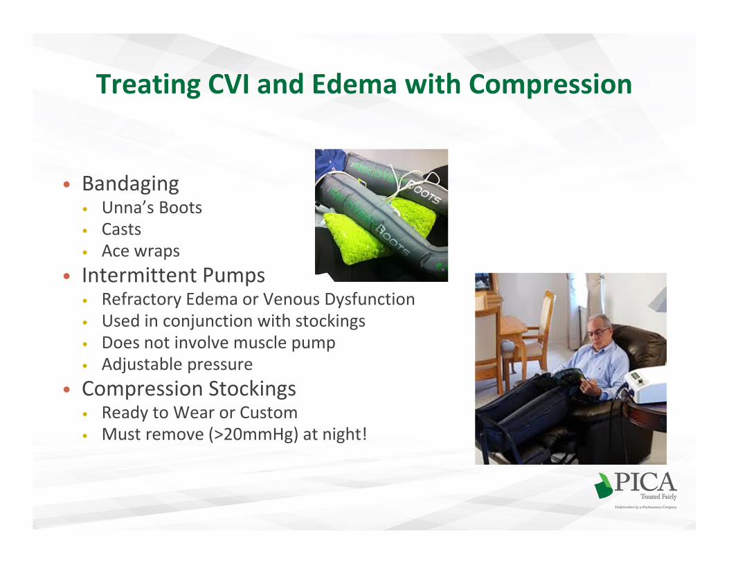
Treating CVI and Edema with Compression
• Bandaging• Unna’s Boots
• Casts
• Ace wraps
• Intermittent Pumps• Refractory Edema or Venous Dysfunction
• Used in conjunction with stockings
• Does not involve muscle pump
• Adjustable pressure
• Compression Stockings• Ready to Wear or Custom
• Must remove (>20mmHg) at night!

Why Compression is Important
• Helps prevent VTE – Venothromboembolism: DVT/PE
• Helps alleviate edema and discomfort
• Reduction of superficial venous pressure
• Decreases the progression of both venous and lymphatic
disease
• Decreases the progression of post thrombosis syndrome
and other venous dermatitis
• Helps soften woody edema
• Helps prevent skin breakdown
• Helps prevent and heal venous ulcers
What does Compression Actually Do??
• Therapeutic Effect:
• Decreases pathologic venous capacity
• Increases (or supports) the function of the insufficient valves
• Increases fibrinolytic activity of the blood
• Increases reabsorption of fluid into the capillaries
• Decreases Retrograde Flow
• Increases Venous Return
• Counters the affects of Ambulatory Venous Hypertension
The Accepted Mechanism of Action
of Gradient Compression
• Reduces Edema
• Allows the dermis to engage the superficial capillary network to improve
function of O2 and waste exchange
• Capillary Action: Improves absorption and decreases leakage
• Increases interstitial pressure
• Decreased capillary leakage of fluids and solutes
• Increase absorption of interstitial fluids – lymph system as well
• Improves local capillary clearance
• Reduces the Diameter of Superficial Veins
• Reduces the capacitance of these veins
• Reduces reflux and venous pooling
• Increase proximal venous flow

Compression of Superficial Veins
• Acts as a “restraint” as superficial veins do not have the
muscular and fascial support system of deep veins
• External compression can compensate for both
superficial and perforator valvular disease
• Return distended, over dilated veins to a more normal
size
• Fabric is an external replacement for lost elasticity and
resistance of skin
Indications for Compression Therapy
• Edema
• Chronic Venous
Insufficiency
• DVT/PE Prevention
• Prevention of ulcer
recurrence
• Management of ulcers • Requires 35mmHg
• Varicose Veins
• Pregnancy
• Post sclerotic therapy
• Superficial
thrombophlebitis
• Tired, aching legs
• Hypertrophic Scar and
dermatitis
• Lymph Edema
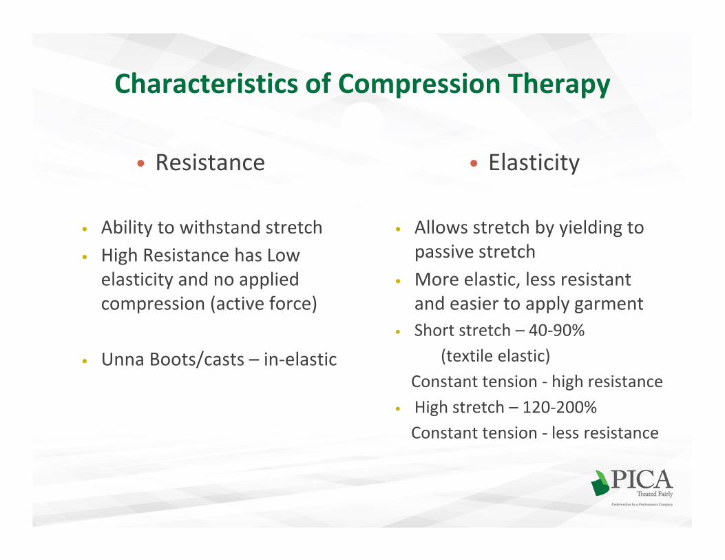
• Resistance • Elasticity
• Ability to withstand stretch
• High Resistance has Low
elasticity and no applied
compression (active force)
• Unna Boots/casts – in-elastic
• Allows stretch by yielding to
passive stretch
• More elastic, less resistant
and easier to apply garment
• Short stretch – 40-90%
(textile elastic)
Constant tension - high resistance
• High stretch – 120-200%
Constant tension - less resistance
Characteristics of Compression Therapy
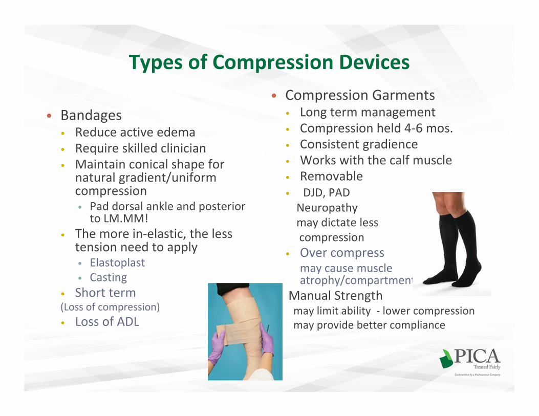
Types of Compression Devices
• Bandages• Reduce active edema
• Require skilled clinician
• Maintain conical shape for natural gradient/uniform compression• Pad dorsal ankle and posterior
to LM.MM!
• The more in-elastic, the less tension need to apply• Elastoplast
• Casting
• Short term (Loss of compression)
• Loss of ADL
• Compression Garments• Long term management
• Compression held 4-6 mos.
• Consistent gradience
• Works with the calf muscle
• Removable
• DJD, PAD
Neuropathy
may dictate less
compression
• Over compressmay cause muscle atrophy/compartment syndrome
Manual Strengthmay limit ability - lower compression
may provide better compliance

• Ankle Anatomy and Sizing• Choosing Level of
Compression mmHg
• Measure ankle & calf
• Range of mmHg is given
as a range of pressure at
ankle based on ankle size
• Abnormal anatomy may
dictate custom for
greater precision
• 10-15 - fatigue, aches
• 15-20 -pregnancy, post surgical, travel, mild varicosities, moderate edema, DVT prophalactis
• 20-30 - moderate to severe varicosities, edema with pregnancy DVT, ulcer prevention, CVI, thrombophlebitis
• 30-40 – Lymph edema, severe CVI symptoms, ulcers
Choosing the Right Compression Garment
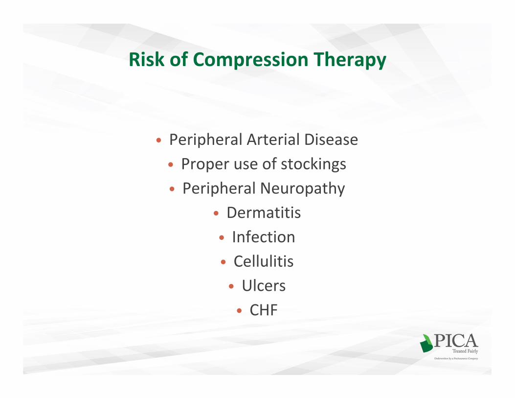
Risk of Compression Therapy
• Peripheral Arterial Disease
• Proper use of stockings
• Peripheral Neuropathy
• Dermatitis
• Infection
• Cellulitis
• Ulcers
• CHF

In Summary….
Lower Extremity Venous Disease affects our patients and
needs as much attention as peripheral arterial disease for
prevention of progression.
Conservative treatment for edema should include
elevation, increasing the muscle pump with activity and
supportive measures to reduce the subjective and objective
symptoms of superficial and deep venous disease.
Thank You