vascularization in tissue engineering
TRANSCRIPT

Vascularization in tissue engineeringJeroen Rouwkema1,2, Nicolas C. Rivron1 and Clemens A. van Blitterswijk1
1 Department of Tissue Regeneration, University of Twente, Drienerlolaan 5, 7522NB Enschede, The Netherlands2 Department of Biomechanical Engineering, University of Twente, Drienerlolaan 5, 7522NB Enschede, The Netherlands
Review
Glossary
Angiogenesis: new blood-vessel formation by the growth and sprouting of
existing blood vessels.
Hypoxia: a state in which the oxygen concentration is lower than the
physiological level.
Matrix metalloproteinases (MMPs): enzymes capable of degrading multiple
extracellular matrix proteins. MMPs are secreted by migrating endothelial cells
in order to break down the extracellular matrix that surrounds vessels and thus
to allow for vessel growth.
Mural cells: the combined term for smooth muscle cells and pericytes.
Perfusion bioreactor: a bioreactor in which medium is perfused through a
construct. This allows the active delivery of nutrients and oxygen to the cells in
the interior of the construct.
Pericytes: the cells that surround endothelial cells in capillaries. The role of
pericytes is similar to the role of smooth muscle cells in bigger vessels.
Prevascular network: in this article this is defined as an engineered vascular
network that lacks the organization of, for instance, a vascular tree. As such, a
prevascular network is comparable to the primitive vascular network that is
formed during vasculogenesis.
Smooth muscle cells: the cells that surround endothelial cells in all blood
vessels, except capillaries. Smooth muscle cells stabilize the vessels and play a
role in the expansion and contraction of the vessels.
Vascular anastomosis: the process by which two vessels are functionally
connected to each other.
Vascular axis: a macrovascular structure that is used for the transport of
blood to and from a certain location. A vascular axis is often associated with a
vascular tree, which distributes blood within a tissue.
Vascular tree: the typical organization of a vascular network within a tissue. A
blood-supplying artery branches into smaller vessels (arterioles) that subse-
quently branch into capillaries. These combine again into venules that
combine into a vein that transports the blood away from the tissue.
Vasculogenesis: the de novo formation of blood vessels by endothelial
progenitor cells.
Vessel maturation: the process in which vessels are stabilized by the
Tissue engineering has been an active field of researchfor several decades now. However, the amount of clinicalapplications in the field of tissue engineering isstill limited. One of the current limitations of tissueengineering is its inability to provide sufficient bloodsupply in the initial phase after implantation. Insufficientvascularization can lead to improper cell integration orcell death in tissue-engineered constructs. This reviewwill discuss the advantages and limitations of recentstrategies aimed at enhancing the vascularization oftissue-engineered constructs. We will illustrate thatcombining the efforts of different research lines mightbe necessary to obtain optimal results in the field.
IntroductionMost tissues in the body rely on blood vessels to supply theindividual cells with nutrients and oxygen. For a tissue togrow beyond 100–200 mm (the diffusion limit of oxygen),new blood-vessel formation is required [1], and this is alsotrue for tissue-engineered constructs. During in vitro cul-ture, larger tissue-engineered constructs can be suppliedwith nutrients, for instance in perfusion bioreactors [2,3].However, after implantation of tissue constructs, thesupply of oxygen and nutrients to the implant is oftenlimited by diffusion processes that can only supply cellsin a proximity of 100–200 mm from the next capillary. Inorder for implanted tissues of greater size to survive, thetissue has to be vascularized, which means that a capillarynetwork capable of delivering nutrients to the cells isformed within the tissue. After implantation, blood vesselsfrom the host generally invade the tissue to form such anetwork, in part in response to signals that are secreted bythe implanted cells as a reaction to hypoxia. However, thisspontaneous vascular ingrowth is often limited to severaltenths of micrometers per day [4], meaning that the timeneeded for complete vascularization of an implant of sev-eral millimeters is in the order of weeks. During this time,insufficient vascularization can lead to nutrientdeficiencies and/or hypoxia deeper in the tissue. Moreover,nutrient and oxygen gradients will be present in the outerregions of the tissue, which could result in non-uniform celldifferentiation and integration and thus decreased tissuefunction [5].
Because the speed of vascularization after implantationis amajor problem in tissue engineering, the successful useof tissue-engineered constructs is currently limited to thinor avascular tissues, such as skin or cartilage, for whichpostimplantation neovascularization from the host is suf-ficient to meet the demand for oxygen and nutrients [6]. To
Corresponding author: Rouwkema, J. ([email protected])
434 0167-7799/$ – see front matter � 2008 Elsevie
succeed in the application of tissue engineering for biggertissues, such as bone and muscle, the problem of vascular-ization has to be solved [7].
Vascularization in tissue engineeringAfter implantation of tissue-engineered constructs, aspontaneous vascularization of the implant is usually seen(Box 1). This is in part due to an inflammatory wound-healing response, which is induced by the surgical pro-cedure. Furthermore, the seeded cells often create ahypoxic state in the implant, which stimulates theendogenous release of angiogenic growth factors [8]. How-ever, this induced vessel ingrowth is often too slow toprovide adequate nutrient transport to the cells in theinterior of the transplanted tissue. Therefore, additionalstrategies for enhancing vascularization are essential toensure the survival of large tissue-engineered grafts.
Several strategies for enhancing vascularization arecurrently under investigation. These include scaffolddesign, the inclusion of angiogenic factors, in vivo prevas-cularization and in vitro prevascularization (see Figure 1).Although all these strategies can in principle enhance
association with mural cells and the synthesis of extracellular matrix. Vessel
maturation is generally accompanied by an inhibition of vessel growth.
r Ltd. All rights reserved. doi:10.1016/j.tibtech.2008.04.009 Available online 26 June 2008

Box 1. Vascularization
Blood vessels are part of the circulatory system. They transport
blood, and thus nutrients and waste products, to and from almost
every part of the body. Three distinct structures can be distin-
guished in the vascular system. These are the (i) macrovessels
(arteries and veins), which branch out into (ii) microvessels
(arterioles and venules) and finally into (iii) capillaries. The
capillaries facilitate the actual distribution of nutrients to the tissues
in the body.
During blood-vessel formation, three processes can be distin-
guished; vasculogenesis, angiogenesis and arteriogenesis [52].
Vasculogenesis is the de novo vessel-forming process, which
takes place during early embryonic development. Endothelial
cells differentiate from their precursors and proliferate within a
previously avascular tissue to form a primitive capillary network
[53]. Vasculogenesis is followed by angiogenesis, when the initial
vascular network is remodeled into more complex networks [54].
During this process, endothelial cells are activated and begin to
degrade their surrounding matrix by the release of matrix
metalloproteinases (MMPs). After this, the endothelial cells
migrate into the gaps, resulting in the formation of capillary
buds and sprouts. Endothelial cells that are located behind the
migrating endothelium proliferate, thereby elongating the newly
developing blood vessel [8]. Arteriogenesis is the process of
structural enlargement and remodeling of preexisting small
arterioles into larger vessels (Figure I) [55]. For a long time, it
was generally accepted that new vessel formation in adults was
limited to angiogenesis and arteriogenesis. However, more recent
data suggest that the basis for native as well as for therapeutic
neovascularization also includes postnatal vasculogenesis pro-
cesses. It has been established that bone-marrow-derived en-
dothelial progenitor cells, which are present in the peripheral
blood, are augmented in response to certain cytokines and/or
tissue ischemia and home into sites of neovascularization, where
they are incorporated [56–58].
Vessel maturation is an important process in blood-vessel
formation. Differentiated pericytes and smooth muscle cells
stabilize vessel structures and suppress endothelial cell growth
[59]. Vessel growth that is not accompanied by vessel maturation
results in disorganized, leaky and hemorrhagic blood vessels that
are prone to regression [6]. Because maturation is accompanied
by a suppression of endothelial cell growth, the timing of
maturation is crucial when designing vascularization strategies.
If maturation starts too early, the vascular network will not be
extensive enough to supply the entire tissue with nutrients.
Conversely, if maturation starts too late, vessels are likely to
regress and thus will not be able to establish a physiological
circulation of blood.
Figure I. Vascular tree development. During vasculogenesis, endothelial
progenitor cells give rise to a primitive vascular network. In the next stages,
termed angiogenesis and arteriogenesis, the network expands, and pericytes
(PCs) and smooth muscle cells (SMCs) are recruited for the stabilization of the
vessels. Finally, a mature organized vascular network emerges. Adapted, with
permission, from Ref. [60].
Review Trends in Biotechnology Vol.26 No.8
vascularization after implantation, the degree to whichthese strategies are capable of enhancing vascularizationvaries, and this is further illustrated in Figure 2. The firsttwo approaches, scaffold design and angiogenic factordelivery, both rely on the ingrowth of host vessels intothe entire implanted construct. Therefore, although thesestrategies are able to increase the rate of vascularization, itwould still take several days to weeks for the center of theimplant to become perfused. By contrast, in vivo prevas-cularization can in principle result in the instantaneousperfusion of a construct after implantation at the final sitebecause the construct is microsurgically connected to thehost vasculature. However, before implantation at the finalsite, a pre-implantation period is necessary, during whichtime the implant has to rely on spontaneous angiogenesisfrom the surrounding vessels into the construct. Therefore,nutrient limitations are likely to occur during this stage. Invitro prevascularization does not result in the instan-taneous perfusion of a construct because vessels have togrow from the host into the construct until they reach thevascular network formed in vitro. The invading vessels canthen anastomose to the present vasculature, resulting inthe perfusion of the entire construct with blood. Comparedto scaffold design and angiogenic factor delivery, thismethod can dramatically decrease the time that is neededto vascularize the implant because host vessels do not haveto grow into the entire construct but only into its outerregions, that is, until the ingrowing vessels meet thepreformed vascular network.
Scaffold design
The architecture and design of a scaffold has a profoundeffect on the rate of vascularization after implantation.First, the pore size of the scaffold is a critical determinantof blood-vessel ingrowth. Druecke et al. showed that vesselingrowth was significantly faster in scaffolds with poresgreater than 250 mm than in those with smaller pores [9].However, it is not only the pore size that is important forvascularization: the interconnectivity of the pores is alsosignificant because cell migration, and thus vasculariza-tion, will be inhibited if pores are not interconnected, evenif the scaffold porosity is high [10–11].
Conventional scaffold fabrication techniques include,amongst others, gas foaming, phase separation, freeze dry-ing and particulate leaching [12]. These fabrication tech-niques have been widely used to produce 3D scaffolds fortissue-engineering applications. Although the shape andthe size of the pores can be varied by changing theparameters of these techniques, the resulting organizationof the pores is random. This can lead to pore pathways thatare only partially connected and that follow contortedroutes, which could impede the supply of nutrients andthe ingrowth of tissue and vessels into the scaffold. Becausethey offer better control and the ability to actively design theporosity and interconnectivity of scaffolds, solid free-formfabrication systems are nowadays at the center of attention[13,14]. These versatile systems are capable of producingcomplex scaffolds with well-defined architecture and opti-mized pore interconnectivity. In addition, distinct regions,which might be of benefit for the recreation of zonal tissueslike cartilage, can easily be created within a single scaffold.
435
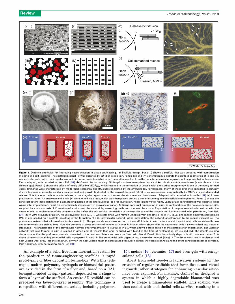
Figure 1. Different strategies for improving vascularization in tissue engineering. (a) Scaffold design. Panel (i) shows a scaffold that was prepared with compression
molding and salt leaching. The scaffold in panel (ii) was obtained by 3D fiber deposition. Panels (iii) and (iv) schematically illustrate the scaffold geometries of (i) and (ii),
respectively. Note that in the irregular scaffold (iii), some pores (depicted in red) cannot be reached from the outside, so vascular ingrowth will be prevented in these pores.
Partly adapted, with permission, from Ref. [51]. (b) Growth factor delivery. Fibrin gel matrices were placed on a chicken chorioallantoic membrane (a membrane of the
chicken egg). Panel (i) shows the effects of freely diffusible VEGF121, which resulted in the formation of vessels with a disturbed morphology. Many of the newly formed
vessel branches were characterized by malformed, corkscrew-like structures (indicated by the arrowheads). Furthermore, many of those branches appeared to abruptly
drain into zones of irregular capillary enlargement and growth (indicated by the arrows). In panel (ii), VEGF121 was released enzymatically by MMPs in a cell-demanded
release. Note that upon cell-demanded release, a more regular organization of the vascular structures can be observed. Adapted, with permission, from Ref. [32]. (c) In vivo
prevascularization. An artery (A) and a vein (V) were joined via a loop, which was then placed around a bone-tissue-engineered scaffold and implanted. Panel (i) shows the
construct before implantation with plastic tubing instead of the arteriovenous loop for illustration. Panel (ii) shows the highly vascularized construct that was obtained eight
weeks after implantation. Panel (iii) schematically depicts in vivo prevascularization. 1: Tissue construct preparation in vitro. 2: Implantation at the prevascularization site,
supplied by a vascular axis. 3: Formation of a microvascular network by vessel ingrowth from the vascular axis. 4: Explantation of the prevascularized construct with the
vascular axis. 5: Implantation of the construct at the defect site and surgical connection of the vascular axis to the vasculature. Partly adapted, with permission, from Ref.
[34]. (d) In vitro prevascularization. Mouse myoblast cells (C2C12) were combined with human umbilical vein endothelial cells (HUVECs) and mouse embryonic fibroblasts
(MEFs) and seeded on a scaffold, resulting in the formation of a 3D prevascular network. After implantation, the network anastomosed to the mouse vasculature. The
prevascular network that is formed in vitro is shown in (i). This picture shows a cross section of the scaffold after in vitro culture in which endothelial cells are stained brown
and muscle cells are stained blue. Note the presence of cross sections of tubular structures in brown, which shows that the endothelial cells have organized into vascular
structures. The anastomosis of the prevascular network after implantation is illustrated in (ii), which shows a cross section of the scaffold after implantation. The vascular
network that was formed in vitro is stained in green and all vessels that were perfused with blood at the time of explantation are stained red. The double staining
demonstrates that the preformed vessels connected to the host vasculature and were perfused with blood. Panel (iii) schematically depicts in vitro vascularization. 1: A
tissue construct containing endothelial cells is prepared in vitro. 2: The endothelial cells organize into a vascular network (blue). 3: The tissue construct is implanted and
host vessels (red) grow into the construct. 4: When the host vessels reach the precultured vascular network, the vessels connect and the entire construct becomes perfused.
Partly adapted, with permission, from Ref. [39].
Review Trends in Biotechnology Vol.26 No.8
An example of a solid free-form fabrication system forthe production of tissue-engineering scaffolds is rapidprototyping or fiber deposition technology. With this tech-nique, molten polymers, hydrogels or biomaterial pastesare extruded in the form of a fiber and, based on a CAD(computer-aided design) pattern, deposited on a stage toform a layer of the scaffold. An entire 3D scaffold can beprepared via layer-by-layer assembly. The technique iscompatible with different materials, including polymers
436
[15], metals [16], ceramics [17] and even gels with encap-sulated cells [18].
Apart from solid free-form fabrication systems for thecreation of regular scaffolds that favor tissue and vesselingrowth, other strategies for enhancing vascularizationhave been explored. For instance, Gafni et al. designed asystem in which a highly degradable biomaterial wasused to create a filamentous scaffold. This scaffold wasthen seeded with endothelial cells in vitro, resulting in a
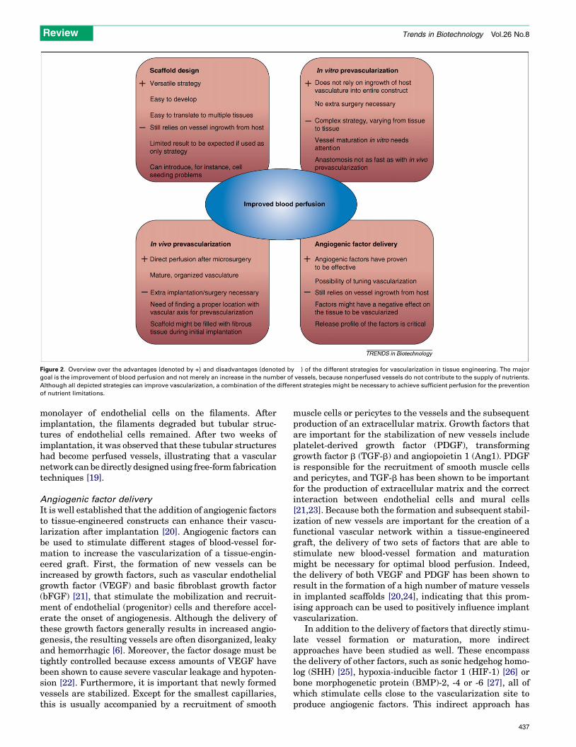
Figure 2. Overview over the advantages (denoted by +) and disadvantages (denoted by �) of the different strategies for vascularization in tissue engineering. The major
goal is the improvement of blood perfusion and not merely an increase in the number of vessels, because nonperfused vessels do not contribute to the supply of nutrients.
Although all depicted strategies can improve vascularization, a combination of the different strategies might be necessary to achieve sufficient perfusion for the prevention
of nutrient limitations.
Review Trends in Biotechnology Vol.26 No.8
monolayer of endothelial cells on the filaments. Afterimplantation, the filaments degraded but tubular struc-tures of endothelial cells remained. After two weeks ofimplantation, it was observed that these tubular structureshad become perfused vessels, illustrating that a vascularnetwork canbedirectly designedusing free-form fabricationtechniques [19].
Angiogenic factor delivery
It is well established that the addition of angiogenic factorsto tissue-engineered constructs can enhance their vascu-larization after implantation [20]. Angiogenic factors canbe used to stimulate different stages of blood-vessel for-mation to increase the vascularization of a tissue-engin-eered graft. First, the formation of new vessels can beincreased by growth factors, such as vascular endothelialgrowth factor (VEGF) and basic fibroblast growth factor(bFGF) [21], that stimulate the mobilization and recruit-ment of endothelial (progenitor) cells and therefore accel-erate the onset of angiogenesis. Although the delivery ofthese growth factors generally results in increased angio-genesis, the resulting vessels are often disorganized, leakyand hemorrhagic [6]. Moreover, the factor dosage must betightly controlled because excess amounts of VEGF havebeen shown to cause severe vascular leakage and hypoten-sion [22]. Furthermore, it is important that newly formedvessels are stabilized. Except for the smallest capillaries,this is usually accompanied by a recruitment of smooth
muscle cells or pericytes to the vessels and the subsequentproduction of an extracellular matrix. Growth factors thatare important for the stabilization of new vessels includeplatelet-derived growth factor (PDGF), transforminggrowth factor b (TGF-b) and angiopoietin 1 (Ang1). PDGFis responsible for the recruitment of smooth muscle cellsand pericytes, and TGF-b has been shown to be importantfor the production of extracellular matrix and the correctinteraction between endothelial cells and mural cells[21,23]. Because both the formation and subsequent stabil-ization of new vessels are important for the creation of afunctional vascular network within a tissue-engineeredgraft, the delivery of two sets of factors that are able tostimulate new blood-vessel formation and maturationmight be necessary for optimal blood perfusion. Indeed,the delivery of both VEGF and PDGF has been shown toresult in the formation of a high number of mature vesselsin implanted scaffolds [20,24], indicating that this prom-ising approach can be used to positively influence implantvascularization.
In addition to the delivery of factors that directly stimu-late vessel formation or maturation, more indirectapproaches have been studied as well. These encompassthe delivery of other factors, such as sonic hedgehog homo-log (SHH) [25], hypoxia-inducible factor 1 (HIF-1) [26] orbone morphogenetic protein (BMP)-2, -4 or -6 [27], all ofwhich stimulate cells close to the vascularization site toproduce angiogenic factors. This indirect approach has
437

Review Trends in Biotechnology Vol.26 No.8
several advantages over the direct delivery of angiogenicgrowth factors. First, the secretion of angiogenic factors bycells is often regulated and thereby ensures that the con-centration of angiogenic factors is in the physiologicalrange and can be adapted according to the requirementsof different stages of vessel formation. Second, the pro-duction of angiogenic factors results in the formation ofgrowth factormicrogradients, which have been shown to beimportant for capillary morphogenesis [28]. Third andlastly, the stimulation with indirect factors often resultsin the secretion of several angiogenic factors that are ableto regulate both vessel formation and maturation. Forexample, SHHwas able to induce interstitialmesenchymalcells to secrete several factors, including VEGF and angio-poietins-1 and -2, and this resulted in the formation ofhighly organized, mature vessels [25].
Several strategies for the delivery of both direct andindirect angiogenic factors have been developed. Theseinclude the addition of recombinant proteins [29] andgenes [30] to biomaterials and the use of cell transplantsthat are genetically engineered to overexpress specificfactors [31]. The addition of recombinant proteins to bio-materials is the easiest method and thus has been mostwidely studied. The delivery of growth factors with bioma-terial matrices is either driven by passive diffusion or canbe coupled to the rate of biomaterial degradation. Bothprocesses usually occur independently from each other andare often not in tune with the actual healing process [22],therefore resulting in a growth factor release profile thatcannot be adjusted or fine tuned. The degree of release canbe varied to some extent by altering the amount of growthfactor added to the matrix. The kinetics of factor releasecan be influenced by varying the degradation rate of thematerial, which depends both on its chemical compositionand its geometry. However, these limited measures areoften insufficient to synchronize the growth factor levelswith actual cellular demands. A novel approach to solvingthis problem utilizes a specific chemical linkage of growthfactors to a gel matrix. The endothelial cells of ingrowingblood vessels secrete matrix metalloproteinases (MMPs)that are able to degrade the matrix to allow the vessels topenetrate the tissue. By degrading the matrix, the cellsthus release the growth factors locally in response tocellular demand. It has been shown that the neovascula-ture that was induced by cell-demanded release of growthfactors showed a higher degree of organization than neo-vasculatures that arose from an uncontrolled growth factorrelease [32,33].
In vivo prevascularization
Another promising strategy for enhancing vascularizationin tissue engineering is in vivo prevascularization. Thismethod, also referred to as tissue prefabrication, involvestwo distinct stages. In the first stage, a tissue-engineeredconstruct is implanted into a region with an artery (orvascular axis) suitable for microsurgical transfer. This canmean that the tissue-engineered graft is either wrapped inan axially vascularized tissue, such as muscle, or that anartery is implanted into the graft. Although this ensuresthat a transplantable macrovessel is present in or aroundthe graft, the graft is not yet supplied with a capillary
438
network at this stage. A vascularization period of severalweeks at this initial implant site will result in the for-mation of a microvascular network in the engineered con-struct, which is supplied with blood by the vascular axis(see Figure 1c) [34]. After this initial stage, the tissue-engineered construct is harvested together with the micro-vascular network and the supplying artery and then reim-planted at the defect site. At this site, the vascular axis isconnected to the local vasculature using microsurgicalvascular-anastomosis techniques, which results in instan-taneous perfusion of the entire construct [35]. Theadvantage of this technique is that after implantation atthe final site, the construct becomes immediately perfusedby surgical anastomosis. However, its drawbacks are thattwo separate surgeries (one to implant the construct at thevascularization site and one to implant the construct at thefinal defect site) are necessary. In addition, a vascular axishas to be removed from the initial implantation site and,furthermore, cells might have to be reseeded beforeimplantation at the final defect site because nutrientlimitations are still likely during the vascularizationperiod at the initial implantation site.
In vitro prevascularization
A strategy for improving vascularization that has gainedinterest recently is in vitro prevascularization. Tissuesthat have been studied include skin [36–38], skeletalmuscle [39], bone [40–43] and cardiac muscle [44,45](Table 1). This strategy is based on the observation thatendothelial cells are able to form prevascular structureswhen they are cultured under the right conditions andenvironment in vitro. During in vitro prevascularization,endothelial cells are added to other tissues in vitro, whichresults in the formation of a prevascular network withinthis tissue. After implantation, this network can thenanastomose spontaneously to the ingrowing vasculatureof the host and supply the construct with nutrients. Withthis approach, host blood vessels do not need to grow intothe entire construct, but only into the outer regions of theconstruct until the prevascular structures are reached.Even though this reduces the time needed for completevascularization fromweeks to days, perfusion is not as fastas with the previous strategy because the vascular networkis not microsurgically connected after implantation. How-ever, future developments in this fieldmight aim to includethe creation of a vascular axis within the in vitro constructthat could be surgically connected to the host vasculature.
For prevascularized tissue engineering, endothelialcells are combined with other cell types to attain a tissueor tissue precursor together with a prevascular network(for instance, muscle cells and endothelial cells for pre-vascularized muscle). It is therefore important to findculture conditions that are suitable for both the organiz-ation of the vascular network as well as the development ofthe tissue that is being engineered. This implies that theuse of angiogenic growth factors has to be minimizedbecause they might negatively influence cells other thanthe target cell type that are present in the treated tissue. Inbone-tissue engineering for instance, the use of VEGFcould result in endothelial instead of osteogenic differen-tiation of the mesenchymal stem cells (MSCs) that are

Table 1. Published examples that used in vitro prevascularization as a strategy for improving vascularization in engineered tissues
Tissue Cells used (Implantation) result Refs
Bladdera Porcine smooth muscle cells and urothelial cells + porcine
endothelial progenitor cells from peripheral blood
Non-endothelialized constructs showed implant thrombosis within
30 min after implantation, whereas endothelialized constructs showed
no thrombosis after 3 h
[47]
Bone Human mesenchymal stem cells from bone marrow +
human umbilical vein endothelial cells
Vascular structures were still present after two weeks of implantation.
However, no perfusion of the implanted structures was observed,
indicating that they were not functional
[41]
Bone Human bone marrow derived fibroblasts + human bone
marrow endothelial cell line (HBMEC-60)
No implantation study performed. The paper points out that
biomaterial composition and surface has an effect on the
differentiation and organization of the co-cultures in vitro
[40]
Bone Human primary osteoblasts or human osteoblast-like cell
line (MG-63) + human dermal microvascular endothelial
cells
No implantation study performed. The paper explores the use of
various 3D bone biomaterials
[42]
Bone Human primary osteoblasts or human osteoblast-like cell
line (MG-63) + outgrowth endothelial cells from human
peripheral blood or human umbilical vein endothelial
cells
No implantation study performed. The paper illustrates that outgrowth
endothelial cells show superior performance with regard to the
formation of a vascular network in vitro
[43]
Cardiac
muscle
Rat cardiomyocytes + human umbilical vein endothelial
cells
Perfusion of the implant after 60 h of implantation was reported, but
the origin of the perfused vessels (host- or implant-derived) was not
determined
[45]
Cardiac
muscle
Human embryonic stem-cell-derived cardiomyocytes +
human umbilical vein endothelial cells; human embryonic
stem-cell-derived endothelial cells + embryonic
fibroblasts
No implantation study performed. The paper is innovative because the
authors demonstrate the formation of a prevascularized tissue from a
single cell source: human embryonic stem cells
[44]
Skeletal
muscle
Mouse myoblast cell line (C2C12) + human umbilical vein
endothelial cells; human embryonic stem-cell-derived
endothelial cells + mouse embryonic fibroblasts
Implant-derived vessels showed anastomosis to the host vasculature.
40% of implant-derived vessels were perfused after two weeks.
Prevascularized structures resulted in increased implant perfusion and
survival
[39]
Skin Human keratinocytes + human dermal fibroblasts +
human umbilical vein endothelial cells
No implantation study performed. However, this was the first paper to
illustrate the possibility of in vitro prevascularization
[36]
Skin Human keratinocytes + human dermal fibroblasts +
human umbilical vein endothelial cells
Prevascularized constructs were perfused with blood more quickly
then non-prevascularized constructs. Perfused implant-derived vessels
could be detected after four days
[37]
Skin Human keratinocytes + human endothelial progenitor
cells from cord blood, peripheral blood or human
umbilical vein endothelial cells
Prevascularized constructs resulted in increased vascularization of the
implant. Implant-derived vessels were coated with mural cells. No
vessel perfusion data was presented
[38]
aThis example differs in that it did not rely on the organization of endothelial cells into vascular structures. Here, decellularized native vascular structures were reseeded with
endothelial cells followed by microsurgical anastomosis of the host vasculature.
Review Trends in Biotechnology Vol.26 No.8
typically used as bone precursor cells [46]. Strikingly, ithas been demonstrated that endothelial cells can organizewithin a tissue without the addition of angiogenic factors[36,38,39,41,45]. This is an important finding because itmight allow for the formation of a prevascular networkwithout disturbing the development of the surroundingtissue.
The efficacy of in vitro prevascularization has beenshown by studies that demonstrated that the prevascularnetworks formed in vitro can connect to the host vascularsystem after implantation [37,39]. Tremblay et al. reportedthat the prevascular network in a skin construct couldanastomose to the host vascular system within four days,whereas vascularization of a non-prevascularized grafttook as long as 14 days [37]. Moreover, Levenberg et al.reported that prevascularization of a skeletal muscle con-struct in vitro significantly enhanced construct vascular-ization, perfusion and survival after implantation [39]. Inbone-tissue engineering, however, the in vivo success of invitro prevascularization has so far been limited. We haveshown that prevascular structures obtained from co-cul-tures of human umbilical vein endothelial cells (HUVECs)and humanMSCs (hMSCs) were stable and organized intoa more mature network after implantation [41]. However,anastomosis to the host vasculature was limited, indicat-ing that in vitro prevascularization might not be successfulin enhancing vascularization in all tissues.
A different strategy for in vitro prevascularized tissueengineering that does not rely on the spontaneous organ-ization of endothelial cells has been reported bySchultheiss et al. for bladder tissue [47]. In this study, asegment of a porcine small bowel that contained a vascularnetwork, supplied by a vascular axis, was decellularized.The matrix was subsequently reseeded with porcinesmooth muscle cells and urothelial cells, whereas thevascular network was reseeded with porcine endothelialprogenitor cells. This resulted in a prevascularized con-struct that could be microsurgically connected to the hostvasculature. After implantation, the prevascularized con-struct was successfully perfused with blood, whereas thenon-prevascularized construct was blocked by blood clotswithin 30 min. This demonstrated the feasibility of reseed-ing endothelial cells in a decellularized vascular networkas an alternative means of prevascularizing an engineeredtissue.
One crucial aspect of in vitro prevascularized tissueengineering is the source of the endothelial cells thatare used for the formation of the prevascular network.Current developments in the field of endothelial progenitorcells, which can be easily isolated from blood, indicate theirgreat potential in forming prevascular networks [48]. Adetailed discussion of the nature of endothelial progenitorcells is outside the scope of this review, but two recentreviews can be consulted for more information [49,50].
439

Review Trends in Biotechnology Vol.26 No.8
Conclusions and future perspectivesVascularization remains one of the main obstacles thatneeds to be overcome before large tissue-engineered con-structs can be applied in clinical applications. Multiplestrategies for improving vascularization in the field oftissue engineering have been developed. These can bedivided into four groups: scaffold design, angiogenic factordelivery, in vivo prevascularization and in vitro prevascu-larization. However, at present it is still uncertain whichwill prove to be the best method for successful in vivoapplications.
When only the speed of vascularization of a tissue-engineered construct after implantation at a defect siteis taken into consideration, in vivo prevascularization isthe most promising strategy because vascularization isinstantaneous thanks to surgical anastomosis. In termsof speed, in vivo prevascularization is followed by in vitroprevascularization, angiogenic factor delivery and scaffolddesign, respectively. However, even with in vivo prevascu-larization, a construct will not be completely vascularizedif the scaffold design does not allow for vascular ingrowth.Moreover, vascularization speed is not the only factor thatwill determine the success of a tissue-engineering strategy.Practicality in the clinic is another important aspect to betaken into account. In this regard, in vivo prevasculariza-tion poses the clear disadvantage that it requires twoseparate surgeries, and in vitro prevascularization isassociated with a complex in vitro culture period thatmight not be easy to perform in a standard hospital situ-ation.
Unfortunately, at present there is no convincing evi-dence that any of the described strategies will be sufficientto sustain tissue-engineered constructs that are largerthan several millimeters after implantation. To increasethe chances of success, researchers should not focus solelyon any one of these strategies but should instead investi-gate the integration of several strategies with the aim ofcombining their strong points and eliminating their weak-nesses. Apart from that, research should not only focus onthe formation of blood vessels but also on the functionalityand maturation of the newly formed vessels. This meansthat histology alone is not sufficient to determine thesuccess of an experiment and that functional tests to assessvessel perfusion and stability will have to be implemented.In the end, it is not the overall number of vessels that isimportant but the number of functional vessels and theamount of blood they can carry.
AcknowledgementsThe research of J.R. and N.C.R. is supported by the Dutch TechnologyFoundation Stichting van de Technische Wetenschappen, the appliedscience division of Nederlandse organisatie voor WetenschappelijkOnderzoek and the Technology Program of the Ministry of EconomicAffairs.
References1 Carmeliet, P. and Jain, R.K. (2000) Angiogenesis in cancer and other
diseases. Nature 407, 249–2572 Janssen, F.W. et al. (2006) A perfusion bioreactor system capable of
producing clinically relevant volumes of tissue-engineered bone: in vivobone formation showing proof of concept. Biomaterials 27, 315–323
3 Portner, R. et al. (2005) Bioreactor design for tissue engineering.J. Biosci. Bioeng. 100, 235–245
440
4 Clark, E.R.C. (1939) Microscopic observations on the growth of bloodcapillaries in the living mammal. Am. J. Anat. 64, 251–301
5 Malda, J. et al. (2004) Oxygen gradients in tissue-engineered PEGT/PBT cartilaginous constructs: measurement and modeling. Biotechnol.Bioeng. 86, 9–18
6 Jain, R.K. et al. (2005) Engineering vascularized tissue. Nat.Biotechnol. 23, 821–823
7 Johnson, P.C. et al. (2007) Strategic directions in tissue engineering.Tissue Eng. 13, 2827–2837
8 Laschke, M.W. et al. (2006) Angiogenesis in tissue engineering:breathing life into constructed tissue substitutes. Tissue Eng. 12,2093–2104
9 Druecke, D. et al. (2004) Neovascularization of poly(ether ester) block-copolymer scaffolds in vivo: long-term investigations using intravitalfluorescent microscopy. J. Biomed. Mater. Res. A 68, 10–18
10 Yang, S. et al. (2001) The design of scaffolds for use in tissueengineering. Part I. Traditional factors. Tissue Eng. 7, 679–689
11 Karageorgiou, V. and Kaplan, D. (2005) Porosity of 3D biomaterialscaffolds and osteogenesis. Biomaterials 26, 5474–5491
12 Hutmacher, D.W. (2001) Scaffold design and fabrication technologiesfor engineering tissues–state of the art and future perspectives.J. Biomater. Sci. Polym. Ed. 12, 107–124
13 Hollister, S.J. (2005) Porous scaffold design for tissue engineering.Nat.Mater. 4, 518–524
14 Hutmacher, D.W. et al. (2004) Scaffold-based tissue engineering:rationale for computer-aided design and solid free-form fabricationsystems. Trends Biotechnol. 22, 354–362
15 Woodfield, T.B. et al. (2004) Design of porous scaffolds for cartilagetissue engineering using a three-dimensional fiber-depositiontechnique. Biomaterials 25, 4149–4161
16 Li, J.P. et al. (2006) Porous Ti6Al4V scaffold directly fabricating byrapid prototyping: preparation and in vitro experiment. Biomaterials27, 1223–1235
17 Wilson, C.E. et al. (2004) Design and fabrication of standardizedhydroxyapatite scaffolds with a defined macro-architecture by rapidprototyping for bone-tissue-engineering research. J. Biomed. Mater.Res. A 68, 123–132
18 Cohen, D.L. et al. (2006) Direct freeform fabrication of seeded hydrogelsin arbitrary geometries. Tissue Eng. 12, 1325–1335
19 Gafni, Y. et al. (2006) Design of a filamentous polymeric scaffold for invivo guided angiogenesis. Tissue Eng. 12, 3021–3034
20 Richardson, T.P. et al. (2001) Polymeric system for dual growth factordelivery. Nat. Biotechnol. 19, 1029–1034
21 Hirschi, K.K. et al. (2002) Vascular assembly in natural and engineeredtissues. Ann. N.Y. Acad. Sci. 961, 223–242
22 Zisch, A.H. et al. (2003) Biopolymeric delivery matrices for angiogenicgrowth factors. Cardiovasc. Pathol. 12, 295–310
23 Carmeliet, P. (2000) Mechanisms of angiogenesis and arteriogenesis.Nat. Med. 6, 389–395
24 Chen, R.R. et al. (2007) Spatio-temporal VEGF and PDGF deliverypatterns blood vessel formation and maturation. Pharm. Res. 24, 258–264
25 Pola, R. et al. (2001) The morphogen Sonic hedgehog is an indirectangiogenic agent upregulating two families of angiogenic growthfactors. Nat. Med. 7, 706–711
26 Dery, M.A. et al. (2005) Hypoxia-inducible factor 1: regulation byhypoxic and non-hypoxic activators. Int. J. Biochem. Cell Biol. 37,535–540
27 Deckers, M.M. et al. (2002) Bone morphogenetic proteins stimulateangiogenesis through osteoblast-derived vascular endothelial growthfactor A. Endocrinology 143, 1545–1553
28 Helm, C.L. et al. (2005) Synergy between interstitial flow and VEGFdirects capillarymorphogenesis in vitro throughagradientamplificationmechanism. Proc. Natl. Acad. Sci. U. S. A. 102, 15779–15784
29 Post, M.J. et al. (2001) Therapeutic angiogenesis in cardiology usingprotein formulations. Cardiovasc. Res. 49, 522–531
30 Rutanen, J. et al. (2001) Clinical applications of vascular gene therapy.Curr. Cardiol. Rep. 3, 29–36
31 Lee, R.J. et al. (2000) VEGF gene delivery to myocardium: deleteriouseffects of unregulated expression. Circulation 102, 898–901
32 Ehrbar, M. et al. (2004) Cell-demanded liberation of VEGF121 fromfibrin implants induces local and controlled blood vessel growth. Circ.Res. 94, 1124–1132

Review Trends in Biotechnology Vol.26 No.8
33 Ehrbar, M. et al. (2005) Endothelial cell proliferation and progenitormaturation by fibrin-bound VEGF variants with differentialsusceptibilities to local cellular activity. J. Control. Release 101, 93–109
34 Kneser, U. et al. (2006) Engineering of vascularized transplantablebone tissues: induction of axial vascularization in an osteoconductivematrix using an arteriovenous loop. Tissue Eng. 12, 1721–1731
35 Kneser, U. et al. (2006) Tissue engineering of bone: the reconstructivesurgeon’s point of view. J. Cell. Mol. Med. 10, 7–19
36 Black, A.F. et al. (1998) In vitro reconstruction of a human capillary-like network in a tissue-engineered skin equivalent. FASEB J. 12,1331–1340
37 Tremblay, P.L. et al. (2005) Inosculation of tissue-engineeredcapillaries with the host’s vasculature in a reconstructed skintransplanted on mice. Am. J. Transplant. 5, 1002–1010
38 Shepherd, B.R. et al. (2006) Vascularization and engraftment of ahuman skin substitute using circulating progenitor cell-derivedendothelial cells. FASEB J. 20, 1739–1741
39 Levenberg, S. et al. (2005) Engineering vascularized skeletal muscletissue. Nat. Biotechnol. 23, 879–884
40 Choong, C.S. et al. (2006) Co-culture of bone marrow fibroblastsand endothelial cells on modified polycaprolactone substrates forenhanced potentials in bone tissue engineering. Tissue Eng. 12,2521–2531
41 Rouwkema, J. et al. (2006) Endothelial cells assemble into a3-dimensional prevascular network in a bone tissue engineeringconstruct. Tissue Eng. 12, 2685–2693
42 Unger, R.E. et al. (2007) Tissue-like self-assembly in cocultures ofendothelial cells and osteoblasts and the formation of microcapillary-like structures on three-dimensional porous biomaterials. Biomaterials28, 3965–3976
43 Fuchs, S. et al. (2007) Microvessel-like structures from outgrowthendothelial cells from human peripheral blood in 2-dimensional and3-dimensional co-cultures with osteoblastic lineage cells. Tissue Eng.13, 2577–2588
44 Caspi, O. et al. (2007) Tissue engineering of vascularized cardiacmuscle from human embryonic stem cells. Circ. Res. 100, 263–272
45 Kelm, J.M. et al. (2006) Tissue-transplant fusion and vascularization ofmyocardial microtissues and macrotissues implanted into chickenembryos and rats. Tissue Eng. 12, 2541–2553
46 Oswald, J. et al. (2004) Mesenchymal stem cells can be differentiatedinto endothelial cells in vitro. Stem Cells 22, 377–384
47 Schultheiss, D. et al. (2005) Biological vascularized matrix for bladdertissue engineering: matrix preparation, reseeding technique and short-term implantation in a porcine model. J. Urol. 173, 276–280
48 Melero-Martin, J.M. et al. (2007) In vivo vasculogenic potential of humanblood-derived endothelial progenitor cells. Blood 109, 4761–4768
49 Kawamoto, A. and Losordo, D.W. (2008) Endothelial progenitor cellsfor cardiovascular regeneration. Trends Cardiovasc. Med. 18, 33–37
50 Roncalli, J.G. et al. (2008) Endothelial progenitor cells in regenerativemedicine and cancer: a decade of research. Trends Biotechnol. 26, 276–283
51 Malda, J. et al. (2004) The effect of PEGT/PBT scaffold architecture onoxygen gradients in tissue engineered cartilaginous constructs.Biomaterials 25, 5773–5780
52 Risau, W. (1997) Mechanisms of angiogenesis. Nature 386, 671–67453 Risau, W. and Flamme, I. (1995) Vasculogenesis. Annu. Rev. Cell Dev.
Biol. 11, 73–9154 Folkman, J. (1995) Angiogenesis in cancer, vascular, rheumatoid and
other disease. Nat. Med. 1, 27–3155 Helisch, A. and Schaper, W. (2003) Arteriogenesis: the development
and growth of collateral arteries. Microcirculation 10, 83–9756 Asahara, T. et al. (1997) Isolation of putative progenitor endothelial
cells for angiogenesis. Science 275, 964–96757 Takahashi, T. et al. (1999) Ischemia- and cytokine-induced
mobilization of bone marrow-derived endothelial progenitor cells forneovascularization. Nat. Med. 5, 434–438
58 Crosby, J.R. et al. (2000) Endothelial cells of hematopoietic originmakea significant contribution to adult blood vessel formation. Circ. Res. 87,728–730
59 Hirschi, K.K. et al. (1999) Endothelial cells modulate the proliferationof mural cell precursors via platelet-derived growth factor-BB andheterotypic cell contact. Circ. Res. 84, 298–305
60 Carmeliet, P. (2005) Angiogenesis in life, disease andmedicine.Nature438, 932–936
441