vascularised fibula osteocutaneous flap for mandibular reconstruction and multiple implant retained...
TRANSCRIPT

ww.sciencedirect.com
med i c a l j o u r n a l a rm e d f o r c e s i n d i a x x x ( 2 0 1 4 ) 1e4
Available online at w
ScienceDirect
journal homepage: www.elsevier .com/locate/mjafi
Case Report
Vascularised fibula osteocutaneous flap formandibular reconstruction and multiple implantretained fixed prosthetic rehabilitation of a patientwith mandibular ameloblastoma
Maj S. Anil Kumar a,*, Brig Nand Kishore Sahoo b,Brig Harbir Singh Sandhu c
aGraded Specialist (Prosthodontics), Command Military Dental Centre, Lucknow, Indiab Professor & Head (Oral and Maxillofacial Surgery), Dept of Dental Surgery, Armed Forces Medical College,
Pune 411040, IndiacCommandant, Armed Forces Dental Centre, New Delhi, India
a r t i c l e i n f o
Article history:
Received 4 September 2013
Accepted 16 February 2014
Available online xxx
Keywords:
Ameloblastoma
Vascularised free fibula graft
Hemimandibulectomy
FP2 prosthesis
* Corresponding author. Tel.:þ91 9999333140E-mail address: [email protected]
Please cite this article in press as: Anil Kuand multiple implant retained fixed proArmed Forces India (2014), http://dx.doi.o
http://dx.doi.org/10.1016/j.mjafi.2014.02.0070377-1237/ª 2014, Armed Forces Medical Se
flap. Taylor
composite
Introduction
Ameloblastomas are rare, benign dental tumours representing
1% of the oral tumours and cysts.1 The most common site for
ameloblastoma is mandibular molar region. It is an aggressive
benign tumour of epithelial origin that has generally been
treated surgically formetastases. Treatment bywide excision is
curative in up to 95% of cases. Chana et al2 proposed a 1e2 cm
normal margin, and the large defect left after resection war-
rants reconstruction preferably with the fibula osteocutaneous
(mobile).om (S. Anil Kumar).
mar S, et al., Vascularissthetic rehabilitation ofrg/10.1016/j.mjafi.2014.
rvices (AFMS). All rights r
in 19753 first described vascularised fibula graft for
reconstruction of the bone and soft tissue defect.
After demonstrating that osteotomies can be performed in
vascularised fibula graftswithout compromising the viability of
the bone segment, these grafts became the state of art recon-
struction method after mandible ablation. The free fibula flap
provides the greatest bone length and is suitable to accept
dental implants. Osseointegrated implants have become
generally accepted for prosthodontic management.4 The
application of endosseous implants in combination with bone
grafting for jaw reconstruction has allowed for improved re-
sults. Different types of osseointegrated implants have been
placed either simultaneously with bone grafts5,6 or at a later
stage after the bone grafts have healed. In this case report a 23-
year-old female patient underwent left hemimandibulectomy
because of ameloblastoma. Fibula was osteotomised and
reconstructed to resemble mandible shape and fixed to
reconstruction plate with intact pedicle. Implants were placed
four months after surgery and prosthetic rehabilitation of the
edentulous site was accomplished.
Case report
A 23-year-old female patient developed left mandibular
swelling, which was diagnosed as ameloblastoma of the left
ed fibula osteocutaneous flap for mandibular reconstructiona patient with mandibular ameloblastoma, Medical Journal02.007
eserved.

Fig. 1 e A: Pre-operative OPG, B: Pre-operative CT scan.
me d i c a l j o u r n a l a rm e d f o r c e s i n d i a x x x ( 2 0 1 4 ) 1e42
side of the mandible. Clinical evaluation revealed expansion
of the buccal plate of the left mandibular body along with the
numbness of lower lip. A panoramic radiograph and CT scan
[Fig. 1A and B] of the mandible revealed a unilocular radiolu-
cent lesion extending from root apex of the left mandibular
third molar to the midline. On CT scan the tumour size and
cortical bone penetration was assessed. In light of the diag-
nosis the patient underwent left hemimandibulectomy
[Fig. 2A] and immediate reconstruction with microvascular
free fibula graft and titanium reconstruction plate [Fig. 2B and
C]. For a fibula osteocutaneous flap, intraoperatively a tour-
niquet was applied and the standard lateral approach
described by Gilbert7 was planned. The skin paddle was
planned and centred over the pre-planned intraoral mucosal
deficit. The anterior margin was raised and the posterolateral
intermuscular septum was exposed to identify the septocu-
taneous branch. The fibula was exposed and a 14 cm segment
was cut with oscillating saw. The distal cut was 6 cm from the
ankle joint so as not to compromise joint stability. The
vascular pedicle was carefully dissected. Four osteotomies
Fig. 2 e A: Resected left body of mandible, B: Harvested microva
mandible with vascularised free fibula graft and titanium plate
Please cite this article in press as: Anil Kumar S, et al., Vascularisand multiple implant retained fixed prosthetic rehabilitation ofArmed Forces India (2014), http://dx.doi.org/10.1016/j.mjafi.2014.
were performedwith the pedicle still attached. Carewas taken
to protect the periosteal branch of the peroneal artery before
performing an osteotomy as described by Jones et al.8 Shaping
of the resected fibula was done according to the preoperative
template. A titaniumminiplate with locking screws (Leibinger
Universal Fixation System, Leibinger Co., Germany) was used
to secure the osteotomized fibula and the mandible. This was
carried out at the right lower limb itself with the pedicle intact.
When the recipient site and vessels were ready, the pedicle
was cut and the newly formed mandible was transferred and
revascularised after selecting the suitable position. The graft
pedicle was anastomosed to the superior thyroid artery and
two tributaries of external jugular vein. After a healing period
of 4 months [Fig. 2D] multiple implant supported fixed pros-
thesis was planned. Four external hex implants with size
4 mm � 9 mm (Biohorizons External Implant Systems, Inc.
Birmingham) were placed in the region of 33, 34, 35 and 36
region [Fig. 3A, B, C]. After a healing period of four months
second stage surgery was performed and abutments were
placed. A multiple implant retained porcelain fused to metal
scular free fibula graft with osteotomy, C: Reconstruction of
, D: Post-operative healing after 4 months.
ed fibula osteocutaneous flap for mandibular reconstructiona patient with mandibular ameloblastoma, Medical Journal02.007
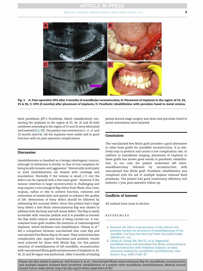
Fig. 3 e A: Post-operative OPG after 4 months of mandibular reconstruction, B: Placement of implants in the region of 33, 34,
35 & 36, C: OPG (4 months) after placement of implants, D: Prosthetic rehabilitation with porcelain fused to metal crowns.
med i c a l j o u r n a l a rm e d f o r c e s i n d i a x x x ( 2 0 1 4 ) 1e4 3
fixed prosthesis (FP-2 Prosthesis, Misch classification) con-
necting the implants in the region of 33, 34, 35 and 36 with
cantilever extending to the region of 31 and 32 were fabricated
and inserted [Fig 3D]. The patientwas reviewed at 2-, 4-, 6- and
12-month interval. All the implants were stable and in good
function with no post-operative complications.
Discussion
Ameloblastoma is classified as a benign odontogenic tumour,
although its behaviour is similar to that of true neoplasia for
being locally invasive and aggressive.9 Historically multicystic
or solid ameloblastoma are treated with curettage and
enucleation. Normally if the tumour is small (<5 cm) the
defect can be repaired with a free bone graft.1 However if the
tumour resection is large reconstruction is challenging and
may require a microsurgical flap either from fibula, iliac crest,
scapula, radius or ribs to achieve function, cosmesis and
restoration of mastication and speech to enhance the quality
of life. Restoration of bony defect should be followed by
addressing the mucosal defect. Since this patient had a large
bony defect a free fibula osteocutaneous flap was chosen to
address both the bony and soft tissue defect. The flap is easily
accessible with vascular pedicle and it is possible to harvest
the flap while tumour resection is being carried out. A vas-
cularised bone graft enables the insertion of osseointegrated
implants, which facilitates oral rehabilitation. Yilmaz et al10
did a comparison between vascularised iliac crest flap and
vascularised free fibular flap in 24 cases. They noticed that less
complication rate, superior functional and aesthetic results
were achieved for those with fibular flap. For this patient
resection of ameloblastoma of left mandible, reconstruction
with vascularised fibula graft and placement of implants in 33,
34, 35 and 36 region was performed. After 4 months of healing
Please cite this article in press as: Anil Kumar S, et al., Vascularisand multiple implant retained fixed prosthetic rehabilitation ofArmed Forces India (2014), http://dx.doi.org/10.1016/j.mjafi.2014.
period second stage surgery was done and porcelain fused to
metal restorations were inserted.
Conclusion
The vascularised free fibula graft provides a good alternative
to other bone grafts for mandible reconstruction. It is rela-
tively easy to perform and carries a low complication rate. In
addition to mandibular shaping, placement of implants in
these grafts has shown good results in prosthetic rehabilita-
tion. In our case the patient underwent left hemi-
mandibulectomy followed by reconstruction with
vascularised free fibula graft. Prosthetic rehabilitation was
completed with the aid of multiple implant retained fixed
prosthesis. The patient had good masticatory efficiency and
esthetics 1 year post-operative follow-up.
Conflicts of interest
All authors have none to declare.
r e f e r e n c e s
1. Bataineh AB. Effect of preservation of the inferior andposterior borders on recurrence of ameloblastomas of themandible. Oral Surg Oral Med Oral Pathol Oral Radiol Endod.2000;90:155e163.
2. Chana JS, Chang YM, Wei FC, et al. Segmentalmandibulectomy and immediate free fibula osteocutaneousflap reconstruction with endosteal implants: an idealtreatment method for mandibular ameloblastoma. PlastReconstr Surg. 2004;113:80e87.
ed fibula osteocutaneous flap for mandibular reconstructiona patient with mandibular ameloblastoma, Medical Journal02.007

me d i c a l j o u r n a l a rm e d f o r c e s i n d i a x x x ( 2 0 1 4 ) 1e44
3. Taylor GI, Miller GD, Ham FJ. The free vascularised bone graft:a clinical extension of microvascular techniques. PlastReconstr Surg. 1975;55(5):533e544.
4. Worthington PH, Branemark P-I, eds. AdvancedOsseointegration Surgery. Applications in the Maxillofacial Region.Chicago: Quintessence; 1992.
5. Shirota T, Schmelzeisen R, Neukam F, Matsui Y, Ohno K,Michi K. Immediate insertion of two types of implants intovascularised bone grafts used for mandibular reconstruction inminiature pigs. Oral Surg Oral Med Oral Pathol. 1994;77:222e231.
6. Shirota T, Ohno K, Michi K, Tachikawa T. An experimentalstudy of healing around hydroxylapatite implants installedwith autogenous iliac bone grafts for jaw reconstruction. JOral Maxillofac Surg. 1991;49:1310e1315.
Please cite this article in press as: Anil Kumar S, et al., Vascularisand multiple implant retained fixed prosthetic rehabilitation ofArmed Forces India (2014), http://dx.doi.org/10.1016/j.mjafi.2014.
7. Gilbert A. Vascularized transfer of the fibular shaft. Int JMicrosurg. 1979;1:100e102.
8. Jones NF, Monstrey S, Gambier BA. Reliability of the fibularosteocutaneous flap for mandibular reconstruction:anatomical and surgical confirmation. Plast Reconstr Surg.1996;97(4):707e716.
9. Kahairi A. Management of large mandibular ameloblastoma-a case report and literature reviews. Arch Orofac Sci.2008;3(2):52e55.
10. Yilmaz M, Vayvada H, Menderes A, Demirdover C,Kizilkaya A. A comparison of vascularised fibular flap andiliac crest flap for mandibular reconstruction. J Craniofac Surg.2008;19(1):227e234.
ed fibula osteocutaneous flap for mandibular reconstructiona patient with mandibular ameloblastoma, Medical Journal02.007