vascular endothelial growth factor levels in the aqueous ...€¦ · anatomy and physiology of the...
TRANSCRIPT

Vascular Endothelial Growth Factor in the Aqueous Humor of Dogs With and Without Intraocular Disease
Christina Ann Sandberg
Thesis submitted to the faculty of the Virginia Polytechnic Institute and State University in partial fulfillment of the requirements for the degree of
Master of Science
In Biomedical and Veterinary Sciences
Ian P. Herring, DVM, MS, DACVO William R. Huckle, MS, PhD
Tanya LeRoith, DVM, PhD, DACVP John H. Rossmeisl, DVM, MS, DACVIM
J. Phillip Pickett, DVM, DACVO
June 8th, 2009 Blacksburg, VA
Keywords: VEGF, pre-iridal fibrovascular membrane, canine, glaucoma

Vascular Endothelial Growth Factor in the Aqueous Humor of Dogs
With and Without Intraocular Disease
Christina A. Sandberg
ABSTRACT
Vascular endothelial growth factor A (VEGF) is a potent mediator of blood vessel
formation throughout the body. Intraocular diseases characterized by inflammation,
hypoxia or neoplasia induce new blood vessel formation within the eye. The end result
of such blood vessel formation may be blinding sequellae such as glaucoma from outflow
obstruction or hyphema from intraocular hemorrhage. Elevated VEGF concentrations in
the aqueous humor and vitreous are documented in a number of human intraocular
disease processes, including tumors, retinal detachment and uveitic glaucoma.
Pharmacotherapy inhibiting VEGF expression demonstrates promise for control of some
of these ophthalmic conditions. We quantified and compared VEGF concentrations in
canine aqueous humor samples from 13 dogs with normal eyes and 226 eyes from 178
dogs with a variety of ophthalmic diseases by ELISA. Dogs with primary cataract,
diabetic cataract, primary glaucoma, uveitic glaucoma, aphakic/pseudophakic glaucoma,
retinal detachment, lens luxation and neoplasia were evaluated. Elevated VEGF
concentrations were found in all disease conditions tested as compared to normal dogs
excepting cataracts and diabetic cataracts. Elevated aqueous humor VEGF
concentrations were found in dogs with pre-iridal fibrovascular membranes (PIFM) as
compared to dogs without PIFM. These results are consistent with the hypothesis that
VEGF has a role in the causation or progression of a variety of canine ocular disorders.

ACKNOWLEDGEMENTS
The author recognizes the Virginia-Maryland Regional College of Veterinary Medicine
Office of Research and Graduate Studies for providing funding for the project. The
author thanks all members of the graduate committee for their assistance throughout the
project and their continued support of the author’s professional career. In addition, the
author greatly appreciates the support of Drs. Phil Pickett, Ian Herring, Jamie Schorling,
Daniel Binder, Bill Miller, Albert Mughannam, Stacy Andrew, Heidi Denis, Sandra van
der Woerdt, and Patricia Smith in providing clinical samples. The author recognizes Dr.
Tanya LeRoith for histopathology review, Dr. Stephen Werre for statistical analyses,
Anne Cinsavich and Pamela Arnold for making available normal canine specimens and
Betsy Midkiff as the Ophthalmology Technician.
iii

TABLE OF CONTENTS
ABSTRACT………………………………………………………………..……………..ii
ACKNOWLEDGEMENTS……………………………………………………………...iii
TABLE OF CONTENTS……………………………………………………………...iv-vi
INTRODUCTION………………………………………………………………..……….1
Anatomy and physiology of the canine uvea……………………………………...1
Aqueous humor production and outflow pathway of the dog……………...……..2
Glaucoma………………………………………………………………..………...3
Uveitis………………………………………………………………..……………4
Cataract………………………………………………………………..…………..5
Intraocular tumor………………………………………………………………….6
Retinal detachment………………………………………………………………..7
Pre-iridal fibrovascular membrane……………………..…………………………7
Vasculogenesis and Angiogenesis………………………………………………...9
Vascular endothelial growth factor……………………………………………….9
OBJECTIVES………………………………………………………………..…………..13
CHAPTER I. LITERATURE REVIEW………………………………………………...14
Role of VEGF in human and canine systemic disease…………………………...14
Neoplasia………………………………………………………………....14
Inflammatory disease………………………………………………….....19
Cardiovascular disease…………………………………………………...19
Anemia…………………………………………………………………...19
Role of VEGF in healing……………………………………………..……...…..20
VEGF in human ocular disease……………………………………………….....20
Intraocular surgery…………………………………………………….....21
iv

Corneal disease…………………..…………………………..…………...21
Intraocular tumors……………………………………..……..…………..21
Glaucoma………………………………………………………………...22
Lens disease………………………………………………………….…..23
Uveitis………………………………………………………………..…..23
Retinal disease…………………………………………………………...24
VEGF inhibitors for ocular disease……………………………………………...26
CHAPTER II. MATERIALS AND METHODS……………………………….……....29
Subject identification………………………………………………………...….29
Sample collection and handling ……………………………………………..….30
VEGF ELISA ……………………………………………………………...…....31
Sample dilution…………………………………………………………………..31
Histopathology…………………………………………………………….....….32
Aqueous humor assay validation……………………………………….......…....32
Assay of sample stability in frost-free freezer…………………………….……..33
Assay of sample stability at room-temperature…………………………...….….33
Statistical analysis………………………………………………………...…..….33
CHAPTER III. RESULTS……………………………………………………….......…35
Aqueous humor assay validation…………………………………………….......35
Assay of sample stability in frost-free freezer………………………….……..…35
Assay of sample stability at room-temperature……………………….…………36
Descriptive data…………………………………………………………….……38
Aqueous humor VEGF concentration…………………………………………...44
Disease comparison………………………………………………………….......49
Plasma VEGF concentration………………………………………………..…...52
Pre-iridal fibrovascular membrane…………………………………………........53
CHAPTER IV. DISCUSSION…………………………..…………………………........56
v

vi
CHAPTER V. CONCLUSIONS…………………………………..……………….....72
LITERATURE CITED………………………………………..………………….........74
APPENDIX …………………………………………………...…………………….....85
VITA……………………………………………………………..……………………107
LIST OF TABLES
Table 1: Case distribution……………………………………………………………….84
Table 2: Mean aqueous humor levels of VEGF…………………….……………..........85
Table 3: PIFM distribution………………………………………….…………..…........86
Table 4: Aqueous Humor and Plasma VEGF Concentration for Samples Within the Limits of Detection and Below the Limits of Detection………………………………. 87
LIST OF FIGURES
Figure 1: Scatter plot of plasma vs aqueous VEGF levels………………………….......88
Figure 2: Bar graph of mean aqueous VEGF level by disease…………………….…....89
Figure 3: Cellular PIFM at 40x magnification.………………………..…………..……90
Figure 4: Vascular PIFM at 40x magnification….………………………………..…….91
Figure 5: Fibrovascular PIFM at 4x magnification..…………………..………..……....92
Figure 6: Fibrovascular PIFM at 10x magnification….……………….……..………....93
Figure 7: Fibrovascular PIFM at 40x magnification………………………..…………..94
SAMPLE SUMMARY…………………………………………………………..……………...95

INTRODUCTION
Anatomy and physiology of the canine anterior uvea and iridocorneal angle
The iris and ciliary body comprise the anterior uvea.1 Embryologically, the iris originates
from the neural crest (iris stroma), mesoderm (vascular endothelium) and neuroectoderm
(epithelium and muscles).2 It extends as a diaphragm over the anterior lens and creates
the pupil and separates the anterior and posterior chambers of the eye. Histologically,
there is an anterior border layer composed of fibroblasts and melanocytes, stroma of
collagen, chromatophores and fibroblasts, smooth sphincter and dilator muscles and a
posterior epithelial layer.1,3 Blood supply is via the terminations of the long posterior
ciliary arteries, which forms an incomplete arterial circle in domestic animals.4 Drainage
is via the radial vessels to the vortex veins.5,6 The iris functions to control light passage
to the posterior segment and is a highly vascular tissue.1
The ciliary body resides posterior to the iris and the posterior iris epithelium is
continuous with the ciliary body epithelium. Ciliary body epithelium originates from
neuroectoderm and mesenchyme.2 It is composed of the anterior pars plicata, consisting
of approximately 75 ciliary processes in the carnivore, and the posterior pars plana.1
Smooth muscle comprises the largest mass of this structure. Vascular supply is via the
long posterior ciliary arteries to the major arterial circle and anterior ciliary arteries.4,5
The ciliary body functions to nourish and maintain ocular rigidity by production of
aqueous humor, allows accommodation and provides attachment for the lens zonules.1
1

The peripheral anterior iris and ciliary body is bounded by the iridocorneal angle, which
is the conventional and major pathway of aqueous humor outflow in the dog.1,5 During
development, the iridocorneal angle develops by separation of the anterior mesenchyme,
trabeculae enlargement, trabecular meshwork clefting and postnatal remodeling.2,7 The
pectinate ligament spans the opening of the ciliary cleft. The trabecular meshwork
resides within the ciliary cleft and is composed of cell-lined collagenous fibers. Aqueous
collecting channels provide outflow from the meshwork and join the angular aqueous
plexus to reach systemic circulation via the vortex veins.1
Aqueous humor production and outflow pathway in dogs
Aqueous humor is a clear ultra-filtrate of plasma produced by the posterior non-
pigmented epithelium of the ciliary processes, primarily by the active transport of solute.8
It is produced in the posterior chamber, then transverses the pupil to the anterior chamber
and exits the eye by the iridocorneal angle.5 In dogs, the major aqueous outflow pathway
is the corneoscleral meshwork; in some other species the uveoscleral or unconventional
pathway predominates.9,10 Balance of aqueous humor production and outflow determines
intraocular pressure. The rate of aqueous production may be affected by the health of the
ciliary epithelium and intraocular pressure in addition to neural, hormonal and
pharmacologic factors. Aqueous outflow may be affected by the health and patency of
the drainage pathways, inflammation as well as intraocular pressure.5,11
2

Glaucoma
Glaucoma is a disease typically identified as elevated intraocular pressure caused by
obstruction of the normal aqueous humor outflow pathway causing damage to the optic
nerve and retinal ganglion cells.12 Sequellae include pain and permanent blindness; these
effects often require surgical removal of the globe or globe contents to alleviate
discomfort. Major causes of glaucoma in dogs include inherited or breed-related
abnormalities of the iridocorneal angle (known also as primary glaucoma) and outflow
obstruction from antecedent intraocular disease (known also as secondary glaucoma).
Primary glaucomas are reported to affect 0.89% of the general canine population, with an
increased incidence for specific breeds and lines.13 Despite anatomical abnormalities of
the iridocorneal angle existing throughout life, most dogs with primary glaucoma are
affected in middle-age. Inflammation and pigment dispersion in the drainage angle, uvea
and retina in the eyes of dogs enucleated for goniodysgenesis-related glaucoma have been
reported.14,15 Tissue ischemia is implicated as cause of retinal injury in dogs.16
Dogs with secondary glaucomas have an acquired ocular disease or primary ocular
disease unrelated to iridocorneal angle malformation impeding aqueous humor outflow
rather than a preexisting structural anomaly of the iridocorneal angle. Secondary
glaucomas were diagnosed in 6.9% of dogs presented for evaluation of ophthalmic
disease in one veterinary teaching hospital in a 5 year period.17 The most frequent causes
of secondary glaucoma in dogs are anterior uveitis, intraocular surgery, lens dislocation,
hyphema, intraocular neoplasia and trauma.17-19 Additionally, breed-related pigmentary
3

changes (pigmentary dispersion in the Cairn terrier, uveal cyst syndrome in the Golden
Retriever and multiple ciliary body cysts in the Boston Terrier and Great Dane) have
been associated with glaucoma.20-23
While both medical and surgical anti-glaucoma therapies are widely used for treatment of
canine glaucoma, these therapies are rarely effective in controlling the disease long term.
Present therapies predominantly reduce aqueous humor production, as exemplified by
carbonic anhydrase inhibitors, beta blockers and cyclodestruction, or increase outflow, as
exemplified by miotics, prostaglandins and gonioimplants. In the case of secondary
glaucomas, therapies are aimed at removing the underlying cause in addition to
controlling the resulting intraocular pressure. Lower success rates for control of uveitic
glaucomas compared to primary glaucomas are described in dogs, presumably due to
secondary effects of ocular inflammation.12
Uveitis
In dogs, uveitis or inflammation of the ocular vascular tunic occurs secondary to tissue
injury.24 The blood-aqueous barrier is created by the tight junctions of the nonpigmented
ciliary epithelium, the tight junctions and gap junctions of the iris vascular endothelium
and the non-fenestrated iridal capillaries.8 Uveal inflammation induces loss of the normal
blood-aqueous barrier via increased vascular permeability and increased vascular supply.
Uveitis has a number of underlying infectious, traumatic, neoplastic, iatrogenic and
immune-mediated etiologies. The most commonly diagnosed causes for anterior uveitis
in dogs are lens-induced, traumatic, and immune-mediated disease.19,25,26 Ocular auto-
4

antigens involved in the initiation of uveitis include lens proteins, and in rare cases,
melanocytes.27,28 Most commonly, an infiltrate of lymphocytes and plasma cells is
present in the iris and ciliary body, but the histological findings vary with specific
cause.18
Sequellae of anterior uveitis include intraocular adhesions (synechiae), cataract,
hyphema, secondary glaucoma, and collapse of the globe from chronic hypotony
(phthisis bulbi). Mechanisms for uveitic glaucoma include pupil seclusion with annular
posterior synechia, angle obstruction with inflammatory debris, peripheral anterior
synechia, pre-iridal fibrovascular membrane or a combination of these factors.24
Therapies for uveitis include treatment of the underlying cause and non-specific
treatment using anti-inflammatory drugs, immunosuppressive agents and
immunomodulatory agents. Additionally, specific treatments for prevention or disruption
of synechiae and fibrin clots may be employed.24,26
Cataract
Cataract is a pathologic opacification of the lens and/or lens capsule and is one of the
most common ophthalmic diseases affecting canine patients. Causes include diseases or
conditions that alter nutrition, metabolism or osmotic balance of the lens.18 Cataract
formation is common in pet dogs, with higher incidence in some breeds and with
increasing age, and cataract extraction surgery is commonly performed by veterinary
ophthalmologists.29,30 Surgical intervention in dogs is indicated for animals with
5
significant visual impairment from advanced cataract. Most dogs have some degree of
intraocular inflammation (phacolytic uveitis) prior to cataract removal.31-33 It is reported
that eyes with clinically significant lens-induced uveitis have a poorer prognosis
following cataract extraction.34 In some cases, cataract formation results in lens capsule
rupture and severe intraocular inflammation (phacoclastic uveitis); this condition may be
associated with rapid cataract development with or without diabetes mellitus.35
Complications of cataract surgery include posterior capsular opacification, retinal
detachment, postoperative hypertension and glaucoma; an increased risk of long-term
glaucoma with hypermature cataract is described.36
Intraocular tumors
In dogs, the most common intraocular neoplasms are uveal melanomas, iridociliary
epithelial tumors and lymphomas.18,37,38 Most primary canine uveal tumors arise from
the iris or ciliary body and most do not spread beyond the globe.39,40 However, even
benign intraocular tumors may cause extensive damage to delicate structures of the eye,
resulting in secondary glaucoma, intraocular hemorrhage and retinal detachment.24 These
sequellae often necessitate enucleation to provide comfort.
Retinal detachment
There are a variety of ophthalmic and systemic conditions contributing to a separation of
the neurosensory retina from the retinal pigmented epithelium. Such conditions include
retinal tears (rhegmatogenous retinal detachment), chorioretinitis from infectious or
autoimmune disease, congenital ocular disease, posterior segment neoplasms, systemic
6

hypertension or vitreal traction bands.41 Chronic, extensive retinal detachments result in
retinal degeneration, non-responsive uveitis and glaucoma.42
Pre-iridal fibrovascular membrane
One key pathologic feature shared by many of the aforementioned diseases is intraocular
neovascularization. Iridal neovascularization as described in human beings, consists
histologically of a network of blood vessels, with or without myofibroblasts, originating
from the superficial iris stroma following antecedent ocular disease. Three stages of
neovascularization of the iris in humans are described: new vessels appearing at the iris
base and pupillary margin (stage 1), vessels penetrating the anterior iris surface and
merging at the collarette (stage 2) and vessels with a connective tissue support (stage 3).43
Causes of iris neovascularization in humans are numerous. Broadly, causes include
vascular disease (e.g. central retinal vein occlusion), ocular disease (e.g. uveitis, retinal
detachment, primary glaucoma and secondary glaucoma), intraocular surgery (e.g. retinal
reattachment surgery and cataract extraction), ocular trauma, intraocular neoplasia and
systemic disease (e.g. diabetes mellitus).43 Iridal neovascularization was noted in 0.5%
of human eyes removed at autopsy and 18-20% of eyes enucleated for therapeutic
purpose.43
A well-recognized form of ocular neovascularization that arises secondary to intraocular
disease in domestic animals is pre-iridal fibrovascular membrane (PIFM). In domestic
7

animals, cellular, vascular and fibrous forms of PIFM are described, and are thought to
represent a temporal continuum.44
Clinically, iridal vascularization may cause reddish discoloration, known as rubeosis
iridis; this feature is most apparent in eyes with light colored irides. This discoloration
may be absent if the fibroblastic component obscures visualization of vessels.45
Contracture of the membrane may cause inward or outward turning of the iridal margin,
known as entropion or ectropion uveae, respectively. PIFMs may grow across the
anterior lens surface to cause pupil seclusion and/or the iridocorneal drainage angle
causing obstruction of normal aqueous passage or may extend to the posterior iris or
ciliary processes. Due to vessel fragility, PIFMs are prone to rupture and cause
intraocular hemorrhage (hyphema). In domestic animals and humans, PIFM formation is
suspected to play an important pathophysiologic role in the development of secondary
glaucomas.18,44,46
PIFMs are routinely noted in dogs with intraocular neoplasms, specifically ciliary body
adenoma, adenocarcinoma, neuroepithelial tumor, uveal spindle-cell tumor, and
osteosarcoma.47-51 Additionally, PIFM formation has been identified in dogs with
intraocular inflammatory and hypoxic disease, including uveodermatologic syndrome,
uveal cyst syndrome, retinal detachment, primary glaucoma, ocular infections and
following cataract extraction by phacoemulsification and extracapsular lens
extraction.18,21,23,28,42,52-55
8

The incidence of iridal vascularization is widely variable, depending on report and cause.
In dogs with retinal detachment, ciliary epithelial neoplasia, chronic glaucoma, ocular
hemorrhage, uveal melanoma, endophthalmitis, and uveal cyst syndrome, PIFMs were
noted in 21%, 19%, 14%, 10%, 10% and 6% of cases, respectively.21,44 Histopathologic
studies of canine globes enucleated or eviscerated for complications following surgery
for cataract removal cite PIFM formation in 86% of cases, with 72% classified as mild,
24% as moderate and 4% as severe.54 The pathogenesis of PIFM formation in domestic
animals has not been elucidated.
Vasculogenesis and Angiogenesis
There are two means of blood vessel growth or development in living systems.
Vasculogenesis is the formation of new blood vessels de novo and angiogenesis is the
development of new blood vessels from pre-existing vasculature. Many factors are
implicated in these processes, including vascular endothelial growth factor, basic
fibroblast growth factor, platelet derived growth factor, transforming growth factors and
angiogenin. Neovascularization by vasculogenesis and/or angiogenesis, plays a key role
in growth, tissue repair, inflammation and tumorogenesis.
Vascular endothelial growth factor
Vascular endothelial growth factor (VEGF, referred to also as VEGF-A or vascular
permeability factor) is a 46kDa glycoprotein consisting of two 23kDa homodimer
proteins, with mitogenic effects on vascular endothelial cell activity causing endothelial
cell growth and angiogenesis.56-58 Both VEGF and its two tyrosine kinase receptors
9

(VEGFR1/Flt-1 and VEGFR2/KDR or Flk-1) are required for life, as demonstrated by
lethality in gene knockout mice models.59-61
The in vivo roles of VEGF are multi-fold, as this factor has the potential to enhance
vascular permeability and to promote new blood vessel formation by aiding the growth
and survival of vascular endothelial cells.62-64 In healthy adult animals, VEGF is
expressed in vascularized tissues and theorized to have a role in vascular maintenance by
stabilization of mature blood vessels.65
There are at least six distinct isoforms of human VEGF, consisting of 121, 145, 165, 183,
189 and 206 amino acids. Alternative exon splicing has a role in phenotypic regulation
for both VEGF and VEGF receptors.66-69 VEGF121 and VEGF165 are considered more
biologically active than other forms. VEGF165 is reported to act as more potent pro-
inflammatory factor than VEGF121.70 VEGF isoforms are similar across species. Canine
VEGF has conserved binding sites and 95.2% and 98.4% sequence homology with
human and feline forms, respectively (EMBL accession number AJ133758 for canine
VEGF, GenBank accession number AF133250 for canine VEGF and AB071947 for
feline VEGF).69,71,72
VEGF expression is induced by hypoxia, ischemia, hypo and hyperglycemia, Placental
Growth Factor, Transforming Growth Factor-β, Tumor Necrosis Factor-α, and reactive
oxygen intermediates.73-81 Prostaglandin analogs, presumably by induction of hypoxia,
also result in elevated VEGF expression.82 VEGF expression is inhibited by
10

corticosteroids, steroid hormones and endostatin.83-86 Reports on the effect of
cycloxygenase inhibitors on VEGF expression are conflicting.84,87,88
VEGF164 is the major isoform expressed in canine tissue.71 A study in normal dogs
employing immunohistochemistry, RT-PCR and real time RT-PCR, revealed strongly
positive VEGF expression in Type I and Type II alveolar cells of the lungs, apocrine
glands of the skin and corpus leutum. Moderately positive VEGF expression is noted in
the Kupffer cells of the liver, lung epithelium, lung smooth muscle, collecting tubule of
the kidney, the heart, glomerulosa and reticularis of the adrenal gland, epidermis, hair and
sebaceous gland of the skin, and bladder epithelium. Weak VEGF expression is noted in
hepatocytes, bile duct, proximal and distal tubule of the kidney, zona fasculata of the
adrenal gland, intestinal and bladder muscularis and lymph node. Negative VEGF
expression was noted in renal glomerulus, adrenal medulla, thyroid, intestinal epithelium
and nerve plexus, pancreas and spleen. The Flt-1 receptor showed similar tissue
expression as VEGF. This study did not evaluate expression in ocular tissue.89
Within the eye of humans and laboratory animal models, VEGF and/or its receptors are
expressed constitutively by numerous tissues apart from vascular endothelium, including
the cornea, conjunctiva, iris pigmented epithelium, retinal pigmented epithelium, retinal
ganglion cells, astrocytes, Müller cells, and choroidal fibroblasts.90-93 An autocrine,
neuroprotective role of VEGF for Müller cell and photoreceptor survival has been
proposed.94
11

In humans, increased ophthalmic VEGF expression is noted in association with a variety
of disease conditions. For instance, VEGF over-expression is noted in corneal
neovascularization and corneal wound healing.95,96 Primary open-angle glaucoma,
neovascular glaucoma, angle closure glaucoma and exfoliative glaucoma are associated
with high levels of VEGF.97,98 Elevated VEGF expression is present with intraocular
tumors, including malignant melanocytes and retinoblastomas.99-101 In the retina,
elevated VEGF is found in central retinal vein occlusion, uveitis, diabetic retinopathy (in
retinal tissue and fibrovascular membranes), retinopathy of prematurity, retinitis
pigmentosa and age-related macular degeneration.102-106 In ischemic retinopathies,
VEGF expression is noted in iris as well as retinal tissue.107
12

OBJECTIVES
There are numerous canine ophthalmic conditions in which increased VEGF expression
may induce intraocular neovascularization and for which control of intraocular
neovascularization may limit disease severity or progression. Clarification of the role of
VEGF in these conditions may provide rationale for adjunctive therapy, including the use
of VEGF inhibitors, in canine patients affected by ocular neovascularization.
The objectives of this study were quantification of aqueous humor VEGF concentrations
in dogs affected by a variety of intraocular disease processes and to compare these
concentrations with aqueous humor VEGF concentrations in healthy dogs without
evidence of ocular disease. Specific ocular disease conditions included primary and
diabetic cataracts, primary glaucoma, uveitic glaucoma, intraocular neoplasia,
aphakic/pseudophakic glaucoma, retinal detachment, uveitis, lens luxation, ocular
trauma, ocular melanosis and uveal cyst disease. Additionally, we sought to compare
aqueous humor VEGF levels in eyes with histopathologically confirmed intraocular
neovascularization to those without neovascularization.
We hypothesized that aqueous humor VEGF concentrations would correlate with the
etiology and chronicity of intraocular disease as well as the presence of intraocular
neovascularization.
13
LITERATURE REVIEW
Role of VEGF in systemic disease
Vascularization mediated by VEGF is implicated in a wide variety of human and canine
diseases. These conditions include neoplasia, immune-mediated disease, infectious
disease and cardiovascular disease.
Neoplasia
Human beings
In people, VEGF is expressed in patients with carcinoma, sarcoma, adenoma, lymphoma,
leukemia and melanoma. For many tumors, VEGF expression can relate to likelihood of
disease progression. In a study by Potti et al, intratumoral VEGF over-expression was
associated with a shorter survival times in patients with soft-tissue sarcoma.108 Another
study by Potti et al determined that 20% of archived melanoma samples over-expressed
VEGF; however, over-expression of VEGF was not shown to have prognostic value.109
In head and neck squamous cell carcinomas studied by Eisma et al, elevated VEGF
expression correlated with aggressive disease, with higher rate of disease recurrence and
shorter disease-free interval in patients with tumors expressing higher levels of VEGF.110
Hui et al reported VEGF expression in 60% of nasopharyngeal tumors; high levels of
tumor VEGF expression was associated with a poor overall survival.111 In breast tumors
evaluated by Toi et al, intratumoral VEGF concentration, especially when soluble VEGF
receptor 1 was considered, was a significant poor prognostic indicator.112 In Jebreel’s
14
study of VEGF expression in thyroid pathologies, elevated VEGF expression was noted
in neoplastic disease compared to autoimmune disease.113
In some human conditions, circulating blood carries VEGF in elevated quantities, acting
as a marker for tumor angiogenesis.114 Like over-expression within tissue, elevated
plasma or serum level of VEGF carries prognostic significance for some diseases. For
instance, in patients with hepatocellular carcinoma studied by Poon et al, serum level of
VEGF higher than 245pg/mL was associated with a shorter overall survival, related to
advanced tumor stage and venous invasion.115 For patients with gastrointestinal cancers
evaluated by Hyodo et al, VEGF in plasma greater than 108pg/mL correlated positively
with metastatic disease and negatively with response to chemotherapy and survival
time.116 In a study by Etto et al, serum VEGF concentrations for patients with non-
Hodgkin’s lymphoma diminished with treatment from a mean of 500pg/mL at time of
diagnosis to 308pg/mL following 6 months of therapy.117
Dogs
VEGF levels have also been studied in dogs with neoplastic disease. VEGF expression
in canine CNS tumors are reported in three studies. In dogs evaluated by Rossmeisl et al,
over-expression of VEGF in tumor homogenates by ELISA was found in dogs with
intracranial astrocytomas (mean value of 2.84ng/mL, equivalent to 2840pg/mL),
oligodendrogliomas (mean value of 0.93ng/mL, equivalent to 930pg/mL) and
meningiomas (mean value of 0.24ng/mL, equivalent to 240pg/mL), with a higher
expression of VEGF in higher grade tumors.118 Platt et al’s study of dogs with
15

intracranial meningiomas revealed VEGF expression by immunohistochemistry, with a
negative correlation of VEGF expression with survival time.119 VEGF expression,
especially isoform VEGF164, was found by Dickinson et al using quantitative real-time
TaqMan RT-PCR, in a retrospective evaluation of dogs with intracranial tumors; again,
tumor grade correlated with VEGF expression.120
VEGF expression in canine mammary tumors has also been investigated. In Qui et al’s
immunohistochemical study of canine mammary tumors, VEGF over-expression was
found more often in mammary gland tumors compared to normal tissues and correlated
significantly with stage and lymph node metastasis.121 In a study by Millanta et al,
clinicopathological variables and prognosis did not correlate with level of VEGF
expression by immunohistochemistry in canine mammary carcinomas.122
In evaluation of canine cutaneous tumors, Maiolino et al’s study found
immunohistochemical expression of VEGF in 15/15 of squamous cell carcinomas, but
only 3/20 basal cell tumors.123 In Al-Dissi et al’s evaluation of cutaneous tumors by
immunohistochemistry, expression of VEGF was found in 89.4% of squamous cell
carcinomas and 70.8% of trichepitheliomas and correlated with histological grade for
both tumor types.124 In Patruno et al’s evaluation of dogs with spontaneous cutaneous
mast cell tumors by ELISA and immunohistochemistry, VEGF expression correlated
with microvascular density of tumors and malignancy grade.125
16

VEGF expression has been also evaluated for other canine tumor types. In a study by
Wolfsberger et al using immunohistochemistry, Western blotting and RT-PCR, VEGF
was expressed by 60% of canine lymphomas; VEGFR-1 and VEGFR-2 were expressed
by 54% and 7%, respectively, of tumor cells.126 In Yonomaru et al’s study,
immunohistochemistry and in-situ hybridization were used to detect VEGF over-
expression in vascular tumors, with a higher relative immunoreactivity in
hemangiosarcomas compared to hemangiomas, correlating tumor VEGF expression with
malignancy.127 VEGF was detected by immunohistochemistry in canine patients with
mastocytoma by Rebuzzi et al, but was not believed to act as an autocrine growth
regulator.128
Circulating and extratumoral VEGF has been evaluated and found to have prognostic
implications in a variety of neoplasms in dogs. In a study by Gentili et al, serum levels of
VEGF by ELISA had prognostic value in lymphoma-affected dogs, positively correlating
with stage B disease and negatively correlating with disease-free interval. Mean serum
VEGF concentrations for stage A disease were 13.74pg/mL; mean serum VEGF
concentration for stage B disease were 38.37pg/mL; mean serum concentrations for
healthy control dogs were below the limits of detection.129 In dogs evaluated by ELISA
by Troy et al, dogs with neoplastic disease were significantly more likely to have
detectable plasma VEGF levels (mean, 17.94pg/mL) compared to healthy dogs (mean,
3.1pg/mL) or dogs with non-neoplastic disease (15/16 dogs below the limits of
detection).130 In dogs with neoplastic and non-neoplastic cavity effusions studied by
Clifford et al by ELISA, elevated VEGF expression was noted in the effusates compared
17

to plasma expression. Pericardial and pleural effusates tended to harbor more VEGF than
peritoneal effusates, with a median concentration of VEGF of 3533pg/mL, 3144pg/mL
and 288pg/mL, respectively.131
In dogs with hemangiosarcoma evaluated by Clifford et al, a higher proportion of tumor-
bearing dogs had detectable plasma VEGF compared to healthy controls (13/16 vs. 1/17).
The mean concentration of plasma VEGF in dogs with hemangiosarcoma was
17.2pg/mL.132 In dogs with mammary tumors studied by Kato et al, circulating VEGF
levels by ELISA in the plasma and serum were significantly higher than in the healthy
control dogs. In healthy dogs, no plasma expression of VEGF was detectable. In
diseased dogs, plasma VEGF levels ranged from non detectable to 895.7pg/mL (mean
2.1pg/mL). In healthy dogs, serum concentrations ranged from non detectable to
27.5pg/mL (mean 8.3pg/mL). Mean serum concentrations of dogs with mammary gland
tumors ranged from non detectable to 992.9pg/mL (mean 14.85pg/mL). Higher
circulating VEGF levels had a positive correlation with malignant tumors and post-
operative metastasis.133 In dogs treated with radiation therapy by Wergin et al, baseline
plasma VEGF concentrations greater than 5pg/mL were a poor prognostic indicator for
outcome. No significant difference between and post radiation treatment plasma VEGF
levels were found.134
18

Inflammatory disease
VEGF is also associated with human autoimmune diseases and systemic inflammatory
conditions of unknown cause, specifically systemic lupus erythematosus, rheumatoid
arthritis, multiple sclerosis and sarcoidosis.135-137 Serum VEGF levels are reported to
correlate with immune reaction type: high levels are noted in autoimmune disease and
low levels are noted in allergic reaction138
Cardiovascular disease
In a study by Hamada et al, inhibitors of VEGF and VEGFR-1 were effective in
inhibiting baseline sera activity in patients with cyanotic congenital heart disease.139 In
Hamamichi’s study of patients with Kawasaki disease, a syndrome of systemic vasculitis,
VEGF expression from neutrophils and mononuclear cells by immunoblot analysis was
noted.140
In a study by Ray et al of experimental heart failure in dogs, increased VEGF expression
was noted by quantitative RT-PCR and was theorized to contribute to altered
vasoreactivity leading to pulmonary hypertension.141
Anemia
Anemic patients have elevated levels of plasma VEGF by ELISA compared to patients
with normal red blood cell count; those with anemia attributed to renal disease had higher
VEGF levels than those with malignant disease (16.2pg/mL in normemic patients vs
19

67.8pg/mL in patients with renal disease vs 49.2pg/mL in patients with malignant
disease).142
Role of VEGF in Healing
VEGF expression, as detected by immunohistochemistry in a rabbit model, was
expressed in epithelial, stromal and endothelial corneal cells during corneal wound
healing irrespective of the presence of granulocytes.96 In Bidder’s study of dogs with
flexor tendon repair by in situ hybridization, a gradient of VEGF expression was noted in
cells at the healing tendon cells, but accumulation within the epitenon was minimal,
suggesting a role for VEGF in postoperative angiogenesis.143 In dogs with experimental
distraction osteogenesis studied by Park et al, elevated VEGF was noted in Schwann cells
and neurovasorum for up to two weeks postoperatively.144
VEGF in Ocular disease
In people, increased ophthalmic VEGF expression is noted in association with a variety
of disease conditions, including corneal neovascularization, multiple forms of glaucoma,
intraocular tumors, uveitis, retinal detachment, central retinal vein occlusion, uveitis,
diabetic retinopathy, retinopathy of prematurity, and age-related macular degeneration.95-
106,145-147
Enzyme-linked immunosorbant assay (ELISA) detection of VEGF from the aqueous
humor and vitreous of human beings and animal models has been reported.97,98,102,148-154
ELISA tests from different manufacturers have variable sensitivity to detection of VEGF.
20

Intraocular Surgery
Aqueous humor VEGF as assessed by a Biochip Array System (Evidence Investigator,
Antrim, N. Ireland) was found by Tu et al to be elevated 18 hours following cataract
surgery as compared to intra-operative levels (mean values of 463pg/mL vs. 67pg/mL,
respectively).151
Corneal disease
VEGF has been associated with corneal neovascular disease. Immunohistochemical
staining was used by Philipp et al to evaluate human corneas with inflammatory disease
and normal controls for expression of VEGF and its receptors, Flt-1 and Flk-1. It was
found that VEGF expression was increased in vascularized corneas, specially on
epithelial cells, vascular endothelial cells and fibroblasts.95 In a rabbit model by Gan et
al using immunohistochemistry of corneal alkali burn, VEGF and its Flk-1 receptor were
found to be expressed at the leading edge of cornea in epithelial, stromal and endothelial
cells during the healing phase; this expression did not require the presence of
inflammatory cells.96
Intraocular tumors
In studies of human anterior uveal melanomas and melanoma cell lines, expression of
high levels of VEGF as assessed by immunohistochemistry, in situ hybridization,
Western blot analysis and ELISA have been noted.101,150,155 In ELISA studies of aqueous
humor samples from eyes with uveal melanomas by Missotten et al and Boyd et al, mean
VEGF levels were 146.5pg/mL and 0.8ng (equivalent to 800pg/mL), respectively. Mean
21

aqueous humor VEGF concentrations of control eyes from these studies were 50.1pg/mL
and 0.1ng (equivalent to 100pg/mL), respectively. Missotten et al utilized an ELISA
from R&D Systems (Abingdon, Oxford, UK); Boyd et al utilized an ELISA from
BioSource International (Camarillo, CA, USA). 150,156
Glaucoma
Two studies have evaluated the expression of VEGF in the eyes of people with glaucoma.
In the first, Tripathi et al quantified VEGF expression from the aqueous humor using
single antibody competitive binding enzyme immunoassay with an assay range from
0.195 to 200ng/mL (CytImmune Sciences, College Park, MD). In this study, VEGF was
detected in all 12 aqueous samples from patients with neovascular glaucoma and yielded
a mean VEGF concentration of 29.267ng/mL, equivalent to 29,267pg/mL. VEGF was
detected in 15/28 samples from patients with primary open angle glaucoma, with a mean
value of 0.726ng/mL, equivalent to 726pg/mL.97
In a later study, Hu et al quantified aqueous humor VEGF levels in patients with primary,
neovascular and uveitic glaucoma using a commercial ELISA kit (R&D Systems,
Minneapolis, MN). In this study, VEGF was detected in all samples. Aqueous humor
from glaucoma eyes harbored significantly higher VEGF levels than cataracts and there
was no significant difference between glaucoma types. In primary open-angle glaucoma
samples, there was a mean VEGF concentration of 140.4pg/mL, in angle-closure
glaucoma, there was a mean VEGF concentration of 142.8pg/mL and in neovascular
glaucoma, there was a mean VEGF concentration of 158.6pg/mL.98
22

In a non-human primate model studied by Tolentino et al, 0.25 to 2.5μg VEGF injected
intravitreally was sufficient to cause iridal neovascularization and neovascular
glaucoma.157
Lens disease
In Tripathi et al’s and Hu et al’s studies, quantification of VEGF in the aqueous humor of
patients with cataracts was also performed. In Tripathi’s study, VEGF was detected in
4/20 aqueous humor samples from patients with cataract with a mean concentration of
0.257ng/mL, equivalent to 2570pg/mL. VEGF was detected in 16/16 plasma samples
from normal subjects with a mean concentration of 20.246ng/mL, equivalent to
20,246pg/mL.97 In Hu’s study, VEGF was detected in all aqueous humor samples and
patients with cataracts had a mean concentration of 102.4pg/mL. VEGF was detected in
all plasma samples with a mean concentration of 79.2pg/mL.98
Uveitis
In rat and mouse models of autoimmune uveitis by Vinores et al, markedly increased
retinal expression of VEGF was found by immunohistochemistry. In the same study,
moderately increased VEGF expression was noted in retinas with induced ischemic
retinopathy.145 In patients with uveitis evaluated by Fine et al using ELISA (R&D
Systems, Minneapolis, MN), a correlation with increased aqueous humor VEGF
concentrations and cystoid macular edema was made. Mean aqueous humor VEGF
concentrations for uveitis patients with and without cystoid macular degeneration were
23

152 and 109.5pg/mL, respectively.149 In people with quiescent uveitis studied by Paroli
et al, elevation in aqueous humor VEGF levels as determined by ELISA (R&D Systems,
Abingdon, UK), compared to control subjects was found with values of 118pg/mL and
83pg/mL, respectively.146
Retinal disease
Alterations in VEGF have been noted for a variety of retinal conditions, including retinal
detachment, diabetic retinopathy and retinitis pigmentosa. In a murine model by Ohno-
Matsui et al, induction of high levels of VEGF correlated with development of retinal
neovascularization, proliferative retinopathy and tractional retinal detachment.158 Two-
fold elevations of retinal VEGF as detected by ELISA (R&D Systems, Minneapolis, MN)
were described by Ishida et al for rats in a model of pathological neovascularization,
compared to rats with physiologic vascular development.159 In a recent study by Salom
et al, patients with retinitis pigmentosa, a degenerative retinal disorder, harbored
significantly lower levels of VEGF in the aqueous humor as compared to patients with
cataracts (mean 94.9pg/mL vs. 336.5pg/mL), as detected by ELISA (Pierce
Biotechnology, Woburn, MA).148
In patients with retinal detachment, elevated levels of VEGF in the subretinal fluid
compared to serum was described by Su et al by use of ELISA (R&D Systems,
Minneapolis, MN). In cases of simple rhegmatogenous retinal detachment, average
VEGF concentration was 355pg/mL, while in cases of proliferative vitreoretinopathy
24

VEGF level was 901pg/mL; in serum samples, mean VEGF concentration was
168pg/mL.160
In a study by Shinoda et al using ELISA (R&D Systems, Minneapolis, MN), mean
aqueous humor levels of VEGF were significantly higher for patients with proliferative
diabetic retinopathy (212 pg/mL) than non-diabetic patients (105pg/mL), patients with
non-proliferative retinopathy (77pg/mL) and those without retinopathy (99pg/mL).102 A
study by Ogata et al found that patients with diabetic proliferative vitreoretinopathy had
significantly higher vitreal levels of VEGF (168μg/mL) than patients with retinal
detachment (11μg/mL) using ELISA (manufacturer not specified).152 Shinoda et al
found no significant correlation between VEGF levels as detected by ELISA (R&D
Systems, Minneapolis, MN) in the aqueous humor in patients with proliferative diabetic
retinopathy and the presence of fibrovascular membrane or tractional retinal
detachment.153 Patel et al described mean vitreal VEGF concentrations of 957pg/mL in
patients with non-proliferative diabetic retinopathy and 596pg/mL in patients with
proliferative diabetic retinopathy; patients with proliferative diabetic retinopathy and
active neovascularization had higher VEGF levels (1036pg/mL) compared with patients
that were in a quiescent phase (303pg/mL).154
Adamis et al found that intravitreal injection of a VEGF neutralizing antibody inhibited
VEGF-mediated capillary endothelial cell proliferation causing iris neovascularization,
demonstrating that VEGF is necessary for iridal neovascularization caused by retinal
ischemia.161
25
There is a paucity of information concerning VEGF in companion animals with naturally
occurring ocular disease. However, one study exists for horses. In horses with
spontaneous equine recurrent uveitis studied by Deeg et al, focal up-regulation of VEGF
was found by Western Blot in the vitreous of affected horses and corresponded to
pigment epithelium derived factor down-regulation.162
VEGF inhibitors
VEGF inhibitors administered by anterior chamber or intravitreal injection are effective
in treatment of some ocular diseases in humans. Intracameral injection of bevacizumab
has been shown to decrease aqueous humor VEGF levels in neovascular glaucoma. In a
case report by Mason et al for three patients with neovascular glaucoma secondary to
rubeosis iridis from central retinal vein occlusion, a single intravitreal injection of
bevacizumab caused vascular regression and resulted in an intraocular pressure within
normal range.163 Davidorf et al described effective use of intravitreal bevacizumab to
resolve iridal vascularization in a patient with neovascular glaucoma associated with a
choroidal melanoma.164 Two cases of neovascular glaucoma, and one case of
iris/iridocorneal angle neovascularization reported by Chilov et al, responded to
intravitreal bevacizumab.165 Wakabayashi et al evaluated the outcome of patients in a
larger clinical trial with ischemic retinal diseases causing iris neovascularization and
neovascular glaucoma and found a favorable outcome for 71% of patients with early
disease.166
26

Pegaptanib (trade name Macugen), a pegylated aptamer that blocks VEGF164, was the
first anti-VEGF therapy approved for ocular use, specifically neovascular (wet) age-
related macular degeneration. Selective binding of the drug is theorized to limit side
effects from suppression of physiologic VEGF expression. This drug has not shown
demonstrable toxicity when administered intravitreally in dogs and rabbits.167
Bevacizumab is a recombinant, full-length, anti-VEGF monoclonal antibody.
Ranibizumab is a monoclonal antibody fragment, humanized and affinity maturated. As
such, ranibizumab has approximately 20 times the binding affinity as bevacizumab and
potentially less immunogenicity in humans. In vitro testing and testing in animal models
have not indicated demonstrable ocular toxicity with intravitreal injection of either
drug.168-170
Pharmokinetics for these drugs show similarities. In rabbits with 1.25mg of bevacizumab
injected intravitreally, drug concentrations in the vitreous declined monoexponentially
with a half-life of 4.32 days and aqueous humor drug concentration peaked at 1 week of
29.4ng/mL; concentrations >10ug/mL remained in the vitreous for 30 days.171 In rabbit
models with 0.5mg ranibizumab injected intravitreally, drug concentrations in the
vitreous declined monoexponentially with a half life of 2.88 days and aqueous humor
drug concentration peaked at 3 days of 17.9ug/mL; concentrations > 0.1ug/mL remained
in the vitreous for 29 days.171
27
The long-term safety of these treatments has yet to be evaluated. In cynomolgus
monkeys injected with 1.25mg bevacizumab intravitreally, immunoreactivity of the drug
was found in blood vessels of the anterior uvea and anterior chamber angle, with the
highest levels within 1-4 days following injection.172 Complications of intravitreal VEGF
inhibitor administration are noted, with RPE tears and inflammation reported but
considered rare.173,174
Bevacizumab is approved in the United States for treatment of metastatic colon cancer
under the trade name Avastin (Genentech, Inc., San Francisco, CA). Use of bevacizumab
in ophthalmic disease is off-label, as this drug is not approved by the Food and Drug
Administration for intraocular use. Currently, ranibizumab (trade name Lucentis) and
pegatanib (trade name Macugen), are the two ophthalmic-approved VEGF inhibitors
available commercially and labeled for use in neovascular age-related macular
degeneration.
A xenogenic VEGF vaccine has been evaluated for use in dogs with soft tissue sarcomas.
In a clinical trial of 9 dogs, vaccination over a 16 week period resulted in induction of
humoral response to human and canine VEGF; an overall tumor response rate of 30%
was noted.175
To the best of our knowledge, at this time, VEGF inhibitors have not been evaluated,
beyond safety studies, for canine ophthalmic disease.
28

MATERIALS AND METHODS
Subject Identification
The study population was dogs with intraocular disease presented to the Ophthalmology
service at the Virginia-Maryland Regional College of Veterinary Medicine and to
participating veterinary ophthalmologists throughout the United States undergoing
enucleation, intraocular surgery or requiring aqueous humor sampling for diagnostic or
therapeutic purpose. Informed consent was obtained from all clients and all procedures
were approved by the Institutional Animal Care and Use Committee of Virginia
Polytechnic Institute and State University.
All study subjects had an ophthalmic examination performed by an ACVO board
certified veterinary ophthalmologist and/or ophthalmology resident to characterize the
ongoing intraocular disease process prior to aqueous humor sampling. Examination
entailed slit-lamp biomicroscopy, tonometry and indirect funduscopy or ocular
ultrasound.
Ophthalmic or systemic steroid administration and ophthalmic prostaglandin
administration within the previous 2 weeks were recorded for each patient. The specific
diseases included were cataract, uveitis, intraocular neoplasia, primary glaucoma, post-
inflammatory glaucoma, aphakic glaucoma, uveal cyst syndrome and retinal detachment.
29

The control population consisted of young, adult male mixed breed dogs euthanized at
the VTH for reasons unrelated to the present study. There was no apparent adnexal or
intraocular disease based on slit-lamp biomicroscopy and indirect funduscopy.
Sample Collection and Handling
Aqueous humor samples were collected by aspiration with a 25 or 27 gauge cannula
following corneal incision for intraocular surgery or by perilimbal paracentesis using a 25
or 27 gauge needle. At the time of sampling, we aimed to collect ≥ 0.2mL of aqueous
humor. For diseased eyes, aqueous humor was obtained at the time of surgical
intervention related to the primary disease process or at the time of aqueous humor
collection for other diagnostic or therapeutic purposes. For aqueous humor collection for
preliminary assay validation and control dog samples, aqueous humor was collected
within 1 hour of euthanasia.
All samples obtained at the Veterinary Teaching Hospital (VTH), were immediately
frozen and maintained at -70º C. Samples obtained at distant sites were immediately
frozen at -20º C and maintained for up to 6 weeks with overnight transport on ice to the
VTH, with subsequent freezing at -70º C. Samples with gross hyphema were centrifuged
to remove blood contamination.
A blood sample was obtained from study dogs where possible from peripheral
venipuncture or by arterial catheter. Whole blood samples were collected in EDTA-
30

containing tubes and centrifuged to obtain plasma. Plasma samples were handled and
stored identically to aqueous humor samples.
In February, 2008, a freezer malfunction resulted in thawing to room temperature of all
previously collected samples. The exact duration of the thaw period was unknown, but
may have been up to four days. The thawed samples were replaced to -70o C until time of
assay.
VEGF ELISA
A sandwich enzyme-linked immunosorbant assay (ELISA) developed for detection of
human VEGF-A in blood, aqueous humor and vitreal samples and previously validated
for detection of VEGF in canine blood was utilized for assaying VEGF concentration in
canine aqueous humor samples and plasma samples (Quantikine, DVE00, R&D Systems,
Minneapolis, MN).130 Assessment of VEGF concentrations in the aqueous humor and
plasma samples was performed in duplicate using 100μL of sample per well according to
manufacturer instruction. The mean minimum detectable level of VEGF for this test is
reported by the manufacturer to be 9.0 pg/mL and the lowest and highest standards
provided are 31.25 pg/mL and 2000pg/mL, respectively. The value of samples below the
limits of detection was established to be 0pg/mL.
Sample dilution
In cases where less than 200μL of sample was available, available sample volume was
diluted with assay diluent RD6U to a total volume of 100μL and the dilution factor was
31

noted and used to calculate total VEGF concentration. Samples with greater than
2000pg/mL of VEGF were reassayed after 1:20 or 1:100 dilution with assay diluent
RD6U and the dilution factors was noted and used to calculate total VEGF concentration.
Histopathology
Enucleated and eviscerated ocular contents were formalin-fixed and paraffin-embedded.
5μm sections were routinely processed and stained with H&E. For the purposes of the
study, a single veterinary pathologist who was unaware of the VEGF level, clinical
diagnosis or original histopathologic diagnosis reviewed all slides to evaluate for
histopathologic evidence of intraocular neovascularization. PIFMs were classified as
cellular, vascular or fibrovascular according to their histopathologic appearance. The
lesions were scored as follows: 0 = normal, 1= cellular, 2= fibrous or vascular, 3=
fibrovascular.
Aqueous Humor Assay Validation
In order to validate the applicability of the VEGF ELISA assay and confirm its sensitivity
and specificity for canine aqueous humor, canine VEGF each was added to pooled canine
aqueous humor from dogs without intraocular disease, pooled canine plasma from dogs
without systemic disease and assay calibrator diluent RD6U to make 500pg/mL solutions.
Solutions were made in triplicate. Assessment of VEGF concentrations in the samples
was performed in duplicate using 100μL of sample. The baseline VEGF level of the
medium (aqueous humor, plasma or diluent) without added purified canine VEGF was
subtracted and the difference was used for subsequent calculations.
32
Assay of stability in frost-free freezer
The VEGF ELISA test kit manufacturer, R&D systems, recommends freezing samples at
temperatures equal to or less than -20º C without repeated freeze-thaw cycles (package
insert). The stability of VEGF in standard commercial (frost-free) freezers was evaluated
by storing aliquots of pooled aqueous humor samples and plasma samples with purified
canine VEGF added to a concentration of 1000pg/mL, in a frost-free freezer for 0, 2, 4,
and 6 weeks with subsequent storage at -70oC until assay. Sample preparation and
storage was performed in duplicate. Assessment of VEGF concentrations in the samples
was performed in duplicate using 100μL of sample.
Assay of stability at room temperature
The VEGF ELISA test kit manufacturer, R&D systems, recommends freezing samples at
temperatures equal to or less than -20º C without repeated freeze-thaw cycles (package
insert). The stability of VEGF at room temperature for up to 4 days was evaluated by
storing aliquots of pooled aqueous humor samples and pooled plasma samples with
purified canine VEGF added to a concentration of 1700pg/mL and 500pg/mL at room
temperature for 0, 1, 2, 3 or 4 days with subsequent storage at -70º C until assay. Sample
preparation and storage was performed in duplicate. Assessment of VEGF
concentrations in the samples was performed in duplicate by using 100μL of sample.
Statistical Analysis
Data analyses were performed by a statistician using SAS software (SAS version 9.2,
Cary, NC, USA). Aqueous humor VEGF concentrations were log transformed to achieve
33

normality. Before hypothesis testing, absence of association between plasma VEGF and
aqueous humor VEGF (and vice versa) was verified using a scatter plot. Two null
hypotheses were stated as: 1) disease condition does not have an effect on aqueous humor
VEGF concentration and 2) aqueous humor VEGF concentration does not have an effect
on PIFM. To test hypothesis number 1, data were modeled by mixed model ANOVA.
Possible confounders comprised categorical (patient sex, breed, systemic disease,
glaucoma, concurrent medication, intraocular neovascularization) and continuous
(duration and age) variables. In a bivariable model, least squares means (for VEGF
concentration) were compared for each of the categorical variables while a regression
coefficient was generated for each of the continuous variables. Subsequently each of the
hypothesized confounders (one at a time) was added to a multivariable model that
included disease and the resulting least squares means (as well as standard errors) for
disease compared with those from a model that had only disease as a fixed effect. Where
required, the Tukey-Kramer adjustment for multiple comparisons was applied. All
models included patient (dog) and sample quality as blocking factors (i.e., random
effects). Hypothesis number 2 was tested as described for hypothesis number 1 with
PIFM as the main independent variable instead of disease. Significance was set at an α
value of 0.05.
34
RESULTS
Aqueous Humor Assay Validation for 500pg/mL
The mean VEGF concentration of aqueous humor with added standardized canine VEGF
was 460pg/mL (95% confidence interval, 433 to 487pg/mL). The mean VEGF
concentration of plasma with added standardized canine VEGF was 398pg/mL (95%
confidence interval, 371 to 425pg/mL). The mean VEGF concentration of assay diluent
RD6U with added standardized canine VEGF were 437pg/mL (95% confidence interval,
410 to 464pg/mL). The concentration of VEGF was significantly greater in aqueous
humor than in plasma (p=0.0176). The concentration of VEGF did not differ
significantly between aqueous humor and diluent (p=0.3756) or diluent and plasma
(p=0.1017).
Assay of stability in frost-free freezer for 1000pg/mL
Aqueous humor samples with added canine VEGF that remained in the -70o freezer at all
times between spiking and assay had a mean VEGF concentration of 1020pg/mL (95%
confidence interval, 931 to 1110pg/mL); plasma samples with added canine VEGF that
remained in the -70o freezer between spiking and assay had a mean VEGF concentration
of 980pg/mL (95% confidence interval, 849 to 1110pg/mL). Aqueous humor samples
stored in a frost-free -20o freezer for two weeks, four weeks and six weeks after spiking
had a mean VEGF concentration of 987pg/mL (95% confidence interval, 899 to
1080pg/mL), 937pg/mL (95% confidence interval, 849 to 1030pg/mL) and 815pg/mL
(95% confidence interval, 727 to 903pg/mL), respectively. Plasma samples stored in a -
35

20o frost-free freezer for two weeks, four weeks and six weeks after spiking had a mean
VEGF concentration of 1040pg/mL (95% confidence interval, 909 to 1170pg/mL),
845pg/mL (95% confidence interval, 714 to 975pg/mL) and 917pg/mL (95% confidence
interval, 786 to 1050pg/mL), respectively.
The concentration of aqueous humor VEGF for samples continuously stored in -70o
freezer and those stored for two weeks in a -20o frost-free freezer were significantly
greater than the concentration at week 6 (p=0.0177 and p=0.0471, respectively). The
concentration of aqueous humor VEGF for samples continuously stored in -70o freezer
did not differ significantly from samples stored in a -20o freezer for two weeks
(p=0.9418), or four weeks (p=0.5084). The concentration of plasma VEGF for samples
stored in a -20o freezer for two weeks, four weeks and six weeks did not differ
significantly from samples maintained at -70o (p=0.8939, p=0.4151 and p=0.8778,
respectively).
Assay of VEGF stability in aqueous humor and plasma at room temperature
Samples spiked with canine VEGF to a concentration of 1700pg/mL
Aqueous humor samples with 1700pg/mL canine VEGF that were continuously stored at
-70o and those thawed to room temperature for 1 day, 2 days, 3 days and 4 days had a
mean VEGF concentration of 1750pg/mL, 1690pg/mL, 1620pg/mL, 1700pg/mL and
1590pg/mL, respectively. Only aqueous humor samples thawed for 4 days showed a
significant difference from aqueous humor samples that were continuously stored at -70o
(p=0.0227).
36

Plasma samples with 1700pg/mL canine VEGF that were continuously stored at -70o and
those thawed to room temperature for 1 day, 2 days, 3 days and 4 days had a mean VEGF
concentration of 1720pg/mL, 1540pg/mL, 1710pg/mL, 1520pg/mL and 1720pg/mL,
respectively. Plasma samples with 1700 pg/mL canine VEGF thawed for 4 days did not
show a significant difference from plasma samples that were continuously stored at -70o
(p=0.9861).
Samples spiked with canine VEGF to a concentration of 500pg/mL
Aqueous humor samples with 500pg/mL of canine VEGF that were continuously stored
at -70o and those thawed to room temperature for 1 day, 2 days, 3 days and 4 days had a
mean VEGF concentration of 580.2pg/mL, 479.5ppg/mL, 460.3pg/mL, 448.8pg/mL and
427.5pg/mL, respectively. Only aqueous humor samples thawed for 4 days showed a
significant decrease from aqueous humor samples that were continuously stored at -70o
(p=0.0024).
Plasma samples with 500pg/mL canine VEGF that were continuously stored at -70o and
those thawed to room temperature for 1 day, 2 days, 3 days and 4 days had a mean VEGF
concentration of 449.2pg/mL, 445.4pg/mL, 426.4pg/mL, 460.3pg/mL and 380.3pg/mL,
respectively. Only plasma samples thawed for 4 days showed a significant decrease from
aqueous humor and plasma samples that were continuously stored at -70o (p=0.0445).
37

Descriptive data
All samples
In total, 252 aqueous humor samples and 101 plasma samples from 191 dogs were
included in the study. 126 aqueous humor samples were obtained from the right eye, 126
aqueous humor samples were obtained from the left eye; in 30 cases, aqueous humor
samples was obtained from both eyes. 43 dogs were mixed breed dogs, 19 were Cocker
Spaniels, 16 were Labrador Retrievers, 12 were Jack Russell Terriers, 8 were Beagles, 7
were Bassett Hounds, 6 each were Bichon Frise or Boston Terriers, 5 were Pugs and 69
were other breeds with less than 5 representatives for the breed. Mean age was 6.9 years
(range, 9 months to 18 years). 86 had glaucoma and 105 did not have glaucoma. 70
were neutered males, 93 were spayed females, 22 were intact males and 6 were intact
females. 140 had no systemic disease, 34 had diabetes mellitus, 4 had cardiac disease, 3
had atopy and 10 had other systemic conditions or disease including pregnancy,
urogenital disease, neoplasia, autoimmune disease and infectious disease.
Control
26 aqueous humor samples were obtained from both eyes of 13 dogs without apparent
ocular disease and receiving no topical or systemic medications. All control dogs were
young adult, intact male, purpose-bred, mixed breed dogs.
Primary cataract
44 aqueous humor samples were obtained from 32 dogs with primary cataract. 21
aqueous humor samples from the right eye, 23 aqueous humor samples from the left eye
38
and 20 plasma samples were obtained. Breeds of dogs with primary cataract included
mixed breed dog (n=4), Bichon Frise (n=3), Boston terrier (n=3), Cocker Spaniel (n=3),
Jack Russell Terrier (n=2), others (n=14), Bassett Hound (n=1), Beagle (n=1) and Pug
(n=1). Mean age was 6.5 years (range, 1 year to 14 years). 15 dogs were spayed female,
16 were castrated males and 1 was intact female. Mean duration of disease was 4.8
months (range, 1 week to 30 months). 11 dogs received only a topical steroid and 18
dogs received a topical and systemic steroid within 2 weeks of sampling.
Diabetic cataract
51 aqueous humor samples were obtained from 30 dogs with cataract associated with
diabetes mellitus. 27 aqueous samples from the right eye, 24 aqueous samples from the
left eye and 17 plasma samples were obtained. Breeds of dogs included Labrador
Retrievers (n=7), mixed breed dogs (n=8), other (n=10), Bichon Frise (n=2), Pug (n=2)
and Beagle (n=1). Mean age was 7.5 years (range, 2 years to 16 years). 15 dogs were
spayed female, 13 were castrated males and 2 were intact males. Mean duration of
disease was 3.7 months (range, 3 weeks to 18 months). 25 dogs received topical steroids
within 2 weeks of sampling.
Lens-induced uveitis
17 aqueous humor samples were obtained from 13 dogs with cataract-related lens-
induced uveitis without glaucoma. These dogs were not represented in previous cataract
groups. 11 aqueous samples from the right eye, 7 aqueous samples from the left eye and
6 plasma samples were obtained. Breeds represented included other (n=9), Cocker
39

Spaniel (n=2), Beagle (n=1) and Labrador Retriever (n=1). Mean age was 7.7 years
(range, 3 to 13 years). 4 dogs were spayed female, 8 were castrated males, and 1 was an
intact male. Mean duration of disease was 1.6 months (range 3 days to 13 months). 12
dogs received topical and 3 dogs received systemic steroids within 2 weeks of sampling.
Aphakic/pseudophakic glaucoma
7 aqueous humor samples were obtained from 7 dogs with aphakic or pseudophakic
glaucoma. 4 aqueous samples from the right eye, 3 aqueous samples from the left eye
and 6 plasma samples were obtained. Breeds represented included other (n=4), Bichon
Frise (n=1), Jack Russell Terrier (n=1) and Cocker Spaniel (n=1). Mean age was 10.1
years (range, 9 years to 12 years). 2 dogs were spayed female and 5 were castrated
males. Mean duration of disease was 1.8 months (range, 4 days to 11 months). 6 dogs
received topical or systemic steroids within 2 weeks of sampling.
Primary glaucoma
41 aqueous humor samples were obtained from 35 dogs with primary glaucoma. 20
aqueous samples from the right eye, 21 aqueous samples from the left eye and 14 plasma
samples were obtained. Breeds of dogs with primary glaucoma included Cocker Spaniels
(n=9), mixed breed dogs (n=7), other (n=7), Bassett Hounds (n=6), Jack Russell Terriers
(n=3), Labrador Retrievers (n=2) and Pug (n=1). Mean age was 8.6 years (range, 1 year
to 14 years). 26 dogs were spayed female, 4 were castrated males, 3 were intact male and
2 were intact female. Mean duration of disease was 2.9 months (range, 3 days to 24
40

months). 10 dogs received topical or systemic steroids within 2 weeks of sampling and
22 dogs received topical prostaglandins within 2 weeks of sampling.
Uveitic glaucoma
24 aqueous humor samples were obtained from 23 dogs with uveitic glaucoma. 11
aqueous humor samples from the right eye, 13 aqueous humor samples from the left eye
and 15 plasma samples were obtained. Breeds represented included other (n=13), mixed
breed dogs (n=3), Jack Russell Terrier (n=2), Cocker Spaniels (n=2), Beagle (n=1),
Labrador Retriever (n=1) and Pug (n=1). Causes of uveitis included immune-mediated
(n=12), trauma (n=3), lens-induced (n=3), corneal perforation (n=2), lymphoma (n=1),
and fungal disease (n=1). Mean age was 7.4 years (range, 2 years to 15 years). 11 dogs
were spayed females, 11 were castrated male and 1 was an intact female. Mean duration
of disease was 0.9 months (range, 1 day to 18 months). 14 dogs received topical steroids,
1 dog received systemic steroids, 1 dog received topical prostaglandin and 3 dogs
received topical steroid and prostaglandin within 2 weeks of sampling.
Retinal detachment
13 aqueous humor samples were obtained from 11 dogs with retinal detachments causing
glaucoma. 6 plasma samples were obtained. Breeds of dogs with retinal detachments
causing glaucoma included Boston Terriers (n=3), Cocker Spaniels (n=2), mixed breed
dogs (n=2), others (n=2), Labrador Retriever (n=1), and Beagle (n=1). Mean age was 5.0
years (range, 9 months to 11 years). 4 dogs were spayed female, 5 dogs were castrated
males and 2 dogs were intact males. Mean duration of glaucoma was 1.3 months (range,
41

1 day to 24 months). 2 dogs received topical steroids alone, 3 dogs received topical
steroids with topical prostaglandin analogs and 2 dogs received systemic steroids within 2
weeks of sampling.
Intraocular tumor
19 aqueous humor samples were obtained from 19 dogs with intraocular tumors. 9
aqueous samples from the right eye, 10 aqueous samples from the left eye and 12 plasma
samples were obtained. Breeds of dogs with intraocular tumors included mixed breed
(n=5), Labrador Retrievers (n=4), Beagles (n=2), Jack Russell Terriers (n=1) and others
(n=7). Tumor types included benign melanomas (n=8), malignant melanomas (n=4),
ciliary body adenomas (n=4), lymphoma (n=1), spindle cell tumor (n=1) and anaplastic
tumor (n=1). Mean age was 9.0 years (range, 2 years to 13 years). 13 were spayed
females, 5 were castrated males and 1 was intact female. Mean duration of disease was
1.0 months (range, 1 day to 30 months). 10 dogs received topical or systemic steroids
within 2 weeks of sampling and 5 dogs received topical prostaglandins within 2 weeks of
sampling.
Lens luxation
7 aqueous humor and 3 plasma sample were obtained from 6 dogs with lens luxation
causing glaucoma. 3 aqueous samples from the right eye, 4 aqueous samples from the
left eye and 3 plasma samples were obtained. Breeds represented included mixed breed
(n=1), Jack Russell Terrier (n=1), Beagle (n=1) and other (n=3). Mean age was 7.8 years
(range, 3 to 18 years). 3 were spayed females, 1 was an intact female and 3 were
42

neutered males. Mean duration of disease was 1 week (range, 1 day to 10 months). 3
dogs received topical prostaglandins and 1 dog received topical steroid within 2 weeks of
sampling.
Other disease conditions
1 aqueous humor and 1 plasma sample were obtained for 1 dog each with ocular
melanosis or uveal cyst syndrome. The dogs represented were a 12 year old male
neutered Boxer dog and 11 year old male neutered Golden Retriever, respectively. The
duration of disease was 1 week, and 1.5 months, respectively. The dog with uveal cyst
syndrome received topical steroids and the dog with ocular melanosis received no
medications.
Sample distribution is listed in Table 1.
43

Aqueous humor VEGF concentration
Control
Vascular endothelial growth factor was detected in 18 of 26 aqueous humor samples from
dogs without intraocular disease. Mean concentration of VEGF in aqueous humor
samples from control dogs was 10.6pg/mL (95% confidence interval, 2.9 to 39.0pg/mL).
Primary cataract
Vascular endothelial growth factor was detected in 37 of 44 aqueous humor samples from
dogs with primary cataract. Mean concentration of VEGF from aqueous humor from
dogs with primary cataract was 28.2pg/mL (95% confidence interval, 9.2 to 86.3pg/mL).
Mean concentration of VEGF from aqueous humor from 14 eyes of 10 dogs with
incomplete cataract was 17.9pg/mL (95% confidence interval, 1.0 to 262pg/mL). Mean
concentration of VEGF from aqueous humor from 13 eyes of 9 dogs with complete
cataract was 46.1pg/mL (95% confidence interval, 3.3 to 642pg/mL). Mean
concentration of VEGF from aqueous humor from 17 eyes of 13 dogs with resorbing
cataract was 14.9pg/mL (95% confidence interval, not detectable to 248pg/mL). There
was no statistically significant difference in aqueous humor VEGF concentration when
comparing cataract stage (incomplete, complete, resorbing) (p=0.4574 to 0.9993).
Diabetic cataract
Vascular endothelial growth factor was detected in 43 of 51 aqueous humor samples from
dogs with cataract secondary to diabetes mellitus. Mean aqueous VEGF concentration
44

from dogs with diabetic cataract was 45.0pg/mL (95% confidence interval, 14.7 to
138pg/mL). Mean aqueous VEGF concentration of 9 eyes of 5 dogs with incomplete
diabetic cataract was 24.3pg/mL (95% confidence interval, 3.2 to 190pg/mL). Mean
aqueous VEGF concentration of 32 eyes of 19 dogs with complete diabetic cataract was
45.6pg/mL (95% confidence interval, 5.3 to 394pg/mL). Mean aqueous VEGF
concentration of 10 eyes of 6 dogs with resorbing diabetic cataract was 37.2pg/mL (95%
confidence interval, 5.0 to 277pg/mL). There was no statistically significant difference in
aqueous humor VEGF concentration when comparing incomplete, complete and
resorbing cataract stage (p=0.7125 to 0.9619).
Lens-induced uveitis
Vascular endothelial growth factor was detected in 18 of 18 aqueous humor samples from
13 dogs with cataract-related lens-induced uveitis. Mean aqueous VEGF concentration
of dogs with lens-induced uveitis was 664pg/mL (95% confidence interval, 187 to
2360pg/mL). Mean aqueous VEGF concentration of 7 eyes of 4 dogs with complete
cataract and lens-induced uveitis was 871pg/mL (95% confidence interval, 266 to
2850pg/mL). Mean aqueous VEGF concentration of 11 eyes of 9 dogs with resorbing
cataract and lens-induced uveitis was 708pg/mL (95% confidence interval, 319 to
1570pg/mL). There was no statistically significant difference in aqueous humor VEGF
concentration when comparing complete and resorbing cataract stage (p=0.7468).
45

Primary glaucoma
Vascular endothelial growth factor was detected in 40 of 41 aqueous humor samples from
35 dogs with primary glaucoma. Mean aqueous VEGF concentration of dogs with
primary glaucoma was 601pg/mL (95% confidence interval, 197 to 1830pg/mL).
Uveitic glaucoma
Vascular endothelial growth factor was detected in 24 of 24 aqueous humor samples from
23 dogs with glaucoma secondary to uveitis. Mean aqueous VEGF concentration of dogs
with uveitic glaucoma was 2150pg/mL (95% confidence interval, 686 to 6710pg/mL).
Aphakic/pseudophakic glaucoma
Vascular endothelial growth factor was detected in 7 of 7 aqueous humor samples from
dogs with glaucoma following cataract extraction. Mean aqueous VEGF concentration of
7 dogs with aphakic or pseudophakic glaucoma was 496pg/mL (95% confidence interval,
100 to 2460pg/mL). Mean aqueous VEGF concentration of 5 dogs with pseudophakic
glaucoma was 1240pg/mL (95% confidence interval 226 to 6830pg/mL). Concentration
of VEGF from aqueous humor of 2 dogs with aphakic glaucoma was 7.8 and 762pg/mL.
There was no statistically significant difference in aqueous humor VEGF concentration
when comparing aphakia and pseudophakic eyes (p=0.0799).
46

Lens luxation
Vascular endothelial growth factor was detected in 7 of 7 aqueous humor samples from 6
dogs with glaucoma secondary to lens luxation. Mean aqueous VEGF concentration of
dogs with lens instability was 1030pg/mL (95% confidence interval, 187 to 5650pg/mL).
Retinal detachment
Vascular endothelial growth factor was detected in 13 of 13 aqueous humor samples from
11 dogs with glaucoma secondary to retinal detachment. Mean aqueous VEGF
concentration of dogs with retinal detachment was 3120pg/mL (95% confidence interval,
816 to 11900pg/mL).
Intraocular tumor
Vascular endothelial growth factor was detected in 17 of 19 aqueous humor samples from
19 dogs with intraocular tumors. Aqueous humor VEGF was not detected in 1 patient
with benign melanoma and 1 patient with malignant melanoma. Mean aqueous VEGF
concentration of dogs with tumors was 1400pg/mL (95% confidence interval, 430 to
4540pg/mL). Mean aqueous VEGF concentration of 8 dogs with benign melanoma was
250pg/mL (95% confidence interval, 12.7 to 4950pg/mL). Mean aqueous VEGF
concentration of 4 dogs with malignant melanoma was 1040pg/mL (95% confidence
interval, 31.1 to 35000pg/mL). Mean aqueous VEGF concentration of 4 dogs with ciliary
body adenoma was 11900pg/mL (95% confidence interval 361 to 38800pg/mL).
Concentrations of VEGF from 1 aqueous humor sample each of 1 dog with lymphoma,
spindle cell tumor and anaplastic tumor were 2700pg/mL, 3670pg/mL and 63400pg/mL,
47

respectively. There was not a statistically significant difference when comparing tumor
types (p=0.3824 to 1.0).
Other disease conditions
Concentrations of VEGF from the aqueous humor of dogs with ocular melanosis and
uveal cyst syndrome were 1250pg/mL, and 815pg/mL, respectively.
Mean aqueous VEGF concentrations are summarized in Table 2 and depicted in Figure 2.
48
Disease comparison
Aqueous humor VEGF concentration was significantly increased compared to normal
dogs in eyes with primary glaucoma (p<0.001), aphakic/pseudophakic glaucoma (p =
0.0018), lens instability (p=0.003), uveitic glaucoma (p<0.001), lens-induced uveitis
(p<0.001), retinal detachment (p<0.001) and intraocular tumors (p<0.0003). There was
no statistically significant difference between dogs without intraocular disease and dogs
with primary cataract (p=0.8237) or diabetic cataract (p=0.3419). There was also no
significant difference for aqueous humor VEGF concentration between primary and
diabetic cataract (p=0.9916).
There was a significant difference for aqueous humor VEGF concentration between
primary cataract and all other diseases (p<0.001 to 0.0172). There was also a significant
difference for aqueous humor VEGF concentration between diabetic cataract and all
other diseases (p<0.01 to 0.0133). There was not a significant difference between
aqueous humor VEGF concentration for any other disease comparison (primary
glaucoma, uveitic glaucoma, aphakic/pseudophakic glaucoma, lens-induced uveitis, lens
luxation, retinal detachment and intraocular tumor) (p=0.2652 to 1.0).
When controlling for disease process, aqueous humor VEGF concentration was not
significantly different in diseased eyes of dogs with glaucoma as compared to dogs
without glaucoma.
49

There was no statistically significant difference for aqueous humor VEGF concentration
for any breed (p=0.5982 to 1.0) and there was no statistically significant difference of
aqueous humor VEGF concentration for any sex excepting intact males as compared to
spayed females (p = 0.0332).
There was no statistically significant difference for aqueous humor VEGF concentration
in dogs without systemic disease and those with any evaluated systemic disease (e.g.
diabetes mellitus, hyperadrenocorticism, cardiac disease, atopy, and hypertension), when
analysis was controlled with respect to intraocular disease (p=0.533 to 1.0).
There was no significant difference in aqueous humor VEGF concentration between dogs
that received no medication and those that received only topical corticosteroids
(p=0.9988). Aqueous humor VEGF concentration in dogs that received topical steroids
were significantly lower than dogs that received only topical prostaglandin analogs
(p=0.023). Aqueous humor VEGF concentration in dogs that received no medications
were significantly higher than in dogs that received topical and systemic steroids
(p=0.0002) Aqueous humor VEGF concentration in dogs that received no medications
were significantly lower than dogs that received a prostaglandin analog (p=0.0153).
There was not a statistically significant difference for aqueous humor VEGF
concentration in dogs treated with topical corticosteroids alone as compared to dogs
treated with topical and systemic corticosteroids (p=0.1215). Dogs treated with only
systemic corticosteroids had a significantly higher aqueous humor VEGF concentration
as compared to dogs treated with a topical and systemic steroid (p=0.0263).
50

When controlling for disease process, aqueous humor VEGF concentration was not
significantly correlated with disease duration (data not shown).
There was a tendency for increased aqueous humor VEGF concentration with increasing
age. A scatter plot fitted with a regression line gave the following regression equation:
Ln[VEGF]AH = 3.2954 + 0.2413 x (age), where [VEGF]AH = concentration of VEGF in
the aqueous humor in pg/mL and (age) = patient age in years.
51

Plasma VEGF concentration
Vascular endothelial growth factor was detected in 9 of 21 plasma samples from dogs
with primary cataract, with a mean plasma VEGF concentration of 3.2pg/mL (range, 0 to
30.6pg/mL). Vascular endothelial growth factor was detected in 12 of 17 plasma samples
from dogs with cataract secondary to diabetes mellitus, with a mean plasma VEGF
concentration of 12.0pg/mL (range, 0 to 989pg/mL). Vascular endothelial growth factor
was detected in 2 of 6 plasma samples from dogs with lens-induced uveitis with a mean
plasma VEGF concentration of 3.7pg/mL (range, 0 to 55.9pg/mL). Vascular endothelial
growth factor was detected in 3 of 14 plasma samples from dogs with primary glaucoma,
with a mean plasma VEGF concentration of 3.0pg/mL (range, 0 to 46.1pg/mL). Vascular
endothelial growth factor was not detected in any of 3 plasma samples from dogs with
glaucoma secondary to lens instability or with aphakic glaucoma. Vascular endothelial
growth factor was detected in 5 of 15 plasma samples from dogs with uveitic glaucoma,
with a mean plasma VEGF concentration of 3.2pg/mL (range, 0 to 44.2pg/mL). Vascular
endothelial growth factor was detected in 3 of 6 plasma samples from dogs with retinal
detachment, with a mean plasma VEGF concentration of 5.2pg/mL (range, 0 to
33.0pg/mL). Vascular endothelial growth factor was detected in 9 of 12 plasma samples
from dogs with intraocular tumor, with a mean plasma VEGF concentration 4.6pg/mL
(range, 0 to 23.4pg/mL). Plasma VEGF concentration from 1 dog each with ocular
melanosis and uveal cyst syndrome were 27.5pg/mL and 17.9pg/mL, respectively.
Plasma VEGF concentration did not correlate with aqueous humor VEGF concentration.
Figure 1 shows an aqueous humor versus plasma VEGF concentration scatter plot with a
52

fitted regression line for 52 samples in which VEGF was within the limits of detection for
both aqueous and plasma samples. A similar scatter plot that included samples where the
VEGF concentration was below detection limits and therefore assigned values of zero
was evaluated and had a similar regression line, indicating lack of correlation between
plasma and aqueous humor VEGF concentrations. Table 4 illustrates the proportions of
aqueous and plasma samples that were within and below the detection limits of the assay
and their mean values.
Pre-iridal fibrovascular membrane
73 globes were evaluated for pre-iridal fibrovascular membranes (PIFMs). No
histopathologic samples were obtained from control dogs or dogs with primary cataract,
diabetic cataract or lens-induced uveitis without glaucoma. PIFMs were detected in 56 of
73 (76.7%) histopathologic samples. Of 73 globes examined, 17 (23.3%) had no
evidence of a PIFM, 26 (35.6%) had cellular PIFMs, 15 (20.5%) had vascular or fibrous
PIFMs and 15 (20.5%) had fibrovascular PIFMs. Of all PIFMs, 46.4% were cellular,
26.8% were vascular or fibrous and 26.8% were fibrovascular.
PIFMs were noted in 10 of 18 (55.6%) histopathologic samples of dogs with primary
glaucoma. PIFMs were predominantly cellular (n=7, 70%), with few vascular or fibrous
(n=2, 20%) or fibrovascular (n=1, 10%).
53

PIFMs were noted in 14 of 19 (73.7%) histopathologic samples of dogs with uveitic
glaucoma. PIFMs were cellular (n=7, 50%), vascular or fibrous (n=2, 14.3%) and
fibrovascular (n=5, 35.7%).
PIFMs were noted in 4 of 4 (100%) histopathologic samples of dogs with glaucoma
secondary to lens instability. PIFMs were cellular (n=3, 75%) and vascular (n=1, 25%).
PIFMs were noted in 7 of 8 (87.5%) histopathologic samples of dogs with glaucoma
secondary to retinal detachment. PIFMs were cellular (n=2, 28.6%), vascular or fibrous
(n=2, 28.6%) and fibrovascular (n=3, 42.8%).
PIFMs were noted in 16 of 18 (88.9%) histopathologic samples of dogs with intraocular
tumors. In dogs with benign melanomas, PIFMs were predominantly vascular or fibrous
(n=4, 57.1%), in addition to cellular (n=1, 14.3%) and fibrovascular (n=2, 28.6%). In
dogs with malignant melanoma, PIFMs were vascular or fibrous (n=1, 50%) and
fibrovascular (n=1, 50%). In dogs with adenomas, PIFMs were cellular (n=1, 25%),
vascular or fibrous (n=2, 50%) and fibrovascular (n=1, 25%).
PIFMs were noted in 1 of 1 histopathologic sample each of dogs with uveal cyst disease
and ocular melanosis, both in the cellular form.
PIFM distribution according to disease is listed in Table 3. Examples of cellular,
vascular and fibrovascular PIFMs are depicted in figures 3, 4, 5, 6 and 7.
54

Dogs without evidence of PIFM had a mean aqueous humor VEGF concentration of 417
pg/mL (95% confidence interval, 57.7 to 3020pg/mL). Dogs with cellular PIFM had a
mean aqueous humor VEGF concentration of 1061pg/mL (95% confidence interval, 144
to 7840pg/mL). Dogs with vascular or fibrous PIFM had a mean aqueous humor VEGF
concentration of 1420pg/mL (95% confidence interval, 196 to 10300pg/mL). Dogs with
fibrovascular PIFM had a mean aqueous humor VEGF concentration of 5720pg/mL
(95% confidence interval, 791 to 41400pg/mL).
Aqueous humor concentration of VEGF differed significantly for dogs without evidence
of PIFM and dogs with a fibrovascular PIFM (p=0.0012). Aqueous humor VEGF
concentration did not differ significantly for dogs without histopathologic evidence of
PIFM as compared to dogs with cellular (p=0.1704) and dogs with vascular or fibrous
(p=0.0667) PIFM. Aqueous humor VEGF concentration did not differ significantly for
dogs with cellular PIFM as compared to vascular or fibrous (p=0.8412) or fibrovascular
(p=0.1096) PIFM, nor for dogs with vascular or fibrous as compared to fibrovascular
PIFM (p=0.5255).
55

DISCUSSION
Aqueous humor VEGF
In the present study of dogs with intraocular disease, VEGF was detected in aqueous
humor in dogs with all disease conditions examined. VEGF expression was below the
limits of detection or detected at low levels in plasma and aqueous humor of normal dogs.
VEGF concentration in all diseased eyes, excepting primary and diabetic cataracts, was
significantly elevated in comparison to non-diseased eyes.
The highest mean aqueous humor VEGF concentrations were found, in descending order,
in eyes with retinal detachment, uveitic glaucoma, intraocular tumors, ocular melanosis,
lens luxation, uveal cyst syndrome, lens-induced uveitis, primary glaucoma,
aphakic/pseudophakic glaucoma. Disease processes as compared to each other were not
significantly different, likely due to the broad range and 95% confidence interval of
aqueous humor VEGF concentrations found for many disease conditions resulting in
overlap between conditions.
The cause of elevated VEGF expression in eyes with these disease conditions may be
related, broadly, to the relative induction of ischemia, hypoxia or reactive oxygen
intermediates with these diseases. Examination of the exact mechanisms at play is
beyond the scope of this study.
56

Normal dogs
Low levels of VEGF were detected in the aqueous humor of normal dogs. Due to the
invasiveness of sampling, aqueous humor VEGF in humans without intraocular disease is
not reported and aqueous humor VEGF concentration of other species is not described
and so interspecies comparison is not possible. For the purposes of this study, euthanized
dogs served as the control population. Despite my best efforts for a timely sampling, in
some cases sampling was delayed for up to 1 hour. Post-mortem hypoxia or cellular
degradation may be the cause of detectable levels of VEGF in our control samples.
While most samples had aqueous humor VEGF concentrations of <20pg/mL, a few
samples had levels of 40 to 60pg/mL. A time delay in sample collection may explain the
disparity but timing of sample collection was not recorded for each sample. Further
study of the post-mortem effects on VEGF in bodily fluids would be required to better
elucidate this effect. Additionally, all eyes were evaluated as best possible for pre-
existing ocular disease before collection, but histopathology was not performed on any
control globes. Some control eyes may have harbored disease below clinical detection
limits affecting aqueous humor VEGF concentration.
Cataract
Aqueous humor VEGF concentrations in dogs with primary cataract and diabetic cataract
were not significantly elevated compared to normal dogs or compared to each other.
Controlled studies in humans often utilize the aqueous humor of patients undergoing
cataract extraction for comparison to other diseases, such as glaucoma and intraocular
neoplasia. In dogs, intraocular inflammation is noted with all stages of cataract.31-33 We
57

found no significant difference in aqueous humor VEGF concentrations in eyes with
cataract and eyes without intraocular disease, suggesting either minimal inflammation in
these eyes or lack of association of VEGF with subclinical lens-induced uveitis. In
humans evaluated with ELISA (R&D Systems, Minneapolis, MN), mean aqueous humor
VEGF concentration of 102.4pg/mL was noted, as compared to 28.2pg/mL in dogs noted
in the present study.98
In relatively few eyes within the cataract groups, aqueous VEGF concentrations reached
a very high level. In one patient with primary cataract, aqueous VEGF concentration was
198pg/mL and in one patient with diabetic cataract, aqueous VEGF concentration was
897pg/mL while the majority of patients did not have levels exceeding 100pg/mL. These
outliers may represent animals that had subclinical uveitis resulting from the cataract or
animals with high levels of oxidative stress at the tissue level.
Subclinical uveitis is reported to be present even in early stages of cataract before
clinically apparent uveitis is present.31-33 Both primary and diabetic cataract samples had
lower mean aqueous VEGF concentration as compared to dogs with clinically apparent
lens-induced uveitis. The lens is reported to produce inhibitor(s) of angiogenesis.176 It is
possible that this inhibitory effect counteracts the inflammation induced as soluble lens
proteins escape the intact lens capsule to produce lens-induced (phacolytic) uveitis. At a
point that the regulatory mechanisms are overwhelmed, and vasculogenesis can no longer
be staunched, high levels of VEGF may be detected and clinically apparent uveitis may
result.
58

Lens-induced uveitis
Dogs with clinical lens-induced uveitis harbored high aqueous VEGF levels as compared
to dogs without disease or with only cataract. Uveitis, or inflammation of the uveal tract,
implies a role for inflammatory mediators in order to perpetuate disease. From the results
of the present study, VEGF appears to be present in response to intraocular inflammation
in dogs. This is similar to what has been described for humans with intraocular
inflammation, although lens-induced uveitis is much less common in human patients than
dogs and assessment of VEGF expression in this condition for people has not been
previously performed, to my best knowledge. Other cytokines are reported to be elevated
in human eyes with lens-induced uveitis as compared to chronic uveitis, specifically IFN-
γ and IL-12.177
Primary glaucoma
Dogs with primary glaucoma had significantly elevated aqueous VEGF concentrations.
One major mechanism of injury to the retina with elevated intraocular pressure is
ischemia of the retinal ganglion cell layer and optic nerve tissue.12 This posterior
segment hypoxia, as well as ischemic damage to the ocular tissues, provides an
explanation for release of a vasogenic factor such as VEGF. Additionally, reactive
oxygen intermediates, known to signal VEGF production and release, are involved in the
pathogenesis of glaucoma in human beings and likely play a role in dogs.81,178 Lastly,
many dogs with primary glaucomas were treated with prostaglandin analogs.
Prostaglandin analogs are reported to induce VEGF expression, although the effects of
local administration on aqueous levels have not been reported. There was a statistically
59
significant increase in aqueous humor VEGF concentration for animals treated with
prostaglandin analogs in this study, when controlling data for the disease state.
In a human study utilizing the same kit ELISA (R&D Systems, Minneapolis, MN) as the
present study, mean aqueous humor VEGF levels in primary open angle glaucoma
patients were 140pg/mL and in angle closure glaucoma were 143pg/mL.98 Primary
glaucomas in dogs are most similar to angle closure glaucoma in humans and the levels
we found were 601pg/mL. The 4.3 fold elevation in aqueous humor VEGF in dogs as
compared to humans is perhaps related to duration and severity of disease, as most of the
canine cases were sampled at the time of salvage surgery, whereas samples from human
patients were collected at the time of filtration surgery, presumably earlier in the disease
process. Biologic differences in degree of VEGF expression between species may also
account for this difference.
Uveitic glaucoma
Uveitic glaucoma shares the ischemia described for primary glaucoma and the
inflammation described for lens-induced uveitis. However, dogs with uveitic glaucoma
were sampled, in most cases, at the time of a salvage procedure. This implies that these
dogs experienced significant, intractable disease. In addition, most dogs were on local
and/or systemic medications at the time of sampling. Although these medications were
not significantly correlated with aqueous VEGF concentration in this study, they are
reported to dampen the production of VEGF.83,87 It is possible that aqueous VEGF levels
60

in this study would be more elevated if topical and systemic steroids had not been
utilized.
Aqueous humor samples from two people with uveitic glaucoma were evaluated in a
study utilizing the same VEGF kit as was utilized in the present study (R&D Systems,
Minneapolis, MN). In that report, mean aqueous humor VEGF levels in humans were
322pg/mL, as compared to 2150pg/mL found in dogs with this condition.98 As was the
case for primary glaucomas, the almost 6-fold elevation in aqueous humor VEGF in dogs
as compared to humans is perhaps related to duration and severity of disease, as most of
the canine cases were sampled at the time of salvage surgery, whereas samples from
human patients were collected at the time of filtration surgery, presumably earlier in the
disease process. Biologic differences in degree of VEGF expression between species
may also contribute to this difference.
Lens instability
The cause of elevated VEGF expression in eyes with lens luxation was most likely
related to effects of glaucoma and the inflammation associated with lens instability. In
addition, some dogs received topical prostaglandins, which may contribute to elevated
aqueous humor VEGF levels, although the contribution of prostaglandins was not found
to be statistically significant. There are no human reports, to my best knowledge, which
document VEGF or inflammatory cytokine activity in this specific condition in humans,
probably due to the uncommon nature of this disease.
61
Retinal detachment
Retinal detachment in dogs was associated with an elevated aqueous humor VEGF
concentration. This finding parallels human disease, where elevated VEGF levels are
found in the vitreous and subretinal fluid in patients with retinal detachment.103,147,160 A
specific form of glaucoma in humans with antecedent retinal ischemia is neovascular
glaucoma.43 In a study using the same commercial ELISA utilized for the present study
(R&D Systems, Minneapolis, MN), one eye with neovascular glaucoma had an aqueous
humor VEGF concentration of 759pg/mL.98 We found mean aqueous VEGF
concentration of 3120pg/mL in dogs with retinal detachment and glaucoma.
Intraocular tumors
In our study, the difference in mean aqueous VEGF concentration between tumor types
was not statistically significant. However, it could be expected that differences between
tumor types would be related to relative amount of tumor vasculogenesis and host tissue
destruction. The cause of elevated VEGF expression in eyes with intraocular tumors may
perhaps relate to tumor factors that up regulate VEGF expression in order to mitigate
tumor growth or host tissue ischemia following tumor invasion.
VEGF is expressed by human ocular tumors and found in elevated levels in the ocular
fluids of eyes with tumor.99,100,150,155 One study evaluated aqueous humor VEGF
concentrations in eyes with uveal melanomas using a similar ELISA kit used for the
present study (R&D Systems, Abingdon, Oxford, UK). In that study, aqueous humor
VEGF concentrations had a median value of 800pg/mL as compared to dogs in the
62

present study with mean of 251pg/mL for benign melanoma and 1040pg/mL for
malignant melanoma.156 In this disease process, the malignant melanomas of dogs are
more likely to behave biologically like uveal melanomas of humans and VEGF
concentrations were indeed similar.
Uveal cyst and ocular melanosis
Pigmentary disease was noted in two patients, one with ocular melanosis and one with
uveal cyst syndrome. Both patients had glaucoma. In both conditions, pigment deposits
in the eye in varying forms. Since inflammation is not a hallmark for either disease, it is
likely that VEGF expression in these conditions is independent of uveitis.20,21
In one human patient with exfoliative glaucoma evaluated by the ELISA kit utilized in
the present study (R&D Systems, Minneapolis, MN), aqueous humor VEGF
concentration of 159pg/mL was detected. In our two patients with pigmentary disease,
aqueous humor VEGF concentrations of 1250pg/mL and 815pg/mL were detected. The
sample numbers are too low to make meaningful correlations.
Plasma VEGF
Plasma VEGF concentrations were measured to address the possibility that VEGF within
the canine eye principally reflects spillover from the systemic circulation. Plasma VEGF
exceeded aqueous VEGF in 2 diabetic patients with cataract. In these patients, systemic
disease likely played a role in circulating VEGF levels, although systemic disease was
not found to be associated with aqueous VEGF concentration when controlling for ocular
63
disease. For one patient, plasma VEGF was 989pg/mL, significantly higher than the 127
and 135 pg/mL found in the right and left eyes. This is the only patient where circulating
VEGF may have influenced aqueous VEGF levels and is unlikely to contribute
significantly to the mean of aqueous or plasma VEGF for this group. The other patient
had a plasma VEGF concentration of 40pg/mL but aqueous VEGF levels were below the
limits of detection. For all other patients, aqueous humor VEGF exceeded plasma VEGF
concentration. Aqueous humor VEGF levels in these patients is therefore unlikely to be
related to circulating VEGF levels and indicates local VEGF production within the eye.
Our findings of mean plasma VEGF concentrations correlate well with what has been
previously described.118,129,130,132,133 Most dogs had undetectable or low VEGF
concentrations in plasma samples. In previous reports, mean plasma utilizing the same
ELISA kit (R&D Systems, Minneapolis, MN), plasma VEGF concentrations were largely
below the limits of detection for normal dogs.130 No study has specifically evaluated
plasma VEGF in dogs with diabetes mellitus, a condition known to affect circulating
VEGF levels in humans.102 In our study, mean plasma VEGF concentration for dogs
with diabetic cataract was 12.0pg/mL.
Pre-iridal fibrovascular membranes
PIFM diagnosis was considerably higher in this study than in previous reports with
similar disease conditions. The grading system employed to characterize PIFMs in the
present study was based on the descriptions by Peiffer et al of PIFM in domestic
animals.44 In our study, PIFMs were detected in 76.7% of ocular samples. The high rate
64
of PIFM detection presently may be related to increased sensitivity due to high clinical
suspicion or an increased severity of disease as compared to previous reports. While case
bias may also be implicated, the spectrum of conditions is similar to that examined by
Peiffer et al.44
More specifically, PIFMs were detected in 56% of cases of primary glaucoma in the
present study, as compared to 14% of cases of chronic glaucoma by Peiffer et al; 88% of
cases of retinal detachment in the present study as compared to 21% of globes by Peiffer
et al; and 89% of cases of intraocular tumor as compared to 19% with ciliary body
adenoma and 10% with intraocular melanoma by Peiffer et al.44 However, PIFMs were
noted in 86% of globes enucleated for glaucoma following cataract extraction in a
previous study, which is comparable to the present report of 75.3% in aphakic or
pseudophakic glaucoma.54 PIFMs were detected in 74% of cases of uveitic glaucoma in
this study, a disease condition that has not been evaluated on a large scale previously in
the dog.
The cellular, vascular and fibrous forms of PIFM are theorized to represent a spectrum of
progression with chronicity of disease.44 Of all PIFMs detected in the present study, the
cellular form predominated. The fibrovascular form was significantly associated with
elevated VEGF concentration as compared to eyes that did not have evidence of PIFM,
which may indicate that aqueous humor VEGF is responsible for the vascular component
of PIFM or that the PIFM is responsible for VEGF expression. The cellular and vascular
forms of PIFM did not vary significantly from the eyes without PIFM or the eyes with
65

fibrovascular PIFM. It would be most advantageous to correlate PIFM with VEGF
and/or VEGF receptor expression to better confirm a causative relationship of VEGF on
PIFM development.
Primary glaucoma
Dogs with primary glaucoma tended to have no evidence of a PIFM (8/18) or have a
cellular PIFM (7/18) on histopathology. Only 1 dog of 18 had a fibrovascular PIFM, the
only PIFM type which correlated with elevated aqueous humor VEGF levels. Although
cellular to fibrovascular forms of PIFMs are thought to represent a continuum, the finding
that many dogs in this group, despite end-stage disease, did not develop a PIFM or show
evidence of progression past a cellular form may indicate that ocular neovascularization
is not an overwhelming response to primary glaucoma in dogs.
Uveitic glaucoma
In dogs with uveitic glaucoma studied here, 14/19 eyes had evidence of a PIFM in a
cellular (7/19) to fibrovascular (5/19) form. The high aqueous humor VEGF
concentration in concert with the high degree of intraocular neovascularization supports a
pathogenic role for VEGF and PIFM in canine uveitic glaucomas.
Lens luxation
In the present study, 4 of 4 dogs with lens luxation had PIFM, 3 in a cellular and 1 in a
vascular form. This demonstrates that few of these dogs have the more progressed
fibrovascular form and parallels the findings for primary glaucoma.
66

Retinal detachment
Dogs with retinal detachment had evidence of PIFM in 7 of 8 histopathology samples.
Fibrovascular PIFM were found in 37.5% of retinal detachment cases, the highest relative
frequency of any disease process we evaluated. The high mean VEGF concentration and
high frequency of fibrovascular PIFM gives strong indication for a causal association
between these two variables in dogs with retinal detachment. These findings explain the
incidence of hyphema and secondary glaucoma in dogs with retinal detachment.41
Intraocular tumors
Overall, dogs with intraocular tumors had a fairly high rate of PIFM. Of 18 dogs, 6/18
dogs had fibrovascular PIFM, and 7/18 had vascular PIFM. Only 2 dogs did not have
evidence of PIFM. Analyzing specific groups, for cases of benign melanomas, 2/8 had
fibrovascular PIFM and 4/8 had vascular PIFM. For malignant melanoma, 1/3 had
fibrovascular and 1/3 had vascular PIFM. For adenoma, 1/4 had fibrovascular and 2/4
had vascular PIFM. Overall, there was a high incidence of fibrovascular and vascular
PIFM as compared to cellular PIFM, again supporting a pathogenic role for VEGF in the
development of complications, particularly secondary glaucoma, from intraocular
neoplasms.
Uveal cyst and ocular melanosis
Dogs with melanosis and uveal cyst syndrome each had cellular form of PIFM.
67

Caveats
Limitations of this study include sampling and patient factors. A limited number of
samples were obtained from some dogs with specific disease conditions, notably uveal
cyst syndrome and ocular melanosis, rendering it impossible to draw concrete
conclusions from these disease groups.
Within disease groups, subgroups of the disease conditions had limited case numbers,
especially tumors and lens regrowth. Increasing the number of dogs with specific
conditions may have allowed elucidation of differences within groups.
There was a wide range of VEGF levels for many disease conditions. This allowed a
large amount of overlap between disease groups and often prevented conclusions
regarding significant differences between groups to be drawn.
As a prospective clinical study, there is an aim for consistency. However, as an
uncontrolled, non-randomized study of clinical patients, control of some factors is not
practical or possible. In this study we reported, but did not control, medications utilized,
patient signalment, duration of disease and systemic disease.
There was a sincere goal to treat all patient samples identically from the time of
collection until assay, but this aim was thwarted by failed refrigeration midway through
the project, thawing all samples collected to that point to room temperature for up to 4
days. We attempted to replicate this room temperature storage for samples spiked with a
68

controlled amount of VEGF. However, these experiments revealed a small, but
statistically significant, decline in sample VEGF, significant for the fourth day. A decline
in aqueous humor VEGF concentration over time would not render this study invalid as it
would not likely change our findings in a clinically significant manner.
69

CONCLUSIONS
The purpose of this study was to evaluate expression of vascular endothelial growth
factor in the aqueous humor of dogs with intraocular disease.
An ELISA kit targeting human VEGF164 was used successfully to detect VEGF in the
aqueous humor of normal dogs and the aqueous humor and plasma of dogs with a variety
of intraocular diseases.
Based on the results of this study, VEGF is expressed at low levels in dogs without
intraocular disease and in dogs with cataract. VEGF expression is significantly elevated
in the aqueous humor of dogs with inflammatory and hypoxic intraocular diseases,
specifically retinal detachment, uveitic glaucoma, intraocular tumors, ocular melanosis,
lens luxation, uveal cyst syndrome, lens-induced uveitis, primary glaucoma,
aphakic/pseudophakic glaucoma. Whether VEGF levels are merely elevated in response
to disease or play a pathogenic role in these conditions likely varies with disease and
remains to be elucidated.
Aqueous humor VEGF concentration is positively correlated with detection of the
fibrovascular form of pre-iridal fibrovascular membrane and likely has a pathogenic role
in that condition.
70
VEGF appears to be produced within the canine eye in response to disease. The tissues
responsible for such production remain to be elucidated.
These results indicate a role for VEGF inhibitors early in the treatment of dogs with
inflammatory and hypoxic ocular disease in order to prevent PIFM formation. It is
warranted to continue research regarding the safety and efficacy of such compounds for
use in companion animals, specifically dogs, as these medications may limit the sequellae
of diseases characterized by intraocular neovascularization, most specifically retinal
detachment, uveal tumors and uveitic glaucoma.
71

LITERATURE CITED
References 1. Samuelson D. Ophthalmic anatomy In: Gelatt KN, ed. Veterinary Ophthalmology. 4th ed. Ames, IA: Blackwell Publishing, 2007;62-88. 2. Cook C. Ocular Embryology and Congenital Malformations In: Gelatt KN, ed. Veterinary Ophthalmology. 4th ed. Ames, IA: Blackwell Publishing, 2007;14-16. 3. Donovan RH, Carpenter RL, Schepens CL, et al. Histology of the normal collie eye II. Uvea. Ann Ophthalmol 1974;6:1175-1178, 1181-1172, 1185-1176 passim. 4. Sharpnack DD, Wyman M, Anderson BG, et al. Vascular pathways of the anterior segment of the canine eye. American Journal of Veterinary Research 1984;45:1287-1294. 5. Gum GG GK, Esson DW. Physiology of the Eye In: Gelatt KN, ed. Veterinary Ophthalmology. 4th ed. Ames, IA: Blackwood Publishing, 2007;158-170. 6. Anderson BG, Anderson WD. Vasculature of the equine and canine iris. Am J Vet Res 1977;38:1791-1799. 7. Samuelson DA, Gelatt KN. Aqueous outflow in the beagle. I. Postnatal morphologic development of the iridocorneal angle: pectinate ligament and uveal trabecular meshwork. Curr Eye Res 1984;3:783-794. 8. Caprioli J. The Ciliary Epithelia and Aqueous Humor In: WM H, ed. Adler's Physiology of the Eye. 9th ed. St Louis, MO: Mosby-Year Book, 1992;228-243. 9. Samuelson DA, Gum GG, Gelatt KN, et al. Aqueous outflow in the beagle: unconventional outflow, using different-sized microspheres. Am J Vet Res 1985;46:242-248. 10. Smith PJ, Samuelson DA, Brooks DE, et al. Unconventional aqueous humor outflow of microspheres perfused into the equine eye. Am J Vet Res 1986;47:2445-2453. 11. Toris CB, Pederson JE. Aqueous humor dynamics in experimental iridocyclitis. Invest Ophthalmol Vis Sci 1987;28:477-481. 12. Gelatt KN BD, Kallberg ME. The Canine Glaucomas In: KN G, ed. Veterinary Ophthalmology. 4th ed. Ames, IA: Blackwell Publishing, 2007;753-802. 13. Gelatt KN, MacKay EO. Prevalence of the breed-related glaucomas in pure-bred dogs in North America. Vet Ophthalmol 2004;7:97-111. 14. Reilly CM, Morris R, Dubielzig RR. Canine goniodysgenesis-related glaucoma: a morphologic review of 100 cases looking at inflammation and pigment dispersion. Vet Ophthalmol 2005;8:253-258. 15. Mangan BG, Al-Yahya K, Chen CT, et al. Retinal pigment epithelial damage, breakdown of the blood-retinal barrier, and retinal inflammation in dogs with primary glaucoma. Vet Ophthalmol 2007;10 Suppl 1:117-124. 16. Savagian CA, Dubielzig RR, Nork TM. Comparison of the distribution of glial fibrillary acidic protein, heat shock protein 60, and hypoxia-inducible factor-1alpha in retinas from glaucomatous and normal canine eyes. Am J Vet Res 2008;69:265-272. 17. Johnsen DA, Maggs DJ, Kass PH. Evaluation of risk factors for development of secondary glaucoma in dogs: 156 cases (1999-2004). J Am Vet Med Assoc 2006;229:1270-1274.
72

18. Grahn BH PR. Fundamentals of Veterinary Ophthalmic Pathology In: Gelatt KN, ed. Veterinary Ophthalmology. 4th ed. Ames, IA: Blackwood Publishing, 2007;402-408. 19. Gelatt KN, MacKay EO. Secondary glaucomas in the dog in North America. Vet Ophthalmol 2004;7:245-259. 20. Petersen-Jones SM, Forcier J, Mentzer AL. Ocular melanosis in the Cairn Terrier: clinical description and investigation of mode of inheritance. Vet Ophthalmol 2007;10 Suppl 1:63-69. 21. Deehr AJ, Dubielzig RR. A histopathological study of iridociliary cysts and glaucoma in Golden Retrievers. Vet Ophthalmol 1998;1:153-158. 22. Corcoran KA, Koch SA. Uveal cysts in dogs: 28 cases (1989-1991). J Am Vet Med Assoc 1993;203:545-546. 23. Spiess BM, Bolliger JO, Guscetti F, et al. Multiple ciliary body cysts and secondary glaucoma in the Great Dane: a report of nine cases. Vet Ophthalmol 1998;1:41-45. 24. Hendrix D. Diseases and Surgery of the Canine Anterior Uvea In: Gelatt KN, ed. 2007. 4th ed. Ames, IA: Blackwell Publishing, 2007;812-849. 25. Massa KL, Gilger BC, Miller TL, et al. Causes of uveitis in dogs: 102 cases (1989-2000). Vet Ophthalmol 2002;5:93-98. 26. Townsend WM. Canine and feline uveitis. Vet Clin North Am Small Anim Pract 2008;38:323-346, vii. 27. Wilcock BP, Peiffer RL, Jr. The pathology of lens-induced uveitis in dogs. Vet Pathol 1987;24:549-553. 28. Sigle KJ, McLellan GJ, Haynes JS, et al. Unilateral uveitis in a dog with uveodermatologic syndrome. J Am Vet Med Assoc 2006;228:543-548. 29. Gelatt KN, Mackay EO. Prevalence of primary breed-related cataracts in the dog in North America. Vet Ophthalmol 2005;8:101-111. 30. Wilkie DA, Colitz CM. Update on veterinary cataract surgery. Curr Opin Ophthalmol 2009;20:61-68. 31. Paulsen ME, Lavach JD, Severin GA, et al. The effect of lens-induced uveitis on the success of extracapsular cataract extraction: a retrospective study of 65 lens removals in the dog. Journal of the American Animal Hospital Association 1986;22:49-56. 32. Dziezyc J, Millichamp NJ, Smith WB. Fluorescein concentrations in the aqueous of dogs with cataracts. Veterinary & Comparative Ophthalmology 1997;7:267-270. 33. Leasure J, Gelatt KN, MacKay EO. The relationship of cataract maturity to intraocular pressure in dogs. Vet Ophthalmol 2001;4:273-276. 34. van der Woerdt A, Nasisse MP, Davidson MG. Lens-induced uveitis in dogs: 151 cases (1985-1990). J Am Vet Med Assoc 1992;201:921-926. 35. Wilkie DA, Gemensky-Metzler AJ, Colitz CM, et al. Canine cataracts, diabetes mellitus and spontaneous lens capsule rupture: a retrospective study of 18 dogs. Vet Ophthalmol 2006;9:328-334. 36. Sigle KJ, Nasisse MP. Long-term complications after phacoemulsification for cataract removal in dogs: 172 cases (1995-2002). J Am Vet Med Assoc 2006;228:74-79. 37. Dubielzig RR. Ocular neoplasia in small animals. Vet Clin North Am Small Anim Pract 1990;20:837-848.
73

38. Krohne SG, Henderson NM, Richardson RC, et al. Prevalence of ocular involvement in dogs with multicentric lymphoma: prospective evaluation of 94 cases. Veterinary & Comparative Ophthalmology 1994;4:127-135. 39. Wilcock BP, Peiffer RL, Jr. Morphology and behavior of primary ocular melanomas in 91 dogs. Vet Pathol 1986;23:418-424. 40. Ryan AM, Diters RW. Clinical and pathologic features of canine ocular melanomas. J Am Vet Med Assoc 1984;184:60-67. 41. Narfstrom K PJS. Diseases of the Canine Ocular Fundus In: KN G, ed. Veterinary Ophthalmology. 4th ed. Ames, IA: Blackwell Publishing, 2007;1007-1012. 42. Grahn BH, Barnes LD, Breaux CB, et al. Chronic retinal detachment and giant retinal tears in 34 dogs: outcome comparison of no treatment, topical medical therapy, and retinal reattachment after vitrectomy. Can Vet J 2007;48:1031-1039. 43. Gartner S, Henkind P. Neovascularization of the iris (rubeosis iridis). Surv Ophthalmol 1978;22:291-312. 44. Peiffer RL, Jr., Wilcock BP, Yin H. The pathogenesis and significance of pre-iridal fibrovascular membrane in domestic animals. Vet Pathol 1990;27:41-45. 45. John T, Sassani JW, Eagle RC, Jr. The myofibroblastic component of rubeosis iridis. Ophthalmology 1983;90:721-728. 46. Barile GR, Chang S, Horowitz JD, et al. Neovascular complications associated with rubeosis iridis and peripheral retinal detachment after retinal detachment surgery. Am J Ophthalmol 1998;126:379-389. 47. Dubielzig RR, Steinberg H, Garvin H, et al. Iridociliary epithelial tumors in 100 dogs and 17 cats: a morphological study. Vet Ophthalmol 1998;1:223-231. 48. Peiffer RL, Jr. Ciliary body epithelial tumours in the dog and cat; a report of thirteen cases. Journal of Small Animal Practice 1983;24:347-370. 49. Hendrix DV, Donnell RL. Lenticular invasion by a ciliary body adenocarcinoma in a dog. Vet Pathol 2007;44:540-542. 50. Aleksandersen M, Bjerkas E, Heiene R, et al. Malignant teratoid medulloepithelioma with brain and kidney involvement in a dog. Vet Ophthalmol 2004;7:407-411. 51. Heath S, Rankin AJ, Dubielzig RR. Primary ocular osteosarcoma in a dog. Vet Ophthalmol 2003;6:85-87. 52. Sapienza JS, Simo FJ, Prades-Sapienza A. Golden Retriever uveitis: 75 cases (1994-1999). Vet Ophthalmol 2000;3:241-246. 53. Zarfoss MK, Dubielzig RR, Eberhard ML, et al. Canine ocular onchocerciasis in the United States: two new cases and a review of the literature. Vet Ophthalmol 2005;8:51-57. 54. Moore DL, McLellan GJ, Dubielzig RR. A study of the morphology of canine eyes enucleated or eviscerated due to complications following phacoemulsification. Vet Ophthalmol 2003;6:219-226. 55. Collinson PN, Peiffer RL, Jr. Pathology of canine cataract surgery complications. N Z Vet J 2002;50:26-31. 56. Gospodarowicz D, Abraham JA, Schilling J. Isolation and characterization of a vascular endothelial cell mitogen produced by pituitary-derived folliculo stellate cells. Proc Natl Acad Sci U S A 1989;86:7311-7315.
74

57. Ferrara N, Henzel WJ. Pituitary follicular cells secrete a novel heparin-binding growth factor specific for vascular endothelial cells. Biochem Biophys Res Commun 1989;161:851-858. 58. Leung DW, Cachianes G, Kuang WJ, et al. Vascular endothelial growth factor is a secreted angiogenic mitogen. Science 1989;246:1306-1309. 59. Ferrara N, Carver-Moore K, Chen H, et al. Heterozygous embryonic lethality induced by targeted inactivation of the VEGF gene. Nature 1996;380:439-442. 60. Fong GH, Rossant J, Gertsenstein M, et al. Role of the Flt-1 receptor tyrosine kinase in regulating the assembly of vascular endothelium. Nature 1995;376:66-70. 61. Shalaby F, Rossant J, Yamaguchi TP, et al. Failure of blood-island formation and vasculogenesis in Flk-1-deficient mice. Nature 1995;376:62-66. 62. Ferrara N, Davis-Smyth T. The biology of vascular endothelial growth factor. Endocr Rev 1997;18:4-25. 63. Alon T, Hemo I, Itin A, et al. Vascular endothelial growth factor acts as a survival factor for newly formed retinal vessels and has implications for retinopathy of prematurity. Nat Med 1995;1:1024-1028. 64. Murata T, Ishibashi T, Khalil A, et al. Vascular endothelial growth factor plays a role in hyperpermeability of diabetic retinal vessels. Ophthalmic Res 1995;27:48-52. 65. Maharaj AS, Saint-Geniez M, Maldonado AE, et al. Vascular endothelial growth factor localization in the adult. Am J Pathol 2006;168:639-648. 66. Tischer E, Mitchell R, Hartman T, et al. The human gene for vascular endothelial growth factor. Multiple protein forms are encoded through alternative exon splicing. J Biol Chem 1991;266:11947-11954. 67. Woolard J, Wang WY, Bevan HS, et al. VEGF165b, an inhibitory vascular endothelial growth factor splice variant: mechanism of action, in vivo effect on angiogenesis and endogenous protein expression. Cancer Res 2004;64:7822-7835. 68. Houck KA, Ferrara N, Winer J, et al. The vascular endothelial growth factor family: identification of a fourth molecular species and characterization of alternative splicing of RNA. Mol Endocrinol 1991;5:1806-1814. 69. Scheidegger P, Weiglhofer W, Suarez S, et al. Vascular endothelial growth factor (VEGF) and its receptors in tumor-bearing dogs. Biol Chem 1999;380:1449-1454. 70. Usui T, Ishida S, Yamashiro K, et al. VEGF164(165) as the pathological isoform: differential leukocyte and endothelial responses through VEGFR1 and VEGFR2. Invest Ophthalmol Vis Sci 2004;45:368-374. 71. Jingjing L, Srinivasan B, Bian X, et al. Vascular endothelial growth factor is increased following coronary artery occlusion in the dog heart. Mol Cell Biochem 2000;214:23-30. 72. Koga L, Kobayashi Y, Yazawa M, et al. Nucleotide sequence and expression of the feline vascular endothelial growth factor. J Vet Med Sci 2002;64:453-456. 73. Shweiki D, Itin A, Soffer D, et al. Vascular endothelial growth factor induced by hypoxia may mediate hypoxia-initiated angiogenesis. Nature 1992;359:843-845. 74. Vasir B, Aiello LP, Yoon KH, et al. Hypoxia induces vascular endothelial growth factor gene and protein expression in cultured rat islet cells. Diabetes 1998;47:1894-1903.
75

75. Matsunaga T, Warltier DC, Weihrauch DW, et al. Ischemia-induced coronary collateral growth is dependent on vascular endothelial growth factor and nitric oxide. Circulation 2000;102:3098-3103. 76. Stein I, Neeman M, Shweiki D, et al. Stabilization of vascular endothelial growth factor mRNA by hypoxia and hypoglycemia and co-regulation with other ischemia-induced genes. Mol Cell Biol 1995;15:5363-5368. 77. Mu H, Zhang XM, Liu JJ, et al. Effect of high glucose concentration on VEGF and PEDF expression in cultured retinal Muller cells. Mol Biol Rep 2008. 78. Bottomley MJ, Webb NJ, Watson CJ, et al. Placenta growth factor (PlGF) induces vascular endothelial growth factor (VEGF) secretion from mononuclear cells and is co-expressed with VEGF in synovial fluid. Clin Exp Immunol 2000;119:182-188. 79. Pertovaara L, Kaipainen A, Mustonen T, et al. Vascular endothelial growth factor is induced in response to transforming growth factor-beta in fibroblastic and epithelial cells. J Biol Chem 1994;269:6271-6274. 80. Ryuto M, Ono M, Izumi H, et al. Induction of vascular endothelial growth factor by tumor necrosis factor alpha in human glioma cells. Possible roles of SP-1. J Biol Chem 1996;271:28220-28228. 81. Kuroki M, Voest EE, Amano S, et al. Reactive oxygen intermediates increase vascular endothelial growth factor expression in vitro and in vivo. J Clin Invest 1996;98:1667-1675. 82. Girsh E, Plaks V, Gilad AA, et al. Cloprostenol, a prostaglandin F(2alpha) analog, induces hypoxia in rat placenta: BOLD contrast MRI. NMR Biomed 2007;20:28-39. 83. Nauck M, Karakiulakis G, Perruchoud AP, et al. Corticosteroids inhibit the expression of the vascular endothelial growth factor gene in human vascular smooth muscle cells. Eur J Pharmacol 1998;341:309-315. 84. Edelman JL, Lutz D, Castro MR. Corticosteroids inhibit VEGF-induced vascular leakage in a rabbit model of blood-retinal and blood-aqueous barrier breakdown. Exp Eye Res 2005;80:249-258. 85. Sibug RM, Helmerhorst FM, Tijssen AM, et al. Estrogen reduces vascular endothelial growth factor(164) expression in the mouse nucleus paraventricularis of the hypothalamus. Neurosci Lett 2002;333:199-202. 86. Takahashi K, Saishin Y, Saishin Y, et al. Intraocular expression of endostatin reduces VEGF-induced retinal vascular permeability, neovascularization, and retinal detachment. Faseb J 2003;17:896-898. 87. Castro MR, Lutz D, Edelman JL. Effect of COX inhibitors on VEGF-induced retinal vascular leakage and experimental corneal and choroidal neovascularization. Exp Eye Res 2004;79:275-285. 88. Takahashi K, Saishin Y, Saishin Y, et al. Topical nepafenac inhibits ocular neovascularization. Invest Ophthalmol Vis Sci 2003;44:409-415. 89. Uchida N, Nagai K, Sakurada Y, et al. Distribution of VEGF and flt-1 in the normal dog tissues. J Vet Med Sci 2008;70:1273-1276. 90. Ambati BK, Patterson E, Jani P, et al. Soluble vascular endothelial growth factor receptor-1 contributes to the corneal antiangiogenic barrier. Br J Ophthalmol 2007;91:505-508. 91. Kim I, Ryan AM, Rohan R, et al. Constitutive expression of VEGF, VEGFR-1, and VEGFR-2 in normal eyes. Invest Ophthalmol Vis Sci 1999;40:2115-2121.
76

92. Kvanta A. Expression and regulation of vascular endothelial growth factor in choroidal fibroblasts. Curr Eye Res 1995;14:1015-1020. 93. Kociok N, Heppekausen H, Schraermeyer U, et al. The mRNA expression of cytokines and their receptors in cultured iris pigment epithelial cells: a comparison with retinal pigment epithelial cells. Exp Eye Res 1998;67:237-250. 94. Saint-Geniez M, Maharaj AS, Walshe TE, et al. Endogenous VEGF is required for visual function: evidence for a survival role on Muller cells and photoreceptors. PLoS ONE 2008;3:e3554. 95. Philipp W, Speicher L, Humpel C. Expression of vascular endothelial growth factor and its receptors in inflamed and vascularized human corneas. Invest Ophthalmol Vis Sci 2000;41:2514-2522. 96. Gan L, Fagerholm P, Palmblad J. Vascular endothelial growth factor (VEGF) and its receptor VEGFR-2 in the regulation of corneal neovascularization and wound healing. Acta Ophthalmol Scand 2004;82:557-563. 97. Tripathi RC, Li J, Tripathi BJ, et al. Increased level of vascular endothelial growth factor in aqueous humor of patients with neovascular glaucoma. Ophthalmology 1998;105:232-237. 98. Hu DN, Ritch R, Liebmann J, et al. Vascular endothelial growth factor is increased in aqueous humor of glaucomatous eyes. J Glaucoma 2002;11:406-410. 99. Vinores SA, Kuchle M, Mahlow J, et al. Blood-ocular barrier breakdown in eyes with ocular melanoma. A potential role for vascular endothelial growth factor/vascular permeability factor. Am J Pathol 1995;147:1289-1297. 100. Notting IC, Missotten GS, Sijmons B, et al. Angiogenic profile of uveal melanoma. Curr Eye Res 2006;31:775-785. 101. Stitt AW, Simpson DA, Boocock C, et al. Expression of vascular endothelial growth factor (VEGF) and its receptors is regulated in eyes with intra-ocular tumours. J Pathol 1998;186:306-312. 102. Shinoda K, Ishida S, Kawashima S, et al. Comparison of the levels of hepatocyte growth factor and vascular endothelial growth factor in aqueous fluid and serum with grades of retinopathy in patients with diabetes mellitus. Br J Ophthalmol 1999;83:834-837. 103. Pe'er J, Shweiki D, Itin A, et al. Hypoxia-induced expression of vascular endothelial growth factor by retinal cells is a common factor in neovascularizing ocular diseases. Lab Invest 1995;72:638-645. 104. Murata T, Nakagawa K, Khalil A, et al. The relation between expression of vascular endothelial growth factor and breakdown of the blood-retinal barrier in diabetic rat retinas. Lab Invest 1996;74:819-825. 105. Matsuoka M, Ogata N, Minamino K, et al. Expression of pigment epithelium-derived factor and vascular endothelial growth factor in fibrovascular membranes from patients with proliferative diabetic retinopathy. Jpn J Ophthalmol 2006;50:116-120. 106. Lutty GA, McLeod DS, Merges C, et al. Localization of vascular endothelial growth factor in human retina and choroid. Arch Ophthalmol 1996;114:971-977. 107. Vinores SA, Youssri AI, Luna JD, et al. Upregulation of vascular endothelial growth factor in ischemic and non-ischemic human and experimental retinal disease. Histol Histopathol 1997;12:99-109.
77

108. Potti A, Ganti AK, Foster H, et al. Immunohistochemical detection of HER-2/neu, c-kit (CD117) and vascular endothelial growth factor (VEGF) overexpression in soft tissue sarcomas. Anticancer Res 2004;24:333-337. 109. Potti A, Moazzam N, Tendulkar K, et al. Immunohistochemical determination of vascular endothelial growth factor (VEGF) overexpression in malignant melanoma. Anticancer Res 2003;23:4023-4026. 110. Eisma RJ, Spiro JD, Kreutzer DL. Vascular endothelial growth factor expression in head and neck squamous cell carcinoma. Am J Surg 1997;174:513-517. 111. Hui EP, Chan AT, Pezzella F, et al. Coexpression of hypoxia-inducible factors 1alpha and 2alpha, carbonic anhydrase IX, and vascular endothelial growth factor in nasopharyngeal carcinoma and relationship to survival. Clin Cancer Res 2002;8:2595-2604. 112. Toi M, Bando H, Ogawa T, et al. Significance of vascular endothelial growth factor (VEGF)/soluble VEGF receptor-1 relationship in breast cancer. Int J Cancer 2002;98:14-18. 113. Jebreel A, England J, Bedford K, et al. Vascular endothelial growth factor (VEGF), VEGF receptors expression and microvascular density in benign and malignant thyroid diseases. Int J Exp Pathol 2007;88:271-277. 114. Poon RT, Fan ST, Wong J. Clinical implications of circulating angiogenic factors in cancer patients. J Clin Oncol 2001;19:1207-1225. 115. Poon RT, Ho JW, Tong CS, et al. Prognostic significance of serum vascular endothelial growth factor and endostatin in patients with hepatocellular carcinoma. Br J Surg 2004;91:1354-1360. 116. Hyodo I, Doi T, Endo H, et al. Clinical significance of plasma vascular endothelial growth factor in gastrointestinal cancer. Eur J Cancer 1998;34:2041-2045. 117. Etto L, Lacerda E, Baiocchi O, et al. Clinical correlations and prognostic relevance of HGF, VEGF AND FGF expression in Brazilian patients with non-Hodgkin lymphoma. Leuk Lymphoma 2008;49:257-264. 118. Rossmeisl JH, Duncan RB, Huckle WR, et al. Expression of vascular endothelial growth factor in tumors and plasma from dogs with primary intracranial neoplasms. Am J Vet Res 2007;68:1239-1245. 119. Platt SR, Scase TJ, Adams V, et al. Vascular endothelial growth factor expression in canine intracranial meningiomas and association with patient survival. J Vet Intern Med 2006;20:663-668. 120. Dickinson PJ, Sturges BK, Higgins RJ, et al. Vascular endothelial growth factor mRNA expression and peritumoral edema in canine primary central nervous system tumors. Vet Pathol 2008;45:131-139. 121. Qiu CW, Lin DG, Wang JQ, et al. Expression and significance of PTEN and VEGF in canine mammary gland tumours. Vet Res Commun 2008;32:463-472. 122. Millanta F, Silvestri G, Vaselli C, et al. The role of vascular endothelial growth factor and its receptor Flk-1/KDR in promoting tumour angiogenesis in feline and canine mammary carcinomas: a preliminary study of autocrine and paracrine loops. Res Vet Sci 2006;81:350-357. 123. Maiolino P, De Vico G, Restucci B. Expression of vascular endothelial growth factor in basal cell tumours and in squamous cell carcinomas of canine skin. J Comp Pathol 2000;123:141-145.
78

124. Al-Dissi AN, Haines DM, Singh B, et al. Immunohistochemical expression of vascular endothelial growth factor and vascular endothelial growth factor receptor associated with tumor cell proliferation in canine cutaneous squamous cell carcinomas and trichoepitheliomas. Vet Pathol 2007;44:823-830. 125. Patruno R, Arpaia N, Gadaleta CD, et al. VEGF concentration from plasma activated platelets rich correlates with microvascular density and grading in canine mast cell tumour spontaneous model. J Cell Mol Med 2008. 126. Wolfesberger B, Guija de Arespacohaga A, Willmann M, et al. Expression of vascular endothelial growth factor and its receptors in canine lymphoma. J Comp Pathol 2007;137:30-40. 127. Yonemaru K, Sakai H, Murakami M, et al. Expression of vascular endothelial growth factor, basic fibroblast growth factor, and their receptors (flt-1, flk-1, and flg-1) in canine vascular tumors. Vet Pathol 2006;43:971-980. 128. Rebuzzi L, Willmann M, Sonneck K, et al. Detection of vascular endothelial growth factor (VEGF) and VEGF receptors Flt-1 and KDR in canine mastocytoma cells. Vet Immunol Immunopathol 2007;115:320-333. 129. Gentilini F, Calzolari C, Turba ME, et al. Prognostic value of serum vascular endothelial growth factor (VEGF) and plasma activity of matrix metalloproteinase (MMP) 2 and 9 in lymphoma-affected dogs. Leuk Res 2005;29:1263-1269. 130. Troy GC, Huckle WR, Rossmeisl JH, et al. Endostatin and vascular endothelial growth factor concentrations in healthy dogs, dogs with selected neoplasia, and dogs with nonneoplastic diseases. J Vet Intern Med 2006;20:144-150. 131. Clifford CA, Hughes D, Beal MW, et al. Vascular endothelial growth factor concentrations in body cavity effusions in dogs. J Vet Intern Med 2002;16:164-168. 132. Clifford CA, Hughes D, Beal MW, et al. Plasma vascular endothelial growth factor concentrations in healthy dogs and dogs with hemangiosarcoma. J Vet Intern Med 2001;15:131-135. 133. Kato Y, Asano K, Mogi T, et al. Clinical significance of circulating vascular endothelial growth factor in dogs with mammary gland tumors. J Vet Med Sci 2007;69:77-80. 134. Wergin MC, Roos M, Inteeworn N, et al. The influence of fractionated radiation therapy on plasma vascular endothelial growth factor (VEGF) concentration in dogs with spontaneous tumors and its impact on outcome. Radiother Oncol 2006;79:239-244. 135. Carvalho JF, Blank M, Shoenfeld Y. Vascular endothelial growth factor (VEGF) in autoimmune diseases. J Clin Immunol 2007;27:246-256. 136. Sekiya M, Ohwada A, Miura K, et al. Serum vascular endothelial growth factor as a possible prognostic indicator in sarcoidosis. Lung 2003;181:259-265. 137. Malemud CJ. Growth hormone, VEGF and FGF: involvement in rheumatoid arthritis. Clin Chim Acta 2007;375:10-19. 138. Ciprandi G, Murdaca G, Colombo BM, et al. Serum vascular endothelial growth factor in allergic rhinitis and systemic lupus erythematosus. Hum Immunol 2008;69:510-512. 139. Hamada H, Ebata R, Higashi K, et al. Serum vascular endothelial growth factor in cyanotic congenital heart disease functionally contributes to endothelial cell kinetics in vitro. Int J Cardiol 2007;120:66-71.
79

140. Hamamichi Y, Ichida F, Yu X, et al. Neutrophils and mononuclear cells express vascular endothelial growth factor in acute Kawasaki disease: its possible role in progression of coronary artery lesions. Pediatr Res 2001;49:74-80. 141. Ray L, Mathieu M, Jespers P, et al. Early increase in pulmonary vascular reactivity with overexpression of endothelin-1 and vascular endothelial growth factor in canine experimental heart failure. Exp Physiol 2008;93:434-442. 142. Dunst J, Becker A, Lautenschlager C, et al. Anemia and elevated systemic levels of vascular endothelial growth factor (VEGF). Strahlenther Onkol 2002;178:436-441. 143. Bidder M, Towler DA, Gelberman RH, et al. Expression of mRNA for vascular endothelial growth factor at the repair site of healing canine flexor tendon. J Orthop Res 2000;18:247-252. 144. Park BW, Kim JR, Lee JH, et al. Expression of nerve growth factor and vascular endothelial growth factor in the inferior alveolar nerve after distraction osteogenesis. Int J Oral Maxillofac Surg 2006;35:624-630. 145. Vinores SA, Chan CC, Vinores MA, et al. Increased vascular endothelial growth factor (VEGF) and transforming growth factor beta (TGFbeta) in experimental autoimmune uveoretinitis: upregulation of VEGF without neovascularization. J Neuroimmunol 1998;89:43-50. 146. Paroli MP, Teodori C, D'Alessandro M, et al. Increased vascular endothelial growth factor levels in aqueous humor and serum of patients with quiescent uveitis. Eur J Ophthalmol 2007;17:938-942. 147. Aiello LP, Avery RL, Arrigg PG, et al. Vascular endothelial growth factor in ocular fluid of patients with diabetic retinopathy and other retinal disorders. N Engl J Med 1994;331:1480-1487. 148. Salom D, Diaz-Llopis M, Garcia-Delpech S, et al. Aqueous humor levels of vascular endothelial growth factor in retinitis pigmentosa. Invest Ophthalmol Vis Sci 2008;49:3499-3502. 149. Fine HF, Baffi J, Reed GF, et al. Aqueous humor and plasma vascular endothelial growth factor in uveitis-associated cystoid macular edema. Am J Ophthalmol 2001;132:794-796. 150. Missotten GS, Notting IC, Schlingemann RO, et al. Vascular endothelial growth factor a in eyes with uveal melanoma. Arch Ophthalmol 2006;124:1428-1434. 151. Tu KL, Kaye SB, Sidaras G, et al. Effect of intraocular surgery and ketamine on aqueous and serum cytokines. Mol Vis 2007;13:1130-1137. 152. Ogata N, Nishikawa M, Nishimura T, et al. Inverse levels of pigment epithelium-derived factor and vascular endothelial growth factor in the vitreous of eyes with rhegmatogenous retinal detachment and proliferative vitreoretinopathy. Am J Ophthalmol 2002;133:851-852. 153. Shinoda K, Ishida S, Kawashima S, et al. Clinical factors related to the aqueous levels of vascular endothelial growth factor and hepatocyte growth factor in proliferative diabetic retinopathy. Curr Eye Res 2000;21:655-661. 154. Patel JI, Tombran-Tink J, Hykin PG, et al. Vitreous and aqueous concentrations of proangiogenic, antiangiogenic factors and other cytokines in diabetic retinopathy patients with macular edema: Implications for structural differences in macular profiles. Exp Eye Res 2006;82:798-806.
80

155. Sahin A, Kiratli H, Tezel GG, et al. Expression of vascular endothelial growth factor a, matrix metalloproteinase 9 and extravascular matrix patterns in iris and ciliary body melanomas. Ophthalmic Res 2007;39:40-44. 156. Boyd SR, Tan D, Bunce C, et al. Vascular endothelial growth factor is elevated in ocular fluids of eyes harbouring uveal melanoma: identification of a potential therapeutic window. Br J Ophthalmol 2002;86:448-452. 157. Tolentino MJ, Miller JW, Gragoudas ES, et al. Vascular endothelial growth factor is sufficient to produce iris neovascularization and neovascular glaucoma in a nonhuman primate. Arch Ophthalmol 1996;114:964-970. 158. Ohno-Matsui K, Hirose A, Yamamoto S, et al. Inducible expression of vascular endothelial growth factor in adult mice causes severe proliferative retinopathy and retinal detachment. Am J Pathol 2002;160:711-719. 159. Ishida S, Usui T, Yamashiro K, et al. VEGF164-mediated inflammation is required for pathological, but not physiological, ischemia-induced retinal neovascularization. J Exp Med 2003;198:483-489. 160. Su CY, Chen MT, Wu WS, et al. Concentration of vascular endothelial growth factor in the subretinal fluid of retinal detachment. J Ocul Pharmacol Ther 2000;16:463-469. 161. Adamis AP, Shima DT, Tolentino MJ, et al. Inhibition of vascular endothelial growth factor prevents retinal ischemia-associated iris neovascularization in a nonhuman primate. Arch Ophthalmol 1996;114:66-71. 162. Deeg CA, Altmann F, Hauck SM, et al. Down-regulation of pigment epithelium-derived factor in uveitic lesion associates with focal vascular endothelial growth factor expression and breakdown of the blood-retinal barrier. Proteomics 2007;7:1540-1548. 163. Mason JO, 3rd, Albert MA, Jr., Mays A, et al. Regression of neovascular iris vessels by intravitreal injection of bevacizumab. Retina 2006;26:839-841. 164. Davidorf FH, Mouser JG, Derick RJ. Rapid improvement of rubeosis iridis from a single bevacizumab (Avastin) injection. Retina 2006;26:354-356. 165. Chilov MN, Grigg JR, Playfair TJ. Bevacizumab (Avastin) for the treatment of neovascular glaucoma. Clin Experiment Ophthalmol 2007;35:494-496. 166. Wakabayashi T, Oshima Y, Sakaguchi H, et al. Intravitreal bevacizumab to treat iris neovascularization and neovascular glaucoma secondary to ischemic retinal diseases in 41 consecutive cases. Ophthalmology 2008;115:1571-1580, 1580 e1571-1573. 167. Foy JW, Rittenhouse K, Modi M, et al. Local tolerance and systemic safety of pegaptanib sodium in the dog and rabbit. J Ocul Pharmacol Ther 2007;23:452-466. 168. Manzano RP, Peyman GA, Khan P, et al. Testing intravitreal toxicity of bevacizumab (Avastin). Retina 2006;26:257-261. 169. Bakri SJ, Cameron JD, McCannel CA, et al. Absence of histologic retinal toxicity of intravitreal bevacizumab in a rabbit model. Am J Ophthalmol 2006;142:162-164. 170. Luthra S, Narayanan R, Marques LE, et al. Evaluation of in vitro effects of bevacizumab (Avastin) on retinal pigment epithelial, neurosensory retinal, and microvascular endothelial cells. Retina 2006;26:512-518. 171. Bakri SJ, Snyder MR, Reid JM, et al. Pharmacokinetics of intravitreal ranibizumab (Lucentis). Ophthalmology 2007;114:2179-2182.
81

172. Peters S, Heiduschka P, Julien S, et al. Immunohistochemical localisation of intravitreally injected bevacizumab in the anterior chamber angle, iris and ciliary body of the primate eye. Br J Ophthalmol 2008;92:541-544. 173. Bakri SJ, Larson TA, Edwards AO. Intraocular inflammation following intravitreal injection of bevacizumab. Graefes Arch Clin Exp Ophthalmol 2008;246:779-781. 174. Bakri SJ, Patel SP. Retinal pigment epithelial tear following intravitreal bevacizumab. Eye 2007;21:424-425. 175. Kamstock D, Elmslie R, Thamm D, et al. Evaluation of a xenogeneic VEGF vaccine in dogs with soft tissue sarcoma. Cancer Immunol Immunother 2007;56:1299-1309. 176. Williams GA, Eisenstein R, Schumacher B, et al. Inhibitor of vascular endothelial cell growth in the lens. Am J Ophthalmol 1984;97:366-371. 177. Somnath B, Vijay S, Scott RAH, et al. Multiplex bead analysis of vitreous humor of patients with vitreoretinal disorders. Investigative Ophthalmology & Visual Science 2007;48:2203-2207. 178. Dong A, Xie B, Shen J, et al. Oxidative stress promotes ocular neovascularization. J Cell Physiol 2009;219:544-552.
82
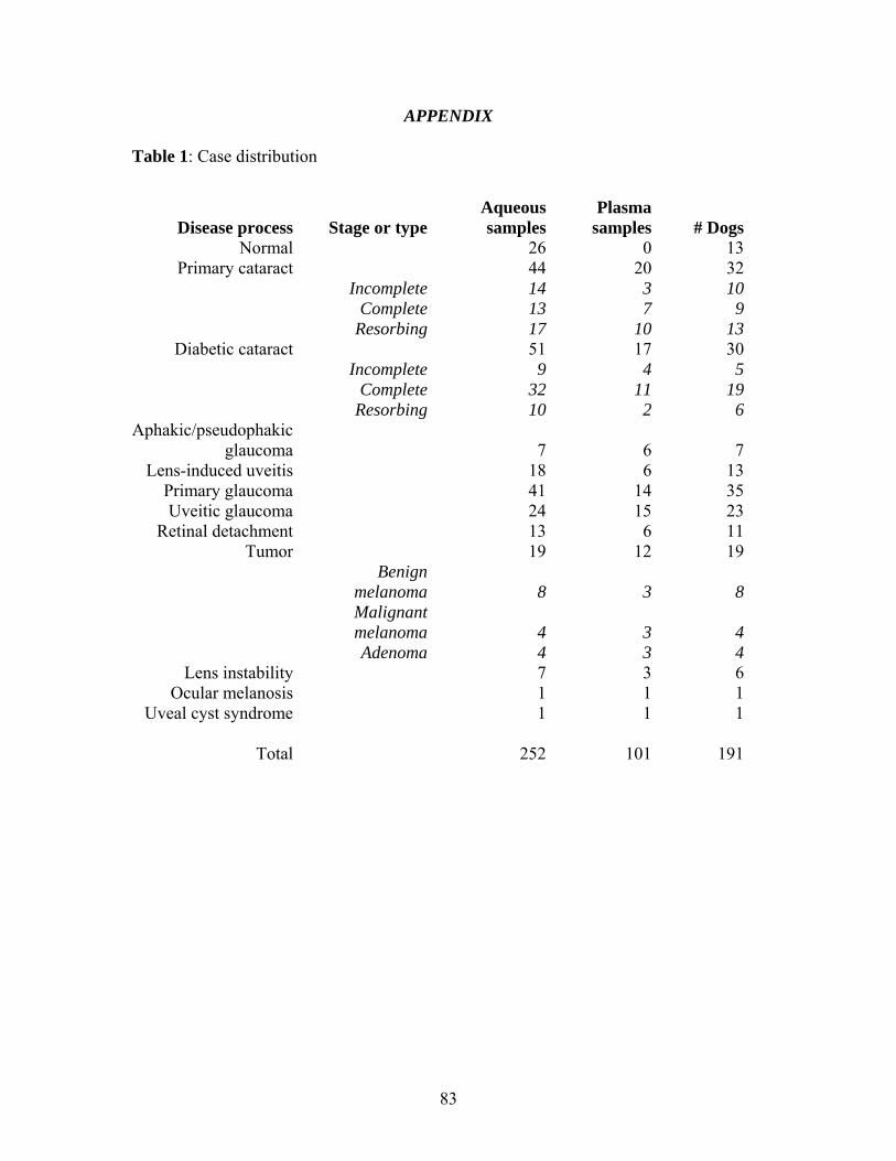
APPENDIX Table 1: Case distribution
Disease process Stage or typeAqueous samples
Plasma samples # Dogs
Normal 26 0 13Primary cataract 44 20 32
Incomplete 14 3 10 Complete 13 7 9 Resorbing 17 10 13
Diabetic cataract 51 17 30 Incomplete 9 4 5 Complete 32 11 19 Resorbing 10 2 6
Aphakic/pseudophakic glaucoma 7 6 7
Lens-induced uveitis 18 6 13Primary glaucoma 41 14 35Uveitic glaucoma 24 15 23
Retinal detachment 13 6 11Tumor 19 12 19
Benign
melanoma 8 3 8
Malignant melanoma 4 3 4
Adenoma 4 3 4Lens instability 7 3 6
Ocular melanosis 1 1 1Uveal cyst syndrome 1 1 1
Total 252 101 191
83
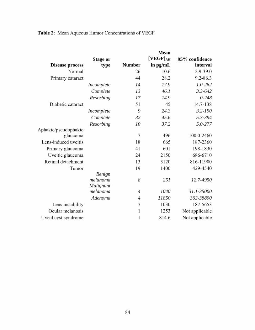
Table 2: Mean Aqueous Humor Concentrations of VEGF
Disease process Stage or
type
Mean [VEGF]AH 95% confidence
intervalNumber in pg/mLNormal 26 10.6 2.9-39.0
Primary cataract 44 28.2 9.2-86.3 Incomplete 14 17.9 1.0-262 Complete 13 46.1 3.3-642 Resorbing 17 14.9 0-248
Diabetic cataract 51 45 14.7-138 Incomplete 9 24.3 3.2-190 Complete 32 45.6 5.3-394 Resorbing 10 37.2 5.0-277
Aphakic/pseudophakic glaucoma 7 496 100.0-2460
Lens-induced uveitis 18 665 187-2360Primary glaucoma 41 601 198-1830Uveitic glaucoma 24 2150 686-6710
Retinal detachment 13 3120 816-11900Tumor 19 1400 429-4540
Benign
melanoma 8 251 12.7-4950
Malignant melanoma 4 1040 31.1-35000
Adenoma 4 11850 362-38800Lens instability 7 1030 187-5653
Ocular melanosis 1 1253 Not applicableUveal cyst syndrome 1 814.6 Not applicable
84

Table 3: Distribution of PIFMs in Histopathology Samples
Disease Type NumberAbsent
(0)Cellular
(1)Vascular or
fibrous (2) Fibrovascular
(3)Aphakic/pseudophakic
glaucoma 4 1 2 1 0Primary glaucoma 18 8 7 2 1Uveitic glaucoma 19 5 7 2 5
Retinal detachment 8 1 2 2 3Tumor 18 2 3 7 6
Benign
melanoma 8 1 1 4 2
Malignant melanoma 3 1 0 1 1
Adenoma 4 0 1 2 1Lens instability 4 0 3 1 0
Ocular melanosis 1 0 1 0 0Uveal cyst syndrome 1 0 1 0 0
Total 73 17 26 15 15
85
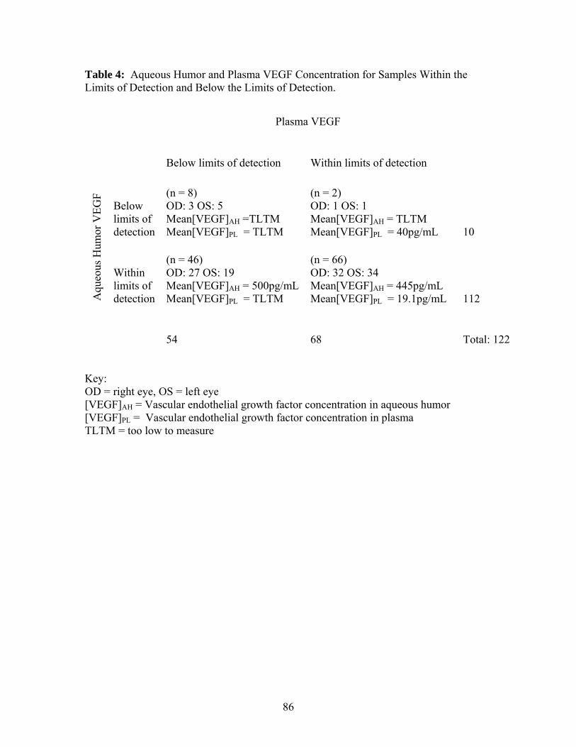
Table 4: Aqueous Humor and Plasma VEGF Concentration for Samples Within the Limits of Detection and Below the Limits of Detection.
Plasma VEGF
Aqu
eous
Hum
or V
EGF
Below limits of detection Within limits of detection
Below limits of detection
(n = 8) OD: 3 OS: 5 Mean[VEGF]AH =TLTM Mean[VEGF]PL = TLTM
(n = 2) OD: 1 OS: 1 Mean[VEGF]AH = TLTM Mean[VEGF]PL = 40pg/mL 10
Within limits of detection
(n = 46) OD: 27 OS: 19 Mean[VEGF]AH = 500pg/mL Mean[VEGF]PL = TLTM
(n = 66) OD: 32 OS: 34 Mean[VEGF]AH = 445pg/mL Mean[VEGF]PL = 19.1pg/mL 112
54 68 Total: 122 Key: OD = right eye, OS = left eye [VEGF]AH = Vascular endothelial growth factor concentration in aqueous humor [VEGF]PL = Vascular endothelial growth factor concentration in plasma TLTM = too low to measure
86

Figure 1: Scatter Plot of Plasma VEGF and Aqueous Humor VEGF Concentration
87

Figure 2: Bar Graph of Mean Aqueous Humor VEGF Concentration by Disease. Significant difference of disease groups from dogs without intraocular disease is denoted with an asterisk (*).
88

Figure 3: Cellular PIFM at 40x magnification. A cellular layer lacking blood vessels covers the anterior iris epithelium. The arrow indicates the interface of the anterior iris and cellular membrane.
89

Figure 4: Vascular PIFM at 40x magnification. A thin cellular layer with blood vessels covers the anterior iris epithelium. A blood vessel is indicated by the arrow in this view.
90

Figure 5: Fibrovascular PIFM at 4x magnification. A thick fibrovascular membrane is covering the anterior surface of the iris and causing entropion uvea, as indicated by the arrow.
91

Figure 6: Fibrovascular membrane at 10x magnification. The membrane is extending over the iridocorneal angle (indicated by arrow).
92

Figure 7: Fibrovascular PIFM at 40x magnification. Evaluation of the structure at this magnification reveals blood vessels and connective tissue covering the anterior iris. The arrow indicates the junction of the anterior iris with the fibrovascular membrane.
93

Sample summary: “patient” refers to patient identification number; “OD”, “OS” and “Pl” refer to the VEGF concentration of the right eye, left eye and plasma, respectively; “ocular disease” refers to ocular disease (1, 2 and 3 refer to incomplete, complete and resorbing cataract, respectively); “age” refer to age in years; “sex” refers to gender (M = intact male, MC= castrated male, F= intact female, FS= spayed female); “duration” refers to ocular disease duration in months; “PIFM” refers to pre-iridal fibrovascular membrane (0=not present; 1= cellular, 2= vascular or fibrous, 3= fibrovascular).
Patient OD OS Pl Ocular Disease Age Breed Sex
Systemic Disease Duration Medications PIFM Glaucoma
DMCR 0 24.3 Control 2 Mixed M None None No IMCR 10.3 19.5 Control 2 Mixed M None None No LMEK 63 39.7 Control 2 Mixed M None None No MAJ 12.4 14.5 Control 2 Mixed M None None No MAP 13.7 7.9 Control 2 Mixed M None None No MAR 12.5 0 Control 2 Mixed M None None No MAY 10.1 12 Control 2 Mixed M None None No MB5 9.5 0 Control 2 Mixed M None None No MBI 64.5 49.7 Control 2 Mixed M None None No MBJ 7.4 11.2 Control 2 Mixed M None None No MBY 0 0 Control 2 Mixed M None None No MCX 14.4 16.5 Control 2 Mixed M None None No MFY 25.2 5 Control 2 Mixed M None None No
95874 28.6 Primary cataract- 1 6
Basset Hound MC None 7
Topical, systemic steroid
No
99213 48.6 191 Primary cataract- 1 7 Beagle MC None 12
Topical, systemic steroid
No
92685 147 37.3 0 Primary cataract- 2 13
Bichon Frise FS Atopy 0.25 Topical steroid 0
No
93064 0 0 Primary cataract- 1 10
Bichon Frise FS None 18 Topical steroid
No
AM3A 36.4 26.1 8.5 Primary cataract- 2 8
Bichon Frise MC None 3
Topical steroid, prostaglandin
No
97313 131 163 Primary cataract- 1 11
Boston Terrier FS None 10
Topical
No
95
97816 155 Primary cataract- 1 11
Boston Terrier FS Atopy 8
Topical
No
99268 63.5 198 16 Primary cataract- 3 7
Boston Terrier FS None 18
Topical
No
99918 0 15.5 Primary cataract- 3 2
Cocker Spaniel F None 2
Topical
No
AM2A 6.2 8.6 Primary cataract- 2 2
Cocker Spaniel FS None 3
Topical steroid, prostaglandin
No
SA3A 0 0 Primary cataract- 1 2
Cocker Spaniel MC None 2 Topical steroid
No
91135 0
Primary cataract- 3
7 JRT MC None 30 Topical, systemic steroid
No
97546 17.1 0 Primary cataract- 3 1 JRT FS Atopy 3.5
Topical, systemic steroid
No
97146 42.3 29.7 Primary cataract- 3 6 Other FS None 0.75
Topical, systemic steroid
No
98474 189.3 12.3 Primary cataract- 1 13 Other FS HAC 12
Topical, systemic steroid
No
91439 54.4 Primary cataract- 2 6 Other FS Other 1.5 Topical steroid
No
68171 0 0 Primary cataract- 3 11 Mixed FS None 2
Topical, systemic steroid
No
AM1A 191 6.5 Primary cataract- 2 14 Mixed MC None 3 Topical steroid
No
SA11A 31.6 Primary cataract- 1 4 Mixed MC None Topical steroid
No
SA5A 553 0 Primary cataract- 2 3.5 Mixed MC None 2 Topical steroid
No
100058 179 5.3 Primary cataract- 2 6 Other FS None 6
Topical, systemic steroid
No
88313 42.2 27.6 Primary cataract- 3 8 Other MC None 24
Topical, systemic steroid
No
97029 38.6 7.7 Primary cataract- 1 6 Other FS None 6
Topical, systemic steroid
No
99975 5.1 51.4 Primary cataract- 1 11 Other MC None 12
Topical, systemic steroid
No
96
96844 116 17.7 Primary cataract- 2 6 Other MC None 2
Topical, systemic steroid
No
99247 89 0 Primary cataract- 3 10 Other
MC None 18
Topical, systemic steroid
No
92855 31.6 0 0 Primary cataract- 2 11 Pug
MC None 8.5
Topical steroid
No
BM14A 24.3 0 Primary cataract- 3 8 Other
MC None
Topical steroid
No
BM16A 26.9 0 Primary cataract- 3 13 Other MC None 24 None
No
93519 0 0 0 Primary cataract- 3 8 Other FS None 12
Topical, systemic steroid
No
BM12A 721 0 Primary cataract- 3 7.5 Other MC None 6 None
No
BM7A 381 23.2 Primary cataract- 2 7 Other FS None 0.5 None
No
92451 94.7 0 Diabetic cataract- 3 6 Beagle FS DM 3 Topical steroid
No
95939 77 94.2 11.6 Diabetic cataract- 2 10
Bichon Frise FS DM 3.5 Topical steroid
No
98081 24.3 31.8 Diabetic cataract- 2 8
Bichon Frise MC DM 1 Topical steroid
No
100022 48.5 23.5 Diabetic cataract- 1 11 JRT MC DM 3.5 Topical steroid
No
99450 11.7 4.6 0 Diabetic cataract- 2 6 JRT MC DM 1.5 Topical steroid
No
89550 127 Diabetic cataract- 3 10 Lab Ret M DM, other 4 Topical steroid
No
94352 0 0 40 Diabetic cataract- 1 7 Lab Ret FS DM 0.8 Topical steroid
No
96458 88.2 89.5 0 Diabetic cataract- 3 10 Lab Ret FS DM 18 Topical steroid
No
96637 94.1 105 Diabetic cataract- 2 11 Lab Ret FS DM 12 Topical steroid
No
99714 83.7 59.7 18.3 Diabetic cataract- 2 10 Lab Ret MC DM 6 Topical steroid
No
97
SA19A 45.1 0 Diabetic cataract- 2 12 Lab Ret FS DM None
No
SA2A 94.2 0 Diabetic cataract- 2 13 Lab Ret FS DM None
No
90840 0 0 Diabetic cataract- 2 3 Mixed FS DM 7 Topical steroid
No
90869 0 0 Diabetic cataract- 3 2 Mixed M DM 6 Topical steroid
No
90903 48 0 Diabetic cataract- 3 3 Mixed FS DM 4 Topical steroid
No
97702 213 Diabetic cataract- 2 8 Mixed FS DM 4 Topical steroid
No
98082 61.5 54 Diabetic cataract- 1 13 Mixed FS DM 1.5 Topical steroid
No
98303 40.8 98.5 24.8 Diabetic cataract- 1 8 Mixed MC DM 6 Topical steroid
No
BM10A 76.8 19.3 Diabetic cataract- 6 Mixed MC DM 6 None
No
BM3A 266 16.9 Diabetic cataract- 2 9 Mixed MC DM 3 None
No
89992 385 150 Diabetic cataract- 2 11 Other FS
DM 10 Topical steroid
No
90931 85.6 0 Diabetic cataract- 2 5 Other MC
DM 6 Topical steroid
No
92116 127 136 989 Diabetic cataract- 2 10 Other FS
DM 8 Topical steroid
No
92571 21.7 30.3 Diabetic cataract- 2 9 Other FS
DM 2 Topical steroid
No
96783 53.2 898 10.5 Diabetic cataract- 1 7 Other FS
DM 3 Topical steroid
No
97601 124 139 Diabetic cataract- 2 6 Other MC
DM 1 Topical steroid
No
97966 30.8 41.7 18.3 Diabetic cataract- 2 16 Other
MC DM 5 Topical steroid
No
99281 106 66.7 Diabetic cataract- 3 10 Other
MC DM 6 Topical steroid
No
98

96434 184 61.2 39.3 Diabetic cataract- 2 5 Pug
MC DM
1.5 Topical steroid
No
BM2A 29.8 14.3 Diabetic cataract- 2 4 Pug
MC DM 2 None
No
HD5A 737 0
Pseudo-phakic glaucoma 10
Bichon Frise
MC
None 1 None Yes
81646 762 0 Aphakic glaucoma 10
Cocker Spaniel
MC None 0.17 Systemic steroid 1 Yes
AM17A 1820 0
Pseudo-phakic glaucoma 12 JRT
MC None
2.5 Topical steroid, prostaglandin 2 Yes
73695 695 0
Pseudo-phakic glaucoma 9 Other MC
None
11 Topical steroid 1 Yes
SV1A 1980 0
Pseudo-phakic glaucoma 12 Other FS DM 3 Topical steroid Yes
72690 7.8 Aphakic glaucoma 9 Other FS Cardiac 1
Systemic steroid, topical prostaglandin Yes
HD1A 1600 0
Pseudo-phakic glaucoma 9 Other MC None 4 Topical steroid Yes
92310 819 747 Lens-induced uveitis- 2 5 Beagle FS DM 0.25
Topical steroid No
HD2A 1120 43.3 Lens-induced uveitis- 3 6
Cocker Spaniel
MC None 1
Topical steroid Yes
HDA7 595 0 Lens-induced uveitis- 3 10
Cocker Spaniel
MC None 7 None
Yes
BM21A 1290 0 Lens-induced uveitis- 3 9 Lab Ret
MC None 3
Topical steroid No
91560 43.1 Lens-induced uveitis- 3 12 Other FS Cardiac 13
Topical steroid Yes
98137 1780 1760 0 Lens-induced uveitis 6 Other
MC None 6
Topical steroid 3 Yes
99
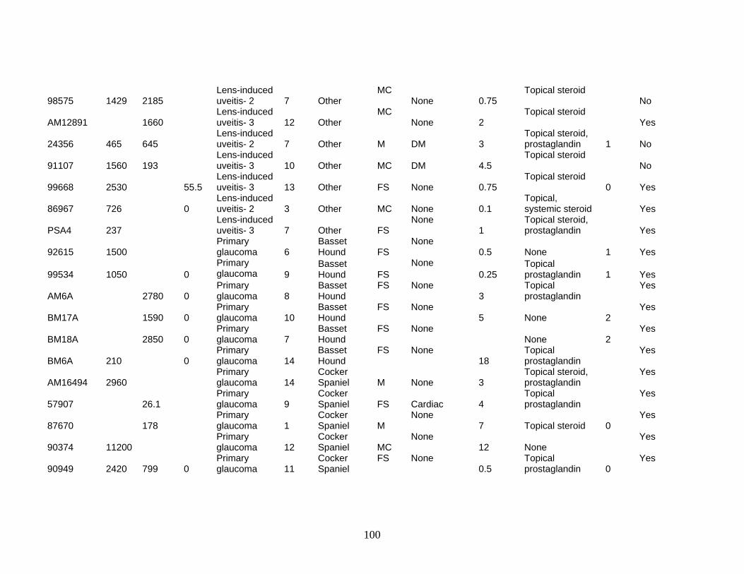
98575 1429 2185 Lens-induced uveitis- 2 7 Other
MC None 0.75
Topical steroid No
AM12891 1660 Lens-induced uveitis- 3 12 Other
MC None 2
Topical steroid Yes
24356 465 645 Lens-induced uveitis- 2 7 Other M DM 3
Topical steroid, prostaglandin 1 No
91107 1560 193 Lens-induced uveitis- 3 10 Other MC DM 4.5
Topical steroid No
99668 2530 55.5 Lens-induced uveitis- 3 13 Other FS None 0.75
Topical steroid 0 Yes
86967 726 0 Lens-induced uveitis- 2 3 Other MC None 0.1
Topical, systemic steroid Yes
PSA4 237 Lens-induced uveitis- 3 7 Other FS
None 1
Topical steroid, prostaglandin Yes
92615 1500 Primary glaucoma 6
Basset Hound FS
None 0.5 None 1 Yes
99534 1050 0 Primary glaucoma 9
Basset Hound FS
None 0.25
Topical prostaglandin 1 Yes
AM6A 2780 0 Primary glaucoma 8
Basset Hound
FS None 3
Topical prostaglandin
Yes
BM17A 1590 0 Primary glaucoma 10
Basset Hound
FS None 5 None 2
Yes
BM18A 2850 0 Primary glaucoma 7
Basset Hound
FS None None 2
Yes
BM6A 210 0 Primary glaucoma 14
Basset Hound
FS None 18
Topical prostaglandin
Yes
AM16494 2960 Primary glaucoma 14
Cocker Spaniel M None 3
Topical steroid, prostaglandin
Yes
57907 26.1 Primary glaucoma 9
Cocker Spaniel FS Cardiac 4
Topical prostaglandin
Yes
87670 178 Primary glaucoma 1
Cocker Spaniel M
None 7 Topical steroid 0
Yes
90374 11200 Primary glaucoma 12
Cocker Spaniel MC
None 12 None
Yes
90949 2420 799 0 Primary glaucoma 11
Cocker Spaniel
FS None 0.5
Topical prostaglandin 0
Yes
100

91249 553 Primary glaucoma 9
Cocker Spaniel
FS None 0.8
Topical prostaglandin 0
Yes
95216 1630 Primary glaucoma 11
Cocker Spaniel FS None 0.1
Topical prostaglandin 1
Yes
PSA1 2010 Primary glaucoma 13
Cocker Spaniel FS Atopy 3
Topical steroid, prostaglandin
Yes
PSA2 398 Primary glaucoma 14
Cocker Spaniel FS
None 2
Topical prostaglandin
Yes
AM19A 0 0 Primary glaucoma 11 JRT FS
None 3
Topical steroid, prostaglandin 0
Yes
HD3A 594 9.2 Primary glaucoma 4 JRT M
None 6 Topical steroid
Yes
HD4A 146 0 Primary glaucoma 10 JRT F
None 6 Topical steroid
Yes
92684 630 1570 14.4 Primary glaucoma 8 Lab Ret MC
None 0.1
Topical prostaglandin
Yes
AM18A 6830 46.1 Primary glaucoma 10 Lab Ret FS
None 2.5
Topical steroid, prostaglandin 0
Yes
69243 307 Primary glaucoma 11 Mixed MC Other 1 None 3
Yes
93705 191 Primary glaucoma 10 Mixed
FS None 12
Topical prostaglandin 0
Yes
96753 5600 146 Primary glaucoma 2 Mixed
FS None 12 None 0
Yes
97866 190 53.5 0 Primary glaucoma 10 Mixed
FS None 7
Topical prostaglandin 1
Yes
98792 76.1 785 Primary glaucoma 13 Mixed
FS Other 1 None 1
Yes
MDLBRT 14500 Primary glaucoma 5 Mixed F Other 0.5
Topical prostaglandin 0
Yes
SA9A 6550 0 Primary glaucoma 9 Mixed
FS None 14
Topical steroid, prostaglandin
Yes
96546 1760 Primary glaucoma 12 Other
FS None 2 None 1
Yes
98370 125 Primary glaucoma 12 Other
FS None 2
Topical prostaglandin
Yes
101

AM13A 32.1 Primary glaucoma 8 Other MC
None 24
Topical steroid, prostaglandin
Yes
BM8A 37.3 0 Primary glaucoma 13.5 Other
FS None 6 Topical steroid
Yes
91435 41900 Primary glaucoma 9 Pug
FS None 2
Topical prostaglandin
Yes
AM27044 4930 Primary glaucoma 8 Other
FS None 1
Topical steroid, prostaglandin
Yes
99287 263 Primary glaucoma 8 Other
FS None 5 None 0
Yes
98274 482 183 Primary glaucoma 10 Other
FS None 12 None
Yes
97734 519 Uveitic glaucoma 7 Beagle
FS Other 0.5
Topical steroid, prostaglandin 3
Yes
94914 1170 0 Uveitic glaucoma 8
Cocker Spaniel MC
None 0.002 Topical steroid 1
Yes
95150 792 0 Uveitic glaucoma 10
Cocker Spaniel MC
None 0.75 Topical steroid 2
Yes
96101 2760 24 Uveitic glaucoma 15 JRT
FS None 0.5 Topical steroid 1
Yes
98944 724 Uveitic glaucoma 7 JRT
FS None 1.5 Topical steroid 0
Yes
96519 5400 Uveitic glaucoma 6 Lab Ret MC
None 0.5 Systemic steroid 3
Yes
BM1A 3070 0 Uveitic glaucoma 3 Other MC
None 3
Topical steroid
Yes
SA4A 33000 0 Uveitic glaucoma 14 Other MC
None 1.5
Topical steroid
Yes
BM13A 6040 11.9 Uveitic glaucoma 12 Other FS
None 0.5 None 3
Yes
AM27865 786 Uveitic glaucoma 8 Mixed
FS None
Topical steroid 2
Yes
95353 224 0 Uveitic glaucoma 8 Mixed
FS None 0.75
Topical steroid 0
Yes
98516 4170 1704 27.2 Uveitic glaucoma 11 Mixed
FS Other 0.75
Topical steroid 0
Yes
102

46253 58000 0
Uveitic glaucoma
9 Other MC None 0.75
Topical steroid
1
Yes
59386 10200 Uveitic glaucoma 9 Other MC DM 0.25
Topical prostaglandin 3
Yes
84804 79.6 Uveitic glaucoma 6 Other FS None 12 None 0
Yes
97757 705 0 Uveitic glaucoma 4 Other FS None 4
Topical steroid, prostaglandin
Yes
BM4A 253 0 Uveitic glaucoma 4 Other F Other 2 Topical steroid 1
Yes
EC2A 3390 35 Uveitic glaucoma 8 Other FS None 9
Topical steroid, prostaglandin 0
Yes
HD6A 47400 0 Uveitic glaucoma 10 Pug MC None 2 Topical steroid
Yes
89181 869 Uveitic glaucoma 8 Other FS None 18 None 1
Yes
96065 37400 44.2 Uveitic glaucoma 10 Other MC DM 0.3 None 3
Yes
98783 1942 Uveitic glaucoma 9 Other FS None 1 Topical steroid 1 No
98273 304 0 Uveitic glaucoma 2 Other MC Other 0.2 Topical steroid 2 Yes
99547 12550 Retinal detachment 4 Beagle FS None 2 Topical steroid 1 No
AM28242 3880 Retinal detachment 4
Boston Terrier MC
None 0.5
Topical steroid, prostaglandin 2 No
86366 257 Retinal detachment 6
Boston Terrier
MC None 5
Topical steroid, prostaglandin No
90493 71.8 102 0 Retinal detachment 5
Boston Terrier
MC None 0.5 None 1
No
SA20A 41800 Retinal detachment 6
Cocker Spaniel FS
None 6 Systemic steroid
No
SA6A 2370 18.2 Retinal detachment 6
Cocker Spaniel FS
None 1 Systemic steroid
No
97815 26500 6790 Retinal detachment 0.75 Lab Ret M
None 9 None 2
No
103

96086 1080 9 Retinal detachment 6 Other MC
None 2.5 None 3
No
98258 1600 0 Retinal detachment 6 Mixed FS
None 0.001 None 0
No
AM16A 20300 0 Retinal detachment 11 Mixed MC
None
Topical steroid, prostaglandin 3
No
90350 12300 33 Retinal detachment 10 Other M
None 24 Topical steroid 3
No
99051 0 Benign melanoma 2 Beagle FS
None 0.001 None 1
No
SA1A 200000 23.4 Malignant melanoma 12 Beagle FS
None 6
Topical prostaglandin Yes
95822 1260 16.5 Benign melanoma 10 Other FS
None 1 Topical steroid 3 No
EC3A 5910 4.7 Adenoma 7 Other FS None None 2 Yes
94941 2020 9.4 Benign melanoma 10 JRT FS
None 6 Topical steroid 2 No
100556 16300 0 Malignant melanoma 12 Lab Ret FS
None 1 None 3 No
90375 270 Benign melanoma 10 Lab Ret FS
None 2 Topical steroid 2 No
92739 46100 11.5 Adenoma 9 Lab Ret MC None
0.25 Topical prostaglandin 2 No
92779 343 Malignant melanoma 8 Lab Ret F
None 0.24 None 2 No
100175 0 0 Malignant melanoma 7 Mixed MC
None 3.5 None 0 No
93911 88.7 7.0 Benign melanoma 8 Mixed FS
None 30 None 2 Yes
94907 2700 3.4 Spindle cell tumor 8 Mixed MC Other 0.5
Topical steroid, prostaglandin 3 Yes
99666 281 Benign melanoma 13 Mixed FS
None 1 Topical steroid 0 Yes
EC1A 63400 0 Anaplastic 13 Mixed FS None
1.5 Topical steroid, prostaglandin 3 Yes
100276 33500 Adenoma 8 Other FS None 0.5 None 3 No
104

105
AM25917 1060 Benign melanoma 12 Other MC
None
2 Topical steroid, prostaglandin 3 No
90742 788 Benign melanoma 13 Other MC
None 2.5 Topical steroid 2 No
98346 3670 11.5 Lymphoma 8 Other FS None 0.5 Topical steroid 1 Yes AM9A 2250 3.9 Adenoma 11 Other FS None 3 Topical steroid 1 No
79800 475 80.7 0 Lens luxation 6 Beagle FS
Hypertension 2.5 Topical steroid, prostaglandin 1 Yes
91853 116 Lens luxation 6 JRT FS None 0.001 None Yes 96105 2570 0 Lens luxation 16 Mixed FS None 0.5 None 1 Yes
77820 2230 0 Lens luxation
7 Other MC None
10 Topical prostaglandin 1 Yes
91584 2530 Lens luxation
3 Other F None
0.5 Topical prostaglandin 2 Yes
28594 684 Lens luxation 18 Other MC None 0.5 Topical steroid Yes