vascular access in adult apheresis - c.ymcdn.com · vascular access in adult apheresis outline •...
TRANSCRIPT
Vascular Access In Adult Apheresis: An Overview & 2017 Update
Jan Hofmann, M.D., M.P.H., M.Sc.
Associate Medical Director,
Apheresis Care Group,
Department of Medicine,
California Pacific Medical Center,
Director, Apheresis Education,
BCP-UCSF Transfusion Medicine Program,
UCSF School of Medicine,
San Francisco, CA
May 4, 2017
2
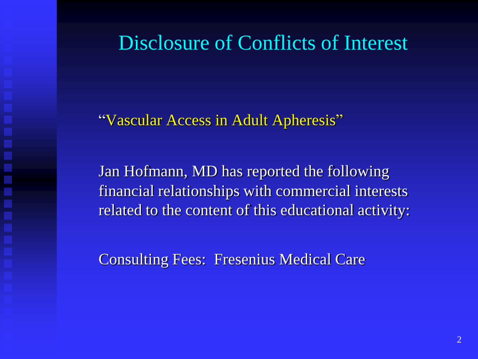
Disclosure of Conflicts of Interest
“Vascular Access in Adult Apheresis”
Jan Hofmann, MD has reported the following
financial relationships with commercial interests
related to the content of this educational activity:
Consulting Fees: Fresenius Medical Care

3
Vascular Access in Adult Apheresis Outline
• Temporary and tunneled double-lumen central venous catheters (CVCs)
- Advantages and disadvantages
- Trialysis CVCs, Power Hickman CVCs
• Placement of CVCs (where?, who?)
• Inpatient care of CVCs (port patency, dressing changes)
• Outpatient care of CVCs (keeping dressing dry & clean)
• Troubleshooting CVC malfunction (obstruction/fibrin sheaths; kinking; exit site inflammation/infection).
• Adverse Events (PTX; line infection; line migration)
• Removal of CVCs (exit site care)
• Ultrasound-guided peripheral access
• Optimization of peripheral access (hydration, patient preparation)
• Emerging forms of vascular access in adults (new ports, angiocaths, radial arterial access, etc).
• Questions & Answers
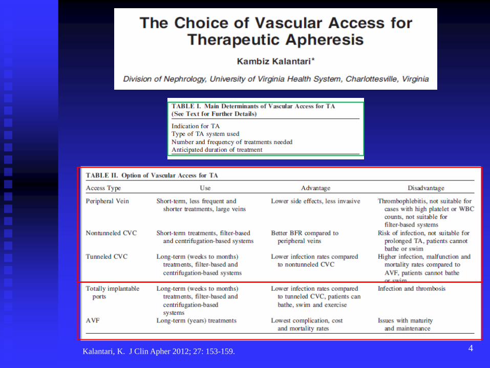
4 Kalantari, K. J Clin Apher 2012; 27: 153-159.

5
Golestaneh L, Mokrzycki, MH.
J Clin Apher 2013; 28: 64-72.

6
Golestaneh L, Mokrzycki, MH. J
Clin Apher 2013; 28: 64-72.
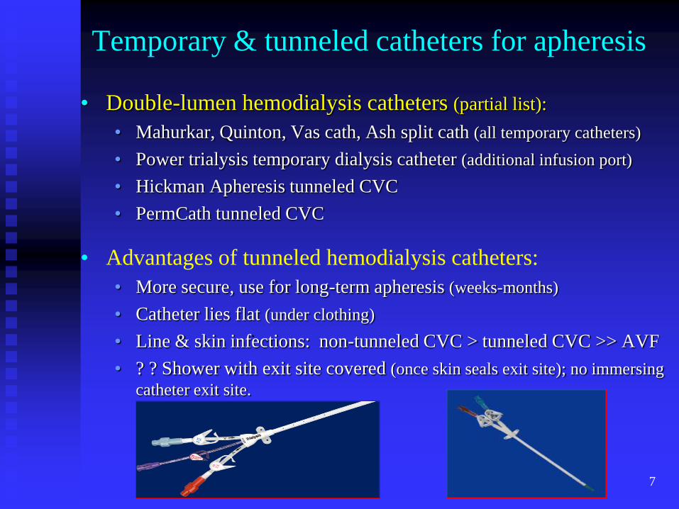
Temporary & tunneled catheters for apheresis
• Double-lumen hemodialysis catheters (partial list):
• Mahurkar, Quinton, Vas cath, Ash split cath (all temporary catheters)
• Power trialysis temporary dialysis catheter (additional infusion port)
• Hickman Apheresis tunneled CVC
• PermCath tunneled CVC
• Advantages of tunneled hemodialysis catheters:
• More secure, use for long-term apheresis (weeks-months)
• Catheter lies flat (under clothing)
• Line & skin infections: non-tunneled CVC > tunneled CVC >> AVF
• ? ? Shower with exit site covered (once skin seals exit site); no immersing
catheter exit site.
7
8
Placement of Apheresis CVCs
• Anatomic locations:
• Most common choices: great vessels (chest); femoral veins
• Right IJ >> Right SC >> Left SC > Left IJ
• Catheter tip: junction of SVC and right atrium; proximal right atrium
• Preparation (NPO for ≥8 hours)
• Placement verification (fluoroscopy; CXR; ultrasound; TEE)
• Who places apheresis CVCs: • Interventional radiologists (IR suite: scheduled; weekend: emerg only)
• Surgeons (OR suite; scheduled: often delayed due to other surgeries)
• Intensivists (ICU; ultrasound guidance; may be faster than IR or OR
• Other (residents; nephrologists)
• Tunneled CVCs (currently interventional radiologists or surgeons only)

Right Internal Jugular (IJ)
Central Venous Access
9
From Heffner AC et al. Overview of Central
Venous Access. Uptodate 2017; 3/16/17: 14.

Advantages & Disadvantages
of Central Vein Approaches
For CVC Placement.
10
From Heffner AC et al. Overview of Central
Venous Access. Uptodate 2017; 3/16/17: 18.

11
Care of Apheresis CVCs
• Inpatient care (for nurses):
• Intra-luminal catheter-locking agents (port patency): • Heparin: usually 1000-5000 U/ml (total of 5-6 ml)
• Studies using 100U/ml; 10,000 U/ml
• Citrate (4%)(5, 30, 47%): similar efficacy; ↓ bleeding risk & cost; no risk of HIT
• rt-PA (recombinant tissue plasminogen activator)
• Tego caps (non-heparin)
• Dressing changes: • usually after each treatment
• antiseptic technique (mask, etc)
• protection of line
• Outpatient care (for patients):
• Temporary lines (keep dressing dry/no showers; care with dislodging line)
• Tunneled lines (first 2-3 weeks: keep dressing dry/no showers)
• Monitoring for site infection
• Compliance with catheter flush schedule
Hemmelgarn BR et al. N Engl J Med 2011; 364: 303-312.
Mokrzycki MH, Lok CE. Kidney Int 2010; 78: 1218-1231.
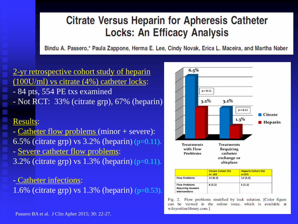
12 Passero BA et al. J Clin Apher 2015; 30: 22-27.
2-yr retrospective cohort study of heparin
(100U/ml) vs citrate (4%) catheter locks:
- 84 pts, 554 PE txs examined
- Not RCT: 33% (citrate grp), 67% (heparin)
Results:
- Catheter flow problems (minor + severe):
6.5% (citrate grp) vs 3.2% (heparin) (p=0.11).
- Severe catheter flow problems:
3.2% (citrate grp) vs 1.3% (heparin) (p=0.11).
- Catheter infections:
1.6% (citrate grp) vs 1.3% (heparin) (p=0.53).

13 Passero BA et al. J Clin Apher 2015; 30: 22-27.
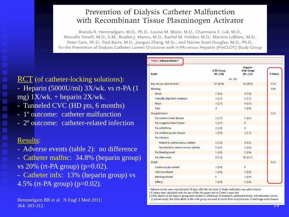
14 Hemmelgarn BR et al. N Engl J Med 2011;
364: 303-312.
RCT (of catheter-locking solutions):
- Heparin (5000U/ml) 3X/wk. vs rt-PA (1
mg) 1X/wk. + heparin 2X/wk.
- Tunneled CVC (HD pts, 6 months)
- 1º outcome: catheter malfunction
- 2º outcome: catheter-related infection
Results:
- Adverse events (table 2): no difference
- Catheter malfnc: 34.8% (heparin group)
vs 20% (rt-PA group) (p=0.02).
- Catheter infx: 13% (heparin group) vs
4.5% (rt-PA group) (p=0.02).

15 Hemmelgarn BR et al. N Engl J Med 2011; 364: 303-312.

16 Mokrzycki MH, Lok CE. Kidney
Int 2010; 78: 1218-1231.
Excellent review:
- 30-35 studies (1998-2010)
evaluating methods to prevent
HD catheter malfunction.
17 Mokrzycki MH, Lok CE. Kidney Int 2010; 78: 1218-1231.
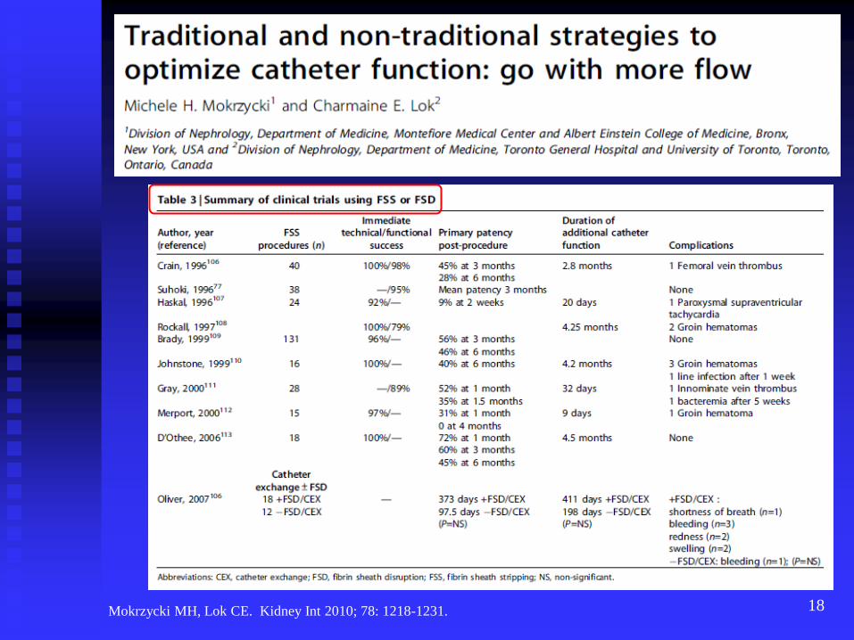
18 Mokrzycki MH, Lok CE. Kidney Int 2010; 78: 1218-1231.

19
Management Strategies for CVC Dysfunction
• Immediate and long-term strategies (for CVC dysfunction):
• Forceful saline flush and reversal of ports (~15% ↓ efficiency):
• Re-positioning patient (ie, catheter port against vessel wall)
• Thrombolytic agents (rTPA) may restore patency: • Short-term success: 22-100%
• Long-term (90-day patency) rate: 25-53%
• Mechanical disruption of fibrin sleeve or stripping (FSS): • Immediate patency rate: 79-98%
• Long term (3-month) patency (after FSS): 45-60%
• Complications: groin hematomas, infection, & venous thrombosis
• Tunneled catheter exchange over a guidewire (after failed TPA):
• Primary patency (@ 3 months): 51%; @ 6 months: 37%
• Low complication rates
Golestaneh L et al. J Clin Apher 2013; 28: 64-72.

20
Vascular Access Complications (associated with CVCs placed for apheresis procedures)
Overall – 0-4% of all CVC placement procedures (for apheresis):
• Site inflammation (or infection)
• Line infection:
• Occult bacteremia to septicemia
• Signs/symptoms include (partial list):
• Fever >38.0 C
• Atrial tachyarrythmias; hypotension; rigors; CNS changes; etc
• Pneumothorax (rare; noticeable during or slightly after line placement, or incidentally):
• Confirmed by CXR; may (or may not) require chest tube
• Line migration
• Hematoma (slightly greater occurrence when pts on anticoagulation therapy)
• Thrombosis
Golestaneh L et al. J Clin Apher 2013; 28: 64-72. Kaplan A. Seminars in Dialysis 2012; 25 (2): 152-158.
Gallieni M et al. J Vasc Access 2011; 12 (4): 273-279. Lorente L et al. Critical Care 2005; 9: R631-R635.
Bromlage CP et al. J Clin Apher 2009; 24 (6): 225-31. Michon B et al. Transfusion 2007; 47 (10): 1837-1842.
Howard MA et al. Transfusion 2006; 46 (1): 154-56. Basic-Jukic N et al. Ther Apher Dial 2005; 9 (5): 391-95.
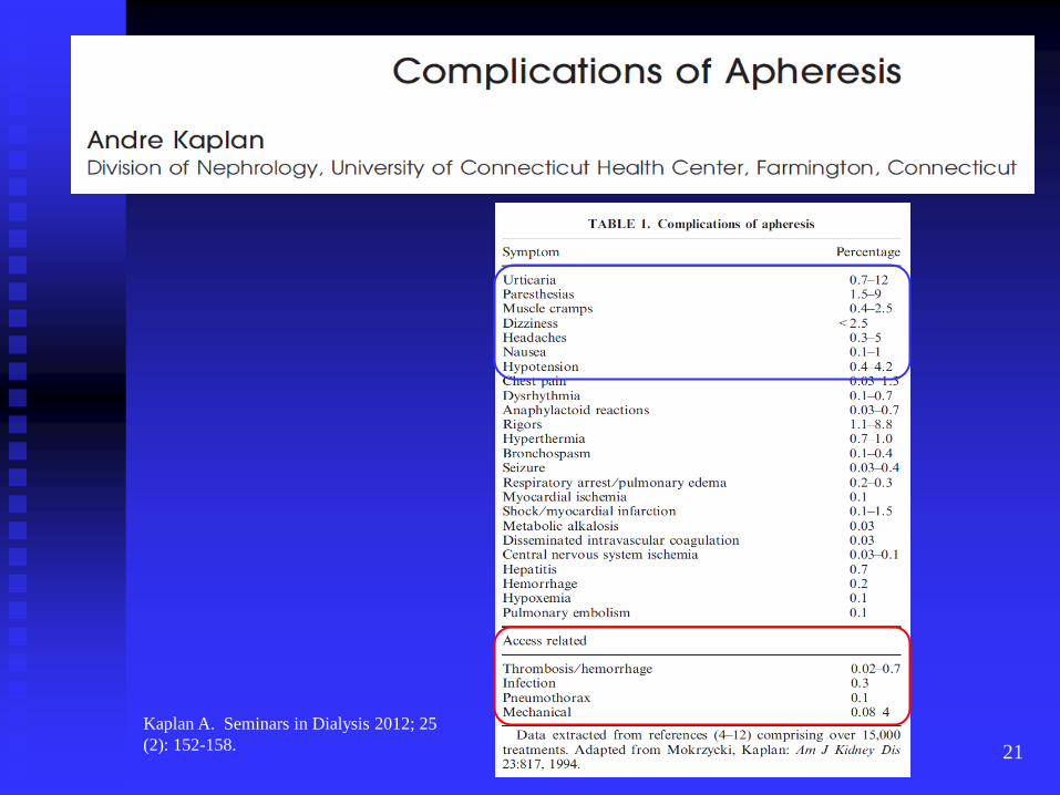
21
Kaplan A. Seminars in Dialysis 2012; 25
(2): 152-158.

Complications of CVC
22
From Heffner AC et al. Overview of Central
Venous Access. Uptodate 2017; 3/16/17: 22.
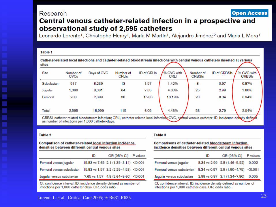
23 Lorente L et al. Critical Care 2005; 9: R631-R635.
24
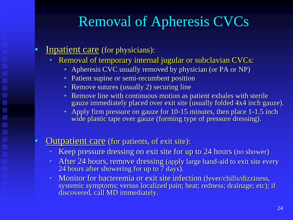
Removal of Apheresis CVCs
• Inpatient care (for physicians):
• Removal of temporary internal jugular or subclavian CVCs: • Apheresis CVC usually removed by physician (or PA or NP)
• Patient supine or semi-recumbent position
• Remove sutures (usually 2) securing line
• Remove line with continuous motion as patient exhales with sterile gauze immediately placed over exit site (usually folded 4x4 inch gauze).
• Apply firm pressure on gauze for 10-15 minutes, then place 1-1.5 inch wide plastic tape over gauze (forming type of pressure dressing).
• Outpatient care (for patients, of exit site):
• Keep pressure dressing on exit site for up to 24 hours (no shower)
• After 24 hours, remove dressing (apply large band-aid to exit site every 24 hours after showering for up to 7 days).
• Monitor for bacteremia or exit site infection (fever/chills/dizziness, systemic symptoms; versus localized pain; heat; redness; drainage; etc); if discovered, call MD immediately.

Ultrasound-guided Peripheral Venous Access
25
Gopalasingam N et al. J Clin Apher 2017; Mar
20. doi: 10:1002/jca.21533.
Putensen D et al. J Clin Apher 2016; 6:
501-506.
Salazar E et al. J Clin Apher 2016; Aug 10.
doi: 10.1002/jca.21493.
Putensen D et al. J Clin Apher 2016; Sep 15.
doi: 10.1002/jca.21508.
Stolz LA et al. J Vasc Access 2015; 16: 321-326.
Mayhew M et al. J Clin Apher 2017; May 4: in press.
O’Leary MF et al. J Clin Apher 2016; Jan 13. jca.21445.
Brass P et al. Cochrane Database Syst Rev 2015. Jan 9.
Hanafusa N et al. J Clin Apher 2015; 30: 380-381.
Liu YT et al. Eur J Emerg Med 2014; 21: 18-23.
Egan G et al. Emerg Med J 2013; 30: 521-526.
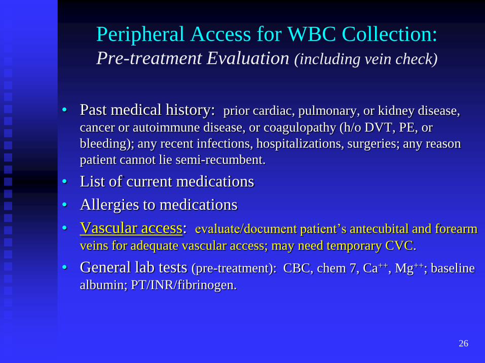
Peripheral Access for WBC Collection: Pre-treatment Evaluation (including vein check)
• Past medical history: prior cardiac, pulmonary, or kidney disease,
cancer or autoimmune disease, or coagulopathy (h/o DVT, PE, or
bleeding); any recent infections, hospitalizations, surgeries; any reason
patient cannot lie semi-recumbent.
• List of current medications
• Allergies to medications
• Vascular access: evaluate/document patient’s antecubital and forearm
veins for adequate vascular access; may need temporary CVC.
• General lab tests (pre-treatment): CBC, chem 7, Ca++, Mg++; baseline
albumin; PT/INR/fibrinogen.
26

Patient Preparation is Key (for maintaining peripheral access during long TA procedure)
2-3 days before scheduled procedure:
Hydrate (water >> juice) for 72 hours prior to scheduled procedure
Avoid strenuous exercise, alcohol, diuretics, or caffeinated drinks or
food for 48-72 hours prior to procedure.
Get plenty of sleep (>7-8 hours/night); attempt to maintain low stress
environment.
Hydration “app” (text messages Q 6 hours X 72 hours); Post-Care “app”
Day of procedure:
Eat breakfast (no coffee, minimal fluids) the morning of procedure
Hold morning medications (esp. BP meds); bring all meds to clinic
Wear loose, comfortable clothing
Distractions (DVDs, audio CDs, supportive friend); conscious relaxation
27

Vascular Access (outpatient WBC collections)
Obtaining peripheral access:
Skill of RN is paramount (vein choice, access skills, warming extremity)
Access (16-17 gauge steel needle, antecubital vein, ID lidocaine)
Return (18-20 gauge angiocath, non-antecubital vein, ID lidocaine)
Maintaining peripheral access (throughout the procedure):
Skill of RN (vein choice, access skills, keeping patient engaged & warm)
Patient preparation, hydration, and ability to comply with protocol; IV hydration (200-1000 ml NS over 30-90 minutes) may be helpful.
Hot packs (pre-warming access site; patient squeezing pack during tx)
Positioning (keeping “access” arm externally rotated to maintain flow)
IV start: cuff pressure 20-30 mm Hg > DBP (~100 mm Hg); ↓ to 40 mm Hg
WB:AC = 12:1 (9:1-14:1)
Vascular spasm (autonomic control)
↑↑ pressure alarms or loss of interface ↑↑ RBC contamination
Monitoring for citrate toxicity (paresthesias, vibratory sensation, etc)
28
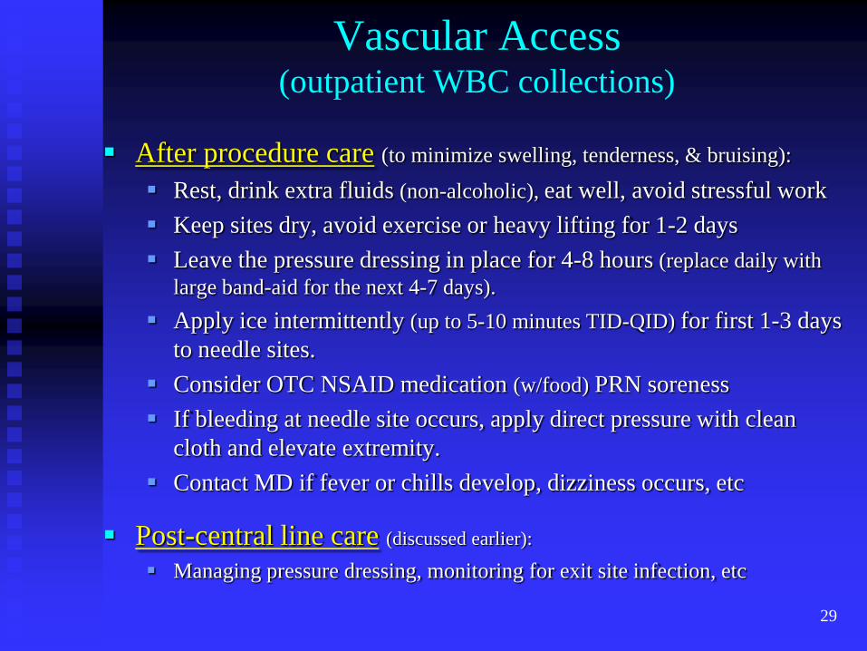
Vascular Access (outpatient WBC collections)
After procedure care (to minimize swelling, tenderness, & bruising):
Rest, drink extra fluids (non-alcoholic), eat well, avoid stressful work
Keep sites dry, avoid exercise or heavy lifting for 1-2 days
Leave the pressure dressing in place for 4-8 hours (replace daily with
large band-aid for the next 4-7 days).
Apply ice intermittently (up to 5-10 minutes TID-QID) for first 1-3 days
to needle sites.
Consider OTC NSAID medication (w/food) PRN soreness
If bleeding at needle site occurs, apply direct pressure with clean
cloth and elevate extremity.
Contact MD if fever or chills develop, dizziness occurs, etc
Post-central line care (discussed earlier):
Managing pressure dressing, monitoring for exit site infection, etc
29

Venous Access for IV Contrast
30
• No decision flowchart
available for choosing most
appropriate venous access
for different types of TA.
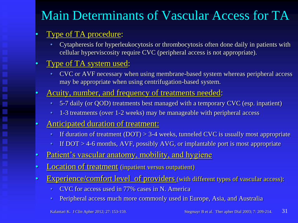
Main Determinants of Vascular Access for TA
31
• Type of TA procedure:
• Cytapheresis for hyperleukocytosis or thrombocytosis often done daily in patients with
cellular hyperviscosity require CVC (peripheral access is not appropriate).
• Type of TA system used:
• CVC or AVF necessary when using membrane-based system whereas peripheral access
may be appropriate when using centrifugation-based system.
• Acuity, number, and frequency of treatments needed:
• 5-7 daily (or QOD) treatments best managed with a temporary CVC (esp. inpatient)
• 1-3 treatments (over 1-2 weeks) may be manageable with peripheral access
• Anticipated duration of treatment:
• If duration of treatment (DOT) > 3-4 weeks, tunneled CVC is usually most appropriate
• If DOT > 4-6 months, AVF, possibly AVG, or implantable port is most appropriate
• Patient’s vascular anatomy, mobility, and hygiene
• Location of treatment (inpatient versus outpatient)
• Experience/comfort level of providers (with different types of vascular access):
• CVC for access used in 77% cases in N. America
• Peripheral access much more commonly used in Europe, Asia, and Australia
Kalantari K. J Clin Apher 2012; 27: 153-159. Stegmayr B et al. Ther apher Dial 2003; 7: 209-214.
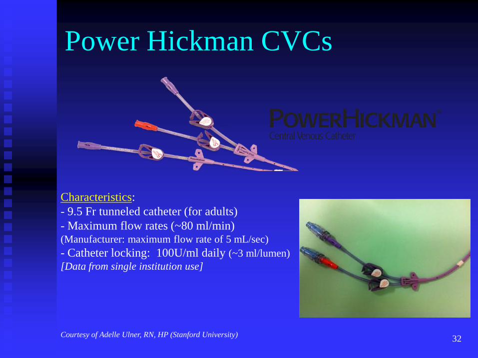
Power Hickman CVCs
32
Characteristics:
- 9.5 Fr tunneled catheter (for adults)
- Maximum flow rates (~80 ml/min) (Manufacturer: maximum flow rate of 5 mL/sec)
- Catheter locking: 100U/ml daily (~3 ml/lumen)
[Data from single institution use]
Courtesy of Adelle Ulner, RN, HP (Stanford University)

Insyte Angiocath (for
peripheral IV access)
33
For peripheral access & return:
- To replace steel needle (esp. with
prior thrombosis).
- Flow rates: 40-50 ml/min (with
18 G angiocath).
- Use AC vein (or non-AC vein in
forearm).
- May use 20 G angiocath (for
return; minimal high pressure alarms).
- Works well for ECP txs
Courtesy of Adelle Ulner, RN, HP (Stanford University)

34
Temporary Radial Artery Peripheral Access: - For long-term, intermittent, usually outpatient TPE treatments
- Placement: 1-2 ml 2% lidocaine local anesthesia
- ARROW Int’l Radial Artery Catheterization Set (18 gauge)
- Flow rates >80 ml/min
- Catheter removal: 2-5 min direct pressure (occasionally ice packs)
- Compatible with Cobe Spectra
Khatri, BO. J Clin Apher 2003; 18; 134.
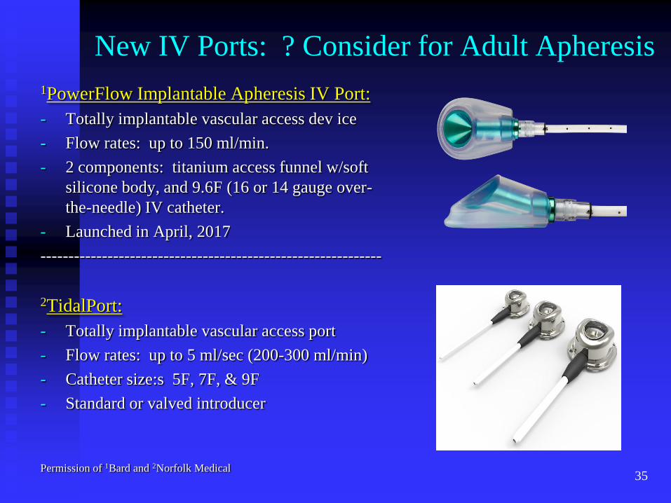
New IV Ports: ? Consider for Adult Apheresis
1PowerFlow Implantable Apheresis IV Port:
- Totally implantable vascular access dev ice
- Flow rates: up to 150 ml/min.
- 2 components: titanium access funnel w/soft
silicone body, and 9.6F (16 or 14 gauge over-
the-needle) IV catheter.
- Launched in April, 2017
-------------------------------------------------------------
2TidalPort:
- Totally implantable vascular access port
- Flow rates: up to 5 ml/sec (200-300 ml/min)
- Catheter size:s 5F, 7F, & 9F
- Standard or valved introducer
Permission of 1Bard and 2Norfolk Medical
35
36 36 36 36 36 36 36
Conclusions / Summary
Main determinants of vascular access for TA depend on type of procedure;
acuity; number, frequency, and anticipated duration of treatment; patient’s
vascular anatomy; and providers’ comfort level.
Catheter locking agents other than heparin (such as citrate and rt-PA) are
showing similar efficacy and safety.
Maintaining peripheral access (especially for lengthy TA procedures, such
as WBC collections) depend on patient hydration, RN venous access skills,
and vein selection and management. Obtaining peripheral access is
enhanced with ultrasound guidance.
New devices for central venous and peripheral access (new ports,
angiocaths, etc) are providing greater choice in technology and improving
ease of use and less exposure to locking anticoagulants.
North and South America have a significantly greater use of CVCs (and
less use of peripheral vascular access) compared to Europe and Australia.
36

37 37 37 37 37 37 37
Selected References
Gopalasingam N et al. J Clin Apher 2017; Mar 20. doi: 10.1002/jca.21533.
Wooster M et al. J Vasc Access 2017; Jan 25. doi: 10.5301/jva.5000644.
Putensen D et al. J Clin Apher 2016; Sep 15. doi: 10.1002/jca.21508.
Salaza E et al. J Clin Apher 2016; Aug 10. doi: 10.1002/jca.21493.
Putensen D et al. J Clin Apher 2016; 31(6): 501-506.
Khatri B, Kramer J. Muscle Nerve 2013; 48(4): 624.
Kalantari K. J Clin Apher 2012; 27: 153-159.
Stegmayer B et al. Ther Apher Dial 2003; 7: 209-214.
Golestaneh L, Mokrzycki, MH. J Clin Apher 2013; 28: 64-72.
Malchesky et al. Ther Apher Dial 2010; 14 (1): ref 4.
Passero BA et al. J Clin Apher 2015; 30: 22-27.
Hemmelgarn BR et al. N Engl J Med 2011; 364: 303-312.
Mokrzycki MH, Lok CE. Kidney Int 2010; 78: 1218-1231.
Heffner AC et al. Overview of Central Venous Access. Uptodate 2017.
Kaplan A. Seminars in Dialysis 2012; 25 (2): 152-158.
Gallieni M et al. J Vasc Access 2011; 12 (4): 273-279.
Lorente L et al. Critical Care 2005; 9: R631-R635.
Bromlage CP et al. J Clin Apher 2009; 24 (6): 225-31.
Michon B et al. Transfusion 2007; 47 (10): 1837-1842.
Howard MA et al. Transfusion 2006; 46 (1): 154-56.
Basic-Jukic N et al. Ther Apher Dial 2005; 9 (5): 391-95.
37
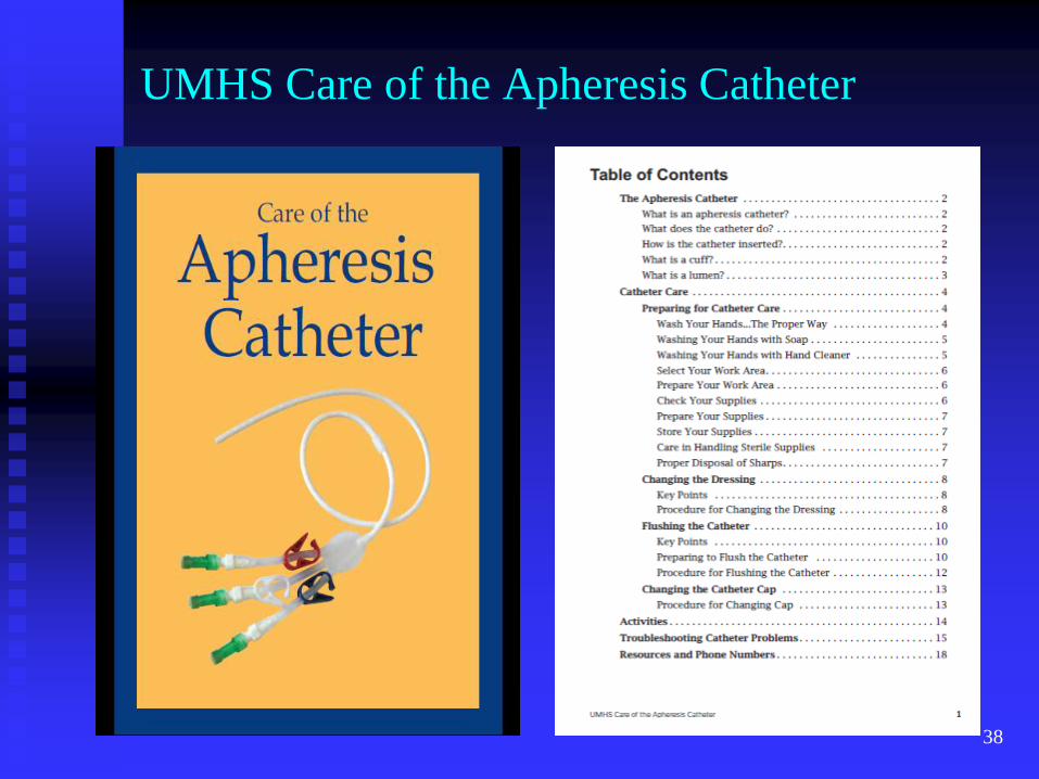
UMHS Care of the Apheresis Catheter
38

Thank you for your attention Questions?
39