variations on a theme: diverse n-acyl homoserine … · variations on a theme: diverse n-acyl...
TRANSCRIPT

Black plate (167,1)
Variations on a theme: diverse N-acyl
homoserine lactone-mediated quorum
sensing mechanisms in Gram-negative
bacteria
Debra Smitha, Jin-Hong Wangb, Jane E. Swattona, Peter Davenporta,
Bianca Pricea, Helga Mikkelsena, Hannah Sticklanda, Kahoko
Nishikawaa,c, Noemie Gardiola, David R. Springd and Martin Welcha
ABSTRACT
Many Gram-negative bacteria employ a mechanism of cell – cell communica-
tion known as quorum sensing (QS). The role of QS is to enable the cells in a
culture to coordinate their gene expression profile with changes in the
population cell density. The best characterized mechanisms of QS employ
N-acylated homoserine lactones (AHLs) as signalling molecules. These
AHLs are made by enzymes known as LuxI homologs, and accumulate in
the culture supernatant at a rate proportional to the increase in cell density.
Once the AHL concentration exceeds a certain threshold value, these ligands
bind to intracellular receptors known as LuxR homologs. The latter are
transcriptional regulators, whose activity alters upon binding the AHL
ligand, thereby eliciting a change in gene transcription. Over the last five
years, it has become increasingly obvious that this is a rather simplistic view
of AHL-dependent QS, and that in fact, there is considerable diversity in the
way in which LuxI-R homologs operate. The aim of the current review is to
describe these variations on the basic theme, and to show how functional
genomics is revolutionizing our understanding of QS-controlled regulons.
Keywords: N-acyl homoserine lactones, biofilms, cell-cell communica-
tion, LuxI, LuxR, quorum sensing, signal transduction
aDepartment of Biochemistry, Building O, Tennis Court Road, Cambridge CB2 1QWbDepartment of Veterinary Medicine, Madingley Road, Cambridge CB3 0EScDepartment of Traumatology and Critical Care Medicine, National Defense MedicalCollege, 3-2 Namiki Tokorozawa, Saitama, 359-8513 JapandDepartment of Chemistry, Lensfield Road, Cambridge CB2 1EW
Science Progress (2006), 89(3/4), 167–211
www.scilet.com 167

Black plate (168,1)
Martin Welch is the author to whom correspondence should be
addressed. He is a Lecturer in Microbiology at Cambridge
University (UK). He received his PhD from the Weizmann
Institute of Science (Israel) in 1994, where he worked on
bacterial chemotaxis. His current research interests focus on
(i) how bacteria respond to antibiotic intervention, (ii) the
development of new antimicrobial agents and quorum sensing
blockers, and (iii) how quorum sensing is controlled during the transition into the
stationary phase. He can be contacted by E-mail at: [email protected]
Introduction: scope of the current review
Over the last decade, it has become increasingly clear that manymicrobes, previously long viewed as being archetypal single-celledorganisms, can coordinate their population behaviour in a processnow known as ‘‘quorum sensing’’ (QS). Cell –cell communicationplays a central role in mediating this cooperative behaviour and thelast few years have witnessed an explosion in research in the area,primarily due to the involvement of QS in the control of virulencefactor production and the formation of antibiotic-insensitivebiofilms by clinically-important pathogens. QS, and the phenotypictraits it controls in different organisms, has been extensivelyreviewed in the past and there are many excellent reports describingthis (see refs. 1–4 for some recent reviews). However, in this review,we aim to emphasize the mechanistic diversity of QS. For reasonsof space limitation and clarity, we will focus mainly on N-acyl-homoserine lactone (AHL)-mediated QS in Gram-negativebacteria, but we will also touch on some other signalling systemsthat impinge on this. QS in Gram-positive organisms has beencomprehensively reviewed recently5,6. The current review is notintended to be exhaustive or fully comprehensive; we cannotpossibly hope to do justice to the full range of QS systems nowbeing investigated. Instead, we will use just a few well-characterizedexamples to illustrate how our preconceptions about QS havechanged in recent years, and in particular, emphasize just howprevalent variations on the basic theme of AHL-mediated QS havebecome.
Cell –cell signalling in Vibrio fischeri: theparadigm for QS
The earliest work on QS as we know it today was done in the 1970sin the lab of Hastings7,8, although the term ‘‘quorum sensing’’ wasnot coined until much later9. Hastings and colleagues were inves-tigating the origins of light production by the Gram-negative
168 Martin Welch et al.
Black plate (169,1)
bioluminescent marine bacterium, Vibrio fischeri. This organism isperhaps best known for its ability to form a symbiotic relationshipwith marine fauna, including several sepiolid squids and mono-centrid fishes, although it is also found in the gut of some marineanimals, and is an occasional pathogen of certain invertebrates. V.fischeri can also exist in the free-living state in sea water, where itlives off suspended and dissolved organic matter, although it rarelyachieves a cell density greater than ca 104 cells per ml in this milieu.In the case of at least one host organism – the Hawaiian bobtailedsquid, Euprymna scalopes – much has been garnered about themechanism of colonization. Newly hatched squid become colonizedby free-living V. fischeri within an hour of hatching. The presenceof free-living bacteria stimulates mucus secretion by the host, andwithin hours, the bacteria form dense aggregates of cells within thismatrix. These cell aggregates migrate through pores into the cryptstructures of the Euprymna light organ. Once there, nurtured by aready source of host-supplied amino acids, the V. fischeri multiply,eventually reaching maximal post-inoculation levels (ca 1010
bacteria per ml) after just 24 h10. By this time, the bacteria in thelight organ bioluminesce strongly, and the squid may exploit thisfeature in a nocturnal counter-illumination strategy designed toevade predator (the role of bioluminescence in V. fischeri hasrecently been discussed11). In the morning, the light organ isperiodically vented, releasing ca 90% of its contents back intothe seawater. The remaining 10% goes on to seed the next cycle ofbioillumination.Hastings et al. found that the lux genes required for light
production by V. fischeri exhibit a pronounced growth phase-dependency in their expression. During the early log-phase ofgrowth, the bacteria exhibited very little lux gene expression, butas the cultures enter the stationary phase of growth, biolumines-cence markedly increases (by a factor of 4104). Crucially, anddifferentiating this expression profile from that of other growth-phase-dependent phenotypes, these workers noted that lux geneexpression could be advanced simply by adding spent stationaryphase culture supernatant to log-phase cells. Moreover, the diffu-sible signal factor responsible for this advancement was species-specific; addition of stationary-phase culture supernatants from V.fischeri to that of a related bioluminescent bacterium, Vibrioharveyi (or vice versa) did not restore light production, so thesignalling molecule(s) involved were denoted ‘‘autoinducers’’. Thatis, the bacteria were concertedly responding en masse to a popula-tion cell density-dependent signal, rather than a growth phase-
www.scilet.com Variations on a theme. 169

Black plate (170,1)
dependent signal. We now know that the autoinducers are N-acylated homoserine lactone (AHL) derivatives; N-(3-oxohexa-noyl)-L-homoserine lactone (OHHL) in the case of V. fischeri,and N-(3-hydroxybutanoyl)-L-homoserine lactone (hydroxyl-BHL)in the case of V. harveyi12,13.The V. fischeri lux genes were subsequently cloned in E. coli14,
where it was found that the bioluminescence functions are encodedin two linked, but divergently transcribed genetic units, separatedby about 150 bp of intergenic DNA. One of these units encodes a250 residue-long transcriptional activator protein, LuxR. Thisprotein is thought to be the receptor for OHHL, and althoughnominally globular and cytoplasmic, is normally found associatedwith the cell membrane. The distal transcriptional unit encodes anoperonic cluster, luxICDABEG. The luxCDABEG structural genesencode the proteins required for production of light per se,although in E. coli, luxG, which encodes an FMN reductase,appears to be dispensable. However, the first gene in the operon,luxI, encodes a protein required for biosynthesis of OHHL, andcan support the synthesis of this molecule when expressed inheterologous hosts. Subsequent work has shown that LuxI utilizesS-adenosylmethionine and 3-oxohexanoyl ACP (probably drainedoff the main pool of fatty acid biosynthetic intermediates15) tosynthesize OHHL. Centered 42.5 bp upstream from the transcrip-tional start site of luxICDABEG is a small (ca 20 bp) invertedrepeat sequence called the lux box, which has been shown to bindactivated (i.e., OHHL-bound) LuxR, initiating transcription of theadjacent luxICDABEG cluster16,17. At the same time, LuxR-binding to this operator region also seems to alter expression ofluxR itself (autoregulation). Proteins homologous to theLuxIyLuxR signal generatoryreceptor pair have been identified ina variety of other organisms, and are frequently arranged in adivergently-transcribed bicistronic pair like that described above.Such LuxIR pairs are not always genetically linked to the genesthey control – this is the case for V. harveyi.The current working model of QS in V. fischeri is that LuxI
constitutively produces OHHL, which passively diffuses across themembrane and accumulates in the culture supernatant at a rateproportional to the increase in population cell density. At the sametime, LuxR is expressed and accumulates intracellularly, bindingOHHL as it does so. Ligand binding is non-cooperative andreversible. Once the bulk AHL concentration exceeds a criticalthreshold value (defined by the Kd (0.1mM) of OHHL for LuxR,and by the expression level of the latter), LuxR undergoes a
170 Martin Welch et al.

Black plate (171,1)
conformational change, which, in turn, leads to a change in themultimeric state of the protein. The multimerized protein thenbinds to the operator region (lux box) upstream of luxICDABEGand stimulates binding of RNA polymerase to the interveningpromoter region17. Detailed analysis of LuxR and its orthologshas been frustrated by the intransigence of these proteins tobiochemical manipulation – most LuxR-type proteins form inso-luble inclusion bodies during over-expression, and do not readilyrefold upon solubilizationyrenaturation. However, molecular-genetic18–21 and more recently, limited biochemical17 analysis ofLuxR has revealed that it is comprised of two domains; an N-terminal OHHL-binding domain, and a C-terminal DNA-bindingdomain containing a helix-turn-helix motif (residues 190–210).Within each domain, mutational analyses have revealed a moresubtle distribution of labour across the molecule, with differentsub-functionalities being associated with defined portions of theLuxR sequence. For example, mutation of residues 10–20 affectsLuxR autoregulation, but not lux box binding. The next 136residues are involved in OHHL binding, since deletion of theseresidues results in OHHL-independent luxICDABEG expression.Residues 79–127 appear to play a role in ligand-binding, whileresidues 116–161 are involved in multimerization. Finally, at theextreme C-terminal end of the molecule, the last 20 residues (230–250) appear to be required for the transcriptional activation ofluxICDABEG, but not for luxR autoregulation. These varioussequence determinants are likely to be generally functionallyconserved across most LuxR-type proteins.
A second class of signal generators
In 1995, a second autoinducer molecule was identified in V. fischeri;N-octanoyl-(L)-homoserine lactone, or OHL22. Intriguingly, thesynthesis of this molecule was found to be catalyzed by a 45.6 kDaprotein, AinS, which has no similarity to LuxI, but does showpartial homology to the LuxM protein from V. harveyii. AinSemploys the same basic substrates for OHL synthesis (SAM andoctanoyl-ACP) as LuxI, although it can also accept CoA deriva-tives in lieu of charged ACPs23. This finding suggests that QSsignalling systems have evolved at least twice during evolutionaryhistory, and that the different systems can be mixed-and-matched.The current model for V. fischeri QS posits that AinS-derived OHLis produced at relatively low cell densities (ca 5108 cells per ml)and is required for the initial stages of light organ colonization10,24
www.scilet.com Variations on a theme. 171

Black plate (172,1)
(Figure 1A,B). At this time, the bacteria do not need to luminesce,so luxICDABEG expression is not activated. Then, via amechanism involving a non-LuxR-type membrane-associatedAHL receptor protein, AinR, and a LuxU-dependent phosphorelaysystem, OHL inactivates LuxO, which normally acts to repress theexpression of litR10,25. The LitR protein is a positive transcriptionalactivator of luxR, and is also required for the expression of earlyfactors necessary for the successful (and species-specific) coloniza-tion of the light organ. At higher cell densities, the induction ofluxR expression by LitR activates the expression of luxICDABEGand the other late-colonization genes. OHL also participatesdirectly in this process since, in the absence of an activating AHLligand, the expressed LuxR will not stimulate luxI expression on itsown. Therefore, even though it binds to LuxR with much loweraffinity than OHHL, the small amount of bound OHL is bothnecessary and sufficient to initiate the positive feedback loop thatrapidly ramps up luxICDABEG expression (Figure 1C). Thus, QSin V. fischeri is a finely-tuned, sequential process that ensures theproper timing of gene expression during colonization.In 2002, the genome sequence of one strain of V. fischeri (ATCC
700601, isolated in the late 1980’s from a Euprymna light organ)was completed, and has yielded a wealth of information about thismodel symbiont and its relationship with host organisms26. Inparticular, the genome sequence opened up the possibility ofusing microarray analysis to define globally the QS regulon(s)controlled by the ain and lux QS systems in V. fischeri10,27, andto screen the genome for genes preceded by potential lux boxoperator sequences. In addition to cataloging which genes areunder the control of QS, these analyses have, by-and-large,confirmed the postulated link between the ain and lux systems,and further, have revealed hitherto unknown QS-controlled regu-lators that likely hardwire QS into other regulatory circuits such asthe rpoS regulon. These and other findings are challenging therather elementary view from the pre-genomic era, that QS systemsare simple modular control units which simply serve to tie in geneexpression to population cell density changes. A good example ofthis is seen in the ongoing work to characterize the interfacebetween QS and virulence in various pathogenic organisms.
QS can be repressive
Members of the Erwinia (Pectobacterium) genus are phytopatho-gens active against a range of plants. One of these, Erwinia
172 Martin Welch et al.

Black plate (173,1)
carotovora subsp. atroseptica (Eca), is responsible for causingblackrot in potato plants and soft-rot in stored tubers28, andconsequently, is of considerable economical importance. Becauseof this, the genome sequence of Eca was recently determined29. Ecais phytopathogenic because it produces a welter of secretedvirulence factors capable of macerating and digesting plant cellwalls, including cellulases (Cels), pectin methylesterases, pectate
www.scilet.com Variations on a theme. 173
Fig. 1. Control of bioluminescence in Vibrio fischeri. (A) In conditions of lowcell density, a phospho-relay system involving LuxQ and LuxO (a signalintegrator) maintain LuxO in a phosphorylated state. LuxO is a transcriptionalactivator which, in the phosphorylated state, increases the expression of several
small RNA species. These, in turn, bind to the litR transcript and target it fordegradation. In the absence of LitR, luxR is not expressed.

Black plate (174,1)
lyases (Pels), pectin lyases, polygalacturonases, and proteases(Prts). As in many other Gram-negative pathogens, the productionof many of these virulence factors is under QS control30,31, makingthis a good target for the development of novel anti-bacterial
174 Martin Welch et al.
Fig. 1. Control of bioluminescence in Vibrio fischeri. (B) At intermediate celldensities (ca 108 cellsyml), AinS synthesizes OHL, which accumulates in the
culture medium. The OHL is sensed by a membrane-associated receptor, AinR,causing it to switch its biochemical activity from being a kinase to being aphosphatase. This decreases the phosphorylation level of LuxU (and conse-
quently, LuxO) leading to an increase in litR expression. LitR activates luxRexpression. The LuxR is able to bind OHL (albeit with low affinity), therebyactivating it. This allows the LuxR-OHL complex to start stimulating the
expression of the lux genes, including luxI.

Black plate (175,1)
strategies32. In line with this, QS mutants of Eca show reducedvirulence factor production and reduced virulence in planta. Onlyone QS signal generator, ExpI, has been identified experimentallyin Eca, and analysis of the genome sequence suggests that this is theonly AHL synthase present29,30,31. Like LuxI, ExpI producesOHHL (along with smaller quantities of the non-3-oxo derivative,HHL), although there is some strain-to-strain variation in thenature of the AHL produced (summarized in reference 33), and
www.scilet.com Variations on a theme. 175
Fig. 1. Control of bioluminescence in Vibrio fischeri. (C) At high cell densities,the LuxI protein generates OHHL, which binds to LuxR with high affinity. The
LuxR-OHHL complex positively auto-regulates luxI expression, thereby rapidlyramping up the expression of the lux gene cluster, giving rise to bioluminescence.

Black plate (176,1)
at least two broad classes of AHL-producing Erwiniae have beenidentified34. For the purposes of clarity, we will restrict ourdiscussion here to QS in the sequenced strain of Eca (SCRI1043);the mechanistic details of QS in other Erwinia strains variesconsiderably, and has been reviewed recently33. The expI gene ispart of a convergently transcribed bicistronic unit, the othercomponent of which is a luxR homolog, expR (Figure 2). The 3’ends of each gene overlap slightly, indicating that they areevolutionarily tightly linked. However, the precise role of ExpRin virulence was questioned for many years because knockouts ofthe corresponding gene had very little impact on any aspect ofvirulence in many strains of Erwinia35,36. In contrast, inactivationof expI essentially abrogated virulence36. This issue was resolved bythe finding (from genome interrogation29) that Eca contains anadditional LuxR homolog, ECA1561, now denoted VirR36. Aswith expR, inactivation of virR had very little effect on virulencefactor production. However, when introduced into an expI mutant(expI and virR are unlinked), the virR mutation fully restoredexoenzyme production, even though OHHL production was abol-ished. This suggested – contrary to expectation – that VirRnormally represses exoenzyme production (presumably throughbinding to some lux-type box at the 5’ end of target genes, orperhaps by controlling the level of some intermediate regulatorsuch as rsmA37,38). A working model is that at low cell densities,VirR binds to operator sequences upstream from its target genes(e.g. celV, pelC, pehA, nip and svx) and prevents their expression,but as the cell density rises, ExpI-derived OHHL complexes withVirR and relieves this repression (Figure 2C). VirR has also beenimplicated in controlling the expression of rsmA, another globalregulator of exoenzyme production36,37,38, and it is possible thatmany of the phenotypic effects of virR mutants are mediated viadisruption of RsmA-mediated signalling (Figure 2A and B). Therole of ExpR in this, if any, is not yet clear, although ExpR hasbeen shown to exert some control over rsmA expression in certainstrains of Erwinia37. Also, recent studies using purified ExpR haveshown that the protein is certainly capable of binding OHHL,albeit with relatively low affinity (Kd¼ 7.5 mM)39. This alone wasgood ‘‘smoking gun’’ evidence that another LuxR homolog wasinvolved, since virulence factor production can be induced at muchlower OHHL concentrations (51 mM). This notwithstanding, thediscovery of VirR is a good example of how genomics can assist inelucidating the components of a QS system. Equally, these findingsalso highlight the value of searching for QS-bypass mutants (i.e.,
176 Martin Welch et al.

Black plate (177,1)
those mutants that restore AHL-dependent phenotypes in luxIhomolog mutants).Eca is not the only phytopathogen to exhibit QS-dependent
negative regulation; indeed, the first such example was describedin Pantoea stewartii subsp. stewartii (Pss), the causative agent ofStewart’s wilt in corn crops40. The disease is caused by accumula-tion of an exopolysaccharide, stewartan, which is thought to blockxylem vessels in the plant, leading to loss of turgor pressure andsubsequently, wilt. Stewartan is a complex polymer composed ofheptameric oligosaccharide repeat units containing glucose, galac-
www.scilet.com Variations on a theme. 177
Fig. 2. Quorum sensing control of exoenzyme production in the plant pathogen,Erwinia carotovora. (A) At low cell density, VirR activates the expression ofRsmA. This protein binds to the exoenzyme transcripts and targets them for
degradation. RsmA is also thought to inhibit the expression of expI.

Black plate (178,1)
tose and glucuronic acid, and is synthesized by a large 14-genecluster (encoding the cps genes). Expression of the cps genes iscontrolled partly by QS; esaI and esaR are convergently transcribedORFs encoding the AHL synthaseyreceptor pair in Pss, and partlyby a multicomponent phosphorelay signal transduction systemcomprised of RcsA, RcsB, RcsC and YoiN41,42. Deletion of esaIresults in abolition of OHHL synthesis by the organism, and loss of
178 Martin Welch et al.
Fig. 2. Quorum sensing control of exoenzyme production in the plant pathogen,Erwinia carotovora. (B) At higher cell densities, other regulatory inputs over-
come the inhibitory effect of RsmA on expI expression, leading to the synthesis ofOHHL. The AHL binds to VirR, causing it to dissociate from the RsmApromoter, thereby reducing RsmA levels and de-repressing exoenzyme synthesis.
The precise role of ExpR, which is also capable of binding OHHL, is not yetknown.

Black plate (179,1)
virulence. However, deletion of esaR or deletion of esaR and esaItogether results in a hypermucoid phenotype in which stewartan isoverexpressed. Subsequent studies have shown that EsaR directlyrepresses the transcription of rcsA. The product of this gene, RcsA,along with RcsB, is required for the activation of cps transcription.In the absence of bound EsaR, RcsA probably positively auto-
www.scilet.com Variations on a theme. 179
Fig. 2. Quorum sensing control of exoenzyme production in the plant pathogen,
Erwinia carotovora. (C) An alternative hypothesis for VirR action is that at lowcell densities, this protein binds directly to the target gene (exoenzyme)promoters and represses expression from these. At higher cell densities,OHHL made by ExpI binds to VirR, causing it to dissociate from the target
promoters, thereby allowing exoenzyme expression.

Black plate (180,1)
regulates itself, but in the repressed state, the concentration of anyRcsA that is made by leaky expression is probably kept in checkby Lon-mediated turnover, keeping the system tightlycontrolled41. EsaR has also been shown to have an autoregulatoryrole, providing further proof for its QS inhibitory activity42. EsaRis one of the few LuxR homologs for which there is goodbiochemical data, and experiments have shown that EsaRdimerizes and binds, independent of OHHL, to an operatorsequence which spans the � 10 region of a s70 promoterconsensus sequence upstream of esaR. This binding is relievedin the presence of OHHL, indicating that, like VirR in Erwinia,EsaR primarily operates as a repressor42.QS-repressive activity has been identified in other systems too (to
date, all of them in the enterobacteriaciae). One of the bestcharacterized of these is in the opportunistic pathogen, Serratiamarcescens, which infects a range of mammalian, insect and planthosts. Many strains of this organism produce copious quantities ofvirulence factors, including secreted secondary metabolites andexoenzymes (such as hemolytic proteases, lipases, nucleases, andchitinases), and these phenotypes are often found to be under QScontrol43,44. In addition, in strain ATCC39006, production of thevivid red pigment, prodigiosin, by a 15-gene biosynthetic cluster(pigA-O) is under QS control45 (Figure 3). ATCC39006 contains aconvergently-transcribed LuxI-R pair denoted smaI and smaR.Inactivation of smaI leads to loss of pigmentation, while inactiva-tion of smaR has no effect on this phenotype. However, pigmentproduction in a smaI–smaR double mutant is restored, indicatingthat SmaR normally represses pigment production at low celldensities, and that this repression is relieved upon binding theAHL ligand45. In line with this, purified SmaR was shown to bindto a DNA fragment encompassing the promoter region upstreamof the carA gene (which is known to be under the control of QS inthis strain), and this binding was essentially abolished in thepresence of AHL46. Intriguingly, although b-galactosidase expres-sion from a pigA::lacZ transcriptional fusion was reduced in a smaImutant, and restored by addition of exogenous AHL, theexogenous signalling molecule did not act to advance expressionof the pig cluster45. Clearly, more is being sensed here than just thepopulation cell density, and the precise timing of pigment produc-tion is influenced by other factors. Recent work has indicated thatpigment production is also subject to regulation by multipleenvironmental cues, mediated by a diverse set of regulatoryproteins including a GacAS-like two-component system, an
180 Martin Welch et al.

Black plate (181,1)
adenylate cyclase, and several pleiotropic regulators of regulators(such as Rap and PigP)46.Unlike E. carotovora and V. fischeri, the SmaI protein encoded
by Serratia sp. ATCC39006 primarily produces butyryl-(L)-homo-
www.scilet.com Variations on a theme. 181
Fig. 3. Quorum sensing in Serratia sp: orphan regulators and surrogate pheno-types. At low cell densities, the SmaR protein represses expression of the pigA-O
cluster and of the carR gene. At higher cell densities, BHL produced by the SmaIprotein accumulates in the culture and binds to SmaR. This causes the latter todissociate from the pig promoter, allowing production of the prodigiosin biosyn-
thetic genes. At the same time the BHL-independent CarR protein binds to thepromoter region in front of the carA-H gene cluster, activating expression of thecarbapenem biosynthetic genes. Additional, multiple regulatory inputs also
impinge on expression of the car and pig clusters.
Black plate (182,1)
serine lactone (BHL)47, rather than OHHL, and this is reflected bythe altered ligand specificity of SmaR45,46. Recent work aimed atengineering an altered specificity in LuxR48 has shed light on howthis altered specificity might have arisen. Presumably, smaI musthave picked up mutations that influenced the ligand it makes(presumably by changing the sizeynature of the acyl-ACP bindingsite). In turn, the smaR gene must have undergone a parallelselection to ensure that it responded appropriately to the newligand. This could have arisen by horizontal transfer of a newluxR homolog with the correct ligand specificity, but given the verytight linkage between smaI and smaR – the genes overlap and areconvergently transcribed – it is far more likely that the existingSmaR underwent a contemporaneous change of ligand specificityitself. [This does not mean that this change in specificity originallyoccurred in a Serratia genetic background – indeed, inATCC39006, the smaIR locus is flanked by remnants of insertionsequences, suggesting that this locus was mobile as a unit in thedistant past47,49.] The recent work with LuxR has provided anexplanation as to how this might have occurred. The most likelyfirst step will have been a relaxation in the specificity of SmaR toenable it to recognize and respond to a range of AHLs. Acquisitionof such ‘‘promiscuous’’ functionality50 is a feature of many evolu-tionary intermediates during ‘‘respecialization’’; it is rare for asingle mutation to confer a new function and at the same time,completely abolish an older one. However, the broader specificityof the intermediate state would almost certainly incur a fitness cost,in which case, there would be a strong selective pressure to accruemutations that minimize the deleterious nature of these, and indoing so, increase the specificity of the protein. Collins et al.48
recently exploited this dual selection route to engineer a LuxRprotein with specificity for decanoyl-(L)-homoserine lactone(DHL) instead of OHHL.
Surrogate phenotypes and orphan regulators
In both Serratia sp. ATCC39006 and in Erwinia carotovora subsp.carotovora (Ecc) strain ATTn10, the QS system controls not onlyvirulence factor production, but also the biosynthesis of a beta-lactam antibiotic, 1-carbapen-2-em-3-carboxylic acid (car). Thisantibiotic is thought to be made in order to reduce competitionfor the nutritional windfall created at the site of an infection. Thegenes required for carbapenem production are encoded by an 8gene cluster denoted carA-H51. The first 5 of these genes, carA-E,
182 Martin Welch et al.

Black plate (183,1)
are required for carbapenem biosynthesis, while carF and carGencode an apparently novel resistance mechanism necessary toprevent the cell from killing itself upon induction of carbapenemproduction. No function has yet been assigned to carH. The carcluster is preceded by 150 bp intergenic region containing a s70-likepromoter and a range of invertedydirect repeats that are candidatebinding sites for regulatory factors. Within the cluster, a weakconstitutive promoter exists between carE and carF, probablymaintaining a constant low-level stream of pre-formed resistanceproteins. However, a much stronger, QS-dependent promoter islocated at the 5’ end of the cluster. Interestingly, in both SerratiaATCC39006 and in Ecc ATTn10, the carA-H genes are locatedadjacent to a divergently transcribed luxR homolog, denotedcarR52 (Figure 3). In the case of Ecc, CarR is a potent OHHL-dependent activator of carbapenem production, and has beenshown to bind directly to the carR-carA intergenic region53.Indeed, CarR was the first LuxR homolog to be purified andbiochemically characterized in any great detail53. The purifiedprotein bound OHHL with a Kd of 1.8 mM, which is within therange of OHHL concentrations (1–3 mM) normally produced bythis organism when grown in LB. Ligand binding was monitoredby exploiting the ability of AHLs to alter the fluorescence of ahighly conserved Trp residue in CarR, and calculations showedthat 1 OHHL molecule was bound per monomeric unit of theprotein. Recent biophysical data have confirmed this (MW andDRS, data not shown). The cognate LuxI homolog which generatesthe OHHL to which CarR responds is unlinked to carR, andcorresponds to the expI gene in Eca (although in Ecc it is known,for historical reasons, as carI). The CarR protein thereforeappeared to be an orphan LuxR homolog, and since CarR is notknown to control any other phenotypes in Ecc, it looks as if thecarRycarA-H locus has been introduced into Ecc from elsewhere(by horizontal gene transfer) and has come under the control of theendogenous QS system. Carbapenem production in Ecc is thereforea ‘‘surrogate’’ QS phenotype. Whether the cluster arrived in anOHHL-responsive state, or whether CarR evolved to becomeresponsive to endogenous OHHL, is not clear. However, althoughnot all strains of Ecc have the potential to produce carbapenem, anumber of them carry ‘‘cryptic’’ carbapenem clusters which can beactivated to produce the antibiotic when functional ATTn10 carRis expressed in trans54. Most of these cryptic strains have apparentlylost their ability to make carbapenem due to the presence of defectsin the endogenous carR genes or in the carR promoter region.
www.scilet.com Variations on a theme. 183

Black plate (184,1)
Whatever the reason for the persistence of this evolutionarywreckage, it shows that it is relatively straightforward to acquirenew surrogate QS-controlled functions – a feature that we willreturn to presently.As in Ecc, carabapenem production in Serratia ATCC39006 is
also controlled by CarR. However, early studies indicated that theSerratia CarR operates in a ligand-independent manner. TheSerratia CarR protein contains a Trp?Cys substitution in itsligand binding site. This eliminates the Trp residue most likelyresponsible for causing AHL-dependent quenching of intrinsic Trpfluorescence in Ecc CarR39,53. Indeed, in a subsequent X-raycrystallographic analysis of the tertiary complex formed betweenTraR (a LuxR homolog from Agrobacterium tumefaciens – seebelow), its cognate AHL (3-oxo-octanoyl-(L)-homoserine lactone,or OOHL), and a target DNA sequence, Vannini et al.55 and Zhanget al.56 independently showed that this highly conserved Trpresidue plays a central role in binding the homoserine lactonemoiety of AHLs53. However, recent mutational analysis of LuxRhas challenged the generality of this conclusion, suggesting insteadthat the Trp side chain may make contacts with the 3-oxo moiety inligand-bound LuxR57. Unexpectedly, in Serratia, expression of thisAHL-independent CarR protein results in AHL-dependent carba-penem production45. That is, in either a carR mutant, or in a smaImutant, transcription of the carA-H cluster is abolished. Slater andcolleagues45 have now elegantly elucidated much of the molecularbasis for this, and have proposed a plausible model for how carRexpression might be controlled by SmaR. In essence, at low celldensities, they propose that SmaR binds to the carR promoterregion, repressing carR transcription. However, at higher celldensities, BHL accumulates in the culture and binds to SmaR,causing it to dissociate from the carR promoter, thereby relievingcarR repression and allowing synthesis of the antibiotic.One simple, but very illuminating recent study has shown just
how easy it is to acquire surrogate QS-dependent phenotypesthrough phage-mediated horizontal gene transfer58. In pigmentedSerratia marcescens, the pig gene cluster is always located betweencueR and copA, whereas in non-pigmented strains, cueR and copAare contiguous, implying that at some ancestral stage, the pigcluster inserted itself between these genes. Sma 274 is a pigmentedenvironmental Serratia isolate which does not carry out AHL-dependent QS, while Sma 12 is a non-pigmented clinical isolatewhich contains an active QS system, but no pig cluster. Coulthurstet al. used a generalized transducing phage, F3M, to introduce the
184 Martin Welch et al.

Black plate (185,1)
pig cluster from Sma 274 into Sma 12. Remarkably, they foundthat not only was the pig cluster expressed in Sma 12, but it alsocame under the control of the endogenous QS system. Conversely,when these workers introduced the smaR gene from Sma 12 intothe non-QS strain Sma 274, they found that pig gene expressioncould be made dependent on exposure to exogenous BHL. Theauthors speculate that SmaR-mediated repression of some pleio-tropic regulator present in all Serratiasmight be responsible for thisapparently facile imposition of QS control. Whatever themechanism involved, this little study illustrates just how easy it isto introduce QS, and therefore population cell density-dependentcontrol of a phenotypic trait. It also suggests that QS is not alwaysstrictly necessary for control, raising the question of just howwidespread the phenomenon of QS really is.LuxIR orthologs have now been identified in a range of a, b and
g Proteobacteria, where they have been shown to be responsible forcontrolling a diversity of phenotypic traits. However, relatively fewProteobacteria (just 4% of genera from all orders at the lastcount59) are known to exploit AHLs for QS, and most of theseare limited to just 3 orders; the enterobacteriaciae, pseudomonaciae,and the rhizobiales. This is remarkable if we accept that the lastcommon ancestor of all five Proteobacterial classes probablycarried a LuxIR-type QS system60, suggesting that over thecourse of time, this particular signalling mechanism has beenlargely dumped. Intriguingly, those species that have kept AHL-mediated QS systems are mostly pathogens, indicating that QS is agood way of controlling virulence factor production.
Extrachromosomal QS systems
So far, all of the QS systems described above have been encoded onthe bacterial chromosome. However, in some cases, such asAgrobacterium tumefaciens and in certain Rhizobial species, QSsystem(s) are plasmid encoded. A. tumefaciens is a more insidiousphytopathogen than the Erwiniaciae; it transfers oncogenic T-DNAfrom tumor-inducing (Ti)-plasmids directly into the nucleus of host(dicotyledonous) plant cells, where it stimulates overproduction ofplant growth hormones and consequent rapid neoplastic tissueproliferation to generate a crown gall tumor61,62. Some 21 virgenes, encoded in six operonic units (denoted virA-G) are locatedon the Ti plasmid and are required for T-DNA processing andtransfer. The expression of these genes is induced by the presence ofplant-derived signals, including pH, monosaccharides and certain
www.scilet.com Variations on a theme. 185
Black plate (186,1)
phenolic compounds (reviewed in ref. 62). In addition, other geneson the Ti plasmid direct the host cell to produce a novel class ofsignalling compound called opines (derived from sugars and aminoacids). The two main classes of opines that concern us here are thenopalines (e.g. agrocinopines) and octopines, and the synthesis ofthese is directed by subtly-different types of Ti-plasmid. Theseopines serve not only as a nutrient source for the bacteria; theyare also plant-derived signals which stimulate Ti plasmid transfer tonearby plasmid-less, avirulent bacteria. These arise because A.tumefaciens that have lost their Ti plasmids have a strong growthadvantage and so soon out-compete organisms that have retainedtheir plasmids; as such, they are evolutionary ‘‘cheats’’ whichpiggy-back on the nutritional windfall created by their plasmid-bearing partners63. However, this process is reversed in late stagegall formation by opine-stimulated conjugal Ti plasmid transferbetween bacterial donors and recipients. Octopines and agrocino-pines bind to opine-responsive transcription factors (OccR andAccR, respectively), thereby relieving repression of the respectiveocc and accyarc operons (Figure 4). These operons mostly containgenes involved in the uptake and catabolism of opines, but inaddition, they also contain a luxR homolog called traR. Therefore,synthesis of the AHL receptor protein is under the control of apathogen-directed host-derived signal molecule (the opine),ensuring that conjugal transfer can only occur in the environmentof the plant tumor. It is of interest to note that the simple insertionof traR into the arc or occ operons introduces an element of cell-density control into this system, highlighting the importance ofgene context.The target genes of TraR are encompassed in two divergently
transcribed tra operons, and in the trb and rep operons. Each ofthese operonic units is preceded by an 18 bp operator region knownas a tra box, analogous to the lux box described earlier64–67. Thetra and trb operons encode functions required for conjugal transfer,while the rep operon encodes the plasmid replication machinery.The first gene within the trb operon (in both nopaline and octopineTi plasmids) is a luxI homolog, traI 66,67. The encoded TraI proteinmakes OOHL, which is the cognate AHL recognized by TraR. Inthe apo-state, TraR is a membrane-associated monomer64.However, the protein is unstable in the absence of the ligand andis rapidly degraded by endogenous proteases65. This is prevented inthe presence of OOHL64,65. It is thought that as the nascent TraRprotein is being extruded from the ribosome, it folds around theAHL molecule, forming an essentially irreversible, but stable active
186 Martin Welch et al.

Black plate (187,1)
www.scilet.com Variations on a theme. 187
Fig. 4. Quorum sensing control of plasmid transfer functions in Agrobacter-
ium. tumefaciens. For simplicity, only nopaline signalling is illustrated, but thesame principles also apply to opine-mediated signalling (see text for details).Plant (host) cells containing T-DNA produce agrocinopine signals which are
taken up by the Agrobacteria. Once inside the cells, the nopaline binds to theAccR receptor protein, causing it to dissociate from the promoter controllingexpression of the arc-traR operon on the Ti-plasmid. This permits expression of
TraR, which, in the monomeric form is membrane-associated and rapidlyturned over by endogenous proteolytic degradation processes. Low-level con-stitutive expression of traI, the gene encoding the OOHL synthase, eventuallygenerates sufficient AHL to allow binding to the TraR. This causes the latter to
dimerize and stabilize, and the resulting transcriptional activator stimulatesexpression of the trayrep functions required for conjugative plasmid trans-feryreplication. TraR also stimulates expression of a negative regulator, TraM
(see text for details).

Black plate (188,1)
complex68. Not all LuxR homologs exhibit this type of beha-viour – others show straightforward reversible binding to theAHL molecules39,42,53. Once bound to OOHL, the protein under-goes a conformational change and dimerizes, permitting the HTHmotif to bind the major groove of the tra box, thereby stimulatingtranscription of the downstream genes55,56. Given that TraRcontrols TraI expression, these proteins positively cooperate torapidly ramp up expression of the genes required for conjugal Tiplasmid transfer, and ensure that conjugation becomes dependenton the population cell density of the donor cells. Quite how the celldensity of plasmid-less recipients is measured – if this is at allimportant for effective conjugal transfer – is not clear, since thesecells do not produce OOHL and therefore should be effectively‘‘invisible’’ to any quorum count. The whole system is driven by thefact that traR and traI both exhibit a low level of constitutiveexpression; in the complete absence of OOHL, TraR would not beable to function as a transcriptional activator. In the pre-quorateperiod, active TraR levels are kept in check by TraM. This smallanti-activator protein, whose expression is under the control ofTraR, binds to the C-terminal HTH-domain in the latter, effec-tively inactivating it until (it is thought) active TraR levels out-titrate TraM70. TraR activity has also been shown to be antag-onized under certain circumstances by TrlR; this LuxR homologcontains a frameshift at residue 182, but encodes a functional,OOHL-binding N-terminus that forms inactive heterodimers withTraR71,72. No other luxIR homologues have been identified in thechromosome of the nopaline A. tumefaciens ‘‘genome’’ strain, C58.In 2000, Zhang and colleagues announced that they had identi-
fied an enzyme in a Bacillus sp., denoted AiiA (for ‘‘autoinducerinactivation’’) that was capable of hydrolyzing the homoserinelactone ring moiety in AHLs73,74. Recent biochemical and struc-tural analyses75,76,77 of AiiA have established that this enzyme is azinc metallo-enzyme, although some workers contest this78.Expression of AiiA in transgenic plants protected them fromattack by Erwinia carotovora74. This exciting finding proved thatsuch ‘‘quorum quenching’’ enzymes hold great biotechnologicalpotential, and confirm QS as an excellent target for antimicrobialintervention. Two years later, the same team showed that anorthologous enzyme, denoted AttM, was able to degrade OOHLin A. tumefaciens79. AttM forms part of an operon comprised ofattK, attL and attM. Expression of this operon is under the controlof the IclR-like regulator, AttJ, and the same workers subsequentlydemonstrated that the relA-mediated stringent response also
188 Martin Welch et al.

Black plate (189,1)
imposes on this system80. However, recent findings81,82 havebrought this area of research full-circle by showing that g-aminobutyric acid (GABA), a plant-derived metabolite produced duringwounding, is able to induce AttJ-dependent transcription of theattKLM cluster, thereby preventing Ti-plasmid conjugal transfer.Quite what advantage this confers upon the bacterium is not clear.AHL-degrading systems have now been identified in a diversevariety of bacteria83,84,85, and not all of them are lactonases86,87,88.One protein that holds particular intrigue is PvdQ fromPseudomonas aeruginosa, which acts as a potent quorum quenchervia its AHL acylase activity89,90.Although the A. tumefaciens QS system may seem convoluted, it
is actually relatively straightforward compared to the systemsinvolved in controlling phenotypes including conjugal plasmidtransfer in some Rhizobiales [reviewed recently in ref. 91]. Forexample, in R. leguminosarum, the cinI-cinR locus has been shownto be a master regulator for at least three subordinate AHL-dependent QS systems (raiI– raiR, rhi– rhiR, and traI– triR),making this one of the most complex QS organisms currentlyknown, and a world away from the simplistic LuxI–LuxR para-digm of just a decade ago.
QS in Pseudomonas aeruginosa – time for aconceptual re-think?
QS in Pseudomonas aeruginosa represents the ultimate pot pouri ofsignalling systems discussed so far; it is hierarchical, hardwired toother regulons, exploits a variety of signalling molecules, andinvolves an orphan LuxR homolog (QscR) that controls its own,discrete set of surrogate phenotypes. P. aeruginosa has become aparadigm for the study of long-term, chronic bacterial infections,although it is also associated with certain acute infections. P.aeruginosa has been associated with some 10% of all nosocomial(hospital-acquired) infections, and of the Gram-negative organ-isms, seems to be the worst offender in this regard92. As succinctlypointed out by Todar in his concise little curriculum vitae for theorganism93, P. aeruginosa is the epitome of an opportunisticpathogen; it rarely causes infections in healthy hosts, but in theimmune-compromised, there is hardly any tissue that it will notattack. This is particularly true for cystic fibrosis (CF) patients,who, by virtue of their defective CFTR pump, accumulate largeamounts of viscous mucus in the lung tissue, which is readilycolonized by P. aeruginosa and members of the B. cepacia
www.scilet.com Variations on a theme. 189

Black plate (190,1)
complex . Nowadays, most CF patients succumb to the effects ofthese long-term, chronic infections, which are a leading cause ofmortality and morbitity in this population. QS plays a central rolein P. aeruginosa pathogenicity, and as a consequence, perhaps moreis known about AHL-signalling in this organism than any other.AHLs have been readily isolated from the sputum of CF patients94,and QS mutants display reduced virulence in model systems95,96.Interestingly, although the presence of a functional QS systemseems to be pre-requisite for effective colonization, it is notrequired for persistence in the host, since many Pseudmonas isolatesfrom CF sputum appear to have at least partially lost their abilityto do QS.Several excellent reviews on P. aeruginosa QS have been
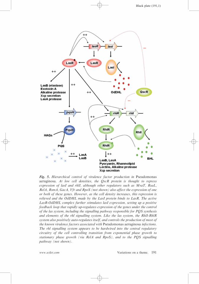
presented recently, and the reader is referred to these for a detaileddescription of the system and the various phenotypic traits underits control2,92,97. Briefly, the QS system in P. aeruginosa is hier-archical, and consists of two AHL-dependent signalling systems,both luxI– luxR homologs (Figure 5). At the top of the regulatoryhierarchy is LasI, which makes 3-oxo-dodecanoyl-(L)-homoserinelactone (OdDHL), and the cognate receptor for this ligand, LasR.On binding OdDHL, LasR multimerizes and becomes active as atranscriptional regulator. Until recently, genes under the control ofthe las system were thought to be preceded by operator-likepalindromic las boxes about 20 bp in length. However, newdata98 indicates that many las boxes lack dyad symmetry, anddisplay notable variation in their LasR-binding properties, parti-cularly with respect to cooperative associations. The heterogenousnature of las operator sequences may indicate that they areresponsive to more subtle variations in LasR concentra-tionyactivity than previously thought. In addition, there is goodevidence that LasR can also repress the expression of certain targetgenes (Wagner et al. identified 222 genes that are QS-repressed99).Subordinate to the las system, and controlled by it, is the RhlI–
RhlR signalling pair (rhl sub-system), which is involved in thegeneration and reception of BHL. The rhl signalling system wasprobably acquired by P. aeruginosa after the las system49. Incontrast to LasR, RhlR requires its cognate ligand for activity asa transcriptional activator, but not for dimerization100. Unlike thegenes controlled by the las system, RhlR promoters absolutelyrequire dyad symmetry in the corresponding rhl box operatorsequences. Many rhl-regulated genes also accept a regulatoryinput from the las system, but it is interesting to note that mostof the virulence-related genes in the P. aeruginosa QS regulon are
190 Martin Welch et al.
Black plate (191,1)
www.scilet.com Variations on a theme. 191
Fig. 5. Hierarchical control of virulence factor production in Pseudomonasaeruginosa. At low cell densities, the QscR protein is thought to repressexpression of lasI and rhlI, although other regulators such as MvaT, RsaL,
RelA, RsmA, GacA, Vfr and RpoS (not shown) also affect the expression of oneor both of these genes. However, as the cell density increases, this repression isrelieved and the OdDHL made by the LasI protein binds to LasR. The active
LasR-OdDHL complex further stimulates lasI expression, setting up a positivefeedback loop that rapidly up-regulates expression of the genes under the controlof the las system, including the signalling pathway responsible for PQS synthesisand elements of the rhl signalling system. Like the las system, the RhlI-RhlR
system also positively auto-regulates itself, and controls the production of most ofthe known virulence factors associated with Pseudomonas aeruginosa infections.The rhl signalling system appears to be hardwired into the central regulatory
circuitry of the cell controlling transition from exponential phase growth tostationary phase growth (via RelA and RpoS), and to the PQS signallingpathway (not shown).

Black plate (192,1)
primarily rhl-controlled97. On the basis of this, Schuster andGreenberg97 have proposed that acquisition of the rhl system wastied in with the adoption of a more pathogenic lifestyle by thisorganism. It is interesting to note that BHL and OdDHL are verydifferent in size and degree of acyl-chain substitution, ensuring thatthese signalling systems do not cross-talk to any appreciable extentthrough inappropriate ligand-binding to the ‘‘wrong’’ LuxRhomolog. Together, the LasI–LasR and RhlI–RhlR signallingsystems control the expression of a large number of phenotypictraits including secretion of extracellular virulence factors such asexotoxins, proteolytic haemolysins, phospholipases and secondarymetabolites (Figure 5), and crucially, also biofilm formation101,102.In the last few years, a third signalling factor, the Pseudomonasquinolone signal (PQS) has become implicated in linking the lasand rhl systems, and probably controls its own subset of pheno-typic traits103–106. A parallel PQS signalling system has recentlybeen reported in Burkholderia sp107. The synthesis of PQS is underthe positive control of the las system, but is negatively controlled bythe rhl system, indicating that the ratio between OdDHL and BHLis critical for its production. PQS is derived from 4-hydroxy-2-alkylquinolones made by the pqsABCDE and phnAB operons,whose expression is controlled by PqsR. The product of thesesteps, 4-hydroxy-2-heptylquinolone, is then converted to PQS byPqsH, whose expression is controlled by LasR. Like OdDHL, PQSis a hydrophobic molecule that is relatively insoluble in aqueoussolutions. However, recently, Mashburn and Whiteley108 reportedthat PQS (but not the AHLs) can be shuttled between individualcells in small vesicles budded off from the cell surface. This vesiclebudding was itself stimulated by PQS production, since it wasabsent in a mutant (pqsH) unable to make PQS. It seems likely thatthe insertion of PQS itself into the outer membrane can stimulatevesicle formation and budding.Plasmodium aeruginosa may be unique in terms of the number of
independent microarray studies that have been done in order tocharacterize the genes under QS control. In three microarraystudies99,109,110, 866 genes were identified (in total) as being QS-induced. This is a conservative figure since one of these studies (thatof Hentzer et al.109) employed a more stringent cut-off (5-fold) thanthe others. However, although the individual studies identifiedbetween 3 and 7% of the genome as being QS-modulated, theoverlap between them was small, with only 97 genes being found incommon. This common set of genes may represent a ‘‘core’’ QSregulon which is expressed in most growth conditions. Moreover,
192 Martin Welch et al.

Black plate (193,1)
the study of Wagner et al. identified4200 genes as being repressed,whereas Schuster et al. only found 38 QS-repressed genes. This mayreflect differences in either the growth conditions employed, ordifferences in the way in which a gene was defined as being QS-regulated. Also, the very large numbers of genes identified in thesemicroarray studies as being QS-regulated may be misleading;transcriptomic analysis alone cannot discriminate between causeand effect, and a large number of the modulated genes are likely tobe only indirectly controlled by QS. Indeed, just 7% of the QS-controlled genes identified by Wagner et al. were preceded by las orrhl boxes99. Although this can be partially accounted for by thepresence of multi-gene operons, each associated with just a singlelasyrhl box, it is noteworthy that ca 9% of all QS-controlled genesin that study were classified as transcriptional regulators or two-component systems of some kind. Therefore, many of the genesidentified as being supposedly ‘‘QS-regulated’’ are likely to becontrolled indirectly by other pleiotropic regulators97. Even rela-tively minor changes in growth conditions may therefore bringabout some variation in the apparent QS regulon99, reinforcing theremarkable adaptability of this organism to environmental chal-lenges111. It seems that in P. aeruginosa, QS has become so deeplyembedded into the existing regulatory networks that it has becomehardwired to the organism’s ‘‘CPU’’ machinery.Another key feature to come out of the microarray analyses
relates to the timing of signal molecule production; not all QS-controlled genes were induced at the same point in the growthcurve110. That is, the ‘‘traditional’’ view of QS in which geneexpression is triggered once a defined AHL concentration isexceeded does not hold here, and there seems to be a continuumof both signal specificities and timing of gene expression in thisorganism110. Furthermore, QS mutants grown in the presence ofsaturating concentrations of AHLs showed similar gene expres-sion timing to the wild-type strain, indicating that the trigger forQS gene activation was not the signal molecule concentration perse. This is reminiscent of events required for progress through theeukaryotic cell cycle – checkpoint steps, which ensure that inap-propriate gene expression is suppressed until the cell is ready tocommit to e.g. virulence factor production. To complicate theissue further, it seems that certain complex media contain a P.aeruginosa-metabolizable substance which inhibits AHL-depen-dent gene expression, which may partially explain the (verylarge) differences between the apparent QS regulons defined fordifferent media112.
www.scilet.com Variations on a theme. 193

Black plate (194,1)
P. aeruginosa also contains an ‘‘orphan’’ LuxR homolog,denoted QscR. Mutants in qscR are hypervirulent, suggestingthat this protein operationally functions as a repressor113. QscRforms heterodimers with LasR and RhlR, and there is goodevidence to suggest that the protein binds OdDHL114. This situa-tion is reminiscent of the TraR-TrlR interaction observed in A.tumefaciens, where TrlR effectively ‘‘titrates’’ TraR and therebysuppresses any phenotypes under its control. However, unlikeTrlR, QscR contains a functional DNA-binding domain, indicatingthat it is capable of controlling the expression of target genes itself.Indeed, studies with purified QscR have shown that it can bindoperator regions with dyad symmetry (ca 20 bp in length) asso-ciated with the promoters of certain genes (e.g. PA1897, adjacent toqscR itself), and that this binding is enhanced by OdDHL115.Therefore, in addition to functioning as a repressor, QscR canalso activate gene expression. Clearly, unlike the rhl sub-system,which is dependent on the las sub-system at a transcriptional level,the QscR regulon is subordinate to the las system because of itsdependency on the OdDHL ligand produced by LasI. Dependingon the promoter, binding affinities vary from 0.3?4 nM, and mayor may not exhibit weak cooperativity115. QscR-dependent promo-ters exhibit similarities to LasR-dependent promoters, although thetwo are clearly distinct. However, QscR appears to have a relaxedsignal specificity compared to LasR115, and may even bind ODHL(3-oxo-decanoyl-(L)-homoserine lactone) with higher affinity thanOdDHL, suggesting that this protein may also respond to signalsproduced by other organisms. A recent study defined the QscRregulon, by using microarray analysis to compare the transcrip-tome of a qscR mutant with that of the wild-type116. Some 424genes showed altered expression – most of them appearing to berepressed by QscR, although, consistent with the in vitro biochem-ical characterization of the protein, the expression of 76 genesappeared to be activated. As might be expected, the QscR regulonsubstantially overlaps those of LasR and RhlR, although withinthis, a subset of genes also appear to be exclusively regulated byQscR itself. Quite how QscR fits in to the overall QS system is notyet clear, although it probably functions to tightly repress QS-controlled virulence factor production during the early (pre-quorate) stages of growth.Perhaps the most important QS-controlled phenotype in P.
aeruginosa is biofilm formation. Although the precise role ofbiofilms in P. aeruginosa pathogenicity has not yet been estab-lished, the more-or-less constant inflammatory response stimulated
194 Martin Welch et al.

Black plate (195,1)
by the presence of bacterial cells in the lung tissue plays a majorrole in the progressive lung degeneration seen in many CF patients.Viewed close-up, P. aeruginosa biofilms grown in flow cells exhibita three-dimensional architecture composed of mushroom-liketowers interspersed by water-filled channels117,118, although thereis considerable variation in biofilm morphology from strain-to-strain and in different growth conditions118. The cells that comprisethe biofilm are embedded within a secreted exopolysccharidematrix consisting largely of alginate, sometimes augmented byprotein and nucleic acids, which apparently acts to glue thewhole structure together117,119. The release of genomic DNAfrom Pseudomonas biofilms is dependent on the AHL and PQSsignalling systems119, and may play a role in adaptive evolution.P. aeruginosa biofilms go through a well-defined series of
developmental stages before achieving their final structural config-uration120. Briefly, planktonic founder cells first establish thecolony by adhering to the solid substratum. The micro-assemblagethen proliferates and subsequently differentiates, in a multi-stepprocess known as ‘‘maturation’’. This is often followed by thedispersal of new planktonic seed cells from discrete pockets on themushroom-like structures. These maturation steps often involveadditional sculpting, which in one case, has been shown to bemediated by a prophage-dependent remodeling process121. Indeed,it is interesting to speculate why biofilms have not evolved furtherinto ‘‘true’’ (i.e., genetically pre-determined, distinct) multicellularentities. Presumably the answer lies in the fact that once formed, amulticellular organism cannot ‘‘dissociate’’ into its componentparts and seed new habitats, whereas biofilm cells can. That is,phenotypic plasticity seems to be favored over phenotypic determi-nation. Mutants affected in QS, particularly the las component ofthe QS system, form flat, undifferentiated biofilms101. However,one recent report has challenged the role of the las system in thisand has posited that the subordinate rhl system makes a greatercontribution to biofilm formation102. Whatever the case, drug-likeQS blockers would almost certainly reduce biofilm formation invivo, and therefore increase the fraction of the bacterial populationthat are susceptible to conventional antibiotic intervention.Promising work done by the GivskovyHoiby groups in Denmarkhas identified a number of furanone-like small molecules thatapparently block QS in P. aeruginosa and indeed, attenuatevirulence in mouse models in vivo and reduce biofilm formationin vitro122,123,124. Several other groups are also working on the
www.scilet.com Variations on a theme. 195

Black plate (196,1)
development of small molecule QS blockers, and this has beenreviewed recently in reference (125).P. aeruginosa-derived AHLs also have pathogenicity-related
effects on the host immune system. For example, OdDHL hasbeen shown to stimulate T-cells to produce interferon-g.Furthermore, and in one of the most exciting developments inthe field in recent years, the P. aeruginosa outer membrane protein,OprF, was found to be a receptor for this cytokine126,127.Interferon-g was found to up-regulate a number of QS phenotypes,and this up-regulation was abolished in rhlI and rhlR mutants. Thisindicated that the binding of IFN-g to OprF leads, by mechanismsunknown, to an up-regulation of rhl-dependent phenotypes(including virulence factor production). In this way, a cycle ofcommunication between the pathogen and the host immune cells isset up, rapidly ramping up the production of virulence factors.Other immune-modulatory effects of OdDHL have also beenreported (reviewed recently in (128).
Biofilms and the possible origins of QS
QS has now been shown to control biofilm formation in manymicrobes, and the Centers for Disease Control (CDC) has recentlyestimated that around 80% of chronic bacterial diseases involvethese structures129. This notwithstanding, remarkably, there is stillcontroversy over precisely what biofilms are. Are they distinctgrowth states, or are they simply extreme versions of an otherwisecontinuum of growth phenotypes? What is their relationship to say,colonies on an agar plate? The latter do not need QS to form,although certain QS mutants of P. aeruginosa often display small-colony morphologies (MW, unpublished observations). Also, thequestion of whether biofilms secrete the same spectrum of virulencefactors as planktonic cells has yet to be addressed. Presumably, theanswers to these fascinating problems will be forthcoming in thefuture. However, rather more is known about how biofilm cellsdiffer from planktonic cells, mainly as a result of several detailedtranscriptomic analyses. For example, in the case of P. aeruginosa,Whitley et al.130 found that the expression of just 78 genes (out of atotal complement of ca 5600 ORFs) was altered between exponen-tial-phase planktonic and mature biofilm cells. This resultsuggested that sessile biofilm cells resemble actively-growing expo-nential-phase cells in most respects, although this remains to beproven. As these authors acknowledge though, biofilms are hetero-genous communities that are likely to exhibit different gene
196 Martin Welch et al.

Black plate (197,1)
expression profiles depending on where in the biofilm the sample isderived. In one detailed study of this issue, it was found that lasIexpression diminishes in biofilms over time, while rhlI expressionremains essentially unaffected. Moreover, both genes are expressedto a greater level at the base of the biofilm, adjacent to thesubstratum131. In contrast, global proteomic analyses of biofilmand planktonic cell cultures present a different story; about half ofthe P. aeruginosa soluble proteome is altered in mature biofilmsrelative to their planktonic counterparts120. This lack of congru-ence between transcriptomic and proteomic studies is notuncommon. However, although it is accepted that most regulationoccurs at the transcriptional level in prokaryotes, the functionalmolecules in the cells are proteins and metabolites, and there arearguments for both technologies being complimentary to oneanother. Interestingly, there are very few reports of global meta-bolomic analysis being applied to the study of biofilms and QS,although studies along these lines are well-underway in the author’slaboratories. These data indicate that QS mutants exhibit well-defined metabolomes, and we can readily discriminate between say,wild-type cells on the one hand, and lasI mutants, rhlI mutants orpqsR mutants on the other, on the basis either of their secretedsecondary metabolome, or their primary metabolome (MW &PWD, unpublished observations). These observations reinforcethe notion that QS has global effects and that it substantiallyimpinges on the central metabolic pathways of the cell.Biofilms need not be composed of a single species of organism.
Indeed, mixed biofilms are probably the norm (especially inenvironmental isolates), and it is becoming increasingly clear thatas many as 99% of all species of bacteria are probably unculturablewhen grown alone132. This raises an interesting issue with regardsto the definition of an ecological ‘‘niche’’, since a niche can nolonger be considered to be the habitat occupied by just one species.Mixed biofilms of ‘‘unculturables’’ potentially permit sharing of themetabolic burden associated with colonization of the niche,allowing greater adaptive flexibility. That is, metabolic productsproduced by one cell can potentially diffuse across to a differentcell where they are metabolized further, and vice versa. Cell –cellcommunication, both through direct physical contact and viadiffusible chemical signals, must play a key role in the developmentof this cooperative behaviour since, at the very least, the playersnecessarily need to coordinate their replication; if one of thepartners in this intercellular metabolic relay dies, so will theother, providing a strong selection pressure for the evolution of
www.scilet.com Variations on a theme. 197

Black plate (198,1)
intercellular signals. That is, the primordial intercellular signal(s)may have arisen from say, a shared metabolite within a mixedbiofilm. A good example of this type of signal is seen with the P.aeruginosa siderophore, pyoverdine, which can function as both aniron scavenger and a signal molecule133. Winzer et al.134 haveprovided a good working definition for a cell –cell signallingmolecule, arguing that 4 simple conditions must be met before amolecule can be regarded as a true signalling agent. Perhaps mostimportantly, they point out that the cellular response, post-recep-tion of the signal, must extend beyond the physiological change(s)required to metabolize or detoxify the compound. Furthermore, fora signal molecule to be classed as a quorum sensing signalling agent,its production needs to be coupled to population growth in theculture. These are crucial points, especially given the recentcontroversy over ‘‘autoinducer-2’’.
Bacterial esperanto? The controversy over AI-2
As outlined in an earlier section, the control of bioluminescence inV. harveyi is under the control of AHL-dependent QS. However,this description is rather overly simplistic since it ignores thecontribution of autoinducer-2 (‘‘AI-2’’), which regulates biolumi-nescence in conjunction with N-3-hydroxybutanoyl-(L)-homo-serine lactone. AI-2 is made by the LuxS enzyme, which hassince been identified in numerous species of bacteria135,136,including pathogens such as V. cholerae, where it has beenimplicated in the control of virulence factor production137. Thefunction of LuxS is to hydrolyze S-ribosylhomocysteine, generatinghomocysteine and 4,5-dihydroxy-2,3-pentanedione (DPD)138,139.As such, it plays an important role in the activated methyl cycleof these cells, since S-ribosylhomocysteine is obtained fromdemethylation of S-adenosylmethionine. Not all bacteria exploitthis pathway, and some, such as P. aeruginosa, use an S-adeno-sylhomocysteine hydrolase to circumvent this step, cleaving SAHto generate homocysteine and adenosine. The role played by DPDand LuxS in central metabolism has frustrated the characterizationof AI-2 as a signal molecule, since luxS mutants would be expectedto be pleiotropically affected in metabolism. [This said, in normallaboratory culture conditions, luxS mutants rarely displayprofound growth defects, although in their natural environment,the selection pressure is likely to be dramatically different.] Onesuggestion has been that while AHLs serve as intra-species QSmolecules, AI-2 could serve as an inter-species signalling agent,
198 Martin Welch et al.
Black plate (199,1)
perhaps functioning to inform those cells with appropriate recep-tors for this molecule about how crowded the neighborhood mightbe, or telling the organism when it has entered a particularenvironmental niche (e.g. the gut). For example, there is goodexperimental evidence to suggest that P. aeruginosa responds to theAI-2 made by other host microflora140. Another possibility, likelyto hold for organisms such as the Vibrios, is that AI-2 acts as partof a ‘‘coincidence’’ sensor, allowing multiple signalling inputs to besimultaneously assessed136. However, at least one study, involvingSerratia marcescens141, showed that there is considerable strain-to-strain variation in the phenotypes of luxS mutants, even within agiven ‘‘species’’, indicating that we are only just scratching thesurface of the potential biology involved.DPD is only a precursor for AI-2, and needs to undergo non-
enzymatic rearrangement to generate the active signallingcompound(s). However, this inter-conversion can generate severaldifferent molecular species (all of them furanones), depending onthe conditions prevailing at the time, so the term AI-2 should beconstrued as referring to a collection of related compounds142.Reinforcing this, two different DPD-derived compounds have beenidentified in co-crystals of AI-2 receptors. In the structure of the V.harveyi LuxPyAI-2 complex, AI-2 appeared to be a furanosylborate diester138. However, in the structure of the Salmonellatyphimurium LsrB protein complexed with AI-2143, the signallingmolecule appeared to be (2R,4S)-2-methyl-2,3,3,4-tetrahydroxyter-ahydrofuran (‘‘R-THMF’’). This molecule does not contain boron,even though, during its preparation for crystallization, there wouldhave been ample opportunity for borate to become complexed withthe ligand. It seems then, that AI-2 is a heterogenous collection ofmolecular species, although these are all dependent upon thepresence of a functional LuxS protein. Whether subtle alterationsto the ligand turn it from being a metabolic intermediate into asignalling molecule remain to be seen. In theory, DPD is a goodprecursor for a QS molecule, since its synthesis, being tightly linkedto a central metabolic flux, ought to be proportional to the rate ofincrease in the cell density. The signal transduction pathwaysmediated by AI-2 in S. typhimurium and V. harveyi have beenreviewed recently142. These systems have so far fallen into twocategories. One, exemplified by signalling in the Vibrios, involvesthe perception of extracellular AI-2 and the subsequent activationof a signal transduction cascade. The other system, characterized inE. coli and several other enterics, involves the uptake of AI-2 intothe cell and its subsequent metabolism. Likewise, P. aeruginosa,
www.scilet.com Variations on a theme. 199

Black plate (200,1)
which does not encode luxS, is also capable of taking up andmetabolizing exogenous AI-2140. It is tempting to speculate thatthese catabolic systems evolved, at least in part, as a means ofscavenging AI-2 made by other organisms. In relation to this,several bacterial species have been shown to not only degrade, butalso to utilize the AHL signalling molecules produced by othermicrobes as a sole C-source (e.g. see ref. 89). This suggests that themetabolism of signal molecules generally may be a widespreadphenomenon.
Concluding remarks
The last 5 years or so have seen a veritable explosion in ourknowledge and understanding of QS in Gram-negative bacteria.Much of this progress has been driven by the ready availability oftechnological platforms that enable facile transcriptional andproteomic profiling. The challenge now will be to try and puttogether the various (currently, rather disparate) studies andgenerate useful, holistic models of QS in various organism(s).Furthermore, by its very nature, many functional genomic analysesare ‘‘hypothesis-generating’’ rather than ‘‘hypothesis-testing’’(although this is not always the case), and it is becoming clearthat a good deal of molecular analysis lies ahead before we willtruly understand the systems concerned. The aim of this review is tohighlight the differences between QS systems, rather than theirsimilarities. If the last few years have taught us anything, it is thatthis diversity is substantial, and much remains to be learned aboutthis fascinating signalling paradigm.
Acknowledgements
Work in the MW and DRS laboratories is supported by theBBSRC, EPSRC and Royal Society. HM wishes to thankAmerada Hess (Faroes) Ltd., BP Amoco Exploration (Faroes)Ltd. and Statoil Færøyene AyS for generous financial support.KN is supported by grants from the Japanese Association forAcute Medicine and from the Isaac Newton Trust (Cambridge).
References
1. Reading, N. C. and Sperandio, V. (2006) Quorum sensing: the manylanguages of bacteria. FEMS Microbiol. Lett., 254, 1–11.
200 Martin Welch et al.

Black plate (201,1)
2. Soberon-Chavez, G., Aguirre-Ramirez, M. and Ordonez, L. (2005) IsPseudomonas aeruginosa only ‘‘sensing quorum’’? Crit. Rev. Microbiol.,
31, 171–182.3. Whitehead, N. A., Barnard, A. M., Slater, H., Simpson, N. J. and
Salmond, G. P. (2001) Quorum-sensing in Gram-negative bacteria.FEMS Microbiol. Rev., 25, 65–404.
4. West, S. A., Griffin, A. S., Gardner, A. and Diggle, S. P. (2006) Socialevolution theory for microorganisms. Nat. Rev. Microbiol., 4, 597–607.
5. Lyon, G. J. and Novick, R. P. (2004) Peptide signalling in Staphylococcus
aureus and other Gram-positive bacteria. Peptides, 25, 1389–1403.6. Sturme, M. H., Kleerebezem, M., Nakayama, J., Akkermans, A. D.,
Vaugha, E. E. and de Vos, W. M. (2002) Cell to cell communication by
autoinducing peptides in Gram-positive bacteria. Antonie Van Leeuwen-hoek, 81, 233–243.
7. Nealson, K. H., Platt, T. and Hastings, J. W. (1970) Cellular control of the
synthesis and activity of the bacterial luminescent system. J. Bacteriol.,104, 313–322.
8. Eberhard, A. (1972) Inhibition and activation of bacterial luciferasesynthesis. J. Bacteriol., 109, 1101–1105.
9. Fuqua, W. C., Winans, S. C. and Greenberg, E. P. (1994) Quorum sensingin bacteria: the LuxR–LuxI family of cell density-responsive transcrip-tional regulators. J. Bacteriol., 176, 269–275.
10. Lupp, C. and Ruby, E. G. (2005) Vibrio fischeri uses two quorum-sensingsystems for the regulation of early and late colonization factors. J.Bacteriol., 187, 3620–3629.
11. Ruby, E. G. and Lee, K. H. (1998) The Vibrio fischeri-Euprymna scolopeslight organ association: current ecological paradigms. Appl. Environ.Microbiol., 64, 805–812.
12. Eberhard, A., Burlingame, A. L., Eberhard, C., Kenyon, G. L., Nealson,
K. H. et al. (1981) Structural identification of autoinducer of Photobacter-ium fischeri. Biochemistry, 20, 2444–2449.
13. Cao, J. G. and Meighen, E. A. (1989) Purification and structural identi-
fication of an autoinducer for the luminescence system of Vibrio harveyi. J.Biol. Chem., 264, 21670–21676.
14. Engebrecht, J. and Silverman, M. (1984) Identification of genes and gene
products necessary for bacterial bioluminescence. Proc. Natl. Acad. Sci.USA, 81, 4154–4158.
15. Parsek, M. R., Val, D. L., Hanzelka, B. L., Cronan, J. E. Jr. and
Greenberg, E. P. (1999) Acyl homoserine-lactone quorum-sensing signalgeneration. Proc. Natl. Acad. Sci. USA, 96, 4360–4365.
16. Fuqua, C., Winans, S. C. and Greenberg, E. P. (1996) Census andconsensus in bacterial ecosystems: the LuxR–LuxI family of quorum-
sensing transcriptional regulators. Ann. Rev. Microbiol., 50, 727–751.17. Urbanowski, M. L., Lostroh, C. P. and Greenberg, E. P. (2004) Reversible
acyl-homoserine lactone binding to purified Vibrio fischeri LuxR protein.
J. Bacteriol., 186, 631–637.18. Hanzelka, B. L. and Greenberg, E. P. (1995) Evidence that the N-terminal
region of the Vibrio fischeri LuxR protein constitutes an autoinducer-
binding domain. J. Bacteriol., 177, 815–817.
www.scilet.com Variations on a theme. 201

Black plate (202,1)
19. Stevens, A. M., Dolan, K. M. and Greenberg, E. P. (1994) Synergisticbinding of the Vibrio fischeri LuxR transcriptional activator domain and
RNA polymerase to the lux promoter region. Proc. Natl. Acad. Sci. USA,91, 12619–12623.
20. Stevens, A. M. and Greenberg, E. P. (1997) Quorum sensing in Vibriofischeri: essential elements for activation of the luminescence genes. J.
Bacteriol., 179, 557–562.21. Egland, K. A. and Greenberg, E. P. (2001) Quorum sensing in Vibrio
fischeri: analysis of the LuxR DNA binding region by alanine-scanning
mutagenesis. J. Bacteriol., 183, 382–386.22. Gilson, L., Kuo, A. and Dunlap, P. V. (1995) AinS and a new family of
autoinducer synthesis proteins. J. Bacteriol., 177, 6946–6951.
23. Hanzelka, B. L., Parsek, M. R., Val, D. L., Dunlap, P. V., Cronan, J. E.Jr. and Greenberg, E. P. (1999) Acylhomoserine lactone synthase activityof the Vibrio fischeri AinS protein. J. Bacteriol., 181, 5766–5770.
24. Lupp, C., Urbanowski, M., Greenberg, E. P. and Ruby, E. G. (2003) TheVibrio fischeri quorum-sensing systems ain and lux sequentially induceluminescence gene expression and are important for persistence in thesquid host. Mol. Microbiol., 50, 319–331.
25. Fidopiastis, P. M., Miyamoto, C. M., Jobling, M. G., Meighen, E. A. andRuby, E. G. (2002) LitR, a new transcriptional activator in Vibrio fischeri,regulates luminescence and symbiotic light organ colonization. Mol.
Microbiol., 45, 131–143.26. Ruby, E. G., Urbanowski, M., Campbell, J., Dunn, A., Faini, M.,
Gunsalus, R., Lostroh, P., Lupp, C., McCann, J., Millikan, D., Schaefer,
A., Stabb, E., Stevens, A., Visick, K., Whistler, C. and Greenberg, E. P.(2005) Complete genome sequence of Vibrio fischeri: a symbiotic bacter-ium with pathogenic congeners. Proc. Natl. Acad. Sci. USA, 102, 3004–3009.
27. Callahan, S. M. and Dunlap, P. V. (2000) LuxR- and acyl-homoserine-lactone-controlled non-lux genes define a quorum-sensing regulon inVibrio fischeri. J. Bacteriol., 182, 2811–2822.
28. Toth, I. K., Bell, K. S., Holeva, M. C. and Birch, P. R. J. (2003) Soft roterwiniae: from genes to genomes. Mol. Plant Pathol., 4, 17–30.
29. Bell, K. S., Sebaihia, M., Pritchard, L., Holden, M. T., Hyman, L. J.,
Holeva, M. C. et al. (2004) Genome sequence of the enterobacterialphytopathogen Erwinia carotovora subsp. atroseptica and characteriza-tion of virulence factors. Proc. Natl. Acad. Sci. USA, 101, 11105–
11110.30. Jones, S., Yu, B., Bainton, N. J., Birdsall, M., Bycroft, B. W., Chhabra, S.
R., Cox, A. J., Golby, P., Reeves, P. J., Stephens, S., Winson, M. K.,Salmond, G. P. C., Stewart, G. S. A. B. and Williams, P. (1993) The lux
autoinducer regulates the production of exoenzyme virulence determi-nants in Erwinia carotovora and Pseudomonas aeruginosa. EMBO J., 12,2477–2482.
31. Pirhonen, M., Flego, D., Heikinheimo, R. and Palva, E. T. (1993) A smalldiffusible signal molecule is responsible for the global control of virulenceand exoenzyme production in the plant pathogen Erwinia carotovora.
EMBO J., 12, 2467–2476.
202 Martin Welch et al.
Black plate (203,1)
32. Toth, I. K., Newton, J. A., Hyman, L. J., Lees, A. K., Daykin, M., Ortori,C., Williams, P. and Fray, R. G. (2004) Potato plants genetically modified
to produce N-acylhomoserine lactones increase susceptibility to soft roterwiniae. Mol. Plant Microbe Interact., 17, 880–887.
33. Barnard, A. M. and Salmond, G. P. (2006) Quorum sensing in Erwiniaspecies. Anal. Bioanal. Chem. (in press).
34. Chatterjee, A., Cui, Y., Hasegawa, H., Leigh, N., Dixit, V. and Chatterjee,A. K. (2005) Comparative analysis of two classes of quorum-sensingsignalling systems that control production of extracellular proteins and
secondary metabolites in Erwinia carotovora subspecies. J. Bacteriol., 187,8026–8038.
35. Andersson, R. A., Eriksson, A. R., Heikinheimo, R., Mae, A., Pirhonen,
M., Koiv, V. et al. (2000) Quorum sensing in the plant pathogen Erwiniacarotovora subsp. carotovora: the role of expR(Ecc). Mol. Plant MicrobeInteract., 13, 384–393.
36. Burr, T., Barnard, A. M., Corbett, M. J., Pemberton, C. L., Simpson, N.J. and Salmond, G. P. (2006) Identification of the central quorum sensingregulator of virulence in the enteric phytopathogen, Erwinia carotovora:the VirR repressor. Mol. Microbiol., 59, 113–125.
37. Cui, Y., Chatterjee, A., Hasegawa, H., Dixit, V., Leigh, N. and Chatterjee,A. K. (2005) ExpR, a LuxR homolog of Erwinia carotovora subsp.carotovora, activates transcription of rsmA, which specifies a global
regulatory RNA-binding protein. J. Bacteriol., 187, 4792–4803.38. Cui, Y., Chatterjee, A., Hasegawa, H. and Chatterjee, A. K. (2006)
Erwinia carotovora subspecies produce duplicate variants of ExpR,
LuxR homologs that activate rsmA transcription but differ in theirinteractions with N-acylhomoserine lactone signals. J. Bacteriol., 188,4715–4726.
39. Welch, M., Dutton, J. M., Glansdorp, F. G., Thomas, G. L., Smith, D. S.,
Coulthurst, S. J., Barnard, A. M., Salmond, G. P. and Spring, D. R.(2005) Structure-activity relationships of Erwinia carotovora quorumsensing signalling molecules. Bioorg. Med. Chem. Lett., 15, 4235–4238.
40. Coplin, D. L. and Majerczak, D. R. (1990) Extracellular polysaccharidegenes in Erwinia stewartii: directed mutagenesis and complementationanalysis. Mol. Plant-Microbe Interact., 3, 286–292.
41. Minogue, T. D., Carlier, A. L., Koutsoudis, M. D. and von Bodman, S. B.(2005) The cell density-dependent expression of stewartan exopolysacchar-ide in Pantoea stewartii ssp. stewartii is a function of EsaR-mediated
repression of the rcsA gene. Mol. Microbiol., 56, 189–203.42. Minogue, T. D., Wehland-von Trebra, M., Bernhard, F. and von
Bodman, S. B. (2002) The autoregulatory role of EsaR, a quorum-sensingregulator in Pantoea stewartii ssp. stewartii: evidence for a repressor
function. Mol. Microbiol., 44, 1625–1635.43. Wei, J. R. and Lai, H. C. (2006) N-acylhomoserine lactone-dependent cell-
to-cell communication and social behaviour in the genus Serratia. Int. J.
Med. Microbiol., 296, 117–124.44. Harris, A. K., Williamson, N. R., Slater, H., Cox, A., Abbasi, S., Foulds,
I., Simonsen, H. T., Leeper, F. J. and Salmond, G. P. (2004) The Serratia
gene cluster encoding biosynthesis of the red antibiotic, prodigiosin, shows
www.scilet.com Variations on a theme. 203

Black plate (204,1)
species- and strain-dependent genome context variation. Microbiology,150, 3547–3560.
45. Slater, H., Crow, M., Everson, L. and Salmond, G. P. (2003) Phosphateavailability regulates biosynthesis of two antibiotics, prodigiosin andcarbapenem, in Serratia via both quorum-sensing-dependent and -inde-pendent pathways. Mol. Microbiol., 47, 303–320.
46. Fineran, P. C., Slater, H., Everson, L., Hughes, K. and Salmond, G. P.(2005) Biosynthesis of tripyrrole and beta-lactam secondary metabolites inSerratia: integration of quorum sensing with multiple new regulatory
components in the control of prodigiosin and carbapenem antibioticproduction. Mol. Microbiol., 56, 1495–1517.
47. Thomson, N. R., Crow, M. A,, McGowan, S. J., Cox, A. and Salmond, G.
P. (2000) Biosynthesis of carbapenem antibiotic and prodigiosin pigmentin Serratia is under quorum sensing control. Molec. Microbiol., 36, 539–556.
48. Collins, C. H., Leadbetter, J. R. and Arnold, F. H. (2006) Dual selectionenhances the signalling specificity of a variant of the quorum-sensingtranscriptional activator LuxR. Nat. Biotechnol., 24, 708–712.
49. Lerat, E. and Moran, N. A. (2004) The evolutionary history of quorum-
sensing systems in bacteria. Molec. Biol. Evolut., 21, 903–913.50. Collins, C. H., Arnold, F. H. and Leadbetter, J. R. (2005) Directed
evolution of Vibrio fischeri LuxR for increased sensitivity to a broad
spectrum of acyl-homoserine lactones. Molec. Microbiol., 55, 712–723.51. McGowan, S. J., Sebaihia, M., O’Leary, S., Hardie, K. R., Williams, P.,
Stewart, G. S. A. B., Bycroft, B. W. and Salmond, G. P. C. (1997)
Analysis of the carbapenem gene cluster of Erwinia carotovora: definitionof the antibiotic biosynthetic genes and evidence for a novel beta-lactamresistance mechanism. Molec. Microbiol., 26, 545–556.
52. McGowan, S., Sebaihia, M., Jones, S., Yu, B., Bainton, N., Chan, P. F.,
Bycroft, B., Stewart, G. S. A. B., Williams, P. and Salmond, G. P. C.(1995) Carbapenem antibiotic production in Erwinia carotovora is regu-lated by CarR, a homologue of the LuxR transcriptional regulator.
Microbiology, 141, 541–550.53. Welch, M., Todd, D. E., Whitehead, N. A., McGowan, S. J., Bycroft, B.
W. and Salmond, G. P. (2000) N-acyl homoserine lactone binding to the
CarR receptor determines quorum-sensing specificity in Erwinia. EMBOJ., 19, 631–641.
54. Holden, M. T., McGowan, S. J., Bycroft, B. W., Stewart, G. S., Williams,
P. and Salmond, G. P. C. (1998) Cryptic carbapenem antibiotic produc-tion genes are widespread in Erwinia carotovora: facile trans activation bythe carR transcriptional regulator. Microbiology, 144, 1495–1508.
55. Vannini, A., Volpari, C., Gargioli, C., Muraglia, E., Cortese, R., De
Francesco, R., Neddermann, P. and Marco, S. D. (2002) The crystalstructure of the quorum sensing protein TraR bound to its autoinducerand target DNA. EMBO J., 21, 4393–4401.
56. Zhang, R. G., Pappas, T., Brace, J. L., Miller, P. C., Oulmassov, T.,Molyneaux, J. M., Anderson, J. C., Bashkin, J. K., Winans, S. C. andJoachimiak, A. (2002) Structure of a bacterial quorum-sensing transcrip-
tion factor complexed with pheromone and DNA. Nature, 417, 971–974.
204 Martin Welch et al.

Black plate (205,1)
57. Koch, B., Liljefors, T., Persson, T., Nielsen, J., Kjelleberg, S. and GivskovM. (2005) The LuxR receptor: the sites of interaction with quorum-sensing
signals and inhibitors. Microbiology, 151, 3589–3602.58. Coulthurst, S. J., Williamson, N. R., Harris, A. K. P., Spring, D. R. and
Salmond G. P. C. (2006) Metabolic and regulatory engineering of Serratiamarcescens: mimicking phage-mediated horizontal acquisition of antibio-
tic biosynthesis and quorum-sensing capacities. Microbiology, 152, 1899–1911.
59. Manefield, M. and Turner, S. L. (2002) Quorum sensing in context: out of
molecular biology and into microbial ecology. Microbiology, 148, 3762–3764.
60. Gray, K. M. and Garey, J. R. (2001) The evolution of bacterial LuxI and
LuxR quorum sensing regulators. Microbiology, 147, 2379–2387.61. Zhu, J., Oger, P. M., Schrammeijer, B., Hooykaas, P. J., Farrand, S. K.
and Winans, S. C. (2000) The bases of crown gall tumorigenesis. J.
Bacteriol., 182, 3885–3895.62. Brencic, A. and Winans, S. C. (2005) Detection of and response to signals
involved in host-microbe interactions by plant-associated bacteria. Micro-biol. Molec. Biol. Rev., 69, 155–194.
63. Von Bodman, S. B., Bauer, W. D. and Coplin, D. L. (2003) Quorumsensing in plant-pathogenic bacteria. Ann. Rev. Phytopathol., 41, 455–482.
64. Qin, Y., Luo, Z. Q., Smyth, A. J., Gao, P., von Bodman, S. and Farrand,
S. K. (2001) Quorum-sensing signal binding results in dimerization ofTraR and its release from membranes into the cytoplasm. EMBO J., 19,5212–5221.
65. Zhu, J. and Winans, S. C. (1999) Autoinducer binding by the quorum-sensing regulator TraR increases affinity for target promoters in vitro anddecreases TraR turnover rates in whole cells. Proc. Natl. Acad. Sci. USA,96, 4832–4837.
66. Fuqua, W. C. and Winans, S. C. (1994) A LuxR-LuxI type regulatorysystem activates Agrobacterium Ti plasmid conjugal transfer in the pre-sence of a plant tumor metabolite. J. Bacteriol., 176, 2796–2806.
67. Hwang, I., Li, P. L., Zhang, L., Piper, K. R., Cook, D. M. et al. (1994)TraI, a LuxI homologue, is responsible for production of conjugationfactor, the Ti plasmid N-acylhomoserine lactone autoinducer. Proc. Natl.
Acad. Sci. USA, 91, 4639–4643.68. Zhu, J. and Winans, S. C. (2001) The quorum-sensing transcriptional
regulator TraR requires its cognate signalling ligand for protein folding,
protease resistance, and dimerization. Proc. Natl. Acad. Sci. USA, 98,1507–1512.
69. Fuqua, C., Burbea, M. and Winans, S. C. (1995) Activity of the Agro-bacterium Ti plasmid conjugal transfer regulator TraR is inhibited by the
product of the traM gene. J. Bacteriol., 177, 1367–1373.70. Hwang, I., Smyth, A. J., Luo, Z. Q. and Farrand, S. K. (1999) Modulating
quorum sensing by antiactivation: TraM interacts with TraR to inhibit
activation of Ti plasmid conjugal transfer genes.Mol. Microbiol., 34, 282–294.
71. Oger, P., Kim, K. S., Sackett, R. L., Piper, K. P. and Farrand, S. K. (1998)
Octopine-type Ti plasmids code for a mannopine-inducible dominant-
www.scilet.com Variations on a theme. 205

Black plate (206,1)
negative allele of traR, the quorum-sensing activator that regulates Tiplasmid conjugal transfer. Molec. Microbiol., 27, 277–288.
72. Chai, Y., Zhu, J. and Winans, S. C. (2001) TrlR, a defective TraR-like protein of Agrobacterium tumefaciens, blocks TraR function invitro by forming inactive TrlR:TraR dimers. Molec. Microbiol., 40,414–421.
73. Dong, Y. H., Xu, J. L., Li, X. Z. and Zhang, L. H. (2000) AiiA, an enzymethat inactivates the acylhomoserine lactone quorum-sensing signal andattenuates the virulence of Erwinia carotovora. Proc. Natl. Acad. Sci. USA,
97, 3526–3531.74. Dong, Y. H., Wang, L. H., Xu, J. L., Zhang, H. B., Zhang, X. F. and
Zhang, L. H. (2001) Quenching quorum-sensing-dependent bacterial
infection by an N-acyl homoserine lactonase. Nature, 411, 813–817.75. Thomas, P. W., Stone, E. M., Costello, A. L., Tierney, D. L. and Fast, W.
(2005) The quorum-quenching lactonase from Bacillus thuringiensis is a
metalloprotein. Biochemistry, 44, 7559–7569.76. Liu, D., Lepore, B. W., Petsko, G. A., Thomas, P. W., Stone, E. M., Fast,
W. and Ringe, D. (2005) Three-dimensional structure of the quorum-quenching N-acyl homoserine lactone hydrolase from Bacillus thuringien-
sis. Proc. Natl. Acad. Sci. USA, 102, 11882–11887.77. Kim, M. H., Choi, W. C., Kang, H. O., Lee, J. S., Kang, B. S., Kim, K. J.,
Derewenda, Z. S., Oh, T. K., Lee, C. H. and Lee, J. K. (2005) The
molecular structure and catalytic mechanism of a quorum-quenching N-acyl-L-homoserine lactone hydrolase. Proc. Natl. Acad. Sci. USA, 102,17606–17611.
78. Wang, L. H., Weng, L. X., Dong, Y. H. and Zhang, L. H. (2004)Specificity and enzyme kinetics of the quorum-quenching N-Acyl homo-serine lactone lactonase (AHL-lactonase). J. Biol. Chem., 279, 13645–13651.
79. Zhang, H. B., Wang, L. H. and Zhang, L. H. (2002) Genetic control ofquorum-sensing signal turnover in Agrobacterium tumefaciens. Proc. Natl.Acad. Sci. USA, 99, 4638–4643.
80. Zhang, H. B., Wang, C. and Zhang, L. H. (2004) The quormonedegradation system of Agrobacterium tumefaciens is regulated by starva-tion signal and stress alarmone (p)ppGpp. Molec. Microbiol., 52, 1389–
1401.81. Wang, C., Zhang, H. B., Wang, L. H. and Zhang, L. H. (2006) Succinic
semialdehyde couples stress response to quorum-sensing signal decay in
Agrobacterium tumefaciens. Molec. Microbiol. (in press).82. Chevrot, R., Rosen, R., Haudecoeur, E., Cirou, A., Shelp, B. J., Ron, E.
and Faure, D. (2006) GABA controls the level of quorum-sensing signal inAgrobacterium tumefaciens. Proc. Natl. Acad. Sci. USA, 103, 7460–7464.
83. Dong, Y. H., Gusti, A. R., Zhang, Q., Xu, J. L. and Zhang, L. H. (2002)Identification of quorum-quenching N-acyl homoserine lactonases fromBacillus species. Appl. Environ. Microbiol., 68, 1754–1759.
84. Park, S. Y., Lee, S. J., Oh, T. K., Oh, J. W., Koo, B. T., Yum, D. Y. andLee, J. K. (2003) AhlD, an N-acyl-homoserine lactonase in Arthrobactersp., and predicted homologues in other bacteria.Microbiology, 149, 1541–
1550.
206 Martin Welch et al.

Black plate (207,1)
85. Lee, S. J., Park, S. Y., Lee, J. J., Yum, D. Y., Koo, B. T. and Lee, J. K.(2002) Genes encoding the N-acyl homoserine lactone-degrading enzyme
are widespread in many subspecies of Bacillus thuringiensis. Appl. Environ.Microbiol., 68, 3919–3924.
86. Huang, J. J., Petersen, A., Whiteley, M. and Leadbetter, J. R. (2006)Identification of QuiP, the product of gene PA1032, as the second acyl-
homoserine lactone acylase of Pseudomonas aeruginosa PAO1. Appl.Environ. Microbiol., 72, 1190–1197.
87. Lin, Y. H., Xu, J. L., Hu, J., Wang, L. H., Ong, S. L., Leadbetter, J. R.
and Zhang, L. H. (2003) Acyl-homoserine lactone acylase from Ralstoniastrain XJ12B represents a novel and potent class of quorum-quenchingenzymes. Molec. Microbiol., 47, 849–860.
88. Leadbetter, J. R. and Greenberg, E. P. (2000) Metabolism of acyl-homoserine lactone quorum-sensing signals by Variovorax paradoxus. J.Bacteriol., 182, 6921–6926.
89. Huang, J. J., Han, J. I., Zhang, L. H. and Leadbetter, J. R. (2003)Utilization of acyl-homoserine lactone quorum signals for growth by a soilpseudomonad and Pseudomonas aeruginosa PAO1. Appl. Environ. Micro-biol., 69, 5941–5949.
90. Sio, C. F., Otten, L. G., Cool, R. H., Diggle, S. P., Braun, P. G., Bos, R.,Daykin, M., Camara, M., Williams, P. and Quax, W. J. (2006) Quorumquenching by an N-acyl-homoserine lactone acylase from Pseudomonas
aeruginosa PAO1. Infect. Immunol., 74, 1673–1682.91. Wisniewski-Dye, F. and Downie, J. A. (2002) Quorum-sensing in Rhizo-
bium. Antonie Van Leeuwenhoek, 81, 397–407.
92. Van Delden, C. and Iglewski, B. H. (1998) Cell-to-cell signalling andPseudomonas aeruginosa infections. Emerg. Infect. Dis., 4, 551–560.
93. http://textbookofbacteriology.net/pseudomonas.html94. Singh, P. K., Schaefer, A. L., Parsek, M. R., Moninger, T. O., Welsh, M.
J. and Greenberg, E. P. (2000) Quorum-sensing signals indicate that cysticfibrosis lungs are infected with bacterial biofilms. Nature, 407, 762–764.
95. Hoffmann, N., Rasmussen, T. B., Jensen, P. O., Stub, C., Hentzer, M.,
Molin, S., Ciofu, O., Givskov, M., Johansen, H. K. and Hoiby, N. (2005)Novel mouse model of chronic Pseudomonas aeruginosa lung infectionmimicking cystic fibrosis. Infect. Immunol., 73, 2504–2514.
96. Wu, H., Song, Z., Hentzer, M., Andersen, J. B., Molin, S., Givskov, M.and Hoiby, N. (2004) Synthetic furanones inhibit quorum-sensing andenhance bacterial clearance in Pseudomonas aeruginosa lung infection in
mice. J. Antimicrob. Chemother., 53, 1054–1061.97. Schuster, M. and Greenberg, E. P. (2006) A network of networks:
quorum-sensing gene regulation in Pseudomonas aeruginosa. Int. J. Med.Microbiol., 296, 73–81.
98. Schuster, M., Urbanowski, M. L. and Greenberg, E. P. (2004) Promoterspecificity in Pseudomonas aeruginosa quorum sensing revealed by DNAbinding of purified LasR. Proc. Natl. Acad. Sci. USA, 101, 15833–15839.
99. Wagner, V. E., Bushnell, D., Passador, L., Brooks, A. I. and Iglewski, B.H. (2003) Microarray analysis of Pseudomonas aeruginosa quorum-sensingregulons: effects of growth phase and environment. J. Bacteriol., 185,
2080–2095.
www.scilet.com Variations on a theme. 207
Black plate (208,1)
100. Ventre, I., Ledgham, F., Prima, V., Lazdunski, A., Foglino, M. andSturgis, J. N. (2003) Dimerization of the quorum sensing regulator RhlR:
development of a method using EGFP fluorescence anisotropy. Mol.Microbiol., 48, 187–198.
101. Davies, D. G., Parsek, M. R., Pearson, J. P., Iglewski, B. H., Costerton,J. W. and Greenberg, E. P. (1998) The involvement of cell-to-cell signals
in the development of a bacterial biofilm. Science, 280, 295–298.102. Favre-Bonte, S., Kohler, T. and Van Delden, C. (2003) Biofilm forma-
tion by Pseudomonas aeruginosa: role of the C4-HSL cell-to-cell signal
and inhibition by azithromycin. J. Antimicrob. Chemother., 52, 598–604.103. Pesci, E. C., Milbank, J. B., Pearson, J. P., McKnight, S., Kende, A. S.,
Greenberg, E. P. and Iglewski ,B. H. (1999) Quinolone signalling in the
cell-to-cell communication system of Pseudomonas aeruginosa. Proc.Natl. Acad. Sci. USA, 96, 11229–11234.
104. Gallagher, L. A., McKnight, S. L., Kuznetsova, M. S., Pesci, E. C. and
Manoil, C. (2002) Functions required for extracellular quinolone signal-ling by Pseudomonas aeruginosa. J. Bacteriol., 184, 6472–6480.
105. Diggle, S. P., Winzer, K., Chhabra, S. R., Worrall, K. E., Camara, M. andWilliams, P. (2003) The Pseudomonas aeruginosa quinolone signal mole-
cule overcomes the cell density-dependency of the quorum sensing hier-archy, regulates rhl-dependent genes at the onset of stationary phase andcan be produced in the absence of LasR.Molec. Microbiol., 50, 29–43.
106. Deziel, E., Gopalan, S., Tampakaki, A. P., Lepine, F., Padfield, K. E.,Saucier, M., Xiao, G. and Rahme, L. G. (2005) The contribution ofMvfR to Pseudomonas aeruginosa pathogenesis and quorum sensing
circuitry regulation: multiple quorum sensing-regulated genes are modu-lated without affecting lasRI, rhlRI or the production of N-acyl-L-homoserine lactones. Molec. Microbiol., 55, 998–1014.
107. Diggle, S. P., Lumjiaktase, P., Dipilato, F., Winzer, K., Kunakorn, M.,
Barrett, D. A., Chhabra, S. R., Camara, M. and Williams, P. (2006)Functional genetic analysis reveals a 2-Alkyl-4-quinolone signallingsystem in the human pathogen Burkholderia pseudomallei and related
bacteria. Chem. Biol., 13, 701–710.108. Mashburn, L. M. and Whiteley, M. (2005) Membrane vesicles traffic
signals and facilitate group activities in a prokaryote. Nature, 437, 422–
425.109. Hentzer, M., Wu, H., Andersen, J. B., Riedel, K., Rasmussen, T. B.,
Bagge, N., Kumar, N., Schembri, M. A., Song, Z., Kristoffersen, P.,
Manefield, M., Costerton, J. W., Molin, S., Eberl, L., Steinberg, P.,Kjelleberg, S., Hoiby, N. and Givskov, M. (2003) Attenuation ofPseudomonas aeruginosa virulence by quorum sensing inhibitors.EMBO J., 22, 3803–3815.
110. Schuster, M., Lostroh, C. P., Ogi, T. and Greenberg, E. P. (2003)Identification, timing, and signal specificity of Pseudomonas aeruginosaquorum-controlled genes: a transcriptome analysis. J. Bacteriol., 185,
2066–2079.111. Smith, R. S. and Iglewski, B. H. (2003) Pseudomonas aeruginosa quorum
sensing as a potential antimicrobial target. J. Clin. Invest., 112, 1460–
1465.
208 Martin Welch et al.

Black plate (209,1)
112. Yarwood, J. M., Volper, E. M. and Greenberg, E. P. (2005) Delays inPseudomonas aeruginosa quorum-controlled gene expression are condi-
tional. Proc. Natl, Acad. Sci. USA, 102, 9008–9013.113. Chugani, S. A., Whiteley, M., Lee, K. M., D’Argenio, D., Manoil, C.
and Greenberg, E. P. (2001) QscR, a modulator of quorum-sensing signalsynthesis and virulence in Pseudomonas aeruginosa. Proc. Natl. Acad. Sci.
USA, 98, 2752–2757.114. Ledgham, F., Ventre, I., Soscia, C., Foglino, M., Sturgis, J. N. and
Lazdunski, A. (2003) Interactions of the quorum sensing regulator QscR:
interaction with itself and the other regulators of Pseudomonas aerugi-nosa LasR and RhlR. Molec. Microbiol., 48, 199–210.
115. Lee, J. H., Lequette, Y. and Greenberg, E. P. (2006) Activity of purified
QscR, a Pseudomonas aeruginosa orphan quorum-sensing transcriptionfactor. Molec. Microbiol., 59, 602–609.
116. Lequette, Y., Lee, J. H., Ledgham, F., Lazdunski, A. and Greenberg, E.
P. (2006) A distinct QscR regulon in the Pseudomonas aeruginosaquorum-sensing circuit. J. Bacteriol., 188, 3365–3370.
117. Costerton, J. W. et al. (1995) Microbial biofilms. Ann. Rev. Microbiol.,49, 711–745.
118. Parsek, M. R. and Greenberg, E. P. (2005) Sociomicrobiology: theconnections between quorum sensing and biofilms. Trends Microbiol.,13, 27–33.
119. Allesen-Holm, M., Barken, K. B., Yang, L., Klausen, M., Webb, J. S.,Kjelleberg, S., Molin, S., Givskov, M. and Tolker-Nielsen, T. (2006) Acharacterization of DNA release in Pseudomonas aeruginosa cultures and
biofilms. Molec. Microbiol., 59, 1114–1128.120. Sauer, K., Camper, A. K., Ehrlich, G. D., Costerton, J. W. and Davies,
D. G. (2002) Pseudomonas aeruginosa displays multiple phenotypesduring development as a biofilm. J. Bacteriol., 184, 1140–1154.
121. Webb, J. S., Thompson, L. S., James, S., Charlton, T., Tolker-Nielsen,T., Koch, B., Givskov, M. and Kjelleberg, S. (2003) Cell death inPseudomonas aeruginosa biofilm development. J. Bacteriol., 185, 4585–
9452.122. Rasmussen, T. B. and Givskov, M. (2006) Quorum sensing inhibitors: a
bargain of effects. Microbiolog., 152, 895–904.
123. Smith, R. S. and Iglewski, B. H. (2003) Pseudomonas aeruginosa quorumsensing as a potential antimicrobial target. J. Clin. Invest., 112, 1460–1465.
124. Smith, K. M., Bu, Y. and Suga, H. (2003) Library screening for syntheticagonists and antagonists of a Pseudomonas aeruginosa autoinducer.Chem. Biol., 10, 563–571.
125. Welch, M., Mikkelsen, H., Swatton, J. E., Smith, D., Thomas, G. L.,
Glansdorp, F. G. and Spring, D. R. (2005) Cell –cell communication inGram-negative bacteria. Mol. Biosyst., 1, 196–202.
126. Wagner, V. E., Frelinger, J. G., Barth, R. K. and Iglewski, B. H. (2006)
Quorum sensing: dynamic response of Pseudomonas aeruginosa to exter-nal signals. Trends Microbiol., 14, 55–58.
127. Wu, L., Estrada, O., Zaborina, O., Bains, M., Shen, L., Kohler, J. E.,
Patel, N., Musch, M. W., Chang, E. B., Fu, Y. X., Jacobs, M. A.,
www.scilet.com Variations on a theme. 209

Black plate (210,1)
Nishimura, M. I., Hancock, R. E., Turner, J. R. and Alverdy, J. C.(2005) Recognition of host immune activation by Pseudomonas aerugi-
nosa. Science, 309, 774–777.128. Pritchard, D. I. (2006) Immune modulation by Pseudomonas aeruginosa
quorum-sensing signal molecules. Int. J. Med. Microbiol., 296, 111–116.129. Davies, D. (2003) Understanding biofilm resistance to antibacterial
agents. Nat. Rev. Drug Discov., 2, 114–122.130. Whiteley, M., Bangera, M. G., Bumgarner, R. E., Parsek, M. R., Teitzel,
G. M., Lory, S. and Greenberg, E. P. (2001) Gene expression in
Pseudomonas aeruginosa biofilms. Nature, 413, 860–864.131. De Kievit, T. R., Gillis, R., Marx, S., Brown, C. and Iglewski, B. H.
(2001) Quorum-sensing genes in Pseudomonas aeruginosa biofilms: their
role and expression patterns. Appl. Environ. Microbiol., 67, 1865–1873.132. Kaeberlein, T., Lerwis, K. and Epstein, S. S. (2002) Isolating ‘‘unculti-
vable’’ microorganisms in pure culture in a simulated natural environ-
ment. Science, 296, 1127–1129.133. Lamont, I. L., Beare, P. A., Ochsner, U., Vasil, A. I. and Vasil, M. L.
(2002) Siderophore-mediated signalling regulates virulence factor pro-duction in Pseudomonas aeruginosa. Proc. Natl. Acad. Sci. USA, 99,
7072–7077.134. Winzer, K., Hardie, K. R. and Williams, P. (2002) Bacterial cell-to-cell
communication: sorry, can’t talk now - gone to lunch! Curr. Opin.
Microbiol., 5, 16–222.135. Surette, M. G., Miller, M. B. and Bassler, B. L. (1999) Quorum sensing in
Escherichia coli, Salmonella typhimurium, and Vibrio harveyi: a new
family of genes responsible for autoinducer production. Proc. Natl.Acad. Sci. USA, 96, 1639–1644.
136. Henke, J. M. and Bassler, B. L. (2004) Three parallel quorum-sensingsystems regulate gene expression in Vibrio harveyi. J. Bacteriol., 186,
6902–6914.137. Miller, M. B., Skorupski, K., Lenz, D. H., Taylor, R. K. and Bassler, B.
L. (2002) Parallel quorum sensing systems converge to regulate virulence
in Vibrio cholerae. Cell, 110, 303–314.138. Chen, X., Schauder, S., Potier, N., Van Dorsselaer, A., Pelczer, I.,
Bassler, B. L. and Hughson, F. M. (2002) Structural identification of a
bacterial quorum-sensing signal containing boron. Nature, 415, 545–549.
139. Schauder, S., Shokat, K., Surette, M. G. and Bassler, B. L. (2001) The
LuxS family of bacterial autoinducers: biosynthesis of a novel quorum-sensing signal molecule. Molec. Microbiol., 41, 463–476.
140. Duan, K., Dammel, C., Stein, J., Rabin, H. and Surette, M. G. (2003)Modulation of Pseudomonas aeruginosa gene expression by host micro-
flora through interspecies communication. Molec. Microbiol., 50, 1477–1491.
141. Coulthurst, S. J., Kurz, C. L. and Salmond, G. P. (2004) luxS mutants of
Serratia defective in autoinducer-2-dependent ‘‘quorum sensing’’ showstrain-dependent impacts on virulence and production of carbapenemand prodigiosin. Microbiology, 150, 1901–1910.
210 Martin Welch et al.

Black plate (211,1)
142. Vendeville, A., Winzer, K., Heurlier, K., Tang, C. M. and Hardie, K. R.(2005) Making ‘sense’ of metabolism: autoinducer-2, LuxS and patho-
genic bacteria. Nat. Rev. Microbiol., 3, 383–396.143. Miller, S. T., Xavier, K. B., Campagna, S. R., Taga, M. E., Semmelhack,
M. F., Bassler, B. L. and Hughson, F. M. (2004) Salmonella typhimuriumrecognizes a chemically distinct form of the bacterial quorum-sensing
signal AI-2. Molec. Cell, 15, 677–687.
www.scilet.com Variations on a theme. 211