vanessa neto - intechcdn.intechopen.com/pdfs/31867/intech-new... · vanessa neto1 et al.*...
TRANSCRIPT

11
New Approaches of Ovarian Tissue Cryopreservation from
Domestic Animal Species
Vanessa Neto1 et al.* Université de Lyon, VetAgro Sup – Veterinary Campus of Lyon, UPSP ICE
‘Interactions between Cells and their Environment’, Team Cryobio France
1. Introduction
Cryopreservation has been attempted for most of the developmental stages of both male and female reproductive cells, ranging from the immature gametes residing in ovarian or testicular tissues through the mature oocytes and spermatozoa.
However, each variant of the reproductive cells has introduced their own problems, and it has been realized that many aspects of the particular physiology of the cells will dictate how they respond to cryopreservation. Both male and female gametes have acquired highly specialized structural components (essential to fertilization and development) that may respond to the freezing process in ways different from that of basic cell structures.
1.1 Cryopreservation of spermatozoa
Semen is one of the most practical means of storing germplasm due to its abundant availability and ease of application (Holt and Pickard, 1999; FAO, 2007). Stored spermatozoa could be introduced back into existing populations either immediately or decades or centuries afterwards. In this way, cryopreservation of spermatozoa associated with artificial insemination (AI) or in vitro fertilization (IVF) facilitates the management of domestic animals herds, especially cow dairy herds where it is now used since 60 years. Cryopreservation better allows the use of semen from genetically superior males of threatened livestock breeds and has the potential to protect existing diversity and maintain heterozygosity while minimizing the movement of living animals and the transmission of venereal diseases (Johnston and Lacy, 1995; Andrabi and Maxwell, 2007).
Spermatozoon is a very small cell containing low amounts of cytoplasm and consequently low quantity of water. Furthermore sperm nuclear material is compacted and protected
*Thierry Joly1, Loris Commin1, Pierre Bruyère1, Anne Baudot2, Gérard Louis2, Pierre Guérin1 and Samuel Buff1 1Université de Lyon, VetAgro Sup – Veterinary Campus of Lyon, UPSP ICE ‘Interactions between Cells and their Environment’, Team Cryobio, France 2Université Paris Descartes, Sorbonne Paris Cité, UFR Biomedical, France
www.intechopen.com

Current Frontiers in Cryopreservation
206
against physical injuries. For these reasons, cryopreservation of spermatozoa gives excellent results in term of viability and fertility, and is today widely used in human and animal assisted reproduction.
Semen from most mammalian and a few avian species has been successfully frozen in the
past several years (FAO, 2007). Indeed, much better results have been obtained with sperm
cryopreservation than with oocytes (and embryos) in term of viability. For these reasons,
sperm cryobanking is used since the middle of the last century in domestic animal species
and lately in human. Numerous storage facilities such as the French National Cryobank
were also created for the preservation of valuable or endangered species. However,
protocols currently used to conserve semen are still suboptimal and cannot be easily applied
across species (Woods et al., 2004). First-service conception rates vary drastically between
different breeding programs, but on average conception rates are fairly high in cattle, pigs,
goats, and sheep. In wild species cryopreservation of gametes is currently used to preserve
endangered species or breeds, and to overcome some fertility troubles. Breed reconstruction
solely from semen is possible through a series of back-cross generations; however, the entire
genetics of the original breed will not be recovered (Boettcher et al., 2005).
In human, gametes cryopreservation has also been developed to overcome fertility troubles
(eg. genital duct problems, sexual dysfunction …). Sperm cryopreservation that has
produced live birth has been available for over 50 years (Sherman and Bunge, 1953).
Anonymous donor sperm banking has been a fundamental concept of reproductive
medicine for several decades, and artificial insemination and donor sperm cryobanking are
widely used in reproductive medicine centers (Critser, 1998). The availability of frozen
donor sperm has become a mainstay for the treatment of serious male infertility worldwide.
More recently, gametes cryopreservation has been used to preserve fertility of men (or
women) submitted to gonadotoxic treatments and elective sperm cryopreservation
programs have been provided from cancer patients all over the world. Using vitrification of
sperm obtained from testicular biopsy, epididymal fluid, or a semen sample after
electroejaculation could create new hope for infertile men (Edge et al., 2006).
1.2 Cryopreservation of testicular tissue
Cryopreservation of testicular tissue has been studied since about ten years in animals
(Jezek et al., 2001; Jahnukainen et al., 2007; Milazzo et al., 2008; Zeng et al., 2009; Abrishami
et al., 2010; Milazzo et al., 2010; Curaba et al., 2011) and human (Bahadur and Ralph, 1999;
Bahadur et al., 2000; Guerin, 2005; Revel and Mejia, 2010). It is the only available solution for
pre-pubertal boys who must receive a gonadotoxic treatment (eg. cancer therapy;(Keros et
al., 2005; Keros et al., 2007; Wyns et al., 2007; Wyns et al., 2008; Wyns et al., 2010).
In contrast to the situation in the ovary, it is well established that spermatogonial stem cells
exist in the testis and are responsible for maintaining spermatogenesis from puberty for the
lifetime of the male. Human testicular cells might be harvested, cryopreserved before a
gonadotoxic treatment and re-introduced into the testis upon its completion. Possibilities
include transplantation back into the inactive testes (ipsigenic germ cell transplantation),
maturation in vivo in another host (xenogenic germ cell transplantation), or in vitro
spermatogenesis. Mature sperm could then be used for fertilization by ICSI.
www.intechopen.com

New Approaches of Ovarian Tissue Cryopreservation from Domestic Animal Species
207
Sperm obtained after stem cell transplantation were shown to be able to fertilize mouse oocytes. Fertile offspring were obtained through artificial reproductive technologies following the establishment of complete spermatogenesis by grafting testis tissue from newborn mice, pigs, or goats into mouse host (Honaramooz et al., 2002; Schlatt et al., 2002). Different freezing protocols have been developed in several species but without a clearly identified procedure (Travers et al., 2011). Testicular tissue from prepubertal boys facing gonadotoxic treatment could be banked for several years for spermatogonial stem cell transplantation. Pregnancies have been achieved with ICSI using immature spermatids and secondary spermatocytes extracted from testicular tissue in men with spermatogenic arrest (Fishel et al., 1997; Vanderzwalmen et al., 1997; Sofikitis et al., 1998).
1.3 Cryopreservation of oocytes
Oocyte cryopreservation offers many advantages. It permits to preserve endangered species
and low effective breeds, and to preserve fertility of high genetic value females. In human it
permits to overcome some fertility problems. Oocyte banks would also enlarge the gene
pool, facilitate several assisted reproductive procedures, salvage female genetics after
unexpected death, and avoid controversy surrounding the preservation of embryos (Ledda
et al., 2001; Checura and Seidel, 2007). Like semen, oocyte cryopreservation is beneficial for
international exchange of germplasm, as it avoids injury and sanitary risks involved in live
animal transportation (Pereira and Marques, 2008). But oocyte cryopreservation gives much
lower results when compared with spermatozoa.
This is due essentially to the much important size and complexity of oocytes. For example,
nuclear material is much more exposed in prophase I or metaphase II oocyte than in
compacted chromatin of sperm cells. Oocytes collected from slaughterhouse derived ovaries
are at the germinal vesicle (GV) stage in which the genetic material is contained within the
nucleus. Since this stage has no spindle present, GVs are assumed to be less prone to
chromosomal and microtubular damage during cryopreservation. However, oocytes can
also be cryopreserved at the metaphase II stage of maturation. During the metaphase II
stage, the cumulus cells surrounding the oocyte are expanded, microfilaments of actin are
involved in cell shape and movements, and microtubules form the spindle apparatus
(Massip, 2003). Moreover, cryopreservation of oocytes necessitates the success of the
following steps: in vitro maturation, in vitro fertilization and embryo culture. Progress in
female gametes cryopreservation has gone hand in hand with that for in vitro maturation
and embryo culture. In livestock animals, oocytes collected by in vivo pickup or at slaughter
can be frozen for extended periods of time for subsequent IVF to produce embryos.
However, in some species such as canidae the collection of oocytes is difficult and in vitro
maturation (IVM), in vitro fertilization (IVF) and embryo culture are not yet under control
(Luvoni et al., 2006). For these reasons precise cooling and thawing rates and the use of a
programmable cell freezer are necessary for oocyte (and also embryo) cryoconservation and
very few studies have been conducted in animals.
Oocytes are extremely sensitive to chilling, and the technique is not as established as in semen or embryos, due to the fact that oocytes typically have a low permeability to cryoprotectants (Woods et al., 2004). The major differences between oocytes and embryos are the plasma membrane, presence of cortical granules, and spindle formation at
www.intechopen.com

Current Frontiers in Cryopreservation
208
metaphase II stage of meiosis (Chen et al., 2006; Salvetti et al., 2010). To date, there has been no consistent oocyte cryopreservation method established in any species, although, there has been significant progress and offspring have been born from frozen-thawed oocytes in cattle, sheep, and horses (Otoi et al., 1996; Maclellan et al., 2002; Woods et al., 2004). During the process of cryopreservation, oocytes suffer considerable morphological and functional damage, although, the extent of cryoinjuries depends on the species and the origin (in vivo or in vitro produced). The mechanism for cryoinjuries is yet to be fully understood, and until more insight is gained, improvement of oocyte cryopreservation will be difficult. Also, it is noticeable that immature oocyte present in primordial follicles seems more resistant to cryopreservation when compared to mature oocyte. Consequently, cryopreservation of ovarian cortex may constitute an interesting alternative to isolated mature oocyte cryopreservation.
1.4 Cryopreservation of ovarian tissue
At birth, the ovaries contain the lifetime complement of primary oocytes which are arrested in the prophase stage of meiosis 1 and are surrounded by a single-layered epithelium to form the primordial follicles. Ovarian cortex presents several advantages when compared with isolated oocytes: (i) it contains the important pool of growing follicles; (ii) it does not necessitate the in vitro maturation /in vitro fertilization /embryo culture steps if it is associated with grafting; (iii) no previous ovarian stimulation is necessary. Consequently cryopreservation of ovarian cortex is an alternative to cryopreservation of isolated oocytes or embryos. It could be used as an emergency preservation and as infertility therapy method for valuable animals. Ovarian cortex cryopreservation has been developed in human in order to preserve fertility in young women submitted to gonadotoxic therapy (Stahler et al., 1976; Gook et al., 2004). In human newborns were obtained after orthotopic autograft of frozen-thawed ovarian cortices (Donnez et al., 2004).
It is obvious that, to achieve successful cryopreservation of ovarian tissue, it is essential to maintain the functional status of the whole mixture of different cell types: oocytes, granulosa cells, epithelial cells, fibroblasts… This represents a major difficulty, because the optimum kinetic of cooling is different for each cell type. Oocytes are large cells, with a low surface to volume ratio, surrounded by zona pellucida. Immediately adjacent to the oocyte are corona radiata cells that have long cytoplasmic extensions which penetrate the zona pellucida, ending in oocyte membrane. These processes and gap junctions are important in the metabolic cooperation between the oocyte and surrounding layers of granulosa cells, which form the cumulus-oocyte complex during the growth phase. Consequently, at the opposite to cryopreservation of isolated cells, a cryopreservation protocol for a tissue represents a compromise between the requirements of the different constitutive cells.
The early work on ovarian tissue cryopreservation was performed in animal studies: rabbit (Smith, 1952) and rat (Parkes and Smith, 1953; Deanesly, 1954). The earliest positive results were obtained when glycerol (15%) plus serum were used as cryoprotective agents (CPAs) for cryopreservation of rabbit granulosa cells, via a slow cooling protocol (Smith, 1952). An equilibration period was necessary to achieve CPA penetration into the tissue. For this reason small samples were recommended. A rapid rewarming by plunging the samples into a water bath at 40°C was the most effective procedure (Parkes and Smith, 1953; 1954). Normal offspring were obtained from mice with orthotopic ovarian grafts of tissue that had
www.intechopen.com

New Approaches of Ovarian Tissue Cryopreservation from Domestic Animal Species
209
been frozen and stored at -79°C (Parrott, 1960). Vitrification of ovarian tissue was also investigated. Nevertheless, Isachenko et al suggested that in human, low freezing protocols were more promising than vitrification protocols (Isachenko et al., 2009).
This technique has also been developed in rabbit (Neto et al., 2007a), mouse (Candy et al.,
1997), rat (Aubard et al., 1998), ewe (Gosden et al., 1994; Demirci et al., 2001), cow (Paynter
et al., 1999). We have obtained newborn rabbits after autografting of cryopreserved ovarian
cortex (Neto et al., 2007b). Also, our team developed this technique in cat (Neto et al., 2006)
and dog (Commin et al., 2011).
Several techniques have been applied to ovarian cortex cryopreservation: slow freezing,
vitrification. Simultaneously to ovarian tissue cryopreservation, numerous researches have
been conducted about ovarian tissue grafting: orthotopic, heterotopic, auto-, allo- and
hetero-grafting (Pullium et al., 2008).
2. Development of optimized methods for the cryopreservation of the ovarian tissue in domestic mammalian species
The most common cryopreservation method is the slow freezing procedure, consisting of an
initial slow, controlled-rate cooling to subzero temperatures followed by rapid cooling as
the sample is plunged into liquid nitrogen for storage (−196°C). At such a low temperature,
biological activity is effectively stopped, and the cells functional status may be preserved for
centuries. However, several physical stresses damage the cells at these low temperatures.
Intracellular ice formation is one the largest contributors to cell death; therefore, freezing
protocols use a combination of dehydration, freezing point depression, supercooling, and
intracellular vitrification in an attempt to avoid cell damage.
Currently used ovarian cortex cryopreservation protocols have been direct, or slight
modifications of the methods developed for isolated oocytes and embryos. There were
primarily developed by trial and error adjustments of cooling and warming rates, and
choice of CPA and CPA concentrations. However, because there are a large number of
protocol variables potentially affecting cell viability, an exhaustive experimental search for
the optimal combination of these parameters has long been considered to be prohibitively
expensive in terms of time and resources.
2.1 Chemical and physical parameters affecting equilibration and freezing processes of ovarian tissue in mammalian species
The result of a cryopreservation process is influenced by several chemophysical parameters affecting directly or not the functions and the integrity of the ovarian cells along the freezing process, from the equilibration to the thawing. Among these parameters, the method of equilibration, the freezing rate, the composition of the freezing solution and notably the nature of the permeating CPAs and the non-permeating CPAs, the concentration of each CPA, the use of serum, or the rate of thawing may be investigated to know the relative influence of each of them and the induced cell injuries.
In general, we can expect coupled flows of water and CPAs when CPAs are added, during freezing, thawing and when CPAs are removed from the cells, resulting in a series of
www.intechopen.com

Current Frontiers in Cryopreservation
210
anisosmotic conditions. During freezing, the cells dehydrate and shrink and remain shrunken during storage, but return to their isosmotic volume upon thawing. Finally, the cells are subjected to potentially lethal swelling upon CPA dilution and removal. During the controlled slow cooling extracellular ice formation is induced (seeding) at a temperature just below the solutions’ freezing point, and then the cooling continues at a given rate in the presence of a growing extracellular ice phase, which raises the extracellular solute concentration in the unfrozen fraction and results in water being removed from the cell via exosmosis.
Permeating CPAs, such as glycerol, dimethyl sulfoxide, ethylene glycol or propylene glycol are typically included in the cryoprotective medium, to protect the cells against injury from the high concentrations of electrolytes that develop as water is removed from the solution as ice. During the equilibration step the inner cell water is partly replaced by the permeating CPAs. However, the CPAs can be damaging to the cells, especially when it is used at high concentrations. The toxicity can be reduced by decreasing the time or the temperature of the equilibration step (Karlsson and Toner, 1996). But equilibration at low temperatures requires increasing the exposition time to freezing solution. Furthermore, the CPAs may have dramatic osmotic effects upon the cells during their addition and their removal. Consequently, the use of several steps of increasing concentrations of CPAs during the equilibration allows reducing the osmotic gradient. The cells exposed to such permeating CPAs undergo initial dehydration, followed by rehydration, and potential gross swelling upon removal. This osmotic shock may generate membrane damages by mechanical means and predisposition of the cell to injuries during the other steps of cryopreservation, or even cell death (Mazur and Schneider, 1986). These kinds of damages could be reduced by using cells surfactant such as serum. During the freezing step, the follicular preservation depends on the nature and the concentration of the CPAs.
Control of the cooling and warming rates is also crucial, as the freezing/thawing rates and the temperature of seeding also influence the ice properties. If cells are cooled too rapidly during the controlled slow cooling process, water does not exit the cells fast enough to maintain equilibrium and, therefore, the oocytes and other ovarian cells freeze intracellularly, resulting in death in most cases. If cooling is too slow, the long duration can cause ‘solution effects’ injury resulting from the high concentration of extra- and intracellular solutes, probably due to the effects of the solutes on the cellular membrane or through osmotic dehydration. During warming the small intracellular ice crystals might subsequently undergo recrystallization, forming bigger ice crystals that rupture the cell membrane, thus leading to fatal damage. Finally, the thawing and the removal of the CPA depend on the temperature and on the presence of non-permeating CPA limiting the osmotic swelling during rinsing.
2.2 Use of fractional experimental design
The influence of the multiple chemical and physical parameters cannot be exhaustively performed as it would require too much time and resources. Even if the number of factors, k, in a design is small, the 2k runs specified for a full factorial can quickly become very large. For example, 25 = 32 runs is for a two-level, full factorial design with five factors. To this design we would need to add a good number of centerpoint runs and we could thus quickly run up a very large resource requirement for runs with only a modest number of factors.
www.intechopen.com

New Approaches of Ovarian Tissue Cryopreservation from Domestic Animal Species
211
Moreover, while the approach is sequential in nature, it is potentially increasing in complexity as the knowledge and understanding of the application and domain evolves. Design of experiments techniques provides a systematic, effective and efficient approach to the investigation of a phenomenon. The main advantages of this strategy were the saving in times and resources expended compared to other approaches and the resulting mathematical models that helps us to better understand the phenomena under investigation more fully. To analyze the response of ovarian cortices from different species to the freeze/thaw process, the authors decided to use fractional (2n-p) experimental designs (Mechakra et al., 1999).
Using this statistical tool, the authors used only a fraction of the runs specified by the full
factorial design; which runs to make and which to leave out was one of our subjects of
picked. The authors used various strategies that ensure an appropriate choice of the runs. As
for an example, fractional experimental designs 2 (5-2) presented in table 1 aim to evaluate the
combined effect of five different factors according to two modalities for each of them. For
each experimental design, eight combinations of factors were performed. For each of them,
the ratio of morphological preservation and the ratio of viability of isolated preantral
follicles were recorded. While the designs were similar for each of the species that were
evaluate, the parameters were chosen according to each species (Table 2 to Table 5).
Variable
Run I X1 X2 X3 X4 X5 X1.X2
1 +1 -1 -1 -1 +1 -1 +1
2 +1 +1 -1 -1 +1 +1 -1
3 +1 -1 +1 -1 -1 +1 -1
4 +1 +1 +1 -1 -1 -1 +1
5 +1 -1 -1 +1 -1 +1 +1
6 +1 +1 -1 +1 -1 -1 -1
7 +1 -1 +1 +1 +1 -1 -1
8 +1 +1 +1 +1 +1 +1 +1
Table 1. Fractional experimental design 2(5-2) used to evaluate the cryopreservation protocols in the doe rabbit, in the queen and in the cow
Variables Level -1 Level +1
X1: Permeating CPA DMSO PROH
X2: Concentration of permeating CPA 1.5M 2M
X3: Non permeating CPA trehalose sucrose
X4: Freezing rate 0.3°C/min 2°C/min
X5: Equilibration 1 step 3 steps
Table 2. Dependent variable list evaluated in the rabbit doe
www.intechopen.com
Current Frontiers in Cryopreservation
212
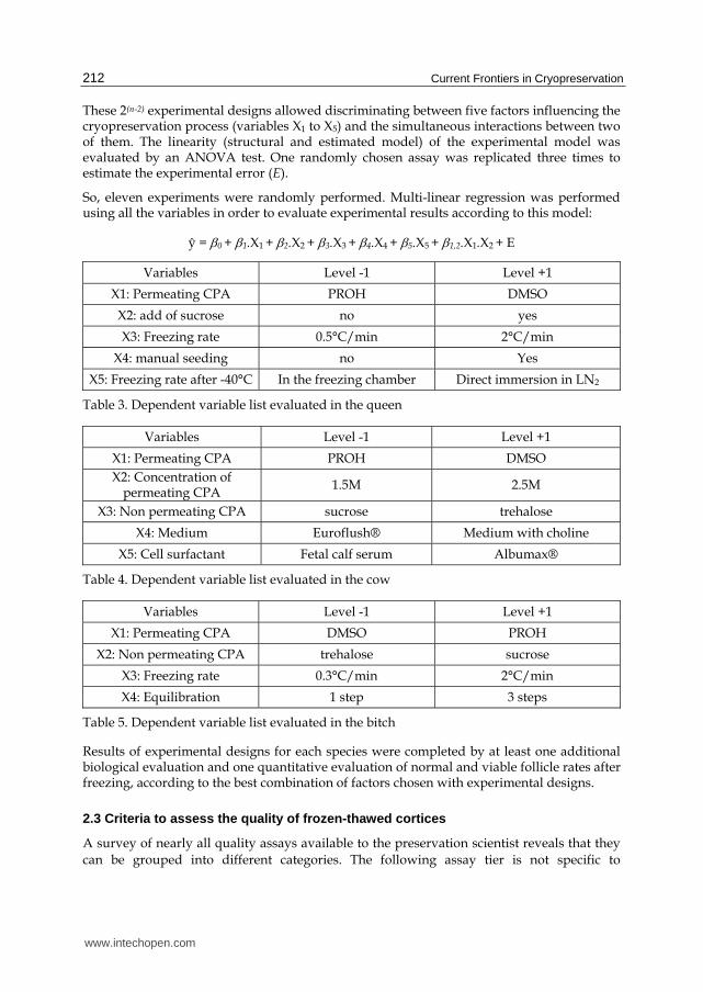
These 2(n-2) experimental designs allowed discriminating between five factors influencing the cryopreservation process (variables X1 to X5) and the simultaneous interactions between two of them. The linearity (structural and estimated model) of the experimental model was evaluated by an ANOVA test. One randomly chosen assay was replicated three times to estimate the experimental error (E).
So, eleven experiments were randomly performed. Multi-linear regression was performed using all the variables in order to evaluate experimental results according to this model:
ŷ = 0 + 1.X1 + 2.X2 + 3.X3 + 4.X4 + 5.X5 + 1,2.X1.X2 + E
Variables Level -1 Level +1
X1: Permeating CPA PROH DMSO
X2: add of sucrose no yes
X3: Freezing rate 0.5°C/min 2°C/min
X4: manual seeding no Yes
X5: Freezing rate after -40°C In the freezing chamber Direct immersion in LN2
Table 3. Dependent variable list evaluated in the queen
Variables Level -1 Level +1
X1: Permeating CPA PROH DMSO
X2: Concentration of permeating CPA
1.5M 2.5M
X3: Non permeating CPA sucrose trehalose
X4: Medium Euroflush® Medium with choline
X5: Cell surfactant Fetal calf serum Albumax®
Table 4. Dependent variable list evaluated in the cow
Variables Level -1 Level +1
X1: Permeating CPA DMSO PROH
X2: Non permeating CPA trehalose sucrose
X3: Freezing rate 0.3°C/min 2°C/min
X4: Equilibration 1 step 3 steps
Table 5. Dependent variable list evaluated in the bitch
Results of experimental designs for each species were completed by at least one additional biological evaluation and one quantitative evaluation of normal and viable follicle rates after freezing, according to the best combination of factors chosen with experimental designs.
2.3 Criteria to assess the quality of frozen-thawed cortices
A survey of nearly all quality assays available to the preservation scientist reveals that they
can be grouped into different categories. The following assay tier is not specific to
www.intechopen.com
New Approaches of Ovarian Tissue Cryopreservation from Domestic Animal Species
213
cryopreservation, but is presented below as a support that can guide those who work with
preserved tissues. The authors decided to assess the quality of the protocols developed in
different species using all or part of alternate test presented below. The morphology and the
viability of the ovarian follicles were systematically assessed, in combination with the
investigation of the ultrastructure of the follicles, and their capacity to resume
folliculogenesis after graft.
2.3.1 Morphology and ultrastructure
Assessment of the morphology of ovarian cells required the ovarian fragments were fixed in a preliminary defined fixative agent adapted to the species, before being processed for classical light microscopy. Primordial to primary follicles – from oocytes surrounded by flattened granulosa cells until oocytes surrounded by one layer of cuboidal granulosa cells (Gougeon and Chainy, 1987) – were usually classified into four types of morphological defects: (Type I) follicle without any morphological defect - follicle is regular, with joined follicular cells; cytoplasm of the oocyte is homogeneous and chromatin is diffused and regular; (Type II) follicle with cytoplasmic defect - cytoplasm of the oocyte is vacuolated or eosinophil; (Type III) follicles with nuclear defect - nucleus of the oocyte is picnotic, without apparent nuclear membrane or with an irregular nuclear membrane; (Type IV) degenerated follicle – oocyte with combined cytoplasmic and nuclear defects or follicle with irregular shape or with disjoined follicular cells or with swelled follicular cells.
The ultrastructure of ovarian follicles was also examined for the presence of apoptotic and para-apoptotic cell death. Ultrastructurally apoptosis is characterized by margination of condensed chromatin, nuclear fragmentation, and the formation of apoptotic bodies. Para-apoptosis, nonclassical apoptosis, is a specific morphologic type of non-necrotic cell death and is characterized by cytoplasmic vacuolization, condensed chromatin (but not early margination of the chromatin), and swollen mitochondria.
2.3.2 Cytolysis live/dead assay
The cytolysis assays have both a very positive and a negative attribute to them. On the positive side, there are a variety of assays that can reveal cell membrane leakage that occurs as a final stage in most forms of cell death. Yet given that cytolysis is the last stage of preservation-induced cell death, these assays do not reveal early-stage mechanisms underlying preservation-induced cell death and thus have limited use in the future as a diagnostic means to develop improved preservation formulations and protocols. The LDH assay continues today to be useful for measuring preservation-induced cytotoxicity. The concept behind this cytolysis assay is simply that if the cell membrane is compromised, then LDH will leak into the extracellular milieu where it can be measured. The trypan blue assay is also one of the most commonly used cytolysis assays. A number of investigators has used the trypan blue exclusion assay in studying preservation efficacy. It does however share the same handicap as the LDH assay given that neither can be analyzed using fluorescence and/or bioluminescence. Currently, the best cytolysis live/dead assays are those that employ fluorescent indicator dyes. Available probes can be subdivided into two different subsets, one of which is trapped by the cell and leaks out only is a membrane rupture occurs. The other subtype, exemplified by ethidium homodimer or propidium iodide, is membrane insoluble and only stains the cell if it gains access through a compromised
www.intechopen.com

Current Frontiers in Cryopreservation
214
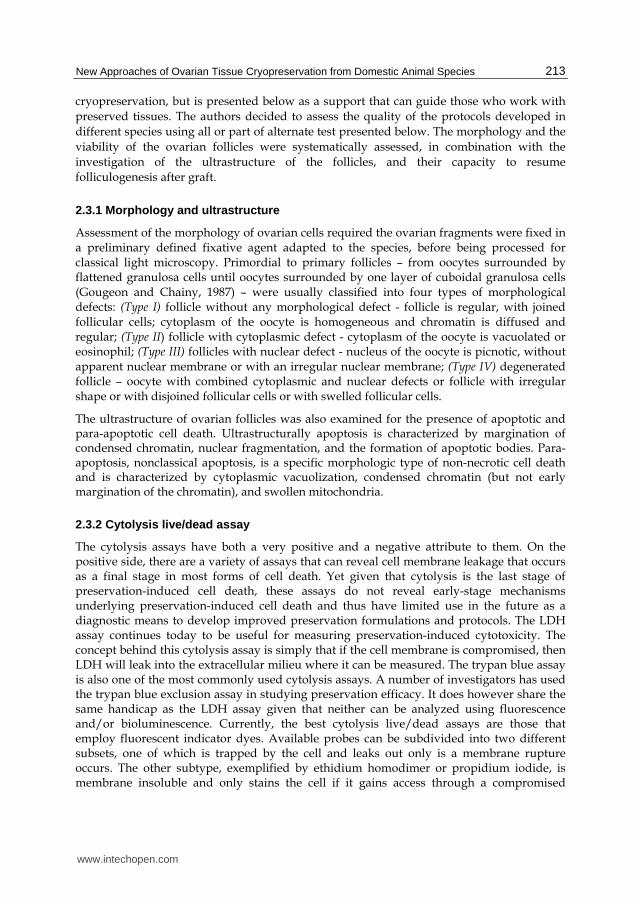
plasma membrane. In this way, the authors did evaluate the viability of the frozen-thawed
follicles using Calcein-AM and Ethidium homodimer I stains (Live/Dead Viability/Cytotoxicity Kit, Molecular Probes) on enzymatically isolated follicles (Fig. 2).
2.3.3 In vivo functionality (autografting model) and in vivo growth potential (xenografting model)
One of the key components of any preservation protocol must be a functional assay that matches the type of cells or tissue being analyzed. In some cases this determination is quite easy. As for example, the sperm motility is a well-accepted functional assay for this system, whereas the capacity to resume folliculogenesis sounds reasonable for the ovarian cortex.
Fresh and cryopreserved rabbit doe ovarian tissues were autografted into young females.
Fresh ovarian tissue was grafted on the ipsilateral ovarian pedicle immediately after ovary
resection (2 grafts per female), contrary to cryopreserved ovarian tissue grafted on the
controlateral ovarian pedicle 24 hours after freezing (one graft per female). Control females
were ovariectomised, according to the same surgery resection that used before graft. From
height week after graft, grafted rabbit doe were inseminated every 3 weeks in case of
negative pregnant diagnosis. Eleven months later, ovarian grafts were removed at necropsy
to observe follicular structures by histology.
In the bitch, the growth ability of frozen-thawed ovarian tissue has also been assessed by
implantation of small pieces of ovarian tissue into adult female SCID mice. After removing
mice ovaries, the canine frozen thawed ovarian tissue was placed intramuscularly in the
gluteal superficial muscle. To study the setting up of graft resumption, the graft was
harvested at one, eight or 16 weeks and the resumption of the ovarian activity was
controlled by vaginal cytology assessment. After harvesting, the grafts were processed for
histological assessment.
2.3.4 Vascularization
In the bitch, the alpha smooth muscle actin (alpha-SMA), a marker of the mature blood
vessel, was used to assess the vessel density within the ovarian tissue, using a primary
antibody directed against alpha-SMA. For each slice, image analysis was performed under
direct light microscope to determine the tissue areas. The stained vessels were counted in
several fields of the same slice and in several slices per animal to deduce a blood vessel
density.
2.4 Experimental results
To illustrate the interest of the use of fractional experimental design, the authors decided to
present some of their results obtained in the doe rabbit, in the queen, in the bitch and in the
cow during the last 6 years. The doe rabbit was used as a model for the human and the
animal applications of the ovarian tissue cryopreservation, because of its biological and
breeding characteristics. The cat was considered as a model for the ovarian tissue
cryopreservation studies of endangered wild felids; from all the felids, the domestic cat is
the only non endangered feline species. The cow was used as model for ruminants, with a
special interest for preserving high valuable individuals in combination to embryos and to
www.intechopen.com

New Approaches of Ovarian Tissue Cryopreservation from Domestic Animal Species
215
semen cryopreservation. Finally, the bitch was studied for preserving the reproductive
potential of future guide dogs submitted to neutering surgery before training.
2.4.1 In the doe rabbit
The experimental variability expressed as the repetition of one single combination, showed
that both the structural and the estimated models of the experimental design were valid
when considering the morphological preservation ratio of the follicles. The concentration of
the permeating CPA (P = 0.67) and the number of equilibration steps (P = 0.19) seemed to
have no significant effect on the morphological preservation ratio of ovarian follicles. The
nature of the permeating and non-permeating CPA seemed to influence the morphological
preservation ratio of the follicles (P = 0.08 and P = 0.07 respectively) although the non-
significant difference. DMSO tended to reduce the morphological preservation ratio, as
compared with PROH. Morphological preservation ratio was increased in the presence of
trehalose compared with sucrose. The freezing rate seemed to be the factor that had the
greatest impact on the morphological preservation ratio of the doe rabbit follicles. At a
freezing rate of 0.3°C/min we observed a significant increasing of the follicular
morphological preservation ratio, as compared with 2°C/min (P<0.01). No significant
interaction was observed between the nature of the permeating CPA and its concentration.
According to the results of the experimental design, the precise evaluation of the best combination of factors influencing positively the morphological preservation ratio (3 steps equilibration protocol, 1.5M DMSO or 1.5M PROH, medium supplemented with either sucrose or trehalose) was performed. Ovarian pieces were treated according to the results obtained with experimental design. Ovarian cortices were equilibrated (3 steps) in the freezing media based on TCM 199 and 10% FCS, at room temperature. The freezing media was supplemented with 1.5 M DMSO or 1.5M PROH and 0.2 M sucrose or 0.2M trehalose. Freezing of ovarian fragments was slowly performed at 0.3°C/min from the temperature of seeding (-7°C/min) up to -35°C. Thawing, histology, viability tests and electron microscopy evaluation process were performed before and after cryopreservation as described previously.
A B
Fig. 1. Rabbit follicular morphology before (A) and after (B) cryopreservation with PROH and trehalose, with a post-seeding freezing rate at 0.3°C/min
www.intechopen.com
Current Frontiers in Cryopreservation
216
A B
Fig. 2. View of rabbit isolated follicles under direct light for selection (A) and under fluorescent light (B) after calcein AM/ethidium homodimer I stains to evaluate viability after cryopreservation with PROH, with a post-seeding freezing rate at 0.3°C/min
In control fragments, we observed 72.6 2.8% and 77.7 3.9% of type I follicles (no
significant difference) for sucrose and trehalose control groups respectively. After
cryopreservation, no statistical difference of the proportions of type I follicles was found
between sucrose and trehalose (50.2 4.1% vs. 51.1 1.8% respectively) when using
DMSO for cryopreservation. When using PROH as permeating CPA, the proportion of
type I follicles was lower after cryopreservation with sucrose as compared to trehalose
(55.0 3.8% vs. 65.0 3.3% respectively; P<0.05). When freezing with trehalose the
proportion of type I follicles was higher with PROH as compared to DMSO (65.0 3.3%
vs. 51.1 1.8% respectively; P<0.01). Nevertheless, the proportions of type I follicles were
significantly reduced after cryopreservation (from P≤0.01 to P<0.001), whatever the
permeating and the non-permeating CPA. No significant difference was observed
between the different groups of frozen ovarian cortices, when considering the
morphological preservation ratio.
According to these results, the cryopreservation protocol based on a post-seeding freezing
rate at 0.3°C/min and using a freezing medium composed of 1.5M PROH, supplemented
with 0.2M trehalose was finally evaluated by orthotopic autografting to observe the
potential of the cryopreserved follicles to resume follicular growth and to be fertilized.
Before freezing, type II follicles represented the most important part of follicles with
morphological defect (19.1 2.9% and 16.1 3.2% in sucrose and trehalose groups
respectively). After cryopreservation, follicular defect of type IV (degenerated follicles) was
the most important type of morphological defect: 32.5 4.8% and 24.0 1.9% after freezing
using DMSO, with sucrose and trehalose respectively; versus 27.2 5.6% and 18.1 3.0%
after freezing using PROH, with sucrose and trehalose respectively. The general aspect of
ovarian tissue before and after cryopreservation showed a good preservation of structural
architecture (follicular structure and connective tissue). Spaces were observed in some case,
www.intechopen.com

New Approaches of Ovarian Tissue Cryopreservation from Domestic Animal Species
217
in the ovarian stroma and the albuginea. Epithelial cells were often absent as compared to
the fresh ovarian tissue.
Ultrastructural analysis of the preantral follicles was performed without preliminary
selection on semi-thin sections. TEM analysis showed that most follicles of control ovarian
tissue had normal ultrastructure, according to mitochondria, nucleus and nuclear
membrane, Golgi apparatus and endoplasmic reticulum cisternae observation. They often
had vacuoles in cytoplasm and vesicles. Nevertheless, vacuoles were not characteristic of
apoptosis. Cellular membranes of the oocyte and follicular cells were in close connection. In
general, ovarian stroma was well organised. Fibroblasts and collagen fibres were
distinguishable (Fig. 3).
After cryopreservation, oocyte ultrastructure appeared to be similar to the control especially
for mitochondria, Golgi apparatus, endoplasmic reticulum, interdigital structure between
oocyte and follicular cells (Fig.3). Vesicles and vacuoles were rarely observed. Chromatin of
the oocyte was diffused and well preserved. Nevertheless, dark follicular cells or follicular
cells without any content were most frequently observed, whereas some follicles showed
partial or total disruption of their nuclear membrane whatever the evaluated cryprotective
solution. The most important damage observed after cryopreservation was the
disorganisation of the ovarian stroma (Fig.3). Fibroblasts showed lack of cytoplasm or
important vacuolisation. In general, these damages were less frequently observed after
cryopreservation using PROH and trehalose.
2.4.2 Investigations in the queen
The experimental variability showed that neither the structural, nor the estimated models
of the experimental design were valid when considering the morphological preservation
ratio of the follicles or the viability preservation ratio. So, global discrimination of the
chemo-physical parameters was not possible. Nevertheless, the influence of the freezing
rate after seeding and after -40°C, and the influence of the addition of sucrose in the
freezing medium composed of 1.5M CPA were evaluated and analyzed by classical
ANOVA test.
Before freezing, ovarian tissue presented 72.2 3.6% and 83.8 2.9% of normal follicles
(type I) for group 2°C/min and 0.5°C/min post-seeding freezing rate respectively. When
freezing with PROH, and whatever the post-seeding freezing rate, proportions of
morphologically normal follicles were not significantly reduced after freezing compared to
before freezing (69.2 9.1% for 2°C/min group vs. 67.4 2.9°C/min for 0.5°C/min group).
After freezing with DMSO, and whatever the post-seeding freezing rate, proportions of
type I follicles were significantly reduced (40.8 6.6% after freezing at 2°C/min and
51.6 5.1% after freezing at 0.5°C/min; P<0.05). Whatever the post-seeding freezing rate,
type III defects were the most frequently observed after freezing. General observation of the
ovarian tissue showed a good preservation of the ovarian stroma cells and structure after
cryopreservation.
Before freezing, ovarian tissue submitted to a free fall into the freezing chamber during the
third phase of the freezing process presented 72.2 3.6% of type I follicles without any
www.intechopen.com

Current Frontiers in Cryopreservation
218
difference with samples directly immersed into the liquid nitrogen (86.8 2.5%). Proportion
of normal follicles decreased significantly after cryopreservation except after freezing with
PROH according to a free fall into the freezing chamber (68.2 9.1% of type I). After
freezing using a direct immersion into liquid nitrogen after -40°C, and whatever the CPA,
proportions of type I follicles were decreased compared to fresh control.
A B
A B
Fig. 3. Rabbit follicular ultrastructure before (A & B) and after (C & D) cryopreservation with PROH and trehalose, with a post-seeding freezing rate at 0.3°C/min
Before freezing, queen ovarian tissue showed 72.2 3.6% and 74.8 6.3% of type I follicles
respectively for group without and with sucrose without any difference between the two
control groups. After freezing, addition of sucrose allowed preserving 63.2 13.6% of
normal follicles versus 68.2 9.1% without sucrose when associated with PROH, without
any significant difference. Contrary to the results after freezing with DMSO, proportion of
type I follicles was not significantly different after freezing with PROH compared to fresh
A B
D C
www.intechopen.com

New Approaches of Ovarian Tissue Cryopreservation from Domestic Animal Species
219
control. Morphological defect of type III was the most frequently observed after freezing
with PROH. Queen ovarian stroma was well preserved.
A B
Fig. 4. Queen follicular morphology before (A) and after (B) cryopreservation with 1.5M PROH and 0.2M sucrose. Post-seeding freezing rate at 2°C/min.
In conclusion, queen ovarian tissue seems to be well preserved, without any difference compared to the fresh control when freezing with PROH (according to a free fall in the freezing chamber, without influence of neither sucrose nor post-seeding freezing rate) (Fig. 4).
2.4.3 Investigation in the bitch
In the bitch, the estimated model was validated when the viability preservation ratio was
considered. The nature of the non-permeating CPA (P = 0.37) did not influence the post
thawing viability rate of the ovarian follicles. However all the other factors investigated in
this experimental design presented a significant effect on the viability rate. The permeating
CPA nature (P<0.0001) was the factor that influenced more the viability rate of the follicles.
Thus, contrarily to the observations in the other species, the DMSO better preserved the
evaluated parameter than PROH. The freezing rate had also a major effect on the viability
rate (P<0.0001) and a slow freezing rate (0.3°C/min) was less deleterious than the rapid
freezing rate for the follicles viability. Moreover the equilibration step also significantly
affected the follicles viability, with a beneficial effect of the one step equilibration compared
to the 3 steps equilibration. However, no interaction was observed in this model. As a result,
the fractional experimental design developed in the bitch, suggested that ovarian tissue
A B
www.intechopen.com

Current Frontiers in Cryopreservation
220
should be cryopreserved in a solution containing 2 M DMSO as permeating CPA
supplemented with 0.2 M sucrose or trehalose in a one-step equilibration and frozen at a
0.3°C/min freezing rate (Fig. 5).
A B
Fig. 5. Bitch follicular morphology before (A) and after (B) cryopreservation
Consequently, theses parameters validated by the fractional experimental design for
viability assessment were used and applied for morphological assessment of frozen-thawed
bitch ovarian tissue and comparison with fresh tissue. So, the morphology of preantral
follicles was compared between fresh and cryopreserved tissue. The histological analysis
revealed that no significant difference was observed between fresh (89.1 6.1 % type I
follicles) and cryopreserved ovarian tissue (82.4 4.4 % type I follicles) when type I follicles
were observed. The main abnormality observed on preantral follicle after cryopreservation
in the bitch was the oocyte nucleus defect (~8%). In this case, the nucleus often appeared
pycnotic, with a reduced size and a densely packed chromatin. Sometimes the nuclear
membrane was ruptured. However, ooplasm defect were rarely observed alone, but
combined with nuclear defects. In some cases few granulosa cells were absent in the
primordial or primary follicles. It is probable that ice crystal formation occurring during
cooling be responsible of this partially denuded pattern, by destroying or dislodging some
granulosa cell during ice expansion.
2.4.4 Investigations in the cow
As for the rabbit doe, the experimental design was valid when considering the
morphological preservation ratio of the follicles, but not when considering the viability ratio
of the follicles. The concentration of the permeating CPA (P = 0.59) and the medium (P =
0.76) seem to have no significant effect on the morphological preservation ratio of ovarian
follicles. The nature of the permeating CPA seemed to influence the morphological
preservation ratio of the follicles (P = 0.07) although the non-significant difference. PROH
tended to improve the morphological preservation ratio, as compared with DMSO. The
nature of the non-permeating CPA (P = 0.002) and the cells surfactant (P = 0.04) had
significant influence. Trehalose and Albumax® improved morphological preservation ratio
www.intechopen.com

New Approaches of Ovarian Tissue Cryopreservation from Domestic Animal Species
221
compared respectively with sucrose and FCS. No significant interaction was observed
between the nature of the permeating CPA and its concentration.
In order to discriminate permeating CPAs, ovarian fragments from 5 cows were frozen using 1.5M DMSO or 1.5M PROH with 4 g/L Albumax® and 0.2M trehalose according to a
post-seeding freezing rate at 0.3°C/min. Before freezing, ovarian tissue showed 40.6 12.6%
of type I preantral follicles. Proportion of type I follicles was reduced to 20.2 3.9% after
cryopreservation using DMSO and to 23.8 3.4% when using PROH. No statistical difference was found between DMSO and PROH. Morphological defects of type II were the
most important kind of defect (47.0 11.7%). Proportion of type IV follicular defects was
significantly improved compared to control for the both CPAs (47.2 7.8% and 44.8 4.4% for DMSO and PROH respectively). Proportion of type III follicles was constant before and after freezing. Ovarian stroma seems to be affected by cryopreservation and shows spaces and disjoined cells.
A B
Fig. 6. Cow follicular morphology before (A) and after (B) cryopreservation using PROH and slow freezing rate
As for the other species, the influence of the post-seeding freezing rate was evaluated when
freezing with 2M PROH. Proportions of morphologically normal follicles were significantly
reduced after freezing compared with fresh tissue, whatever the post-seeding freezing rate
17.6 6.2% after freezing at 2°C/min vs. 57.8 13.0% before freezing and 17.8 6.5% after
freezing at 0.3°C/min vs. 60.0 4.9% before freezing).
2.4.5 In vivo follicle resumption from cryopreserved ovarian tissue
In the doe rabbit, nine pups were born from cryopreserved grafted group, suggesting the
efficiency of the cryopreservation protocols based on PROH and trehalose and using very
slow freezing rate. At necropsy, follicular structures were observed in most of females.
In the bitch, the cryopreservation method optimized with the fractional experimental design and validated by in vitro assessment (morphology, viability, and toxicity) was then evaluated by heterotopic xenografting to determine whether the ovarian tissue integrity and
www.intechopen.com

Current Frontiers in Cryopreservation
222
the follicular growth potential were maintained. The frozen-thawed tissues were grafted to female SCID mice as previously described. The histological assessments of the follicle population revealed a significant increase in the density and distribution of secondary follicles from eight weeks post grafting compared to the follicle population at 1 week (P<0.05). Consequently, the shift from primordial-primary follicles to secondary follicles occurred in a time laps of eight weeks. Moreover morphologically normal follicles were observed until 16 weeks post transplantation and intact secondary follicles with more than 3 layers of granulosa cells and a normal oocyte surrounded by a well-defined zona pellucida were present at this time. Despite a massive follicular loss touching particularly the early follicles and occurring just after grafting, the graft survived long term xenografting. Similarly, after an important loss just after grafting (one week grafts) the stromal cell number increased during the graft period, to reach a density comparable to fresh ovarian tissue at 16 weeks. Otherwise, the vascularization setting-up was assessed by immunohistochemistry as developed previously. The vessel density analysis revealed that already at one week post grafting the vessel density within the graft was comparable to the fresh control ovarian tissue. Moreover, the vessel density tended to increase at 16 weeks post grafting compared to the other groups (fresh control, one and eight weeks, P<0.05) even if no significant difference was registered. No antral follicles was present at the end of the graft time, however, persisting vaginal cornification was noticed on the recipients from 5-9 days post grafting, and estrus behavior was observed several times during the graft period in the recipients cages indicating an hormonal activity resulting from the graft. Taken together, these in vivo results confirm the good preservation of the bitch ovarian tissue by applying our cryopreservation method.
2.5 Discussion
The greater challenge of studies related to the cryopreservation of the ovarian tissue is to
define a freezing protocol adapted to the different cell types such as oocytes, follicular cells,
stroma cells, etc. One of the objectives of our team was to compare the effects of different
freezing parameters based on the morphology and the viability of the follicles, the
evaluation of the ultrastructure of the ovarian tissue, the DNA fragmentation of the oocytes
or the graft of the ovarian tissue. When the mathematical model was validated, the use of
experimental fractional designs allowed us to know simultaneously the individual and the
relative effects of different chemo-physical freezing parameters for each species. This
statistical method firstly allows a global evaluation of cryopreservation protocols and
discriminates the most valuable factors. Finally, the factors which seemed to have a
discriminating effect on follicular morphological preservation were evaluated in a wider
population. (Neto et al., 2008).
The results of the experimental design in the doe rabbit and in the bitch show that the post-seeding freezing rate is one of the most important chemophysical factors influencing the morphology or the viability of ovarian follicles. A slow freezing rate (0.3°C/min) seems to be more appropriate for the cryopreservation of the doe rabbit and bitch ovarian tissue. Nevertheless, no influence of the freezing rate was observed in the queen and in the cow. Most of the authors use a very slow freezing rate, which is derived from embryo freezing protocols. However, few studies show the importance of this freezing parameter. In contrast to our results in the bitch and in the doe rabbit, but in accordance with our result in the
www.intechopen.com

New Approaches of Ovarian Tissue Cryopreservation from Domestic Animal Species
223
queen and in the cow, Demirci et al. observed a high (but not significant) proportion of follicles without any morphological defect after a post-seeding freezing rate of 2°C/min in the ewe (Demirci et al., 2001). Nevertheless, Gook et al. also observed a better follicular preservation when using a slow freezing rate (0.3°C/min) with human ovarian tissue (Gook et al., 1999). Whereas Cleary et al. observed no difference in terms of follicular growth after grafting, between a conventional embryo freezing protocol (0.3°C/min) and a passive cooling at 1°C/min from 0°C to -84°C on the mouse ovarian tissue (Cleary et al., 2001). Although these two cooling rates (0.3°C/min and 2°C/min) could be considered as slow, these results may be explained by a difference in cell dehydration during the post-seeding step. With rapid cooling rates, we can hypothesise that time required for the exosmose of the cell water is insufficient and consequently promotes the formation of lethal intracellular ice. While at very slow cooling rates, high level of dehydration occurs with concomitant increasing in solute concentration (salting out). Investigations on the freezing rate were extended in the queen with the evaluation of the third freezing phase. When associated with PROH, slow cooling in solid phase seems to be more appropriate. Nevertheless, several authors use cryopreservation protocols with a direct immersion into liquid nitrogen after –40°C, such as Rodrigues et al. (2004) in the goat, Lucci et al. (2004) in the zebu cow, Santos et al. (2006) in the ewe or Lima et al. (2006) in the cat. Births had been obtained after graft of ovarian fragments frozen with such a protocol in the rabbit doe (Almodin et al., 2004b) and in the ewe (Almodin et al., 2004a), but not in the cat where in vivo follicular growth were observed when using a freezing protocol with controlled third phase (Bosch et al., 2004).
The experimental designs revealed a crucial role of the permeating CPA in the doe rabbit, in
the cow and in the bitch, added to a crucial role of the non permeating CPA in the doe rabbit
and in the cow. Among the various freezing protocols described in the literature, those
using DMSO or PROH as permeating CPA seems to be more efficient, whatever the species.
Our results suggest that PROH improve the follicular quality after freezing in the doe rabbit
and in the queen. Results of experimental design obtained in the cow suggested a better
morphological preservation rate when using PROH. However, in this species, standard
comparison between DMSO and PROH doesn’t confirm these results. Contrary to that, bitch
ovarian tissue seems to be better preserved in freezing medium composed of DMSO. These
results were confirmed by the follicular growth observed after xenograft. It can be
hypothesized that DMSO penetrate better within the tissue than PROH. Indeed, the bitch
ovarian tissue as the goat or the ewe is rich in collagenous fibbers and more fibrous than the
doe rabbit ovarian tissue for example. Therefore, a good ability to penetrate within the
ovarian tissue is an important characteristic for the chosen CPA Nevertheless, both CPA
have sensibly the same molecular weight (PROH: 76.10 g/mol, DMSO: 78.14 g/mol) with a
lower weight for PROH. Thus a better penetration of the DMSO cannot be explained by this
physical parameter. The explanation may come from the toxicity of both CPAs. Our team
also investigated the toxicity of the both CPAs on bitch ovarian tissue after equilibration
steps at room temperature without freezing and registered a deleterious effect of PROH
compared to DMSO on preantral follicle viability in this species.
Except in canine and feline models, addition of non-permeating CPA in the freezing
medium seems to improve the protective effect of CPAs. The protective effect of PROH
seems to be improved when it is associated with trehalose, in the doe rabbit and in the cow.
This observation was confirmed by electron microscopy evaluation of the doe rabbit ovarian
www.intechopen.com

Current Frontiers in Cryopreservation
224
tissue subjected to cryopreservation. Ultrastructure of doe rabbit follicles after
cryopreservation was well preserved, but stromal cells and fibroblasts were damaged. Such
alterations have been observed in human tissues after cryopreservation (Navarro-Costa et
al., 2005; Santos et al., 2010). However, fibroblasts can easily be reproduced by cell division,
indicating that damage to the stroma can be repaired. Collagen fibers did not seem to be
damaged by cryopreservation in this study, but they were sparse in the doe rabbit ovary.
This observation may explain the fragility of doe rabbit ovarian tissue during the
equilibration, freezing and thawing steps (Neto et al., 2005).
Sugar are not systematically associated with permeating CPA, but Marsella et al. (2008), showed the advantageous effect of sucrose. Trehalose has been frequently used in embryo cryopreservation, but not in ovarian tissue cryopreservation. Sucrose and trehalose share the property to stabilise cellular membranes and proteins via the formation of hydrogen bonds with polar residues of phospholipidic membrane. This property allows preserving the membrane integrity under anhydrous conditions. Moreover, it modifies the temperature at which the separation of lipid phase occurs during cooling (Crowe et al., 1984; Crowe et al., 1985; Crowe et al., 2001). As compared to other sugars, trehalose seems to have a higher capacity to preserve biomolecules, cellular membrane and cells in a drying or in a freezing state (Crowe et al., 1996; Storey et al., 1998; Sano et al., 1999; Welsh and Herbert, 1999).
Few comparable studies have been reported in the cryopreservation of different mammalian species. Despite encouraging results in the different studied species, and except in the queen, none of evaluated protocols allows preserving the same proportion of normal follicles than before freezing. Most of authors observed a reduction of normal follicles in frozen/thawed ovarian tissue compared with fresh control when using similar freezing protocols in the mouse (Candy et al., 1997), the goat (Rodrigues et al., 2004), the cow (Lucci et al., 2004), and the ewe (Demirci et al., 2002). As for the queen, no morphological difference was observed in human follicles before and after cryopreservation (Hovatta et al., 1996; Fabbri et al., 2006) Newton observed similar proportions of “viable” follicles after freezing when using DMSO, ethylene glycol or PROH and xenografting (Newton et al., 1996).
Live births in the rabbit doe and follicular growths observed in the bitch after grafting of cryopreserved ovarian tissue shows the efficacy of evaluated freezing protocols. Almodin et al. obtained live offspring after grafting of small fragments of cryopreserved rabbit ovarian tissue using 1.5 M DMSO and a very slow post seeding freezing rate (Almodin et al., 2004b). In the bitch, results about in vivo growth obtained after cryopreservation are relatively scarce compared to other species. Ishijima et al. (2006) tried to transplant vitrified ovarian tissue (2M DMSO, 3M PROH, 1M acetamide) into immunodeficient mice during 4 weeks and observed signs of growth in the early follicles (primordial-primary follicles). However, they noticed an important follicular loss occurring just after grafting which is in accordance with our results obtained with slow-frozen tissue. The time necessary for setting up of the neovascularization within the grafted tissue seems to be more deleterious for the cells than the cryopreservation technique itself. Except our results obtained on the bitch ovarian tissue cryopreservation no other results using slow freezing of female germ cells was obtained in this species. Furthermore, live birth has not yet been obtained after ovarian tissue cryopreservation, but the difficulties to mastered in vitro maturation and fertilization steps in canines do not contribute to the development of this technique.
www.intechopen.com
New Approaches of Ovarian Tissue Cryopreservation from Domestic Animal Species
225
For the first time a complete evaluation process of important factors influencing the morphology and the viability of preantral follicles has been performed after equilibration process and freezing in different species. These results suggest that cryopreservation of ovarian tissue is a promising and suitable technique that could be used as complementary tool to embryo cryopreservation, to preserve the animals’ genetic resources by the female pathway. Doe rabbit could also be used as a biomedical model to investigate the long term consequences of cryopreservation on ovarian follicles and the birth of future progenies.
3. Perspectives
In definitive, the use of factorial fractional experimental design approach allowed us to develop suitable cryopreservation protocol in different species, while reducing the number of experiments and increasing the number of parameters evaluated. However it can be noticed in our model species, but also in the literature, that results can be radically different according to the species. Moreover, among the numerous articles published on ovarian tissue cryopreservation heterogeneous results can be observed in the same species after applying roughly or widely different slow-freezing protocols. One of the candidates to explain such disparity in the obtained results is the amount of ice crystals formed during slow freezing. Indeed, the strategy of slow cooling is to decrease cell temperature enough slowly to allow removal of most of the freezable intracellular water before reaching the ice nucleation temperature. The main objective of this method is to avoid intracellular ice crystal formation which is known to be lethal. However ice crystallization still occurs extracellularly with the risk of tissue shrinkage or disorganization of the tissue components.
As ice formation and melting are exothermic and endothermic phenomena respectively,
they can be objectified and studied by thermodynamical measures. Among the various
physical methods of analysis, Differential Scanning Calorimetry (DSC) is an interesting tool.
In fact, DSC gives the opportunity to measure important parameters of a cryopreservation
solution under dynamic conditions. A cryopreservation solution can thus be characterized
by its thermal properties such as temperatures of phase transitions and quantity of ice
crystallized and melted. Two types of DSC are commonly used: power-compensation DSC
and heat-flux DSC. Our team chose the first one in order to study cryopreservation solutions
with a more fundamental approach than with biological methods.
The power compensation DSC is based on the “zero balance principle” as explained as
follow. The sample and a reference are placed in two microfurnaces which are continuously
cooled by liquid nitrogen. The temperature of each microfurnace can be, on the one hand,
precisely measured by a temperature sensor and, on the other hand, precisely adjusted by a
heating resistor. Each microfurnace contains one sensor and one resistor. The principle of
the power compensation DSC is to maintain the two microfurnaces under the same
temperature regardless of phase transitions or reactions occurring in the sample. Thus,
when a phase transition occurs in the sample, the heat released or absorbed by the sample
has to be compensated by the heating resistor which is below the sample. Consequently, the
calorimeter measures a difference between the heating powers provided by the two
resistors. This difference reveals the phase transition. When this phase transition is
crystallization, this difference allows also us to measure the quantity of ice formed.
www.intechopen.com

Current Frontiers in Cryopreservation
226
Since recent years, several strategies are developed to avoid deleterious effects of the ice crystal formation during cooling, and thermodynamical approaches are more and more associated to these strategies.
Fig. 7. Scheme of power-compensation DSC
The specimen and reference temperatures are controlled independently using separate (identical) ovens. The temperature difference between the sample and reference is maintained to zero by varying the power input to the two furnaces. This energy is then a measure of the enthalpy or hat capacity changes in the test specimen (relative to the reference).
3.1 Prevent the ice formation
The physical definition of vitrification is the glass-like solidification of solutions at low temperatures, without the formation of intracellular ice crystals. The vitrification technique is the solidification of a liquid without ice crystal formation. This phase is obtained by increasing the solute concentration of the vitrification media (increased viscosity that makes water solidify without expansion) or by using very fast cooling rate to avoid molecular rearrangement into ice crystals but into an amorphous glass. (Vajta, 2000). Consequently, the risk of injuries due to intra- or extracellular ice crystallization is avoided, which constitutes the main advantage of this technique.
Despite the fact that slow freezing is the most widely used cryopreservation technique, vitrification is a viable and promising alternative that is increasingly becoming more attractive to the commercial sector. Vitrification has been used for oocyte and tissue cryopreservation since the 1980s (Rall and Fahy, 1985). Since the first use of this approach in ovarian tissue cryopreservation, it has been well optimized, particularly by decreasing the concentration of CPAs used to reduce their toxicity, but also by increasing the cooling rate applied (Chen et al., 2006; Keros et al., 2009). These improvements of vitrification in ovarian
www.intechopen.com

New Approaches of Ovarian Tissue Cryopreservation from Domestic Animal Species
227
tissue but also in embryo and oocyte vitrification place the vitrification as a gold standard method for germ cells cryopreservation (Saragusty and Arav, 2011). It has been suggested that with time, conventional slow freezing will be replaced entirely by vitrification techniques (Vajta and Kuwayama, 2006).
As mentioned above, DSC can be a precious aid for the study of vitrification. In fact, DSC allows the detection of phase transitions, both crystallization or glass transition, and can thus determine three thermal properties of vitrification solutions: the vitreous transition temperature, the critical cooling rate and the critical warming rate (Baudot et al., 2007). These three thermal properties are specific to each vitrification solution and could be used for a better utilization of the solutions. The critical cooling rate and the critical warming rate are the cooling and warming speeds above which the crystallization cannot occur between the vitreous transition temperature and the crystallization temperature. The calculation of these critical cooling and warming rates is based on a semi-empiric model developed by Boutron in 1986 according to the “classic” theory of crystallization (Boutron, 1986). This model reproduces well analytically the experimental results, but some approximations are introduced.
3.2 Limit the ice formation
As described in the introductive part, extracellular crystallization occurs during slow
freezing. The penetrating CPAs like DMSO, PROH or ethylene glycol, are mainly used to
reduce the volume of freezable ice within the cells, but their respective toxicity for the
cells limit the potential concentration usable. However, since the last decades, non-
penetrating CPAs like disaccharides have been widely used in cryoprotective solution to
improve cell preservation and reduce the penetrating CPA amount necessary. To our
knowledge, the first use of sucrose as cryoprotectant of ovarian tissue was in 1996 with
human ovaries (Hovatta et al., 1996). Sugars have been proved to play a role on ice crystal
formation and stability (Kuleshova et al., 1999). Previous studies on mouse embryos
revealed that sugars increase the homogeny ice transition temperature in freezing
solutions. By this way, dehydration of the cells occurs when they are still permeable to
water without reaching ice nucleation temperature. Moreover, the trehalose was reported
to reduce the size of ice crystals and shorten their elongation during freezing (Sei et al.,
2002).
On the basis of the contribution of DSC in other fields of cryopreservation [vitrification of
ovary (Baudot et al., 2007), slow-freezing or vitrification of plants (Volk and Walters, 2006;
Skyba et al., 2011), slow-freezing of aquatic crustaceans (Issartel et al., 2006)], our team chose
a thermodynamic approach to study slow-freezing solutions for ovarian tissue. In fact, DSC
allows the measure of the maximal quantity of ice formed in a solution (Qmax). Qmax
corresponds to the definition of Boutron : “The heat of solidification are represented by the
numbers q of grams of ice whose solidification at 0°C would liberate the same amount of
heat as that from 100g of solution on crossing the corresponding peaks. They are close to the
real quantities of ice crystallized in % (w/w) of the solution when it is ice which crystallizes.
One obtains the heat in calories per 100g of solution by multiplying q by 79.78” (Boutron,
1984). Our team has thus quantified the quantity of ice formed in solutions used for the
cryopreservation of ovarian tissue of different species (table 6).
www.intechopen.com

Current Frontiers in Cryopreservation
228
Species Qmax in percentage (w/w) of solution
Bitch 36.81
Doe rabbit 45.79
Cow 37.64
Table 6. Maximal quantity of ice crystallized (Qmax) in solutions used for the cryopreservation of ovarian tissue of bitch, doe rabbit and cow.
Then, our team decided to explore two research areas. On the one hand, DSC can compare the quantity of ice formed in two different cryopreservation solutions. Thus, it seems possible to select the most suitable cryopreservation solutions for slow freezing methods. The first results obtained in doe rabbit seem to confirm this hypothesis. In fact between two cryopreservation solutions tested, those for which the quantity of ice formed was the lowest, was also the one with the best biological results. On the other hand, for a given solution, DSC allows the measure of the quantity of ice formed for different freezing kinetics. Consequently, it seems also possible to select the most suitable kinetics according to the cryopreservation solutions.
3.3 Promote the formation of a non-vulnerable extracellular ice
Another approach to optimize cryopreservation process should be to control the ice crystal growth and shape in order to promote the less deleterious crystallization. It is already assumed that intracellular ice formation is lethal. Nonetheless, the recent observations of rabbit ovarian tissue by cryoscanning electronic microscopy and cryofracture reveal that depending on the cooling rate, the extracellular ice shape is modified. Moreover, according to these results the seeding temperature influences the shape and regularity of the ice crystals resulting in large uniform crystals when seeding was induced close to the solution melting point (Gosden et al., 2010).
Otherwise, a better understanding of intracellular ice formation can also be advantageous to improve cryopreservation processes. Indeed, Han et al (2009) investigated the size of intracellular ice crystals in mouse oocytes by cryomicroscopy. They conclude that increasing the concentration of macromolecules in the cells by increasing the extracellular non permeating solute concentration significantly reduced the required permeating CPA concentration for intracellular vitrification. Moreover they also observed that intracellular ice melting point was always lower than extracellular ice. Taken together, this information can be helpful to optimize the cryopreservation protocols.
Regarding DSC, a recent study showed that it is possible to differentiate the crystallization of intra and extracellular ice depending on freezing kinetics (Seki et al., 2009). Consequently, in addition to the measure of the quantity of crystallized ice, DSC can provide a better control of ice formation.
4. Acknowledgment
The results presented in this chapter were partially supported financially by Région Rhône-Alpes and the breeding center (CESECAH) of the French Guide Dog Federation (FFAC). The authors also thank the LLC Hycole (Marcoing, France) for their technical support.
The authors want to thank equally regards the Electronic Microscopy Center of the University of Lyon, the Laboratory of Pathological Anatomy of the Veterinary Campus of
www.intechopen.com

New Approaches of Ovarian Tissue Cryopreservation from Domestic Animal Species
229
Lyon and the Institute Claude Bourgelat for access to their facilities. The authors are also grateful to all veterinarians which allowed the ovary collection.
5. References
Abrishami, M.; Anzar, M.; Yang, Y.&Honaramooz, A. (2010). Cryopreservation of immature
porcine testis tissue to maintain its developmental potential after xenografting into
recipient mice. Theriogenology, Vol.73, No.1, pp: 86-96,
Almodin, C. G.; Minguetti-Camara, V. C.; Meister, H.; Ceschin, A. P.; Kriger, E.&Ferreira,
J. O. (2004a). Recovery of natural fertility after grafting of cryopreserved
germinative tissue in ewes subjected to radiotherapy. Fertil Steril, Vol.81, No.1,
pp: 160-164,
Almodin, C. G.; Minguetti-Camara, V. C.; Meister, H.; Ferreira, J. O.; Franco, R. L.;
Cavalcante, A. A.; Radaelli, M. R.; Bahls, A. S.; Moron, A. F.&Murta, C. G. (2004b).
Recovery of fertility after grafting of cryopreserved germinative tissue in female
rabbits following radiotherapy. Hum Reprod, Vol.19, No.6, pp: 1287-1293,
Andrabi, S. M.&Maxwell, W. M. (2007). A review on reproductive biotechnologies for
conservation of endangered mammalian species. Anim Reprod Sci, Vol.99, No.3-4,
pp: 223-243,
Aubard, Y.; Newton, H.; Scheffer, G.&Gosden, R. (1998). Conservation of the follicular
population in irradiated rats by the cryopreservation and orthotopic autografting
of ovarian tissue. Eur J Obstet Gynecol Reprod Biol, Vol.79, No.1, pp: 83-87,
Bahadur, G.&Ralph, D. (1999). Gonadal tissue cryopreservation in boys with paediatric
cancers. Hum Reprod, Vol.14, No.1, pp: 11-17,
Bahadur, G.; Chatterjee, R.&Ralph, D. (2000). Testicular tissue cryopreservation in boys.
Ethical and legal issues: case report. Hum Reprod, Vol.15, No.6, pp: 1416-1420,
Baudot, A.; Courbiere, B.; Odagescu, V.; Salle, B.; Mazoyer, C.; Massardier, J.&Lornage, J.
(2007). Towards whole sheep ovary cryopreservation. Cryobiology, Vol.55, No.3, pp:
236-248,
Boettcher, P. J.; Stella, A.; Pizzi, F.&Gandini, G. (2005). The combined use of embryos and
semen for cryogenic conservation of mammalian livestock genetic resources. Genet
Sel Evol, Vol.37, No.6, pp: 657-675,
Bosch, P.; Hernandez-Fonseca, H. J.; Miller, D. M.; Wininger, J. D.; Massey, J. B.; Lamb, S.
V.&Brackett, B. G. (2004). Development of antral follicles in cryopreserved cat
ovarian tissue transplanted to immunodeficient mice. Theriogenology, Vol.61, No.2-
3, pp: 581-594,
Boutron, P. (1984). More accurate determination of the quantity of ice crystallised at low
cooling rates in glycerol and 1,2-propanediol aqueous solutions : comparison with
equilibrium. Cryobiology, Vol.21, 183-191,
Boutron, P. (1986). Comparison with the theory of the kinetics and extent of ice
crystallization and of glass-forming tendency in aqueous cryoprotective solutions.
Cryobiology, Vol.23, 88-102,
Candy, C. J.; Wood, M. J.&Whittingham, D. G. (1997). Effect of cryoprotectants on the
survival of follicles in frozen mouse ovaries. J Reprod Fertil, Vol.110, No.1, pp: 11-
19,
www.intechopen.com

Current Frontiers in Cryopreservation
230
Checura, C. M.&Seidel, G. E., Jr. (2007). Effect of macromolecules in solutions for
vitrification of mature bovine oocytes. Theriogenology, Vol.67, No.5, pp: 919-930,
Chen, S. U.; Chien, C. L.; Wu, M. Y.; Chen, T. H.; Lai, S. M.; Lin, C. W.&Yang, Y. S. (2006).
Novel direct cover vitrification for cryopreservation of ovarian tissues increases
follicle viability and pregnancy capability in mice. Hum Reprod, Vol.21, No.11, pp:
2794-2800,
Cleary, M.; Snow, M.; Paris, M.; Shaw, J.; Cox, S. L.&Jenkin, G. (2001). Cryopreservation of
mouse ovarian tissue following prolonged exposure to an Ischemic environment.
Cryobiology, Vol.42, No.2, pp: 121-133,
Commin, L.; Rosset, E.; Allard, A.; Joly, T.; Guérin, P.; Neto, V.&Buff, S. (2011). Assesment of
tissue functionality after xenotransplantation of canine ovarian cortex to severe
combined immunodeficient mice (SCID mice), Theriogenology Conference and
Symposia, pp: Milwaukee, Wisconsin
Critser, J. K. (1998). Current status of semen banking in the USA. Hum Reprod, Vol.13 Suppl
2, 55-67; discussion 68-59,
Crowe, J. H.; Crowe, L. M.&Chapman, D. (1984). Preservation of membranes in
anhydrobiotic organisms: the role of trehalose. Science, Vol.223, No.4637, pp: 701-
703,
Crowe, J. H.; Crowe, L. M.; Oliver, A. E.; Tsvetkova, N.; Wolkers, W.&Tablin, F. (2001). The
trehalose myth revisited: introduction to a symposium on stabilization of cells in
the dry state. Cryobiology, Vol.43, No.2, pp: 89-105,
Crowe, L. M.; Reid, D. S.&Crowe, J. H. (1996). Is trehalose special for preserving dry
biomaterials? Biophys J, Vol.71, No.4, pp: 2087-2093,
Crowe, L. M.; Crowe, J. H.; Rudolph, A.; Womersley, C.&Appel, L. (1985). Preservation of
freeze-dried liposomes by trehalose. Arch Biochem Biophys, Vol.242, No.1, pp: 240-
247,
Curaba, M.; Verleysen, M.; Amorim, C. A.; Dolmans, M. M.; Van Langendonckt, A.;
Hovatta, O.; Wyns, C.&Donnez, J. (2011). Cryopreservation of prepubertal mouse
testicular tissue by vitrification. Fertil Steril, Vol.95, No.4, pp: 1229-1234 e1221,
Deanesly, R. (1954). Immature rat ovaries grafted after freezing and thawing. J Endocrinol,
Vol.11, No.2, pp: 197-200,
Demirci, B.; Lornage, J.; Salle, B.; Frappart, L.; Franck, M.&Guerin, J. F. (2001). Follicular
viability and morphology of sheep ovaries after exposure to cryoprotectant and
cryopreservation with different freezing protocols. Fertil Steril, Vol.75, No.4, pp:
754-762,
Demirci, B.; Salle, B.; Frappart, L.; Franck, M.; Guerin, J. F.&Lornage, J. (2002).
Morphological alterations and DNA fragmentation in oocytes from primordial and
primary follicles after freezing-thawing of ovarian cortex in sheep. Fertil Steril,
Vol.77, No.3, pp: 595-600,
Donnez, J.; Dolmans, M. M.; Demylle, D.; Jadoul, P.; Pirard, C.; Squifflet, J.; Martinez-
Madrid, B.&van Langendonckt, A. (2004). Livebirth after orthotopic transplantation
of cryopreserved ovarian tissue. Lancet, Vol.364, No.9443, pp: 1405-1410,
Edge, B.; Holmes, D.&Makin, G. (2006). Sperm banking in adolescent cancer patients. Arch
Dis Child, Vol.91, No.2, pp: 149-152,
www.intechopen.com
New Approaches of Ovarian Tissue Cryopreservation from Domestic Animal Species
231
Fabbri, R.; Pasquinelli, G.; Bracone, G.; Orrico, C.; Di Tommaso, B.&Venturoli, S. (2006).
Cryopreservation of human ovarian tissue. Cell Tissue Bank, Vol.7, No.2, pp: 123-
133,
FAO (2007). The state of the world's animal genetic resources for food and agriculture, Food
and Agriculture Organization of the United Nations, Commission on Genetic
Resources for Food and Agriculture, Rome, Italy
Fishel, S.; Green, S.; Hunter, A.; Lisi, F.; Rinaldi, L.; Lisi, R.&McDermott, H. (1997). Human
fertilization with round and elongated spermatids. Hum Reprod, Vol.12, No.2, pp:
336-340,
Gook, D. A.; Edgar, D. H.&Stern, C. (1999). Effect of cooling rate and dehydration regimen
on the histological appearance of human ovarian cortex following cryopreservation
in 1, 2-propanediol. Hum Reprod, Vol.14, No.8, pp: 2061-2068,
Gook, D. A.; Edgar, D. H.&Stern, C. (2004). Cryopreservation of human ovarian tissue. Eur J
Obstet Gynecol Reprod Biol, Vol.113 Suppl 1, S41-44,
Gosden, R. G.; Baird, D. T.; Wade, J. C.&Webb, R. (1994). Restoration of fertility to
oophorectomized sheep by ovarian autografts stored at -196 degrees C. Hum
Reprod, Vol.9, No.4, pp: 597-603,
Gosden, R. G.; Yin, H.; Bodine, R. J.&Morris, G. J. (2010). Character, distribution and
biological implications of ice crystallization in cryopreserved rabbit ovarian tissue
revealed by cryo-scanning electron microscopy. Hum Reprod, Vol.25, No.2, pp: 470-
478,
Gougeon, A.&Chainy, G.B.N. (1987). Morphometric studies of small follicles in ovaries of
women at different ages. J Reprod Fertil, Vol.81, 433-442,
Guerin, J. F. (2005). [Testicular tissue cryoconservation for prepubertal boy: indications and
feasibility]. Gynecol Obstet Fertil, Vol.33, No.10, pp: 804-808,
Han, X.&Critser, J. K. (2009). Measurement of the size of intracellular ice crystals in mouse
oocytes using a melting point depression method and the influence of intracellular
solute concentrations. Cryobiology, Vol.59, No.3, pp: 302-307,
Holt, W. V.&Pickard, A. R. (1999). Role of reproductive technologies and genetic resource
banks in animal conservation. Rev Reprod, Vol.4, No.3, pp: 143-150,
Honaramooz, A.; Snedaker, A.; Boiani, M.; Scholer, H.; Dobrinski, I.&Schlatt, S. (2002).
Sperm from neonatal mammalian testes grafted in mice. Nature, Vol.418, No.6899,
pp: 778-781,
Hovatta, O.; Silye, R.; Krausz, T.; Abir, R.; Margara, R.; Trew, G.; Lass, A.&Winston, R. M.
(1996). Cryopreservation of human ovarian tissue using dimethylsulphoxide and
propanediol-sucrose as cryoprotectants. Hum Reprod, Vol.11, No.6, pp: 1268-1272,
Isachenko, V.; Isachenko, E.; Weiss, J. M.; Todorov, P.&Kreienberg, R. (2009). Cryobanking
of human ovarian tissue for anti-cancer treatment: comparison of vitrification and
conventional freezing. Cryo Letters, Vol.30, No.6, pp: 449-454,
Ishijima, T.; Kobayashi, Y.; Lee, D. S.; Ueta, Y. Y.; Matsui, M.; Lee, J. Y.; Suwa, Y.; Miyahara,
K.&Suzuki, H. (2006). Cryopreservation of canine ovaries by vitrification. J Reprod
Dev, Vol.52, No.2, pp: 293-299,
Issartel, J.; Voituron, Y.; Odagescu, V.; Baudot, A.; Guillot, G.; Ruaud, J. P.; Renault, D.;
Vernon, P.&Hervant, F. (2006). Freezing or supercooling: how does an aquatic
www.intechopen.com

Current Frontiers in Cryopreservation
232
subterranean crustacean survive exposures at subzero temperatures? J Exp Biol,
Vol.209, No.Pt 17, pp: 3469-3475,
Jahnukainen, K.; Ehmcke, J.; Hergenrother, S. D.&Schlatt, S. (2007). Effect of cold storage
and cryopreservation of immature non-human primate testicular tissue on
spermatogonial stem cell potential in xenografts. Hum Reprod, Vol.22, No.4, pp:
1060-1067,
Jezek, D.; Schulze, W.; Kalanj-Bognar, S.; Vukelic, Z.; Milavec-Puretic, V.&Krhen, I. (2001).
Effects of various cryopreservation media and freezing-thawing on the
morphology of rat testicular biopsies. Andrologia, Vol.33, No.6, pp: 368-378,
Johnston, L. A.&Lacy, R. C. (1995). Genome resource banking for species conservation:
selection of sperm donors. Cryobiology, Vol.32, No.1, pp: 68-77,
Karlsson, J.O.M.&Toner, M. (1996). Long-term storage of tissues by cryopreservation :
critical issues. Biomaterials, Vol.17, 243-256,
Keros, V.; Rosenlund, B.; Hultenby, K.; Aghajanova, L.; Levkov, L.&Hovatta, O. (2005).
Optimizing cryopreservation of human testicular tissue: comparison of protocols
with glycerol, propanediol and dimethylsulphoxide as cryoprotectants. Hum
Reprod, Vol.20, No.6, pp: 1676-1687,
Keros, V.; Hultenby, K.; Borgstrom, B.; Fridstrom, M.; Jahnukainen, K.&Hovatta, O. (2007).
Methods of cryopreservation of testicular tissue with viable spermatogonia in pre-
pubertal boys undergoing gonadotoxic cancer treatment. Hum Reprod, Vol.22, No.5,
pp: 1384-1395,
Keros, V.; Xella, S.; Hultenby, K.; Pettersson, K.; Sheikhi, M.; Volpe, A.; Hreinsson,
J.&Hovatta, O. (2009). Vitrification versus controlled-rate freezing in
cryopreservation of human ovarian tissue. Hum Reprod, Vol.24, No.7, pp: 1670-
1683,
Kuleshova, L. L.; MacFarlane, D. R.; Trounson, A. O.&Shaw, J. M. (1999). Sugars exert a
major influence on the vitrification properties of ethylene glycol-based solutions
and have low toxicity to embryos and oocytes. Cryobiology, Vol.38, No.2, pp: 119-
130,
Ledda, S.; Leoni, G.; Bogliolo, L.&Naitana, S. (2001). Oocyte cryopreservation and ovarian
tissue banking. Theriogenology, Vol.55, No.6, pp: 1359-1371,
Lima, A. K.; Silva, A. R.; Santos, R. R.; Sales, D. M.; Evangelista, A. F.; Figueiredo, J.
R.&Silva, L. D. (2006). Cryopreservation of preantral ovarian follicles in situ from
domestic cats (Felis catus) using different cryoprotective agents. Theriogenology,
Vol.66, No.6-7, pp: 1664-1666,
Lucci, C. M.; Kacinskis, M. A.; Lopes, L. H.; Rumpf, R.&Bao, S. N. (2004). Effect of different
cryoprotectants on the structural preservation of follicles in frozen zebu bovine
(Bos indicus) ovarian tissue. Theriogenology, Vol.61, No.6, pp: 1101-1114,
Luvoni, G. C.; Chigioni, S.&Beccaglia, M. (2006). Embryo production in dogs: from in vitro
fertilization to cloning. Reprod Domest Anim, Vol.41, No.4, pp: 286-290,
Maclellan, L. J.; Carnevale, E. M.; Coutinho da Silva, M. A.; Scoggin, C. F.; Bruemmer, J.
E.&Squires, E. L. (2002). Pregnancies from vitrified equine oocytes collected from
super-stimulated and non-stimulated mares. Theriogenology, Vol.58, No.5, pp: 911-
919,
www.intechopen.com

New Approaches of Ovarian Tissue Cryopreservation from Domestic Animal Species
233
Marsella, T.; Sena, P.; Xella, S.; La Marca, A.; Giulini, S.; De Pol, A.; Volpe, A.&Marzona, L.
(2008). Human ovarian tissue cryopreservation: effect of sucrose concentration on
morphological features after thawing. Reprod Biomed Online, Vol.16, No.2, pp: 257-
267,
Massip, A. (2003). Cryopreservation of bovine oocytes: current status and recent
developments. Reprod Nutr Dev, Vol.43, No.4, pp: 325-330,
Mazur, P.&Schneider, U. (1986). Osmotic responses of preimplantation mouse and bovine
embryos and their cryobiological implications. Cell Biophys, Vol.8, No.4, pp: 259-
285,
Mechakra, A.; Auberger, B.; Remeuf, F.&Lenoir, J. (1999). Optimisation d'un milieu de
culture pour la production d'enzymes protéolytiques acides pas Penicilium
camemberti. Sciences des aliments, Vol.19, 663-675,
Milazzo, J. P.; Vaudreuil, L.; Cauliez, B.; Gruel, E.; Masse, L.; Mousset-Simeon, N.; Mace,
B.&Rives, N. (2008). Comparison of conditions for cryopreservation of testicular
tissue from immature mice. Hum Reprod, Vol.23, No.1, pp: 17-28,
Milazzo, J. P.; Travers, A.; Bironneau, A.; Safsaf, A.; Gruel, E.; Arnoult, C.; Mace, B.; Boyer,
O.&Rives, N. (2010). Rapid screening of cryopreservation protocols for murine
prepubertal testicular tissue by histology and PCNA immunostaining. J Androl,
Vol.31, No.6, pp: 617-630,
Navarro-Costa, P.; Correia, S. C.; Gouveia-Oliveira, A.; Negreiro, F.; Jorge, S.; Cidadao, A. J.;
Carvalho, M. J.&Plancha, C. E. (2005). Effects of mouse ovarian tissue
cryopreservation on granulosa cell-oocyte interaction. Hum Reprod, Vol.20, No.6,
pp: 1607-1614,
Neto, V.; Joly, T.&Salvetti, P. (2007a). Ovarian tissue cryopreservation in the doe rabbit:
from freezing to birth Cryobiology, Vol.55, No.3, pp: 344,
Neto, V.; Guerin, P.; Lornage, J.; Corrao, N.; Buff, S.&Joly, T. (2005). [Follicular morphology
after ovarian cortex cryopreservation in the rabbit doe(Oryctolagus cuniculus)].
Gynecol Obstet Fertil, Vol.33, No.10, pp: 793-798,
Neto, V.; Joly, T.; Sailley, F.; Corrao, N.; Guérin, P.&Buff, S. (2006). Assessment of
different chemo-physical factors influencing the cryopreservation of queen’s
ovarian cortex, 5th Biannual EVSSAR Congress, 7-9 April, pp: 283-284, Budapest,
Hungary
Neto, V.; Buff, S.; Lornage, J.; Bottollier, B.; Guerin, P.&Joly, T. (2008). Effects of different
freezing parameters on the morphology and viability of preantral follicles after
cryopreservation of doe rabbit ovarian tissue. Fertil Steril, Vol.29, No.5, pp: 1348-
1356,
Neto, V.; Joly, T.; Salvetti, P.; Lefranc, A.C.; Corrao, N.; Guerin, P.&Buff, S. (2007b). Ovarian
tissue cryopreservation in the Doe rabbit: from freezing to birth, 44th annual
Congress of the Society of Cryobiology, pp: Lake Louise, Canada
Newton, H.; Aubard, Y.; Rutherford, A.; Sharma, V.&Gosden, R. (1996). Low temperature
storage and grafting of human ovarian tissue. Hum Reprod, Vol.11, No.7, pp: 1487-
1491,
www.intechopen.com

Current Frontiers in Cryopreservation
234
Otoi, T.; Yamamoto, K.; Koyama, N.; Tachikawa, S.&Suzuki, T. (1996). A frozen-thawed in
vitro-matured bovine oocyte derived calf with normal growth and fertility. J Vet
Med Sci, Vol.58, No.8, pp: 811-813,
Parkes, A. S.&Smith, A. U. (1953). Regeneration of rat ovarian tissue grafted after exposure
to low temperatures. Proc R Soc Lond B Biol Sci, Vol.140, No.901, pp: 455-470,
Parkes, A. S.&Smith, A. U. (1954). Preservation of ovarian tissue at -79 degrees C. for
transplantation. Acta Endocrinol (Copenh), Vol.17, No.1-4, pp: 313-320,
Parrott, D. M. (1960). The fertility of mice with orthotopic ovarian grafts from frozen tissue.
J. Reprod. Fertil, Vol.1, 230-241,
Paynter, S. J.; Cooper, A.; Fuller, B. J.&Shaw, R. W. (1999). Cryopreservation of bovine
ovarian tissue: structural normality of follicles after thawing and culture in vitro.
Cryobiology, Vol.38, No.4, pp: 301-309,
Pereira, R. M.&Marques, C. C. (2008). Animal oocyte and embryo cryopreservation. Cell
Tissue Bank, Vol.9, No.4, pp: 267-277,
Pullium, J. K.; Milner, R.&Tuma, G. A. (2008). Pregnancy following homologous prepubertal
ovarian transplantation in the dog. J Exp Clin Assist Reprod, Vol.5, No.1, pp: 1,
Rall, W. F.&Fahy, G. M. (1985). Ice-free cryopreservation of mouse embryos at -196 degrees
C by vitrification. Nature, Vol.313, No.6003, pp: 573-575,
Revel, A.&Mejia, J. (2010). Testicular tissue cryopreservation, In: Testicular tissue
cryopreservation, In: Fertility cryopreservation, (Ed.)^(Eds), Cambridge University
Press, 978-0-521-51778-2, United Kingdom
Rodrigues, A. P.; Amorim, C. A.; Costa, S. H.; Matos, M. H.; Santos, R. R.; Lucci, C. M.; Bao,
S. N.; Ohashi, O. M.&Figueiredo, J. R. (2004). Cryopreservation of caprine ovarian
tissue using dimethylsulphoxide and propanediol. Anim Reprod Sci, Vol.84, No.1-2,
pp: 211-227,
Salvetti, P.; Buff, S.; Afanassieff, M.; Daniel, N.; Guerin, P.&Joly, T. (2010). Structural,
metabolic and developmental evaluation of ovulated rabbit oocytes before and
after cryopreservation by vitrification and slow freezing. Theriogenology, Vol.74,
No.5, pp: 847-855,
Sano, F.; Asakawa, N.; Inoue, Y.&Sakurai, M. (1999). A dual role for intracellular trehalose in
the resistance of yeast cells to water stress. Cryobiology, Vol.39, No.1, pp: 80-87,
Santos, R. R.; Rodrigues, A. P.; Costa, S. H.; Silva, J. R.; Matos, M. H.; Lucci, C. M.; Bao, S. N.;
van den Hurk, R.&Figueiredo, J. R. (2006). Histological and ultrastructural analysis
of cryopreserved sheep preantral follicles. Anim Reprod Sci, Vol.91, No.3-4, pp: 249-
263,
Santos, R. R.; Amorim, C.; Cecconi, S.; Fassbender, M.; Imhof, M.; Lornage, J.; Paris, M.;
Schoenfeldt, V.&Martinez-Madrid, B. (2010). Cryopreservation of ovarian tissue: an
emerging technology for female germline preservation of endangered species and
breeds. Anim Reprod Sci, Vol.122, No.3-4, pp: 151-163,
Saragusty, J.&Arav, A. (2011). Current progress in oocyte and embryo cryopreservation by
slow freezing and vitrification. Reproduction, Vol.141, No.1, pp: 1-19,
Schlatt, S.; Kim, S. S.&Gosden, R. (2002). Spermatogenesis and steroidogenesis in mouse,
hamster and monkey testicular tissue after cryopreservation and heterotopic
grafting to castrated hosts. Reproduction, Vol.124, No.3, pp: 339-346,
www.intechopen.com

New Approaches of Ovarian Tissue Cryopreservation from Domestic Animal Species
235
Sei, T.; Gonda, T.&Arima, Y. (2002). Growth rate and morphology of ice crystals growing in
a solution of trehalose and water. Journal of Crystal Growth, Vol.240, 218-229,
Seki, S.; Kleinhans, F. W.&Mazur, P. (2009). Intracellular ice formation in yeast cells vs.
cooling rate: predictions from modeling vs. experimental observations by
differential scanning calorimetry. Cryobiology, Vol.58, No.2, pp: 157-165,
Sherman, J. K.&Bunge, R. G. (1953). Effect of glycerol and freezing on some staining
reactions of human spermatozoa. Proc Soc Exp Biol Med, Vol.84, No.1, pp: 179-180,
Skyba, M.; Faltus, M.; Zàmecnik, J.&Cellàrovà, E. (2011). Thermal analysis of cryopreserved
Hypericum perforatum L. shoot tips: Cooling regime dependent dehydration and
ice growth. Thermochimica acta, Vol.514, 22-27,
Smith, A.U. (1952). Culture of ovarian granulosa cells after cooling to very low
temperatures. Exp. Cell. Res., Vol.3, 574-583,
Sofikitis, N.; Mantzavinos, T.; Loutradis, D.; Yamamoto, Y.; Tarlatzis, V.&Miyagawa, I.
(1998). Ooplasmic injections of secondary spermatocytes for non-obstructive
azoospermia. Lancet, Vol.351, No.9110, pp: 1177-1178,
Stahler, E.; Sturm, G.; Spatling, L.; Daume, E.&Buchholz, R. (1976). Investigations into the
preservation of human ovaries by means of a cryoprotectivum. Arch Gynakol,
Vol.221, No.4, pp: 339-344,
Storey, B. T.; Noiles, E. E.&Thompson, K. A. (1998). Comparison of glycerol, other polyols,
trehalose, and raffinose to provide a defined cryoprotectant medium for mouse
sperm cryopreservation. Cryobiology, Vol.37, No.1, pp: 46-58,
Travers, A.; Milazzo, J. P.; Perdrix, A.; Metton, C.; Bironneau, A.; Mace, B.&Rives, N. (2011).
Assessment of freezing procedures for rat immature testicular tissue.
Theriogenology, Vol.76, No.6, pp: 981-990,
Vajta, G. (2000). Vitrification of the oocytes and embryos of domestic animals. Anim Reprod
Sci, Vol.60-61, 357-364,
Vajta, G.&Kuwayama, M. (2006). Improving cryopreservation systems. Theriogenology,
Vol.65, No.1, pp: 236–244,
Vanderzwalmen, P.; Zech, H.; Birkenfeld, A.; Yemini, M.; Bertin, G.; Lejeune, B.; Nijs, M.;
Segal, L.; Stecher, A.; Vandamme, B.; van Roosendaal, E.&Schoysman, R. (1997).
Intracytoplasmic injection of spermatids retrieved from testicular tissue: influence
of testicular pathology, type of selected spermatids and oocyte activation. Hum
Reprod, Vol.12, No.6, pp: 1203-1213,
Volk, G. M.&Walters, C. (2006). Plant vitrification solution 2 lowers water content and alters
freezing behavior in shoot tips during cryoprotection. Cryobiology, Vol.52, No.1, pp:
48-61,
Welsh, D. T.&Herbert, R. A. (1999). Osmotically induced intracellular trehalose, but not
glycine betaine accumulation promotes desiccation tolerance in Escherichia coli.
FEMS Microbiol Lett, Vol.174, No.1, pp: 57-63,
Woods, E. J.; Benson, J. D.; Agca, Y.&Critser, J. K. (2004). Fundamental cryobiology of
reproductive cells and tissues. Cryobiology, Vol.48, No.2, pp: 146-156,
Wyns, C.; Van Langendonckt, A.; Wese, F. X.; Donnez, J.&Curaba, M. (2008). Long-term
spermatogonial survival in cryopreserved and xenografted immature human
testicular tissue. Hum Reprod, Vol.23, No.11, pp: 2402-2414,
www.intechopen.com

Current Frontiers in Cryopreservation
236
Wyns, C.; Curaba, M.; Vanabelle, B.; Van Langendonckt, A.&Donnez, J. (2010). Options for
fertility preservation in prepubertal boys. Hum Reprod Update, Vol.16, No.3, pp: 312-
328,
Wyns, C.; Curaba, M.; Martinez-Madrid, B.; Van Langendonckt, A.; Francois-Xavier,
W.&Donnez, J. (2007). Spermatogonial survival after cryopreservation and short-
term orthotopic immature human cryptorchid testicular tissue grafting to
immunodeficient mice. Hum Reprod, Vol.22, No.6, pp: 1603-1611,
Zeng, W.; Snedaker, A. K.; Megee, S.; Rathi, R.; Chen, F.; Honaramooz, A.&Dobrinski, I.
(2009). Preservation and transplantation of porcine testis tissue. Reprod Fertil Dev,
Vol.21, No.3, pp: 489-497,
www.intechopen.com

Current Frontiers in CryopreservationEdited by Prof. Igor Katkov
ISBN 978-953-51-0302-8Hard cover, 462 pagesPublisher InTechPublished online 14, March, 2012Published in print edition March, 2012
InTech EuropeUniversity Campus STeP Ri Slavka Krautzeka 83/A 51000 Rijeka, Croatia Phone: +385 (51) 770 447 Fax: +385 (51) 686 166www.intechopen.com
InTech ChinaUnit 405, Office Block, Hotel Equatorial Shanghai No.65, Yan An Road (West), Shanghai, 200040, China
Phone: +86-21-62489820 Fax: +86-21-62489821
Almost a decade has passed since the last textbook on the science of cryobiology, Life in the Frozen State,was published. Recently, there have been some serious tectonic shifts in cryobiology which were perhaps notseen on the surface but will have a profound effect on both the future of cryobiology and the development ofnew cryopreservation methods. We feel that it is time to revise the previous paradigms and dogmas, discussthe conceptually new cryobiological ideas, and introduce the recently emerged practical protocols forcryopreservation. The present books, "Current Frontiers in Cryobiology" and "Current Frontiers inCryopreservation" will serve the purpose. This is a global effort by scientists from 27 countries from allcontinents and we hope it will be interesting to a wide audience.
How to referenceIn order to correctly reference this scholarly work, feel free to copy and paste the following:
Vanessa Neto, Thierry Joly, Loris Commin, Pierre Bruyère, Anne Baudot, Gérard Louis, Pierre Guérin andSamuel Buff (2012). New Approaches of Ovarian Tissue Cryopreservation from Domestic Animal Species,Current Frontiers in Cryopreservation, Prof. Igor Katkov (Ed.), ISBN: 978-953-51-0302-8, InTech, Availablefrom: http://www.intechopen.com/books/current-frontiers-in-cryopreservation/new-approaches-of-ovarian-tissue-cryopreservation-from-domestic-animal-species