value of a lower limb immobilization device for spect/ct...
TRANSCRIPT
Value of a lower limb immobilization device for SPECT/CT image
fusion optimization
Joana do Mar F. Machado1,2,3, Marina S. Monteiro2, Victor Fernandes Vieira2, Jean-
Aybert Collinot2, John O. Prior2, Lina Vieira3, and José A. Pires-Jorge1
1Haute École de Santé Vaud - Filière TRM, University of Applied Sciences and Arts
Western Switzerland, Lausanne, Switzerland;
2Nuclear Medicine Department, Lausanne University Hospital, Lausanne, Switzerland.
3Área Científica de Medicina Nuclear, Escola Superior de Tecnologia da Saúde de
Lisboa, Instituto Politécnico de Lisboa, Lisbon, Portugal
Disclaimer: None
Short running title: Lower limb immobilization SPECT/CT
Corresponding author: José A. Pires Jorge Professeur HES-S2
Haute École de Santé Vaud
Avenue de Beaumont 21, CH-1011 Lausanne, Switzerland
Phone: +41 21 316 81 53, FAX: +41 21 316 80 01
Email: [email protected], Website: http://www.hesav.ch
First author: Joana do Mar F. Machado
Haute École de Santé Vaud
Avenue de Beaumont 21, CH-1011 Lausanne, Switzerland
Phone +41 21 316 81 53, FAX +41 21 316 81 41
E-mail: [email protected]
First author is a Nuclear Medicine Technologist student
Word Count: 2,995 word
J of Nuclear Medicine Technology, first published online April 9, 2015 as doi:10.2967/jnmt.114.145771by on May 30, 2018. For personal use only. tech.snmjournals.org Downloaded from
1
Abstract
The foot and the ankle are small structures commonly affected by disorders, and their
complex anatomy represent significant diagnostic challenges. SPECT/CT Image
fusion can provide missing anatomical and bone structure information to functional
imaging, which is particularly useful to increase diagnosis certainty of bone pathology.
However, due to SPECT acquisition duration, patient’s involuntary movements may
lead to misalignment between SPECT and CT images. Patient motion can be reduced
using a dedicated patient support. We aimed at designing an ankle and foot
immobilizing device and measuring its efficacy at improving image fusion.
Methods: We enrolled 20 patients undergoing distal lower-limb SPECT/CT of the
ankle and the foot with and without a foot holder. The misalignment between
SPECT and CT images was computed by manually measuring 14 fiducial markers
chosen among anatomical landmarks also visible on bone scintigraphy. Analysis of
variance was performed for statistical analysis. Results: The obtained absolute
average difference without and with support was 5.1±5.2 mm (mean±SD) and 3.1±2.7
mm, respectively, which is significant (p<0.001).
Conclusion: The introduction of the foot holder significantly decreases misalignment
between SPECT and CT images, which may have clinical influence in the precise
localization of foot and ankle pathology.
Key words: SPECT/CT, misalignment, lower-limb, patient-motion
by on May 30, 2018. For personal use only. tech.snmjournals.org Downloaded from
2
Introduction
Single photon emission computed tomography/computed tomography (SPECT/CT) is
an imaging technique combining both functional and anatomical information (1-5) in the
identification and characterization of different disorders (2), including endocrine and
neuroendocrine diseases, infection and inflammation (2,4,6-8), benign and malignant
bone diseases (2,4). SPECT/CT is currently on the main focus of growing interest in
the assessment of musculoskeletal disorders (5).
The high sensitivity provided by the single photon emission computed tomography
(SPECT) combined with the increased specificity provided by the computed
tomography (CT) (7,9) can increase diagnostic accuracy and confidence in areas
with special diagnostic difficulties, like the foot and the ankle (7,9,10). Indeed, in
clinical examination, it can be challenging to find the origin of the pain (10), even for
the most experienced clinicians (4), mainly because of the variety of etiologies
producing similar patient complaints and clinical abnormalities (8). The foot and the
ankle are composed by a complex anatomy of small structures (1,9,10), including
bones, ligaments and tendons (11), which can be subject to inflammatory and
degenerative diseases producing severe disability (12).
To make specific diagnoses and deliver appropriate treatments, small or focal
pathologic changes must be well localized (7). Currently, magnetic resonance imaging
(MRI) is the most widely used imaging technique in evaluating chronic foot and ankle
pain, although SPECT/CT can play an important role in assessing the origin of pain
(4,10) and early stages of the disease (13). On the other hand, in early degenerative
changes in the varus and valgus misaligned hind foot, SPECT/CT is useful before
conventional scintigraphy and CT scans (14).
by on May 30, 2018. For personal use only. tech.snmjournals.org Downloaded from
3
Aligning two different modality datasets is not a simple task due to differences in
imaging resolutions, patient’s alignment (15), as well as differences between the
information obtained from each technique (16).
Some authors have shown that imaging misalignment resulting from patients
movement (6,17) occurs in the majority of studies and that even 1 pixel misalignment
can already be visible on corrected SPECT images (18). Based on the CT
information on tissue absorption, SPECT images can be corrected for tissue
and bone attenuation. The pixel values must be scaled to match attenuation
coefficients appropriate and SPECT images must be precisely aligned and
CT-based attenuation correction is sensitive to spatial misalignment between
CT and SPECT and can result in artifacts in the attenuation corrected SPECT
scan (19). In order to avoid these issues, it is important to have an optimal patient
preparation, which includes appropriate and comfortable support, as well as to select
established and validated protocols (20,21).
The aim of this study was to manufacture and implement a dedicated foot
immobilization support during SPECT/CT acquisitions of foot and ankle to decrease
the likelihood of movement between both studies, thus contributing to the
improvement of alignment.
Materials and Methods
Patient Population
We evaluated twenty patients (mean 50±15 y, range 18–73 y, 16 men, 4 women)
referred for various foot and ankle problems, investigated for persistent pain after
by on May 30, 2018. For personal use only. tech.snmjournals.org Downloaded from
4
post-traumatic injury. Between January and May 2014, they underwent distal
lower-limb SPECT/CT imaging at the end of a standard 3-phase bone scintigraphy.
According to the Swiss legislation (Art. 2 of the Federal Act on Research Involving
Human Beings), the Ethics Commission ruled that the present study on imaging quality
did not require approval by a Swiss Ethics Committee on research involving humans
and waived the requirement to obtain written informed consent.
Patients were divided into two groups: group G1: 10 patients, in whom the SPECT/CT
was performed without the foot holder; and group G2: 10 patients, in whom the
SPECT/CT was performed with the foot holder.
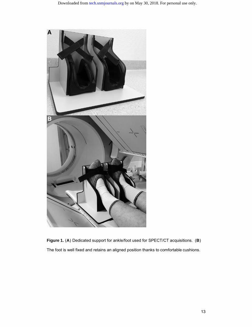
Foot Support
The support consists of a 40-cm (L) 22-cm (W) × ×15-cm (H) compact panel
composed of synthetic materials (0.8- and 1-cm thickness, 200–300 HU) built for this
study to achieve firm, but comfortable immobilization of the foot and the ankle during
the examination (Figure 1). The inner parts were padded with soft material (2- to 3-
cm thickness, HU –950) in order to reduce patient discomfort. The support was not
found to minimally attenuate significantly the gamma radiation at 140 keV energies
(about 13–16%) (22). More importantly, the support was entirely in the field of view of
the CT, which is important as to not introduce truncation artifacts during attenuation
correction (17).
Each patient received instructions to avoid any movements during the
procedure. The support was manufactured to resemble the ankle and the foot MRI
array coil used by our Radiology Department to ease SPECT/MRI fusion, when
needed. When in use with patients, we confirmed that both feet were always securely
fixed with the built-in strap to increase the accuracy and to make individuals feel
comfortable during image acquisition.
by on May 30, 2018. For personal use only. tech.snmjournals.org Downloaded from
5
Image acquisition
All patients underwent classical technetium-99m-3,3-diphosphono-1,2- propanedicar-
boxylic acid bone scintigraphy after intravenous injection of a mean activity of
840±100 MBq (range, 590–950 MBq), followed by SPECT/CT imaging of both foot and
ankle 5.4±0.9 hours (range 3.8–7.8 hours) after tracer injection.
SPECT/CT (GE Discovery NM/CT 670, GE Healthcare, Milwaukee, WI) image
acquisition was performed using high-resolution low-energy collimator with auto
contour rotation mode (15 seconds/frame, 60 views over 360º, step-and-shoot mode,
matrix size 128x128, zoom factor 1.00) and a 15% energy window was centered on
140 keV. Helical CT acquisitions used a voltage of 120 kV, a current of 90 mA, a slice
thickness of 0.625 mm and a 512 matrix for reconstruction.
SPECT images were reconstructed using iterative reconstruction (ordered subset
expectation maximization), 2 iterations, 10 subsets, using PSF-recovery “Evolution for
bone” algorithm” and CT for attenuation correction on a Xeleris 3.0 workstation (GE
Healthcare, Milwaukee, WI).
Image analysis
SPECT and CT image datasets were processed using the Integrated Registration tool
on an Advantage workstation (GE Healthcare, Milwaukee, WI). The misalignment
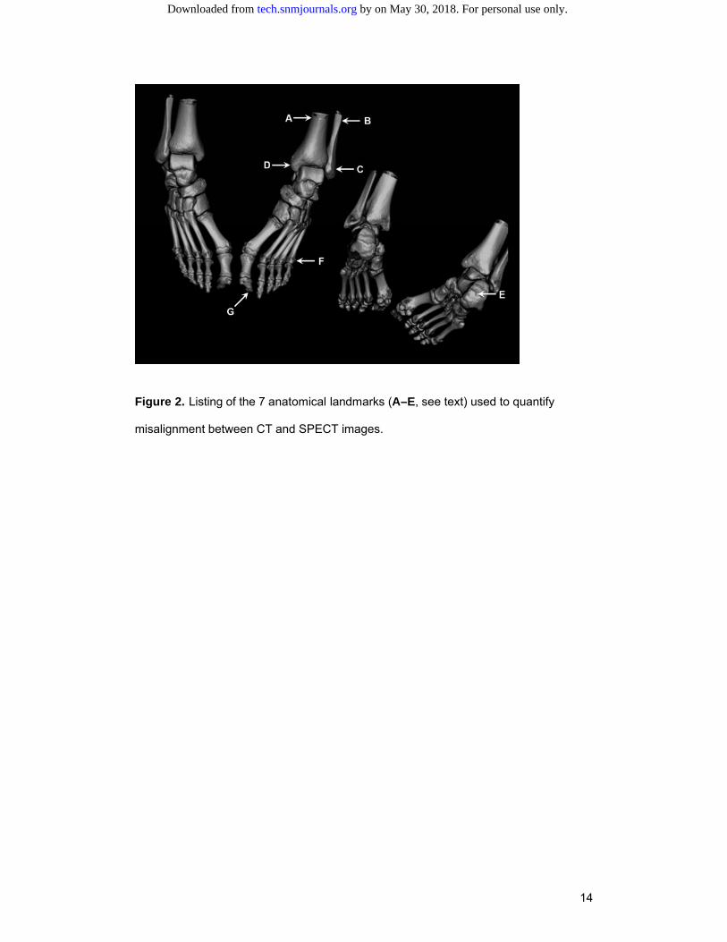
between SPECT and CT acquisitions was quantified by manually placing 14 fiducial
markers chosen among anatomical landmarks (tibia, fibula, medial malleolus, lateral
malleolus, insertion of the Achilles tendon, tuberosity of the 5th metatarsal and distal
phalanx of great toe of right and left foot/ankle) on each SPECT and CT images
(Figure 2). For optimal visualization of the different landmarks, varying the color look-
up table of the images was allowed. The fiducial marker placement was processed by
a nuclear technologist and subsequently checked by two nuclear physicians.
by on May 30, 2018. For personal use only. tech.snmjournals.org Downloaded from
6
Statistical Analysis
The results were analyzed using Stata 13.1 (StataCorp, College Station, TX). Mean
and standard deviation are presented unless specified otherwise. Box plots were
generated to display the absolute difference between patients group. Differences
between the 2 datasets according to the presence or absence of support and the
fiducial marker number (1–14, 7 for the right foot and 7 for the left one) were assessed
using analysis of variance. A p-value of 0.05 was considered as the threshold for
significance.
Results
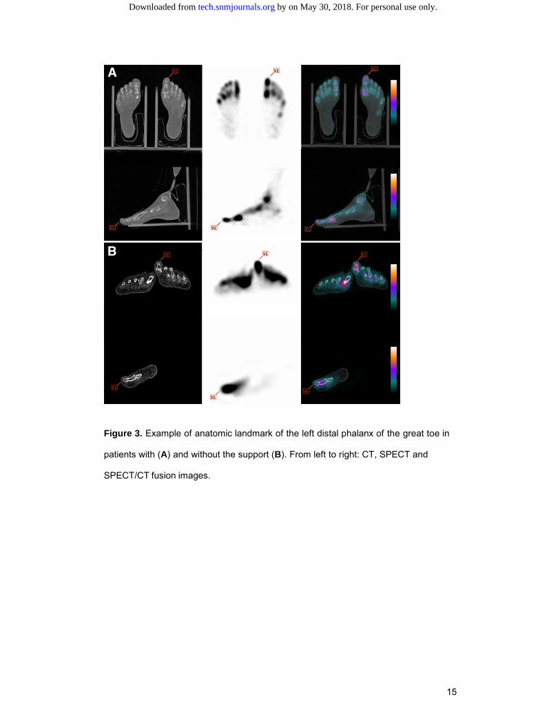
A total of 280 anatomical landmarks were analyzed in twenty patients (as illustrated in
Figure 3 for the great toe landmark on SPECT and CT images for a patient with the
support and without the support. The absolute difference for patients without and
with support was 5.1±5.2 mm vs. 3.1±2.7 mm, respectively. Thus, the alignment
between SPECT and CT images was better with support, as reported in Figure 4.
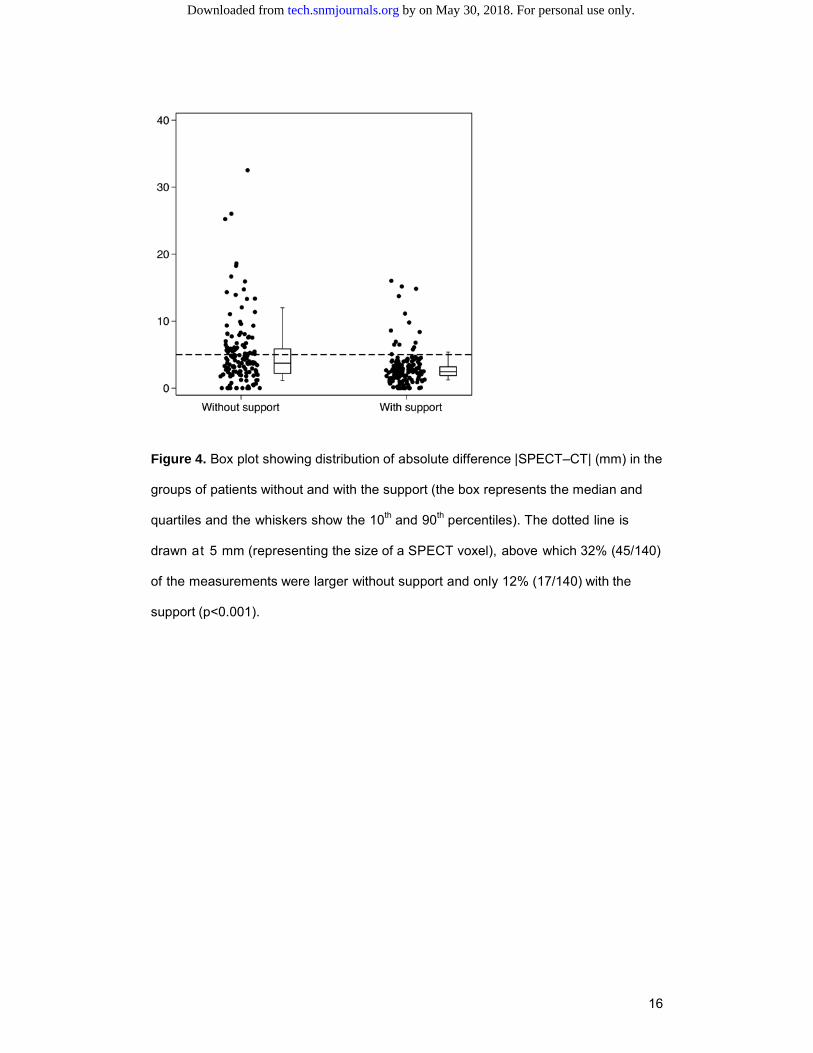
When considering a misalignment threshold of 5 mm as a reference for appropriate
clinical evaluation, most individuals using the support were found below this empirical
value (dotted line) as opposite to patients without the support.
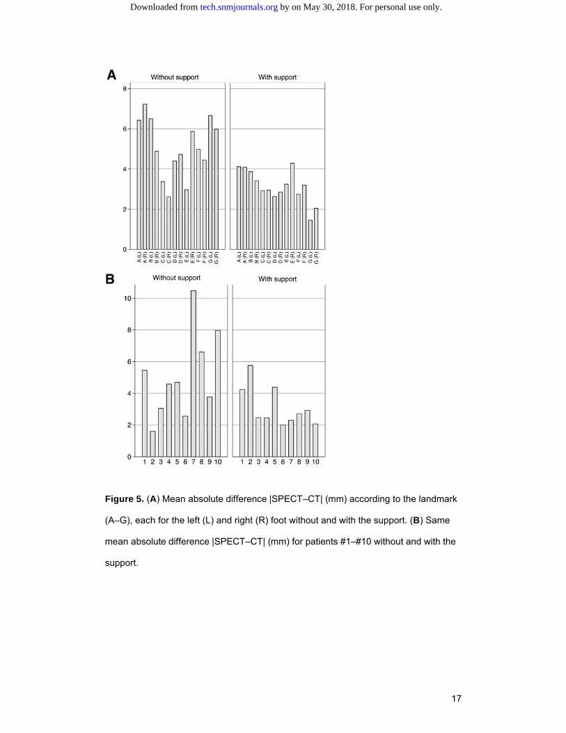
Results of the analysis of variance showed that misalignments in the group using the
support were significantly lower than for those without support (p<0.0001). Moreover,
no dependence of the misalignment on the fiducial marker was observed (p=0.54)
(Figure 5A).
by on May 30, 2018. For personal use only. tech.snmjournals.org Downloaded from
7
Discussion
This is the first study that quantitatively evaluates the value of a lower limb
immobilization device during SPECT/CT acquisitions. Our results show a significant
reduction in misalignment of SPECT/CT images between patients with and without the
dedicated foot support, presumably due to a more stable and comfortable patient
position using the support. However, a certain degree of misalignment is still present
because of uncontrollable movements and the support may need to be adjusted for
each patient (e.g. the distal extremities of the toes depend on the foot size).
Beyer et al. assessed the effectiveness of different supports to reduce misregistration
in the head and the neck during whole-body positron emission tomography/computed
tomography studies using anatomical landmarks (20). They observed that the motion
likelihood can be reduced and that an improvement in accuracy can be observed
during coregistration (the misalignment was reduced to a minimum of 1.4 mm for a
head holder fitted with a vacuum-lock bag). However, our study shows larger
misalignments even when the support is applied, which could be explained by
SPECT’s worse spatial resolution and greater partial volume effect. Moreover, head
and the neck structures maybe more stable when compared with lower extremities, as
the ones addressed in this article.
An important limitation of manual alignment is the focally increased pathological
uptake observed in certain patients, which increased uncertainty when assigning
anatomical landmarks close to these structures. The simultaneous analysis of CT
images increases accuracy when using this approach, emphasizing the previously
mentioned limitation. Structures such as malleoli or the one concerning the insertion of
the Achilles tendon were particularly difficult to identify but the task seemed to be
easier when the support was used. In these conditions, the implementation of the
by on May 30, 2018. For personal use only. tech.snmjournals.org Downloaded from
8
support seems to improve the clinical performance of the test, allowing for better
characterization in case of small focal abnormalities. It is worth mentioning that SPECT
spatial resolution was worse than CT’s leading to difficulties to precisely identify
anatomical landmarks; this could therefore mask smaller differences.
Gayed et al. reported an increased quality in diagnostic when using SPECT/CT,
when compared to SPECT (8). Considering this, misregistration between SPECT and
CT would need to be kept to a minimum by using a support like ours. For instance,
when considering the horizontal red line in Figure 3, drawn at 5 mm (this represents
the size of a SPECT voxel or about half of the spatial FWHM SPECT resolution), 32%
(45/140) of the values for the class of patients without support is above it while this
number reduces to 12% (17/140), for patients with the support which is significantly
different (p<0.001).
One limitation of this current support design is a slight increase in the distance
between the foot/ankle and the detector due to the intrinsic geometry of the support
which was manufactured to look alike a foot ankle MR array coil to allow easy
SPECT/MR fusion. This could lead to a slight decrease in spatial resolution. A final
limitation of our study would be a relatively small number of patients in each group
(npatients=10), with a number of anatomical landmarks in each patient (nlandmarks=14)
quite high making a large number of measurements in each group (nmeasurements=140 in
total), however. Future research w i l l include comparing the comfort with and
without support in the same patient, as well as simultaneously evaluating image
quality.
by on May 30, 2018. For personal use only. tech.snmjournals.org Downloaded from
9
Conclusion
Our study shows that a foot immobilization device can significantly reduce
misalignment between SPECT and CT images during hybrid SPECT/CT. This may
positively influence localization and clinical diagnosis in areas of complex foot and ankle
anatomy.
Acknowledgments
We are indebted to Mr. Michel Gillieron from our institution’s wood workshop for
manufacturing the support.
by on May 30, 2018. For personal use only. tech.snmjournals.org Downloaded from
10
References
1. van Dijk M, Lavalaye J. Nuclear imaging in orthopaedic decision making with focus
on ankle and foot. Tijdschr Nucl Geneesk. 2012;34:968-976.
2. Mariani G, Bruselli L, Kuwert T, et al. A review on the clinical uses of SPECT/CT.
Eur J Nucl Med Mol Imaging. 2010;37:1959-1985.
3. Jacene HA, Goetze S, Patel H, Wahl RL, Ziessman HA. Advantages of Hybrid
SPECT/CT vs SPECT Alone. Open Med Imaging J. 2008;2:67-69.
4. Singh VK, Javed S, Parthipun A, Sott AH. The diagnostic value of single photon-
emission computed tomography bone scans combined with CT (SPECT-CT) in
diseases of the foot and ankle. Foot Ankle Surg. 2013;19:80-83.
5. Pagenstert GI, Barg A, Leumann AG, et al. SPECT-CT imaging in degenerative
joint disease of the foot and ankle. J Bone Joint Surg Br. 2009;91:1191-1196.
6. Filippi L, Uccioli L, Giurato L, Schillaci O. Diabetic foot infection: usefulness of
SPECT/CT for 99mTc-HMPAO-labeled leukocyte imaging. J Nucl Med.
2009;50:1042-1046.
7. Scharf S. SPECT/CT imaging in general orthopedic practice. Semin Nucl Med.
2009;39:293-307.
8. Gayed I, Wan D, Joseph U, Awad J, John S. Impact of bone SPECT-CT imaging
on evaluation of lower extremities' pathology. J Nucl Med. 2011;52:456.
9. Huellner MW, Strobel K. Clinical applications of SPECT/CT in imaging the
extremities. Eur J Nucl Med Mol Imaging. 2014;41 Suppl 1:S50-58.
10. Mohan HK, Gnanasegaran G, Vijayanathan S, Fogelman I. SPECT/CT in imaging
foot and ankle pathology-the demise of other coregistration techniques. Semin
Nucl Med. 2010;40:41-51.
11. Navas A. Role of computed tomography in chronic ankle and foot pain. Tijdschr
Nucl Geneesk. 2012;34:961-967.
12. Robinson AH, Bird N, Screaton N, Wraight EP, Meggitt BF. Coregistration imaging
of the foot. A new localisation technique. J Bone Joint Surg Br. 1998;80:777-780.
by on May 30, 2018. For personal use only. tech.snmjournals.org Downloaded from
11
13. Knupp M, Pagenstert GI, Barg A, Bolliger L, Easley ME, Hintermann B. SPECT-CT
compared with conventional imaging modalities for the assessment of the varus
and valgus malaligned hindfoot. J Orthop Res. 2009;27:1461-1466.
14. Saha S, Burke C, Desai A, Vijayanathan S, Gnanasegaran G. SPECT-CT:
applications in musculoskeletal radiology. Br J Radiol. 2013;86:20120519.
15. Psiuk-Maksymowicz K, Borys D, Gorczewski K, Steinhof K, d’Amico A. CT/SPECT
image fusion in patients treated with iodine-131. J Med Informatics &
Technologies. 2004;8:7-13.
16. Majumder DD, Ray D. Approaches of Multimodal Medical Images Registration and
Fusion: Efficacy on Diagnostic and Therapeutic Planning. IETE J Res.
2011;57:498-514.
17. Płachcińska A, Siennicki J, Kovacevic-Kuśmierek K, Bieńkiewicz M, Kuśmierek J.
Effect of attenuation correction on normal 99mTc-MIBI myocardial perfusion
scintigrams acquired with a hybrid SPECT/CT camera. Nuclear Med Rev.
2008;11:59-66.
18. Fricke H, Fricke E, Weise R, Kammeier A, Lindner O, Burchert W. A method to
remove artifacts in attenuation-corrected myocardial perfusion SPECT Introduced
by misalignment between emission scan and CT-derived attenuation maps. J Nucl
Med. 2004;45:1619-1625.
19. Seo Y, Mari C, Hasegawa BH. Technological development and advances in single-
photon emission computed tomography/computed tomography. Semin Nucl Med.
2008;38:177-198.
20. Gnanasegaran G, Cook G, Adamson K, Fogelman I. Patterns, variants, artifacts,
and pitfalls in conventional radionuclide bone imaging and SPECT/CT. Semin Nucl
Med. 2009;39:380-395.
21. Beyer T, Tellmann L, Nickel I, Pietrzyk U. On the use of positioning aids to reduce
misregistration in the head and neck in whole-body PET/CT studies. J Nucl Med.
2005;46:596-602.
by on May 30, 2018. For personal use only. tech.snmjournals.org Downloaded from

12
22. Brown S, Bailey DL, Willowson K, Baldock C. Investigation of the relationship
between linear attenuation coefficients and CT Hounsfield units using
radionuclides for SPECT. Appl Radiat Isot. 2008;66:1206-1212.
by on May 30, 2018. For personal use only. tech.snmjournals.org Downloaded from
13
Figure 1. (A) Dedicated support for ankle/foot used for SPECT/CT acquisitions. (B)
The foot is well fixed and retains an aligned position thanks to comfortable cushions.
by on May 30, 2018. For personal use only. tech.snmjournals.org Downloaded from
14
Figure 2. Listing of the 7 anatomical landmarks (A–E, see text) used to quantify
misalignment between CT and SPECT images.
by on May 30, 2018. For personal use only. tech.snmjournals.org Downloaded from
15
Figure 3. Example of anatomic landmark of the left distal phalanx of the great toe in
patients with (A) and without the support (B). From left to right: CT, SPECT and
SPECT/CT fusion images.
by on May 30, 2018. For personal use only. tech.snmjournals.org Downloaded from
16
Figure 4. Box plot showing distribution of absolute difference |SPECT–CT| (mm) in the
groups of patients without and with the support (the box represents the median and
quartiles and the whiskers show the 10th and 90th percentiles). The dotted line is
drawn at 5 mm (representing the size of a SPECT voxel), above which 32% (45/140)
of the measurements were larger without support and only 12% (17/140) with the
support (p<0.001).
by on May 30, 2018. For personal use only. tech.snmjournals.org Downloaded from
17
Figure 5. (A) Mean absolute difference |SPECT–CT| (mm) according to the landmark
(A–G), each for the left (L) and right (R) foot without and with the support. (B) Same
mean absolute difference |SPECT–CT| (mm) for patients #1–#10 without and with the
support.
by on May 30, 2018. For personal use only. tech.snmjournals.org Downloaded from
Doi: 10.2967/jnmt.114.145771Published online: April 9, 2015.J. Nucl. Med. Technol. and José A. Pires-JorgeJoana do Mar F. Machado, Marina S. Monteiro, Victor Fernandes Vieira, Jean-Aybert Collinot, John O. Prior, Lina Vieira Value of a lower limb immobilization device for SPECT/CT image fusion optimization
http://tech.snmjournals.org/content/early/2015/04/09/jnmt.114.145771This article and updated information are available at:
http://tech.snmjournals.org/site/subscriptions/online.xhtml
Information about subscriptions to JNMT can be found at:
http://tech.snmjournals.org/site/misc/permission.xhtmlInformation about reproducing figures, tables, or other portions of this article can be found online at:
and the final, published version.proofreading, and author review. This process may lead to differences between the accepted version of the manuscript
ahead of print area, they will be prepared for print and online publication, which includes copyediting, typesetting,JNMTcopyedited, nor have they appeared in a print or online issue of the journal. Once the accepted manuscripts appear in the
. They have not beenJNMT ahead of print articles have been peer reviewed and accepted for publication in JNMT
(Print ISSN: 0091-4916, Online ISSN: 1535-5675)1850 Samuel Morse Drive, Reston, VA 20190.SNMMI | Society of Nuclear Medicine and Molecular Imaging
is published quarterly.Journal of Nuclear Medicine Technology
© Copyright 2015 SNMMI; all rights reserved.
by on May 30, 2018. For personal use only. tech.snmjournals.org Downloaded from