valproic acid-induced changes in gene expression during neurulation in a mouse model
TRANSCRIPT

TERATOLOGY 54284-297 (1996)
Valproic Acid-Induced Changes in Gene Expression During Neurulation in a Mouse Model BOGDAN C. WLODARCZYK,1*2 JOHANNAC. CRAIG: GREGORY D. BENNETT? JAMES A. CALVIN,S AND RICHARD H. FI"ELL2* INational Veterinary Research Institute, Department of Pharmacology and Toxicology, Pulawy, Poland 2Department of Veterinary Anatomy and Public Health, Texas A&M University, College Station, Texas 3Department of Statistics, Texas A&M University, College Station, ? h a s
ABSTRACT The teratogenic potential of val- proic acid has been well established both in experimen- tal models and in human clinical studies. As with all human teratogens, there are genetically determined differences in individual susceptibility to the induction of congenital defects. Using a mouse model of valproate- induced neural tube defects, a study was undertaken to examine differential changes in gene expression for selected transcription factor (Pax-3, Emx-1, Emx-2, c-fos, c-jun, creb) and cell cycle checkpoint genes (bcl-2, p53, wee-1) during neural tube closure. In general, exposure to teratogenic concentrations of valproic acid elicited GD 9:12 control levels of transcrip- tion factor mRNA expression in GD 9:0 embryos of both strains. This accelerated developmental profile is marked by significant elevation of Ernx-1 , Ernx-2, c-fos, c-jun, and creb expression. There was also a significant overexpression of the cell cycle genes p53 and bcl-2 in the LMDc embryos in response to the teratogenic insult. Examination of the ratio of expression of these genes clearly favored bcl-2, which supports the hypoth- esis that altered neuroepithelial cell proliferation rates, rather than increased apoptosis, is the underlying mechanism by which valproic acid alters normal neural tube rnorphogenesis. An investigation into interactive effects of these genes on the molecular profile of GD 9:0 embryos further validated this observation. That is, the overall proliferative state among the control em- bryos was prematurely modified into a more differenti- ated state following teratogenic insult. These results suggest that alterations in the expression of multiple genes are most likely responsible for valproic acid- induced neural tube defects. Teratology 54284-297, 1996. o 1997 ~ i ~ e y - ~ i s s , Inc.
Teratogens are believed to act in a number of differ- ent pathways exploiting multiple targets and mecha- nisms to alter normal embryogenesis (Wilson, '77). Relatively little is known about the consequences of a teratogenic insult on the molecular homeostasis of the developing embryo. Such information is only now being rapidly acquired. What is apparent from a number of different experimental systems is that a teratogenic
insult can disrupt important developmental processes controlled by multiple genes under an elaborate regula- tory structure. One approach that has been successfully applied to investigate complex developmental events and their coordinate genetic regulation has been to disrupt normal morphogenesis using specific terato- genic agents and then to analyze carefully the physi- ological and molecular consequences. We used this approach to examine the expression patterns for se- lected transcription factors and cell cycle checkpoint genes and attempted to correlate any changes in gene expression with observations at the morphological lev- els following teratogenic treatment with the anticonvul- sant medication, valproic acid (Depakene, Abbott Labo- ratories, Abbott Park, IL). The period during which neural tube closure occurs was selected as the target developmental timeframe, as the well-documented mor- phogenetic events occurring during this period of devel- opment demands complex tissue organization within rigid temporal and spatial requirements, and under- scores the complexity of the processes involved (Geelen and Langman, '77; Kaufman, '79; Macdonald et al., '89; Finnell, '91; Golden and Chernoff, '93).
Structurally unrelated to any of the other anticonvul- sant medications, valproic acid (VPA) has a demon- strated teratogenic effect in a wide variety of animal species, including the mouse, rat, hamster, rabbit, and rhesus monkey (for review, see Nau and Hendrickx '87). The principal malformation associated with VPA expo- sure in utero in experimental animals has been neural tube defects (NTDs), including exencephaly and spina bifida. Nau ('85) observed exencephaly in mouse em- bryos from dams exposed to sufficiently high dosages of
Contract grant sponsor: National Institute of Dental Research, con- tract grant number DE 11303; contract grant sponsor: National Institute of Environmental Health Sciences, contract grant number ES07165; contract grant sponsor: NIH.
*Correspondence to: Dr. Richard H. Finnell, Department of Veterinary Anatomy and Public Health, College of Veterinary Medicine, Texas A&M University, College Station, TX 77843-4458. E-mail: [email protected]
Received 12 September 1996; Accepted 17 December 1996
o 1997 WILEY-LISS, INC.

VALPROIC ACID TERATOGENICITY AND GENE EXPRESSION 285
VPA to produce maternal plasma concentrations in excess of 230 pg/ml, irrespective of the route of adminis- tration. This concentration represents a two- to fivefold increase over the recommended human therapeutic level (Niedermeyer, '83). Ehlers et al. ('92a,b) demon- strated that VPA administered (200 mg/kg, IP) 6 hr apart, beginning on gestational day 9, produced a 10% response frequency of spina bifida occulta, which in- creased to 95% affected fetuses concomitant with a VPA dosage increase to 500 mgkg body weight. A significant degree of malformation of the ribs and vertebrae was apparent when the above-treated fetuses were exam- ined following Alcian blue-alizarin red skeletal stain- ing (Ehlers et al., '92a,b). A low frequency (4-6%) of spina bifida aperta was also induced by the same VPA treatment regimen in the Han:NMRI mouse strain, resulting in a highly disorganized and necrotic spinal cord within the vertebral canal in the lumbosacral region of the developing fetus. The absence of neuronal tissue in these affected fetuses indicates an almost complete localized ablation of the neural tube in the VPA-exposed fetuses (Ehlers et al., '92b).
Clinically, infants exposed in utero to VPA are a t elevated risk of an NTD (Robert, '82; Robert and Guibaud, '82; Lindhout et al., '92). An estimated 2% of all infants exposed to VPA during early pregnancy will have spina bifida, a 20-fold increased prevalence over that observed in the general population (Lammer et al., '87). In addition to NTDs, a characteristic pattern of craniofacial abnormalities, including a flat nasal bridge with upturned nasal tip, thin vermilion borders, a shallow philtrum, and downturning mouth, has been described among valproate-exposed infants (DiLiberti et al., '84; Ardinger et al., '88). As the mechanism by which VPA induces NTDs is poorly understood, it remains difficult to predict which infants exposed in utero to the drug will develop spina bifida. Clearly, fetuses that have some genetically determined predispo- sition are at increased risk.
Efforts to learn more about the genetic basis of susceptibility to VPA-induced NTDs have relied on data obtained from murine model systems. Finnell et al. ('88) treated dams from several inbred mouse strains with a single IP injection of VPA (600 mg/kdday) on gesta- tional day 8:12, and observed a strain-dependent hierar- chy of susceptibility to NTDs that paralleled the pat- tern of susceptibility observed for mice exposed to elevated intrauterine temperatures (Finnell et al., '86). That is, the SWV mice were very sensitive, with 35% of fetuses exencephalic, while the LhUBc embryos were much more resistant (20% NTDs), and the C57BU6J and the DBA/2J mice were completely resistant (Finnell et al., '88). The underlying basis of this strain difference was examined with respect to folate metabolism. Weg- ner and Nau ('91, '92) determined total folate and several folate metabolite concentrations in mice follow- ing teratogenic exposure to VPA. These investigators found a significant alteration in the concentrations of several formylated tetrahydrofolates, specifically the 5-
and 10-formyl-THF, as well as the 5-CH3-THF metabo- lites. When these metabolites were measured in VPA- exposed embryos from the NTD-sensitive (SWV) or -resistant (DBA/2J) strains, a 86-92% inhibition of the 5-CHO-THF and 5-CH3-THF metabolites was found in the SWV embryos, while the DBN2J embryos had no alterations in their 5-CH3-THF, and only a 50% inhibi- tion in the 5-CHO-THF metabolite. The LM/Bc strain embryos were intermediate between the SWV and DBA/2J embryos both in susceptibility to VF'A-induced NTDs and in their profile of metabolic inhibition (R.H. Finnell and H. Nau, unpublished data). This drug- induced alteration in folate metabolism could poten- tially result in a decreased rate of methylation of essential developmentally regulated genes during criti- cal periods of embryogenesis. This would significantly curtail the expression of certain genes, which in turn might enhance the sensitivity of the embryos to specific malformations. Such a difference in the methylation patterns between embryos of different strains might explain their differential sensitivity to VPA-induced NTDs.
The present study was undertaken to monitor the expression level of a population of six transcription factor and three cell cycle checkpoint genes in SWV and LM/Bc embryos in order to (1) document changes in gene expression that occur over time in normal murine embryos during a period of rapid morphogenesis, (2) identify altered patterns of gene expression occurring subsequent to a teratogenic exposure to VPA that may contribute to the abnormal pathogenesis observed in embryos, and (3) determine a possible molecular basis for the strain difference in susceptibility to VPA- induced NTDs. The six transcription factor genes (Pax-3, Emx-1, Emx-2, c-fos, c-jun, and creb) selected for inves- tigation are all believed to be centrally involved in directing or regulating a cascade of morphogenetic events critical to early embryogenesis. The cell cycle checkpoint genes examined in these embryos included bcl-2, p53, and wee-1. These genes are known to be important modifiers of cell proliferation and are devel- opmentally regulated in the mouse embryo.
MATERIALS AND METHODS Experimental animals
The highly inbred SWV and LM/Bc mouse strains were selected for these studies on the basis of their known differences in sensitivity to VPA-induced NTDs (Finnell et al., '88). The mice were maintained on a 12-h light cycle in the Laboratory Animal Resources and Research Facility a t the College of Veterinary Medicine, Texas A&M University. The animals were pathogen- free with no apparent health problems. Up to five healthy females were housed per polycarbonate cage and were allowed free access to Wayne TekLad rodent chow and tap water. Virgin females, 40-60 days of age, were bred overnight to experienced males of their respective strain. Females were examined the next morning for the presence of vaginal plugs. The begin-
286 B.C. WLODARCZYK ET AL.
ning of gestation (day 0) was set at 10 PM of the previous evening, the midpoint of the dark cycle (Snell et al., '48).
Teratogen treatments For the teratology studies, no less than five dams
were randomly assigned to each treatment group and exposed to a single intraperitoneal injection of VPA (600 mgkg) or the distilled water vehicle a t GD 8.5. The drug was dissolved in distilled water immediately before use and administered in volumes of 0.1 my10 g body weight. Following treatment, the dams are re- turned to their home cages until the desired gestational day for the embryo collection. At the assigned hour (GD 8:18, 9.0, and 9:12), pregnant dams were killed by cervical dislocation, the abdomen opened, and the uterine contents removed. The location of all viable embryos or fetuses and resorption sites were recorded. Using watchmaker's forceps, the embryos were dis- sected free of the decidual capsule and its chorion and amnion while in cold phosphate-buffered saline (PBS), under a Wild M8 dissecting microscope (Heerbrugg, Switzerland).
Embryo collection and morphological staging Embryos were collected and grossly examined mor-
phologically to facilitate their classification on the basis of their stage of neural tube closure, using previously described standardized staging criteria (Cole and Trader, '80; Macdonald et al., '89; Golden and Chernoff, '93). Embryos with obviously delayed neural tube clo- sure, or completely open neural folds that clearly were developing NTDs, were selected from the teratogen- exposed litters for use in the in situ transcription studies. All control embryos for these experiments were developing normally toward the completion of anterior neural tube closure.
Removal of neural tube from neural tube closure stage embryos
For the gene expression studies, no less than three embryos per litter from a minimum of five litters per treatment group were used as the basis of the dataset. The embryos were collected as described above. With the aid of watchmaker's forceps, the neural tube proper was dissected away from supporting paraxial mesoder- mal tissue under the dissecting microscope (Taylor et al., '95; Wlodarczyk et al., '96). Once dissected free, it was examined to ensure that only the intact neural tube tissue had been collected, free from extraneous tissues. In preparation for the in situ studies, this tissue was placed in a hybridization buffer containing 5 mM DTT, 100 units RNasin (Promega, Madison, WI), and 0.1% digitonin. Following a brief pulse with a sonic dismembranator, an additional 50 units of RNasin was added to the buffer, and the tissue was frozen at -80°C until further processed.
In situ transcription and aRNA amplification In situ transcription and antisense RNA amplifica-
tion (RT/aRNA) procedures were performed according
to methods that have been described in detail else- where (Eberwine et al., '92a,b; Taylor et al., '95; Wlodar- czyk et al., '96). The initial steps involved making an RNA/DNA hybrid molecule from the population of mRNAs present in each of the isolated neural tube samples with the addition of avian myeloblast reverse transcriptase (Seikagaku America, Bethesda, MD), and an oligo-dT-T7 oligonucleotide primer that hybridizes to the poly-A tail of the mRNAs. The purified single stranded cDNA was then made double stranded by the Gubler-Hoffman method, the resulting hairpin loop was digested with S1 nuclease, and then blunt-ended using standard procedures. The cDNA template was further purified by a spin column procedure, and then used t o produce radiolabeled, amplified, antisense RNA (aRNA) by the addition of T7 RNA polymerase (Epicen- tre Technologies, Madison, WI) in the presence of [32Pl-CTP. The aRNA, representing the entire popula- tion of mRNAs from the neural tube tissue, was then hybridized to "reverse" Northern blots in order to determine quantitatively the pattern of gene expres- sion for the candidate genes selected for investigation (see below). This approach was selected over more conventional reverse transcriptase-polymerase chain reaction (RT-PCR) procedures, given the linear nature of the RT/aRNA amplifications.
Genetic expression profiling Equimolar concentrations of the cDNA clones of the
nine genes of interest (Pax-3, Emx-1, Emx-2, c-fos, c-jun, web, bcl-2, p53, and wee-1) were immobilized on a nylon membrane (Zetaprobe, BioRad, Richmond, CA), along with an internal control cDNA (cyclophilin), using a BioRad slotting apparatus following the manu- facturer's protocol. The resulting slot blots were hybrid- ized with an aRNA probe using conditions detailed previously by Taylor et al. ('95). Following hybridiza- tion, the slot blots were washed at medium stringency down to 0.1X SSC containing 0.1% sodium dodecyl sulfate (SDS) at 42"C, dried, wrapped in plastic wrap, and placed in an Ambis 101 two-dimensional radioana- lytics imaging detector (Scanalytics, Billerica, MA) that directly measured the radioactivity (CPMs) of each slot on the reverse Northern blots. The individual signals were normalized to cyclophilin gene expression. The selection of cyclophilin as the normalizing cDNA simply enables us to make comparisons between different blots, as the individual hybridization intensities of individual cDNA on each blot can be expressed as a ratio of its expression to cyclophilin. There is no particu- lar significance to the selection of this gene, other than the fact that it is constituitively expressed and found in high abundance in the mouse embryo (Danielson et al., '88). This makes it an excellent internal standard for use in multiprobe assays when examining low abun- dance messages.
Basis for selection of candidate genes The nine cDNA clones selected for this particular
analysis encode three cell cycle genes (wee-I, p53, and
VALPROIC ACID TERATOGENICITY AND GENE EXPRESSION 287
bcl-2) and six transcription factor genes (Pax-3, Emx-1, Emx-2, c-fos, c-jun, creb). The basis for selection of the cell cycle cDNA clones, as well as c-fos, c-jun, and creb was their documented interactive effects on epithelial cell differentiation and proliferation through the cell cycle. The remaining transcription factors, Pax-3, Emx-1, and Emx-2 are known to play important roles in patterning during early embryogenesis. The selected cell cycle genes encode checkpoint proteins that operate at the Gz (wee-I) or the GI (p53 and bcl-2) cell cycle phases, and are involved in various aspects of assess- ment and regulation of DNA replication and repair (Ellege and Lee, '95). wee-1 encodes a protein kinase that negatively regulates entry into mitosis by inactiva- tion of the maturation promoting factor (MPF) (Davey et al., '95). Overexpression of wee-1 inhibits the cellular transition from Gz to mitosis in the cell cycle, ulti- mately leading to apoptotic cell death (Igarashi et al., '91; Davey et al., '95). At the GI cell cycle phase, it has been reported that the protein product of bcl-2 mediates the growth-inhibiting and apoptotic effects of p53 pro- tein product (Miyashita et al., '94). All three of these cell cycle genes are expressed in a number of different tissues, especially in "epithelium-like" cells. p53 is expressed in the developing neural tube and has impor- tant implications in orchestrating neural tube closure, via cell cycle regulation (Sah et al., '95), while bcl-2 is normally highly expressed in fetal central nervous system (CNS) tissues, as well as in rapidly proliferating epithelial cells (Hockenberry, '95; Veis-Novak and Kors- meyer, '94).
The transcription factors Pax3, creb, Emx-1, and Emx-2 serve as the initial triggers in a cascade of molecular events that result in changing the relative abundance of many responsive downstream genes. With the exception of creb, all these genes are involved in embryonic pattern formation. Emx-1 and Emx-2 are both homeobox-containing genes expressed in restric- tion regions of the developing forebrain, including the presumptive cerebral cortex (Simeone et al., '92). The products of these homeobox containing genes, namely homeodomain proteins, serve to regulate gene expres- sion by directly binding to cis-acting DNA sequences (Breier et al., '88). The Pax3 gene encodes a 56-kd protein that contains a paired-type homeodomain in addition to the paired box domain (Goulding et al., '91). Throughout neural development, Pax3 expression was coincident with regions of the neural tube from which neural crest cells are known to arise and migrate.
c-jun has the general properties of most oncogenes in that it is involved in regulating either cellular prolifera- tion or differentiation (Weinberg, '85). Specifically, c-jun encodes for a nuclear phosphoprotein that is a transcrip- tion factor, comprising a major component of the AP-1 transcription complex, which also includes the Fos protein (Angel et al., '88). These immediate-early- response genes are believed to play a vital role in cellular proliferation and the regulation of the cell cycle, as it was demonstrated that these genes are
induced within minutes of a mitogenic stimulation (Kovary and Bravo, '91). These transcription factors bind to specific DNA consensus sequences known as AP- 1 recognition sites, or TPA-response elements (Bohmann et al., '87). This binding enables the preferen- tial binding of RNA polymerase, and thus the rapid transcription of specific genes that have bound these factors (Ptashne, '88). Taken together, the interassocia- tions of these nine genes may aid in regulating cellular proliferation and establishing future embryonic axis patterning and polarity in the developing neural tube. Such interactions may have important implications for neuroepithelial cell function during neural plate fold- ing and neural fold fusion, as well as organization of future CNS differentiation events.
Statistical analysis The data for the statistical analysis were generated
from LM/Bc/Fnn and SWV/Fnn embryos collected at GDs 8:12, 9:0, and 9:12, under control and VPA treat- ment conditions. All statistical analysis of these data was performed using SAS (Statistical Analysis System) and completed in three stages. The first of these stages involved morphological analysis of somite pairs and determination of neural tube closure stage frequency distribution. The second and third stages involved univariate and multivariate analyses, respectively, of the candidate genes.
Morphological analysis The mean number of somite pairs per embryo, as well
as the frequency distribution of the stages of neural tube closure were subjected to a two-way analysis of variance (ANOVA) and a chi-square goodness of fit test. This was done in order to determine any VPA treatment effects at a given timepoint on embryonic growth and development (Sokal and Rohlf, '81). Follow-up analyses of significant effects observed in the ANOVA testing were performed using a Bonferroni multiple compari- son test.
Univariate analysis These procedures involved determination of statisti-
cal significance (P < 0.05) in the gene expression profil- ing studies, both within and between strains. Because of the factorial nature of the treatment combinations, the testing of these experiments required investigation of the interactive effects of treatment, strain and gesta- tional stage. Statistical tests to determine treatment differences within both strains at each of the time- points, followed by tests to determine treatment differ- ences across strains and timepoints and simple ratios of gene expression, were conducted. All strain, treatment, and temporal interaction comparisons were evaluated by analysis of variance and the least-square means (LSMEANS) option in the general linear models (GLM) procedure. The LSMEANS option computes model- based estimates of arithmetic means when there are unequal sizes of the experimental groups. Therefore,

288 B.C. WLODARCZYK ET AL.
GLM was used to determine the level of significance for means among treatment classes, while adjusting for unbalanced sample sizes. For comparisons within each strain, LSMEANS was used to calculate the adjusted means for each control and VPA-treated experimental group, and contrasted these treatment means sepa- rately for each timepoint. To examine strain differences in terms of transcriptional activity, the LSMEANS procedure calculated the adjusted means of the differ- ences between the control and the teratogen-treated means separately for each timepoint and strain. This procedure then contrasted these mean differences (rep- resenting specific treatments, timepoints and strains) to test the hypothesis that the effect of W A treatment on transcription factor or cell cycle checkpoint gene expression is consistent across strains. Statistical sig- nificance for all of the univariate analyses was set at the (I‘ < 0.05) level (Sokal and Rohlf, ’81). For a descrip- tion of the least square estimation formula, see Neter et al. (’89), and for a summary of the LSMEANS option see SAS (SAS Institute, ’90). In order to avoid confusion, the adjusted means computed by LSMEANS in these analyses will be referred to throughout the remainder of the text as “means.”
Multivariate analysis Principal components analysis (PCA), an exploratory
multivariate procedure, was performed in this study to examine the coordinate interaction of the transcription factor and cell cycle checkpoint genes, for the purpose of gaining new insight into their potential combined influ- ence during neural tube development. PCA is a statisti- cal technique that looks at a group of data, with the goal of creating new variables: (1) a recombination of the original data, and (2) capable of providing the same information as the original data, but which is also more insightful when trying to assess interdependence among the original measurements. This technique is con- cerned with finding linear, or additive combinations of the original measurements, called principal compo- nents (PCs), which are based on the eigenvectors of the covariance matrix based on the original dataset. The goal is to find such a transformation, which is both biologically and statistically meaningful. For a discus- sion of PCA see Rao (‘731, Rohlf and Bookstein (‘go), or Johnson and Wichern (’92).
As a technical footnote, PCA is usually performed using observations which are independent. This is not our situation, but due to the extensive controlled breed- ing program for these highly inbred strains, it is reasonable, in this exploratory setting, to set aside the embryo interdependence when looking for target gene relationships. A separate PCA was performed on the control embryos for each of the inbred strains. Since we started with expression data on nine genes, the PCA results consisted of nine new variables (PCs), which are linear combinations of the original measurements and, for convenience, sorted based on their variability. The first new variable (PCl), is the most variably expressed
combination of the original measurements, whereas PC9 is the combination which is the least variably expressed. Therefore, the latter PCs are significantly more consistent in their respective values, among all embryos within the selected embryo group. As such, they make good candidates for describing interdepen- dencies among the original measurements which are consistently expressed for all embryos within a given group. For each control group, PC1 through PC9 were computed, and the combination of genes going into each PC was assessed for biological significance. The SAS procedure PRINCOMP was used for these calculations.
Follow-up analysis Once a specific PC was deemed to be informative, the
interdependence was simplified by eliminating any genes that had minimal impact on the PC (eigenvector values <0.2). This new, reduced combination of the original measurements was then constructed as a mul- tigene, or complex ratio, for all embryos, regardless of treatment. To graphically assess the change in the PC from control to VPA treated in each strain, a plot of the log of the numerator versus the log of the denominator was produced. If the PC distinguished among the treatment groups, the grouping for one treatment looked quite different from the other, with respect to either the location of the points or the pattern of the points within a treatment group. Simple descriptive statistics were calculated for this ratio, and a test of equal variances was performed using Hartley’s F-Max test (Mason et al., ’89) for each treatment group. To ensure that the exploratory process did not misstate the treatment relationship due to the lack of adjustment for correla- tion due to our sampling, the new PC was also assessed using the same univariate procedures described for the second stage.
RESULTS Morphological studies
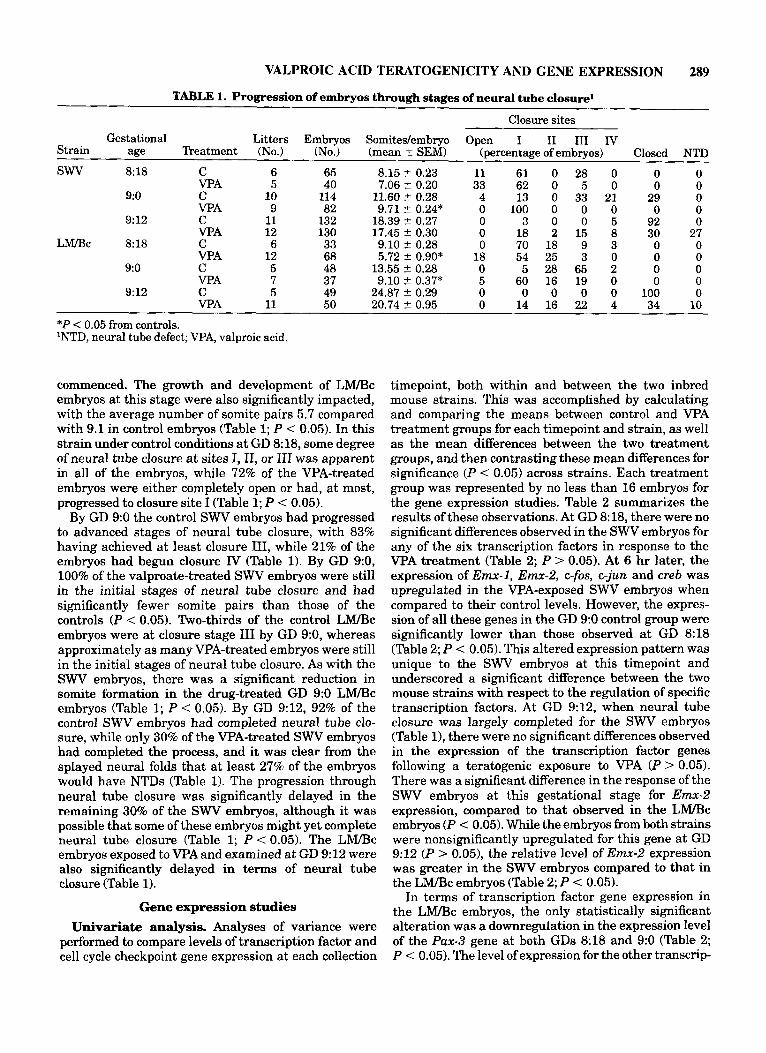
Mouse embryos from 99 litters collected between GD 8:18 and 9:12 were grossly examined for the number of somites present, and the stage or extent of neural tube closure achieved at each of the collection timepoints under control and teratogen treatment conditions (Table 1). For each of these treatment groups, no fewer than 33 embryos were examined. Any embryo whose neural folds had not yet fused at any site was considered “open.” Embryonic growth, as determined by the mean number of somite pairs present in a given specimen, was found to differ significantly according to the gesta- tional age (P < 0.05) and the teratogenic treatment (P < 0.05). VPA-treated embryos on GDs 8:18 and 9:0 had significantly fewer somite pairs relative to the controls (Table 1; P < 0.05). Of the SWV embryos examined at GD 8: 18, the valproate-treated embryos were developmentally delayed as compared to the controls, with 33% of the embryos having completely open neural tubes, while 28% of the control embryos had progressed to the point where closure at site I11 had
VALPROIC ACID TERATOGENICITY AND GENE EXPRESSION 289
TABLE 1. Progression of embryos through stages of neural tube closure1
Closure sites Gestational Litters Embryos Somitedembryo Open I I1 I11 Iv
Strain age Treatment (No.) (No.) (mean f SEM) (percentage of embryos) Closed NTD
SWV 8% C 6 65 8.15 2 0.23 11 61 0 28 0 0 0 VPA 5 40 7.06 t 0.20 33 62 0 5 0 0 0
9:o C 10 114 11.60 ? 0.28 4 13 0 33 21 29 0 VPA 9 82 9.71 t 0.24* 0 100 0 0 0 0 0
9:12 C 11 132 18.39 2 0.27 0 3 0 0 5 92 0 VPA 12 130 17.45 -C 0.30 0 18 2 15 8 30 27
L r n c 8:18 C 6 33 9.10 t 0.28 0 70 18 9 3 0 0 VPA 12 68 5.72 ? 0.90* 18 54 25 3 0 0 0
9:o C 5 48 13.55 2 0.28 0 5 28 65 2 0 0 VPA 7 37 9.10 t 0.37* 5 60 16 19 0 0 0
9:12 C 5 49 24.87 2 0.29 0 0 0 0 0 100 0 VPA 11 50 20.74 2 0.95 0 14 16 22 4 34 10
*P < 0.05 from controls. 'NTD, neural tube defect; VPA, valproic acid.
commenced. The growth and development of LM/Bc embryos at this stage were also significantly impacted, with the average number of somite pairs 5.7 compared with 9.1 in control embryos (Table 1; P < 0.05). In this strain under control conditions at GD 8:18, some degree of neural tube closure at sites I, 11, or I11 was apparent in all of the embryos, while 72% of the VPA-treated embryos were either completely open or had, a t most, progressed to closure site I (Table 1; P < 0.05).
By GD 9:0 the control SWV embryos had progressed to advanced stages of neural tube closure, with 83% having achieved at least closure 111, while 21% of the embryos had begun closure IV (Table 1). By GD 9:0, 100% of the valproate-treated SWV embryos were still in the initial stages of neural tube closure and had significantly fewer somite pairs than those of the controls ( P < 0.05). Two-thirds of the control LM/Bc embryos were at closure stage I11 by GD 9:0, whereas approximately as many VPA-treated embryos were still in the initial stages of neural tube closure. As with the SWV embryos, there was a significant reduction in somite formation in the drug-treated GD 9:0 LM/Bc embryos (Table 1; P < 0.05). By GD 9:12, 92% of the control SWV embryos had completed neural tube clo- sure, while only 30% of the VPA-treated SWV embryos had completed the process, and it was clear from the splayed neural folds that at least 27% of the embryos would have NTDs (Table 1). The progression through neural tube closure was significantly delayed in the remaining 30% of the SWV embryos, although it was possible that some of these embryos might yet complete neural tube closure (Table 1; P < 0.05). The L W C embryos exposed to VPAand examined at GD 9:12 were also significantly delayed in terms of neural tube closure (Table 1).
Gene expression studies Univariate analysis. Analyses of variance were
performed to compare levels of transcription factor and cell cycle checkpoint gene expression at each collection
timepoint, both within and between the two inbred mouse strains. This was accomplished by calculating and comparing the means between control and VPA treatment groups for each timepoint and strain, as well as the mean differences between the two treatment groups, and then contrasting these mean differences for significance (P < 0.05) across strains. Each treatment group was represented by no less than 16 embryos for the gene expression studies. Table 2 summarizes the results of these observations. At GD 8: 18, there were no significant differences observed in the SWV embryos for any of the six transcription factors in response to the VPA treatment (Table 2; P > 0.05). At 6 hr later, the expression of E m - 1 , Emx-2, c-fos, c-jun and creb was upregulated in the VPA-exposed SWV embryos when compared to their control levels. However, the expres- sion of all these genes in the GD 9:0 control group were significantly lower than those observed at GD 8:18 (Table 2; P < 0.05). This altered expression pattern was unique to the SWV embryos at this timepoint and underscored a significant difference between the two mouse strains with respect to the regulation of specific transcription factors. At GD 9:12, when neural tube closure was largely completed for the SWV embryos (Table l), there were no significant differences observed in the expression of the transcription factor genes following a teratogenic exposure to VPA (P > 0.05). There was a significant difference in the response of the SWV embryos at this gestational stage for Emx-2 expression, compared to that observed in the LhUBc embryos (P < 0.05). While the embryos from both strains were nonsignificantly upregulated for this gene at GD 9:12 (P > 0.05), the relative level of E m - 2 expression was greater in the SWV embryos compared to that in the LM/Bc embryos (Table 2; P < 0.05).
In terms of transcription factor gene expression in the LM/Bc embryos, the only statistically significant alteration was a downregulation in the expression level of the Pax-3 gene at both GDs 8:18 and 9:0 (Table 2; P < 0.05). The level of expression for the other transcrip-

290 B.C. WLODARCZYK ET AL. TABLE 2. Least-square mean response of transcription factor gene expression
Strain Genes
SWV Pax-3 E m - 1 E m - 2 c-fos c-jun creb
L r n c Pax-3 Emx-1 Ems-2 c-fos c-jun creb
Gestational day
8:18 treatment 9:0 treatment 9:12 treatment
Control VPA Control W A Control VPA
1.14 1.05 1.06 1.07 1.14 1.10 1.22 1.3 1.21 1.11 1.14 1.15 1.31 1.05* 1.13 1.13 0.80 0.65 0.89 0.91 0.87 0.81 0.98 1.17
0.96 0.88 0.75 0.83 0.80 0.77 1.4 1.16 0.86 0.98 0.88 1.03
1.08l 1.11" 0.99"J 1.05*J 1.06**' 1.03*3' 1.0* 1.07 0.72 0.86 0.84 1.02
1.09 1.15 0.97 1.11 1.15 1.15 1.09 1.07 0.69 0.86 0.85 1.06
1.2 1.16 1.13l 1.17 1.18 1.19 1.1 1.05 0.81 0.96 0.92 1.08
*P < 0.05 from controls. 1SWV transcriptional activity is significantly different from LM/Bc embryos.
TABLE 3. Least-square mean response of cell cycle gene expression
Gestational day 9 0 treatment 912 treatment 818 treatment
Strain Genes Control VPA Control VPA Control VPA
SWV bcl-2 2.65 1.70 1.55 1.75 3.50 3.10 P53 1.92 1.31 1.12 1.40 2.60 2.40 wee-1 2.77 2.40 1.50 1.93 5.20 3.50*
LhUBc bcl-2 4.80 10.70*J 2.80 5.80**' 1.60 4.30*!' P53 3.40 7.00*J 1.30 4.05**' 1.20 3.10*J wee-1 1.89 1.98 2.08 2.36 3.5 2.60*
*P < 0.05 from controls. lSWV transcriptional activity significantly different from that of LM/Bc embryos.
tion factors remained statistically unaltered, indepen- dent of treatment (Table 2; P > 0.05). At GD 9:0, there was a significant difference in the response to the VPA treatment between the two strains for Pax-3 expression (P < 0.05). While the SWV strain embryos manifested a slight elevation in the expression of this gene, the LM/Bc embryos had a reduction of nearly 30% in Pax-3 expression (Table 2).
The cell cycle checkpoint genes were similarly evalu- ated for changes in teratogen-induced expression (Table 3). Overall, the expression of these genes tended t o be lower in the SWV embryos in response to VPA exposure at GD 8:18 and 9:12, although these reductions were not statistically significant (Table 3; P > 0.05). The sole exception was the significant reduction in the expres- sion of wee-1 at GD 9:12 (P < 0.05). Although the SWV embryos collected at GD 9:0 showed a trend towards increased gene expression of all three cell cycle check- point genes, none were significantly altered (P > 0.05).
With respect to the LMBc embryos, the VPA treat- ment significantly upregulated the expression of both bcl-2 and p53 at all three embryo collection timepoints (Table 3; P < 0.05). This was particularly pronounced at the earlier timepoints, with the expression increas- ing two- to threefold over that observed in the control
embryos. Furthermore, the expression levels of bcl-2 and p53 in the drug-exposed LM5c embryos differed significantly from those observed in the SWV embryos at all three timepoints (P < 0.05). In the LIVI/Bc em- bryos, wee-1 expression remained relatively constant following teratogenic treatment at the early time- points, showing slight but nonsignificant increases (Table 3; P > 0.05). However, a t GD 9:12, as was observed in the SWV strain, there was a significant decrease in the expression of this gene in the valproate- exposed embryos (Table 3; P < 0.05).
From the initial univariate analysis, it was clear that the functional consequences of alterations in mRNA abundance must be viewed as a composite, rather than focusing on isolated changes in individual gene expres- sion in response to the VPAexposure. Such an overview was essential in order to dissect out the genetic interac- tions regulating the morphogenetic processes involved in neural tube closure. To attain such a profile, we compared the ratios of mRNA abundance of the three cell cycle genes, which were significantly altered at all three embryo collection timepoints in the LMlJ3c strain. The transcription factor genes were not subjected to this gene ratio analysis, given that there were signifi- cant alterations in the gene expression at only one

VALPROIC ACID TERATOGENICITY AND GENE EXPRESSION 291
TABLE 4. Ratios of least-square mean responses of cell cycle genes expression
Gestational day
8:18 9:o 9:12
Stain Genes Control VPA Control VPA Control VPA
SWV bcl-2 lp53 1.37 1.27 1.39 1.25*J 1.38 1.28
bcl-2lwee-1 0.97 0.74l 1.06 0.901 0.68 0.93 LM/Bc bcl-2 lp53 1.47 1.48 2.07 1.43* 1.37 1.36
wee-1 lp53 0.71 0.37 1.58 0.64" 3.05 0.93* bcl-2lwee-1 2.60 4.97* 1.35 2.52* 0.51 1.60*
wee-1 lp53 1.50 2.04**' 1.40 1.45l 2.3 1.901
*P < 0.05 from controls. 'SWV significantly different from LM/Bc embryos.
embryonic timepoint and only in the SWV embryos. In the GD 8:18 SWV embryos, there was a significant increase in the gene expression ratio of wee-1 top53 (1.5 to 2.04; P < 0.05). Furthermore, the magnitude of the change was significantly different between the two strains, with the ratio of this gene pair significantly increasing in the SWV embryos (P < 0.051, while de- creasing in the LM/Bc embryos. Just the opposite was observed at GD 8:18 for bcl-2 to wee-1 expression, which nonsignificantly decreased in the SWV embryos (Table 4; P > 0.051, and significantly increased in the L W c embryos (P < 0.05) following the VPA treatment. The magnitude of the change differed significantly between the two strains (P < 0.05).
At GD 9:0, there was a significant decrease in the ratio of bcl-2 to p53 gene expression in both the SWV and the LM/Bc embryos (Table 4; P < 0.05). The remain- ing two ratios of cell cycle genes were not significantly altered in the valproic acid-exposed SWV embryos at this time point (P > 0.05). However, in the LMlBc embryos at GD 9:0, the ratio of wee-1 top53 expression was significantly decreased, while that of bcl-2 to wee-1 significantly increased (P < 0.05). All of the gene expres- sion ratios examined in the LM/Bc embryos at this timepoint were significantly different from those ob- served in SWV embryos (Table 4; P < 0.05).
As neural tube closure progressed toward comple- tion, there were no significant shifts in the ratio of cell cycle gene expression in the SWV embryos in response to the teratogenic insult (P > 0.05). However, in the LM/Bc embryos at GD 9:12, the gene expression ratio of wee-1 top53, and bcl-2 to wee-1 were both significantly altered ( P < 0.05). That is, there was significantly more p53 and bcl-2 expression relative to wee-1 in these embryos, and the magnitude of the expression change for these two genes differed significantly between the SWV and LM/Bc mouse strains (Table 4; P < 0.05).
Multivariate analysis. Given that it is not feasible to envision all possible relationships among the genes of interest by looking at each gene individually, PCA was used to provide insight into more complicated aspects of analyzing gene expression. From the nine original measurements, nine PCs were computed for the gene expression data generated from the LMlBc and SWV/Fnn embryos collected at GD 9:0 under con-
TABLE 5. Eigenvector values for the influential PCs: PC7 and PC9 for the PCAs on LM/Bc and SWV control
groups, respectively'
Eigenvectors Strains
(% variance) PC7 (1.1%) PC9 (0.23%)
Genes wee-1 0.403l 0.148 bcl-2 0.061 0.637'
0.161 -0.663l -0.436l 0.107
P53 Pax-3 c-fos -0.339l 0.141 c-jun -0.5301 -0.021 creb 0.158 0.105
Principal components LM/Bc SWV
Emx-1 0.197 -0.174 E m - 2 0.398l -0.246l
'Heavily weighted genes. The PCAs are based on the expres- sion values of the three cell cycle checkpoint and six transcrip- tion factor genes.
trol conditions. The eigenvectors for the selected PCs, along with their respective percent of original variabil- ity, are given in Table 5. In the PCA using the L W c control group, PC7 was deemed the most biologically interesting. Similarly, PC9 was selected for the PCA using the SWV control group. These PCs contained lim- ited original variability, and thus are expressed at very consistent levels within all embryos comprising these groups. Inspecting the eigenvector weights associated with each gene, we decided that a simplification involv- ing only wee-1, Em-2, Pax-3, c-fos, and c-jun adequately represented PC7 for the LM/Bc group, while bcl-2, p53, and Emx-2 represented PC9 for the SWV group. These gene subsets are used in all subsequent discussions of PCs 7 and 9. Thus, for the LhVBc plot of ln(numerator) (natural log numerator) versus Iddenominator), wee-1 and Em-2 are in the numerator and Pax-3, c-fos, and c-jun are in the denominator (Fig. la). Similarly, bcl-2 is in the numerator, and p53 and Emx-2 are in the denominator of the SWV plot (Fig. lb). The small variability associated with these combinations can be interpreted by observing that as expression of the numerator genes increases, so too does the expression in the denominator, thereby keeping the ratio constant.

292 B.C. WLODARCZYK ET AL.
a
0.4 I
0.2
0
-0.2
-0.4
Pax4 + efos + eiun
b
0.75 1 0.5
0.25
0
-0.25
W
H controlLWBc
0 VPA-treated LM/Bc
H controlSW
0 VPA-treated SWV
H I I I
d 2 v! 0 9
p53 + Em-2
Fig. 1. a: PCA plot of valproic acid-treated and control L W c embryos from GD 9:0 in the new coordinate space defined by PC7, using expression data for six transcription factor and three cell cycle genes. Lines drawn through the treatment groups identify the embryo distribution boundaries. *WA-treated PCA ratio means significantly different (P < 0.05) from control PCA ratio means. b PCA plot of valproic acid-treated and control SWV embryos from GD 9:0 in the
new coordinate space defined by PC9, using expression data for six transcription factor and three cell cycle genes. The lines drawn through the treatment groups identify the embryo distribution bound- aries. *WA-treated PCAratio means significantly different (P < 0.05) from control PCA ratio means. GD, gestational day; PC, principal components.

VALPROIC ACID TERATOGENICITY AND GENE EXPRESSION 293
In Figure la,b, note that the control embryos for both strains are more tightly clustered around their respec- tive line, indicating the consistency in the expression of this gene combination. Also note that the clusters for these control embryos are noticeably different than are the clusters for their respective treatment groups, indicating an alteration in the gene relationships. The variability of treatment and control groups for each strain are not significantly different (P > 0.05), as determined by Hartley's F-Max test for constant vari- ance. However, a follow-up test for equality of means rejects at P < 0.05, with the mean for the GD 9:0 control embryos significantly distinct from the GD 9:0 VPA treatment means for both strains (Fig. la,b). Taken together, these findings suggest that the selected gene combinations for the control groups in both strains remain predictable following VPA exposure, as demon- strated by their unaltered distributions following treat- ment. However, their tolerance threshold limits appear to have been modified by VPA treatment, as suggested by the shift in the mean of the selected gene combina- tions.
DISCUSSION Several hypotheses have been proposed to explain
how VPA may disrupt normal morphogenetic mecha- nisms leading to the development of a NTD. In addition to the hypotheses concerning potential changes in embryonic folate metabolism (Wegner and Nau, '91, '92), it has also been suggested that VPA may exert its teratogenic effects by inducing cellular senescence and inhibiting normal proliferative events within the neu- ral folds. This is based on work by Nau and Scott ('86, '871, who have proposed that weak acids, like VPA, can accumulate in the developing embryo because of its naturally alkaline medium, which is approximately 0.4 pH units above the maternal blood pH during the period of neural tube closure. The elevated fetal concen- trations of VPA may decrease the pH of the embryonic environment below a threshold which allows for cell proliferation, although the precise mechanism by which this drug alters normal development remains highly speculative.
In the present study, we used molecular techniques for nucleic acid amplification in order to explore the possibility that the underlying basis for the difference in susceptibility to VPA-induced NTDs could be due to the differential expression of selected transcription factor or cell cycle checkpoint genes, which are molecu- lar targets of VPA. Alterations in transcription factor gene expression were considered important because they serve as the initial triggers in a cascade of molecular events that result in changing the relative abundance of responsive genes. By altering the amount of any given transcription factor, the VPA treatment will likely alter the transcription rate of multiple downstream genes. This occurs because transcription factors function by binding to the promoter or enhancer region of a gene so as to alter the efficiency of RNA
polymerase activity. Cell cycle checkpoint genes were considered important to include in this initial panel of candidate genes, given the previously published work from our laboratory, demonstrating a marked delay in embryonic development following exposure to terato- genic concentrations of VPA (Finnell, '91).
In the analysis of transcription factor mRNA changes, it is important to note that the corresponding proteins encoded by these genes can form multimeric functional proteins, either hetero- or homodimers (Kovary and Bravo, '91). The gene product of c-fos can form het- erodimers with that of c-jun, while the c-jun protein product is also capable of forming heterodimers with the gene product of creb. Both cjun and creb gene products can also form homodimers. If it is assumed that relative mRNA levels for these molecules parallel the protein levels, and that hetero- and homodimers of these molecules form with equal affinity, it is possible to predict the stochastic ratio of hetero- and homodimers in the population of transcription factors (Ryseck and Bravo, '91). Intuitively, if a single transcription factor that forms homodimers is altered, the transcription level of those genes regulated by this factor will be similarly altered. Since heterodimers are composed of two different transcription factors that have differing selectivity or affinity for specific cis DNA-binding sites, the alteration of any single transcription factor may significantly alter the relative amounts of hetero- and homodimers, which in turn can have a much broader molecular influence affecting a myriad of genes other than those simply regulated by the homodimers (Ryseck and Bravo, '91).
Based on gross morphological examination of neural tube closure, it appears as if VPA significantly delayed normal developmental events as well as impacting the overall growth of the embryo (Table 1). As a general rule, the molecular data presented in this study suggest that exposure to VPA consistently elicited GD 9:12 control levels of transcription factor mRNA expression in the embryos on GD 9:0. That is, the normal temporal pattern of gene expression is altered, and the teratogen- treated embryos express mRNA levels for candidate genes comparable to what would normally be observed 12 hr later under control conditions (Fig. 2). This was true for both inbred mouse strains, although it was more pronounced in the SWV embryos. This acceler- ated development was marked by a significant eleva- tion in the level of mRNA for most of the transcription factors (Emx-1, Emx-2, c-fos, c-jun, and creb) in the SWV embryos, and a decrease in the expression of Pux-3 in the LM/Bc embryos at GD 9:0 (Table 2). The increase in these transcription factors suggested a generalized activation of gene expression occurring at this gestational timepoint. Since the relative levels of gene expression are normalized to cyclophilin, which is present in all cells, the alteration in mRNA levels is in direct response to the teratogenic treatment and is not merely due to an increase in the number of cells within the developing embryo.

294 B.C. WLODARCZYK ET AL.
Fig. 2. Relative levels of transcription factor gene expression, depicted as least-square means, as observed in neuroepithelial tissue obtained from SWV embryos. a,b Expression patterns of control GD 9:0 (hatched), valproic acid-treated GD 9:O (stippled) and valproic acid-treated GD 9: 12 embryos (solid black). GD, gestational day; PC, principal components.
In response to the teratogenic VPAexposure, we also observed a significant over-expression of the cell cycle checkpoint genes, p53 and bcl-2 in the neural tubes of L m c embryos collected at all three gestational time- points. In the NTD-sensitive SWV embryos, the expres-
sion of these two genes was depressed at GD 8:18 and GD 9:12 and was only slightly elevated at GD 9:0 (Table 3). The peak expression of these genes occurs when the neural tube has initiated fusion at the prosencephalon- mesencephalon border. Coincidentally, this is also the

VALPROIC ACID TERATOGENICITY AND GENE EXPRESSION 295
time when another teratogen, arsenate, produced a significant delay in neurulation in the SWV and L W c embryos (Wlodarczyk et al., '96). Two cellular functions ascribed to the p53 and bcl-2 gene products are the inhibition of cellular proliferation, and the induction of apoptosis (Miyashita et al., '94). Although, no direct measurements of either apoptosis or cell proliferation were made in this study, the expression pattern of these two genes is consistent with a drug-induced inhibition in cell proliferation, rather than inducing apoptosis. The apoptotic functions of p53 and bcl-2 gene products are thought to be activated when the equilibrium between the expression of these genes favors p53 (Miyashita et al., '94; Wang et al., '93). In the current study, the ratio of the expression of bcl-2lp53 clearly favored bcl-2 and argues against the induction of apoptosis (Table 4). The concomitant peak expression of p53 and bcl-2 observed in this study further discounts the involvement of an apoptotic pathway, since p53- induced apoptosis can be blocked by an elevation in the level of the bcl-2 gene product (Wang et al., '93). Taken together, these data strongly suggest that teratogenic concentrations of VPA inhibit the cellular proliferation of the developing neuroepithelium during the early stages of neural tube closure. This produces a highly significant delay in development from which the em- bryo never fully recovers. The data from the present study support the hypothesis that a teratogenic treat- ment with VPA has highly specific effects on a number of different gene targets involved with the regulation of cell proliferation, in order to adversely affect morphoge- netic processes critical to normal neural tube closure.
The PCA analysis provided an additional level of insight into the gene expression patterning, by reveal- ing interactions among the transcription factors and cell cycle genes that distinguished among embryo treat- ment groups at GD 9:0. In addition, unlike the results obtained from the univariate analyses, we found that the mitotic inhibitor gene wee-1 was a highly significant factor in characterizing the LM/Bc control GD 9:0 embryos. While the transcription factor genes were significantly upregulated in the WA-exposed SWV embryos, as determined by the univariate analyses, their coordinate expression did not distinguish between the two treatment groups of this strain. The most noteworthy observation was the distinct shiR in the mean location of the WA-treated embryos, relative to the control group in both strains, with respect to their combined expression ratios (Fig. la,b). This shiR may signify that a certain proportion of the embryos had fallen outside the control tolerance limits for this particular gene combination, and thus may be indica- tive of those embryos at an increased risk of developing an NTD. Therefore, it can be inferred that the gene associations revealed by these analyses must remain constant during neural tube closure to ensure normal morphogenesis.
These genes are potentially involved in regulating the transition between neuroepithelial differentiation
and proliferation, via cell cycle regulation or dorsoven- tral (DV) and anteroposterior (AP) patterning to facili- tate neural tube closure. The PCA yielded gene combi- nation for each inbred strain that provided a biologically plausible interpretation of neuroepithelial cell activity. Had we relied solely on the univariate mean and ratio analyses (Table 21, it is likely that these combinations would have been overlooked. Furthermore, PCA re- vealed gene interassociations that broadened our view of the process of neural tube gene regulation and generated novel hypotheses concerning genetic control over the cell cycle in these tissues. PCA also enabled us to observe more complicated ratios that potentially have significantly greater resolving power, in terms of describing the behavior of genes during early embryo- genesis. As such, the ratio relationship of these genes can be used t o explain apparent contrasts in their regulatory behavior within an embryo group. Specifi- cally, such an interpretation involves contrasting the behavior of the genes in the numerator with that of genes in the denominator. For example, all the GD 9:0 embryos in the LM/Bc control group responded simi- larly to the ratio of wee-1, E m - 2 , to Pax-3, c-fos, and cjun, such that if the numerator genes either increased or decreased their combined expression levels, the expression levels of the denominator genes would be altered in parallel, for each embryo. The significant mean differences between the control and VPA-treated groups in both strains for their respective ratios, indi- cated that these control embryos expressed a different tolerance threshold level of the gene combination. The similar variances between the two treatment groups in each strain illustrates that the strain-specific numera- tor and denominator relationships among these genes was expressed in parallel in all the embryos. Thus, the results of the analysis revealed a dramatic distinction in terms of tolerance limits for the combined expression of the respective ratio relationships between the GD 9:0 control and W A treatment groups in both inbred strains.
At GD 9:0, proliferation is the main cellular state among the neuroepithelial cells of the neural tube. During this time, critical cell cycling and patterning events are occurring in preparation for postmitotic differentiation processes. The gene combinations se- lected to characterize active neural tube closure in each strain are consistent with cellular growth and the establishment of axis patterning in the developing neural tube, That is, the mathematical relationship of the genes selected to describe the LM/Bc control group, such that wee-I, and E m - 2 are in the numerator and Pax-3, c-fos, and c-jun are in the denominator, can be aligned with their biological relationship to provide meaningful insight into cell cycle regulation and pat- terning in the neuroepithelium. Specifically, it has been shown that upregulation of wee-1 results in the induc- tion of apoptosis in certain cells lines (Davey et al., '95). Previous work indicates that wee-1 expression is low at GD 9:0 in the LM/Bc strain (Wiodarczyk et al., '96).

296 B.C. WLODARCZYK ET AL.
Taken together, we can speculate that the low level of wee-1 reflects the minimal impact of its protein product a t GB. This enables the neuroepithelial cells to rapidly progress through the cell cycle, thus complementing the growth inducing actions of the c-fos and c-jun gene products at GI. Thus, the contrasting assemblage of these three genes could be reflective of a mitotic stimu- lation that counteractively decreases the activity of wee-1 and stimulates the expression of c-fos and c-jun, thereby favoring cellular proliferation. The remaining two genes in the L W c combination are Pax-3 and Emx-2, both of which are involved in various aspects of embryonic patterning and are initially expressed early in mitotically active neuroepithelial cells. Pax-3 expres- sion is restricted to the dorsal neural tube along the entire length of the AP region delineating the hindbrain- midbrain boundary. This gene has also been implicated in influencing the DV polarity of the neural tube. Emx-2 is expressed in the dorsal and ventral neural tube and plays a'role in the patterning in the positional specifica- tion of cells along the DV axis. Therefore, the simulta- neous contrasting expression of these two genes may be reflective of their opposing patterning roles during axis formation, with the gene product of Pax-3 specifying the AP gradient, and the product of Emx-2 regulating the DV axis. Taken together, the simultaneous expres- sion of these five genes creates a picture of the cellular efforts necessary to establish and maintain neuroepithe- lial proliferation, while initiating patterning events vital t o pre-neural tube closure embryos.
In the SWV embryos, the selected combination in- cluded bcl-2 in the numerator andp53 and Emx-2 in the denominator. As with the L W c embryos, this gene combination has implications in neuroepithelial cell proliferation and patterning. The contrasting regula- tory effects of thep53 and bcl-2 gene products in the cell cycle are well documented and have been described above. The current multivariate analysis may be reflec- tive of a regulatory counter balance of these two genes, such that the growth arrest effects of p53 are held in check by bcl-2, enabling the cells to cycle uninhibited. Meanwhile, Emx-2 may be functioning reciprocally to initiate cellular establishment of AV patterning, once the proliferative phase is complete.
From this study it is clear that the exposure to teratogenic concentrations of VPA results in a signifi- cant alteration in the relative expression of both tran- scription factors and cell cycle checkpoint genes. Over- all, the data support the hypothesis that multiple gene targets of VPA are responsible for teratogen-induced changes during normal morphogenesis. It is the coordi- nate change of several molecules, which together are capable of producing the adverse phenotypic changes, that appear to be involved in disrupting embryogenesis during the period of neural tube closure. The mRNA levels observed in this study may be reflective of altered transcription rates of the corresponding genes, or of changes in mRNA stability. Irrespective of the mecha- nism, the fact that coordinate changes do occur second-
ary to an environmental influence on the developing murine embryo, suggests a number of areas of future investigation. It may be possible to limit or reduce the developmental influence of teratogens by artifically altering the embryonic expression profile in an attempt to retain or restore the normal embryonic pattern of gene expression. Experiments in which cDNA synthesis is restricted to individual cells of the neural tube, particularly in the region of closure site 11, may permit greater sensitivity in detecting the most relevant changes in gene expression. Additionally, protein levels of expression may be examined using immunohisto- chemistry, radioimmunoassays, and functional protein assays to ascertain whether the changes we have documented in mRNA levels are translated into compa- rable differences in protein levels. Such comparisons deserve immediate experimental attention and are currently in progress in our laboratory.
ACKNO-DGMENTS The contents of this work are solely the responsibility
of the authors and do not necessarily represent the official views of the NIEHS. The authors express their appreciation to Ms. Aine Miller for her expert clerical and editorial assistance. We thank the following for providing cDNA clones: J.H. Ebenvine, T. Curran, S.A. Mackler, M. Chao, E. Boncinelli, D. Trasler, M. Pisano, and S.J. Korsemeyer.
LITERATURE CITED Ardinger, H.H., J.F. Atkin, R.D. Blackston, L.J. Elsas, S.K. Clarren, S.
Livingstone, D.B. Flannery, J.M. Pellock, M.J. Harrod, and E.J. Lammer (1988) Verification of the fetal valproate syndrome pheno- type. Am. J. Med. Genet., 29:171-185.
Angel, P., K. Hattori, T. Smeal, and M. Karin (1988) The jun proto-oncogene is positively autoregulated by its product, JudAP-1. Cell, 55.475-885.
Bohmann, D., T.J. Bos, A. Admon, T. Nishimura, and P.K. Vogt (1987) Human proto-oncogene encodes a DNA binding protein with struc- tural and functional properties of transcription factor AP-1. Science,
Breier, G., G.R. Dressler, and P. Gruss (1988) Primary structure and developmental expression pattern of Hox 3.1, a member of the murine Hox 3 homeobox gene cluster. EMBO J., 7:1329-1336.
Cole, W.A., and D.G. Trasler (1980) Gene-teratogen interaction in insulin induced mouse exencephally. Teratology, 22:125-139.
Danielson, P.E., S. Forss-Petter, M.A. Brow, et al. (1988) plB15: A cDNAclone of the rat mRNA encoding cyclophilin. DNA, 7:261-267.
Davey, S., and D. Beach (1995) RACH2, a novel human gene that complements a fission yeast cell cycle checkpoint mutation. Mol. Biol. Cell, 6r1411-1421.
DiLiberti, J.H., P.A. Farndon, N.R. Dennis, and C.J.R. Curry (1984) The fetal valproate syndrome. Am. J . Med. Genet., 19:473-481.
Eberwine, J.H., H. Yeh, K. Miyashiro, Y. Cao, S. Nair, R.H. Finnell, M. Zeittel, and P. Coleman (1992a)Analysis of gene expression in single live neurons. Proc. Natl. Acad. Sci. U.S.A., 89:3010-3014.
Eberwine, J.H., C.M. Spencer, K. Miyashiro, S.A. Mackler, and R.H. Finnell(1992b) cDNA synthesis in situ: Methods and applications. Methods Enzymol . , 2 16230-100.
Ehlers, K., H. Sturje, H. Merker, and H. Nau (1992a) Valproic acid-induced spina bifida: A mouse model. Teratology, 45:145-154.
Ehlers, K., H. Sturje, H. Merker, and H. Nau (1992b) Spina bifida aperta induced by valproic acid and by all-trans-retinoic acid in the mouse: Distinct differences in morphology and periods of sensitivity. Teratology, 46:117-130.
238:1386-1392.

VALPROIC ACID TERATOGENICITY AND GENE EXPRESSION 297
Ellege, R.M., and W.H. Lee (1995) Life and death. Bioessays, 17:923- 930.
Finnell, R.H. (1991) Genetic differences in susceptibility to anticonvul- sant drug-induced developmental defects. Pharmacol. 'Ibxicol., 69:
Finnell, R.H., S.P. Moon, L.C. Abbott, J.A. Golden, and G.F. Chernoff (1986) Strain differences in heat induced neural tube defects in mice. Teratology, 33~247-252.
Finnell, R.H., G.D. Bennett, S.B. Karras, and V.K. Mohl (1988) Common hierarchies of susceptibility to the induction of neural tube defects by valproic acid and its 4-propyl-4-pentenoic acid metabolite. Teratology, 38:313-320.
Geelen, J.G., and J. Langman (1977) Closure of the neural tube in the cephalic region of the mouse embryo. Anat. Rec., 189:62M40.
Golden, J.A., and G.F. Chernoff (1993) Intermittent pattern of neural tube closure in two strains of mice. Teratology, 47:73-80.
Goulding, M.D., G. Chalepakis, U. Deutsch, J.R. Erselius, and P. Gruss (1991) Pax-3, a novel murine DNA binding protein expressed during early neurogenesis. EMBO J., 10:1135-1147.
Hockenberry DM (1995) bcl-2, a novel regulator of cell death. Bioes- says, 17:631-638.
Igarashi, M., A. Nagata S. Jinno, K. Suto, and H. Okayama (1991) Weel+-like gene in human cells. Nature, 353:80-83.
Johnson, R.A., and D.W. Wichern (1992) In: Applied Multivariate Statistical Analysis. S.R. Conmy, eds. Prentice-Hall, Englewood Cliffs, NJ, pp. 35-95.
Kaufman, M.H. (1979) Cephalic neurulation and optic vessel forma- tion in the early mouse embryo. Am. J. Anat., 155:425444.
Kovary, K., and R. Bravo (1991) Expression of different Jun and Fos proteins during the G,GI transition of mouse fibroblasts: in vitro and in vivo associations. Mol. Cell. Biol., 11~2451-2459.
Lammer, E.J., L.E. Sever, and G.P. Oakley, Jr. (1987) Teratogen update: Valproic acid. Teratology, 35~465-473.
Lindhout, D., H. Meinardi, J.W.A. Meijer, and H. Nau (1992) Antiepi- leptic drugs and teratogenesis in two consecutive cohorts: Changes in prescription policy paralleled by changes in pattern of malforma- tions. Neurology, 42:94-110.
Macdonald, K.B., D.M. Juriloff, and M.J. Harris (1989) Developmental study of neural tube closure in a mouse stock with high incidence of exencephaly. Teratology, 39:195-213.
Mason, R.L., R.F. Gunst, and J.L. Hess (1989) Statistical Design and Analysis of Experiments. New York: John Wiley and Sons.
hliyashita,T.,S. Krajewski,M.Krajewska,H.G. Wang,H.K. Lin, D.A. Liebermann, B. Hoffman, and J.C. Reed (1994) Tumor supressor p53 is a regulator of Bcl-2 and bax gene expression in vitro and in vivo. Oncogene, 9:1799-1805.
Nau, H. (1985) Teratogenic valproic acid concentrations: Infusion by implanted minipumps vs conventional regimen in the mouse. Toxi- col Appl Pharmacol., 80~243-250.
Nau, H., and W.J. Scott (1986) Weak acids may act as teratogens by accumulating in the basic milieu of the early mammalian embryo. Nature, 323.276-278.
Nau, H., and W.J. Scott (1987) Teratogenicity of valproic acid and related substances in the mouse: Drug accumulation and pHi in the embryo during organogenesis and structureactivity consider- ations. Arch. Toxicol., 11:128-139.
Neter, J., W. Wasserman, and M.H. Kutner (1989) Linear regression with one independent variable. In: Applied Linear Regression Models. R.D. Irwin, eds. Irwin, Boston, pp. 10-40.
223-227.
Niedermeyer, E. (1983) Epilepsy Guide: Diagnosis and Treatment of Epileptic Seizure Disorders. Urban and Schwarzenberg, Baltimore, p. 173.
Prakash, M., and M.N. Murty (1995) A genetic approach for selection of (Near-) Optimal subsets of principal components of discrimina- tion. Pattern Recog. Lett., 16~781-787.
Ptashne, M. (1988) How eukaryotic transcriptional activators work. Nature, 335:683-689.
Rao, C.R. (1973) Linear Statistical Inference and Its Applications. 2nd Ed. John Wiley & Sons, New York, pp. 78-147.
Robert, E. (1982) Valproic acid and spina bifida: A preliminary report-France. CDC M.M.W.R. 31:565-566.
Robert, E., and P. Guibaud P (1982) Maternal valproic acid and congenital neural tube defects. Lancet, 2:937.
Rohlf, J.F., and F.L. Bookstein (1990) Traditional Morphometrics. In: Proceedings of the Michigan Morphometrics Workshop. J.F. Rohlf, and F.L. Bookstein, eds. University of Michigan Museum of Zoology, Ann Arbor, pp. 77-125.
Ryseck, R.P., and Bravo, R. (1991) CJun , Jun-b and Jun-d differ in their binding affinities to AP-1 and cre consensus sequences; effect of Fos proteins. Oncogene, 6:533-542.
Sah, V.P., L.D. Attardi, G.J. Mulligan, B.O. Williams, R.T. Bronson, and T.A. Jacks (1995) Subset of p53-deficient embryos exhibit exencephaly. Nature Genet., 10~175-180.
SAS Institute (1990) in SAWSTAT User's Guide Release 6.03. SAS Institute, Cary, NC, pp. 677-771,891-996,1241-1263.
Simeone, A., D. Acampora, M. Gulisano, A. Stornaiuolo, and E. Bondnelli (1992) Nested expression domains of four homeobox genes in developing rostra1 brain. Nature, 358:687-690.
Snell, G.D., E. Fekete, and K-P. Hummel (1948) The relation of mating, ovulation, and the estrus smear in the house mouse to the time of day. Anat. Rec., 76:30-54.
Sokal, R.R., and F.J. Rohlf (1981) Biometry. 2nd Ed. W.H. Freeman, San Francisco.
Taylor, L.E., G.D. Bennett, and R.H. Finnell (1995) Altered gene expression in murine branchial arches following in utero exposure to retinoic acid. J . Cranio. Genet. Dev. Biol., 1513-25.
Veis-Novack, D., and S.J. Korsmeyer (1994) Bcl-2 protein expression during murine development. Am. J . Pathol., 145:61-73.
Wang, Y., L. Szekely, I. Okan, G. Klein, and K.G. Wiman (1993) Wild-type p53-triggered apoptosis is inhibited by Bcl-2 in a v-myc- induced T-cell lymphoma line. Oncogene, 8:3427-3431.
Wegner, C., and H. Nau (1991) Diurnal variation of folate concentra- tions in mouse embryo and plasma: The protective effect of folinic acid on valproic acid-induced teratogenicity is time-dependent. Reprod. Toxicol., 5~465-471.
Weinberg, R.A. (1985) The action of oncogenes in the cytoplasm and nucleus. Science, 230~770-778.
Wegner, C., and H. Nau (1992) Alteration of embryonic folate metabo- lism by valproic acid during organogenesis: Implications for mecha- nism of teratogenesis. Neurology, 42:17-24.
Wilson, J.G. (1977) Current status of teratology. In: Handbook of Teratology: General Principles and Etiology. J.G. Wilson, and F.C. Fraser, eds. Plenum Press, New York, pp. 47-74.
Wlodarczyk, B., G.D. Bennett, J.A. Calvin, and R.H. Finnell (1996) Arsenic induced changes in transcription factor gene expression: Im- plications for abnormal neural development. Dev. Genet., 18~306-315.