validation of a strategy for cancer therapy: delivering
TRANSCRIPT

Instructions for use
Title Validation of a Strategy for Cancer Therapy: Delivering Aminoglycoside Drugs to Mitochondria in HeLa Cells
Author(s) Abe, Jiro; Yamada, Yuma; Harashima, Hideyoshi
Citation Journal of Pharmaceutical Sciences, 105(2), 734-740https://doi.org/10.1002/jps.24686
Issue Date 2016-02
Doc URL http://hdl.handle.net/2115/64433
Rights © 2016. This manuscript version is made available under the CC-BY-NC-ND 4.0 licensehttp://creativecommons.org/licenses/by-nc-nd/4.0/
Rights(URL) http://creativecommons.org/licenses/by-nc-nd/4.0/
Type article (author version)
File Information manuscript.pdf
Hokkaido University Collection of Scholarly and Academic Papers : HUSCAP

1
Validation of a strategy for cancer therapy, delivering aminoglycoside
drugs to mitochondria in HeLa cells
Jiro Abe1,2,3, Yuma Yamada2,3, Hideyoshi Harashima2,*
1Department of Pediatrics, Graduate School of Medicine, Hokkaido University, Kita-15, Nishi 7,
Kita-ku, Sapporo 060-8638, Japan
2Laboratory for molecular design of pharmaceutics, Faculty of Pharmaceutical Sciences, Hokkaido
University, Kita-12, Nishi-6, Kita-ku, Sapporo 060-0812, Japan.
3 These authors equally contribute this study.
*Corresponding author: Laboratory for molecular design of pharmaceutics, Faculty of
Pharmaceutical Sciences, Hokkaido University, Kita-12, Nishi-6, Kita-ku, Sapporo 060-0812, Japan
Tel: +81-11-706-3919 Fax: +81-11-706-4879
E-mail: [email protected]
Keywords: targeted drug delivery; liposomes; nanotechnology; biomaterials; cancer

2
Abstract
Mitochondria in human cancer cells have been implicated in cancer cell proliferation, invasion,
metastasis and even drug resistance mechanisms, making them a potential target organelle for the
treatment of human malignancies. Gentamicin (GM), an aminoglycoside drug, is a small molecule
that functions as an antibiotic and has ototoxic and nephrotoxic characteristics. Thus, the delivery of
GM to mitochondria in cancer cells would be an innovative anticancer therapeutic strategy. In this
study, we attempted mitochondrial delivery of GM in HeLa cells derived from a human cervical
cancer. For the mitochondrial delivery, we used MITO-Porter, a liposomal nanocarrier for
mitochondrial delivery via membrane fusion. We first encapsulated GM in the aqueous phase of the
carrier to construct GM–MITO-Porter. Flow cytometry analysis and fluorescent microscopy
observations permitted us to confirm that the GM–MITO-Porter was efficiently taken up by HeLa
cells and accumulated in mitochondria, while naked GM was not taken up by the cells. Moreover,
cell viability assays using HeLa cells showed that the GM–MITO-Porter induced strong cytotoxic
effects related to mitochondrial disorder. This finding is the first report of the mitochondrial delivery
of an aminoglycoside drug to cancer cells for cancer therapeutic strategy.

3
1. Introduction
Waksman discovered streptomycin from Streptomyces spp. in 19441, and such
aminoglycoside drugs (AGs) are now clinically used in treating infections caused by gram-negative
bacteria with a broad antibiotic spectrum all over the world. Some AGs, for example gentamicin
(GM), are preferred for use because of their low cost and higher safety by therapeutic drug
monitoring, although AGs can cause irreversible hearing loss and renal toxicity caused by
mitochondrial damage, the detailed mechanisms responsible for this remain unclear2. Mitochondria
in the epidermal cells in the organ of Corti or kidneys are vulnerable to AGs3, thus the patients with
mitochondrial disease have a higher risk for AGs4. Actually unexpected adverse effects of AGs have
been reported among patients with undiagnosed mitochondrial disease5.
On the other hand, mitochondria in human cancer cells are closely related to cancer cell
proliferation, invasion, metastasis and even drug resistance mechanisms, because of the power
exerted by cancer cells for the production of usable energy 6-8. Thus, we hypothesized that the
cellular uptake of AGs could kill cancer cells via mitochondrial toxicity, which would lead to an
effective strategy for treating cancer in a different way from conventional anticancer agents. It is
well known that AGs are efficiently internalized into ear hair cells3 and kidney distal tubule cells9
via non-selective cation channels such as the Transient receptor potential vanilloid 4 (TRPV4)10, the
studies regarding cellular uptake of AGs by cancer cells have not been examined. To date, our
knowledge of the anti-tumor effect of AG remains limited, probably because the internalization of
AG into cancer cells without specific channels would be difficult. To overcome the first barrier to
kill cancer cells via the mitochondrial toxicity of AG, the efficient cellular uptake of AG is required.
We previously developed MITO-Porter, a nanocarrier to deliver nucleic acids, proteins,
or certain types of small molecules to mitochondria via membrane fusion 11-14. To date, we showed
that the MITO-Porter was efficiently taken up by HeLa cells derived from a human cervical cancer,
and bio functional cargoes that regulate mitochondrial function were delivered to mitochondria. Here,
we validated that the mitochondrial delivery of GM (a model AG) using the MITO-Porter resulted in
the killing of HeLa cells via mitochondrial pharmacodynamics (Fig. 1). Octaarginine (R8)15,16
modified on the surface of the MITO-Porter plays important roles in cellular uptake and
mitochondrial targeting. It is expected that GM would be delivered to the mitochondrial interior, thus
permitting GM to exert a pharmacological effect.
We first prepared a MITO-Porter encapsulating GM conjugated with 7-nitrobenz-2-oxa-1,
3-diazole (NBD) (a fluorescent-dye), GM-MITO-Porter and evaluated its physicochemical
properties including diameters and ζ potentials. We then performed flow cytometry to assess the
cellular uptake of the GM-MITO-Porter, and observed the intracellular trafficking of the carrier. We
4
also quantified the mitochondrial targeting rate based on obtained images. Moreover, a comparison
of cell toxicity between the GM-MITO-Porter and naked NBD-GM was performed by a cell viability
assay. Finally, we validated the correlation between the mitochondrial delivery of NBD-GM and cell
toxicity.
2. Materials and Methods
2.1. Materials
Gentamicin Sulfate was purchased from Wako Pure Chemical Industries, Ltd (Osaka,
Japan) and 4-fluoro-7-nitro-2,1,3-benzoxadiazole (NBD-F) from Tokyo Chemical Industry Co., Ltd
(Tokyo, Japan). The Gentamicin Sulfate was conjugated with NBD-F to synthesize NBD-GM,
which was purified as previously reported17, and then freeze-dried (see Supplementary materials for
the details). In this experiment, NBD-GM was used as a naked GM and packaged in the
MITO-Porter. 1,2-Dioleoyl-sn-glycero-3-phosphatidyl ethanolamine (DOPE), egg
phosphatidylcholine (EPC) were purchased from Avanti Polar lipids (Alabaster, AL).
Sphingomyelin (SM) was purchased from Sigma (St. Louis, MO). Stearylated octaarginine
(STR-R8) was obtained from Kurabo Industries Ltd. (Osaka, Japan). HeLa human cervix carcinoma
cells were obtained from RIKEN Cell Bank (Tsukuba, Japan). Dulbecco’s modified Eagle’s medium
(DMEM), fetal bovine serum (FBS) and MitoTracker Deep Red FM were purchased from the
Invitrogen Corporation (Carlsbad, CA). All other chemicals used were commercially available
reagent-grade products.
2.2. Construction of GM–MITO-Porter
We prepared GM–MITO-Porter by the reverse phase evaporation method 18. The
GM-MITO-Porter contains NBD-GM in the aqueous phase of the carrier. A suspension containing
500 μl of chloroform containing 2.75 μmol lipids [DOPE/SM = 9:2 (molar ratio)], 500 μl of
diisopropyl ether and 500 μl of 10 mM HEPES buffer (pH 7.4) including 5 μmol NBD-GM, was
prepared in a glass tube. The resulting suspension was sonicated via a small-size digital homogenizer
(20KHz; Emerson Electric Co., St Louis, MO) and evaporation of the organic solvent, followed by
sonication using a bath-type sonicator (85 W; Aiwa Company, Tokyo, Japan) and mixed with an
STR-R8 solution (10mol% of total lipid) to prepare the GM–MITO-Porter. The lipid compositions
of the GM-MITO-Porter and control liposomes (GM-R8-EPC-LP, GM-DOPE-LP) are summarized
in Table 1. Unencapsulated NBD-GM was separated by ultracentrifugation (74,000g, 20 min, 4oC)
three times using HIMAC (Hitachi Koki Co., Ltd., Tokyo, Japan). The encapsulation efficiency of

5
the NBD-GM into MITO-Porter was estimated, as described in the Supplementary materials. The
diameters and ζ-potentials were measured using Zetasizer Nano ZS (Malvern Instruments,
Worcestershire, UK), as previously reported22.
2.3. Evaluation of the cellular uptake of the GM–MITO-Porter by flow cytometry
Cellular uptake of the GM-MITO-Porter was quantified, as previously reported (see the
reference 22 for the detailed methods). Briefly, HeLa cells (1 × 10⁵ cells/well) were seeded on a
six-well plate (Corning Inc., Corning, NY) with DMEM, containing 10% FBS, under an atmosphere
of 5% CO2 /air at 37oC for 24 h. The carriers were added to the HeLa cells (final concentration of the
total lipid, 27.5 μM) for the experiment of Fig. 2. The final concentrations of NBD-GM encapsulated
in carriers were used at 4, 6, 10 μM for the experiment of Fig. 4B. To validate the cellular uptake of
the naked NBD-GM (Fig. 2), the cells were also treated with only NBD-GM at a high applied dose
(final concentration of NBD-GM, 1000 μM). The cells were then incubated in serum-free medium
for 1 h, followed by removing the medium. After the cells were washed, the cells were analyzed by
flow cytometry (FACScan, Becton Dickinson, Franklin Lakes, NJ) and CellQuest software (Becton
Dickinson). The cells were excited with a 488 nm light, and the fluorescence detection channel was
set to a 530 nm FL1 filter. The cellular uptake is expressed as the mean fluorescence intensity (MFI).
2.4. Intracellular observation of GM–MITO-Porter using confocal laser scanning microscopy
(CLSM)
The intracellular trafficking of carriers was observed using CLSM, as previously reported
(see the reference22 for the detailed methods). Briefly, HeLa cells (5× 10⁴ cells/well) were cultured
in 3.5 cm glass-base dish (Iwaki, ASAHI GLASS Co., Ltd, Tokyo, Japan) with DMEM, which
contained 10% FBS, under an atmosphere of 5% CO2/air at 37oC for 24 h. The carriers (final
concentration of total lipid, 13.75 μM) were added to the HeLa cells for the experiments of Figs. 3A,
3B. The final concentrations of NBD-GM encapsulated in GM-MITO-Porter were used at 4, 6, 10
μM for the experiment of Fig. 3C. The cells were incubated in phenol red-free medium without
serum under an atmosphere of 5% CO2/air at 37oC for 1 h, the medium was then replaced with fresh
phenol red-free medium containing serum, followed by further a 100 min incubation. Before
observation, mitochondria were stained with MitoTracker Deep Red FM (final concentration, 100
nM) for 10 min incubation. After washing the cells, the intracellular trafficking was observed by
CLSM (FV10i- LIV; Olympus Corporation, Tokyo, Japan) equipped with a water-immersion
6
objective lens (UPlanSApo 60×/NA = 1.2) and a dichroic mirror (DM473/635). The cells were
excited with a 473nm light for detecting NBD-GM, and a 635nm light for detecting MitoTracker
Deep Red FM from a LD laser. The two fluorescence detection channels (Ch) were set to the
following filters: Ch1: BP 490-540 (green color) for NBD–GM, and Ch2: BP 660-710 (red color) for
MitoTracker Deep Red FM.
2.5. Calculation of mitochondrial targeting rate and mitochondrial occupation rate
We estimated mitochondrial targeting rate and mitochondrial occupation rate of the
carriers using Image Pro-Plus software (ver7.0.; Ropper Industries, Sarasota, FL), as described
below. Fluorescent images were captured using a FV10i- LIV microscope, as shown in Fig. 3A. The
yellow pixel areas where NBD-GM (green color) co-localized with mitochondria (red color) were
marked in each image. The yellow, green and red pixel areas of each cluster in the cell were
separately summed for each image, and the values in each image were further summed, as
previously reported (see the reference 19 for the detailed methods). S (yellow), S (green) and S (red)
represent the total area of the NBD-GM that was colocalized with the mitochondria, all of the
NBD-GM inside the cell and the mitochondrial region in the total cell. Mitochondrial targeting rate
(Fig. 3B) was calculated as follows;
Mitochondrial targeting rate (%) = S (yellow) / (S (yellow) + S (green)) × 100
This value indicates the mitochondrial targeting efficiency of the carriers, which is expected to be
dependent on the intracellular trafficking of carriers to access mitochondria, not the cellular uptake
process. Mitochondrial occupation rate (Fig. 3C) was calculated as follows;
Mitochondrial occupation rate (%) = S (yellow) / S (red) × 100
This value was used to evaluate the accumulation of the NBD-GM in mitochondria and is affected
by the cellular uptake of the NBD-GM.
2.6. Evaluation of Cell Viability
The cell viability was evaluated using WST-1 reagent (Takara BIO Inc., Shiga, Japan), as
previously reported (see the reference22 for the detailed methods). Briefly, HeLa cells (1 × 10⁴
cells/well) were incubated in a 24 well plate (Greiner Bio-One GmbH, Frickenhausen, Germany)

7
with DMEM containing 10% FBS, under an atmosphere of 5% CO2 /air at 37oC for 24 h. The cells
were incubated with sample suspensions in 250 μl of serum-free DMEM under an atmosphere of 5%
CO2 /air at 37oC for 2 h, and the medium was then replaced with DMEM containing serum, followed
by incubation for a further 48 h.
The cell viability was measured using a WST-1 Reagent by an EnSpireTM 2300
Multilabel Reader (PerkinElmer, Waltham, MA). The value of cell viability was calculated as
follows;
Cell viability (%) = VS/VU × 100,
where VS and VU represent the cell viability for treated and untreated cells with samples,
respectively.
2.7. Statistical analysis
The cellular uptake of NBD-GM by the carriers were evaluated in Fig. 2B, and the
statistical significance between the non treatment and others were calculated by one-way ANOVA,
followed by the Bonferroni/Dunn test. In Fig. 3B, the mitochondrial targeting rates were evaluated
and the statistical significance between the GM-MITO-Porter and GM-R8-EPC-LP were calculated
by the student t-test. In Fig. 4A, cellular viabilities were evaluated, and statistical significances were
calculated between naked NBD-GM and others by one-way ANOVA, followed by the
Bonferroni/Dunn test. In Fig. 4B, the cellular uptake was evaluated and statistical significances
between the GM-MITO-Porter and GM-R8-EPC-LP were calculated by the student t-test. Levels of
p values < 0.05 were considered to be significant.
3. Results
3.1. Construction of the GM-MITO-Porter
We used NBD-GM for the encapsulation in the MITO-Porter, where the
mitochondria-fusogenic lipid composition was used11,12. After the synthesis and purification of the
NBD-GM, we performed a competitive enzyme immunoassay to evaluate the antibacterial activity
of NBD-GM using a MaxSignal Gentamicin ELISA Test Kit (Bio Scientific Corporation, Austin,
TX). The findings showed that the activity of the NBD-GM was 52 ± 19% of the antibacterial
activity of the unconjugated GM (n=3).
Using a reverse phase evaporation method, we succeeded in preparing a
GM-MITO-Porter in which the nanoparticles were positively charged (147 ± 6 nm, 44 ± 4 mV, n=3)

8
and the encapsulation efficiency of the GM in the MITO-Porter was 6 ± 2% (n=3). Although the
encapsulation efficiency appeared to be low, it was within the value expected for encapsulating very
hydrophilic and cationic low-weight molecules20. We checked the stability of the GM-MITO-Porter,
and confirmed that the diameter and ζ-potential were stable in the liquid at 4oC for one month (Fig.
S1). The GM-MITO-Porter was equipped with STR-R8 as a cellular uptake device 15,16. In this
experiment, we also prepared a GM-R8-EPC-LP with high cellular uptake and low mitochondrial
fusion activities and a GM-DOPE-LP with low cellular uptake and mitochondrial fusion activities.
The diameters of the prepared carriers were comparable (approximately 100-200 nm) as shown in
Table 1. The ζ-potentials of the GM-MITO-Porter and the R8-EPC-LP were positively charged,
suggesting that the cationic R8 was displayed on the carrier surface.
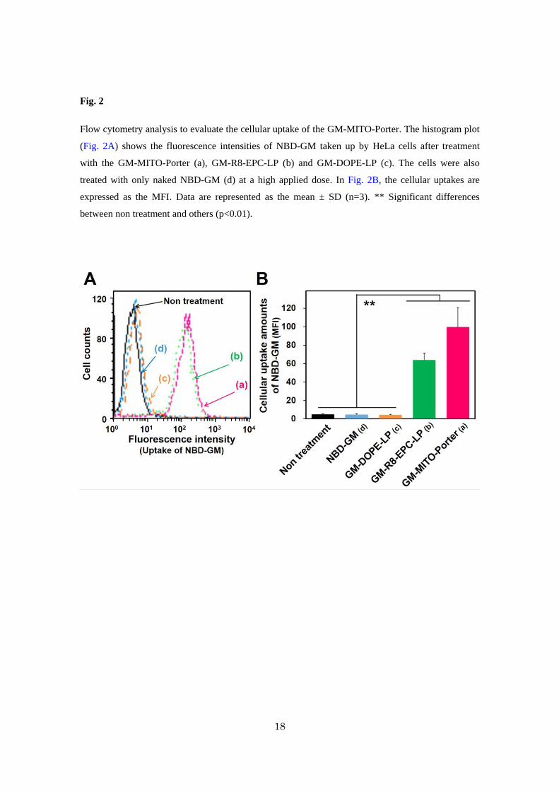
3.2. Evaluation of cellular uptake of GM-MITO-Porter
Although it is well known that GM is efficiently internalized into ear hair cells and
kidney distal tubule cells3,9, it has not been demonstrated that GM can be taken up by cancer cells.
Therefore, we first evaluated the cellular uptake of NBD-GM as naked GM using HeLa cells as
model cancer cells by flow cytometry (Fig. 2). The fluorescence intensities of the NBD-GM taken
up by the cells were comparable to that of non-treated cells, indicating a low cellular uptake of naked
NBD-GM in the case of HeLa cells. Intracellular observations also showed that NBD-GM was not
taken up by the HeLa cells (Fig. S2A). On the other hand, in the case of Pheochromocytoma cell 12
(PC12) cells where TRPV4 was found to be highly expressed21, the NBD-GM was efficiently
internalized into the cells (Fig. S2B). We also performed an RT-PCR assay to detect TRPV4-mRNA,
resulting in the observation of the gene expression of TRPV4 in the case of PC12 cells, while this
was not observed in the case of HeLa cells (Fig. S3). These results indicate that the first barrier for
killing cancer cells via mitochondrial toxicity by mitochondrial delivery of GM is the plasma
membrane.
We previously reported that the MITO-Porter, modified with STR-R8, was efficiently
internalized into HeLa cells22,23. Based on these reports, we hypothesized that MITO-Porter would
likely permit GM to be efficiently internalized into HeLa cells. As shown in Fig. 2A, the
fluorescence intensities of the NBD-GM taken up by the cells were relatively high in the case of
GM-MITO-Porter and GM-R8-EPC-LP, although the value for the GM-DOPE-LP that was not
modified with STR-R8 was comparable to that of naked NBD-GM. As shown in Fig. 2B, both the
cellular uptakes of GM-MITO-Porter and GM-R8-EPC-LP were significantly higher than that for
GM-DOPE-LP and naked NBD-GM. These results indicate that the R8 modified carriers are able to

9
internalize NBD-GM into HeLa cells. We used the GM-MITO-Porter and GM-R8-EPC-LP in
subsequent experiments.
3.3. Intracellular observation of GM-MITO-Porter using CLSM
The final process for mitochondrial delivery includes the introduction of the cargos into
the mitochondrial compartment. We observed the intracellular trafficking of the GM-MITO-Porter,
GM-R8-EPC-LP and NBD-GM in HeLa cells after staining the mitochondria red (Fig. 3A). When
the GM-MITO-Porter was added to HeLa cells, several yellow clusters were observed, indicating
that the green labeled GM (NBD-GM) co-localized with the red stained mitochondria. Only little
yellow clusters were observed in the case of GM-R8-EPC-LP, which has a low mitochondrial fusion
activity.
We quantified the mitochondrial targeting rate based on the obtained images, showing
that the value for the GM-MITO-Porter (12±3% (n=32)) was significantly higher than that of
GM-R8-EPC-LP (4±1% (n=32)) (Fig. 3B). Moreover, we calculated the mitochondrial occupation
rate (Fig. 3C), and confirmed that, in mitochondria that contained the GM-MITO-Porter, its content
increased with increasing applied dose. At an applied dose of 10 μM NBD-GM, the mitochondrial
occupation rate of GM-MITO-Porter was about 2.5%, while the value for GM-R8-EPC-LP was
about 1/50 that of the GM-MITO-Porter (0.05 ± 0% (n=30)).
3.4. Evaluation of cellular toxicity after mitochondrial delivery of NBD-GM using the MITO-Porter
system
Cellular toxicity was evaluated by means of a cell viability assay of HeLa cells after the
mitochondrial delivery of NBD-GM by MITO-Porter system. The results showed that cell viability
was decreased with increasing applied dose of the GM-MITO-Porter, and the effective concentration
50 (EC50) was 2.2 μM of NBD-GM (circles in Fig. 4A). Meanwhile, cell viability was not decreased
in the case of GM-R8-EPC-LP (squares) or NBD-GM (diamonds) (Fig. 4A), although the cellular
uptake of the GM-R8-EPC-LP were comparable with the GM-MITO-Porter at same applied doses
(Fig. 4B). We also observed no cell toxicity, when the empty MITO-Porter was used (Fig. S4). We
also validated the relationship between cellular toxicity and the mitochondrial accumulation of
NBD-GM as shown in Fig. 4C. The results showed that the toxicity steadily increased with the
amount of NBD-GM in mitochondria (Fig. 4C).

10
4. Discussion
In previous study, it has been reported that AGs have a high affinity for ribosomal RNA
and permeability transition in mitochondria, resulting in apoptosis via the inhibition of mitochondrial
transcription or exaggerated oxidative stress, which are consistent with the molecular mechanisms
for the ototoxicity and nephrotoxicity induced by AGs24-26. AGs have been clinically available as
antibiotics for many years, and, unfortunately, can exert some irreversible adverse-effects, especially
in the patients with a mitochondrial disease. AGs are not only antibiotics, but also function as latent
mitochondrial toxic agents. In this study, we validated whether AGs would have anticancer activity
once they are delivered into mitochondria.
However, this validation would be difficult using naked AGs because they cannot readily
penetrate the cellular membrane without selective channels such as TRPV49,21. As shown in Fig. 3A,
NBD-GM as a model AG was not internalized into HeLa cells, which do not express high levels of
TRPV4. Thus, we designed the GM-MITO-Porter, in which NBD-GM was encapsulated by
mitochondrial fusogenic envelopes modified with STR-R8, i.e., a cellular uptake device. We could
confirm that the GM-MITO-Porter achieved mitochondrial delivery of NBD-GM in cancer HeLa
cells (Figs. 2, 3).
The mitochondrial delivery of NBD-GM using the MITO-Porter system resulted in a
decreased cell viability and the effect was dose-dependent (Fig.4A), and the more the mitochondrial
accumulation of NBD-GM increased, the lower was the cellular viability (Fig.4C). Irrespective of
the low encapsulation efficiency, the GM-MITO-Porter had significant cellular toxicity; the EC50
was 2.2 μM of NBD-GM where the total lipid concentration was theoretically only about 6 μM.
Based on this value, the GM-MITO-Porter would be predicted to have a therapeutic effect in the
human body as a liposomal drug. Under an ideal scenario, only about 30 μmol of the total amount of
lipid would be needed for each drip-in-vein to reach the EC50 of the blood GM concentration,
assuming that the total circulating blood volume is 5 L. On the other hand, Doxil, which is a
clinically available liposomal drug encapsulating doxorubicin, requires about 1 mmol of the total
lipid amount for each drip-in-vein27. The GM-MITO-Porter might have some drug advantages, in
that a smaller amount of total lipids is needed for each drip-in-vein.
It was reported that the mitochondrial toxicity of GM in ear hair cells could be induced
by the inhibition of mitochondrial transcription by binding to ribosomal RNA24 or by opening
permeability transition pores by induced oxidative stress and metabolic change25. Thus, we
hypothesized that the delivery of NBD-GM into mitochondria of cancer HeLa cells might lead to
cell death via a mechanism similar to that reported for ear hair cells. Attention should be paid to the

11
typical adverse effects of GM, i.e., ototoxicity and renal toxicity, in the case of the clinical use of the
GM-MITO-Porter. In this study, we simply validated whether the GM-MITO-Porter had the ability
to kill cancer cells in in vitro experiments. Considering the administration of GM-MITO-Porter into
body via systemic injection, it is expected that conventional liposomes such as GM-MITO-Porter
would not reach inner hairy cells across the blood-labyrinthine barrier, which naked GM molecules
could cross in some way9. Moreover, conventional liposomes with sizes of more than 100 nm access
renal tubular epidermal cells with great difficulty, because the carrier needs to pass through the small
pores in blood endothelial cells28. It is probable that the GM-MITO-Porter might prevent hearing
loss and renal injury of a GM molecule, like liposomal amphotericin B, a clinically available
AmBisome29. We plan to investigate the toxicity of the GM-MITO-Porter in in vivo experiments in
the near future.
There have been many reports regarding multidrug resistant cancer cells and the complex
mechanisms associated with this effect30, e.g. P-glycoproteins as efflux pumps of anticancer drugs31,
which suggests that there are some limitations concerning clinically untreatable cancers by using
conventional chemotherapy. The use of a MITO-Porter system for delivering agents that are toxic to
mitochondria, such as GMs, may be an alternative anticancer therapeutic strategy. It is possible that
the chronic administration of the GM-MITO-Porter might induce emerging bacteria that are resistant
to antimicrobial therapies resulting from the transformation of in vivo microbiota. However it is
likely that the liposomal GM would be less likely to induce emerging bacteria resistant to antibiotics
than naked clinically available GM due to the fact that the GM is encapsulated by lipid bilayers and
has a narrower drug biodistribution, which would efficiently isolate GM away from in vivo
microbiota, such as human flora in the skin, digestive, or respiratory tracts.
To use the GM-MITO-Porter as a clinical application for cancer therapy, the selective
targeting to cancer cells in body is required. Thus, we plan to equip the GM-MITO-Porter with in
vivo cancer targeting ability, including the surface modification of carriers with poly ethylene-glycol
to enhance tumor accumulation via the enhanced permeability retention effect32-34 and ligand
modification, e.g. the use of the RGD peptide35 for integrin αvβ3, HER2 antagonistic peptide
modified liposome36 for a HER2 receptor, which some tumors specifically overexpress37. These
studies are currently under way.
5. Conclusion
In order to investigate the intracellular dynamics and cellular toxicity of AGs using
cancer HeLa cells, we constructed a GM-MITO-Porter, where NBD-GM (a model AG) with low

12
cellular internalization, was encapsulated into the MITO-Porter. Flow cytometry analysis and
fluorescent microscopy observations permitted us to confirm that the GM–MITO-Porter achieved
mitochondrial delivery of NBD-GM in cancer HeLa cells. Moreover, we showed that
GM–MITO-Porter induced a strong cytotoxicity, suggesting that NBD-GM has toxicity related to
mitochondrial disorder. The findings indicate that the GM-MITO-Porter has the potency of an
anticancer drug via its mitochondrial toxicity.
Acknowledgments
This work was supported, in part by, a Grant-in-Aid for Scientific Research (B) [Grant
No. 26282131 (to Y.Y.)] from the Ministry of Education, Culture, Sports, Science and Technology,
the Japanese Government (MEXT), and Kobayashi Foundation for Cancer Research (to Y.Y.). We
also thank Dr. Milton Feather for his helpful advice in writing the manuscript.

13
References
1. Comroe JH, Jr. 1978. Pay dirt: the story of streptomycin. Part I. From Waksman to Waksman.
The American review of respiratory disease 117(4):773-781.
2. Kalghatgi S, Spina CS, Costello JC, Liesa M, Morones-Ramirez JR, Slomovic S, Molina A,
Shirihai OS, Collins JJ 2013. Bactericidal antibiotics induce mitochondrial dysfunction and oxidative
damage in Mammalian cells. Science translational medicine 5(192):192ra185.
3. Ward DT, Maldonado-Perez D, Hollins L, Riccardi D 2005. Aminoglycosides induce acute cell
signaling and chronic cell death in renal cells that express the calcium-sensing receptor. Journal of the
American Society of Nephrology : JASN 16(5):1236-1244.
4. Qian Y, Guan MX 2009. Interaction of aminoglycosides with human mitochondrial 12S rRNA
carrying the deafness-associated mutation. Antimicrobial agents and chemotherapy 53(11):4612-4618.
5. Skou AS, Tranebjaerg L, Jensen T, Hasle H 2014. Mitochondrial 12S ribosomal RNA A1555G
mutation associated with cardiomyopathy and hearing loss following high-dose chemotherapy and
repeated aminoglycoside exposure. The Journal of pediatrics 164(2):413-415.
6. Ishikawa K, Takenaga K, Akimoto M, Koshikawa N, Yamaguchi A, Imanishi H, Nakada K,
Honma Y, Hayashi J 2008. ROS-generating mitochondrial DNA mutations can regulate tumor cell
metastasis. Science 320(5876):661-664.
7. Ashkenazi A 2015. Targeting the extrinsic apoptotic pathway in cancer: lessons learned and
future directions. The Journal of clinical investigation 125(2):487-489.
8. Chamberlain GR, Tulumello DV, Kelley SO 2013. Targeted delivery of doxorubicin to
mitochondria. ACS Chem Biol 8(7):1389-1395.
9. Wang Q, Steyger PS 2009. Trafficking of systemic fluorescent gentamicin into the cochlea and
hair cells. J Assoc Res Otolaryngol 10(2):205-219.
10. Lee JH, Park C, Kim SJ, Kim HJ, Oh GS, Shen A, So HS, Park R 2013. Different uptake of
gentamicin through TRPV1 and TRPV4 channels determines cochlear hair cell vulnerability. Exp Mol
Med 45:e12.
11. Yamada Y, Akita H, Kamiya H, Kogure K, Yamamoto T, Shinohara Y, Yamashita K, Kobayashi
H, Kikuchi H, Harashima H 2008. MITO-Porter: A liposome-based carrier system for delivery of
macromolecules into mitochondria via membrane fusion. Biochimica et biophysica acta
1778(2):423-432.
12. Yamada Y, Harashima H 2008. Mitochondrial drug delivery systems for macromolecule and
their therapeutic application to mitochondrial diseases. Adv Drug Deliv Rev 60(13-14):1439-1462.
13. Furukawa R, Yamada Y, Kawamura E, Harashima H 2015. Mitochondrial delivery of antisense
RNA by MITO-Porter results in mitochondrial RNA knockdown, and has a functional impact on
mitochondria. Biomaterials 57:107-115.
14. Yamada Y, Harashima H 2015. Targeting the Mitochondrial Genome via a Dual Function

14
MITO-Porter: Evaluation of mtDNA Levels and Mitochondrial Function. Methods in molecular biology
1265:123-133.
15. Futaki S, Suzuki T, Ohashi W, Yagami T, Tanaka S, Ueda K, Sugiura Y 2001. Arginine-rich
peptides. An abundant source of membrane-permeable peptides having potential as carriers for
intracellular protein delivery. J Biol Chem 276(8):5836-5840.
16. Nakase I, Niwa M, Takeuchi T, Sonomura K, Kawabata N, Koike Y, Takehashi M, Tanaka S,
Ueda K, Simpson JC, Jones AT, Sugiura Y, Futaki S 2004. Cellular uptake of arginine-rich peptides: roles
for macropinocytosis and actin rearrangement. Mol Ther 10(6):1011-1022.
17. Isoherranen N, Soback S 2000. Determination of gentamicins C(1), C(1a), and C(2) in plasma
and urine by HPLC. Clin Chem 46(6 Pt 1):837-842.
18. Szoka F, Jr., Papahadjopoulos D 1978. Procedure for preparation of liposomes with large
internal aqueous space and high capture by reverse-phase evaporation. Proc Natl Acad Sci U S A
75(9):4194-4198.
19. Kawamura E, Yamada Y, Yasuzaki Y, Hyodo M, Harashima H 2013. Intracellular observation
of nanocarriers modified with a mitochondrial targeting signal peptide. Journal of bioscience and
bioengineering 116(5):634-637.
20. Morgan JR, Williams KE 1980. Preparation and properties of liposome-associated gentamicin.
Antimicrobial agents and chemotherapy 17(4):544-548.
21. Jang Y, Jung J, Kim H, Oh J, Jeon JH, Jung S, Kim KT, Cho H, Yang DJ, Kim SM, Kim IB,
Song MR, Oh U 2012. Axonal neuropathy-associated TRPV4 regulates neurotrophic factor-derived
axonal growth. J Biol Chem 287(8):6014-6024.
22. Yamada Y, Kawamura E, Harashima H 2012. Mitochondrial-targeted DNA delivery using a
DF-MITO-Porter, an innovative nano carrier with cytoplasmic and mitochondrial fusogenic envelopes. J
Nanopart Res 14(8):1013-1027.
23. Yamada Y, Nakamura K, Furukawa R, Kawamura E, Moriwaki T, Matsumoto K, Okuda K,
Shindo M, Harashima H 2013. Mitochondrial delivery of bongkrekic acid using a MITO-porter prevents
the induction of apoptosis in human hela cells. J Pharm Sci-Us 102(3):1008-1015.
24. Hobbie SN, Bruell CM, Akshay S, Kalapala SK, Shcherbakov D, Bottger EC 2008.
Mitochondrial deafness alleles confer misreading of the genetic code. Proc Natl Acad Sci U S A
105(9):3244-3249.
25. Jensen-Smith HC, Hallworth R, Nichols MG 2012. Gentamicin rapidly inhibits mitochondrial
metabolism in high-frequency cochlear outer hair cells. PloS one 7(6):e38471.
26. Alharazneh A, Luk L, Huth M, Monfared A, Steyger PS, Cheng AG, Ricci AJ 2011. Functional
hair cell mechanotransducer channels are required for aminoglycoside ototoxicity. PloS one
6(7):e22347.
27. Fujisaka Y, Horiike A, Shimizu T, Yamamoto N, Yamada Y, Tamura T 2006. Phase 1 clinical

15
study of pegylated liposomal doxorubicin (JNS002) in Japanese patients with solid tumors. Jpn J Clin
Oncol 36(12):768-774.
28. Dolman ME, Harmsen S, Storm G, Hennink WE, Kok RJ 2010. Drug targeting to the kidney:
Advances in the active targeting of therapeutics to proximal tubular cells. Adv Drug Deliv Rev
62(14):1344-1357.
29. Walsh TJ, Goodman JL, Pappas P, Bekersky I, Buell DN, Roden M, Barrett J, Anaissie EJ 2001.
Safety, tolerance, and pharmacokinetics of high-dose liposomal amphotericin B (AmBisome) in patients
infected with Aspergillus species and other filamentous fungi: maximum tolerated dose study.
Antimicrobial agents and chemotherapy 45(12):3487-3496.
30. Baguley BC 2010. Multiple drug resistance mechanisms in cancer. Molecular biotechnology
46(3):308-316.
31. Abdallah HM, Al-Abd AM, El-Dine RS, El-Halawany AM 2015. P-glycoprotein inhibitors of
natural origin as potential tumor chemo-sensitizers: A review. Journal of advanced research 6(1):45-62.
32. Matsumura Y, Maeda H 1986. A new concept for macromolecular therapeutics in cancer
chemotherapy: mechanism of tumoritropic accumulation of proteins and the antitumor agent smancs.
Cancer Res 46(12 Pt 1):6387-6392.
33. Maeda H, Sawa T, Konno T 2001. Mechanism of tumor-targeted delivery of macromolecular
drugs, including the EPR effect in solid tumor and clinical overview of the prototype polymeric drug
SMANCS. J Control Release 74(1-3):47-61.
34. Hatakeyama H, Akita H, Harashima H 2013. The polyethyleneglycol dilemma: advantage and
disadvantage of PEGylation of liposomes for systemic genes and nucleic acids delivery to tumors. Biol
Pharm Bull 36(6):892-899.
35. Zhen Z, Tang W, Chen H, Lin X, Todd T, Wang G, Cowger T, Chen X, Xie J 2013.
RGD-modified apoferritin nanoparticles for efficient drug delivery to tumors. ACS nano
7(6):4830-4837.
36. Stefanick JF, Ashley JD, Bilgicer B 2013. Enhanced cellular uptake of peptide-targeted
nanoparticles through increased peptide hydrophilicity and optimized ethylene glycol peptide-linker
length. ACS nano 7(9):8115-8127.
37. Fernandes E, Ferreira JA, Andreia P, Luis L, Barroso S, Sarmento B, Santos LL 2015. New
trends in guided nanotherapies for digestive cancers: A systematic review. J Control Release
209:288-307.

16
Table 1 Characteristics of the carriers used in this study.
Carrier-type Lipid composition
(mol ratio)
Diameter
(nm)
PDI ζ-potential
(mV)
Property
GM-MITO-Porter DOPE/SM/STR-R8=9/2/1 147±6 0.16±0.03 44±4 High mitochondrial
fusogenic envelopes
with cellular uptake
device (R8)
GM-R8-EPC-LP EPC/SM/STR-R8=9/2/1 119±17 0.25±0.09 32±5 Low mitochondrial
fusogenic envelopes
with cellular uptake
device (R8)
GM-DOPE-LP DOPE/SM=9/2 205±5 0.30±0 1±2 Low mitochondrial
fusogenic envelopes
All types of carriers contained NBD-GM. Data denote the mean ± S.D.(n=6).

17
Fig. 1
Schematic image of the mitochondrial delivery of NBD-GM by the MITO-Porter system, leading to
mitochondrial toxicity.
18
Fig. 2
Flow cytometry analysis to evaluate the cellular uptake of the GM-MITO-Porter. The histogram plot
(Fig. 2A) shows the fluorescence intensities of NBD-GM taken up by HeLa cells after treatment
with the GM-MITO-Porter (a), GM-R8-EPC-LP (b) and GM-DOPE-LP (c). The cells were also
treated with only naked NBD-GM (d) at a high applied dose. In Fig. 2B, the cellular uptakes are
expressed as the MFI. Data are represented as the mean ± SD (n=3). ** Significant differences
between non treatment and others (p<0.01).

19
Fig. 3
Intracellular trafficking analysis of the GM-MITO-Porter. Intracellular observation of NBD-GM
(green color) encapsulated in GM-MITO-Porter (upper panels) and GM-R8-EPC-LP (lower panels)
using CLSM (A). NBD-GM is seen to co-localize with red stained mitochondria in HeLa cells,
observed as yellow signals in the merged images. Scale bars, 20μm. The mitochondrial targeting rate
was calculated based on the obtained images (B). Data are represented by the mean ± S.E.M. (n =
32-35). ** Significant differences (p<0.01). Mitochondrial occupation rate of GM-MITO-Porter at
various applied doses of NBD-GM (C). Data are represented by the mean ± S.E.M. (n = 30). The
value of GM-R8-EPC-LP was 0.05 ± 0% at the final concentration of 10 μM NBD–GM (n=30).

20
Fig. 4
Evaluation of the cellular toxicity of the GM-MITO-Porter in HeLa cells and investigation of the
relationship between the toxicity and mitochondrial accumulation of NBD-GM. Comparison of
cellular toxicity among the GM-MITO-Porter, R8-GM-EPC-LP and naked NBD-GM at various
applied doses (A). The circles, squares and diamonds represent the values corresponding to cell
viability (%) of the GM-MITO-Porter, R8-GM-EPC-LP and naked NBD-GM. Data are represented
by the mean ± S.D. (n = 6). ** Significant differences between naked NBD-GM and the others in
each concentration (p<0.01). Comparison of cellular uptake of GM-MITO-Porter and
R8-GM-EPC-LP at various applied dose (B). Data are represented by the mean ± S.D. (n = 3). There
are no significant differences at each concentration. Relationship between mitochondrial occupation
rate and cell viability (C). For the x-axis (mitochondrial occupation rate), we used data shown in Fig.
3C. For the y-axis (cell viability), we used data shown in Fig. 4A.

21
Supplementary data
Supplementary Methods
1. Synthesis and purification of NBD-GM.
The Gentamicin Sulfate was conjugated with NBD-F, which labels primary and
secondary amines, to synthesis NBD-GM by chemical bonding between the amino
groups of Gentamicin Sulfate and NBD-F as previously reported 1. The synthesis was
performed in a 1.5 ml tube by mixing 10 μl of 100 mM Gentamicin Sulfate
water-solution (5 eq.), 2 μl of 100 mM NBD-F ethanol-solution (1 eq.) and 18 μl of 100
mM boric buffer (pH=9.0). After stirring the reaction mixture at 60oC for 10 min in the
dark, the mixture was neutralized by adding 470 μl of a 5 mM HCl solution 4oC to stop
the reaction. The crude NBD-GM was was purified by reversed-phase high performance
liquid chromatography (reversed-phase HPLC), using an InertSustain C18 (5 μm)
column (4.6 x 250 mm, GL Sciences, Tokyo, Japan) with a 30% acetonitrile
water-solution. The HPLC profile was detected at 336 nm. A new peak eluted at 2 - 2.5
min, and this peak was collected as the NBD-GM, and was then freeze dried.
2. Estimation of encapsulation efficiency of the NBD-GM into MITO-Porter.
To estimate the encapsulation efficiency of the NBD-GM, a GM-MITO-Porter

22
containing a lipid film labeled with rhodamine-DOPE (Avanti Polar lipids, Alabaster,
AL) was prepared (1 mol% rhodamine-DOPE of total lipids). The GM-MITO-Porter
was separated from the unencapsulated NBD-GM by ultracentrifugation (74,000g, 20
min, 4oC) three times using HIMAC (Hitachi Koki Co., Ltd., Tokyo, Japan) and the
recovered NBD-GM and the recovered lipids were then determined after treatment with
SDS (final concentration, 1%). The applied NBD-GM and the applied lipids were also
determined before ultrafiltration. The amounts of NBD-GM and the amounts of lipid
were determined by measuring the fluorescent intensities of the NBD-GM (excitation at
460 nm and emission at 534 nm) and the rhodamine-DOPE (excitation at 560 nm and
emission at 590 nm), respectively. The encapsulation efficiency was calculated using
equation shown below:
encapsulation efficiency (%) = (recovered NBD-GM / recovered lipid)/ (applied
NBD-GM / applied lipid) x 100.
3. Analysis of TRPV-4 gene expression
Total RNA extraction from confluent HeLa cells, PC-12 cells or HaCaT cells
(human skin keratinocyte)2 was performed with the TRIzol Reagent (Invitrogen,
Carlsbad, CA) and purification was carried out according to the conventional ethanol

23
precipitation followed by DNase-I treatment to digest any DNA contamination in the
sample. The concentration of the obtained RNA was measured using a supersensitive
NanoDrop Lite spectrophotometer (ThermoScientific, Waltham, MA). Reverse
transcription was performed using the High Capacity RNA-to-cDNA Kit (Applied
Biosystems, Foster City, CA). In brief, cDNA was generated from 1 μg of RNA by
means of an RT Enzyme Mix according to the manufacturer's instructions on a PCR
thermal cycler (C1000 Touch Thermal Cycler, Bio-Rad, Berkeley, CA). The resulting
cDNA was diluted to a final concentration of 10 ng/μl for PCR amplification.
Customized oligonucleotide PCR primers (Sigma Genosys Japan, Ishikari, Japan) to
detect TRPV-4 and GAPDH (internal control), and cDNA templates were mixed with
DNA polymerase (AmpliTaqGold 360 Master Mix, Applied Biosystems) according to
the manufacture instructions in PCR amplification, carried out by using a PCR thermal
cycler. The following primers were used for a human HeLa cell line and human HaCaT
cell line: Homo sapience TRPV4 (Forward 5'-AGATGTACGACCTGCTGCTG-3’,
Reverse 5'-TCCCGCAGCAGTTCATTGAT-3), Homo sapience GAPDH (Forward
5’-CCTCTGACTTCAACAGCGAC-3’, Reverse
5’-CGTTGTCATACCAGGAAATGAG-3’). The following primers were used for rat
PC-12 cell line: Rattus norvegicus TRPV4 (Forward

24
5'-ACCACGGTGGACTACCTGAG-3, Reverse
5'-GATTCAGGAGGGTGACCAGA-3’), Rattus norvegicus GAPDH (Forward
5’-GGCAAGTTCAACGGCACAGT-3’, Reverse
5’-ATGGGTTTCCCGTTGATGAC-3’). Each 4 μL of PCR products were used for
electrophoresis in polyacrylamide gels in TBE (89 mM Tris-HCl, 89 mM boric acid, 2
mM EDTA). The bands were visualized by UV after staining with GelRed Nucleic Acid
Stain (Biotium, Hayward, CA, USA).

25
Supplementary Figures
Figure S1. Stability of the GM-MITO-Porter in terms of diameter and ζ-potential
Fig. S1. GM-MITO-Porter was incubated in 10 mM HEPES buffer (pH=7.4) at 4oC, the
diameters (A) and ζ-potentials (B) were measured at weekly intervals. Date are the
mean ± S.D. (n=3).

26
Figure S2. Intracellular observation of naked NBD-GM in HeLa cells and PC12 cells.
Fig. S2. Intracellular observation of NBD-GM (green color) in HeLa cells (A) and
PC12 cells (B). The cells were treated with only naked NBD-GM at high applied dose
(final concentration of NBD-GM, 1000 μM), and mitochondrial were stained with
MitoTracker Deep Red FM (red color) prior to intracellular observation. Scale bars,
20μm.

27
Figure S3. Gel images of the RT–PCR detection of TRPV4-mRNA.
We performed RT-PCR assays to detect TRPV4-mRNA and GAPDH-mRNA
(internal control) in the total RNA of HeLa cells (a human TRPV-4 negative cell), PC12
cells (a rat TRPV-4 positive cell) and HaCaT cells (a human TRPV-4 positive cell)2.
Upper panel in Fig. S3 shows that the PCR product derived from rat TRPV-4 (403 bp)
was present in PC12 cells (lane 4), while that from human TRPV-4 was not observed in
HeLa cells (lane 2). We also confirmed that a band corresponding to human TRPV-4
(352 bp) was observed in HaCaT cell (a human TRPV-4 positive cell). In lower panel in
Fig. 3, the PCR products derived from GAPDH-mRNA (internal control) were observed
in all kinds of cells (lanes 2, 4, 6). These results suggest that the gene expression of
TRPV4 was observed in the case of PC12 cells, but not in the case of HeLa cells.
Moreover, gel images of RT–PCR detection showed that the target DNA bands
disappeared in the absence of reverse transcription (RT(-)) (lanes 3, 5, 7), suggesting
that contaminating DNA was not present when this RT–PCR assay was performed.
Fig. 3. Total RNA was purified from HeLa cells (lanes 2,3), PC-12 cells (lanes 4,5) or
HaCaT cells (lanes 6,7) and PCR was done with specific primers for TRPV4 mRNA
(upper panel) and GAPDH mRNA (lower panel) with reverse transcription (RT (+),
lanes 2,4,6) and without reverse transcription (RT (-), lanes 3,5,7). In upper panel, the
electrophoresis shows the PCR products derived from human TRPV-4 and rat TRPV-4
at 352 bp and 403 bp, respectively. In the lower panel, the electrophoresis shows the
PCR products derived from human GAPDH and rat GAPDH at 102 bp and 80 bp,
respectively. The PCR products were detected by the GelRed Nucleic Acid Stain after
separation by electrophoresis. Lane 1, DNA Ladder.

28
Figure S4. Cell viability of empty MITO-Porter in HeLa cells.
Fig. S4. Empty-MITO-Porters were added to HeLa cells (final concentration of the total
lipid, 27.5 μM), and the cellular toxicity was evaluated at 48 hr after the incubation.
Data are represented by the mean ± S.D. (n = 3). Significant differences were calculated
by the student t-test and no significant difference (N.S.) were found. Note: lipid
concentration of GM-MITO-Porter at 10 μM NBD-GM as shown in Figure 4A was
about 10 μM.

29
References
1. Isoherranen N, Soback S 2000. Determination of gentamicins C(1), C(1a), and
C(2) in plasma and urine by HPLC. Clin Chem 46(6 Pt 1):837-842.
2. Fusi C, Materazzi S, Minocci D, Maio V, Oranges T, Massi D, Nassini R 2014.
Transient receptor potential vanilloid 4 (TRPV4) is downregulated in keratinocytes in
human non-melanoma skin cancer. J Invest Dermatol 134(9):2408-2417.