valence tautomerism in synthetic models of cytochrome p450 · valence tautomerism in synthetic...
TRANSCRIPT

Valence tautomerism in synthetic models ofcytochrome P450Pradip Kumar Dasa,1, Subhra Samantaa,1, Ashley B. McQuartersb, Nicolai Lehnertb,2, and Abhishek Deya,2
aDepartment of Inorganic Chemistry, Indian Association for the Cultivation of Science, Kolkata 700032, India; and bDepartment of Chemistry, University ofMichigan, Ann Arbor, MI 48109
Edited by Harry B. Gray, California Institute of Technology, Pasadena, CA, and approved May 2, 2016 (received for review January 16, 2016)
CytP450s have a cysteine-bound heme cofactor that, in its as-isolated resting (oxidized) form, can be conclusively described as aferric thiolate species. Unlike the native enzyme, most syntheticthiolate-bound ferric porphyrins are unstable in air unless the axialthiolate ligand is sterically protected. Spectroscopic investigationson a series of synthetic mimics of cytP450 indicate that a thiolate-bound ferric porphyrin coexists in organic solutions at roomtemperature (RT) with a thiyl-radical bound ferrous porphyrin, i.e.,its valence tautomer. The ferric thiolate state is favored by greaterenthalpy and is air stable. The ferrous thiyl state is favored byentropy, populates at RT, and degrades in air. These ground statescan be reversibly interchanged at RT by the addition or removal ofwater to the apolar medium. It is concluded that hydrogen bondingand local electrostatics protect the resting oxidized cytP450 activesite from degradation in air by stabilizing the ferric thiolate groundstate in contrast to its synthetic analogs.
valence tautomerism | cytochrome P450 | synthetic model |entropic contribution | hydrogen bonding
Heme bound to a cysteine ligand is found in the active sites ofseveral key enzymes in biology, including cytochrome P450
(cytP450) and nitric oxide synthase (NOS), which broadly com-prise the cytP450 superfamily (1, 2). Members of the cytP450family of enzymes mediate key transformations in the synthesisof hormones and play an important role in the catabolism ofmetabolites and drugs (3). This subclass of heme enzymes dis-tinguishes themselves by their ability to oxidize substrates usingmolecular O2. The resting forms of their active sites are bestdescribed as an oxidized ferric heme bound to a thiolate ligand(the coordinating functional group of cysteine). The active sitesin their resting ferric state are stable and only react with O2 whenthey are reduced to the ferrous state (4, 5). This is in sharpcontrast to the behavior of synthetic thiolate-bound ferric por-phyrin complexes. Unless the thiolate is sterically protected,these synthetic ferric porphyrin complexes degrade in O2 (6).This phenomenon has remained an enigma ever since the initialreports of synthetic thiolate-bound ferric porphyrin complexes byHolm and coworkers (7) and Collman and Sorrell (8) four de-cades ago. Similar sensitivity to O2 by a formally oxidized metalcenter is exhibited by the active sites of ammine oxidase andintradiol dioxygenases (9, 10). The sensitivity to O2 is signifi-cantly reduced in thiolate-bound ferric porphyrins in aqueousenvironments. In this case, recent work shows that heme–thiolatemodel systems can catalytically hydroxylate inert C–H bondsusing molecular O2 with >200 turnovers, i.e., bioinspired func-tional mimics of cytP450 (11). In addition, cytP450 enzymes formstable diamagnetic FeIII–NO adducts (12). In contrast, attemptsto form stable FeIII–NO adducts of thiolate-bound iron porphyrinmodel systems inevitably results in the NO attacking the thiolatesulfur and not the iron. This is despite the fact that theoreticalcalculations predict these thiolate-bound porphyrin FeIII–NOspecies to be stable entities (13). Curiously, similar sensitivity to O2is also exhibited by most nonheme ferric thiolate and many nickelthiolate complexes, which necessitates their handling in an inertatmosphere (14–16). This is particularly well established in the
nonheme iron enzyme nitrile hydratase and its related syntheticmodels. Although the active site, when isolated in the absence ofair, bears three cysteine thiolates and is inactive, its reaction withO2 leads to the active form, which bears a cysteine sulfenate and acysteine sulfinate as its ligands (17, 18). Similar transformationshave been reproduced in synthetic model complexes (19).The existing paradox of O2 sensitivity of synthetic thiolate-
bound ferric heme complexes thus requires an unusual electronicstructure to be present in its genesis. Resonance Raman (rR),electron paramagnetic resonance (EPR), and 1H NMR areuseful tools to investigate the electronic structures of ferric hemecomplexes. In particular, the oxidation and spin state markermodes in the rR spectra of iron porphyrin complexes findabundant use in inorganic/bioinorganic spectroscopy (20). Syn-thetic iron porphyrin complexes show characteristic sets of ν4(1,340–1,375 cm−1) and ν2 (1,540–1,575 cm−1) vibrations withvery high fidelity (SI Appendix, Table S1). A series of iron por-phyrin complexes with thiolate axial ligands (Fig. 1) areinvestigated here using a combination of rR, EPR, and 1H NMRspectroscopy. These complexes are previously characterized andvary in the nature of the thiolate ligand; the aliphatic complex(Fig. 1A) is unstable in air and has an alkyl thiolate ligand (Fig. 1),the bulky aliphatic complex (Fig. 1B) has a sterically protectedalkyl thiolate and is stable in air, the benzylic complex (Fig. 1C)is stable in air, and the aromatic thiolate (Fig. 1D) is unstable inair. Excitingly, the data presented here clearly suggest that thesecomplexes exist as a mixture of two valence tautomers, namely,ferric thiolate and ferrous thiyl species, in the solid state andin solution.
Significance
Cytochrome P450 is a mammalian enzyme responsible for thecatabolism of organic molecules (food, drug, etc.) as well asbiosynthesis of hormones and cholesterol. Unfortunately, instark contrast to the stability of the enzyme, its syntheticmimics rapidly degrade on being exposed to oxygen impedingerstwhile efforts of the scientific community to understand itschemistry in detail. Here the discovery of a valence tautomer-ism between a ferric thiolate and an arcane ferrous-thiyl spe-cies in synthetic models at room temperature is proposed to beat the root of this three-decade-old mystery. Hydrogen bond-ing to the thiolate sulfur, also present in the enzyme active site,restrains the system to the ferric thiolate form imparting itsstability in the presence of oxygen.
Author contributions: N.L. and A.D. designed research; P.K.D., S.S., and A.B.M. performedresearch; S.S., N.L., and A.D. contributed new reagents/analytic tools; P.K.D., A.B.M., N.L.,and A.D. analyzed data; and P.K.D., N.L., and A.D. wrote the paper.
The authors declare no conflict of interest.
This article is a PNAS Direct Submission.1P.K.D. and S.S. contributed equally to this work.2To whom correspondence may be addressed. Email: [email protected] or [email protected].
This article contains supporting information online at www.pnas.org/lookup/suppl/doi:10.1073/pnas.1600525113/-/DCSupplemental.
www.pnas.org/cgi/doi/10.1073/pnas.1600525113 PNAS | June 14, 2016 | vol. 113 | no. 24 | 6611–6616
CHEM
ISTR
Y
Dow
nloa
ded
by g
uest
on
June
4, 2
020
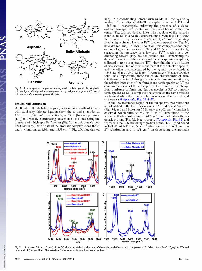
Results and DiscussionrR. rR data of the aliphatic complex (excitation wavelength, 413.1 nm)with axial alkyl-thiolate ligation show the ν4 and ν2 modes at1,361 and 1,554 cm−1, respectively, at 77 K [low temperature(LT)] in a weakly coordinating solvent like THF, indicating thepresence of a high-spin FeIII center (Fig. 2 A and B, blue dashedline). Similarly, the rR data of the aromatic complex shows the ν4and ν2 vibrations at 1,361 and 1,553 cm−1 (Fig. 2D, blue dashed
line). In a coordinating solvent such as MeOH, the ν4 and ν2modes of the aliphatic-MeOH complex shift to 1,369 and1,567 cm−1, respectively, indicating the presence of a six-co-ordinate low-spin FeIII center with methanol bound to the ironcenter (Fig. 2A, red dashed line). The rR data of the benzyliccomplex at LT in a weakly coordinating solvent like THF showthe presence of ν2 modes at 1,552 and 1,565 cm−1 originatingfrom a high-spin and low-spin FeIII species, respectively (Fig. 2C,blue dashed line). In MeOH solution, this complex shows onlyone set of ν4 and ν2 modes at 1,365 and 1,565 cm−1, respectively,suggesting the presence of a low-spin FeIII species in a co-ordinating solvent (Fig. 2C, red dashed line). Importantly, rRdata of this series of thiolate-bound ferric porphyrin complexes,collected at room temperature (RT), show that there is a mixtureof two species. One of them is the parent ferric thiolate species,and the other is characterized by the ν4 and the ν2 bands at1,343–1,346 and 1,540–1,543 cm−1, respectively (Fig. 2 A–D, bluesolid line). Importantly, these values are characteristic of high-spin ferrous species. Although rR intensities are not quantitative,the relative intensities of the ferrous and ferric species at RT arecomparable for all of these complexes. Furthermore, the changefrom a mixture of ferric and ferrous species at RT to a mostlyferric species at LT is completely reversible as the same mixtureis obtained when the frozen solution is warmed up to RT andvice versa (SI Appendix, Fig. S1 A–D).In the low-frequency region of the rR spectra, two vibrations
are identified in the C–S region; one at 655 and one at 662 cm−1
(Fig. 3A, red and blue). At 77 K, only the 662 cm−1 vibration isobserved, which shifts to 657 cm−1 on S34 substitution of thearomatic thiolate sulfur and to 647 cm−1 on deuterating the ar-omatic protons (Fig. 3B, blue to green; SI Appendix, Fig. S2) andrepresents the C–S stretching vibration of the PhS− ligand boundto FeTPP. At RT, the 655 cm−1 vibration shifts to 653 cm−1 onS34 substitution and to 651 cm−1 on deuterating the aromatic
Fig. 1. Iron porphyrin complexes bearing axial thiolate ligands. (A) Aliphaticthiolate ligand, (B) aliphatic thiolate protected by bulky t-butyl groups, (C) benzylthiolate, and (D) aromatic phenyl thiolate.
Fig. 2. rR data (413.1 nm, 10 mW) of the (A) aliphatic, (B) bulky aliphatic, (C) benzylic, and (D) aromatic complexes in THF (black) and MeOH (gray) at RT (boldline) and LT (dashed line). The asterisks (*) represent plasma lines from the laser.
6612 | www.pnas.org/cgi/doi/10.1073/pnas.1600525113 Das et al.
Dow
nloa
ded
by g
uest
on
June
4, 2
020

protons (Fig. 3C, blue to green to violet) and represents the C–Sstretching mode of the PhS· ligand bound to reduced Fe(II)porphyrin at RT (resonance enhancement by Soret not likely ifnot bound to the porphyrin). The Fe–S vibration for the Fe(III)–SR state is mixed with the ν8 mode as evidenced by a 8 cm−1 shiftin the ν8 from 396 to 388 cm−1 on S34 substitution of the thiolate(Fig. 3D, red to green). The vibration at 341 cm−1 is a Fe(III)–SRstretching vibration and it shifts to 334 cm−1 on S34 substitution(Fig. 3D, red to green). Additionally, at RT, a vibration is observedat 206 cm−1 that shifts to 198 cm−1 on S34 substitution and couldrepresent a Fe–S stretching mode (Fig. 3D, red to green). Thus, the655 cm−1 and the 206 cm−1, only observed at RT, and sensitive toS34 substitution may be assigned to the Fe–S and C–S vibration ofthe species at RT. The dramatic lowering of the Fe–S vibration inthis species relative to the ferric thiolate species signifies substantialweakening of the Fe–S vibration from the Fe(III)–SR state to theFe(II) species formed at RT.
EPR Spectroscopy. EPR data of the same series of thiolate-boundcomplexes at LT show that the iron centers in the complexesexist in its high-spin (S = 5/2) or low-spin (S = 1/2) ferric state innoncoordinating or coordinating solvents, respectively (Fig. 4).The iron center in the aliphatic complex is high spin in THF (Fig.4, blue) and low spin in MeOH (Fig. 4, green). Thus, both the rRand EPR data collected at LT indicate that these thiolate-bound
iron porphyrins can be described as ferric thiolate complexesconsistent with previous reports. However, the rR data suggestthat there is a significant decrease in the population of the ferricspecies and increase in population of a ferrous species in solutionat RT relative to LT. The corresponding EPR data at RT showthe increase in population of a radical species characterized by asignal at g = 2 (Fig. 4, red) relative to the data obtained at LT.Although, expectedly, the EPR data at RT do not show signifi-cant intensity of the g = 6 signal from the high-spin ferric speciespresent in solution, the solution rR data under the same condi-tions clearly demonstrate the presence of both ferric and ferrousporphyrin species. These data suggest that, apart from the ferricthiolate species, a ferrous ligand radical species exists at RT. Notethat the ferrous ligand radical species is a valence tautomer of theferric thiolate complex. Spin quantification of this radical signal atRT indicates that the population of this species for the aliphatic,bulky aliphatic, benzylic, and aromatic thiolate complexes are 52%,50%, 46%, and 45%, respectively.
NMR Spectroscopy. 1H NMR is routinely used as a probe for theoxidation and spin state of ferric porphyrin complexes. Theβ-pyrrole resonances are shifted between 60 and 80 ppm forhigh-spin ferrous and ferric porphyrin complexes at 298 K (21).The imidazole-bound iron porphyrin complex (PIM) shows onebroad β-pyrrole resonance at 80 ppm (SI Appendix, Fig. S3), asexpected for a high-spin ferric porphyrin at 289 K. On the con-trary, all of the thiolate-bound porphyrins show two sets of res-onances at 298 K in the 40- to 80-ppm region, which mightcorrespond to the individual signals of the high-spin ferrous andferric porphyrin complexes in solution. This, however, wouldrequire that the switch between the two ground states would beslow enough at RT to be resolved on the NMR timescale, and itis not clear at this point whether this requirement is fulfilled. Forexample, the data for the aliphatic complex, obtained at 273 K,show the FeIII pyrrole resonances at 85 ppm (SI Appendix, Fig.S3). There is a resonance at ∼45 ppm characteristic of a high-spinferrous porphyrin, consistent with the rR data, but these reso-nances may be effected by the resonances of the ligand radicalpresent. On the other hand, for the aromatic complex, the signalat 57–60 ppm at RT is consistent with previous reports (22).
Variable-Temperature and Radical Trapping Experiments. The con-version of the mixture of ferrous and ferric porphyrins at RT to theferric porphyrin at LT is continuous as reflected in the variabletemperature (VT) rR data. The VT rR data of the aliphatic complexin THF show that the ν4 and ν2 bands at 1,344 and 1,536 cm−1,corresponding to the high-spin ferrous porphyrin, lose intensity as thetemperature is gradually lowered and those for the high-spin ferricspecies (1,361 and 1,554 cm−1) increase (SI Appendix, Fig. S4, Left).Similarly, the ν2 and the ν4 bands of the benzylic and aromaticcomplexes corresponding to the high-spin ferrous component loseintensity with decreasing temperature and those of the ferric por-phyrin gain intensity (Fig. 5). This appearance of the radical signalat RT and its reversibility is further indicated from VT EPR data(SI Appendix, Fig. S4, Right). The radical signal at g = 2.05–2.03observed at RT reduces in intensity as the temperature is gradually
Fig. 3. rR data (413.1 nm, 5 mW) of (A) the aromatic complex at RT (red) and77 K (blue), (B) 34S isotopic substitution of aromatic complex at 77 K (green),(C) deuterated version of aromatic complex at RT (violet), and (D) differencespectrum in the Fe–S region of the aromatic complex in THF at RT (black).
Fig. 4. EPR data of the (A) aliphatic, (B) benzylic, and (C) aromatic complexes in toluene at RT (red) and LT (blue). Note that the LT spectrum in C is measured at 10 K.
Das et al. PNAS | June 14, 2016 | vol. 113 | no. 24 | 6613
CHEM
ISTR
Y
Dow
nloa
ded
by g
uest
on
June
4, 2
020

reduced. The radical signal regains intensity when the temperatureof the sample is gradually increased to RT. Similar transitions be-tween ferric and radical signals are observed for the benzylic andaromatic thiolate-bound complexes as well (SI Appendix, Figs. S6and S7). VT absorption data on the aliphatic thiolate-bound com-plexes is obtained in THF solution. The data indicate that transi-tions at 320, 419, 522, and 670 nm gain intensity as the temperatureis lowered with isosbestic points at 330 and 600 nm (SI Appendix,Fig. S8). Analyses of these data indicate that the ferric thiolate statefor the aliphatic complex is favored by ΔH of 5.6 kcal/mol, whereasthe ferrous-thiyl state is favored by ΔS of 21.8 cal·mol−1·K−1.The spectroscopic data are consistent with the presence of
valence tautomerism in thiolate-bound iron porphyrin modelcomplexes, irrespective of the nature of the thiolate ligand. Thepossibility of a FeIII + RS− = FeII + RSSR process is eliminatedby the fact that these changes are reversible with temperature andthe resultant disulfide species [free thiyl radical has t1/2 <10−9 s(23)] is diamagnetic and, hence, would not give rise to the signifi-cant population of the radical at RT. Furthermore, when ethanethiol is added to a solution of FeIII porphyrin, the resultant solutionshows FeII porphyrin (SI Appendix, Fig. S9) irrespective of thetemperature, further indicating that a FeIII + RS− = FeII + RSSRequilibrium is not responsible for the reversible conversion be-tween the FeIII and FeII states observed here. Porphyrin cationradicals are characterized by very weak bands in the rR spectrum,with ν4 and ν2 vibrations at 1,351 and 1,532 cm−1, respectively.These are not consistent with our data, which show clear rR signa-tures of high-spin ferrous porphyrins for the additional speciesobserved at RT. This raises the possibility of a FeIII–RS− ↔ FeII–RS·valence tautomerism in these complexes. This would require theradical species, observed at RT, to be a thiyl radical. The g values ofthis radical signal observed at room temperature in solution rangefrom 2.05 to 2.03 typical for metal-bound thiyl radicals in solution(24, 25). Furthermore, on incubation of the aliphatic complex with5,5-dimethyl-1-pyrroline-N-oxide (DMPO), the EPR data show adominate radical signal, even at LT, indicating the formation of athiyl adduct of DMPO, and only a very weak signal correspondingto FeIII is detected (Fig. 5B, blue). The corresponding rR data ofthis species show the ν4 and ν2 vibrations at 1,344 and 1,541 cm−1,suggesting the presence of a high-spin Fe(II) center in this complex(Fig. 5C, blue). One might expect the spin on the thiyl radical tocouple with the S = 2 FeII center. It is possible that either therelaxation of the signal arising from the FeII center is too fast atRT to be detected in EPR or the coupling between the metal spinand the ligand spin is too weak as has been reported in sometransition metal complexes (26–28). Although density functionaltheory (DFT) calculation cannot provide optimized geometry ofspecies with higher entropy, a potential energy scan of the Fe–Sbond of the aliphatic thiol complex indicates (SI Appendix, Fig. S10)that the spin density on the thiolate sulfur increases above 0.5, im-plying increase in thiyl character, when the Fe–S bond is 2.5 Å, i.e.,∼0.2 Å longer than the DFT-calculated equilibrium Fe–S distanceof the ferric thiolate state. Note that the proposed weak Fe–S vi-bration at 206 cm−1 observed at RT is consistent with the formationof a FeII-thiyl species.
To confirm that the radical is indeed a thiyl radical, a deuteratedversion of the benzylic complex was prepared where the benzylicand the aromatic protons are replaced with deuterium. A vibrationat 1,260 cm−1 is observed to shift to 914 cm−1 upon deuteration ofthe thiolate ligand. This mode, which is only observed at RT and isthus unique to the radical state, represents a benzylic CH2 waggingmode, coupled to the C–S stretch. The observed H/D isotope shiftis consistent with that predicted by DFT calculations on a benzylthiyl radical (SI Appendix, Fig. S11). The rR data of this complexshow that it exists almost exclusively as a ferrous species at RT andreversibly converts to a ferric species at low temperatures (Fig. 5A,red to blue). The entropic contribution leading to the change ofground state at RT thus seems to be derived from the population oflow-lying C–H vibrational modes of the thiolate ligand in the fer-rous thiyl state. The substitution of these C–H units by C–D lowersthe energy of these modes, increasing their populations and leadingto a greater entropic stabilization of the ferrous thiyl radical state atRT. A similar gain in entropy is proposed to stabilize the entaticstate of blue Cu proteins with axial methionine ligands, which re-sults in low-frequency vibrational and rotational modes involvingthe methyl and methylene side chains of the methionine (29). Ourdata provide direct evidence for the presence of FeIII–RS−↔ FeII–RS·valence tautomerism in these model complexes similar to thosereported for some metal dithiolenes.The conversion of FeII-thiyl to FeIII-thiolate at low temperatures
could be arrested when CO is added to a solution of the bulky al-iphatic complex in THF at RT. A sharp Soret band at 418 nm and aQ band at 538 nm are observed, suggesting the formation of a Fe–CO species which dominates the absorption spectrum (SI Appendix,Fig. S12A). These features are quite different from the ferrous COcomplex, which features a Soret band at 445 nm and a Q band at567 nm (30). The rR data of this CO adduct show a major specieswith ν4 and ν2 bands at 1,366 and 1,564 cm−1 and two new bands at522 and 1,953 cm−1 (SI Appendix, Fig. S12 B–D). A minor high-spinferric species with a ν2 at 1,555 cm−1 is also present. The EPR dataof this complex show a sharp radical signal at RT with a g value of2.04 consistent with a thiyl radical (Fig. 5B, gray). Spin quantificationof this signal against TEMPO indicates 78 ± 3% population at RT.Note that the EPR data of these complexes in the solid state
show the presence of the same radical signal at RT as well as atLT (SI Appendix, Fig. S13). The inability of these complexes toexhibit reversible temperature-dependent valence tautomerismin the solid state implies that the process entails a change ingeometry of these complexes that is only possible in solution.
Fig. 5. (A) rR data of the benzylic thiolate (red) and the deuterated benzyl thiolate (blue) at RT in THF. (B) EPR spectra of the DMPO adduct of the aliphaticthiolate (blue) and CO-bound bulky aliphatic complex (green) in toluene at RT and (C) rR data (413.1 nm, 10 mW) of the aliphatic complex (red) and aliphaticplus DMPO (blue) in THF.
Fig. 6. Schematic representation of the valence tautomerism equilibrium.
6614 | www.pnas.org/cgi/doi/10.1073/pnas.1600525113 Das et al.
Dow
nloa
ded
by g
uest
on
June
4, 2
020

Thermal population of an excited state does not require a changein geometry and would occur in the solid state as well. Similarly,spin quantification of the radical signal at RT indicated morethan 50% population at RT. Thus, it seems most likely that thevalence tautomers are in equilibrium with each other in solution(change in ground state, Fig. 6). Using the ΔH and ΔS values,obtained from the VT observation data, the Keq at RT is esti-mated to be 4.57, 4.30, and 4.20 for the aliphatic, benzylic, andaromatic thiolate complexes, respectively, in THF.
The Role of Hydrogen Bonds. rR data of the bulky aliphatic andbenzylic complexes when attached to self-assembled monolayers ofthiols on Ag surfaces (SI Appendix, Fig. S14 A and B, blue) anddipped in aqueous solution show the presence of only ferric thiolatespecies (30, 31). In contrast to the data observed in organic solution(SI Appendix, Fig. S14 A and B, red), the ν4 and ν2 modes at 1,343and 1,542 cm−1 corresponding to ferrous species are not observed inthese cases. The equilibrium of ferrous thiyl and ferric thiolatespecies in solution is completely shifted to a ferric thiolate statewhen hydrated with only 0.5%H2O (by volume) in THF at RT (Fig.7A, black to dashed line). The equilibrium could be reinstated (Fig.7A, black to gray) by dehydrating the THF solution using a moistureadsorbent (MgSO4). Thus, the aqueous environment stabilizes theferric thiolate state only. In these heterogeneous systems, thethermodynamic reduction potentials (E0 FeIII/II = 0.0 V vs. NHE)are shifted toward positive potentials relative to analogous syn-thetic complexes in organic medium (−0.3 V vs. NHE) consistentwith the presence of hydrogen bonding to the axial thiolate ligand(32, 33). We propose that similar hydrogen bonding interactionof the thiolate ligand with water shifts the equilibrium to theFeIII–thiolate state. The conversion to the ferric thiolate stateindeed imparts tolerance to O2 in these systems as indicated bythe fact that, although the aliphatic thiolate complex readily de-grades in O2 in THF resulting in sulfur oxidation (S–O vibration at1,080 and 918 cm−1; Fig. 7B, dashed line), this complex is quitestable in the presence of 0.5% H2O (Fig. 7B, black line). Inparallel, the Fe–S and C–S vibrations in the rR spectrum of thiscomplex dissolved in THF are lost when exposed to O2 but re-tained in the presence of 0.5% H2O (Fig. 7C).
ConclusionsIn summary, rR, H1 NMR, and EPR data, collected at LT, in-dicate that thiolate-bound model complexes are best describedas high-spin ferric porphyrins, consistent with previous literaturereports. Synthetic models fit the description of the cytP450 activesite. However, the VT, rR, H1 NMR, and EPR data show thepresence of a temperature-dependent valence tautomerism be-tween the ferric thiolate and a ferrous thiyl species, with signif-icant amounts of ferrous thiyl species present in solution at RT.The thiyl radical of the latter species could be trapped as aDMPO adduct, and formation of this species was further drivenby CO coordination to the heme. The presence of the ferrous-thiyl state helps explain the mysteriously high sensitivity of syn-thetic thiolate-bound ferric porphyrin mimics of cytP450 towardO2, which is not exhibited by ferric porphyrins having any otherinnocent axial ligand, as well as their instability toward a freeradical like nitric oxide. O2 sensitivity exhibited by thiolate bound
nonheme iron systems (e.g., Nitrile hydratase) and in syntheticcomplexes may have very similar origins. This situation is com-parable to O2 activation by the cupric active site of ammineoxidase and the ferric site of intradiol dioxygenase, where a va-lence tautomer of the resting oxidized state involving an oxidizedligand radical and the reduced metal center is heralded as theactive form (34, 35). The enzyme active sites bearing cysteine-bound hemes, on the contrary, are established to exist in their ferricthiolate forms and hence are stable in O2. Although the proteinactive sites of these enzymes are hydrophobic, quite like the or-ganic solvents used here, there is at least one hydrogen-bondinginteraction present between the protein backbones to the cys-teine thiolate sulfur (Fig. 8, Left, dashed line) (36–38). In thepast, much research has been devoted toward understanding thefunction and significance of these hydrogen bonds for P450 ca-talysis (39–41). Overlay of structures of several members of thecytP450 family shows that the secondary structure near the cys-teine ligand is quite conserved (Fig. 8, Right, GXCY where X andY are hydrophobic residues). The stereochemistry of this loop is assuch that three amide dipoles present in this loop are pointedtoward the cysteine S (the NH amide between GX corresponds tothe conserved hydrogen bond to the cysteine). This results in apositive electrostatic potential near the cysteine sulfur (Fig. 8,Right). It is conceivable that the hydrogen bond and the peptideelectrostatics stabilize the ferric thiolate state relative to the fer-rous thiyl state due to the presence of higher charge separation inthe former (42). Accordingly, we have observed that thiolate-bound iron porphyrin complexes dissolved in organic solventcontaining 0.5% water and when immobilized on the surface ofelectrodes in an aqueous environment show the presence of adominant ferric thiolate ground state at RT. It is possible thathydrogen bonding in these systems mimic those present in theprotein active sites resulting in the stabilization of the ferric thi-olate state in an aqueous environment in contrast to exhibiting theferric thiolate and ferrous thiyl radical equilibrium in nonpolarorganic solvents, which are devoid of any hydrogen-bonding in-teractions. In systems where the thiolate sulfur is sterically pro-tected (e.g., the bulky aliphatic thiolate complex), the ferrouscenter generates O2
−, which degrades the porphyrin macrocycle(SI Appendix, Fig. S15). Our results, therefore, indicate that one ofthe most important functions of the hydrogen bond to the cysteineligand could be the stabilization of the ferric-thiolate ground state
Fig. 7. rR data of the air stable (A) bulky aliphatic complex in THF (black) and in the presence of 0.5%H2O (dashed line) and after dehydrationwith activatedMgSO4 (gray),(B) IR data of the aliphatic complex, and (C) rR data of the aliphatic complex in oxygenated THF (gray) and in 0.5% H2O/THF (small dashes) at RT as well as at LT (black).
Fig. 8. (Left) The conserved secondary structure near the coordinated cysteineand (Right) the resulting electrostatic potential experienced by the cysteine sulfur.
Das et al. PNAS | June 14, 2016 | vol. 113 | no. 24 | 6615
CHEM
ISTR
Y
Dow
nloa
ded
by g
uest
on
June
4, 2
020

of the heme, to prevent catastrophic degradation of the CytP450active site in its (oxidized) resting state by O2.
Materials and MethodsThe organic solvents used are purified by distilling them in the presence of theappropriate drying agent. The solvents were degassed by three to fivefreeze–pump–thaw cycles and stored in an MBraun N2 glove box to preparethe solutions. All reactions were performed under inert conditions usingSchlenk techniques. The aliphatic, bulky aliphatic, and benzylic complexeswere synthesized as previously described in refs. 30 and 43. The details of thedata obtained under heterogeneous conditions are provided in refs. 31 and44. The synthesis of the [Fe(TPP)(SPh)] (aromatic) complex was carried outusing solvents that were dried over CaH2 and distilled under argon. The pu-rified solvents were degassed via six freeze–pump–thaw cycles and stored in anMBraun glove box equipped with a circulating purifier (O2, H2O < 0.1 ppm).The resonance Raman experiments were performed using a Coherent Sabre Krion laser and a Princeton Instruments Trivista 555 triple monochromatorspectrograph fitted with a Pixis Excelion CCD camera for all complexes. TheEPR data were collected on a Jeol instrument at the IACS EPR facility. The 1HNMR spectra were obtained on a Bruker DPX-300 or a DPX-500 instrument atRT (25 °C) as well as LT (−60 °C) in CDCl3 solvent. The absorption data werecollected on an Agilent Technology 8453 spectrophotometer.
The resonance Raman data were collected using 413.1-nm laser excitation byirradiating the sample with <10-mW power. Normally, the data are collectedusing a 45° backscattering configuration. The stray light is rejected using thefirst two stages of the Trivista monochromator as a tunable band pass. The VTdata are collected using a home-built setup where the temperature of a sampleis maintained by controlling the flow of He gas cooled by passing it through aCu tube immersed in liquid N2. The temperature is adjusted by controlling theflow of the cold N2 gas and measured in situ using an alcohol thermometer.The typical sample concentration is 1 mM. It is very important to avoid airleaking into the sample tubes during these experiments. Spin quantification(double integration of the EPR signal) of the samples were carried out at RTagainst a 1 mM solution of (2,2,6,6-tetramethylpiperidin-1-yl)oxyl or (2,2,6,6-tetramethylpiperidin-1-yl)oxidanyl (TEMPO). The TEMPO solution in turn wascalibrated against a 1 mM solution of Cu2+ standard solution (1 mM CuSO4 in1 N HClO4) at 77 K. The spin quantification of the CO complexes was carried outagainst 1 mM Cu2+ standard solution at 77 K.
ACKNOWLEDGMENTS. We acknowledge Prof. Harry B. Gray (California In-stitute of Technology) for his insightful comments and suggestions on the man-uscript. This research was funded by SB/S1/IC-25/2013 (to A.D.); Department ofScience and Technology, Government of India; and National Science FoundationGrant CHE-1464696 (to N.L.).
1. Denisov IG, Makris TM, Sligar SG, Schlichting I (2005) Structure and chemistry of cy-tochrome P450. Chem Rev 105(6):2253–2277.
2. Sono M, Roach MP, Coulter ED, Dawson JH (1996) Heme-containing oxygenases.Chem Rev 96(7):2841–2888.
3. Whitehouse CJC, Bell SG, Wong L-L (2012) P450(BM3) (CYP102A1): Connecting thedots. Chem Soc Rev 41(3):1218–1260.
4. Dawson JH, Sono M (1987) Cytochrome P-450 and chloroperoxidase: Thiolate-ligatedheme enzymes. Spectroscopic determination of their active-site structures andmechanistic implications of thiolate ligation. Chem Rev 87(5):1255–1276.
5. Meunier B, de Visser SP, Shaik S (2004) Mechanism of oxidation reactions catalyzed bycytochrome p450 enzymes. Chem Rev 104(9):3947–3980.
6. Higuchi T, Uzu S, Hirobe M (1990) Synthesis of a highly stable iron porphyrin co-ordinated by alkylthiolate anion as a model for cytochrome P-450 and its catalyticactivity in oxygen-oxygen bond cleavage. J Am Chem Soc 112(19):7051–7053.
7. Koch S, Tang SC, Holm RH, Frankel RH, Ibers JA (1975) Letter: Ferric porphyrin thiolates.Possible relationship to cytochrome P-450 enzymes and the structure of (p-nitro-benzenethiolato)iron(III) protoporphyrin IX dimethyl ester. J Am Chem Soc 97(4):916–918.
8. Collman JP, Sorrell TN (1975) Letter: A model for the carbonyl adduct of ferrous cy-tochrome P450. J Am Chem Soc 97(14):4133–4134.
9. Ghosh S, et al. (2008) Spectroscopic and electronic structure studies of phenolate Cu(II)complexes: Phenolate ring orientation and activation related to cofactor biogenesis.J Am Chem Soc 130(48):16262–16273.
10. Pau MYM, Lipscomb JD, Solomon EI (2007) Substrate activation for O2 reactions byoxidized metal centers in biology. Proc Natl Acad Sci USA 104(47):18355–18362.
11. Sengupta K, Chatterjee S, Samanta S, Bandyopadhyay S, Dey A (2013) ResonanceRaman and electrocatalytic behavior of thiolate and imidazole bound iron porphyrincomplexes on self assembled monolayers: Functional modeling of cytochrome P450.Inorg Chem 52(4):2000–2014.
12. Yang H, Gandhi H, OstromNE, Hegg EL (2014) Isotopic fractionation by a fungal P450 nitricoxide reductase during the production of N2O. Environ Sci Technol 48(18):10707–10715.
13. Hunt AP, Lehnert N (2015) Heme-nitrosyls: Electronic structure implications forfunction in biology. Acc Chem Res 48(7):2117–2125.
14. Kovacs JA, Brines LM (2007) Understanding how the thiolate sulfur contributes to thefunction of the non-heme iron enzyme superoxide reductase. Acc Chem Res 40(7):501–509.
15. Chohan BS, Shoner SC, Kovacs JA, Maroney MJ (2004) Ligand oxidations in high-spinnickel thiolate complexes and zinc analogues. Inorg Chem 43(24):7726–7734.
16. Grapperhaus CA, Darensbourg MY (1998) Oxygen capture by sulfur in nickel thiolates.Acc Chem Res 31(8):451–459.
17. Dey A, et al. (2006) Sulfur K-edge XAS and DFT calculations on nitrile hydratase: Geometricand electronic structure of the non-heme iron active site. J Am Chem Soc 128(2):533–541.
18. Noguchi T, Nojiri M, Takei K, Odaka M, Kamiya N (2003) Protonation structures of Cys-sulfinic and Cys-sulfenic acids in the photosensitive nitrile hydratase revealed byFourier transform infrared spectroscopy. Biochemistry 42(40):11642–11650.
19. Kovacs JA (2004) Synthetic analogues of cysteinate-ligated non-heme iron and non-corrinoid cobalt enzymes. Chem Rev 104(2):825–848.
20. Burke JM, et al. (1978) Structure-sensitive resonance Raman bands of tetraphenyl and“picket fence” porphyrin-iron complexes, including an oxyhemoglobin analog. J AmChem Soc 100(19):6083–6088.
21. Walker FA (1999) Magnetic spectroscopic (EPR, ESEEM, Mossbauer, MCD and NMR)studies of low-spin ferriheme centers and their corresponding heme proteins. CoordChem Rev 185-186(0):471–534.
22. Arasasingham RD, et al. (1990) Proton NMR studies of iron(III) porphyrins with axialphenoxide or thiophenoxide ligands. Inorg Chem 29(10):1847–1850.
23. Tripathi GNR, Sun Q, Armstrong DA, Chipman DM, Schuler RH (1992) ResonanceRaman spectra and structure of phenylthiyl radical. J Phys Chem 96(13):5344–5350.
24. Sievers C, Deters D, Hartmann H-J, Weser U (1996) Stable thiyl radicals in dried yeastCu(I)6-thionein. J Inorg Biochem 62(3):199–205.
25. Kimura S, Bill E, Bothe E, Weyhermüller T, Wieghardt K (2001) Phenylthiyl radicalcomplexes of gallium(III), iron(III), and cobalt(III) and comparison with their phenoxylanalogues. J Am Chem Soc 123(25):6025–6039.
26. Verma P, Pratt RC, Storr T, Wasinger EC, Stack TDP (2011) Sulfanyl stabilization ofcopper-bonded phenoxyls in model complexes and galactose oxidase. Proc Natl AcadSci USA 108(46):18600–18605.
27. Buchanan RM, Pierpont CG (1980) Tautomeric catecholate-semiquinone interconversionvia metal-ligand electron transfer. Structural, spectral, and magnetic properties of(3,5-di-tert-butylcatecholato)(3,5-di-tert-butylsemiquinone)(bipyridyl)cobalt(III), a complexcontaining mixed-valence organic ligands. J Am Chem Soc 102(15):4951–4957.
28. Konarev DV, et al. (2015) Synthesis, structures, and properties of crystalline salts with radicalanions of metal-containing and metal-free phthalocyanines. Chemistry 21(3):1014–1028.
29. Ghosh S, et al. (2009) Thermodynamic equilibrium between blue and green copper sites andthe role of the protein in controlling function. Proc Natl Acad Sci USA 106(13):4969–4974.
30. Samanta S, et al. (2013) O2 reduction reaction by biologically relevant anionic ligandbound iron porphyrin complexes. Inorg Chem 52(22):12963–12971.
31. Chatterjee S, Sengupta K, Samanta S, Das PK, Dey A (2015) Concerted proton-electrontransfer in electrocatalytic O2 reduction by iron porphyrin complexes: Axial ligandstuning H/D isotope effect. Inorg Chem 54(5):2383–2392.
32. Dey A, et al. (2007) Sulfur K-edge X-ray absorption spectroscopy and density func-tional theory calculations on superoxide reductase: Role of the axial thiolate in re-activity. J Am Chem Soc 129(41):12418–12431.
33. Dey A, et al. (2005) Sulfur K-edge XAS and DFT calculations on P450 model complexes:Effects of hydrogen bonding on electronic structure and redox potentials. J Am ChemSoc 127(34):12046–12053.
34. Janes SM, et al. (1990) A new redox cofactor in eukaryotic enzymes: 6-Hydroxydopa atthe active site of bovine serum amine oxidase. Science 248(4958):981–987.
35. Costas M, Mehn MP, Jensen MP, Que L, Jr (2004) Dioxygen activation at mononuclearnonheme iron active sites: Enzymes, models, and intermediates. Chem Rev 104(2):939–986.
36. Yano JK, Hsu M-H, Griffin KJ, Stout CD, Johnson EF (2005) Structures of human mi-crosomal cytochrome P450 2A6 complexed with coumarin and methoxsalen. NatStruct Mol Biol 12(9):822–823.
37. Sligar SG (2010) Chemistry. Glimpsing the critical intermediate in cytochrome P450oxidations. Science 330(6006):924–925.
38. Raag R, Martinis SA, Sligar SG, Poulos TL (1991) Crystal structure of the cytochromeP-450CAM active site mutant Thr252Ala. Biochemistry 30(48):11420–11429.
39. de Visser SP, Ogliaro F, Sharma PK, Shaik S (2002) Hydrogen bonding modulates theselectivity of enzymatic oxidation by P450: Chameleon oxidant behavior by com-pound I. Angew Chem Int Ed Engl 41(11):1947–1951.
40. GalinatoMGI, Spolitak T, Ballou DP, Lehnert N (2011) Elucidating the role of the proximalcysteine hydrogen-bonding network in ferric cytochrome P450cam and correspondingmutants using magnetic circular dichroism spectroscopy. Biochemistry 50(6):1053–1069.
41. Roach MP, Pond AE, Thomas MR, Boxer SG, Dawson JH (1999) The role of the distaland proximal protein environments in controlling the ferric spin state and in stabi-lizing thiolate ligation in heme systems: Thiolate adducts of the myoglobin H93Gcavity mutant. J Am Chem Soc 121(51):12088–12093.
42. Ogliaro F, Cohen S, de Visser SP, Shaik S (2000) Medium polarization and hydrogenbonding effects on compound I of cytochrome P450: What kind of a radical is it re-ally? J Am Chem Soc 122(51):12892–12893.
43. Das PK, Chatterjee S, Samanta S, Dey A (2012) EPR, resonance Raman, and DFT cal-culations on thiolate- and imidazole-bound iron(III) porphyrin complexes: role of theaxial ligand in tuning the electronic structure. Inorg Chem 51(20):10704–10714.
44. Chatterjee S, Sengupta K, Samanta S, Das PK, Dey A (2013) Electrocatalytic O2 reductionreaction by synthetic analogues of cytochrome P450 and myoglobin: in-situ resonanceRaman and dynamic electrochemistry investigations. Inorg Chem 52(17):9897–9907.
6616 | www.pnas.org/cgi/doi/10.1073/pnas.1600525113 Das et al.
Dow
nloa
ded
by g
uest
on
June
4, 2
020