vacterl as primary, polytopic developmental field defects
TRANSCRIPT
VACTERL as Primary, Polytopic DevelopmentalField Defects
Marıa Luisa Martınez-Frıas1* and Jaime L. Frıas2
1ECEMC and Departamento de Farmacologıa, Facultad de Medicina, Universidad Complutense, Madrid, Spain2Department of Pediatrics, University of South Florida, Tampa, Florida
Previously we proposed that the VACTERLassociation represents a dysmorphogeneticresponse of the primary developmentalfield, i.e., polytopic developmental field de-fects (DFD). As such, it should conform tothe essential attributes of a DFD, namely,heterogeneity, homology, and phylogeneity.To study its heterogeneity, we analyzed thedata of the Spanish Collaborative Study ofCongenital Malformations (ECEMC). Ourresults confirm the observations indicatingthat the different patterns of defects thatconstitute this entity are not only clinicallyvariable but also causally heterogeneous.This causal heterogeneity, which is of cru-cial importance in defining developmentalfields, gives additional credence to the hy-pothesis that VACTERL constitutes a pri-mary polytopic DFD. Am. J. Med. Genet. 83:13–16, 1999. © 1999 Wiley-Liss, Inc.
KEY WORDS: VACTERL; primary develop-mental field defect; malfor-mations
INTRODUCTION
Previously we proposed [Martınez-Frıas et al., 1998]that the VACTERL association constitutes a dysmor-phogenetic response of the primary developmentalfield, i.e., a (primary) polytopic developmental field de-fect (DFD). If this is the case, it should exhibit theessential attributes of DFD, namely, heterogeneity, ho-mology, and phylogeneity.
To further confirm causal heterogeneity of the differ-ent patterns of defect that constitute this entity, weanalyzed the data of the Spanish Collaborative Studyof Congenital Malformations (ECEMC).
MATERIAL AND METHODS
The ECEMC is a congenital defect registry with amethodology based on an ongoing hospital-based, case-control study and surveillance system. Physicians ex-amine all children born in the participating hospitalsfrom across Spain during the first 3 days of life to iden-tify major and/or minor/mild defects. For each case, thenext nonmalformed infant of the same sex born in thesame hospital is selected as a control subject. Once thecases and control infants have been identified, thesame physician interviews the mothers to gather infor-mation on family history, obstetrical data, and prena-tal exposures, following a unique and strict methodol-ogy. In many instances, photographs, karyotypes, im-aging studies, pathology reports, and results of otherstudies are also available for review [Martınez-Frıas etal., 1991].
As reported previously [Martınez-Frıas, 1994, 1995;Martınez-Frıas and Urioste, 1994], to facilitate theanalysis of multiple congenital anomalies (MCA) pat-terns, we have designed a multiple level coding system.This system permits the coding of the different pat-terns of anomaly that we recognize among all the con-genital defects observed in each child, such as specificDFDs and associations. We also code the global patternof defects observed in each child. Thus, all individualanomalies and patterns of anomaly can be identified inchildren who have been coded as having known syn-dromes or other known or unknown patterns of defect,regardless of their cause.
This coding system allows us to analyze highly cor-related or contiguous defects (such as DFDs) as dis-crete units, independent of their clinical variability, aswell as their distribution among different categories ofmalformed infants in which they were observed. Inother words, we can easily identify all children with agiven pattern of defect and separate those who onlyhave that pattern from those who have it as part of orin addition to syndromes or other patterns of defect ofdifferent etiologies. We have used this system to dem-onstrate that the different patterns of VACTERL ob-served in our study population are clinically variableand constitute different expressions of the dysmorpho-genetic response of the primary developmental field todifferent causes.
The present study was performed using a series of
*Correspondence to: Dr. M.L. Martınez-Frıas, ECEMC, Fac-ultad de Medicina, Universidad Complutense, 28040 Madrid,Spain. E-mail: [email protected]
Received 25 June 1998; Accepted 9 November 1998
American Journal of Medical Genetics 83:13–16 (1999)
© 1999 Wiley-Liss, Inc.

26,481 (25,967 live and 514 stillborn) malformed chil-dren identified by the ECEMC. Our program surveyeda total population of 1,431,368 livebirths between April1976 and June 1997 and 9,174 stillbirths betweenJanuary 1980 and June 1997. We coded an infant ashaving a VACTERL pattern when (s)he had three ormore of the following components: vertebral anomalies,anal atresia, cardiac septal defects, tracheoesophagealfistula, renal anomalies, and radial limb deficiencies.In our study population we identified 56 malformedinfants, 51 liveborn and five stillborn, who had three ormore of the VACTERL components.
RESULTS
Among the series of 26,481 malformed infants, weidentified 56 (0.21%) with 3 or more VACTERL compo-nents, 51 among the 25,967 liveborn and five amongthe 514 stillborn malformed infants. This correspondsto global prevalence figures of 0.36 per 10,000 live-births and 5.45 per 10,000 stillbirths.
Table I depicts the distribution of infants with threeor more VACTERL defects by diagnostic category. The1,300 DFDs appearing in this table include those thatare well recognized as such (e.g., holoprosencephaly,acrorenal defects, conotruncal heart malformations,laterality defects, etc.) and, because our classificationwas designed for heuristic purposes, we included otherpatterns that may be considered DFDs (e.g., centralnervous system anomalies, defects of the first bran-chial arches, spine defects, caudal dysgenesis, etc.).The 18 cases with VACTERL identified in the DFDscategory are malformed infants who only have VAC-TERL-type defects (isolated VACTERL patterns),whereas those in the rest of the groups have VACTERLanomalies together with other defects (associated VAC-TERL patterns). As shown in Table I, the frequency ofthree or more VACTERL components among the dif-ferent types of malformed children may be consideredsimilar in all groups because the differences are notstatistically significant. It is clear from this table thatVACTERL is causally heterogeneous. The case withVACTERL observed among the 27 infants with com-plexes, had the axial mesodermal dysplasia complexand was published previously as a sequence [Martınez-Frıas and Gomar, 1994]. However, we think that, con-sidering that we do not know the pathogenetic pro-cesses that produce these defects, this case should
rather be classified as a separate complex. The VAC-TERL defects that this child had were VACRL (hemi-vertebra, anal atresia, atrial septal defects, renal dys-plasia, and radial limb deficiencies).
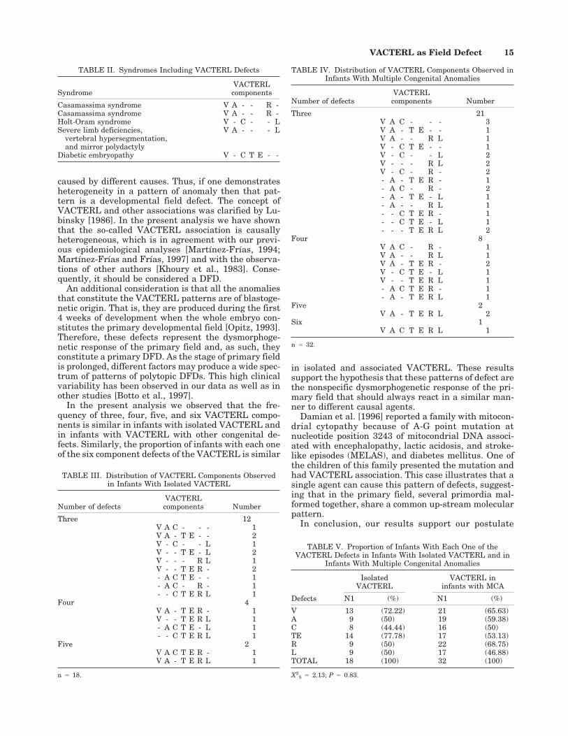
The following tables analyze the clinical variabilityof VACTERL. Table II lists the syndromes present infive infants who had 3 or more VACTERL defects andindicates the type of VACTERL defects observed ineach one of them. We published the syndrome of severelimb deficiencies, vertebral hypersegmentation, andmirror polydactyly in four infants [Urioste et al., 1996;Martınez-Frıas et al., 1997]. In 1981, Casamassima etal. described an entity different from Jarcho-Levin syn-drome that had spondylocostal dysostosis associatedwith anal and urogenital anomalies. The data of TableII should be considered minimal estimates because weregistered other infants with suspected syndromes inwhom the lack of additional studies did not permit adefinitive diagnosis (e.g., suspected trisomy 18 with 3or more VACTERL defects, but without chromosomalstudies). These cases were included in the group of in-fants with MCA.
Table III shows the type of VACTERL defects ob-served in each case with isolated VACTERL and thenumber of infants with three, four, and five of thesedefects. Table IV contains the same information but forthe VACTERL components observed in infants withMCA. These two tables demonstrate the marked clini-cal variability of VACTERL. If we compare the numberof cases with three, four, five, and six VACTERL com-ponents among infants with isolated VACTERL (TableIII) with those observed in infants with MCA (TableIV), the differences are not statistically significant (X2
34 0.94; P 4 0.82).
Finally, Table V shows that the number of childrenwith each one of the six component defects of VAC-TERL are observed in similar proportion among in-fants with isolated VACTERL and in infants with MCA(X2
5 4 2.13; P 4 0.83).
DISCUSSION
Opitz [1993] defined developmental fields as mor-phogenetically reactive units of the developing organ-ism that lead to final structure and are malformed insimilar manner by different causes. Clinically [Opitz,1993], a field is recognized when an identical malfor-mation of complex anatomical structure is proven to be
TABLE I. Distribution of Infants With Three or More VACTERL Componentsby Diagnostic Category*
Diagnostic categoriesTotalcases
With 3 or moreVACTERLcomponents
% Confidenceintervals 95%
(LL–UL)
Developmental field defects (DFD) 1,300 18 1.38 (2.18–2.46)Complexes 27 1 3.70 (0.09–18.97)Multiple congenital anomalies 1,998 32 1.60 (1.10–2.25)Autosomal dominant syndromes 222 1 0.45 (0.01–2.48)Autosomal recessive syndromes 207 2 0.97 (0.11–3.45)Syndromes of unknown cause 152 1 0.66 (0.02–3.61)Embryo-fetopathies 157 1 0.64 (0.02–3.50)
*X 26 4 4.69; P 4 0.58
LL, lower confidence limit; UL, upper confidence limit.
14 Martınez-Frıas and Frıas
caused by different causes. Thus, if one demonstratesheterogeneity in a pattern of anomaly then that pat-tern is a developmental field defect. The concept ofVACTERL and other associations was clarified by Lu-binsky [1986]. In the present analysis we have shownthat the so-called VACTERL association is causallyheterogeneous, which is in agreement with our previ-ous epidemiological analyses [Martınez-Frıas, 1994;Martınez-Frıas and Frıas, 1997] and with the observa-tions of other authors [Khoury et al., 1983]. Conse-quently, it should be considered a DFD.
An additional consideration is that all the anomaliesthat constitute the VACTERL patterns are of blastoge-netic origin. That is, they are produced during the first4 weeks of development when the whole embryo con-stitutes the primary developmental field [Opitz, 1993].Therefore, these defects represent the dysmorphoge-netic response of the primary field and, as such, theyconstitute a primary DFD. As the stage of primary fieldis prolonged, different factors may produce a wide spec-trum of patterns of polytopic DFDs. This high clinicalvariability has been observed in our data as well as inother studies [Botto et al., 1997].
In the present analysis we observed that the fre-quency of three, four, five, and six VACTERL compo-nents is similar in infants with isolated VACTERL andin infants with VACTERL with other congenital de-fects. Similarly, the proportion of infants with each oneof the six component defects of the VACTERL is similar
in isolated and associated VACTERL. These resultssupport the hypothesis that these patterns of defect arethe nonspecific dysmorphogenetic response of the pri-mary field that should always react in a similar man-ner to different causal agents.
Damian et al. [1996] reported a family with mitocon-drial cytopathy because of A-G point mutation atnucleotide position 3243 of mitocondrial DNA associ-ated with encephalopathy, lactic acidosis, and stroke-like episodes (MELAS), and diabetes mellitus. One ofthe children of this family presented the mutation andhad VACTERL association. This case illustrates that asingle agent can cause this pattern of defects, suggest-ing that in the primary field, several primordia mal-formed together, share a common up-stream molecularpattern.
In conclusion, our results support our postulate
TABLE III. Distribution of VACTERL Components Observedin Infants With Isolated VACTERL
Number of defectsVACTERLcomponents Number
Three 12V A C - - - 1V A - T E - - 2V - C - - L 1V - - T E - L 2V - - - R L 1V - - T E R - 2- A C T E - - 1- A C - R - 1- - C T E R L 1
Four 4V A - T E R - 1V - - T E R L 1- A C T E - L 1- - C T E R L 1
Five 2V A C T E R - 1V A - T E R L 1
n 4 18.
TABLE V. Proportion of Infants With Each One of theVACTERL Defects in Infants With Isolated VACTERL and in
Infants With Multiple Congenital Anomalies
Defects
IsolatedVACTERL
VACTERL ininfants with MCA
N1 (%) N1 (%)
V 13 (72.22) 21 (65.63)A 9 (50) 19 (59.38)C 8 (44.44) 16 (50)TE 14 (77.78) 17 (53.13)R 9 (50) 22 (68.75)L 9 (50) 17 (46.88)TOTAL 18 (100) 32 (100)
X25 4 2.13; P 4 0.83.
TABLE II. Syndromes Including VACTERL Defects
SyndromeVACTERLcomponents
Casamassima syndrome V A - - R -Casamassima syndrome V A - - R -Holt-Oram syndrome V - C - - LSevere limb deficiencies,
vertebral hypersegmentation,and mirror polydactyly
V A - - - L
Diabetic embryopathy V - C T E - -
TABLE IV. Distribution of VACTERL Components Observed inInfants With Multiple Congenital Anomalies
Number of defectsVACTERLcomponents Number
Three 21V A C - - - 3V A - T E - - 1V A - - R L 1V - C T E - - 1V - C - - L 2V - - - R L 2V - C - R - 2- A - T E R - 1- A C - R - 2- A - T E - L 1- A - - R L 1- - C T E R - 1- - C T E - L 1- - - T E R L 2
Four 8V A C - R - 1V A - - R L 1V A - T E R - 2V - C T E - L 1V - - T E R L 1- A C T E R - 1- A - T E R L 1
Five 2V A - T E R L 2
Six 1V A C T E R L 1
n 4 32.
VACTERL as Field Defect 15

[Martınez-Frıas et al., 1998] that VACTERL consti-tutes the dysmorphogenetic response of the primarydevelopmental field, which results in the different ex-pressions of this primary, polytopic DFD in response todifferent causes.
REFERENCESBotto LD, Khoury MJ, Mastroiacovo P, Castilla E, Moore CA, Skjaerven R,
Mutchinick OM, Borman B, Cocchi G, Czeizel AE, Goujard J, IrgensLM, Lancaster PAL, Martınez-Frıas ML, Merlob P, Ruusinen A, StollC, Sumiyoshi Y. 1997. The spectrum of congenital anomalies of theVATER association: an international study. Am J Med Genet 71:8–15.
Casamassima AC, Morton CC, Nance WE, Kodroff M, Caldwell R, Kelly T,Wolf B. 1981. Spondylocostal dysostosis associated with anal and uro-genital anomalies in a Mennonite sibship. Am J Med Genet 8:117–127.
Damian MS, Seibel P, Schachenmayr W, Reichmann H, Dorndorf W. 1996.VACTERL with the mitocondrial 3243 point mutation. Am J MedGenet 62:398–403.
Khoury MJ, Cordero JF, Greenberg F, James LM, Erickson JD. 1983. Apopulation study of the VACTERL association: evidence for its etiologicheterogeneity. Pediatrics 71:815–820.
Lubinsky M. 1986. Current concepts: VATER and other associations: his-torical perspectives and modern interpretations. Am J Med Genet(Suppl) 2:9–16.
Martınez-Frıas ML. 1994. Developmental field defects and associations:epidemiological evidence of their relationship. Am J Med Genet 49:45–51.
Martınez-Frıas ML. 1995. Primary midline developmental field I: clinicaland epidemiological characteristics. Am J Med Genet 56:374–381.
Martınez-Frıas ML, Arroyo I, Bermejo E, Espinosa J, Garcıa MJ. 1997.Severe limb deficiencies, vertebral hypersegmentation, and mirrorpolydactyly: two additional cases that expand the phenotype to a moregeneralized effect on blastogenesis. Am J Med Genet 73:205–209.
Martınez-Frıas ML, Frıas JL. 1997. Primary developmental field III: clini-cal and epidemiological study of blastogenetic anomalies and their re-lationship to different MCA patterns. Am J Med Genet 70:11–15.
Martınez-Frıas ML, Frıas JL, Opitz JM. 1998. Errors of morphogenesisand developmental field theory. Am J Med Genet 76:291–296.
Martınez-Frıas ML, Frıas JL, Rodrıguez-Pinilla E, Urioste M, Bermejo E,Cereijo A, Gaya F. 1991. Value of clinical analysis in epidemiologicalresearch: the Spanish registry experience. Am J Med Genet 41:192–195.
Martınez-Frıas ML, Gomar JL. 1994. New case of axial mesodermal dys-plasia sequence: epidemiological evidence of a single entity. Am J MedGenet 49:74–76.
Martınez-Frıas ML, Urioste M. 1994. Segmentation anomalies of the ver-tebrae and ribs: a developmental field defect: epidemiologic evidence.Am J Med Genet 49:36–44.
Opitz JM. 1993. Blastogenesis and the ‘primary field‘ in human develop-ment. BD:OAS vol. XXIX. New York: Alan R. Liss, Inc., for the NationalFoundation March of Dimes. p 3–37.
Urioste M, Lorda-Sanches I, Blanco M, Buron E, Aparicio P, Martınez-Frıas ML. 1996. Severe congenital limb deficiencies, vertebral hyper-segmentation, absent thymus, and mirror polydactyly: a defect expres-sion of a developmental control gene? Hum Genet 97:214–217.
16 Martınez-Frıas and Frıas