ustilago maydis effectors and their impact on virulenceŸe 10, 35043 marburg, germany. 2present...
TRANSCRIPT
Microbial pathogens, such as fungi, oomycetes, bacteria and viruses, are estimated to cause approximately 10% of pre-harvest crop losses worldwide, and this num-ber is likely to increase, in particular for crops that are grown under conditions of high productivity1. To feed an increasing world population, it is necessary to combat pathogens and improve the resistance of crops to dis-eases. To achieve this goal, the mechanisms by which plant pathogenic microorganisms colonize plants need to be elucidated.
In general, when microorganisms attempt to invade plants they are recognized by the plant immune sys-tem through pathogen-associated molecular patterns (PAMPs) or microorganism-associated molecular pat-terns (MAMPs), which are highly conserved molecules such as flagellin in bacteria, glucans in oomycetes and chitin in fungi2. The recognition of such molecules by pattern recognition receptors (PRRs) that reside in the plant plasma membrane triggers an innate immune response known as PAMP-triggered immunity (PTI; orMAMP-triggered immunity (MTI)). Successful invaders are able to dampen or overcome PTI (or MTI) through the secretion of pathogen effectors that either remain at the plant–pathogen interface (apoplastic effec-tors) or are taken up by host cells (translocated or cyto-plasmic effectors). Effectors function in various ways: they can shield the pathogen, inactivate plant enzymes or toxic compounds that are harmful to the pathogen, prevent the elicitation of plant immune responses, or change the physiology of the infected plant to support the growth and development of the pathogen3. In particular, eukary otic pathogens produce a large arsenal of several
hundred effectors, which seem to be produced in consec-utive expression waves during the course of an infection and/or are associated with pathogenic transitions3.
In addition to PTI (MTI), plants have a second line of defence in the form of resistance proteins, which are able to recognize single effectors and activate effector- triggered immunity (ETI). Thus, plant–pathogen inter-actions are driven by a co-evolutionary ‘arms race’ that involves the continuous selection for novel pathogenic strains that overcome ETI through the help of effectors and new plant genotypes that restore ETI by modifying host molecules that are targeted by effectors2–4.
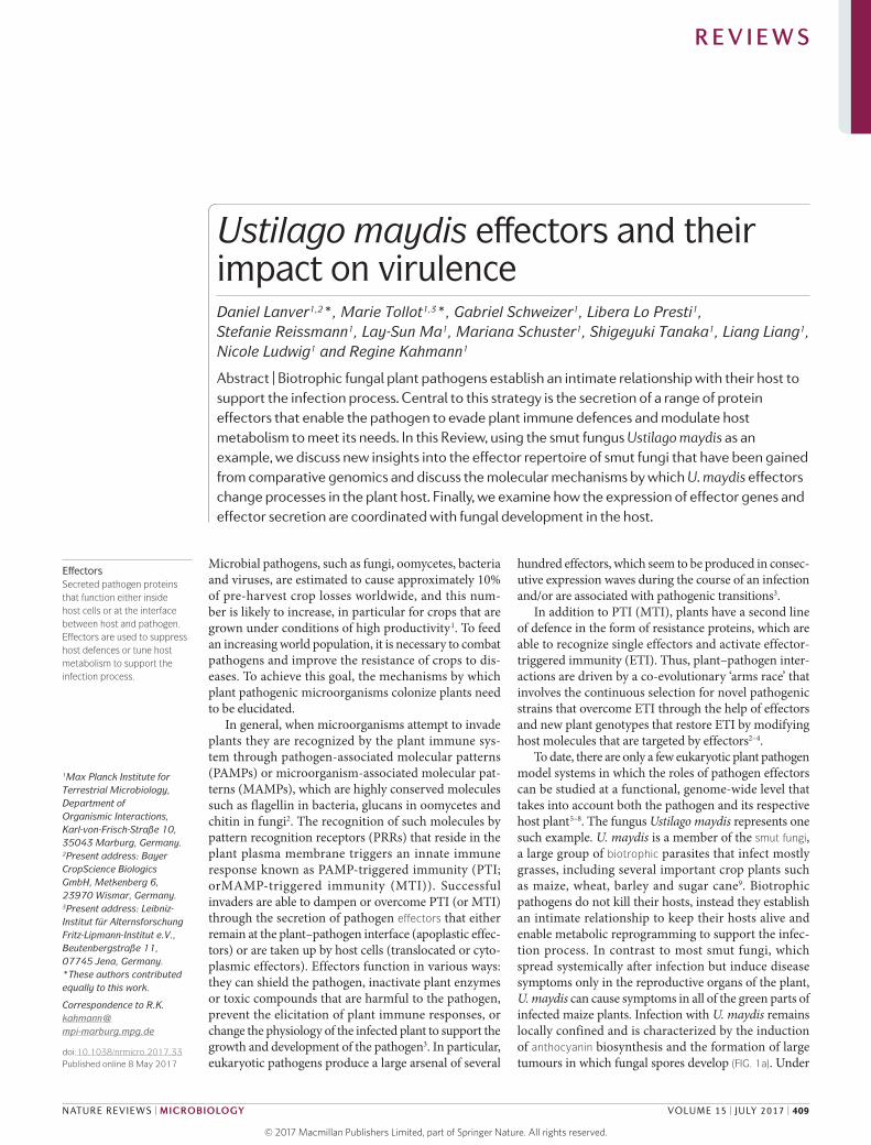
To date, there are only a few eukaryotic plant patho gen model systems in which the roles of pathogen effectors can be studied at a functional, genome-wide level that takes into account both the pathogen and its respective host plant5–8. The fungus Ustilago maydis represents one such example. U. maydis is a member of the smut fungi, a large group of biotrophic parasites that infect mostly grasses, including several important crop plants such as maize, wheat, barley and sugar cane9. Biotrophic pathogens do not kill their hosts, instead they establish an intimate relationship to keep their hosts alive and enable metabolic reprogramming to support the infec-tion process. In contrast to most smut fungi, which spread systemically after infection but induce disease symptoms only in the reproductive organs of the plant, U. maydis can cause symptoms in all of the green parts of infected maize plants. Infection with U. maydis remains locally confined and is characterized by the induction of anthocyanin biosynthesis and the formation of large tumours in which fungal spores develop (FIG. 1a). Under
1Max Planck Institute for Terrestrial Microbiology, Department of Organismic Interactions, Karl-von-Frisch-Straße 10, 35043 Marburg, Germany.2Present address: Bayer CropScience Biologics GmbH, Metkenberg 6, 23970 Wismar, Germany.3Present address: Leibniz-Institut für Alternsforschung Fritz-Lipmann-Institut e.V., Beutenbergstraße 11, 07745 Jena, Germany.*These authors contributed equally to this work.
Correspondence to R.K. kahmann@ mpi-marburg.mpg.de
doi:10.1038/nrmicro.2017.33Published online 8 May 2017
EffectorsSecreted pathogen proteins that function either inside host cells or at the interface between host and pathogen. Effectors are used to suppress host defences or tune host metabolism to support the infection process.
Ustilago maydis effectors and their impact on virulenceDaniel Lanver1,2*, Marie Tollot1,3*, Gabriel Schweizer1, Libera Lo Presti1, Stefanie Reissmann1, Lay-Sun Ma1, Mariana Schuster1, Shigeyuki Tanaka1, Liang Liang1, Nicole Ludwig1 and Regine Kahmann1
Abstract | Biotrophic fungal plant pathogens establish an intimate relationship with their host to support the infection process. Central to this strategy is the secretion of a range of protein effectors that enable the pathogen to evade plant immune defences and modulate host metabolism to meet its needs. In this Review, using the smut fungus Ustilago maydis as an example, we discuss new insights into the effector repertoire of smut fungi that have been gained from comparative genomics and discuss the molecular mechanisms by which U. maydis effectors change processes in the plant host. Finally, we examine how the expression of effector genes and effector secretion are coordinated with fungal development in the host.
R E V I E W S
NATURE REVIEWS | MICROBIOLOGY VOLUME 15 | JULY 2017 | 409
© 2017
Macmillan
Publishers
Limited,
part
of
Springer
Nature.
All
rights
reserved.

Nature Reviews | Microbiology
10 μm
Diploid sporeSeptum
Promycelium
Appressorium
Plant cell wallPlant plasma membrane
Growing tipRetraction septa
Dikaryon
Plant cell penetration Hyphal branching
Clamp-likestructure
Conjugation tube
Apoplastic cavity with aggregated hyphae Spore development
Haploid cell
a
d
g h i
j k
e f
b c
Polysaccharide matrix
Smut fungiBiotrophic basidiomycete plant pathogens of the order Ustilaginales that undergo sexual reproduction only during the infection of a host plant. They produce dark pigmented spores that look similar to coal dust or ‘smut’ when released into the environment.
BiotrophicIn a biotrophic interaction, plant pathogens establish a compatible interaction with their hosts, during which the plant stays fully alive, provides nutrients to the pathogen and enables the pathogen to complete its life cycle.
AnthocyaninA red plant pigment that is synthesized through the phenylpropanoid pathway and protects cells from high-light damage, acts as an antioxidant or is an attractant for pollinators.
Figure 1 | The life cycle of Ustilago maydis. a | Symptoms of a cob infection by U. maydis in a maize field. The infection is locally confined and characterized by the induction of anthocyanin biosynthesis and the formation of large tumours in which fungal spores develop. b | Diploid spores are released when tumours break open. They are dark coloured owing to their high melanin content and have a characteristic round shape and surface ornamentation. Meiosis takes place in germinating spores; the four resulting haploid nuclei migrate into a promycelium, in which they become delineated by septa. c | Following mitotic divisions, haploid cells bud off from these compartments. d | After detection of a compatible mate, the budding programme ceases and cells develop conjugation tubes that are directed towards each other. e | After cell fusion, a filamentous cell cycle-arrested dikaryon is produced. Only the growing tip of this filament is filled with cytoplasm (yellow), whereas older parts are vacuolated (grey) and become sealed off by regularly spaced septa. These retraction septa enable filament elongation and the formation of an infective structure (appressorium) in extended infectious hyphae129. f,g | Hyphal tip cells develop appressoria in specific locations on the leaf surface and then penetrate plant cells. h | During early stages of infection, the cell cycle arrest is released, hyphae begin to branch and clamp-like structures (orange) assure correct segregation of the two nuclei12. During these intracellular stages, hyphae are completely encased by the host plasma membrane13 (red). i | With the onset of plant tumour formation, fungal hyphae are mainly detected intercellularly17. j | Subsequently, the two nuclei of the dikaryon fuse, followed by the substantial proliferation of diploid cells that form huge aggregates in apoplastic cavities17,18. Aggregated hyphae become embedded in a gelatinous polysaccharide matrix19 (pink). k | Hyphae then fragment and undergo spore development. In all panels, white and dark grey nuclei indicate that they are haploid and have different mating-type genes. Nuclei that are half white and half dark grey indicate diploid nuclei that are generated through the fusion of white and dark grey nuclei. Image in part a courtesy of R. Rösser, Max Planck Institute for Terrestrial Microbiology, Marburg, Germany.
R E V I E W S
410 | JULY 2017 | VOLUME 15 www.nature.com/nrmicro
© 2017
Macmillan
Publishers
Limited,
part
of
Springer
Nature.
All
rights
reserved. ©
2017
Macmillan
Publishers
Limited,
part
of
Springer
Nature.
All
rights
reserved.

AppressoriaSpecialized cells that develop at the hyphal tips of many fungal plant pathogens that are used to infect host plants either by mechanical pressure or with the help of the localized secretion of plant cell wall-degrading or loosening enzymes.
ClampA hook-like structure formed by the dikaryotic hyphal tip cells of basidiomycete fungi that assures the correct segregation of the two different nuclei and the maintenance of the dikaryotic state in growing hyphae.
appropriate conditions, such as ambient temperature and humidity, the diploid spores germinate, undergo meio-sis and form a promycelium into which the four haploid nuclei migrate. Septation then produces compartments that contain one haploid nucleus (FIG. 1b). Following mitosis, haploid cells bud off from the promycelium and enter the vegetative life cycle, during which they prolif-erate by budding (FIG. 1b,c). On the leaf surface, haploid cells of different mating types fuse and form a filamen-tous cell cycle-arrested dikaryon, which is the pathogenic form10,11 (FIG. 1d,e). These filaments differentiate into infection structures known as appressoria (FIG. 1f). After penetration of the epidermal layer of the plant, cell cycle arrest ceases and clamp-like structures assure the correct segregation of the two nuclei12 in the growing dikaryotic hyphae (FIG. 1 g,h). Hyphae that colonize the epidermal and immediate underlying tissues remain intracellular and are completely encased by the host plasma mem-brane13, thereby establishing an intimate and extended host– pathogen interaction zone that is enriched in endo-membrane structures14. Notably, U. maydis does not develop haustoria, which are characteristic structures that mediate signal exchange and nutrient acquisition in other
biotrophic fungal plant pathogens6, and it is likely that this inter action zone fulfils these functions. As the infec-tion progresses, hyphae reach mesophyll tissue and often grow along and inside the veins, presumably to access nutrients. Concomitantly with the formation of plant tumours, during which plant cells enlarge and resume mitotic division15,16, fungal hyphae are mainly detected inter cellularly17 (FIG. 1i). Subsequently, the two nuclei of the dikaryon fuse, which is followed by substantial pro-liferation of diploid cells and the formation of huge aggre-gates in apoplastic cavities17,18. Aggregated hyphae become embedded in a gelatinous polysaccharide matrix19 (FIG. 1j). Hyphal morphology changes to highly intertwined struc-tures20, which is followed by hyphal fragmentation and the development of dark coloured ornamented spores (FIG. 1k). Owing to the unique morphological features of an infection with U. maydis, together with the molecular toolbox that has been developed for reverse genetics, cell biology, comparative genomics and functional studies (BOX 1), U. maydis has become an important model system for the study of biotrophic fungal pathogens.
Pathogen effectors are considered to be secreted proteins that modify host cell structure and function to promote the success of a pathogen4. Defining the functional effector repertoire of a pathogen would require genome-wide knockout mutants for all effector candidates, a goal that has not yet been accomplished, even in the most advanced eukaryotic systems. In addi-tion, the mutants should have virulence phenotypes. However, in many cases, virulence is unaffected by deleting single effector genes, which presumably reflects redundancy4,6,21–23. To identify and prioritize effector can-didates of eukaryotic microorganisms, they are usually predicted to contain a signal peptide using bioinformat-ics. In addition, other criteria are used to identify candi-date effectors, such as the presence of certain sequence motifs, small size (less than 200 or 300 amino acids), richness in cysteine residues, expression being linked to the stage of plant colonization and/or the absence of known structural or functional domains4. However, all of these criteria have limitations; specific sequence motifs may be absent, proteins secreted in an unconventional manner that lack a signal peptide can be effectors24, proteins that are larger than 200 or 300 amino acids can function as effectors25–27, the expression of effector genes may already occur before contact with the plant and effectors can have functional protein domains27–29. In U. maydis, about 40% of all candidate secreted proteins are completely novel, as they lack known structural or functional domains30. Many of the respective genes reside in gene clusters in the genome31,32, similarly to patho-genicity islands in bacteria, and they are expressed only during plant colonization31,33. In this Review, we use the term effector for all U. maydis proteins that are predicted to be secreted through the classic endoplasmic reticulum (ER)–Golgi route and lack known domains (novel effec-tors), as well as for secreted proteins with a confirmed virulence function that contain a known domain, are transcriptionally induced following plant colonization and are not required for growth or morphology when U. maydis is propagated in axenic culture.
Box 1 | Tools to study effector function
The first clue in regard to the role of an effector is generally obtained by analysing the virulence phenotype of the corresponding deletion mutant. The highly efficient homologous recombination system of Ustilago maydis has enabled the generation of numerous effector mutants through gene replacement13,15,22,27,31,34,42,50,51,57,71,111. However, functional redundancy in effector families has slowed their characterization, despite the establishment of the flippase-recombinase-based marker recycling system39. In future, the recently adapted CRISPR–Cas9 system112, which has now been engineered to enable multiplexing30, promises to be less tedious and time-consuming for the mutation of effector gene families. A prerequisite for understanding the function of an effector is to know where it localizes. In contrast to oomycete effectors, fungal effectors generally lack characteristic uptake motifs104,105. Live fluorescence microscopy of maize leaves that were infected with U. maydis has been useful to visualize the secretion of effector–GFP or effector–mCherry fusion proteins, but proved unsuccessful for demonstrating effector translocation13,15,27,34,71. Until recently, the most reliable approach to demonstrate effector translocation was immunoelectron microscopy (immuno-EM), using epitope-tagged effectors15,27. A newly developed assay that is based on in vivo biotinylation now promises to be a valuable alternative to immuno-EM for the high-throughput screening of effector uptake106. The assay uses the ability of the bacterial biotin ligase BirA to biotinylate any protein that has a short peptide termed the Avitag. Transgenic maize lines that express BirA in the cytoplasm are infected with engineered strains of U. maydis that secrete effectors tagged with the Avitag. Translocated effectors (that is biotinylated effectors) can then be detected using streptavidin immunoprecipitation from infected leaves106.
It is important to distinguish between potentially relevant and spurious plant interactors, which are often identified using yeast two-hybrid screening and are validated by co-immunoprecipitation or bimolecular fluorescence complementation in transient expression experiments in Nicotiana benthamiana15,27,51,57,71. The availability of constitutive and inducible promoters113,114 also enables the purification of effectors from U. maydis culture supernatants. Purified effectors can then be used to pull down potential plant interactors from infected leaf extracts or to test their ability to inhibit the activity of target proteins in vitro51,57,71. Ultimately, the biological relevance of the effector–target interaction must be demonstrated; for example, by downregulating the expression of the maize interactor either transiently through virus-induced gene silencing57,115 or by generating transgenic plants that are silenced for the respective gene. The relevance of the interaction can also be substantiated by narrowing down the domain that mediates the interaction and by demonstrating that this effector domain is necessary and sufficient to restore the virulence of the deletion mutant51.
R E V I E W S
NATURE REVIEWS | MICROBIOLOGY VOLUME 15 | JULY 2017 | 411
© 2017
Macmillan
Publishers
Limited,
part
of
Springer
Nature.
All
rights
reserved. ©
2017
Macmillan
Publishers
Limited,
part
of
Springer
Nature.
All
rights
reserved.

Nature Reviews | Microbiology
Num
ber o
f sec
rete
d pr
otei
ns
300
400
500
200
100
0U. maydis S. scitamineum S. reilianum U. hordei M. pennsylvanicum
Secreted accessory protein with structural or functional domainSecreted core protein with structural or functional domain
Secreted novel accessory effectorSecreted novel core effector
158
467
45
107
157
165
467
46
108
148
132
404
37
92
143
67
24
72
303
29
75
132
166
464
44
104
150
SecretomeThe totality of all proteins of a species that are predicted to be, or shown to be, secreted into the extracellular space.
In initial studies, to detect an effect on virulence, entire effector gene clusters were deleted in U. maydis31; however, the effects turned out to be additive, as single deletions of such clustered effector genes had a relatively modest or no contribution to virulence22. In addition, whereas the effectors of filamentous eukaryotic plant pathogens often exhibit a high degree of redundancy4, U. maydis proved quite unique in that several mutants that lacked single effector genes were markedly attenu-ated in virulence or became unable to cause any disease symptoms13,26,34,35. In this Review, we summarize our current knowledge of the effector repertoire that has been gained from comparative genomics in smut fungi. We also discuss the molecular mechanisms by which the effectors of U. maydis change host cell processes to dampen plant defences, how the respective genes are regulated and how effectors evolve.
Effectors of U. maydisThe effector repertoire. Of the 6,784 proteins expressed by U. maydis, 467 are potentially secreted (that is, they contain a signal peptide and lack transmembrane domains) and of these 203 are completely novel, which
means that they lack functional or structural domains30. As detailed below, these novel effectors have been ana-lysed extensively (BOX 1) and many of these effectors were shown to contribute to virulence.
In smut fungi that are related to U. maydis, such as the maize head smut pathogen Sporisorium reilianum, the sugar cane pathogen Sporisorium scitamineum and the barley pathogen Ustilago hordei, similar numbers of secreted proteins and novel secreted effectors have been predicted30 (FIG. 2). Melanopsichium pennsylvanicum, a smut fungus that infects dicot plants of the genus Persicaria, has a substantially smaller secretome and also a reduced set of novel effectors, which may be the consequence of adaptation to a dicot host36. A compar-ative analysis of the secretomes of these five smut fungi (FIG. 2) enabled the identification of the effector reper-toire in these species. A core secreted protein family is considered to contain orthologues in all five species and may contain additional paralogues. Families of secreted proteins that are found in only a subset of species are designated as accessory30. According to this analysis, the five smut fungi that were compared in this study have 72 core families of secreted proteins with known domains. Among this set are at least two effector protein families that contain the chorismate mutase Cmu1 and a novel type of hydrophobin, Hum3, in which the hydrophobin domain is fused to a repetitive domain30,37. Of the candi-date effectors without recognizable domains, 24 families are considered core effectors30, and of these, 12 fami-lies have only one representative in all five species. In this latter group is PEP1, which is a crucial effector for virulence in U. maydis that is functionally conserved in U. hordei and M. pennsylvanicum13,38. In addition, 10 members of the EFF1 effector gene family, which redundantly contribute to virulence39, are predicted to be novel core effectors, whereas one member of this family is in the core group of secreted proteins with domains30. The number of novel smut core effectors is in a range that is comparable to the core effectors in plant patho-genic bacteria40,41. The core effectors that have not yet been studied represent promising candidates for future functional analyses because they are likely to have crucial functions for virulence in all smut fungi.
Of the accessory effectors, 45, 33, 43, 52 and 42 are strictly species-specific and exist only in U. maydis, S. scitamineum, S. reilianum, U. hordei and M. pennsylvanicum, respectively30. Five effector families have one or more homologues in the four grass-infecting smuts but are absent in related species30, which makes them interesting effector candidates that have grass-specific functions. Although effectors that are required for host selectivity have not yet been identified in smut fungi, some U. maydis effectors have been shown to have organ-specific functions; that is, they are expressed in an organ-specific manner and are required for virulence only in tassel, the male inflorescence, or in seedlings33,42. Both U. maydis and S. reilianum infect maize, but only S. reilianum can colonize maize systemically following root infection43, which suggests that some of the 38 effectors in S. reilianum that are absent in U. maydis30 might promote this infection strategy.
Figure 2 | Effector repertoire of Ustilago maydis and related smut fungi. A comparative analysis of the secretomes of five smut fungi (the corn pathogen Ustilago maydis, the maize head smut pathogen Sporisorium reilianum, the sugar cane pathogen Sporisorium scitamineum, the barley pathogen Ustilago hordei and Melanopsichium pennsylvanicum, which infects dicot plants of the genus Persicaria) enabled the identification of the effector repertoires in these species. The total number of predicted secreted proteins in each species is indicated at the top of each bar. The secretomes of the indicated smut fungi were sorted into the four categories after constructing families of all secreted proteins in the indicated smut fungi. A core secreted protein family is defined as containing orthologues in all five species and may contain additional paralogues. Families of secreted proteins that are found in only a subset of species are classified as accessory. Numbers of individual secreted proteins in each category are indicated for each species. 24 families of novel core effectors were detected (light grey band across the figure, labelled 24), and for secreted proteins that have predicted domains 72 core families were present in all five smut genomes (light grey band across bottom of the figure, labelled 72)30. The total number of secreted proteins indicated in the core groups in each species is higher than 24 and 72 owing to additional existing paralogues. Data from REF. 30.
R E V I E W S
412 | JULY 2017 | VOLUME 15 www.nature.com/nrmicro
© 2017
Macmillan
Publishers
Limited,
part
of
Springer
Nature.
All
rights
reserved. ©
2017
Macmillan
Publishers
Limited,
part
of
Springer
Nature.
All
rights
reserved.

CystatinA member of a family of conserved proteins that act as cysteine protease inhibitors.
Reactive oxygen species(ROS). Chemically reactive chemical species that contain oxygen and have important roles in the development of, and defence responses in, plants.
ApoplastThe compartment outside the plant plasma membrane that is formed by the continuum of the cell walls of adjacent cells and the extracellular spaces, through which water and solutes can diffuse.
In U. maydis and the related smut fungi, most effector genes are more poorly conserved than the rest of the genome32. This accelerated evolution probably reflects the ongoing adaptation of effectors to their host targets, as well as their need to escape detection by the host immune system4,44,45 (BOX 2). Whether such highly diver-sified effectors target the same plant molecules in the different smut pathosystems is not yet known.
Apoplastic effectors of U. maydis. Apoplastic effectors may function in several ways to disarm plant defence responses. They can scavenge fungal elicitors (for exam-ple, cell wall fragments that would otherwise activate the plant defence response) or they can inhibit plant lytic enzymes, such as proteases, chitinases or glucanases, to prevent the release of fungal elicitors. Apoplastic effec-tors can also directly detoxify antimicrobial compounds that are released during the defence response46.
One of the most widespread mechanisms used by pathogenic fungi to dampen host immune signalling is the secretion of LysM effectors. These effectors bind to soluble chitin oligomers to prevent their recognition by plant immune receptors47. U. maydis produces only one protein that has LysM domains, but there is currently no indication that this protein positively affects virulence48. This indicates that U. maydis must have other mechan-isms to sequester soluble chitin or to protect itself from secreted plant chitinases. Candidate effectors for this
function are Hum3 and Rsp1, which may be structur-ally related to the repetitive repellent protein Rep1 that confers surface hydrophobicity37,49. REP1 encodes 11 short repellent peptides that result from the cleavage of a secreted precursor protein by the Kex2 protease49. These peptides are tightly bound to the cell wall of U. maydis hyphae and form amyloid-like fibrils50. Hum3 consists of a repetitive domain that is fused to a hydrophobin-like domain, whereas Rsp1 consists of only repetitive domains. Hum3 and Rsp1 are also processed by Kex2, which suggests that the biologically active molecules that are generated from these proteins may also be short peptides 21–36 amino acids in length37. Mutants that are deficient in both Hum3 and Rsp1 are able to penetrate into, but fail to spread within, the plant, and necrotic spots develop at the site of infection, which indicates that plant defence responses are induced and virulence is abrogated37. Although the molecular mechanism by which these peptides contribute to virulence remains to be elucidated, we speculate that they might either localize to the hyphal cell wall, shielding or masking the invading fungal hyphae from recognition by the plant immune sys-tem, or that they might function as signalling molecules that downregulate plant defence responses.
In regard to effectors that target lytic enzymes, U. maydis secretes the novel effector Pit2, which inhib-its a set of at least four apoplastic cysteine proteases, the activities of which are directly linked to salicylic acid-associated plant defences51 (FIG. 3a). Pit2 inhibits these cysteine proteases through a 14-amino acid motif that is conserved in Pit2 orthologues but has no resem-blance to any other cysteine protease inhibitor38. Mutant strains that lack PIT2 elicit strong defence responses in their host and the spread of hyphae and the formation of tumours are severely attenuated34. Effector proteins that function as cysteine protease inhibitors were first discovered in Passalora fulva (formerly known as Cladosporium fulvum), in which Avr2, an effector that inhibits the apoplastic cysteine protease Rcr3 of tomato plants, promotes virulence52,53. The pathogenic oomy-cete Phytophthora infestans secretes cystatin-like proteins, which inhibit host cysteine proteases that have been implicated in plant defence54. Intriguingly, U. maydis not only secretes a cysteine protease inhibitor but also stimulates the expression of the maize cystatin CC9, which inhibits the same class of cysteine proteases as Pit2 (REF. 55). This highlights that plant cysteine proteases are crucial virulence targets in the U. maydis–maize system. It remains to be shown whether these proteases need to be inhibited because they directly attack fungal compo-nents (for example, effectors), or because they activate endogenous plant processes that are involved in defence against pathogens.
The initiation of plant defence reactions following fungal infection is often accompanied by the accumu-lation of reactive oxygen species (ROS) at the site of pen-etration56. ROS can be toxic to the fungus or function as second messengers to reinforce plant defence responses. The novel U. maydis effector Pep1 inhibits the maize per-oxidase POX12, which is one of the major generators of H2O2 in the apoplast57 (FIG. 3b). Mutants that lack PEP1
Box 2 | Effector evolution
Effectors and their plant target molecules evolve in a molecular ‘arms race’ — a process of ongoing adaptation and counter-adaptation4. Effector genes in plant pathogenic fungi are thus under strong evolutionary pressure and their evolution is accelerated compared with the rest of the genome. Accelerated evolution can be achieved through the compartmentalized localization of effector genes in highly mutagenic genomic regions. Depending on the fungal species, the strategy of compartmentalization varies, with the most prominent examples being effector localization in repeat-rich and gene-sparse regions116,117, effector localization in AT-rich isochores in which effector genes are subject to repeat-induced point mutations118, effector localization close to chromosomal breakpoints119 and effector localization on dispensable chromosomes120–122.
In smut fungi, the genomes of which are highly compact, effector genes are often arranged in gene clusters. About 25% of the effectors of Ustilago maydis are arranged in such clusters, with the largest cluster comprising 24 genes31. Clusters probably arose through the tandem duplication of genes for which, owing to the low amount of repetitive elements in the genome, subsequent dispersal did not occur123. Among the sequenced smut fungi, Ustilago hordei has the highest content of repetitive elements124. As a likely consequence, U. hordei has fewer clustered effector genes than other smut fungi123. A hallmark of effector clusters is their low sequence conservation between species in an overall syntenic and higher conserved genomic context32. Moreover, clustered effector genes are associated with a class of interspersed repeats that are not yet characterized123, which suggests that effector gene clustering is another way of genome compartmentalization in which specific transposable elements may restrict their activity on effector genes.
Unexpectedly, it is emerging that several novel effectors that have crucial functions for virulence in U. maydis are neither duplicated in clusters nor do they have paralogues elsewhere in the genome. This group includes PEP1 (REFS 13,38,57), PIT2 (REFS 34,51), APB73 (REF. 26) and STP1 (REF. 35). Compared with other effectors, Pep1, Apb73 and Stp1 are more highly conserved, which is inconsistent with their involvement in the ‘arms race’ with their host targets. We speculate that these effectors, which incidentally are all highly expressed, may be under strong selective pressure to evolve translational robustness, a feature that constrains sequence evolution in accordance with the protein misfolding avoidance hypothesis125.
R E V I E W S
NATURE REVIEWS | MICROBIOLOGY VOLUME 15 | JULY 2017 | 413
© 2017
Macmillan
Publishers
Limited,
part
of
Springer
Nature.
All
rights
reserved. ©
2017
Macmillan
Publishers
Limited,
part
of
Springer
Nature.
All
rights
reserved.

Defence
Defence
Nature Reviews | Microbiology
Plant transcriptionfactor (ZmR1)
Protein kinase(ZmTTK1)
UbiquitinylatedZmTTK1
Cmu1
Plant cell wall
Apoplast
Fungal hyphae
See1SGT1
Tin2
Pit2 effector
Host cysteine proteases(CP2, CP1A, CP1B and XCP2)
Pep1
Plant peroxidase(POX12)
Defence
H2O
2
O2
ChorismatePrephenate
Plasma membrane
Plasma membrane
Phenylalanine,tyrosine
Anthocyanin
Lignin
p-Coumaric acid
p-Coumaric acid
?
Nucleus
Chorismate
Salicyclic acid
Chorismate
Salicyclic acid
P
Plant cytoplasm
P
P
P
?
Stimulationof celldivisionin leaves
Defence
SG20
0Δ
PIT2
SG20
0Δ
PEP1
SG20
0Δ
CM
U1
SG20
0Δ
TIN
2SG
200
ΔSE
E1
a
b
c
d
e
R E V I E W S
414 | JULY 2017 | VOLUME 15 www.nature.com/nrmicro
© 2017
Macmillan
Publishers
Limited,
part
of
Springer
Nature.
All
rights
reserved. ©
2017
Macmillan
Publishers
Limited,
part
of
Springer
Nature.
All
rights
reserved.

Shikimate pathwayA metabolic route for the biosynthesis of the aromatic amino acids phenylalanine, tyrosine and tryptophan.
Guard cellsSpecialized epidermal cells that surround the stomatal pore and enable it to open and close.
PlasmodesmataMicroscopic channels that traverse the cell walls of plant cells and enable transport and communication between them.
induce strong plant defence responses, including the accumulation of H2O2, and this completely blocks the mutant from penetrating maize epidermal cells13. Virus-induced gene silencing of POX12 resulted in par-tial complementation of the penetration defect of the PEP1 mutant strain, which provided further functional evidence for the role of peroxidase inhibition in the virulence of U. maydis57. A recent study demonstrated that the virulence defect of the PEP1 mutant could be complemented by orthologous genes from related smut species, including the orthologue of the dicot pathogen M. pennsylvanicum, which emphasizes that PEP1 is a genuine core effector (FIG. 2). Furthermore, transgenic barley plants that expressed PEP1 of U. maydis showed increased sensitivity to infections with Blumeria graminis f. sp. hordei, which illustrates that peroxidase inhibition is a feature that also benefits other pathogens38.
Translocated effectors of U. maydis. Effectors that are taken up by plant cells modulate host cell biology; they may inhibit plant stress and defence responses, manip-ulate hormone signalling and/or synthesis, interfere with signalling cascades downstream of plant immune receptors and/or reprogramme the metabolism of host cells4,6,58. A common feature of biotrophic pathogens is the downregulation of plant salicylic acid signalling59.
Salicylic acid is usually produced in response to infec-tion and leads to the increased expression of defence genes and local cell death60–64. The synthesis of salicylic acid occurs in the plant chloroplasts, in which choris-mate, the end product of the shikimate pathway, func-tions as precursor65,66. U. maydis secretes the active chorismate mutase Cmu1, which is an effector that has a known enzyme domain. Cmu1 is a translocated effector that was shown, using immunoelectron micros-copy, to localize in the cytosol of infected plant cells27
(FIG. 3c). Plants that are infected with a CMU1-deletion strain accumulate substantially higher levels of salicylic acid than plants that are infected with the correspond-ing wild-type strain27. It has been postulated that Cmu1 decreases salicylic acid levels by reducing the pool of chorismate and channelling the shikimate pathway towards the production of phenylalanine and tyrosine27. Microscopy analysis of maize leaves that transiently express a non-secreted Cmu1–mCherry fusion protein revealed that Cmu1–mCherry could spread from the cells that express the protein to neighbouring cells that do not, which suggests that Cmu1 metabolically primes plant cells for an upcoming infection27. Spread to neigh-bouring cells was never observed when guard cells (which lack plasmodesmata) were transformed67, which suggests that Cmu1 passes through plasmodesmata27. Cell-to-cell spread of translocated effectors is also observed in the Magnaporthe oryzae–rice system68 and may thus be a general feature to sensitize uncolonized plant cells to pathogen infection.
Surprisingly, in Cmu1-deficient mutants, virulence was only weakly affected27, which suggests the exist-ence of additional mechanisms to counteract salicylic acid-dependent plant defence responses. U. maydis encodes the salicylate hydrolase Shy1, which is sub-strate-induced and can degrade salicylic acid. However, a mutant that lacked both SHY1 and CMU1 did not show an enhanced virulence defect69. The identifica-tion of Rss1, a salicylic acid-sensing transcription factor in U. maydis, might lead to the discovery of additional proteins that are involved in the degradation of salicylic acid during plant infection70. Recently, an alternative strategy to decrease salicylic acid levels was uncovered in Phytophthora sojae and Verticillium dahliae. These plant pathogens have unconventionally secreted isochor-ismatases that are targeted to chloroplasts and disrupt the plant salicylic acid biosynthetic pathway by hydro-lysing isochorismate, which is the immediate precursor of salicylate24. Interestingly, U. maydis also encodes an isochorismatase (X. Han and R. K., unpublished obser-vations). Whether this enzyme works together with Cmu1 to attenuate salicylic acid-mediated responses is currently under investigation. Alternatively, salicylic acid-induced defence responses can be suppressed by the inhibition of secreted salicylic acid-induced plant cysteine proteases, an activity that was demonstrated for the apoplastic U. maydis effector Pit2 (REF. 51).
Another novel translocated effector of U. maydis is Tin2, which contributes to virulence, but the most remarkable phenotype of an U. maydis TIN2deletion strain is the complete lack of the accumulation of
Figure 3 | Functionally characterized effectors of Ustilago maydis. a–e | Of the 203 novel effector proteins in U. maydis, the molecular functions of two apoplastic effectors, Pit2 and Pep1, as well as two translocated effectors, Tin2 and See1, have been determined. The functionally characterized translocated effector Cmu1 belongs to the class of effectors that have a functional domain. The left panel shows photographs of representative maize leaves 12 days post-infection with the solopathogenic wild-type strain SG200 and the respective effector deletion mutant. The right panel contains schematics that illustrate the molecular function of the respective effector protein. a | The secreted U. maydis effector Pit2 inhibits host cysteine proteases, which have been implicated as crucial virulence targets. Mutant strains that lack PIT2 elicit strong defence responses in the host, and the spread of hyphae and tumour formation are severely attenuated. b | Pep1 inhibits the maize peroxidase POX12, which is one of the major producers of H2O2 in the apoplast. Mutants that lack PEP1 induce strong plant defence responses, including the accumulation of H2O2, which completely blocks the ability of the fungus to penetrate maize epidermal cells. c | Cmu1 in the plant cytosol decreases salicylic acid levels, a signal to activate host defences, by reducing the pool of the salicylic acid precursor chorismate and increasing the production of phenylalanine and tyrosine. Virulence is only weakly attenuated in Cmu1-deficient mutants, which suggests that additional mechanisms exist to counteract salicylic acid-dependent plant defence responses. d | Tin2 interacts with a phosphodegron-like motif of the cytoplasmic maize protein kinase ZmTTK1, which prevents its ubiquitylation and proteasome-dependent degradation. The Tin2-stabilized protein kinase triggers the nuclear localization of ZmR1, a maize transcription factor that induces the expression of genes involved in the biosynthesis of anthocyanin. Tin2-mediated induction of the anthocyanin pathway may deplete p-Coumaric acid, which is also a precursor for the biosynthesis of lignin, thus decreasing the physical lignin-containing barrier that restricts pathogen spread. The question mark indicates that the phosphorylation of ZmR1 remains to be shown experimentally. e | See1 localizes to the plant cytoplasm and nucleus and contributes to tumour progression in maize leaves by reactivating host DNA synthesis and cell division. See1 interacts with maize SGT1 and interferes with its phosphorylation. SGT1 proteins have important roles in plant resistance. How this is connected to the observed See1-dependent reactivation of cell cycle progression remains to be investigated. The question mark indicates that the kinase responsible for the phosphorylation of SGT1 remains to be identified. The images in parts a, b, c and e are courtesy of S. Winterberg, Max Planck Institute for Terrestrial Microbiology, Marburg, Germany. The image in part d is courtesy of S. Tanaka, Max Planck Institute for Terrestrial Microbiology, Marburg, Germany.
◀
R E V I E W S
NATURE REVIEWS | MICROBIOLOGY VOLUME 15 | JULY 2017 | 415
© 2017
Macmillan
Publishers
Limited,
part
of
Springer
Nature.
All
rights
reserved. ©
2017
Macmillan
Publishers
Limited,
part
of
Springer
Nature.
All
rights
reserved.

Non-host resistanceA term that defines the broad-spectrum resistance of a particular plant species against all isolates of a pathogen that can cause disease in other plant species.
anthocyanin in infected maize tissue22,71 (FIG. 3d). The production of anthocyanin can be complemented by the transient expression of Tin2 in the cytoplasm of plant cells that are pre-infected with an U. maydis TIN2-deletion strain71, which revealed that Tin2 func-tions inside maize cells. Tin2 interacts with a phos-phodegron-like motif of the cytoplasmic maize protein kinase ZmTTK1, which prevents its ubiquitylation and proteasome-dependent degradation71 (FIG. 3d). The Tin2-stabilized protein kinase triggers the nuclear locali-zation of ZmR1, a maize transcription factor that induces anthocyanin biosynthetic genes72,73. The stabilization of host proteins by preventing their proteasomal degrada-tion is a common strategy that is also used by other plant pathogenic microorganisms and symbionts74,75.
The induction of anthocyanin biosynthesis is pro-posed as a strategy by which U. maydis decreases the biosynthesis of lignin71 (FIG. 3d). Anthocyanin and lignin biosynthetic pathways share the precursor p-Coumaric acid, and the Tin2-mediated induction of the anthocya-nin pathway may deplete precursors for the biosynthesis of lignin. Lignification of the plant cell wall is considered to constitute a physical barrier that restricts pathogen spreading76. Interestingly, TIN2deletion mutants fail to colonize vascular bundles, a tissue that is known to con-tain substantial amounts of lignin. Furthermore, maize brown midrib mutants, which have defects in lignin bio-synthesis, have enhanced susceptibility to infection with U. maydis71. This suggests that Tin2 channels metabolites into the anthocyanin pathway to decrease their availa-bility for other defence pathways. Although the accu-mulation of anthocyanin is a macroscopic symptom that is observed in several plant–microorganism interaction systems77,78, it is unclear whether it also functions to counteract defences in other systems.
A gene expression profiling-based approach of dif-ferent maize organs infected by U. maydis identified the novel seedling-specific effector See1 (REF. 42). Following translocation, See1 localizes to the cytoplasm and nucleus of plant cells15. Consistent with its seedling-specific expression pattern, See1 contributes to tumour progres-sion in maize leaves15 (FIG. 3e). In contrast to wild-type strains, SEE1-deletion mutants fail to reactivate host DNA synthesis and cell division in leaf tissue, an essen-tial element of tumour formation and subsequent fungal proliferation. As plant cell proliferation is constitutively active in immature tassel tissue, this could explain why SEE1 is dispensable during tassel infection15. Remarkably, strains that overexpressed SEE1 developed tumours in vegetative tissue, such as the tassel peduncle, which cor-roborates the contribution of See1 to tumour formation15. Furthermore, SEE1-related effector genes are found in several other smut fungi, but these do not induce tumours. Interestingly, the SEE1-related gene of U. hordei is unable to functionally complement the U. maydis SEE1-deletion mutant, which suggests that See1 may have acquired a different function in the U. hordei–barley system79. In this context, it will be interesting to investigate whether the SEE1-related gene from M. pennsylvanicum, which is another tumour-inducing smut fungus36, contributes to tumour formation when introduced into U. maydis.
See1 directly interacts with the maize homologue of the Saccharomyces cerevisiae cell cycle regulator SGT1 (REF. 15). In plants, SGT1 proteins have an important role in host and non-host resistance80, and it has been shown in tobacco plants that SGT1 is phosphorylated by the defence-activated mitogen-activated protein kinase (MAPK) SIPK81. See1 was shown to block the phos phory lation of the maize SGT1 protein15 (FIG. 3e). Whether this is connected to the observed reactiva-tion of cell cycle progression remains to be elucidated. Interestingly, bacterial effectors also target SGT1 and thereby suppress plant defences82,83. SGT1 thus repre-sents an effector hub, the multifaceted activities of which are exploited by pathogens with different lifestyles.
Effector gene regulationConnecting regulation and development. The path-ogenic development of U. maydis is initiated through the mating of haploid strains. This involves the sens-ing of pheromone signals by the cell surface receptors Pra1 and Pra2, which leads to the activation of cyclic AMP (cAMP) signalling as well as MAPK signalling84–86 (FIG. 4a). Both signalling cascades converge to activate the pheromone response factor Prf1 (REFS 86,87). A crucial function of the pheromone pathway is the expression of the b mating type genes to enable the formation of a bEast (bE)–bWest (bW) heterodimer after cell fusion88,89 (BOX 3). The bE–bW heterodimer regulates a network of hierarchically ordered transcription factors90 (FIG. 4a). Artificial overexpression of either bE–bW or the down-stream zinc-finger transcription factor RBF1 induces the expression of 14 novel effector genes31,90.
Surface hydrophobicity and the presence of cutin monomers, the two signals that induce the formation of the appressorium in U. maydis91, promote the expression of 47 novel effector genes, including almost all of the effectors that were expressed following overexpression of bE–bW92. Two putative sensors of plant surface cues, the tetra-span membrane protein Sho1 and the transmem-brane mucin Msb2, both of which are essential to form appressoria on a hydrophobic surface93, are required for the surface cue-induced expression of 26 effector genes92. This set of genes includes the effectors PEP1, PIT2 and CMU1, which are important for the establishment of the biotrophic interaction with the host. The signalling cas-cade involves the Sho1-mediated and the Msb2-mediated expression of the genes that encode the homeodomain transcription factor Hdp2 and the zinc-finger transcrip-tion factor Biz1 downstream of bE–bW and Rbf1 (REF. 92) (FIG. 4a). The mechanism by which Sho1 and Msb2 stim-ulate the expression of HDP2 and BIZ1 independently from the bE–bW heterodimer and Rbf1 is currently unknown. Another transcription factor that is involved in the expression of a subset of the surface-induced effec-tors, including PEP1, is the zinc-finger transcription factor Mzr1 (REF. 94). However, in contrast to HDP2 and BIZ1, which are essential for pathogen icity90,95, mzr1 can be deleted without affecting virulence94. Thus, during the growth of U. maydis on the surface of a plant, the combi-nation of the pheromone response, the bE–bW cascade and the sensing of plant surface cues produces a specific
R E V I E W S
416 | JULY 2017 | VOLUME 15 www.nature.com/nrmicro
© 2017
Macmillan
Publishers
Limited,
part
of
Springer
Nature.
All
rights
reserved. ©
2017
Macmillan
Publishers
Limited,
part
of
Springer
Nature.
All
rights
reserved.

bE
Nature Reviews | Microbiology
Hdp2 Biz1 Mzr1 Fox1Ros1
?
Rbf1
Prf1
bW
bE
Rbf1
Cib1bW
cAMP MAPK
Pra1,Pra2 ? Msb2 Sho1
Surface hydrophobicityand cutin monomers
Pheromone Unknownplant signals
Early effectors
Effector expression Effector secretion
Late effectors
Cellcycle
ER stress signals
a
bClp1 Cib1 Unspliced CIB1
transcript
output that culminates in the formation of appressoria and the expression of a specific set of effectors (FIG. 4a). This first wave of effectors is presumed to prepare the invading hyphae to deal with plant defence responses, which are triggered following initial colonization of the leaf surface by U. maydis17.
The expression of effector genes may also be spatially controlled by retrograde signalling that involves motor-driven early endosomes. Early endosomes in filamen-tous fungi represent the first endocytic compartment and their movement along microtubules is necessary for hyphal growth96. Mutants that exhibit defects in the motility of early endosomes show substantially decreased expression of the effector genes CMU1, PIT2 and PEP1 (REF. 97). Blocking the motility of early endosomes is thought to interfere with the endocytic uptake of plant cues and with relaying those signals to the nucleus97.
Whereas the transcription factors Hdp2, Biz1 and Mzr1 are responsible for the expression of the first wave of effectors, other transcription factors have roles in the progression of disease (FIG. 4a). The forkhead transcrip-tion factor Fox1 is upregulated after the biotrophic inter-action with the host is established98. Fox1 is necessary for full virulence and the suppression of host defence, and this phenotype can be at least partially attributed to the Fox1-dependent upregulation of several novel effector genes98. In the later stages of infection, Ros1, which is a WOPR protein, takes over as master regulator of the devel-opmental switch that leads to the formation of teliospores. Ros1 triggers a substantial change in the gene expression profile that affects about 62% of the total secretome, including most novel effectors18. Among the effector genes that are downregulated by Ros1 are several important effectors that are involved in suppressing plant defence responses during early colonization, such as CMU1, PIT2 and STP1 (REFS 27,34,35,51). In addition, Ros1 is necessary for the late expression of a set of 51 novel effector genes. Approximately 40% of the Ros1-regulated novel effector genes are direct targets. The differential regulation of the other effector genes probably results from interference with the bE–bW cascade, presumably through the down-regulation of RBF1 and BIZ1 by Ros1 (REF. 18) (FIG. 4a). It is currently unknown how U. maydis can survive in the environment of the plant with decreased levels of this large set of effectors. It is tempting to speculate that the Ros1-dependent production of the mucilaginous poly-saccharide matrix that embeds the sporogenous hyphae may provide a protective shield against plant defence mechanisms and may substitute for effectors18. In several biotrophic and hemibiotrophic systems (in which an ini-tial biotrophic phase is followed by a necrotrophic phase), it has been demonstrated that effector genes are expressed in successive waves throughout the infection cycle, which prob ably indicates their temporal need during discrete stages of an infection3. Therefore, it is likely that the cur-rent classification of U. maydis effectors into early and late classes (FIG. 4) will need to be reconsidered once more comprehensive effector expression data become available.
In recent years, it has become evident that the expres-sion of effector genes in U. maydis is organ-specific; that is, the expressed effector repertoire in infected seedlings
Figure 4 | The regulatory network that controls effector gene expression and secretion. a | Pheromone signals are sensed by the mating type-specific cell surface receptors Pra1 or Pra2, and the signals are transmitted through the cyclic AMP (cAMP) and mitogen-activated protein kinase (MAPK) cascades (left pathway; indicated in yellow). Both signalling cascades converge to activate the pheromone response factor Prf1, which, in turn, induces the expression of the b mating type genes to enable the formation of a bEast (bE)–bWest (bW) heterodimer following cell fusion. The bE–bW heterodimer regulates a network of hierarchically ordered transcription factors. On the leaf surface, cutin monomers and surface hydrophobicity are sensed by the transmembrane proteins Sho1 and Msb2 (and one or more unidentified proteins, indicated by the question mark), which results in MAPK signalling, Prf1 activation and the formation of a bE–bW heterodimer (middle pathway). The combination of pheromone response and plant surface sensing leads to the expression of HDP2 and BIZ1. These transcription factors, together with another transcription factor, Mzr1, induce the expression of early effector genes when hyphae adhere and differentiate on the surface of the leaf. The question mark indicates that the regulation of Mzr1 through Msb2 and Sho1 is still hypothetical. After penetration, unknown plant or developmental signals (right pathway) induce the expression of the transcription factors Fox1 and Ros1, which leads to the expression of late effectors and the downregulation of early effectors by Ros1, possibly by blocking the bE–bW cascade. The light green box indicates signalling components that are collectively repressed by Ros1. b | Concerted regulation of cell cycle and effector secretion. Before penetration, dikaryotic Ustilago maydis hyphae that grow on the leaf surface are cell cycle arrested. This cell cycle arrest is maintained by the transcription factor Rbf1. Release of cell cycle arrest after penetration requires CLP1. Clp1 (green), which is itself a transcriptional target of bE–bW, represses the activity of Rbf1 and bE–bW through direct interaction, constituting an autoregulatory feedback loop. After hyphae have penetrated the plant epidermis, the unfolded protein response (UPR) is activated and the active form of the central UPR transcription factor Cib1 accumulates after differential splicing of its mRNA. Cib1 directly interacts with, and thus stabilizes, Clp1, thereby linking the UPR pathway and the bW–bE-regulatory cascade. Moreover, some effector genes contain UPR elements in their promoter regions and Cib1 promotes their expression in an endoplasmic reticulum (ER) stress-dependent manner. Thus, the release from cell cycle arrest and extensive effector secretion are synchronized in U. maydis. Transcription factors are depicted in orange and red. Dotted lines indicate unknown pathway parts.
R E V I E W S
NATURE REVIEWS | MICROBIOLOGY VOLUME 15 | JULY 2017 | 417
© 2017
Macmillan
Publishers
Limited,
part
of
Springer
Nature.
All
rights
reserved. ©
2017
Macmillan
Publishers
Limited,
part
of
Springer
Nature.
All
rights
reserved.

Retrograde signallingIn the context used, it refers to the propensity of early endosomes to deliver signals from the growing hyphal tip back to the nucleus.
Early endosomesDistinct membrane-bound endocytic organelles that constitute a central compartment in the endocytic pathway; in filamentous fungi early endosomes move along microtubuli, and this process supports hyphal growth
WOPR proteinProteins of the WOPR family constitute a class of fungal- specific transcriptional regulators that bind to DNA through their amino-terminal WOPR box.
TeliosporesDiploid resting spores that can survive extended periods of time under harsh environmental conditions.
differs from the repertoire that is expressed in infected tassels or older leaves33. One of these organ-specific effectors is SEE1, which is implicated in cell prolifer ation in leaves but is neither expressed nor required when tumours develop in tassels15 (see above). Furthermore, a series of mutants that were generated for effectors with an organ-specific expression profile revealed that several effectors that are expressed specifically in seed-lings function as virulence factors in seedlings but not in tassels, whereas tassel-specific effectors have important functions for virulence in tassels but not in seedlings42. This demonstrates that U. maydis tailors its effector ‘cocktail’ according to the infected plant organ. Neither the signals nor the transcription factors that are involved in ensuring the site-specific expression of effectors are currently known.
The cell cycle, unfolded protein response and effec-tor production. The substantial production of secreted effector proteins during pathogenesis imposes an enor-mous stress on the ER. The unfolded protein response (UPR) is a conserved eukaryotic signalling pathway that detects misfolded proteins in the ER, and responds with the synthesis of ER chaperones and lipids for ER expan-sion99,100. In U. maydis, the UPR is activated immediately after hyphae have penetrated the plant epidermis, as evidenced by the accumulation of the activated spliced form of the central transcription factor in the UPR, Cib1 (REFS 101,102). Interestingly, some effector genes, includ-ing PEP1 and PIT2, contain UPR elements in their pro-moter regions, and Cib1 promotes their expression in an ER stress-dependent manner103 (FIG. 4b). In addition, a remarkable crosstalk between the UPR pathway and the bW–bE-regulatory cascade is established. Cib1 directly interacts with the bE–bW induced Clp1 protein, a pro-tein that is required for clamp formation during cell divi-sion of dikaryotic hyphae12,102. This interaction stabilizes
Clp1 and thereby contributes to the release of the cell cycle arrest102 (FIG. 4b). Thus, the release from cell cycle arrest and extensive effector secretion are synchronized in U. maydis specifically during penetration of the plant. An attractive scenario could be that plant-derived signals enhance effector gene expression, and the resulting ER stress induces the UPR. Through this crosstalk, prolifera-tion of the fungus could be stalled until sufficient amounts of effector proteins have reached their destination and have set the stage for biotrophic development.
OutlookDespite the fact that both the pathogen and the plant can be experimentally manipulated, and that many fungal effectors have a measurable effect on virulence in the U. maydis–maize system, we are only beginning to under-stand which plant processes are modulated by effectors and how this occurs at a molecular level. Currently, we do not know which effectors are required for the forma-tion of tumours and why U. maydis is among the few smut species that are able to induce such substantial disease symptoms. One of the major future tasks will be to group effectors according to their expression profile, which might reveal that effectors are expressed in a time- dependent and space-dependent manner. This could provide a new entry point for the functional analysis of effectors and enable the identification of co-regulated transcription factors, which could enable us to link these to the regulatory networks that were already established for the early and late stages of infection.
The next largely unexplored issue lies in the identi-fication of the signals that trigger the expression of effector genes. At present, it is not known whether the proposed signals are derived from the plant or whether they are of fungal origin and connected to specific stages of fungal development in the host tissue. The recent finding that the Ros1 transcription factor, which regu-lates spore formation in U. maydis, largely changes the pattern of effector gene expression18 supports the idea that progressive fungal development could be a crucial signal per se.
The narrow host range of smut fungi, which usu-ally infect only one or few plant species, makes this group of pathogens attractive to study host specificity. Species-specific effectors are promising candidates for determining host selectivity. High-quality genome sequences of additional species that have overlapping host ranges could yield a more comprehensive picture of species-specific, host-specific and core effectors.
The mechanism that underlies effector delivery into plant cells is an enigma for most eukaryotic phytopatho-gens. Translocated fungal effectors, even in the same species, lack common motifs that could act as a signature for cell entry6,104,105. This suggests that the mechanism of uptake is either nonspecific or that the uptake motif cannot be recognized at the level of the primary amino acid sequence. In this respect, the biotinylation-based uptake assay106, which enables the identification of addi-tional translocated U. maydis effectors, in combination with structural information, may eventually lead to the identification of shared features.
Box 3 | Ustilago maydis and the mating type loci
In U. maydis and related smut fungi, pathogenic development is strictly coupled to sexual development and this is determined by two mating-type loci. The bi-allelic a locus encodes a pheromone–pheromone receptor system and thereby regulates cell recognition, conjugation tube formation and cell fusion126. The subsequent generation of an infectious filament requires the b mating locus. This multi-allelic locus encodes two homeodomain transcription factors, bEast (bE) and bWest (bW), which dimerize when they are derived from different alleles to produce an active transcription factor127. These features enabled the generation of solopathogenic strains that express an active bE–bW heterodimer and therefore do no not require a mating partner to infect plants and cause disease31,128. Artificial overexpression of the active bE–bW heterodimer induces a switch from yeast-like budding to cell cycle-arrested filamentous growth114. Transcriptional profiling at this stage revealed a multilayered transcriptional network downstream of bE–bW90. Only the minority of the 345 identified b-regulated genes are direct targets of the bE–bW heterodimer. 90% require the zinc-finger transcription factor RBF1 for their expression, which itself is a direct target of bE–bW90. Rbf1 induces the expression of another set of transcription factors, namely HDP1, HDP2 and BIZ1, which are required for filament formation, appressorium formation and penetration, respectively90,92,95. Another direct target of bE–bW is CLP1 (REFS 12,90). Clp1 counteracts the activity of bE–bW and Rbf1, and this negative feedback regulation is necessary to release the cell cycle arrest when the fungus enters the host plant101. The hierarchical order of transcription factors potentially enables the integration of diverse signals to precisely coordinate pathogenic development.
R E V I E W S
418 | JULY 2017 | VOLUME 15 www.nature.com/nrmicro
© 2017
Macmillan
Publishers
Limited,
part
of
Springer
Nature.
All
rights
reserved. ©
2017
Macmillan
Publishers
Limited,
part
of
Springer
Nature.
All
rights
reserved.

With the exception of See1, which triggers the reac-tivation of plant DNA synthesis in the infected tissue15, the other U. maydis effectors that have been character-ized to date either directly or indirectly downregulate plant defences; that is, effectors that have an effect on general cellular processes, such as the metabolic repro-gramming of the plant, have not yet been identified. One of the bottlenecks for the functional characteriza-tion of effectors in U. maydis is the laborious identi-fication of physiologically relevant plant interaction partners. We consider interaction screens in which effectors are delivered by U. maydis without overexpres-sion and with all post-translational modifications, pre-cipitated from infected tissue and then the interactors identified by mass spectrometry a promising avenue to achieve this. Furthermore, the established effector translocation assay based on biotinylation106 provides the possibility to immunoprecipitate the translocated fraction of effectors exclusively, as this assay enables screening for interacting partners in a spatially defined compartment. Another promising avenue to study effectors could be to directly screen for their function
through reporter assays that enable monitoring how biotic stress responses or general cellular processes are affected by the presence of certain effectors.
Novel core effectors of U. maydis that have essential functions for virulence present promising targets for the development of new chemical fungicides, as these com-pounds would not interfere with beneficial fungi that lack these effectors. Recently, it was discovered that a substan-tial proportion of cytoplasmic plant resistance proteins contain integrated effector binding domains. These domains originate from effector targets and now func-tion as effector traps107–109. It was recently suggested that synthetic versions of such molecules could be designed as a disease-intervention strategy110. If the core cytoplasmic effectors that are essential for smut disease were identi-fied, the engineering of synthetic resistance proteins that contain the interaction domains for these effectors could confer sustainable resistance, not just to U. maydis but to smut fungi in general. The elucidation of the functions of cytoplasmic effectors and defining their interacting domains in host proteins could thus directly translate into developing new strategies for crop protection.
1. Oerke, E. C. Crop losses to pests. J. Agric. Sci. 144, 31–43 (2006).
2. Cook, D. E., Mesarich, C. H. & Thomma, B. P. Understanding plant immunity as a surveillance system to detect invasion. Annu. Rev. Phytopathol. 53, 541–563 (2015).
3. Toruno, T. Y., Stergiopoulos, I. & Coaker, G. Plant–pathogen effectors: cellular probes interfering with plant defenses in spatial and temporal manners. Annu. Rev. Phytopathol. 54, 419–441 (2016).This review provides an excellent overview of the spatial and temporal effector gene expression profiles in various systems.
4. Win, J. et al. Effector biology of plant-associated organisms: concepts and perspectives. Cold Spring Harb. Symp. Quant. Biol. 77, 235–247 (2012).This review gives a comprehensive overview of the general concepts and perspectives in microbial effector research.
5. Dean, R. et al. The top 10 fungal pathogens in molecular plant pathology. Mol. Plant Pathol. 13, 414–430 (2012).
6. Giraldo, M. C. & Valent, B. Filamentous plant pathogen effectors in action. Nat. Rev. Microbiol. 11, 800–814 (2013).
7. Rovenich, H., Boshoven, J. C. & Thomma, B. P. Filamentous pathogen effector functions: of pathogens, hosts and microbiomes. Curr. Opin. Plant Biol. 20, 96–103 (2014).
8. Stotz, H. U., Mitrousia, G. K., de Wit, P. J. & Fitt, B. D. Effector-triggered defence against apoplastic fungal pathogens. Trends Plant Sci. 19, 491–500 (2014).
9. Agrios, G. Plant Pathology 5th edn (Elsevier/Academic, 2005).
10. Snetselaar, K. M. & Mims, C. W. Sporidial fusion and infection of maize seedlings by the Smut fungus Ustilago maydis. Mycologia 84, 193–203 (1992).
11. García-Muse, T., Steinberg, G. & Pérez-Martín, J. Pheromone-induced G2 arrest in the phytopathogenic fungus Ustilago maydis. Eukaryot. Cell 2, 494–500 (2003).
12. Scherer, M., Heimel, K., Starke, V. & Kamper, J. The Clp1 protein is required for clamp formation and pathogenic development of Ustilago maydis. Plant Cell 18, 2388–2401 (2006).
13. Doehlemann, G. et al. Pep1, a secreted effector protein of Ustilago maydis, is required for successful invasion of plant cells. PLoS Pathog. 5, e1000290 (2009).This study identifies Pep1, the first essential virulence-promoting effector of U. maydis, and uses maize lines that express a fluorescently tagged plant plasma membrane to carry out live cell imaging of U. maydis during host cell infection.
14. Bauer, R., Oberwinkler, F. & Vanky, K. Ultrastructural markers and systematics in smut fungi and allied taxa. Can. J. Bot. 75, 1273–1314 (1997).
15. Redkar, A. et al. A secreted effector protein of Ustilago maydis guides maize leaf cells to form tumors. Plant Cell 27, 1332–1351 (2015).In this paper, the See1 effector of U. maydis is shown to induce mitotic cell division in infected maize leaf tissue, thus providing a link to tumour formation.
16. Matei, A. & Doehlemann, G. Cell biology of corn smut disease — Ustilago maydis as a model for biotrophic interactions. Curr. Opin. Microbiol. 34, 60–66 (2016).
17. Doehlemann, G. et al. Establishment of compatibility in the Ustilago maydis/maize pathosystem. J. Plant Physiol. 165, 29–40 (2008).
18. Tollot, M. et al. The WOPR protein Ros1 is a master regulator of sporogenesis and late effector gene expression in the maize pathogen Ustilago maydis. PLoS Pathog. 12, e1005697 (2016).This work describes the identification of the master regulator of sporogenesis Ros1 and reveals a substantial Ros1-dependent shift in expression of fungal effectors, including the downregulation of effectors that are essential during the early stages of infection.
19. Snetselaar, K. M. & Mims, C. W. Light and electron-microscopy of Ustilago maydis hyphae in maize. Mycol. Res. 98, 347–355 (1994).
20. Banuett, F. & Herskowitz, I. Discrete developmental stages during teliospore formation in the corn smut fungus, Ustilago maydis. Development 122, 2965–2976 (1996).
21. Saitoh, H. et al. Large-scale gene disruption in Magnaporthe oryzae identifies MC69, a secreted protein required for infection by monocot and dicot fungal pathogens. PLoS Pathog. 8, e1002711 (2012).
22. Brefort, T. et al. Characterization of the largest effector gene cluster of Ustilago maydis. PLoS Pathog. 10, e1003866 (2014).
23. Ali, S. et al. An immunity-triggering effector from the barley smut fungus Ustilago hordei resides in an Ustilaginaceae-specific cluster bearing signs of transposable element-assisted evolution. PLoS Pathog. 10, e1004223 (2014).
24. Liu, T. et al. Unconventionally secreted effectors of two filamentous pathogens target plant salicylate biosynthesis. Nat. Commun. 5, 4686 (2014).
25. Nishimura, T. et al. Magnaporthe oryzae glycine-rich secretion protein, Rbf1 critically participates in pathogenicity through the focal formation of the biotrophic interfacial complex. PLoS Pathog. 12, e1005921 (2016).
26. Stirnberg, A. & Djamei, A. Characterization of ApB73, a virulence factor important for colonization of Zea
mays by the smut Ustilago maydis. Mol. Plant Pathol. 17, 1467–1479 (2016).
27. Djamei, A. et al. Metabolic priming by a secreted fungal effector. Nature 478, 395–398 (2011).This work shows that the chorismate mutase Cmu1 effector is translocated to plant cells and interferes with the synthesis of the plant defence hormone salicylic acid.
28. Sanz-Martín, J. M. et al. A highly conserved metalloprotease effector enhances virulence in the maize anthracnose fungus Colletotrichum graminicola. Mol. Plant Pathol. 17, 1048–1062 (2016).
29. Ökmen, B. et al. Detoxification of α-tomatine by Cladosporium fulvum is required for full virulence on tomato. New Phytol. 198, 1203–1214 (2013).
30. Schuster, M., Schweizer, G. & Kahmann, R. Comparative analyses of secreted proteins in plant pathogenic smut fungi and related basidiomycetes. Fungal Genet. Biol. http://dx.doi.org/10.1016/ j.fgb.2016.12.003 (2017).
31. Kamper, J. et al. Insights from the genome of the biotrophic fungal plant pathogen Ustilago maydis. Nature 444, 97–101 (2006).This paper demonstrates that in U. maydis many effectors reside in gene clusters and that these gene clusters are crucial virulence determinants.
32. Schirawski, J. et al. Pathogenicity determinants in smut fungi revealed by genome comparison. Science 330, 1546–1548 (2010).
33. Skibbe, D. S., Doehlemann, G., Fernandes, J. & Walbot, V. Maize tumors caused by Ustilago maydis require organ-specific genes in host and pathogen. Science 328, 89–92 (2010).The study reveals that U. maydis effectors are expressed in an organ-specific manner and that the colonization of different maize organs by U. maydis requires organ-specific effectors.
34. Doehlemann, G., Reissmann, S., Assmann, D., Fleckenstein, M. & Kahmann, R. Two linked genes encoding a secreted effector and a membrane protein are essential for Ustilago maydis-induced tumour formation. Mol. Microbiol. 81, 751–766 (2011).
35. Schipper, K. Charakterisierung eines Ustilago maydis Genclusters, das für drei neuartige sekretierte Effektoren kodiert. Thesis, Philipps Univ. Marburg (2010).
36. Sharma, R., Mishra, B., Runge, F. & Thines, M. Gene loss rather than gene gain is associated with a host jump from monocots to dicots in the smut fungus Melanopsichium pennsylvanicum. Genome Biol. Evol. 6, 2034–2049 (2014).
37. Müller, O., Schreier, P. H. & Uhrig, J. F. Identification and characterization of secreted and pathogenesis-related proteins in Ustilago maydis. Mol. Genet. Genomics 279, 27–39 (2008).
R E V I E W S
NATURE REVIEWS | MICROBIOLOGY VOLUME 15 | JULY 2017 | 419
© 2017
Macmillan
Publishers
Limited,
part
of
Springer
Nature.
All
rights
reserved. ©
2017
Macmillan
Publishers
Limited,
part
of
Springer
Nature.
All
rights
reserved.

38. Hemetsberger, C. et al. The fungal core effector Pep1 is conserved across smuts of dicots and monocots. New Phytol. 206, 1116–1126 (2015).
39. Khrunyk, Y., Münch, K., Schipper, K., Lupas, A. N. & Kahmann, R. The use of FLP-mediated recombination for the functional analysis of an effector gene family in the biotrophic smut fungus Ustilago maydis. New Phytol. 187, 957–968 (2010).
40. Lindeberg, M., Cunnac, S. & Collmer, A. Pseudomonas syringae type III effector repertoires: last words in endless arguments. Trends Microbiol. 20, 199–208 (2012).
41. Deslandes, L. & Genin, S. Opening the Ralstonia solanacearum type III effector tool box: insights into host cell subversion mechanisms. Curr. Opin. Plant Biol. 20, 110–117 (2014).
42. Schilling, L., Matei, A., Redkar, A., Walbot, V. & Doehlemann, G. Virulence of the maize smut Ustilago maydis is shaped by organ-specific effectors. Mol. Plant Pathol. 15, 780–789 (2014).
43. Mazaheri-Naeini, M., Sabbagh, S. K., Martinez, Y., Séjalon-Delmas, N. & Roux, C. Assessment of Ustilago maydis as a fungal model for root infection studies. Fungal Biol. 119, 145–153 (2015).
44. Jones, J. D. & Dangl, J. L. The plant immune system. Nature 444, 323–329 (2006).
45. Dong, S. M. et al. Effector specialization in a lineage of the Irish potato famine pathogen. Science 343, 552–555 (2014).
46. Doehlemann, G. & Hemetsberger, C. Apoplastic immunity and its suppression by filamentous plant pathogens. New Phytol. 198, 1001–1016 (2013).
47. Sánchez-Vallet, A. et al. Fungal effector Ecp6 outcompetes host immune receptor for chitin binding through intrachain LysM dimerization. eLife 2, e00790 (2013).
48. Stolle, N. Funktionelle Charakterisierung eines LysM-Proteins von Ustilago maydis. Thesis, Philipps Univ. Marburg (2013).
49. Wösten, H. A. et al. A novel class of small amphipathic peptides affect aerial hyphal growth and surface hydrophobicity in Ustilago maydis. EMBO J. 15, 4274–4281 (1996).
50. Teertstra, W. R. et al. Repellents have functionally replaced hydrophobins in mediating attachment to a hydrophobic surface and in formation of hydrophobic aerial hyphae in Ustilago maydis. Microbiology 152, 3607–3612 (2006).
51. Mueller, A. N., Ziemann, S., Treitschke, S., Assmann, D. & Doehlemann, G. Compatibility in the Ustilago maydis–maize interaction requires inhibition of host cysteine proteases by the fungal effector Pit2. PLoS Pathog. 9, e1003177 (2013).This paper shows that the essential effector Pit2 inhibits apoplastic plant cysteine proteases through a domain that is not found in other cysteine protease inhibitors.
52. Rooney, H. C. et al. Cladosporium Avr2 inhibits tomato Rcr3 protease required for Cf-2-dependent disease resistance. Science 308, 1783–1786 (2005).
53. van Esse, H. P. et al. The Cladosporium fulvum virulence protein Avr2 inhibits host proteases required for basal defense. Plant Cell 20, 1948–1963 (2008).
54. Tian, M. et al. A Phytophthora infestans cystatin-like protein targets a novel tomato papain-like apoplastic protease. Plant Physiol. 143, 364–377 (2007).
55. van der Linde, K. et al. The maize cystatin CC9 interacts with apoplastic cysteine proteases. Plant Signal Behav. 7, 1397–1401 (2012).
56. Camejo, D., Guzmán-Cedeño, A. & Moreno, A. Reactive oxygen species, essential molecules, during plant–pathogen interactions. Plant Physiol. Biochem. 103, 10–23 (2016).
57. Hemetsberger, C., Herrberger, C., Zechmann, B., Hillmer, M. & Doehlemann, G. The Ustilago maydis effector Pep1 suppresses plant immunity by inhibition of host peroxidase activity. PLoS Pathog. 8, e1002684 (2012).In this study, the essential effector Pep1 is shown to act as inhibitor of maize peroxidase.
58. Le Fevre, R., Evangelisti, E., Rey, T. & Schornack, S. Modulation of host cell biology by plant pathogenic microbes. Annu. Rev. Cell Dev. Biol. 31, 201–229 (2015).
59. Glazebrook, J. Contrasting mechanisms of defense against biotrophic and necrotrophic pathogens. Annu. Rev. Phytopathol. 43, 205–227 (2005).
60. Zhao, Y. et al. Virulence systems of Pseudomonas syringae pv. tomato promote bacterial speck disease
in tomato by targeting the jasmonate signaling pathway. Plant J. 36, 485–499 (2003).
61. DebRoy, S., Thilmony, R., Kwack, Y. B., Nomura, K. & He, S. Y. A family of conserved bacterial effectors inhibits salicylic acid-mediated basal immunity and promotes disease necrosis in plants. Proc. Natl Acad. Sci. USA 101, 9927–9932 (2004).
62. Halim, V. A. et al. Salicylic acid is important for basal defense of Solanum tuberosum against Phytophthora infestans. Mol. Plant Microbe Interact. 20, 1346–1352 (2007).
63. Mach, J. Effector XopD suppresses tissue degeneration in Xanthomonas-infected tomato leaves. Plant Cell 20, 1731 (2008).
64. Makandar, R. et al. Salicylic acid regulates basal resistance to Fusarium head blight in wheat. Mol. Plant Microbe Interact. 25, 431–439 (2012).
65. Wildermuth, M. C., Dewdney, J., Wu, G. & Ausubel, F. M. Isochorismate synthase is required to synthesize salicylic acid for plant defence. Nature 414, 562–565 (2001).
66. Dempsey, D. A., Vlot, A. C., Wildermuth, M. C. & Klessig, D. F. Salicylic acid biosynthesis and metabolism. Arabidopsis Book 9, e0156 (2011).
67. Wille, A. C. & Lucas, W. J. Ultrastructural and histochemical studies on guard cells. Planta 160, 129–142 (1984).
68. Khang, C. H. et al. Translocation of Magnaporthe oryzae effectors into rice cells and their subsequent cell-to-cell movement. Plant Cell 22, 1388–1403 (2010).
69. Rabe, F., Ajami-Rashidi, Z., Doehlemann, G., Kahmann, R. & Djamei, A. Degradation of the plant defence hormone salicylic acid by the biotrophic fungus Ustilago maydis. Mol. Microbiol. 89, 179–188 (2013).
70. Rabe, F. et al. Phytohormone sensing in the biotrophic fungus Ustilago maydis — the dual role of the transcription factor Rss1. Mol. Microbiol. 102, 290–305 (2016).
71. Tanaka, S. et al. A secreted Ustilago maydis effector promotes virulence by targeting anthocyanin biosynthesis in maize. eLife 3, e01355 (2014).This study describes the translocated Tin2 effector that stimulates the biosynthesis of anthocyanin, which probably promotes spread of the fungus in the plant by suppressing lignification.
72. Ludwig, S. R. & Wessler, S. R. Maize R gene family: tissue-specific helix–loop–helix proteins. Cell 62, 849–851 (1990).
73. Dooner, H. K., Robbins, T. P. & Jorgensen, R. A. Genetic and developmental control of anthocyanin biosynthesis. Annu. Rev. Genet. 25, 173–199 (1991).
74. Bos, J. I. et al. Phytophthora infestans effector AVR3a is essential for virulence and manipulates plant immunity by stabilizing host E3 ligase CMPG1. Proc. Natl Acad. Sci. USA 107, 9909–9914 (2010).
75. Plett, J. M. et al. Effector MiSSP7 of the mutualistic fungus Laccaria bicolor stabilizes the Populus JAZ6 protein and represses jasmonic acid (JA) responsive genes. Proc. Natl Acad. Sci. USA 111, 8299–8304 (2014).
76. Vance, C. P., Kirk, T. K. & Sherwood, R. T. Lignification as a defence mechanism of disease resistance. Annu. Rev. Phytopathol. 18, 259–288 (1980).
77. Eaton, C. J. et al. Disruption of signaling in a fungal–grass symbiosis leads to pathogenesis. Plant Physiol. 153, 1780–1794 (2010).
78. Himeno, M. et al. Purple top symptoms are associated with reduction of leaf cell death in phytoplasma-infected plants. Sci. Rep. 4, 4111 (2014).
79. Redkar, A., Villajuana-Bonequi, M. & Doehlemann, G. Conservation of the Ustilago maydis effector See1 in related smuts. Plant Signal Behav. 10, e1086855 (2015).
80. Peart, J. R. et al. Ubiquitin ligase-associated protein SGT1 is required for host and nonhost disease resistance in plants. Proc. Natl Acad. Sci. USA 99, 10865–10869 (2002).
81. Hoser, R. et al. Nucleocytoplasmic partitioning of tobacco N receptor is modulated by SGT1. New Phytol. 200, 158–171 (2013).
82. Cui, H. et al. Pseudomonas syringae effector protein AvrB perturbs Arabidopsis hormone signaling by activating MAP kinase 4. Cell Host Microbe 7, 164–175 (2010).
83. Bhavsar, A. P. et al. The Salmonella type III effector SspH2 specifically exploits the NLR co-chaperone activity of SGT1 to subvert immunity. PLoS Pathog. 9, e1003518 (2013).
84. Dürrenberger, F., Wong, K. & Kronstad, J. W. Identification of a cAMP-dependent protein kinase catalytic subunit required for virulence and morphogenesis in Ustilago maydis. Proc. Natl Acad. Sci. USA 95, 5684–5689 (1998).
85. Andrews, D. L., Egan, J. D., Mayorga, M. E. & Gold, S. E. The Ustilago maydis ubc4 and ubc5 genes encode members of a MAP kinase cascade required for filamentous growth. Mol. Plant Microbe Interact. 13, 781–786 (2000).
86. Müller, P., Weinzierl, G., Brachmann, A., Feldbrugge, M. & Kahmann, R. Mating and pathogenic development of the Smut fungus Ustilago maydis are regulated by one mitogen-activated protein kinase cascade. Eukaryot. Cell 2, 1187–1199 (2003).
87. Kaffarnik, F., Müller, P., Leibundgut, M., Kahmann, R. & Feldbrugge, M. PKA and MAPK phosphorylation of Prf1 allows promoter discrimination in Ustilago maydis. EMBO J. 22, 5817–5826 (2003).
88. Kämper, J., Reichmann, M., Romeis, T., Bölker, M. & Kahmann, R. Multiallelic recognition: nonself-dependent dimerization of the bE and bW homeodomain proteins in Ustilago maydis. Cell 81, 73–83 (1995).
89. Hartmann, H. A., Kahmann, R. & Bölker, M. The pheromone response factor coordinates filamentous growth and pathogenicity in Ustilago maydis. EMBO J. 15, 1632–1641 (1996).
90. Heimel, K. et al. The transcription factor Rbf1 is the master regulator for b-mating type controlled pathogenic development in Ustilago maydis. PLoS Pathog. 6, e1001035 (2010).
91. Mendoza-Mendoza, A. et al. Physical-chemical plant-derived signals induce differentiation in Ustilago maydis. Mol. Microbiol. 71, 895–911 (2009).
92. Lanver, D. et al. Plant surface cues prime Ustilago maydis for biotrophic development. PLoS Pathog. 10, e1004272 (2014).This is the first report of fungal plasma membrane receptors that specifically trigger effector gene expression in response to surface hydrophobicity.
93. Lanver, D., Mendoza-Mendoza, A., Brachmann, A. & Kahmann, R. Sho1 and Msb2-related proteins regulate appressorium development in the smut fungus Ustilago maydis. Plant Cell 22, 2085–2101 (2010).
94. Zheng, Y. et al. The Ustilago maydis Cys2His2-type zinc finger transcription factor Mzr1 regulates fungal gene expression during the biotrophic growth stage. Mol. Microbiol. 68, 1450–1470 (2008).
95. Flor-Parra, I., Vranes, M., Kämper, J. & Pérez-Martin, J. Biz1, a zinc finger protein required for plant invasion by Ustilago maydis, regulates the levels of a mitotic cyclin. Plant Cell 18, 2369–2387 (2006).
96. Steinberg, G. Endocytosis and early endosome motility in filamentous fungi. Curr. Opin. Microbiol. 20, 10–18 (2014).
97. Bielska, E. et al. Long-distance endosome trafficking drives fungal effector production during plant infection. Nat. Commun. 5, 5097 (2014).This paper describes an unexpected link between effector gene expression and the motor-dependent retrograde motility of early endosomes.
98. Zahiri, A., Heimel, K., Wahl, R., Rath, M. & Kämper, J. The Ustilago maydis forkhead transcription factor Fox1 is involved in the regulation of genes required for the attenuation of plant defenses during pathogenic development. Mol. Plant Microbe Interact. 23, 1118–1129 (2010).
99. Cox, J. S., Chapman, R. E. & Walter, P. The unfolded protein response coordinates the production of endoplasmic reticulum protein and endoplasmic reticulum membrane. Mol. Biol. Cell 8, 1805–1814 (1997).
100. Travers, K. J. et al. Functional and genomic analyses reveal an essential coordination between the unfolded protein response and ER-associated degradation. Cell 101, 249–258 (2000).
101. Heimel, K., Scherer, M., Schuler, D. & Kämper, J. The Ustilago maydis Clp1 protein orchestrates pheromone and b-dependent signaling pathways to coordinate the cell cycle and pathogenic development. Plant Cell 22, 2908–2922 (2010).This study identifies an unprecedented connection between developmental transcription factors and components of the unfolded protein response in the regulation of effector gene expression and effector secretion.
R E V I E W S
420 | JULY 2017 | VOLUME 15 www.nature.com/nrmicro
© 2017
Macmillan
Publishers
Limited,
part
of
Springer
Nature.
All
rights
reserved. ©
2017
Macmillan
Publishers
Limited,
part
of
Springer
Nature.
All
rights
reserved.

102. Heimel, K. et al. Crosstalk between the unfolded protein response and pathways that regulate pathogenic development in Ustilago maydis. Plant Cell 25, 4262–4277 (2013).
103. Hampel, M. et al. Unfolded protein response (UPR) regulator Cib1 controls expression of genes encoding secreted virulence factors in Ustilago maydis. PLoS ONE 11, e0153861 (2016).
104. Rafiqi, M., Ellis, J. G., Ludowici, V. A., Hardham, A. R. & Dodds, P. N. Challenges and progress towards understanding the role of effectors in plant–fungal interactions. Curr. Opin. Plant Biol. 15, 477–482 (2012).
105. Petre, B. & Kamoun, S. How do filamentous pathogens deliver effector proteins into plant cells? PLoS Biol. 12, e1001801 (2014).
106. Lo Presti, L. et al. An assay for entry of secreted fungal effectors into plant cells. New Phytol. 213, 956–964 (2017).This paper describes an assay that enables the detection of effector translocation into plant cells based on in vivo biotinylation.
107. Cesari, S., Bernoux, M., Moncuquet, P., Kroj, T. & Dodds, P. N. A novel conserved mechanism for plant NLR protein pairs: the “integrated decoy” hypothesis. Front. Plant Sci. 5, 606 (2014).
108. Nishimura, M. T., Monteiro, F. & Dangl, J. L. Treasure your exceptions: unusual domains in immune receptors reveal host virulence targets. Cell 161, 957–960 (2015).
109. Sarris, P. F., Cevik, V., Dagdas, G., Jones, J. D. & Krasileva, K. V. Comparative analysis of plant immune receptor architectures uncovers host proteins likely targeted by pathogens. BMC Biol. 14, 8 (2016).
110. Ellis, J. G. Integrated decoys and effector traps: how to catch a plant pathogen. BMC Biol. 14, 13 (2016).
111. Kämper, J. A PCR-based system for highly efficient generation of gene replacement mutants in Ustilago maydis. Mol. Genet. Genom. 271, 103–110 (2004).
112. Schuster, M., Schweizer, G., Reissmann, S. & Kahmann, R. Genome editing in Ustilago maydis using the CRISPR–Cas system. Fungal Genet. Biol. 89, 3–9 (2016).
113. Spellig, T., Bottin, A. & Kahmann, R. Green fluorescent protein (GFP) as a new vital marker in the phytopathogenic fungus Ustilago maydis. Mol. Gen. Genet. 252, 503–509 (1996).
114. Brachmann, A., Weinzierl, G., Kämper, J. & Kahmann, R. Identification of genes in the bW/bE regulatory cascade in Ustilago maydis. Mol. Microbiol. 42, 1047–1063 (2001).
115. van der Linde, K., Kastner, C., Kumlehn, J., Kahmann, R. & Doehlemann, G. Systemic virus-induced gene silencing allows functional characterization of maize genes during biotrophic interaction with Ustilago maydis. New Phytol. 189, 471–483 (2011).
116. Haas, B. J. et al. Genome sequence and analysis of the Irish potato famine pathogen Phytophthora infestans. Nature 461, 393–398 (2009).
117. Raffaele, S., Win, J., Cano, L. M. & Kamoun, S. Analyses of genome architecture and gene expression reveal novel candidate virulence factors in the secretome of Phytophthora infestans. BMC Genomics 11, 637 (2010).
118. Rouxel, T. et al. Effector diversification within compartments of the Leptosphaeria maculans genome affected by repeat-induced point mutations. Nat. Commun. 2, 202 (2011).
119. de Jonge, R. et al. Extensive chromosomal reshuffling drives evolution of virulence in an asexual pathogen. Genome Res. 23, 1271–1282 (2013).
120. Han, Y., Liu, X., Benny, U., Kistler, H. C. & VanEtten, H. D. Genes determining pathogenicity to pea are clustered on a supernumerary chromosome in the fungal plant pathogen Nectria haematococca. Plant J. 25, 305–314 (2001).
121. Ma, L. J. et al. Comparative genomics reveals mobile pathogenicity chromosomes in Fusarium. Nature 464, 367–373 (2010).
122. Stukenbrock, E. H. et al. Whole-genome and chromosome evolution associated with host adaptation and speciation of the wheat pathogen Mycosphaerella graminicola. PLoS Genet. 6, e1001189 (2010).
123. Dutheil, J. Y. et al. A tale of genome compartmentalization: the evolution of virulence clusters in smut fungi. Genome Biol. Evol. 8, 681–704 (2016).
124. Laurie, J. D. et al. Genome comparison of barley and maize smut fungi reveals targeted loss of RNA silencing components and species-specific presence of transposable elements. Plant Cell 24, 1733–1745 (2012).
125. Drummond, D. A., Bloom, J. D., Adami, C., Wilke, C. O. & Arnold, F. H. Why highly expressed proteins evolve slowly. Proc. Natl Acad. Sci. USA 102, 14338–14343 (2005).
126. Bölker, M., Urban, M. & Kahmann, R. The a mating type locus of U. maydis specifies cell signaling components. Cell 68, 441–450 (1992).
127. Gillissen, B. et al. A two-component regulatory system for self/non-self recognition in Ustilago maydis. Cell 68, 647–657 (1992).
128. Bölker, M., Bohnert, H. U., Braun, K. H., Gorl, J. & Kahmann, R. Tagging pathogenicity genes in Ustilago maydis by restriction enzyme-mediated integration (REMI). Mol. Gen. Genet. 248, 547–552 (1995).
129. Freitag, J. et al. Septation of infectious hyphae is critical for appressoria formation and virulence in the smut fungus Ustilago maydis. PLoS Pathog. 7, e1002044 (2011).
AcknowledgementsThe authors thank R. Rösser for providing the photograph that depicts a field example of a maize cob infected with Ustilago maydis and S. Winterberg for carrying out infec-tions using U. maydis effector mutant strains and providing photographs. The authors apologize to colleagues whose original work could not be cited owing to space limitations. Research in the author’s laboratories was supported by grants from the Deutsche Forschungsgemeinschaft (DFG) in the frame of the collaborative research center SFB593, through the LOEWE program SYNMIKRO of the state of Hesse, and through the Max Planck Society.
Competing interests statementThe authors declare no competing interests.
Publisher’s noteSpringer Nature remains neutral with regard to jurisdictional claims in published maps and institutional affiliations.
R E V I E W S
NATURE REVIEWS | MICROBIOLOGY VOLUME 15 | JULY 2017 | 421
© 2017
Macmillan
Publishers
Limited,
part
of
Springer
Nature.
All
rights
reserved. ©
2017
Macmillan
Publishers
Limited,
part
of
Springer
Nature.
All
rights
reserved.