using vaast to identify an x-linked disorder resulting in
TRANSCRIPT

ARTICLE
Using VAAST to Identify an X-Linked DisorderResulting in Lethality in Male InfantsDue to N-Terminal Acetyltransferase Deficiency
Alan F. Rope,1 Kai Wang,2,19 Rune Evjenth,3 Jinchuan Xing,4 Jennifer J. Johnston,5 Jeffrey J. Swensen,6,7
W. Evan Johnson,8 Barry Moore,4 Chad D. Huff,4 Lynne M. Bird,9 John C. Carey,1
John M. Opitz,1,4,6,10,11 Cathy A. Stevens,12 Tao Jiang,13,14 Christa Schank,8 Heidi Deborah Fain,15
Reid Robison,15 Brian Dalley,16 Steven Chin,6 Sarah T. South,1,7 Theodore J. Pysher,6 Lynn B. Jorde,4
Hakon Hakonarson,2 Johan R. Lillehaug,3 Leslie G. Biesecker,5 Mark Yandell,4 Thomas Arnesen,3,17
and Gholson J. Lyon15,18,20,*
We have identified two families with a previously undescribed lethal X-linked disorder of infancy; the disorder comprises a distinct
combination of an aged appearance, craniofacial anomalies, hypotonia, global developmental delays, cryptorchidism, and cardiac
arrhythmias. Using X chromosome exon sequencing and a recently developed probabilistic algorithm aimed at discovering disease-
causing variants, we identified in one family a c.109T>C (p.Ser37Pro) variant in NAA10, a gene encoding the catalytic subunit of the
major human N-terminal acetyltransferase (NAT). A parallel effort on a second unrelated family converged on the same variant. The
absence of this variant in controls, the amino acid conservation of this region of the protein, the predicted disruptive change, and
the co-occurrence in two unrelated families with the same rare disorder suggest that this is the pathogenic mutation. We confirmed
this by demonstrating a significantly impaired biochemical activity of the mutant hNaa10p, and from this we conclude that a reduction
in acetylation by hNaa10p causes this disease. Here we provide evidence of a human genetic disorder resulting from direct impairment of
N-terminal acetylation, one of the most common protein modifications in humans.
Introduction
Researchers have used exon capture and high-throughputsequencing (exome sequencing) to identify the molecularetiology of several Mendelian disorders.1–5 It has beenused in studies of a multigenerational pedigree6 and a denovo disorder,1 and it has been applied in molecular diag-nostics.7 Most of these efforts have focused on character-izing previously described and recognizable geneticsyndromes, such as Kabuki (MIM 147920),1 Miller (MIM263750),2,8 and TARP (MIM 311900)9 syndromes. Theseefforts to search for the molecular etiology of knownsyndromes benefited from years of clinical evaluations,allowing researchers to combine unrelated individuals inthe same cohort for analysis.In contrast, exon capture and sequencing can help to
identify previously unrecognized syndromes. We havecharacterized such a syndrome, in which the afflictedboys have an aged appearance, craniofacial anomalies,
hypotonia, global developmental delays, cryptorchidism,and cardiac arrhythmias. To determine the genetic basisof this syndrome, we used X chromosome exon captureand sequencing and a recently developed probabilisticalgorithm aimed at discovering disease-causing variants.We also demonstrate that this phenotype results froma decrease in the function of an enzyme involved inN-terminal acetylation of proteins.
Subjects and Methods
We describe two parallel genetic research efforts that converged on
the same gene variant. The two groups working on this project
became aware of each other’s work after the genetic analyses had
beencompleted, andtherefore themethodologiesusedvaried some-
what. When these methods differed, they are described separately.
The sample collection and analyses of families 1 and 2 were
approved by the institutional review boards at the University of
Utah and the National Human Genome Research Institute,
1Department of Pediatrics (Medical Genetics), University of Utah School of Medicine, Salt Lake City, UT 84112, USA; 2Center for Applied Genomics,Children’s Hospital of Philadelphia, Philadelphia, PA 19104, USA; 3Department of Molecular Biology, University of Bergen, N–5020 Bergen, Norway;4Eccles Institute of Human Genetics, University of Utah, Salt Lake City, UT 84112, USA; 5Genetic Disease Research Branch, National Human GenomeResearch Institute, National Institutes of Health (NIH), Bethesda, MD 20892, USA; 6Department of Pathology, University of Utah, Salt Lake City, UT84112, USA; 7ARUP Laboratories, Salt Lake City, UT 84112, USA; 8Department of Statistics, Brigham Young University, Provo, UT 84602, USA; 9RadyChildren’s Hospital and University of California, San Diego, Department of Pediatrics, San Diego, CA 92123, USA; 10Department of Obstetrics and Gyne-cology, University of Utah, Salt Lake City, UT 84112, USA; 11Department of Neurology, University of Utah, Salt Lake City, UT 84112, USA; 12Department ofPediatrics, University of Tennessee College of Medicine, Chattanooga, TN 38163, USA; 13BGI-Shenzhen, Shenzhen 518083, China; 14Genome ResearchInstitute, Shenzhen University Medical School, Shenzhen 518060, China; 15Department of Psychiatry, University of Utah, Salt Lake City, UT 84112,USA; 16Huntsman Cancer Institute, Salt Lake City, UT 84112, USA; 17Department of Surgery, Haukeland University Hospital, N-5021 Bergen, Norway;18New York University Child Study Center, New York, NY 10016, USA19Present Address: Zilkha Neurogenetic Institute, Department of Psychiatry and Preventive Medicine, University of Southern California, Los Angeles, CA90089, USA20Present Address: Center for Applied Genomics, Children’s Hospital of Philadelphia, Philadelphia, PA 19104, USA*Correspondence: [email protected] 10.1016/j.ajhg.2011.05.017. !2011 by The American Society of Human Genetics. All rights reserved.
28 The American Journal of Human Genetics 89, 28–43, July 15, 2011

respectively. Written informed consent was obtained from the
parents of affected children for collected blood samples and ex-
tracting DNA from stored samples. Blood samples were collected
and genomic DNA extracted with alkaline lysis and ethanol
precipitation (Gentra Puregene, QIAGEN, USA). TwoDNA samples
from family 1 were extracted from stored formalin-fixed-paraffin-
embedded (FFPE) tissues from the prior autopsies of two of the
deceased boys. Slices from FFPE tissue blocks were digested over-
night in proteinase K and then boiled for 10 min to inactivate
the enzyme. The crude lysates were diluted 1:10 for PCR.
Genomic Microarrays and Copy-NumberVariation AnalysisCopy-number variation in individual III-4 in family 1 was evalu-
ated with two oligonucleotide-based genomic microarray
platforms, the U-array Cyto6000 (manufactured by Agilent Tech-
nologies, Santa Clara, CA) and the SignatureChip Oligo Solution
microarray (manufactured by Roche NimbleGen). Both platforms
use a custom design with approximately 10 kb coverage for many
clinically relevant syndromes and telomere and pericentromeric
regions,; the genome-wide coverage is 75 kb for the U-array and
35 kb for the SignatureChipOS. The U-array data were analyzed
with DNA Analytics 4.0.76 software from Agilent Technologies
with the ADM-1 algorithm. The SignatureChipOS data
were analyzed with Genoglyphix software (Signature Genomics,
Spokane, WA).
X-Chromosome Exon Capture and SequencingExon capture for family 1 was carried out with a commercially
available Agilent in-solution method (SureSelect Human X chro-
mosome kit, Agilent) as per the manufacturer guidelines with
minor modifications. Randomly fragmenting the pure and high-
molecular-weight genomic DNA samples with a Covaris S series
Adaptive Focused Acoustics machine resulted in DNA fragments
with a base pair peak of 150 to 200 bps. Adaptors were then ligated
to both ends of the resulting fragments. The adaptor-ligated
templates were purified by the Agencourt AMPure SPRI beads.
Adaptor-ligated products were amplified by PCR with Phusion
polymerase (six cycles) and subsequently purified with a QIAGEN
QIAquick PCR purification kit. The quality and size distribution of
the amplified product was assessed on an Agilent Bioanalyzer DNA
1000 chip. Amplified library (500 ng) with a size range of 200–
400 bp was hybridized to SureSelect Biotinylated RNA Library
(BAITS) for enrichment. Hybridized fragments were bound to
the strepavidin beads whereas nonhybridized fragments were
washed out after a 24 hr hybridization. The captured library was
enriched by amplification by PCR (12 cycles), and the amplified
product was subsequently qualified on an Agilent High Sensitivity
DNA Bioanalyzer chip to verify the size range and quantity of the
enriched library. Each captured library was sequenced with
76 bp single-end reads on one lane each of the Illumina GAIIx
platform. Raw image files were processed by Illumina Pipeline
v1.6 for base-calling with default parameters. All coordinates are
based on the human genome build hg18. The pseudoautosomal
regions were defined based on annotations within the
UCSC genome browser. Specifically, these are the regions
chrX:1–2709520, chrX:154584238–154913754, chrY:1–2709520,
and chrY:57443438-57772954. For family 2, solution hybridiza-
tion selection of the X exome (Sure Select, Agilent, Santa Clara,
CA) was used to produce a paired-end sequencing library
(Illumina, San Diego, CA) as previously described.9 Paired-end
75 bp reads were generated from the target-selected DNA library
in one lane of an Illumina GAIIx. Coding changes were predicted
with custom-designed software.9
Sanger Sequence-Confirmation AnalysisAmplification by PCR and Sanger sequencing were performed as
described so that mutations and the cosegregation could be
confirmed.10 Mutation numbering was performed according to
Human Gene Variation Society nomenclature with reference
NM_003491.2.
Sequence AlignmentFor family 1, sequence reads were converted from Illumina fastq
format to fastq files that conform to the Sanger specification for
base-quality encoding with perl scripts. Burroughs Wheeler align-
ment (BWA)11 version 0.5.8 was used to align the sequencing
reads, and the default parameters were used for fragment reads,
to the human genome sequence build 36 downloaded from the
UCSC Genome Browser or the 1000 Genomes Project websites.12
Alignments were converted from SAM format to sorted, indexed
BAM files with SamTools.13 The picard tool was used to remove
invalid alignments and remove duplicate reads from the BAMfiles.
Regions surrounding potential indels were realigned with the
GATK IndelRealigner tool.14 Variants were called with SamTools
pileup commandwith the default parameters except that no upper
limit was placed on the depth of coverage for calling variants. In
addition, haploid chromosomes were called with the -N 1 options
SamTools pileup. Variants were annotated with respect to their
impact on coding features with perl scripts and the knownGenes
and RefGenes tracks from the UCSC Genome Browser.15
Genotype CallingRegions surrounding potential indels were realigned with the
GATK IndelRealigner tool.14 Genotypes were called with both
SamTools (pileup command) and the GATK UnifiedGenotypeCal-
ler and IndelCaller.14 We then analyzed the union of single nucle-
otide variant (SNV) and indel variant calls from GATK and Sam-
Tools with the ANNOVAR program16 to identify exonic variants
and to identify variants not previously reported in the 1000
Genomes Project and the dbSNP version 130. All variant calls
not present on the X chromosome were removed given the
X-linked nature of the disease.We analyzed the location and geno-
type of variants for each individual to locate the subset of variants
on the X chromosome that were heterozygous in female carriers,
hemizygous in individual III-4, and a hemizygous reference in
the unaffected males. Each candidate variant was also screened
for presence or absence in dbSNP 130,17 the 10Gen Dataset18
and variant data from the 1000 Genomes Project.12
We also used the read-mapping and SNV-calling
algorithmGNUMAP19 independently to align the reads from the
Illumina.qseq files to the X chromosome (human sequence
build 36) and to simultaneously call SNVs. GNUMAP utilizes
a probabilistic pair-hiddenMarkovmodel (PHMM) for base calling
and SNV detection that incorporates base uncertainty on the basis
of the quality scores from the sequencing run, as well as mapping
uncertainty from multiple optimal and suboptimal alignments of
the read to a given location to the genome. In addition, this
approach applies a likelihood ratio test that provides researchers
with straightforward SNV-calling cutoffs based on a p value cutoff
or a false discovery control. Reads were aligned and SNVs called for
the five samples. SNV calls for individual III-4, his brother, and his
The American Journal of Human Genetics 89, 28–43, July 15, 2011 29
uncle weremade assuming a haploid genome (because the calls are
on the X chromosome), whereas heterozygous calls were allowed
for the mother and grandmother. SNVs were selected based on
a p value cutoff of 0.001. Because of the X-linked nature of the
disease, candidate SNVs were selected that are heterozygous in
the mother and grandmother and different between the uncle
and brother and individual III-4.
Variant Annotation, Analysis,and Selection Tool AnalysisThe SNV filtering was performed with the simple selection tool
(SST) module in variant annotation, analysis, and selection tool
(VAAST) on the basis of on the SNV position. Our analysis applied
a disease model that did not require complete penetrance or locus
homogeneity. We restricted the expected allele frequency of puta-
tive disease-causing variants within the control genomes to 0.1%
or lower. The background file used in the analysis is composed
of variants from dbSNP (version 130), 189 genomes from the
1000 Genomes Project,12 the 10Gen dataset,18 184 Danish
exomes,20 and 40 whole genomes from the Complete Genomics
Diversity Panel. VAAST candidate-gene prioritization analysis
was performed with the likelihood ratio test under the domi-
nant-inheritance model. An expected allele frequency of 0.1% or
lower was assumed for the causal variant in the general popula-
tion. After masking out loci of potentially low variant quality,
SNVs in each gene were scored as a group. The significance
level was assessed with individual permutation tests (the
following VAAST analysis parameters were used ‘‘-m lrt -c X -g 4
-d 1.E8 -r 0.001 -x 35bp_se.wig -less_ram -inheritance d’’).
Short-Tandem-Repeat GenotypingGenotyping on family 1 was performed on DNA extracted from
peripheral blood or FFPE tissues with a panel of 16 polymorphic
short-tandem-repeat (STR) genotyping markers spanning the
long arm of chromosome X (Applied Biosystems LinkageMapping
Set v2.5- MD10 and custom primers DXS8020, DXS1275,
DXS6799, DXS1203, DXS8076, and DXS8037). Fluorescently
labeled PCR products were separated with an Applied Biosystems
3130XL Genetic Analyzer and analyzed with GeneMapper Soft-
ware version 3.7. Genotyping and haplotype analysis for family
2 was performed for a subset of markers on the X chromosome
(DXS9900, DXS9896, DXS8016, DXS1003, DXS2132, DXS990,
DXS6797, DXS8057, GATA31E08, DXS7127, and DXS1073) as
described.21
Analysis of Haplotype SharingUsing GNUMAP software and the X chromosome exon sequence
data from family 1, we derived 1322 polymorphic markers on
the mother’s X chromosomes. We then determined the regions
shared or not shared by individual III-4 and his unaffected brother.
Screening for Frequency of Causal VariantsWemanually screened for the presence or absence of c.109T>C in
NAA10 in dbSNP (version 130), 401 participants in the ClinSeq
project,22 180 genomes from the 1000 Genomes Project,12 the
10Gen dataset,18 184 Danish exomes,20 and 40 whole genomes
from the Complete Genomics Diversity Panel.
Sorting Intolerant From Tolerant AnalysisThe sorting intolerant from tolerant (SIFT) sequence was usedwith
the sequence of hNaa10p (NP_003482) as input. The following
parameters were used: the database searched was the UniProt-Swis-
sProt þ TrEMBL 2010_09 and the median conservation of
sequences was 3.00 (between 0 and 4.32, between 2.75 and 3.25
is recommended). The substitution at position 37 from S to P
was predicted to affect protein function with a score of 0.00. The
median sequence conservation was 3.02; 119 sequences were
represented at this position. The threshold for intolerance is
0.05 (less is predicted not tolerated).
Plasmid Construction, Mutagenesis,Protein Production, Purification,and In Vitro Acetyltransferase AssaysThe cDNA encoding hNaa10p WT was cloned into the pETM-41
vector (Maltose Binding Protein [MBP]/His-fusion) (from G. Stier,
EMBL, Heidelberg, Germany) for expression in Escherichia coli.
The plasmid encoding hNaa10p p.Ser37Pro was made by
site-directed mutagenesis with the primers hNAA10 T109C F: 50-C
TTCTACCATGGCCTTCCCTGGCCCCAGCTC-30 and hNAA10
T109C R: 50-GAGCTGGGGCCAGGGAAGGCCATGGTAGAAG-30
according to the instruction manual (QuikChange Site-Directed
Mutagenesis Kit, Stratagene). Correct cloning and mutagenesis
were verified by DNA sequencing and the plasmids were trans-
formed into E. coli BL21 Star (DE3) cells (Invitrogen) by heat shock.
The 200 ml cell cultures were grown in Luria Bertani (LB) medium
to an OD600 nm of 0.6 at 37"C and subsequently transferred to
20"C. After 30 min of incubation, protein production was induced
by adding IPTG to a final concentration of 0.5 mM. After 17 hr of
incubation, the cells were harvested by centrifugation, and the
bacterial pellets were stored at #20"C. E. coli pellets containing
recombinant proteins were thawed at 4"C and lysed by sonication
and French press in lysis buffer (1 mM DTT, 50 mM Tris-HCl
[pH 7.4], 300 mM NaCl, and one tablet EDTA-free protease Inhib-
itor cocktail [Roche] per 50 ml). The cell extracts were applied on
a metal affinity fast protein liquid chromatography column
(HisTrap HP, GE Healthcare, Sweden). Fractions containing
recombinant protein were pooled and subjected to size-exclusion
chromatography (Superdex 75 16/60, GE Healthcare). Fractions
containing the monomeric recombinant protein were pooled,
and the protein purity was analyzed by SDS-PAGE gel electro-
phoresis. The protein concentrations were determined by
OD280 nm measurements.
In the assays investigating the N-terminal acetyltransferase
activity toward selected substrate peptides, purifiedMBP-hNaa10p
WT or p.Ser37Pro were mixed with oligopeptides, acetyl-CoA and
acetylation buffer. After incubation at 37"C, the acetylation reac-
tion products were quantified with RP-HPLC as described previ-
ously.11,13 In the assays investigating the N-terminal acetyltrans-
ferase activity toward the acidic N termini of actins (DDDIA or
EEEIA), 15 nM of purified MBP-hNaa10p WT or S37P were mixed
with 250 mM of oligopeptides. In the assays where the AVFAD and
SESSS based oligopeptides were tested, 250 nM of purified enzyme
were mixed with 200 mM of mentioned oligopeptides. All samples
were furthermixedwith 400 mMacetyl-CoA and acetylation buffer
(1 mM DTT, 50 mM Tris-HCl [pH 8.5], 800 uM EDTA, 10% glyc-
erol) in a total volume of 100 ml. The samples with actin-based oli-
gopeptides as substrates were incubated at 37"C for 15 min,
whereas the SESSS and AVFAD reactions were incubated at 37"C
for 20 min. In the time-course acetylation experiments, the
same conditions as mentioned above were used, and samples
were collected at indicated times. For all aliquots collected, the
enzyme activities were quenched by adding 5 ml of 10% TFA.
30 The American Journal of Human Genetics 89, 28–43, July 15, 2011

The acetylation reaction products were quantified with RP-HPLC
as described previously.11 All peptides were custom-made (Bio-
genes) to a purity of 80%–95%. All peptides (24-mers) used
as substrates contain seven unique N-terminal amino acids
because these are the major determinants influencing N-terminal
acetylation. The next 17 amino acids are essentially identical
to the ACTH peptide sequence (RWGRPVGRRRRPVRVYP);
however, lysines were replaced by arginines so that any potential
interference by N3-acetylation would be minimized. The
following oligopeptides were used, and proteins from which
the seven N-terminal amino acids are derived are indicated:
[H]AVFADLDRWGRPVGRRRRPVRVYP[OH], RNaseP protein p30
(P78346); [H]SESSSKSRWGRPVGRRRRPVRVYP[OH], high-mobility
group protein A1 (P17096); [H]DDDIAALRWGRPVGRRRRPVRVYP
[OH], b-actin (NP 001092); and [H]EEEIAALRWGRPVGRRRRPVRVYP
[OH], g-actin (NP 001092).
Clinical ReportsFamily 1
Individual II-1 was born at 371/2 weeks of gestation to a healthy
19-year-old G1P0/1. There had been concern for placental insuffi-
ciency. His birth weight was 2140 g (3rd#10th centile), and his
length was 47 cm, (25th#50th centile); his orbitofrontal circumfer-
ence (OFC) was 32 cm, (10th#25th centile), and his Apgar scores
were 11 and 1.5 His perinatal course had been complicated by
meconium aspiration leading to central depression. A number of
distinctive features were noted at birth: large anterior and poste-
rior fontanels, prominent eyes, large ears, flared nares, a narrow
palate, a short neck, right cryptorchidism, fifth finger clinodactyly,
relatively large great toes, metatarsus valgus, and very little subcu-
taneous fat.
At 6 months of age, his weight was 4.34 kg, (<< 5th centile), and
his OFC was 39 cm, (<< 5th centile). He had severe global delays
and had only achieved a social smile as a developmental mile-
stone. His clinical course had been complicated by frequent apneic
episodes, poor feeding, and eczema. Diagnostic studies had
included karyotype analysis (46,XY), TORCH titers (which were
normal), a computed tomography (CT) scan of the head demon-
strating cerebral atrophy with enlarged ventricles and echocardi-
ography suggesting peripheral pulmonary stenosis and possibly
a septal defect.
At 111/2 months old, he presented after 2–3 weeks of emesis and
diarrhea. Upon admission he was noted to have an electrolyte
imbalance, arrhythmias (premature ventricular complexes
[PVCs], premature atrial contractions [PACs], supraventricular
tachycardia [SVT], and ventricular tachycardia [Vtach]), and cardi-
omegaly, and went into cardiopulmonary arrest from which he
could not be resuscitated. It should be noted that dysrhythmias
occurred after the electrolyte imbalance had been corrected.
Individual II-6 was born at 381 weeks of gestation to a healthy
27-year-old G6P5/6. There had been concern for placental insuffi-
ciency, and his birth weight was 3065 g, (50th centile); his length
was 47 cm, (25th#50th centile), and his OFC was 33.75 cm, (50th
centile). His Apgar scores were 41 and 6.5 His perinatal course
was complicated by respiratory distress and recognition of
multiple minor anomalies: large anterior and posterior fontanels,
prominent eyes, large ears, flared nares, a short neck, hypotonia,
neurologic depression, and very little subcutaneous fat. Echocardi-
ography was performed because of a murmur, which demon-
strated persistence of the ductus arteriosus.
At 9 months of age, his weight was 6.38 kg, (<5th centile), his
length was 68 cm, (5th centile), and his OFC was at 44.2 cm,
(10th#25th centile). He had moderate to severe global develop-
mental delays, achieving only a social smile and the ability to
raise his head and roll over. He was described as being very
fussy and irritable. His clinical course had been complicated by
being a poor feeder, experiencing frequent otitis media, iron
deficiency anemia, and mild eczema. His physical examination
was notable because the anterior fontanel was open and he had
down-slantingpalpebral fissures; prominenteyes; long lashes; large
ears (75th#97th centile); flarednares; a short columella; a short phil-
trumwith anoverhangingupper lip; a thinvermillionborderof the
lips; anarrowpalate;microretrognathia; anumbilicalhernia; small,
‘‘high riding’’ testes; long fingers; fullness to the dorsumof the feet;
metatarsus valgus; capillary malformation over the glabella,
eyelids, philtrum, and nape of neck; and hypertonia.
Diagnostic studies had included karyotype analysis (46, XY),
very long-chain fatty acids (normal), glycerine kinase (normal),
total carnitine (slightly elevated), urine organic acids (normal), bi-
otinidase activity (normal), an EKG that was somewhat irregular,
and a CT scan of the head demonstrating cerebral atrophy versus
dysgenesis. At 91/2 months of age, he died after presenting to the
hospital with multiple apneic episodes.
Individual III-7 was born at 33 weeks of gestation to a 21-year-
old G1P0/1, reported as having gestational diabetes; he was deliv-
ered by elective C-section because of concern that he suffered from
polycystic kidneys, oligohydramnios, and pulmonary hypoplasia.
His birth weight was 1559 g, (10th#25th centile), and he had
a length of 39 cm, (10th centile); his OFC was 27 cm, (3th#10th
centile), and his Apgar scores were 41 and 8.5 He was noted to
have distinctive facial features and a large anterior fontanel and
right-sided cryptorchidism and inguinal hernia. His newborn
course was complicated by polycythemia, jaundice, low cortisol,
and mild pulmonary hypoplasia. He spent approximately 5 weeks
in the neonatal intensive care unit (NICU); he required assisted
ventilation for 3 days with the rest of the time dedicated to feeding
and growing. Diagnostic studies conducted during the neonatal
period included renal ultrasonography (which was normal) and
echocardiography, demonstrating a small persistent ductus arte-
riosus, a mildly decreased left ventricular systolic function, an
abnormal appearing aortic valve, and an enlargement of the right
ventricle, decreased right ventricular systolic function, and persis-
tence of the foramen ovale.
At 51/2 months of age, he was in his usual state of health until
2 days prior to admission, when he developed rhinorrhea; one
day prior to admission he developed a fever. He presented to the
hospital with increased irritability. At first he did not appear very
ill, but he quickly progressed to shock and death.
At the autopsy he was noted to have a congenital hypotonia-
lymphedema sequence; hypertelorism; a high, broad forehead
with a frontal furrow; micrognathia; cardiomegaly (the right half
of the heart was greater than left) and the persistence of the
foramen ovale; adrenomegaly; and bilateral enlarged kidneys;
and abnormal development, including fetal lobulations, glomeru-
locystic change, sclerotic glomeruli, micronodular distal tubule
proliferation, hypoplastic or small testes, a right-sided inguinal
hernia, and right-sided cryptorchidism.
Individual III-4 (Figures 1 and 2B) was born at 373 weeks of
gestation to a 28-year-old G4P3/4, reported as having gestational
diabetes. She had experienced preterm labor at 35 weeks; there
had been decreased fetal movements, and concern for intrauterine
growth retardation. His birth weight was 2410 g, (10th#25th cen-
tile), and he had a length of 44 cm (<10th centile), an OFC of
32 cm (10th#25th centile), and Apgar scores of 6,1 7,5 and 9.10
The American Journal of Human Genetics 89, 28–43, July 15, 2011 31

He was noted to have large anterior fontanels, prominent eyes,
large ears, flared nares, a short neck, and very little subcutaneous
fat. His newborn course had been complicated by hyperbilirubine-
mia, thrombocytopenia, and polycythemia.
At 31/2 months of age his weight was 3.18 kg (<5th centile), and
he had a length of 48 cm (<5th centile), and an OFC of 35 cm (2nd
centile). He was able to smile. His clinical course was complicated
by feeding difficulties and growth failure, but he had had no
significant illness. He was noted to have a number of distinctive
features, including prominent eyes, downslanted palpebral
fissures, ocular hypertelorism, prominence of the cheeks, rela-
tively large ears, a short nose, a longer philtrum, a narrow palate,
microretrognathia, hypotonia, redundancy of the nuchal skin,
skin laxity, and little subcutaneous fat.
At 15 months of age, his weight was 9.06 kg (<3rd centile), and
he had a length of 78 cm (25th#50th centile) and an OFC of 43 cm
(<3rd centile). He was able to smile. His clinical course had been
complicated by severe scoliosis leading to restrictive lung disease,
dysphagia, and eczema. He was unable to sit up, and he could not
take food by mouth. He had been hospitalized on four separate
occasions for apnea, a viral respiratory tract infection, an aspira-
tion pneumonia, and a complication of surgery infection. He
was noted to have a number of distinctive features including an
anterior fontanel closed with fibrous tissue, a palpable metopic
ridge, relative ocular hypertelorism (75th centile), deep-set eyes,
downslanting palpebral fissures, long eyelashes, a bifid and
depressed nasal tip, prominent nares, prominent ears, a short
neck with excess nuchal skin, torticollis, pectus excavatum, scoli-
osis, and a small scrotal sack with left cryptorchidism. He had been
able to build up subcutaneous fat stores, presumably because of G
tube feeding,. He developed a very thick, dark head of hair by his
first birthday.
Diagnostic studies had included urine organic acids (normal);
serum immunoglobulins (normal); comparative genomic hybrid-
ization U-array 44 k platform (normal); and an MRI of the brain
with spectroscopy that demonstrated bilateral symmetric globus
pallidus T2 prolongation without diffusion restriction, a nonspe-
cific elevation of choline and prominence of the Sylvian fissures,
immature myelination of the splenium, and moderate lateral
and third ventricular dilation without identified cause. An MRI
of the spine demonstrated convex-right curvature reflecting
compensation for moderately severe convex right C-shaped
thoracic neuromuscular scoliosis without any noted congenital
vertebral segmentation anomalies.
Figure 1. Triptych of Individual III-4from Family 1These pictures demonstrate the promi-nence of eyes, down-slanted palpebralfissures, thickened lids, large ears, flarednares, hypoplastic alae, short columella,protruding upper lip, and microretrogna-thia.
He had renal ultrasonography (normal),
and echocardiography demonstrated a
small perimembranous ventricular septal
defect (VSD), patent foramen ovale or
small secundum atrial septal defect,
a mild left atrial enlargement, a trivial
pulmonary stenosis, and a trivial bilateral
branch pulmonary artery stenosis. Cranial
radiography demonstrated a small sella turcica. Holter monitoring
and the EKG had been entirely normal, but he eventually devel-
oped a nonspecific T-wave abnormality.
He died at 15 months of age after a protracted hospitalization
during which he originally presented with multiple hypoxic epi-
sodes.Hehad a surgical correctionof his severe scoliosis to optimize
his pulmonary function. He developed a bradyarrhythmia and
hypoxia and eventual pulseless electrical activity. After then suffer-
ing a full arrest and resuscitation, he was supported by mechanical
ventilation until his parents chose to withdraw support.
An extensive autopsy of individual III-4 revealedmultipleminor
external anomalies, severe scoliosis, an enlarged heart with a form
fruste of a perimembranous VSD, and several findings that could
be attributed to hypoxia and ischemia during individual III-4’s
terminal course, including slight waviness of myocardial fibers
(early ischemic change), pulmonary congestion and intra-alveolar
edema, serous effusions in all body cavities, mid- to central zone
hepatocytic necrosis, and acute neuronal ischemia in the brain.
Focal segmental and global glomerulosclerosis in the outer renal
cortex suggested a more chronic or remote injury that might or
might not be related to the underlying genetic disorder, but no
other histologic or ultrastructural lesion was identified.
Individual III-6 was recently born at 354 weeks of gestation to
a 28-year-old G2P1/2, reported as having gestational diabetes.
His birth weight was 2604 g, (50th#75th centile), and he had
a length of 48 cm, (75th centile), an OFC of 32.5 cm, (50th#75th
centile), and an Apgar scores of 41 and 9.5 He was admitted directly
to the NICU because of respiratory distress and remained there
because of feeding difficulties and mild hyperbilirubinemia. He
was noted to have a number of distinctive features including a rela-
tively large anterior fontanel, prominent eyes, downslanted palpe-
bral fissures, ocular hypertelorism, prominent cheeks, relatively
large ears, a short nose, a longer philtrum, a narrow palate, micro-
retrognathia, left cryptorchidism, a left inguinal hernia and a large
right hydrocele, skin laxity, little subcutaneous fat, broad great
toes, and hypotonia. Echocardiography demonstrated a thickened
bicuspid aortic valve and mild pulmonary hypertension. A CT
scan of the head was remarkable only for a cephalohematoma.
His clinical course has been complicated thus far by continued
feeding difficulties and growth failure.
Family 2
Family 2 presented originally in 1977 when individual II-1
(Figure 2D) was born at 43 weeks of gestation to his 25-year-old
primigravid mother by Cesarean section because of cephalopelvic
32 The American Journal of Human Genetics 89, 28–43, July 15, 2011

disproportion. He weighed 3.3 kg (25th#50th centile), measured
51 cm in length (50th#75th centile), and had a head circumference
of 32 cm (<5th centile); he was assigned Apgar scores of 61 and 8.5
His testicles were undescended. He had poor feeding, jaundice
with a peak bilirubin of 15 mg/dl, and vasomotor instability. He
was transferred to a tertiary hospital at 8 days of age. At 7 months
of age, he weighed 7.33 kg, measured 59 cm, and had a head
circumference of 40.75 cm (all<5th centile). His ears and palpebral
fissures were large for his chronological age, and he appeared to
have bilateral ptosis. His had coarse facial features, horizontal
wrinkles on his forehead, lax skin with irregular fat deposits
beneath, and increased facial and body hair. His phallus was large
at 4 cm, and his testes had descended. A skeletal survey showed de-
layed osseous development. His psychomotor development was
delayed. A diagnosis of Donohue syndrome (MIM 246200) was
suggested. At 8 months of age, he was admitted for traction of
a congenitally dislocated left hip. Traction was insufficient to
reduce the dislocation so a closed reduction under anesthesia
was performed, and he was placed in a half-body cast. On the
evening after the procedure, he apparently aspirated and had
a respiratory arrest, and then had generalized seizures and an
apparent posthypoxia encephalopathy. He recovered from this
and was discharged but died at home several days later.
In 2005, individual III-2 (Figure 2D) was born at 39.7 weeks of
gestation to a 25 year-old primigravida. The pregnancy was
complicated by fetal supraventricular tachycardia, which occurred
at 20 weeks of gestation and was controlled with maternal admin-
istration of flecainide. At birth he weighed 2.66 kg (5th centile),
measured 46 cm (5th#10th centile), had a head circumference of
32 cm (5th#10th centile), and was assigned Apgar scores of 71
and 9.5 He had infraorbital creases, a horizontal crease across his
chin, and prominent nasolabial folds. He had a glabellar vascular
stain; his forehead was wrinkled, giving him a worried look, and
he appeared aged. He had prominent areolae and nipples, which
were normally spaced, but no palpable breast tissue. He had
a generalized increase in fine body hair. He had bilateral inguinal
Figure 2. Pedigree Drawing and Pictures of Families 1 and 2(A) Pedigree drawing for family 1. The most recent deceased individual, III-4, is the most well-studied subject in the family and is indi-cated by an arrow. Genotypes are marked for those in which DNA was available and tested. The following abbreviations are used: SB,stillborn; þ, normal variant; mt, rare mutant variant.(B) Pictures of four affected and deceased boys in this family, showing the aged appearance.(C) Sanger sequencing results of NAA10 in individual III-4 from family 1.(D) Pedigree for family 2. Individual III-2 is the most well-studied subject in the family and is indicated by an arrow.(E) Picture of individuals II-I and III-2 in family 2 at ~1 year of age.
The American Journal of Human Genetics 89, 28–43, July 15, 2011 33

hernias. The first and second toes appeared widely spaced. His
karyotype was normal (500 bands). His copper and ceruloplasmin
levels were low, but his plasma catecholamine levels were normal,
ruling out Menkes syndrome (MIM 309400). His carbohydrate-
deficient transferrin levels were normal. An echocardiogram re-
vealed no structural or functional abnormalities. Inguinal hernior-
rhaphywas performed uneventfully at 6 weeks of age. At 5months
of age, he weighed 4.35 kg, measured 56.2 cm, and had a head
circumference of 39 cm (all <5th centile). Despite hypertonia
and hyperreflexia, his development was proceeding normally. He
had eczema that was responding to topical corticosteroids. At
7.5 months, he weighed 5 kg, measured 61.3 cm and had a head
circumference of 40 cm (all <5th centile), and his weight-for-
length was 5%. He had little subcutaneous fat, which gave him
a wizened look. His ears measured 4.6 cm (50th centile), appeared
large for the size of his body, and had fleshy lobules. His philtrum
measured 1.5 cm and appeared long and prominent. He was
socially engaging, vocalized mostly with vowels, and could roll
over, but could not sit independently. Subtelomeric fluorescence
in situ hybridization analysis was normal. Eye evaluation showed
only mild lagophthalmos. By 11 months of age, two incisors had
erupted; a pincer grasp was emerging, and he could maintain
a sitting position if placed into one. He vocalized with screeching
and squealing but did not babble. At 11.5 months of age, he pre-
sented with symptoms of acute gastroenteritis and was admitted
for rehydration. Clostridium difficile toxin testing was positive.
He experienced three tonic-clonic seizures, and there was decere-
brate posturing during the last. After loading with phenobarbital
was performed, no further seizures occurred, and he returned to
baseline. He continued to have excessive stool output, but electro-
lyte levels remained normal. He experienced increased difficulty in
breathing and poor perfusion and was transferred to the prenatal
intensive care unit, where he had a wide complex rhythm at
120–140 beats per minute. Ultimately, the rhythm became irreg-
ular, pulses were not palpable, and attempts to resuscitate him
were unsuccessful. The autopsy showed severe micro- and macro-
vesicular steatosis. Because of the prenatal SVT and the rhythm
disturbance without any causal electrolyte disturbance at the
end of his life, his heart was evaluated postmortem for abnormal-
ities of the conduction system. This analysis showed focal suben-
docardial scarring, but no cardiomyopathy or conduction system
abnormalities were found. Rare inflammatory infiltrates suggested
the possibility of a resolved infection.
In 2007, individual III-4 (Figure 2D) was born at 34 weeks of
gestation to his 24-year-old mother, who had a 3-year-old
daughter at the time. At 6.5 months of age, he had an episode of
aspiration, after which gastrostomy and a tracheostomytubes
were placed. At the age of 15 months, he was hospitalized for
5 weeks for an infection. The family was told a defibrillator was
needed and surgery was done for this. Two days after his discharge,
he died at home at the age of 16 months.
Results
Family 1Individuals II-1 and II-6 from family 1 (Figure 1 andFigure 2) presented in the mid-1980s to the University ofUtah Medical Center. These boys had striking similarity toeach other with an array of sharedmanifestations (Table 1).Both subsequently died in infancy. At that time no specificdiagnosis could be made, and the inheritance pattern wasuncertain, though autosomal-recessive and X-linkedinheritance modes were considered. X-linked inheritancewas confirmed in the next generation, when individualsIII-4 (Figure 1) and III-7 (Figure 2A) presented. The agedappearance was the most striking part of the disease.
Family 1 X-Chromosome Exon Captureand SequencingTwo genomic microarray analyses in individual III-4 didnot show any likely causal copy-number variants. Accord-ingly, X chromosome exon capture and sequencing wereused to screen for variants within coding regions. We
Table 1. Features of the Syndrome in Family 1
Category Features
Growth postnatal growth failure
Development global, severe delays
Facial wrinkled foreheads; prominence of eyes,down-sloping palpebral fissures, thickened lids;large ears; flared nares, hypoplastic alae, shortcolumella; protruding upper lip; microretrognathia
Skeletal delayed closure of fontanels;broad great toes
Integument redundancy/laxity of skin, minimal subcutaneousfat, cutaneous capillary malformations, very finehair and eyebrows
Cardiaca structural anomalies (ventricular septal defect,atrial level defect, pulmonary artery stenoses),arrhythmias (Torsade de points, PVCs, PACs,SVtach, Vtach), death usually associated withcardiogenic shock preceded by arrhythmia.
Genitala inguinal hernia, hypo- or cryptorchidism
Neurologica hypotonia progressing to hypertonia, cerebral atrophyneurogenic scoliosis
a Features of the syndrome demonstrating more variability. Though variablefindings of the cardiac, genital and neurologic systems were observed, allaffected individuals manifested some pathologic finding of each.
Table 2. Coverage Statistics in Family 1 Based on GNUMAP
RegionRefSeqTranscripts
UniqueExons
Percent ExonCoverage R 13
Percent ExonCoverage R 103
UniqueGenes
Average BaseCoverage
VAASTCandidate SNVs
X chromosomea 1959 7486 97.8 95.6 913 214.6 1 (NAA10)
chrX:10054434–40666673 262 1259 98.1 95.9 134 213.5 0
chrX:138927365–153331900 263 860 97.1 94.9 132 177.1 1 (NAA10)
a On the X chromosome, there are 8222 unique RefSeq exons. Of these exons, 736 were excluded from the SureSelect X-Chromosome Capture Kit because theywere designated as pseudoautosomal or repetitive sequences (UCSC Genome Browser).
34 The American Journal of Human Genetics 89, 28–43, July 15, 2011

established the following filtering criteria for five samplesto determine the final set of variant calls: variants mustbe present on the X chromosome in individual III-4 (hemi-zygous), heterozygous in individual II-2 (the mother),heterozygous in individual I-2 (the grandmother), andabsent in individuals III-2 and II-8 (the unaffectedbrother and unaffected uncle). Table 2 shows the coveragestatistics of the X chromosome exon capture. The exonsequencing reads were processed by three independentvariant-calling pipelines to increase the accuracy forSNVs and/or microindels (Table 3). These three variant-calling pipelines converged on a small list of candidatevariants, which were annotated by ANNOVAR16 for func-tional importance. One mutation had not been seenpreviously in dbSNP or the 1000 Genomes Project data-base. It is missense mutation c.109T>C in NAA10, whichpredicts p.Ser37Pro (MIM 300013) and was confirmed bySanger sequencing (Figure 2C). We confirmed that thismutation was not present in 401 participants (90% whiteand mixed European descent and 96% nonlatino) in theClinSeq project22 nor was it seen in a combination of~6000 genomes or exomes (the majority of white andmixed European descent) collected in ongoing projects atChildren’s Hospital of Philadelpha, University of Utah,and/or BGI.
VAAST AnalysisWe also used a recently developed tool (VAAST),23 whichidentifies disease-causing variants, to analyze the exon
capture data from family 1. VAAST annotates the SNVson the basis of their effect on coding sequences, selectsthe variants compatible with the pedigree, and performsa statistical analysis on the X chromosome genes to iden-tify the variant(s) most likely to be disease-causing. Inthe candidate-gene identification step, VAAST uses a likeli-hood ratio test that incorporates both amino acid substitu-tion frequencies and allele frequencies to prioritize candi-date genes on the basis of SNVs present in those genes.The analyses by VAAST of the variant sets generated fromthe three variant-calling pipelines yielded similar resultsand identified the same causal mutation in NAA10. WithSNVs from the affected child (individual III-4) alone,VAAST was able to narrow the candidate-gene list to fewerthan five genes (Table 4). We then filtered the data by onlyselecting SNVs shared by the mother (individual II-2) andthe affected child (individual III-4). This subset resultedin three, two, and two candidate genes in the GATK, Sam-tools, and GNUMAP datasets, respectively (Table 4). Next,we filtered the data by selecting SNVs shared by themother(individual II-2), the maternal grandmother (individualI-2), and the affected child (individual III-4). This subset re-sulted in two, two, and one hits in the three datasets,respectively, and NAA10 ranked first in all three lists.When we further excluded the SNVs that were present inthe unaffected brother and uncle, VAAST identified a singlecandidate disease-causing variant in NAA10 in the GATKand the Samtools datasets and two candidate disease-causing variants in NAA10 and RENBP in the GNUMAPdataset (Table 4).
Confirmation in Other Family MembersDNA samples from other members of family 1 wereobtained from the medical examiner; the samples include
Table 3. The SNV Count, Nonsynonymous Coding SNV Count, andTi/Tv Ratio for Each Individual for Each Variant Analysis Pipeline inFamily 1
Sample Pipeline SNV Count Nonsynonymous Ti/Tv
III-4 samtools 1499 114 2.0
GATK 1546 þ 236a 146 þ 6(nonsynonymous þframe)
2.0
GNUMAP 2168 155 2.0
II-2 samtools 2512 219 1.6
GATK 1999 þ 270a 168 þ 8 2.1
GNUMAP 2893 183 2.0
III-2 samtools 1491 106 2.0
GATK 1509 þ 252a 134 þ 10 2.0
GNUMAP 2062 131 2.0
I-2 samtools 2637 229 1.5
GATK 2032 þ 278a 160 þ 10 2.0
GNUMAP 2920 183 1.9
II-8 samtools 1513 108 1.9
GATK 1572 þ 243a 136 þ 8 1.9
GNUMAP 1924 139 2.0
a Microindels ascertained with GATK pipeline.
Table 4. Summary of the Filtering Procedure and CandidateGenes with VAAST
SNV-Calling Pipeline GATK Samtools GNUMAP
III-4 (total SNVs) 1546 1499 2168
III-4 (nsSNVs) 146 114 155
VAAST candidate genes(NAA10 ranking)
4 (3) 3 (2) 5 (2)
Present in III-4 and mother II-2(nsSNVs)
122 107 116
VAAST candidate genes(NAA10 ranking)
3 (2) 2 (1) 2 (2)
Present in III-4, mother II-2,and grandmother I-2 (nsSNVs)
115 95 104
VAAST candidate genes(NAA10 ranking)
2 (1) 2 (1) 1 (1)
Present in III-4, II-2, and I-2,absent in brother III-2 anduncle II-8 (nsSNVs)
8 6 8
VAAST candidate genes(NAA10 ranking)
1 (1) 1 (1) 2 (1)
The American Journal of Human Genetics 89, 28–43, July 15, 2011 35

Table 5. Haplotype Analysis in Affected Males from Family 1: FineMapChX_STR Results
Sample Allele Markera hg18 Position Chromosome
III-4 219 DXS1275 (AFM261ZH5) 68,431,124 Xq13.1
III-6 225
II-6 (FFPE tissue) ?
III-7 (FFPE tissue) 225
III-4 253 DXS8037 (AFMA285XG5) 74,040,314 Xq13.3
III-6 257
II-6 (FFPE tissue) 242
III-7 (FFPE tissue) 257
III-4 162 DXS986 (AFM116XG1) 79,267,784 Xq21.1
III-6 164
II-6 (FFPE tissue) 168
III-7 (FFPE tissue) 164
III-4 106 DXS8076 (AFMB357XE5) 82,666,040 Xq21.1
III-6 98
II-6 (FFPE tissue) 98
III-7 (FFPE tissue) 98
III-4 217 DXS1203 (AFM262VG1) 92,654,766 Xq21.32
III-6 217
II-6 (FFPE tissue) 217
III-7 (FFPE tissue) 217
III-4 131 DXS990 (AFM136YC7) 92,887,320 Xq21.32
III-6 127
II-6 (FFPE tissue) 127
III-7 (FFPE tissue) 127
III-4 253 DXS6799 (GATA29G07) 97,265,664 Xq21.33
III-6 257
II-6 (FFPE tissue) 257
III-7 (FFPE tissue) 257
III-4 201 DXS8020 (AFMA162TC1) 99,455,440 Xq22.1
III-6 212
II-6 (FFPE tissue) 203
III-7 (FFPE tissue) 212
III-4 128 DXS1106 (AFM263WE1) 102,618,641 Xq22.2
III-6 130
II-6 (FFPE tissue) 126
III-7 (FFPE tissue) 130
III-4 317 DXS8055 (AFMB291YE5) 114,561,258 Xq23
III-6 315
II-6 (FFPE tissue) 317
III-7 (FFPE tissue) 315
36 The American Journal of Human Genetics 89, 28–43, July 15, 2011
DNA isolated from FFPE tissues from two of the deceasedboys. Sanger sequencing in 14 DNA samples derivedfrom blood or FFPE tissue from the family confirmed thatthe mutation in NAA10 cosegregated according to thededuced affection status or carrier status in all membersof the family (Figure 2A and Figures S1–S5, availableonline). During the course of this work, another boy(individual III-6) was born to individual II-3 in family 1(Figure S6). This boy was clinically diagnosed as havingthe syndrome and also was confirmed to have the muta-tion by Sanger sequencing.
Haplotype AnalysisHaving identified a putative causative mutation from theX chromosome exon analysis, we performed haplotypeanalysis on family1 toexclude regionsof theXchromosomefrom which we could have possibly missed a mutationbecause of a lack of exon capture or a lack of sequencing
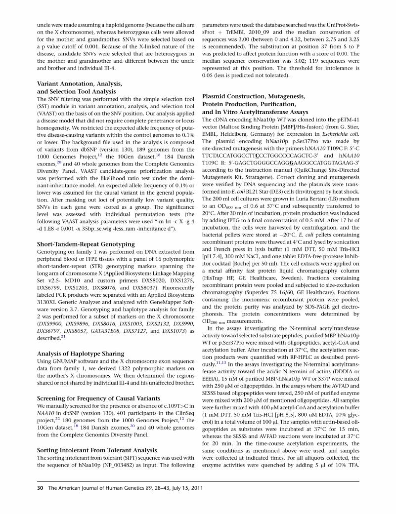
coverage. We performed haplotype analysis by using twodata sets: (1) the SNVs derived from X chromosome exonsequencing of individual III-4 and (2) STR genotyping ofindividual III-4, two other affected males (individuals II-6and III-7) in the family, and carrier females I-2, II-2, andII-5. Haplotype analysis narrowed the possible candidateregions to X chromosome positions 10,054,434–40,666,673 (~30 Mb) and 138,927,365–153,331,900(~14 Mb) (Table 5 and Table S1). The sequencing datashowed that of the ~155 Mb on the X chromosome, theaffected brother (III-4) and unaffected brother (III-2) wereidentical at ~71% and were recombinant at only 29%. The~14 Mb region is located on the telomeric end of the longarmof theX chromosome and includesNAA10. Subsequentaddition of STR genotyping from thenewborn affectedmale(III-6) further narrowed this interval to chrX:143,836,276–154,913,754 (telomere) (~11 Mb), which still includesNAA10 (Table 5).
Table 5. Continued
Sample Allele Markera hg18 Position Chromosome
III-4 206 DXS1001 (AFM248WE5) 119,720,593 Xq24
III-6 199
II-6 (FFPE tissue) 206
III-7 (FFPE tissue) 199
III-4 165 DXS1047 (AFM150XF10) 128,902,983 Xq25
III-6 165
II-6 (FFPE tissue) 165
III-7 (FFPE tissue) 160
III-4 90 DXS1227 (AFM317YE9) 140,630,173 Xq27.2
III-6 82
II-6 (FFPE tissue) 90
III-7 (FFPE tissue) 82
III-4 170 DXS8043 (AFMB018WD9) 143,836,276 Xq27.3
III-6 156
II-6 (FFPE tissue) 170
III-7 (FFPE tissue) 170
III-4 83 DXS8091 (AFM345WG9) 147,410,588 Xq28
III-6 83
II-6 (FFPE tissue) 83
III-7 (FFPE tissue) 83
c.109T>C in NAA10 ~152,850,921b Xq28
III-4 310 DXS1073 (AFM276XH9) 153,482,054 Xq28
III-6 310
II-6 (FFPE tissue) 310
III-7 (FFPE tissue) 310
a Genomic position represents a single nucleotide within the STR.b Maximum Shared Region (hg18): chrX:143,836,276–154,913,754 (telomere) (11 Mb).
The American Journal of Human Genetics 89, 28–43, July 15, 2011 37

Coverage AnalysisExon capture techniques cannot capture all exonic regionsbecause of design issues with replicative regions; this cantherefore result in an incomplete set of variants. The Xchromosome Agilent design consists of over 3 Mb ofexon intervals. We evaluated the capture and sequencingcoverage for all the currently known 1959 chrX transcriptsin RefSeq (hg18). On the X chromosome, there are 8222unique RefSeq exons. Of these exons, 736 were excludedfrom the SureSelect X-Chromosome Capture Kit becausethey were designated as pseudoautosomal or repetitivesequences (UCSC Genome Browser). The remaining 7486exons have an average length of 358.9 bases and arecovered on average by 5.7 bait probes per exon (bait probeswere 120 bases long). The average read coverage in theseregions was 214.6 reads per base; in these regions anaverage of 97.8% of the bases were covered by one ormore reads, and 95.6% were covered at 103 or better.We also analyzed the average coverage for each gene
(calculated as the total read bases within exons dividedby the exon length) more specifically in the affected haplo-type in the 14 Mb critical region derived from analysis offamily 1 (Table S2). The average coverage was 1853 amongthe 167 nonpseudoautosomal or duplicative RefSeq tran-scripts (110 unique genes) in the 14 Mb region. These167 transcripts consist of 864 exons, of which 860 hadsome read coverage and 809 had reads covering morethan 95% of the bases in the exon. Most of the exonportions in the 14 Mb region that were not covered byreads were from the 89 exons not included in the SureSe-lect X-Chromosome Capture Kit because they were in thepseudoautosomal or duplicative regions. There was somecoverage on a handful of these exons as well as intronicand intergenic regions flanking covered exons as a conse-quence of the experimental protocol. For example, if anexcluded exon or intronic sequence is close (~500 bases)to an exon that is captured, the long fragment size (andthe fact that we only sequence the end of the fragment)can lead to reads for the exon or intron. In the NAA10haplotype region, we had greater than 103 coverage for86% of the exons in the region for family 1; the corollaryof this means that 97 exons do not have 103 coverage inthe NAA10 region. However, of the 856 exons in the 14Mb region that did have >103 coverage, the GNUMAPand VAAST approach found only two candidate SNVsthat met our criteria (present in proband, not present inhis brother and uncle, polymorphic in his mother andgrandmother, nonsynonymous, not in the 1000 genomesor dbSNP), and in the exons in the 30 Mb region, no SNVsmet our criteria. Theoretically, we could have nonethelessmissed a crucially important variant, so we needed to useparallel approaches to prove causality of the variant inNAA10.
A Second FamilyDuring preparation of a manuscript reporting the aboveresults, another group (L.M.B., J.J.J, L.G.B.) communicated
to us that they had also identified the NAA10 c.109T>Cmutation in an apparently unrelated family (herein desig-nated as family 2) with an indistinguishable phenotype(see Figure 2D and 2E). Haplotype analysis had been per-formed for family 2, and it identified a shared regionof chrX:140,061,918–154,913,754 (~14 Mb) in threecarrier females (I-2, II-2, and II-3) that was not presentin an unaffected male (III-5). The borders of the regionwere defined by recombinant marker GATA31E08 andthe q terminus.Massively parallel sequencing of X chromosome exons
was done on DNA samples from an obligate carrier andher unaffected child as previously described.9 The entireX chromosome exon region target sequence was2,784,426 bp, and theX chromosome exon region oligonu-cleotide library was designed to target 2,264,175 bp of this(81.3%). The target-selected DNA libraries from one femaleheterozygote (M87_4) and one unaffected male child(M87_5) were sequenced on one lane each of an IlluminaGAIIx in paired-end 75 bp configuration, which yielded,respectively, 75,382,114 and 65,015,016 reads (separateresults are given for each sample) or 5,729,040,664 and4,941,141,216 bp of total sequence. Of the filtered aligningsequence, 20.9% and 11.9% could be uniquely aligned tothe entire X chromosome exon region target (2,784,426bases). This aligned sequence yielded a gross overallcoverage of 20583 and 17753 of the entire X chromosomeexon region target. The capture efficiency varied across thetargets; 2,538,791 and 2,464,367 bp (89.8%) of the entireX chromosome exon region yielded R13 coverage,2,305,571 and 2,251,868 bp (81.8%) yielded R103coverage, and 2,254,446 and 2,183,075 bp (79.7%) yieldedR203 coverage. The Most Probable Genotype (MPG)variant-calling software24 was able to make base calls on2,295,223 and 2,378,985 bp of this sequence (82.4% and85.4%).The genotypes were filtered on the basis of several attri-
butes that were felt to be appropriate for this disorder(Table 6). Heterozygosity was used because the test subjectwas an unaffected female carrier for an X-linked trait. Weperformed further filtering to include nonsynonymous,splice-site, frame-shifting, and nonsense variants. Filteringto exclude variants present in dbSNP or in the ClinSeqcohort (401 control individuals)22 was also performed.Finally, a filter that bounded the variants genomicallywithin the shared 14 Mb haplotype region was applied.This left one variant, a single missense mutation,c.109T>C in NAA10. This variant was confirmed in theother individuals, and mutation status segregated withaffection status and carrier status (Figure 2D).
Genome-wide SignificanceWe next assessed whether adding members of family 2would improve the power of the VAAST analysis. Wecombined variants from individual III-4 in family 1 withthe obligate carrier mother (II-2) in family 2 and performedVAASTanalysis on the 216 coding SNVs present in either of
38 The American Journal of Human Genetics 89, 28–43, July 15, 2011
the two individuals. After incorporating the mother fromfamily 2, NAA10 was the only candidate gene, and theresult was statistically significant (p value ¼ 3.8 3 10#5;Bonferroni corrected p value ¼ 3.83 10#5 3 729 ¼ 0.028).
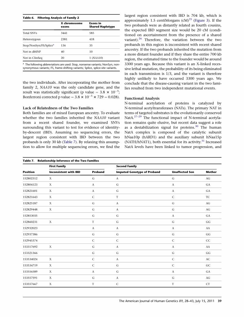
Lack of Relatedness of the Two FamiliesBoth families are of mixed European ancestry. To evaluatewhether the two families inherited the NAA10 variantfrom a recent shared founder, we examined SNVssurrounding this variant to test for evidence of identity-by-descent (IBD). Assuming no sequencing errors, thelargest region consistent with IBD between the twoprobands is only 30 kb (Table 7). By relaxing this assump-tion to allow for multiple sequencing errors, we find the
largest region consistent with IBD is 704 kb, which isapproximately 1.5 centiMorgans (cM)25 (Figure 3). If thetwo probands were as distantly related as fourth cousins,the expected IBD segment size would be 20 cM (condi-tioned on ascertainment from the presence of a sharedvariant).26 Therefore, the variation between the twoprobands in this region is inconsistent with recent sharedancestry. If the two probands inherited the mutation froma more distant founder and if they share the entire 700 kbregion, the estimated time to the founder would be around3300 years ago. Because this variant is an X-linked reces-sive lethal mutation, the probability of its being eliminatedin each transmission is 1/3, and the variant is thereforehighly unlikely to have occurred 3300 years ago. Weconclude that the disease-causing variant in the two fami-lies resulted from two independent mutational events.
Functional AnalysisN-terminal acetylation of proteins is catalyzed byN-terminal acetyltransferases (NATs). The primary NAT interms of targeted substrates is the evolutionarily conservedNatA.27–29 The functional impact of N-terminal acetyla-tion remains quite elusive, but recent data suggest a roleas a destabilization signal for proteins.30 The humanNatA complex is composed of the catalytic subunithNaa10p (hARD1) and the auxiliary subunit hNaa15p(NATH/hNAT1), both essential for its activity.31 IncreasedNatA levels have been linked to tumor progression, and
Table 6. Filtering Analysis of Family 2
X chromosomeexons
Exons inShared Haplotype
Total SNVs 3441 585
Heterozygous 2381 418
Stop/NonSyn/FS/Splicea 136 35
Not in dbSNP 40 10
Not in ClinSeq 20 1 (NAA10)
a The following abbreviations are used: Stop, nonsense variants; NonSyn, non-synonymous variants; FS, frame-shifting variants; Splice, splice-site variants.
Table 7. Relationship Inference of the Two Families
Position
First Family Second Family
Inconsistent with IBD Proband Imputed Genotype of Proband Unaffected Son Mother
152802312 X G A G AG
152804123 X A G A GA
152821601 X A G A GA
152821643 X C T C TC
152825187 X G A G AG
152829448 X G A G AG
152853035 G G A GA
152860231 X T G G GG
152932023 A A A AA
152937386 G G G GG
152945374 C C C CC
153317692 X G A A AA
153321366 G G G GG
153534024 X C A C AC
153534719 X C G C GC
153556589 X A G A GA
153557591 X G A G AG
153557667 X T C T CT
The American Journal of Human Genetics 89, 28–43, July 15, 2011 39

depletion of NatA subunits from cancer cells induces cell-cycle arrest and apoptosis.32 Human Naa10p is a proteinof 235 amino acid residues, of which the first 178 residuescompose a globular region, whereas the latter 57 residuesare predicted to form an unstructured and flexibleC-terminal tail.33 Thus, Ser37 is located in a structuredpart of hNaa10p. For many soluble globular proteins, Prois known to be potentially disruptive for secondary struc-ture elements such as the alpha-helix and beta-sheets.34
Thus, although the structure of hNaa10p is undetermined,the p.Ser37Pro mutation could indeed affect the structureof hNaa10p and thereby the catalytic activity. Ser37 andits surrounding residues are highly conserved amongeukaryotes,31 suggesting an essential function. A SIFT anal-ysis predicting whether an amino acid substitution affectsprotein function35 strongly suggested that a substitutionfrom Ser to Pro at position 37would affect protein functionwith a score of 0.00.In order to directly assess the functional consequences of
the hNaa10p p.Ser37Pro mutation, we analyzed the wild-type (WT) and the mutant proteins by a quantitative in vi-tro N-terminal acetylation assay. The enzyme activities ofhNaa10p WT and hNaa10p p.Ser37Pro were determinedwith four unique peptides as substrate; these peptideshaving been previously shown to be acetylated byhNaa10p and NatA (Figure 4). Compared to hNaa10pWT, hNaa10p p.Ser37Pro displayed a 60%–80% reductionin NAT activity toward the in vivo substrate RNasePprotein p30 (AVFAD-) and toward b-actin (DDDIA-) andg-actin (EEEIA-). In contrast, the activity toward the NatAsubstrate high-mobility group protein A1 (SESSS-) wasonly reduced by 20% (Figure 4). The oligopeptides AVFAD-and SESSS- represent classical cotranslational NatAsubstrates, being the N termini of proteins that are partially(AVFAD-) or fully (SESSS-) acetylated by NatA in HeLacells.27 On the other hand, b-actin (DDDIA-) and g-actin(EEEIA-) are nonclassical substrates recently shown to beacetylated more efficiently by hNaa10p/NatA than theclassical substrates were, representing a posttranslationalNAT activity.36
Discussion
The data presented here show that a mutation in anenzyme involved in N-terminal acetylation of proteins
leads to a distinct, previously undescribed X-linked pheno-type in humans and that males who carry the hypomor-phic hNaa10p p.Ser37Pro allele die in infancy with cardiacarrhythmias. N-terminal acetylation is one of the mostcommon protein modifications in humans, occurring onapproximately 80% of all human proteins27 It is catalyzedby several distinct NATenzymes, of which the major one isNatA.29 The catalytic subunit of the NatA complex,hNaa10p, is essential for survival in the organismsDrosophila melanogaster,37 Trypanosoma brucei,38 and Cae-norhabditis elegans.39 It is presumed that an amorphicNAA10 mutation would lead to embryonal lethality in hu-mans, although one can only prove this by analyzingtissues from pregnancies that did not survive to term.The strong conservation of Naa10p Ser37, the predictedfunctional effect of p.Ser37Pro due to structural distortion,and the demonstrated disruption of catalytic activity byp.Ser37Pro (Figure 4) strongly imply that the hemizygousmales with hNaa10p p.Ser37Pro have impaired NatA func-tion. Thus, a variety of protein N termini, both those thatare cotranslationally and those that are posttranslationallyacetylated (e.g., actins), are likely to be insufficiently acet-ylated. Most likely, the serious consequences of thep.Ser37Pro mutation are caused by the lack of N-terminalacetylation for one or several proteins strictly requiringthis modification for function or for maintenance ofadequate amounts in the cell. Because hNaa10p has alsobeen suggested to performN-lysine (N-epsilon) acetylationof proteins such as beta-catenin,40 a lack of N-lysine acety-lation of selected substrates could also cause the observedeffects. Finally, proposed noncatalytic functions ofhNaa10p41,42 could be affected in the p.Ser37Pro mutantand thereby also play a role in the observed phenotypes.We have demonstrated herein that a probabilistic
disease-causing variant discovery algorithm can readilyidentify and characterize the genetic basis of a previouslyunrecognized X-linked syndrome. We have also shownthat this algorithm, when used in parallel with high-throughput sequencing, can identify variants with highprioritization for causing disease with as few as two indi-viduals. In this instance, we screened ~150 variants distrib-uted among ~2000 transcripts on the X chromosome inone sample from individual III-4 in family 1. With no priorfiltering, we prioritized three to five possible candidategenes, and the mutation in NAA10 ranked second overall(Table 4). Including exon capture data from relatives
Figure 3. IBD Analysis of the Two Fami-lies in the Genomic Region Surroundingc.109T>CInformative SNVs consist of two classes:rare SNVs present in the proband fromfamily 1 in genomic regions with highcoverage in the second family, and
common SNVs present in the first proband and at least one member of the second family. Variant c.109T>C is indicated by the triangle.We imputed the genotype of the proband from family 2 from the genotypes of the mother and unaffected sibling of the second family(see Table 7). SNVs inconsistent with IBD, in which the imputed genotype of the second proband does not match the first proband, areindicated with an X. After allowing for multiple sequencing errors, the largest genomic segment consistent with IBD is around 700 kb inlength.
40 The American Journal of Human Genetics 89, 28–43, July 15, 2011

increased NAA10’s ranking to first overall in just onefamily. Furthermore, after combining variants from theproband in family 1 with the obligate carrier mother infamily 2, VAAST identified NAA10 as the only statisticallysignificant candidate.Although we have noted that the affected infants have
an aged appearance, we have not established any directlink with progeria or other progeroid syndromes. Theautopsies did not reveal any premature arteriosclerosis ordegeneration of vascular smooth muscle cells, as is seenin Hutchinson-Gilford progeria syndrome (MIM176670).43,44 Cell lines now being derived from family 1and possibly future animal models will provide importantinsights about the pathophysiology underlying this previ-ously unrecognized syndrome.
Supplemental Data
Supplemental Data include six figures and two tables and can be
found with this article online at http://www.cell.com/AJHG/.
Acknowledgments
We express our gratitude to the families for their extraordinary
cooperation and assistance. We also thank David Nix, Nina
Glomnes, andWhitney Fitts for advice and/or technical assistance
with family 1. Exon capture and sequencing for family 1 was paid
for by the Department of Psychiatry, University of Utah (to
G.J.L.). Collection of DNA and phenotyping for family 1 was sup-
ported by the Clinical Genetics Research Program: Phenotyping
Core, under CCTS grant UL1RR025764 at the University of Utah.
The University of Utah Microarray and Genomic Analysis core
facility was supported by award number P30CA042014 from the
National Cancer Institute. Functional analyses were supported by
the Research Council of Norway (grant 197136 to T.A.) and the
Norwegian Cancer Society (to J.R.L. and T.A.). B.M. and M.Y. were
supported by National Human Genome Research Institute
(NHGRI) 1RC2HG005619, K.W. by a pilot/methodological study
award from NIH/National Center for Research Resources grant
UL1 RR025774, J.X. by NIH/NHGRI K99HG005846, and W.E.J. by
NHGRI 5R01HG5692. The research on family 2 was supported by
Intramural Funds of theNHGRI, NIH (L.G.B.). These authors thank
Danielle Brinkman for the initial consenting and records gathering
for family2 andCaitlinKrause and JamieTeer for technical support.
Received: April 30, 2011
Revised: May 18, 2011
Accepted: May 19, 2011
Published online: June 23, 2011
Web Resources
The URLs for data presented herein are as follows:
ANNOVAR Software, http://www.openbioinformatics.org/
annovar/
Complete Genomics Diversity Panel, http://www.
completegenomics.com/sequence-data/download-data/
GATK Software, http://www.broadinstitute.org/gsa/wiki/index.
php/The_Genome_Analysis_Toolkit
GNUMAP, http://dna.cs.byu.edu/gnumap/
Figure 4. NAT Activity of Recombinant hNaa10p WTor p.Ser37Pro toward Synthetic N-Terminal Peptides(A and B) Purified MBP-hNaa10p WT or p.Ser37Pro were mixedwith the indicated oligopeptide substrates (200 mM for SESSS and250 mM for DDDIA) and saturated levels of acetyl-CoA (400 mM).Aliquots were collected at indicated time points and the acetyla-tion reactions were quantified with reverse phase HPLC peptideseparation. Error bars indicate the standard deviation based onthree independent experiments. The five first amino acids in thepeptides are indicated, for further details see Subjects andMethods. Time-dependent acetylation reactions were performedto determine initial velocity conditions when comparing the WTand Ser37Pro NAT activities toward different oligopeptides.(C) Purified MBP-hNaa10p WTor p.Ser37Pro were mixed with theindicated oligopeptide substrates (200 mM for SESSS and AVFADand 250 mM for DDDIA and EEEIA) and saturated levels ofacetyl-CoA (400 mM) and incubated for 15 min (DDDIA andEEEIA) or 20 min (SESSS and AVFAD) at 37"C in acetylation buffer.The acetylation activity was determined as above. Error barsindicate the standard deviation based on three independent exper-iments. Black bars indicate the acetylation capacity of theMBP-hNaa10p WT, whereas white bars indicate the acetylationcapacity of the MBP-hNaa10p mutant p.Ser37Pro. The five firstamino acids in the peptides are indicated.
The American Journal of Human Genetics 89, 28–43, July 15, 2011 41

Online Mendelian Inheritance in Man(OMIM), http://www.
omim.org
Picard, http://sourceforge.net/projects/picard/
SIFT (Sorting Intolerant From Tolerant) Analysis, http://sift.bii.
a-star.edu.sg/www/SIFT_seq_submit2.html
UCSC Genome Browser, http://genome.ucsc.edu/
References
1. Ng, S.B., Bigham, A.W., Buckingham, K.J., Hannibal, M.C.,
McMillin, M.J., Gildersleeve, H.I., Beck, A.E., Tabor, H.K.,
Cooper, G.M., Mefford, H.C., et al. (2010). Exome sequencing
identifies MLL2 mutations as a cause of Kabuki syndrome.
Nat. Genet. 42, 790–793.
2. Ng, S.B., Buckingham, K.J., Lee, C., Bigham, A.W., Tabor, H.K.,
Dent, K.M., Huff, C.D., Shannon, P.T., Jabs, E.W., Nickerson,
D.A., et al. (2010). Exome sequencing identifies the cause of
a mendelian disorder. Nat. Genet. 42, 30–35.
3. Choi, M., Scholl, U.I., Ji, W., Liu, T., Tikhonova, I.R., Zumbo,
P., Nayir, A., Bakkalo!glu, A., Ozen, S., Sanjad, S., et al.
(2009). Genetic diagnosis by whole exome capture and
massively parallel DNA sequencing. Proc. Natl. Acad. Sci.
USA 106, 19096–19101.
4. Pierce, S.B., Walsh, T., Chisholm, K.M., Lee, M.K., Thornton,
A.M., Fiumara, A., Opitz, J.M., Levy-Lahad, E., Klevit, R.E.,
and King, M.C. (2010). Mutations in the DBP-deficiency
protein HSD17B4 cause ovarian dysgenesis, hearing loss, and
ataxia of Perrault Syndrome. Am. J. Hum. Genet. 87, 282–288.
5. Bilguvar, K., Ozturk, A.K., Louvi, A., Kwan, K.Y., Choi, M., Tatli,
B., Yalnizo!glu, D., Tuysuz, B., Ca!glayan, A.O., Gokben, S., et al.
(2010). Whole-exome sequencing identifies recessive WDR62
mutations in severe brainmalformations.Nature467, 207–210.
6. Hedges, D.J., Burges, D., Powell, E., Almonte, C., Huang, J.,
Young, S., Boese, B., Schmidt, M., Pericak-Vance, M.A., Martin,
E., et al. (2009). Exome sequencing of a multigenerational
human pedigree. PLoS ONE 4, e8232.
7. Bonnefond, A., Durand, E., Sand, O., De Graeve, F., Gallina, S.,
Busiah, K., Lobbens, S., Simon, A., Bellanne-Chantelot, C.,
Letourneau, L., et al. (2010). Molecular diagnosis of neonatal
diabetes mellitus using next-generation sequencing of the
whole exome. PLoS ONE 5, e13630.
8. Roach, J.C., Glusman, G., Smit, A.F., Huff, C.D., Hubley, R.,
Shannon, P.T., Rowen, L., Pant, K.P., Goodman, N., Bamshad,
M., et al. (2010). Analysis of genetic inheritance in a family
quartet by whole-genome sequencing. Science 328, 636–639.
9. Johnston, J.J., Teer, J.K., Cherukuri, P.F., Hansen, N.F., Loftus,
S.K., Chong, K., Mullikin, J.C., and Biesecker, L.G.; NIH Intra-
mural Sequencing Center (NISC). (2010). Massively parallel
sequencing of exons on the X chromosome identifies
RBM10 as the gene that causes a syndromic form of cleft
palate. Am. J. Hum. Genet. 86, 743–748.
10. Johnston, J.J., Olivos-Glander, I., Killoran, C., Elson, E.,
Turner, J.T., Peters, K.F., Abbott, M.H., Aughton, D.J., Ayls-
worth, A.S., Bamshad, M.J., et al. (2005). Molecular and clin-
ical analyses of Greig cephalopolysyndactyly and Pallister-
Hall syndromes: Robust phenotype prediction from the type
and position of GLI3 mutations. Am. J. Hum. Genet. 76,
609–622.
11. Evjenth, R., Hole, K., Ziegler, M., and Lillehaug, J.R. (2009).
Application of reverse-phase HPLC to quantify oligopeptide
acetylation eliminates interference from unspecific acetyl
CoA hydrolysis. BMC Proc 3 (Suppl 6 ), S5.
12. Durbin, R.M., Abecasis, G.R., Altshuler, D.L., Auton, A.,
Brooks, L.D., Durbin, R.M., Gibbs, R.A., Hurles, M.E., and
McVean, G.A.; 1000 Genomes Project Consortium. (2010). A
map of human genome variation from population-scale
sequencing. Nature 467, 1061–1073.
13. Evjenth, R., Hole, K., Karlsen, O.A., Ziegler, M., Arnesen, T.,
and Lillehaug, J.R. (2009). HumanNaa50p (Nat5/San) displays
both protein N alpha- and N epsilon-acetyltransferase activity.
J. Biol. Chem. 284, 31122–31129.
14. McKenna, A., Hanna, M., Banks, E., Sivachenko, A., Cibulskis,
K., Kernytsky, A., Garimella, K., Altshuler, D., Gabriel, S., Daly,
M., and DePristo, M.A. (2010). The Genome Analysis Toolkit:
A MapReduce framework for analyzing next-generation DNA
sequencing data. Genome Res. 20, 1297–1303.
15. Fujita, P.A., Rhead, B., Zweig, A.S., Hinrichs, A.S., Karolchik, D.,
Cline, M.S., Goldman, M., Barber, G.P., Clawson, H., Coelho,
A., et al. (2011). The UCSC Genome Browser database: Update
2011. Nucleic Acids Res. 39 (Database issue), D876–D882.
16. Wang, K., Li, M., and Hakonarson, H. (2010). ANNOVAR:
Functional annotation of genetic variants from high-
throughput sequencing data. Nucleic Acids Res. 38, e164.
17. Sayers, E.W., Barrett, T., Benson, D.A., Bolton, E., Bryant, S.H.,
Canese, K., Chetvernin, V., Church, D.M., Dicuccio,M., Feder-
hen, S., et al. (2010). Database resources of the National
Center for Biotechnology Information. Nucleic Acids Res. 38
(Database issue), D5–D16.
18. Reese, M.G., Moore, B., Batchelor, C., Salas, F., Cunningham,
F., Marth, G.T., Stein, L., Flicek, P., Yandell, M., and Eilbeck,
K. (2010). A standard variation file format for human genome
sequences. Genome Biol. 11, R88.
19. Clement, N.L., Snell, Q., Clement, M.J., Hollenhorst, P.C.,
Purwar, J., Graves, B.J., Cairns, B.R., and Johnson, W.E.
(2010). The GNUMAP algorithm: Unbiased probabilistic
mapping of oligonucleotides from next-generation
sequencing. Bioinformatics 26, 38–45.
20. Li, Y., Vinckenbosch, N., Tian, G., Huerta-Sanchez, E., Jiang,
T., Jiang, H., Albrechtsen, A., Andersen, G., Cao, H., Kornelius-
sen, T., et al. (2010). Resequencing of 200 human exomes
identifies an excess of low-frequency non-synonymous
coding variants. Nat. Genet. 42, 969–972.
21. Kurpinski, K.T., Magyari, P.A., Gorlin, R.J., Ng, D., and
Biesecker, L.G. (2003). Designation of the TARP syndrome
and linkage to Xp11.23-q13.3 without samples from affected
patients. Am. J. Med. Genet. A. 120A, 1–4.
22. Biesecker, L.G., Mullikin, J.C., Facio, F.M., Turner, C., Cheru-
kuri, P.F., Blakesley, R.W., Bouffard, G.G., Chines, P.S., Cruz,
P., Hansen, N.F., et al; NISC Comparative Sequencing
Program. (2009). The ClinSeq Project: Piloting large-scale
genome sequencing for research in genomic medicine.
Genome Res. 19, 1665–1674.
23. Yandell, M., Huff, C.D., Hu, H., Singleton,M.,Moore, B., Xing,
J., Jorde, L.B., and Reese, M.G. (2011). A probabilistic disease-
gene finder for personal genomes. Genome Res. 21 10.1101/
gr.123158.111.
24. Teer, J.K., Bonnycastle, L.L., Chines, P.S., Hansen, N.F.,
Aoyama, N., Swift, A.J., Abaan, H.O., Albert, T.J., Margulies,
E.H., Green, E.D., et al; NISC Comparative Sequencing
Program. (2010). Systematic comparison of three genomic
enrichment methods for massively parallel DNA sequencing.
Genome Res. 20, 1420–1431.
25. Kong, A., Gudbjartsson, D.F., Sainz, J., Jonsdottir, G.M., Gud-
jonsson, S.A., Richardsson, B., Sigurdardottir, S., Barnard, J.,
42 The American Journal of Human Genetics 89, 28–43, July 15, 2011

Hallbeck, B., Masson, G., et al. (2002). A high-resolution
recombination map of the human genome. Nat. Genet. 31,
241–247.
26. Huff,C.D.,Witherspoon,D.J., Simonson,T.S.,Xing, J.,Watkins,
W.S., Zhang, Y., Tuohy, T.M., Neklason, D.W., Burt, R.W., Guth-
ery, S.L., et al. (2011).Maximum-likelihoodestimationof recent
shared ancestry (ERSA). Genome Res. 21, 768–774.
27. Arnesen, T., Van Damme, P., Polevoda, B., Helsens, K.,
Evjenth, R., Colaert, N., Varhaug, J.E., Vandekerckhove, J.,
Lillehaug, J.R., Sherman, F., and Gevaert, K. (2009). Proteo-
mics analyses reveal the evolutionary conservation and diver-
gence of N-terminal acetyltransferases from yeast and
humans. Proc. Natl. Acad. Sci. USA 106, 8157–8162.
28. Mullen, J.R., Kayne, P.S., Moerschell, R.P., Tsunasawa, S.,
Gribskov, M., Colavito-Shepanski, M., Grunstein, M.,
Sherman, F., and Sternglanz, R. (1989). Identification and
characterization of genes and mutants for an N-terminal
acetyltransferase from yeast. EMBO J. 8, 2067–2075.
29. Polevoda, B., Arnesen, T., and Sherman, F. (2009). A synopsis
of eukaryotic Nalpha-terminal acetyltransferases: Nomencla-
ture, subunits and substrates. BMC Proc 3 (Suppl 6 ), S2.
30. Hwang, C.S., Shemorry, A., and Varshavsky, A. (2010).
N-terminal acetylation of cellular proteins creates specific
degradation signals. Science 327, 973–977.
31. Arnesen, T., Anderson, D., Baldersheim, C., Lanotte, M., Var-
haug, J.E., and Lillehaug, J.R. (2005). Identification and char-
acterization of the human ARD1-NATH protein acetyltransfer-
ase complex. Biochem. J. 386, 433–443.
32. Gromyko, D., Arnesen, T., Ryningen, A., Varhaug, J.E., and
Lillehaug, J.R. (2010). Depletion of the human Na-terminal
acetyltransferase A induces p53-dependent apoptosis and
p53-independent growth inhibition. Int. J. Cancer 127,
2777–2789.
33. Sanchez-Puig, N., and Fersht, A.R. (2006). Characterization of
the native and fibrillar conformation of the human Nalpha-
acetyltransferase ARD1. Protein Sci. 15, 1968–1976.
34. Chou, P.Y., and Fasman, G.D. (1974). Conformational param-
eters for amino acids in helical, beta-sheet, and random coil
regions calculated from proteins. Biochemistry 13, 211–222.
35. Ng, P.C., and Henikoff, S. (2003). SIFT: Predicting amino acid
changes that affect protein function. Nucleic Acids Res. 31,
3812–3814.
36. Van Damme, P., Evjenth, R., Foyn, H., Demeyer, K., De Bock,
P.J., Lillehaug, J.R., Vandekerckhove, J., Arnesen, T., and
Gevaert, K. (2011). Proteome-derived peptide libraries allow
detailed analysis of the substrate specificities of N{alpha}-ace-
tyltransferases and point to hNaa10p as the post-translational
actin N{alpha}-acetyltransferase. Mol. Cell. Proteomics 10,
M110, 004580.
37. Wang, Y., Mijares, M., Gall, M.D., Turan, T., Javier, A., Borne-
mann, D.J., Manage, K., and Warrior, R. (2010). Drosophila
variable nurse cells encodes arrest defective 1 (ARD1), the cata-
lytic subunit of the major N-terminal acetyltransferase
complex. Dev. Dyn. 239, 2813–2827.
38. Ingram, A.K., Cross, G.A., and Horn, D. (2000). Genetic
manipulation indicates that ARD1 is an essential N(infinity)-
acetyltransferase in Trypanosoma brucei. Mol. Biochem. Para-
sitol. 111, 309–317.
39. Sonnichsen, B., Koski, L.B., Walsh, A., Marschall, P.,
Neumann, B., Brehm, M., Alleaume, A.M., Artelt, J., Betten-
court, P., Cassin, E., et al. (2005). Full-genome RNAi profiling
of early embryogenesis in Caenorhabditis elegans. Nature
434, 462–469.
40. Lim, J.H., Park, J.W., and Chun, Y.S. (2006). Human arrest
defective 1 acetylates and activates beta-catenin, promoting
lung cancer cell proliferation. Cancer Res. 66, 10677–10682.
41. Hua, K.T., Tan, C.T., Johansson, G., Lee, J.M., Yang, P.W., Lu,
H.Y., Chen, C.K., Su, J.L., Chen, P.B., Wu, Y.L., et al. (2011).
N-a-acetyltransferase 10 protein suppresses cancer cell metas-
tasis by binding PIX proteins and inhibiting Cdc42/Rac1
activity. Cancer Cell 19, 218–231.
42. Lee, C.F., Ou, D.S., Lee, S.B., Chang, L.H., Lin, R.K., Li, Y.S.,
Upadhyay, A.K., Cheng, X., Wang, Y.C., Hsu, H.S., et al.
(2010). hNaa10p contributes to tumorigenesis by facilitating
DNMT1-mediated tumor suppressor gene silencing. J. Clin.
Invest. 120, 2920–2930.
43. Capell, B.C., Tlougan, B.E., and Orlow, S.J. (2009). From the
rarest to the most common: Insights from progeroid
syndromes into skin cancer and aging. J. Invest. Dermatol.
129, 2340–2350.
44. Liu, G.H., Barkho, B.Z., Ruiz, S., Diep, D., Qu, J., Yang, S.L.,
Panopoulos, A.D., Suzuki, K., Kurian, L., Walsh, C., et al.
(2011). Recapitulation of premature ageing with iPSCs from
Hutchinson-Gilford progeria syndrome. Nature 472, 221–225.
The American Journal of Human Genetics 89, 28–43, July 15, 2011 43