using auditory and visual stimuli to investigate the
TRANSCRIPT
Using Auditory and Visual Stimuli to Investigate the Behavioral andNeuronal Consequences of Reflexive Covert Orienting
Andrew H. Bell, Jillian H. Fecteau, and Douglas P. MunozCentre for Neuroscience Studies, Canadian Institutes of Health Research Group in Sensory-Motor Systems, Department of Physiology,Queen’s University, Kingston, Ontario K7L 3N6, Canada
Submitted 6 November 2003; accepted in final form 17 December 2003
Bell, Andrew H., Jillian H. Fecteau, and Douglas P. Munoz. Usingauditory and visual stimuli to investigate the behavioral and neuronal conse-quences of reflexive covert orienting. J Neurophysiol 91: 2172–2184, 2004.First published December 31, 2003; 10.1152/jn.01080.2003. Reflexivelyorienting toward a peripheral cue can influence subsequent responsesto a target, depending on when and where the cue and target appearrelative to each other. At short delays between the cue and target[cue-target onset asynchrony (CTOA)], subjects are faster to respondwhen they appear at the same location, an effect referred to asreflexive attentional capture. At longer CTOAs, subjects are slower torespond when the two appear at the same location, an effect referredto as inhibition of return (IOR). Recent evidence suggests that thesephenomena originate from sensory interactions between the cue- andtarget-related responses. The capture of attention originates from astrong target-related response, derived from the overlap of the cue-and target-related activities, whereas IOR corresponds to a weakertarget-aligned response. If such interactions are responsible, thenmodifying their nature should impact the neuronal and behavioraloutcome. Monkeys performed a cue-target saccade task featuringvisual and auditory cues while neural activity was recorded from thesuperior colliculus (SC). Compared with visual stimuli, auditory re-sponses are weaker and occur earlier, thereby decreasing the likeli-hood of interactions between these signals. Similar to previous stud-ies, visual stimuli evoked reflexive attentional capture at a shortCTOA (60 ms) and IOR at longer CTOAs (160 and 610 ms) withcorresponding changes in the target-aligned activity in the SC. Audi-tory cues used in this study failed to elicit either a behavioral effect ormodification of SC activity at any CTOA, supporting the hypothesisthat reflexive orienting is mediated by sensory interactions betweenthe cue and target stimuli.
I N T R O D U C T I O N
A sudden change in our environment, such as a mousescurrying across the floor, reflexively grabs our attention. Theconsequences of this reflexive capture of attention can beinvestigated using the cue-target saccade task (Posner andCohen 1984). In this task, a brief flash of light or burst of noisein the periphery is used to draw the subject’s attention to itslocation. After a variable delay, a target appears at the same oropposite location as the cue with equal probability to which thesubject must generate a response. Measuring the time requiredby the subject to generate a response to the target can assess thebehavioral consequences of the initial reflexive shift in atten-tion (Jonides 1981; Maylor and Hockey 1985; Posner 1978;Posner and Cohen 1984).
The location of the cue relative to the target and the time that
elapses between their respective onsets [defined as the cue-target onset asynchrony (CTOA)] both influence the subject’sresponse time. At short CTOAs [i.e., �200 ms for humans,�80 ms for monkeys (Fecteau and Munoz 2003)], subjectsrespond faster when the cue and target appear at the samelocation compared with when they appear at opposite loca-tions. This same-location advantage is thought to represent the“reflexive capture of attention” (Jonides 1981; Posner andCohen 1984; Remington et al. 1992; see Egeth and Yantis 1997for review). At longer CTOAs [i.e., �200 ms for humans(Fecteau and Munoz 2003); �80 ms for monkeys (Dorris et al.2002; Fecteau and Munoz 2003)], subjects respond moreslowly when the cue and target appear at the same location.This has been labeled “inhibition of return” (IOR; Posner et al.1985; see also Maylor and Hockey 1985; Posner and Cohen1984) and it is thought to represent the adaptive bias ofobservers to explore novel locations in the environment.
Recent studies have demonstrated a neurophysiological cor-relate to these consequences of reflexive orienting. Dorris andcolleagues (2002) demonstrated that increased saccadic reac-tion times (SRTs) to previously cued locations were associatedwith a decrease in the magnitude of the neural response to thetarget in the superior colliculus (SC). Fecteau and Munoz(2003) expanded on these results by demonstrating that reflex-ive attentional capture was associated with an increase in themagnitude of the target-aligned response. Importantly, thesetwo studies further demonstrated that these changes in activitywere limited to the target-aligned response and were not ob-served in the motor burst accompanying the saccade. Thesedata suggest that the behavioral consequences of covert orient-ing, in this case the reflexive capture of attention and IOR, maybe linked to bottom-up, sensory processes. The reflexive cap-ture of attention originates from the overlap of the cue- andtarget-aligned responses at short CTOAs, whereas IOR origi-nates from a reduced target-aligned response at longer CTOAs.
If such sensory interactions between the cue- and target-aligned responses are involved in producing the reflexive cap-ture of attention and IOR, then modifying these interactionsshould directly influence the neuronal and behavioral out-comes. We assessed this possibility by training monkeys toperform a cue-target saccade task in which both auditory andvisual cues preceded visual targets. While the animals per-formed this task, we recorded single-unit activity from neuronsin the intermediate layers of the SC. Compared with visualstimuli, initial phasic responses to auditory stimuli in the SC
Address for reprint requests and other correspondence: D. Munoz, Depart-ment of Physiology, Queen’s University, Kingston, Ontario K7L 3N6, Canada(E-mail: [email protected]).
The costs of publication of this article were defrayed in part by the paymentof page charges. The article must therefore be hereby marked ‘‘advertisement’’in accordance with 18 U.S.C. Section 1734 solely to indicate this fact.
J Neurophysiol 91: 2172–2184, 2004.First published December 31, 2003; 10.1152/jn.01080.2003.
2172 0022-3077/04 $5.00 Copyright © 2004 The American Physiological Society www.jn.org

occur much earlier, are weaker, and are shorter in duration(Bell et al. 2001; Wallace et al. 1996). Accordingly, we predictthat these properties of auditory responses will provide lessopportunity for the auditory cue–aligned activity and visualtarget–aligned activity to interact and the reflexive capture ofattention will not be observed in behavior at short CTOAs.Likewise, if the behavioral expression of IOR depends on anattenuated sensory response to the target that occurs as a resultof the same neurons responding to both the visual cue andtarget early in sensory processing, then no such attenuationshould occur after auditory cues and IOR should not be seen.Moreover, if the differential response properties for visual andauditory stimuli are responsible for the inability of auditorystimuli to induce reflexive attentional capture or IOR, this mayexplain why crossmodal IOR is difficult to observe in behavior(see Reuter-Lorenz and Rosenquist 1996; Spence et al. 1998),even though it is demonstrated easily within individual modal-ities (e.g., Poliakoff et al. 2002; Spence et al. 2000).
Preliminary data were previously presented in abstract form(Bell and Munoz 2002).
M E T H O D S
Preparation of experimental animals
All procedures were approved by the Queen’s University AnimalCare Committee and were in accordance with the Canadian Councilon Animal Care policy for the use of laboratory animals. Two adultmale rhesus monkeys (Macaca mulatta), weighing about 7 and 10 kg,were used in this study. Animals were prepared for chronic experi-ments in one aseptic surgical session (see Munoz and Istvan 1998 fordetails). Scleral search coils to monitor eye movements and a head-restraint device were implanted. A stainless steel recording chamber,centered on the midline and tilted 38–40° posterior of vertical, wasimplanted to allow recordings from both the right and left SC. Ani-mals were given a course of antibiotic and analgesic treatment andmonitored closely after surgery. They were allowed a recovery periodof at least 2 wk before behavioral training was initiated.
Behavioral paradigm
The monkeys were trained to perform a nonpredictive cue-targetsaccade task including auditory and visual cues combined with visualtargets (Fig. 1). The onset of each trial was signaled by the removal ofa background light and presentation of a laser- or light-emitting diode(LED)–generated central fixation point (FP) back-projected onto atangent screen facing the animal. The animal was required to fixate theFP for a period of 600–800 ms, after which an auditory or visual cuewas presented in the periphery for 50 ms. The cue was extinguishedand the animal was required to maintain central fixation for anadditional 10, 110, or 560 ms (corresponding to CTOAs of 60, 160,and 610 ms). The FP was extinguished and a visual target appeared atthe SAME location as the peripheral cue or at the diametrically OPPOSITE
location, with equal probability. The three CTOAs were selected onthe basis of a pilot behavioral study using a wide range of CTOAs(Dale MK, AH Bell, and DP Munoz, unpublished observations), toobtain behavioral correlates of reflexive attentional capture and IOR.
The visual cue and target stimuli were generated using LEDs (0.05cd/m2). The auditory-cue stimulus was composed of a white noiseburst (43.5 dB, A-scale, measured from the monkey’s head), producedby small 4-cm, 8.0-� speakers suspended in front of the tangentscreen, facing the animal. The speakers were positioned immediatelyadjacent to where the visual stimuli would appear so as not to obstructthem. The intensities of the auditory and visual stimuli were selected
because they are known to evoke spatially dependent multisensoryinteractions (Bell et al. 2001, 2002) and the strength of these interac-tions are known to be strongest for pairings of weaker stimuli (seeStein and Meredith 1993). All trial types (auditory vs. visual cue;SAME vs. OPPOSITE; 60-, 160-, and 610-ms CTOA) were randomlyinterleaved within a single block of trials. Monkeys were given aliquid reward if they maintained central fixation (i.e., kept their eyesstable within 2° of the FP) for the duration of the fixation period andgenerated a saccade to the visual target. They worked until fullysatiated, at which point they were returned to their home cages. Dailyrecords of animal weight and water intake were kept and the animals’health was closely monitored by the institute veterinarian.
Recording techniques and receptive field mapping
Single-neuron activity was recorded extracellularly in both SC withtungsten microelectrodes (Frederick Haer) having impedances of0.5–3 M� at 1 kHz. Electrodes were lowered by a hydraulic micro-drive (Narishige) through stainless steel guide tubes supported by aDelrin grid placed inside the recording chamber (Crist et al. 1988).Single-neuron activity was sampled at 1 kHz after passing through awindow discriminator (Bak Electronics), which excluded action po-tentials that did not meet both amplitude and temporal constraints.Control of the behavioral paradigms as well as storage of eye positionand neural data was controlled by Pentium PC running a real time dataacquisition software package (REX Ver. 5.4; Hays et al. 1982). Eyeposition was sampled at 500 Hz.
To map the extent of a neuron’s visual receptive field, a handheldophthalmoscope was used to back-project moving spots and bars oflight onto the tangent screen while the monkey maintained centralfixation. In addition, visual stimuli were systematically presentedthroughout the visual field. The center of the receptive field wasapproximated as the point where the maximum visual response waselicited. Because previous studies have shown that auditory receptivefields in the primate SC tend to be very large (45–180° in thecontralateral hemifield in primates; Wallace et al. 1996), no attemptwas made to map the extent of the auditory receptive fields and theauditory stimuli were placed immediately adjacent to the visual stim-uli for each of the cue positions.
FIG. 1. Schematic representation of the nonpredictive cue-target saccadetask (see METHODS for details). Monkeys are required to maintain stable gazeon a central fixation point while a visual (V) or auditory (A) cue is presentedinto the receptive field of the neuron (indicated by the dashed circle) or to theopposite location. After which, the cue is extinguished and the animals mustmaintain fixation for an additional period until the fixation point is removedand the visual target is presented, either to the same location as the cue or tothe diametrically opposite location, with equal probability. Monkeys are thenrequired to generate a saccade to the target. Temporal delay between cue andtarget onset is defined as the cue-target onset asynchrony (CTOA). SRT,saccadic reaction time.
2173SUPERIOR COLLICULUS AND REFLEXIVE COVERT ORIENTING
J Neurophysiol • VOL 91 • MAY 2004 • www.jn.org

Data analysis
All data analysis was carried out on a Sun Ultra 60 Sparcstationusing user-generated programs and a Pentium PC running MatLabsoftware (Mathworks). Data were first run through an automatedsaccade-detection program, which identified the beginning and end ofeach saccade based on velocity and acceleration template matching(Waitzman et al. 1991). All marks were later verified by the experi-menter and adjusted if necessary. Before analysis, all incorrect trialswere rejected (monkey generated saccade to cued location in OPPOSITE
trials, i.e., “direction error”; see following text; or generated saccadebefore the removal of the FP). Likewise, saccades with latenciesbelow 70 ms or above 500 ms were excluded.
Neuronal responses were analyzed by constructing spike densityfunctions based on an exponential growth/decay function. The spikedensity waveform was obtained by convolving each spike with thefollowing function (Thompson et al. 1996)
A�t� � 1 � exp��t
�g� exp��t
�d� (1)
where the activation level (A) varies as a function of time (t), accord-ing to �g, the growth time constant that was set to 1 ms, and �d, thedecay time constant that was set at 20 ms. The individual pulses werethen summed to generate a single spike density function for each trial.To determine a neuron’s responsiveness to the different stimulusmodalities, the peak response from 0 to 150 ms after cue presentationat the 610-ms CTOA trials was measured. A given neuron wasclassified as a visual and/or auditory-responsive neuron when themagnitude of its cue-aligned activity was significantly greater thanbaseline (defined as mean activity 100–0 ms before cue onset; Wil-coxon rank-sum test, P � 0.05). To determine a neuron’s saccade-aligned response, the peak activity �10 ms surrounding saccade onsetwas measured. A neuron was defined as saccade-related when thepeak saccade-aligned activity consistently exceeded 80 spikes/s forsaccades to the neuron’s preferred direction. The experimenter laterverified all classifications to ensure accuracy and consistency.
The cue-response onset latency was defined as the point where theactivation level exceeded baseline (defined as the mean activity 400–200 ms before cue onset) plus 3 SDs. The activity had to remain abovethis level for a minimum of 10 ms to be classified as a valid response.To quantify the magnitude of activity after target presentation (i.e., the“peak of the target-aligned burst”), the absolute peak spike density50–150 ms after target presentation was measured. To estimate themagnitude of the “target-related response” (i.e., the magnitude of thesensory response to the target, independent of differences in baselineactivity produced by the cue; see RESULTS), the pretarget activity(mean activity 35–40 ms after target presentation) was subtractedfrom the target-aligned burst of activity.
For the population analysis of each dependent variable, repeated-measures ANOVAs, including the variables: cue modality (visual vs.auditory), cue condition (SAME vs. OPPOSITE location), and CTOA (60,160, 610 ms) were used. Simple effects were analyzed with pairwiseWilcoxon signed rank-sum tests. Individual sessions were analyzedwith Wilcoxon rank-sum tests. In all instances, an alpha of 0.05 waschosen as significant. For display purposes only, spike density func-tions are shown as floating averages of 10-ms bin widths.
Only the data from correct trials were included in the analysesdescribed below. Error trials were divided into one of two types:anticipatory responses, where the saccade was generated to the correctlocation but with an SRT �70 ms; and direction errors, defined assaccades initially generated away from the target (e.g., to the cuedlocation in the OPPOSITE condition). Monkeys generated a total of1,072 anticipation errors (8%), with the majority being generated inresponse to the visual cue and at the 160-ms CTOA [cue modality:F(1,55) � 9.885, P � 0.005; cue condition: F(1,55) � 0.207, P �0.50; CTOA: F(2,54) � 5.333, P � 0.01]. Monkeys generated a total
of 520 direction errors (4%), with the majority being generated inresponse to the visual cue at a CTOA of 610 ms and in the OPPOSITE
condition [cue modality: F(1,55) � 26.464, P � 0.001; cue condition:F(1,55) � 13.308, P � 0.001; CTOA: F(2,54) � 15.763, P � 0.001].
R E S U L T S
Behavior
Two monkeys completed a total of 12,305 correct trials overthe course of the recording sessions. Because both monkeysexhibited similar behavior, data have been grouped across thetwo subjects. Cue modality (visual vs. auditory), cue condition(SAME vs. OPPOSITE), and CTOA (60, 160, 610 ms) interactedwith one another to influence SRT [3-way interaction:F(2,112) � 20.059, P � 0.0001]. To facilitate the presentationof these data, results for each cue modality are describedseparately.
On visual-cue trials (Fig. 2A; black lines) at the shortestCTOA (60 ms), monkeys generated saccades with significantlyshorter SRTs when the cue and target appeared at the SAME
FIG. 2. Behavior in the nonpredictive cue-target saccade task. A: mean (andSE) saccadic reaction times for the two monkeys. Visual-cue trials shown inblack; auditory-cue trials shown in gray. Solid lines indicate trials where thecue and target appeared in the SAME location; dashed lines represent trialswhere the cue and target appeared in OPPOSITE locations. B: mean SRT for theSAME condition subtracted from the mean SRT for the OPPOSITE condition.Values � 0 indicate shorter SRTs in the SAME condition (i.e., facilitatedresponses); values � 0 indicate shorter SRTs in the OPPOSITE condition (i.e.,inhibition of return). Asterisks indicate statistically significant differencesbetween the SAME and OPPOSITE conditions for the given cue modality (Wil-coxon rank-sum test, P � 0.05).
2174 A. H. BELL, J. H. FECTEAU, AND D. P. MUNOZ
J Neurophysiol • VOL 91 • MAY 2004 • www.jn.org

location (solid black lines) compared with when the stimuliappeared at OPPOSITE locations (dashed black lines; Wilcoxonrank-sum test, P � 0.0001). At the intermediate CTOA (160ms), this effect was reversed and mean SRTs in the SAME
condition were significantly longer compared with the OPPOSITE
condition (P � 0.0001). Note this difference was attributedboth to a significant increase in mean SRT for the SAME
condition (P � 0.0001) and a significant decrease in mean SRTfor the OPPOSITE condition (P � 0.0001), compared with thecorresponding trial type at the 60-ms CTOA. At the longestCTOA tested (610 ms), a significant albeit reduced opposite-location advantage was present (P � 0.0001). Subtracting themean SRT in the SAME condition from that of the OPPOSITE
condition for each CTOA (Fig. 2B, black line) revealed abehavioral facilitation, defined here as reflexive attentionalcapture, at a CTOA of 60 ms and IOR at CTOAs of 160 and610 ms.
By contrast, when an auditory cue preceded the visual target(Fig. 2; gray lines), no differences between the SAME andOPPOSITE conditions were obtained at any of the CTOAs tested(Wilcoxon rank-sum test, P values � 0.50). Saccades gener-ated to visual targets after auditory cues had very similar SRTs,regardless of the location of the cue relative to the target or theCTOA. Interestingly, mean SRT for auditory-cue trials consis-tently fell between those obtained for the two visual-cue con-ditions. That is, at the 60-ms CTOA, presenting an auditory cuebefore a visual target did not provide as great a behavioraladvantage as a visual cue and target presented to the SAME
position but, rather, elicited shorter SRTs compared with avisual cue and target presented to OPPOSITE locations. At thelonger CTOAs (160 and 610 ms), auditory cues had little effecton behavior—effectively serving as a neutral condition, andtherefore emphasizing how the IOR effect obtained after visualcues was likely attributable to both an inhibitory influenceacting in the SAME condition and a facilitatory effect acting inthe OPPOSITE condition.
We also assessed the effect of cue modality, cue condition,and CTOA on the execution of saccades by comparing peaksaccadic velocity and the accuracy of the saccadic endpointsacross the different conditions. Main sequence plots (saccadicamplitude vs. peak saccadic velocity; Bahill et al. 1975) wereconstructed for each trial condition (not shown). Peak saccadicvelocities were not influenced by cue modality, cue condition,or CTOA [3-way: F(2,112) � 1.845, P � 0.158]. For a givensaccadic amplitude, a similar peak saccadic velocity wasachieved, regardless of the individual trial conditions.
To assess the accuracy of the saccades, we calculated boththe saccadic gain (saccadic amplitude/target eccentricity) andendpoint error (magnitude of the vector between saccadicendpoint and true target location; not shown). Neither of thesefactors was influenced by cue condition, modality, or CTOA[3-way: Gain: F(2,112) � 0.641, P � 0.527; Motor Error:F(2,112) � 1.773, P � 0.170]. All saccades were equallyaccurate, regardless of the modality of the cue, the location ofthe target relative to the cue, or the delay between cue andtarget onset.
In summary, reflexively shifting attention to a particularlocation by means of a visual cue resulted in early reflexiveattentional capture at the short CTOA (60 ms) followed by IORat the longer CTOAs (160 and 610 ms). These effects werelimited to changes in SRT; peak saccadic velocity and endpoint
accuracy were unaffected. Auditory cues were unable to eliciteither reflexive attentional capture or IOR at any CTOA tested,showing no significant change in SRT across any of thetrial types.
Activity in the SC recorded from the nonpredictivecue-target saccade task
Sixty-one neurons were recorded from the intermediate lay-ers of both colliculi of two monkeys (29 and 32 from monkeysR and H, respectively). Eighty-four percent (51/61) were re-sponsive to sensory stimuli (visual only: n � 37; auditory only:n � 2; bimodal: n � 12) and were considered for furtheranalysis. Of these, 86% (44/51) had significant saccade-relatedactivity (see METHODS for classification criteria).
The behavioral consequences of nonpredictive cueing wereclosely reflected in changes in the magnitude of the target-aligned burst of activity of neurons in the intermediate layers ofthe SC [3-way interaction between cue modality, cue condi-tion, and CTOA: F(2,96) � 11.099, P � 0.001]. An analysis ofthe magnitude of the saccadic burst showed no differenceacross the different trial conditions [main effect of cue condi-tion: F(1,48) � 0.008, P � 0.50; cue modality: F(1,48) �0.965, P � 0.32; CTOA: F(2,96) � 0.783, P � 0.45; or 3-way:F(2,96) � 1.649, P � 0.19]. When considered in conjunctionwith the lack of effect of cue modality, condition, or CTOA onthe peak velocity and saccadic endpoint accuracy, these datasuggest that reflexively orienting by means of a nonpredictivecue influences the neural processes involved in saccadic initi-ation but not those associated with the actual execution of thesaccade. We now describe how the early and late consequencesof reflexive covert orienting (i.e., attentional capture and IOR)were correlated to the peak target-aligned burst of activity inthe SC.
Neural correlates of reflexive attentional capture
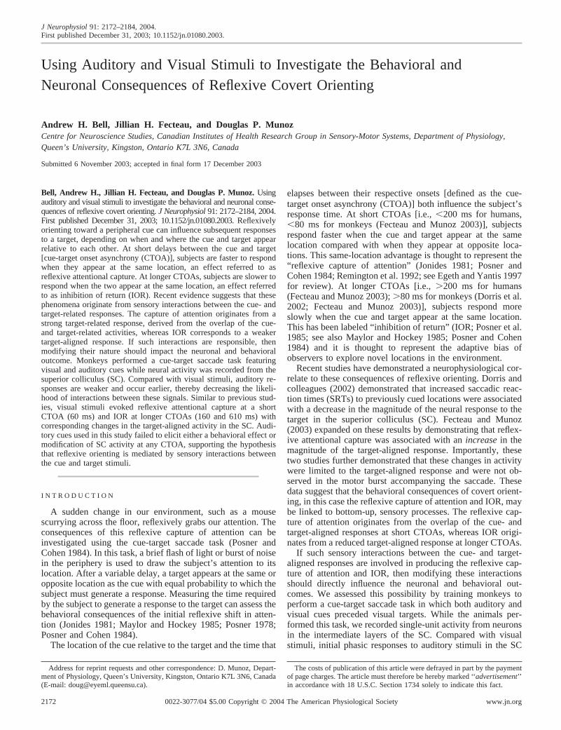
At the shortest CTOA tested (60 ms), monkeys exhibited asame-location advantage when the target was preceded by avisual cue and no difference in behavior when an auditory cuewas presented (Fig. 2). Does the presence of the same-locationadvantage for visual cues correspond to an overlap of the cue-and target-aligned responses that does not occur after auditorycues?
Figure 3 shows the activity of a single sensory-motor neuronin the intermediate layers of the SC when visual or auditory-cue stimuli were presented at a 60-ms CTOA. When the visualcue appeared in the receptive field of this neuron (Fig. 3, A andE), a strong burst of action potentials occurred, beginningabout 65 ms after cue onset. When the visual target appeared atthe SAME location as the visual cue (Fig. 3A), the neuralresponse to the target added to the residual activity from thecue, resulting in a stronger target-aligned burst compared withwhen the cue was presented on the opposite side (Fig. 3C).
This overlap between the cue- and target-aligned responsesdid not occur after auditory cues (Fig. 3, B, D, F, and H). Thisparticular neuron had a weak, but significant, response to theauditory cue that was long over by the time the visual responseto the target began. As such, there was no opportunity for thecue- and target-aligned responses to overlap when both stimuliappeared in the same receptive field (Fig. 3B). Moreover, there
2175SUPERIOR COLLICULUS AND REFLEXIVE COVERT ORIENTING
J Neurophysiol • VOL 91 • MAY 2004 • www.jn.org
was no other observable change in the target-aligned responseafter the appearance of the auditory cue. Therefore it seemsunlikely that any significant multisensory interactions (Steinand Meredith 1993) took place after presentation of the audi-tory cue and visual target at the 60-ms CTOA.
Similar effects of cue modality and cue condition were seen
across the population of neurons examined. Figure 4 shows themean peak of the target-aligned burst for each visually respon-sive neuron (n � 49; Fig. 4). Note that for the remainder of theanalysis, the auditory-only neurons (n � 2) have been removedbecause they had no target-related activity. As demonstrated inFig. 3, the overlap of the visual-cue and target-aligned re-
FIG. 3. Neuronal correlates of reflexive attentional capture in thesuperior colliculus (SC). Activity for an individual sensory-motor neuronrecorded from the intermediate layers of the SC, for trials at the 60-msCTOA. All traces are aligned on cue onset. When a visual cue and targetappear in the same response field (A, black line), there is a substantialincrease in the magnitude of the target-aligned activity compared withwhen the cue appears at the OPPOSITE location, outside the receptive fieldof the neuron (C). The auditory-cue response, on the other hand, providedvery little residual cue activity on which the visual target response couldbuild and so no facilitation occurred (B). Vertical arrows indicate themean SRT for the SAME (solid arrow) and OPPOSITE (dashed arrow)conditions.
FIG. 4. Influence of cue condition and modality on neu-ronal activity at the 60-ms CTOA. Each point represents themean peak in the target-aligned burst of activity for anindividual visually responsive neuron, after a visual (A) orauditory (B) cue. Points lying below the unity line indicategreater activity in the SAME condition and vice versa. Acrossthe population, there was a highly significant trend for astronger burst of target-aligned activity in the SAME condi-tion compared with the OPPOSITE condition after visual (A,solid points, Wilcoxon signed rank-sum test; P � 0.005) butnot auditory cues (B, open points; P � 0.50).
2176 A. H. BELL, J. H. FECTEAU, AND D. P. MUNOZ
J Neurophysiol • VOL 91 • MAY 2004 • www.jn.org

sponses at the 60-ms CTOA resulted in a significantly strongerburst of activity in the SAME condition compared with theOPPOSITE condition for the majority (36/49; 73%) of the neuronsexamined (Fig. 4A; P � 0.005). This was associated with asame-location advantage in behavior (Fig. 2).
No change in the target-aligned burst was observed after thepresentation of auditory cues (Fig. 4B). Relatively few neuronsin our sample responded to auditory stimuli (14/61; 23%) andthose that did exhibited weaker responses (Wilcoxon rank-sumtest; P � 0.05) that occurred earlier in time compared withvisual responses (Wilcoxon rank-sum test; P � 0.0001; Table1). As such, there was no opportunity for the auditory cue– andvisual target–aligned responses to interact. In the auditory-cuecondition, the magnitude of the target-aligned burst of activitydid not change on the basis on the cue’s location (i.e., SAME vs.OPPOSITE; Fig. 4B; P � 0.50).
To summarize, the same-location advantage that was ob-tained in SRT for visual cues at the 60-ms CTOA was associ-ated with an increase in the peak of the target-aligned burst ofactivity that originated from the overlap of the cue- and target-aligned responses. The inability of auditory stimuli to evoke asimilar pattern of behavior appears to be a result of both a lackof significant cue-aligned response across the population onwhich the incoming target signals can build and the absence ofsignificant multisensory interactions between the cue and targetresponses.
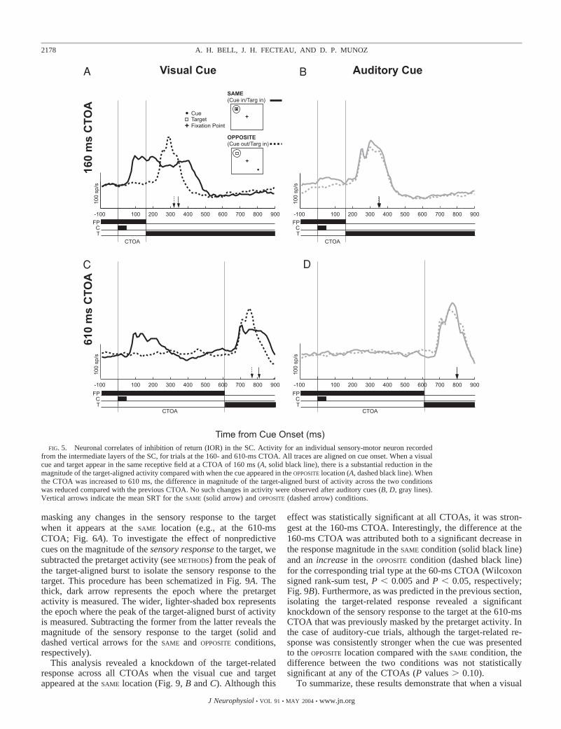
Neural correlates of inhibition of return
At the 160- and 610-ms CTOAs, an opposite-locationadvantage in behavior emerged for visual but not auditorycues (Fig. 2). Figure 5 shows the activity of a sensory-motorneuron recorded from the intermediate layers of the SC atthe 160- and 610-ms CTOAs. In the case of visual-cue trials,the opposite-location advantage in behavior was associatedwith a reduction in the magnitude of the target-aligned burstof activity in the SAME condition compared with when thetwo were presented to OPPOSITE locations. This reduction oftarget-aligned activity was greater at the 160-ms CTOA(Fig. 5A) compared with the 610-ms CTOA (Fig. 5C), aswas the behavioral IOR effect (Fig. 2). For auditory cues,neither the relative location of the cue nor the CTOA had aneffect on behavior (Fig. 2) or the target-aligned burst ofactivity (Fig. 5, B and D).
Similar trends were observed across the population of neu-rons sampled (Fig. 6). At the 160-ms CTOA, visual-cue trialsconsistently exhibited weaker target-aligned bursts of activity
(Fig. 6A; Wilcoxon signed-rank test, P � 0.0001) in the SAME
condition. At the longest CTOA (610 ms), although a signifi-cant opposite-location advantage persisted in behavior, thedifference in activity between the SAME and OPPOSITE conditionswith visual cues was no longer significant (Fig. 6C; P � 0.20).It is possible that an increase in pretarget activity observedafter the cue response might have masked a difference in theactual sensory response to the target. This possibility is ex-plored further in the next section. Again, no difference inactivity was observed after auditory cues at either CTOA (Fig.6, B and D).
To illustrate these data more clearly, we plotted the neuralactivity in a similar manner as SRT (compare Figs. 7 and 2).This subtraction plot shows that a greater peak target-alignedburst of activity in the SAME condition was associated with asame-location advantage in behavior, whereas weaker activitywas associated with an opposite-location advantage (compareFigs. 7B and 2B). As predicted on the basis of behavior, theopposite-location advantage in mean SRT at the 160-ms CTOAfor visual-cue trials corresponded to both a significant reduc-tion in the target-aligned burst in the SAME condition and asignificant increase in the OPPOSITE condition, relative to thevisual-cue trials at the 60-ms CTOA (Wilcoxon signed-ranktest, P values � 0.001 and 0.001, respectively).
To confirm the existence of a relationship between SRT andthe magnitude of the target-aligned burst of activity, we cal-culated the Pearson correlation coefficient for SRT versus thepeak of the target-aligned burst of activity on a trial-by-trialbasis for each neuron in our sample population. For this anal-ysis, we grouped trials from all CTOAs and both cue modal-ities and conditions. Figure 8A shows the correlation betweenSRT and the peak of the target-aligned burst of activity on atrial-by-trial basis for a single sensory-motor neuron (blackpoints, Fig. 8A). This particular neuron showed a significantnegative relationship such that as the magnitude of the peakactivity increased, SRT decreased (Pearson correlation coeffi-cient: r � �0.42; P � 0.05). This was also true for thepopulation (Fig. 8B), which exhibited a negative bias in cor-relation coefficients and of which over half of the neuronsassessed (32/49; 65%) were significantly correlated (P � 0.05).
These data demonstrate that sensory interactions betweenthe cue and target influence the magnitude of the target-alignedburst of neurons in the intermediate layers of the SC and howthese changes are related to behavior. Reflexive attentional cap-ture was associated with an increase in activity, arising from theoverlap of the cue- and target-related responses. IOR was associ-ated with a reduction in the peak of the target-aligned burst ofactivity in the SAME condition and an increase in the OPPOSITE
condition. Auditory cues produced neither of these neural trends,which could account for why no crossmodal reflexive attentionalcapture or IOR was observed in our task.
Effect of nonpredictive cues on target activity in the SC
Although there is a clear effect of nonpredictive cues on theabsolute peak of the target-aligned burst of activity shortlyafter target onset (Fig. 7), what is still uncertain is its effect onthe actual sensory response to the target, independent of pre-target activation. An increase in pretarget activation that oftenfollows the visual-cue response raises the baseline for incom-ing sensory signals related to target presentation, potentially
TABLE 1. Properties of the cue-related response
CuesCue Response Onset
Latency, msCue Response
Magnitude, sp/s
Visual 75 � 2 (52–118) 1301 � 10 (43–342)60 ms CTOA 76 � 2 (52–121) 132 � 10 (43–335)160 ms CTOA 75 � 2 (53–118) 133 � 10 (41–369)610 ms CTOA 74 � 2 (49–115) 129 � 9 (43–325)
Auditory 44 � 3 (30–67) 94 � 12 (47–189)60 ms CTOA 44 � 3 (30–67) 95 � 13 (43–204)160 ms CTOA 44 � 3 (30–68) 96 � 12 (53–191)610 ms CTOA 44 � 3 (31–67) 90 � 12 (45–173)
Values are mean � SE. For visually responsive neurons (n � 49); for allaurally responsive neurons (n � 14). CTOA, cue-target onset asynchrony.
2177SUPERIOR COLLICULUS AND REFLEXIVE COVERT ORIENTING
J Neurophysiol • VOL 91 • MAY 2004 • www.jn.org
masking any changes in the sensory response to the targetwhen it appears at the SAME location (e.g., at the 610-msCTOA; Fig. 6A). To investigate the effect of nonpredictivecues on the magnitude of the sensory response to the target, wesubtracted the pretarget activity (see METHODS) from the peak ofthe target-aligned burst to isolate the sensory response to thetarget. This procedure has been schematized in Fig. 9A. Thethick, dark arrow represents the epoch where the pretargetactivity is measured. The wider, lighter-shaded box representsthe epoch where the peak of the target-aligned burst of activityis measured. Subtracting the former from the latter reveals themagnitude of the sensory response to the target (solid anddashed vertical arrows for the SAME and OPPOSITE conditions,respectively).
This analysis revealed a knockdown of the target-relatedresponse across all CTOAs when the visual cue and targetappeared at the SAME location (Fig. 9, B and C). Although this
effect was statistically significant at all CTOAs, it was stron-gest at the 160-ms CTOA. Interestingly, the difference at the160-ms CTOA was attributed both to a significant decrease inthe response magnitude in the SAME condition (solid black line)and an increase in the OPPOSITE condition (dashed black line)for the corresponding trial type at the 60-ms CTOA (Wilcoxonsigned rank-sum test, P � 0.005 and P � 0.05, respectively;Fig. 9B). Furthermore, as was predicted in the previous section,isolating the target-related response revealed a significantknockdown of the sensory response to the target at the 610-msCTOA that was previously masked by the pretarget activity. Inthe case of auditory-cue trials, although the target-related re-sponse was consistently stronger when the cue was presentedto the OPPOSITE location compared with the SAME condition, thedifference between the two conditions was not statisticallysignificant at any of the CTOAs (P values � 0.10).
To summarize, these results demonstrate that when a visual
FIG. 5. Neuronal correlates of inhibition of return (IOR) in the SC. Activity for an individual sensory-motor neuron recordedfrom the intermediate layers of the SC, for trials at the 160- and 610-ms CTOA. All traces are aligned on cue onset. When a visualcue and target appear in the same receptive field at a CTOA of 160 ms (A, solid black line), there is a substantial reduction in themagnitude of the target-aligned activity compared with when the cue appeared in the OPPOSITE location (A, dashed black line). Whenthe CTOA was increased to 610 ms, the difference in magnitude of the target-aligned burst of activity across the two conditionswas reduced compared with the previous CTOA. No such changes in activity were observed after auditory cues (B, D, gray lines).Vertical arrows indicate the mean SRT for the SAME (solid arrow) and OPPOSITE (dashed arrow) conditions.
2178 A. H. BELL, J. H. FECTEAU, AND D. P. MUNOZ
J Neurophysiol • VOL 91 • MAY 2004 • www.jn.org

target is preceded by a visual cue presented to the SAME
location, the sensory response to that target, ignoring the effectof changes in the pretarget activity, is attenuated comparedwith when the cue is presented to the OPPOSITE location. How-ever, when this response builds on elevated levels of pretargetactivity, this attenuation is masked, which can partially coun-teract the IOR effect (Fig. 2). When a visual target is presentedto the OPPOSITE location as a visual cue at a CTOA of 160 ms,the target-related response is enhanced compared with theresponse at the previous CTOA, which can further increase themagnitude of the IOR effect.
D I S C U S S I O N
The goal of this study was to assess how interactions be-tween the cue- and target-related responses contribute to thebehavioral consequences of the reflexive orienting of attention.Monkeys were trained to perform a nonpredictive cue-targetsaccade task while single-unit activity was recorded from theintermediate layers of the SC. When presented with visual cuesat a 60-ms CTOA, monkeys show a same-location advantagein SRT (Fig. 2), consistent with the behavior previously de-scribed in the human literature (Jonides 1981; Posner andCohen 1984; Wright and Richard 2000). This reflexive captureof attention was associated with a stronger target-aligned burstof activity in the SC originating from the overlap of the cue-and target-aligned activities (Figs. 3 and 4). At the 160- and610-ms CTOAs, monkeys exhibited a same-location disadvan-tage, consistent with the human literature (Maylor and Hockey1985; Posner and Cohen 1984; Wright and Richard 2000). ThisIOR effect was associated with a reduction in the magnitude of
the target-aligned burst of activity (Figs. 5 and 6). Auditorycues used in this study did not elicit either behavioral effect,nor were any significant differences in the target-aligned burstobserved at any of the CTOAs tested.
On the basis of these observations, we hypothesize thatinteractions between the cue- and target-related sensory signalsproduce these changes in behavior. Compared with visualstimuli, auditory stimuli evoke weaker responses that occurearlier in time (Table 1). We propose that these differentcharacteristics do not allow for the necessary interactions be-tween the auditory cue and the visual target responses to takeplace. We discuss how our data support this hypothesis by firstshowing how these interactions lead to changes in the magni-tude of the target-aligned response followed by a description ofhow changes in this activity might lead to changes in behavior.We also discuss why the auditory stimuli used in this study failto elicit attentional capture or IOR.
Attentional capture corresponds with overlap of cueand target responses
Numerous behavioral studies have demonstrated that thesudden onset of a stimulus reflexively captures our attention(Jonides 1981; Jonides and Yantis 1988; Yantis and Jonides1984, 1990). The early appearance of this effect and its inde-pendence from the intentions of the observer (Remington et al.1992; Theeuwes 1991, 1992) suggest that the capture of atten-tion is a stimulus-driven, “bottom-up” mechanism (Fecteauand Munoz 2003).
Using the cue-target task, we have shown that the capture ofattention is linked to a strong target-aligned signal that origi-
FIG. 6. Influence of cue condition and modality onneuronal activity at the 160- and 610-ms CTOA. Eachpoint represents the mean peak in the target-alignedburst of activity for an individual visually responsiveneuron, after a visual (A, C) or auditory (B, D) cue.Points lying below the unity line indicate greater activityin the SAME condition and vice versa. Across the popu-lation, there was a highly significant trend for a strongerburst of target-aligned activity in the OPPOSITE conditioncompared with the SAME condition after visual cues atthe 160-ms CTOA (A, solid points, Wilcoxon signedrank-sum test; P � 0.005) that was no longer significantat the 610-ms CTOA (C, P � 0.20). No change inactivity was observed after auditory cues at either CTOA(B, D, open points).
2179SUPERIOR COLLICULUS AND REFLEXIVE COVERT ORIENTING
J Neurophysiol • VOL 91 • MAY 2004 • www.jn.org

nates from the interaction of the cue- and target-aligned activ-ities. This interaction is schematized in Fig. 10. Visual cueselicit a robust sensory response in SC neurons that occurs about75 ms after the appearance of the cue. This response has been
shown for the population of visually responsive neurons in Fig.10A. Because this initial phasic response to the visual cuepersists for �50–100 ms, cue-related activity will still bepresent when the target-related response is registered by theneuron after a 60-ms CTOA. This is shown as a solid black linein Fig. 10B. The overlap between the cue- and target-alignedactivities allows the incoming target signal to build on thecue-related activity resulting in a significantly stronger target-aligned burst of activity compared with when the cue and targetappear at OPPOSITE locations (“Target,” Fig. 10B). The magni-tude of the target-aligned burst in the OPPOSITE condition(dashed black line, Fig. 10B) is further reduced, presumablyattributable to the local inhibitory network in the SC (Munozand Istvan 1998) that inhibits other areas of the SC when onesubpopulation is active (i.e., when the contralateral SC isresponding to the cue, the ipsilateral SC will be inhibited),shown as the drop in baseline activity in Fig. 10B. Thereforebecause the level of activity is reduced, additional time isrequired to trigger a saccade.
The consequences of auditory cues, or lack thereof, stronglysupport our contention. Compared with visual stimuli, auditorystimuli elicit weaker responses that occur about 45 ms after thestimulus appeared (Table 1). Moreover, so few neurons re-sponded to the auditory stimuli resulting in a much weakerresponse across the population, shown for the population ofaurally responsive neurons in Fig. 10A. Therefore there will beno residual activity left for the target response to build on andconsequently no modulation of the response after auditorycues; hence no change in behavior is observed (gray lines,Fig. 10B) and so the capture of attention is not observed inour study.
IOR corresponds to a weaker target-aligned response
Involvement of the SC in IOR was first proposed by Posnerand colleagues (1985) (see also Rafal et al. 1988) on the basisof clinical findings. They found that patients suffering fromprogressive supranuclear palsy, a Parkinson’s-like disorder thataffects various brain stem structures including the SC (Burnand Lees 2002), did not exhibit IOR under conditions whennormal subjects or patients with Parkinson’s disease did. Sincethen, converging lines of evidence from both clinical andneurophysiological studies suggest a role for the SC in IOR(Dorris et al. 2002; Sapir et al. 1999). However, although we
FIG. 7. Population activity in nonpredictive cue-target saccade task. Mean(and SE) peak target-aligned activity for all visually responsive neurons (A).Visual-cue trials are shown in black; auditory-cue trials are shown in gray.Solid lines indicate trials where the cue and target appeared in the SAME
location; dashed lines represent trials where the cue and target appeared inOPPOSITE locations (target in receptive field). B: mean activity for the OPPOSITE
condition subtracted from that of the SAME condition for peak target-alignedactivity. Values � 0 indicate greater activity in the OPPOSITE condition;values � 0 indicate greater activity in the SAME condition. Asterisks indicatestatistically significant differences between the SAME and OPPOSITE conditions(Wilcoxon rank-sum test, P � 0.05).
FIG. 8. Correlation between saccadic reaction time andneural activity in the SC. A: trial-by-trial correlation of SRTvs. peak magnitude of the target-aligned burst of activity fora single sensory-motor neuron recorded from the intermedi-ate layers of the SC at the 160-ms CTOA. This neurondisplayed a significant negative correlation between SRT andneural activity. B: histogram of the Pearson correlation co-efficients for SRT vs. target-aligned burst of activity for thepopulation of visually responsive neurons. Negative valuesindicate that shorter SRTs are correlated with greater activ-ity. Individual neurons with significant correlations (P �0.05) are represented as solid bars.
2180 A. H. BELL, J. H. FECTEAU, AND D. P. MUNOZ
J Neurophysiol • VOL 91 • MAY 2004 • www.jn.org

observe a neural correlate of IOR in the SC, what specific roledoes the SC play?
When a visual target appears at the SAME location as the
visual cue, the magnitude of the target-aligned burst was sig-nificantly reduced compared with when the cue and targetappear at OPPOSITE locations (Fig. 7; see also Dorris et al. 2002;
FIG. 9. Effect of nonpredictive cues on the sensory response to the target.A: activity of a sensory-motor neuron showing how pretarget activation aftervisual-cue onset elevates the baseline for the target response in the SAME
condition. Solid and dashed vertical arrows indicate the target-related response(i.e., equal to the pretarget activity subtracted from the peak target-alignedburst of activity) for the SAME and OPPOSITE conditions, respectively. B: mean(and SE) target-related response magnitude for all visually responsive neurons.Visual-cue trials are shown in blackl auditory-cue trials are shown in gray.Solid lines indicate trials where the cue and target appeared in the SAME
locationl dashed lines represent trials where the cue and target appeared inOPPOSITE locations. C: mean activity for the OPPOSITE condition subtracted fromthat of the SAME condition for peak target-aligned activity. Values � 0 indicategreater activity in the OPPOSITE condition; values � 0 indicate greater activity in theSAME condition. Asterisks indicate statistically significant differences between theSAME and OPPOSITE conditions (Wilcoxon rank-sum test, P � 0.05).
FIG. 10. Proposed mechanism for attentional capture and inhibition ofreturn in the SC. A: visual and auditory cue–aligned responses for the popu-lation of visual and bimodal neurons (n � 49). Visual cues evoke a robust,phasic sensory response that is often followed by increasing pretarget activity.Auditory cues evoke short latency, weak, and abrupt responses. B: proposedmechanism for attentional capture. When a visual cue (solid black line) ispresented into the response field of neurons in the SC, they respond with aburst of action potentials. This suppresses the activity of neurons that are notresponding to the cue (shown as the drop in baseline for the OPPOSITE condition,dashed black line). When the target later appears, summation of the cue- andtarget-aligned responses increases the level of activity so that when themotor-related activity begins to accumulate; a saccade can be triggered soonercompared with the other conditions. No such overlap of the cue- and target-alignedresponses occurs after auditory cues; hence no attentional capture is seen. Like-wise, because of the suppression in the OPPOSITE condition, the level of activity isreduced and additional time is required to trigger a saccade. C: inhibition of returnin response to visual cues occurs because of the reduction in the target-alignedresponse, which, despite the presence of residual cue activity, still results insignificantly reduced activity. This reduces the level of activity on which themotor-related activity will build and consequently increases the time required tosurpass saccadic threshold and trigger a saccade. There is no modulation of theresponse after auditory cues; hence no change in behavior is observed.
2181SUPERIOR COLLICULUS AND REFLEXIVE COVERT ORIENTING
J Neurophysiol • VOL 91 • MAY 2004 • www.jn.org

Fecteau and Munoz 2003). This reduction occurred despite anincrease in pretarget activity (Fig. 9) so that the net activity wasstill weaker compared with when the two stimuli are presentedto OPPOSITE locations, particularly at the 160-ms CTOA. Aweaker target-aligned burst of activity provides a lower amountof activity on which the motor-related activity will build and assuch, the time required to trigger a saccade is increased (Fig.10C, see following text). This reduction in activity was notobserved after auditory cues, which accounts for why the SRTsfor auditory-cue trials were unaffected. Furthermore, it ap-peared that the OPPOSITE condition was facilitated (see follow-ing text), which further augmented the IOR effect. These datarepresent a link between a sensory event and the ultimatebehavior, supporting the theory that IOR in this particularoculomotor task is driven by sensory interactions between thecue and target.
One question that has yet to be addressed is what is/are thephysiological mechanism(s) underlying this modulation of thetarget-related response after a visual but not auditory cue? Oneimmediately obvious possibility is that visuomotor neurons inthe SC are directly inhibited, either from local sources (e.g.,fixation neurons in the rostral SC; Munoz and Wurtz 1993) orby external projections (e.g., substantia nigra pars reticulata;Hikosaka and Wurtz 1983; Wurtz and Hikosaka 1986). How-ever, Dorris and colleagues (2002) provided critical evidencesuggesting this is not the case. In a similar cue-target saccadetask as used in the current study, they applied microstimulationto the SC instead of presenting a target for the purposes ofevoking a saccade. Contrary to what might be expected if theSC were being directly inhibited, they found that evokedlatencies were shorter when stimulation was applied to theSAME side as that which responded to the cue, which is likelyattributable to the microstimulation combining with increasesin pretarget activity.
Another possibility therefore is the SC is receiving reducedvisual inputs from structures earlier in the visual pathway (e.g.,retina, lateral geniculate nucleus, primary visual cortex). Thisis supported by previous findings showing similar reductions inthe target responses of neurons in the superficial layers of theSC (Dorris et al. 2002; Robinson and Kertzman 1995) andparietal cortex (Robinson et al. 1995) and could thereforerepresent a common feature of the magnocellular pathway.Neurons in this pathway may be unable to respond fully to thetarget when presented to the SAME location. Because this path-way is less concerned with physical properties of visual stimuli(e.g., color, shape) and more with spatial and temporal rela-tionships between stimuli, this sensory-based mechanism mayhelp account for the observations that led researchers to theo-rize that IOR is an adaptive strategy meant to maximize theefficiency of visual search (see Klein 2000). The absence ofsuch an effect after auditory cues further supports a reductionin visual inputs driving the knockdown of the visual responsein IOR. Neurons in the intermediate layers of the SC representone of the earliest sites of crossmodal convergence at the levelof the individual neuron. Sensory neurons earlier in the visualand auditory pathways will respond to visual or auditory stim-uli—but not both—and so the auditory cue should have nodirect effect on the discharge properties of neurons earlier inthe visual pathway.
How do changes in the target-aligned responses link tochanges in SRT?
In the preceding sections, we have described how interac-tions between the cue and target result in changes to themagnitude of the target-aligned burst. How then do thesechanges result in changes in behavior? Previous studies havesuggested that neural activity must accumulate toward a certainlevel of activity, defined as the saccadic threshold, before asaccade will be initiated (Carpenter and Williams 1995; Goldand Shadlen 2000; Hanes and Schall 1996). Factors that affectwhen saccadic threshold is exceeded will therefore have astrong influence on SRT.
Shortly after the onset of the target-aligned burst, activityrelated to the generation of the motor output will begin toaccumulate toward saccadic threshold (“Motor”; Fig. 10, B andC). Even though the target-aligned burst may have decayedsomewhat by this time, the “starting point” (horizontal dashedlines, Fig. 10, B and C) for the motor activity will still be linkedto the magnitude of the initial target-aligned burst. When thetarget-aligned response is strong, as is the case with the SAME
condition at the 60-ms CTOA (Fig. 10B) and with the OPPOSITE
condition at the 160- and 610-ms CTOAs (Fig. 10C), the neuralactivity will exceed saccadic threshold first, thus triggering asaccade with a shorter SRT.
Facilitation to novel locations?
Another facet of IOR that we have not considered up to nowis the facilitated responding to the OPPOSITE side (i.e., novellocations; see Bennett and Pratt 2001; Pratt and Abrams 1999).At the 160-ms CTOA, where IOR was strongest, in addition toobserving a reduction in the magnitude of the target-alignedactivity in the SAME condition that was associated with anincrease in SRT, we also observed an increase in the magni-tude of the target-aligned activity (Fig. 7) coupled with adecrease in SRT for the OPPOSITE condition (Fig. 2) comparedwith the auditory-cue trials or the visual-cue trials at the otherCTOAs. One possibility is that this facilitation is being gener-ated locally, within the SC. While one subpopulation of neu-rons in the SC are responding to the cue, neurons in theopposite side SC are being inhibited because of the widespreadinhibitory network (Mize et al. 1991; Munoz and Istvan 1998).When the visual target later appears at the OPPOSITE location asthe cue, neurons that were previously inhibited may be able torespond with greater activity because of a postinhibitory re-bound/excitation mechanism (e.g., Nishimura et al. 1992;Okada et al. 1990; Syed et al. 1990). Biophysical studies havedemonstrated that, after a period of inhibition, neurons candischarge with increased frequency compared with whether noprevious inhibition was present. For example, in a study ex-amining saccadic suppression, the phenomenon whereby visualinputs are suppressed during saccadic eye movements, Zhu andLo (1996) demonstrated that stimulating the deeper layers ofthe SC resulted in inhibition of neurons in the lateral geniculatenucleus that was followed by a period of facilitation shortlyafter the initial stimulation pulse. If such a mechanism werefunctioning in our task, this could be expressed as an enhancedtarget-related response over that obtained in the neurons thatresponded to the cue. Further investigation will be necessary tosupport or refute this theory but it nonetheless illustrates how
2182 A. H. BELL, J. H. FECTEAU, AND D. P. MUNOZ
J Neurophysiol • VOL 91 • MAY 2004 • www.jn.org

sensory processes related to the cue response might result inchanges in the magnitude of the target-aligned burst of activityand subsequent changes in behavior.
Why no crossmodal attentional biases in our task?
The monkeys in our study did not exhibit either reflexiveattentional capture or IOR in response to auditory cues. Earlycrossmodal facilitation has been demonstrated in detectiontasks (e.g., Macaluso et al. 2000; McDonald et al. 2000).However, in the current study that used an oculomotor task, theauditory cues used failed to evoke reflexive attentional capture,presumably because the amount of residual activity in the SCafter the auditory cue was insufficient to significantly affect themagnitude of target-aligned activity at the 60-ms CTOA,which would have directly affected the motor output of the SC.The lack of IOR after auditory cues can also be accounted forby the differences in the visual versus auditory-cue responseproperties and their subsequent effect on the target-relatedresponse, as outlined in the previous sections.
This does not, however, explain why several other studiesusing humans have been able to elicit crossmodal IOR afterpresentation of an auditory cue (e.g., Reuter-Lorenz et al. 1996;Spence and Driver 1998; Tassinari et al. 2002). The specifictask conditions in these aforementioned studies differ signifi-cantly from our own and it is likely that these differences mayaccount for their ability, and our failure, to evoke crossmodalIOR. For example, using a visual detection task, Reuter-Lorenzand Rosenquist (1996) were unable to induce IOR after audi-tory cues unless subjects were instructed to first make saccadesto the cue and then return to fixation before target presentation.They argued that auditory stimuli alone were unable to suffi-ciently activate the requisite oculomotor systems necessary toinduce IOR. However, by having subjects generate an oculo-motor response to the cue, the oculomotor system was engagedand therefore IOR was presumably more likely to occur.
Similarly, Spence and Driver (1998) demonstrated IOR to avisual target after an auditory cue if an auditory fixation pointwas used to redirect attention back to fixation after cue pre-sentation. IOR was not observed, however, if a visual fixationpoint was used to redirect attention. In this case, the auditorymodality is given increased salience, not because it was thetarget for a saccade but because it was being used to drawattention to several different locations in a given trial and wascritical to the subjects’ being able to perform the task correctly.It should be noted that in both above cases, subjects wererequired to generate responses to auditory stimuli, whetherthey were overt orienting movements or detection responses.We would argue that this fact alone introduces additionalinfluences that will affect the outcome.
In conclusion, altogether we have shown that the summationof overlapping sensory signals in the SC can result in atten-tional capture. We have confirmed that IOR corresponds toreductions in the target-aligned response that scale with thedifference in SRT. Finally, we have provided a neurophysio-logical basis for why the auditory stimuli used in our study failto evoke either effect. Although these results support the in-volvement of the SC in reflexive covert orienting, it remains tobe seen what is driving the changes in the target-related re-sponses.
A C K N O W L E D G M E N T S
We thank A. Lablans, R. Pengally, and F. Paquin for invaluable assistanceand technical expertise; and I. T. Armstrong, S. Boehnke, B. Coe, J. Gore, R.Marino, and K. Rodgers for commenting on earlier versions of this manuscript.
G R A N T S
This work was supported by a Human Frontiers Science Program GrantRG0174/1998-B. A. H. Bell was supported by a Doctoral Research Awardfrom the Canadian Institutes of Health Research. J. H. Fecteau was supportedby a Postdoctoral Fellowship from the Natural Sciences and EngineeringResearch Council of Canada.
R E F E R E N C E S
Bahill A, Clark M, and Stark L. The main sequence, a tool for studyinghuman eye movements. Math Biosci 24: 191–204, 1975.
Bell AH, Corneil BD, Meredith MA, and Munoz DP. The influence ofstimulus properties on multisensory processing in the awake primate supe-rior colliculus. Can J Exp Psychol 55: 123–132, 2001.
Bell AH, Corneil BD, Munoz DP, and Meredith MA. Engagement of visualfixation suppresses sensory responses and multisensory integration in theprimate superior colliculus. Eur J Neurosci 18: 2867–2873, 2003.
Bell AH, Meredith MA, Van Opstal AJ, and Munoz DP. Role of the primatesuperior colliculus in mediating multimodal orienting behaviour. NeuralControl Move Abstr 2002.
Bell AH and Munoz DP. Activity in the primate superior colliculus related toperformance in predictive vs. non-predictive multimodal cued-saccadetasks. Soc Neurosci Abstr 27: 560.5, 2002.
Bennett PJ and Pratt J. The spatial distribution of inhibition of return.Psychol Sci 12: 76–80, 2001.
Burn DJ and Lees AJ. Progressive supranuclear palsy: where are we now?Lancet Neurol 1: 359–369, 2002.
Carpenter RH and Williams ML. Neural computation of log likelihood incontrol of saccadic eye movements. Nature 377: 59–62, 1995.
Crist CF, Yamasaki DS, Komatsu H, and Wurtz RH. A grid system and amicrosyringe for single cell recording. J Neurosci Methods 26: 117–122,1988.
Dorris MC, Klein RM, Everling S, and Munoz DP. Contribution of theprimate superior colliculus to inhibition of return. J Cogn Neurosci 14:1256–1263, 2002.
Egeth HE and Yantis S. Visual attention: control, representation, and timecourse. Annu Rev Psychol 48: 269–297, 1997.
Fecteau JH and Munoz DP. Neurophysiological correlates of covert orientingto uninformative and informative cues in the primate superior colliculus.Cogn Neurosci Annu Meet 10: 2003.
Gold JI and Shadlen MN. Representation of a perceptual decision in devel-oping oculomotor commands. Nature 404: 390–394, 2000.
Hanes DP and Schall JD. Neural control of voluntary movement initiation[see comments]. Science 274: 427–430, 1996.
Hays AV, Richmond BJ, and Optican LM. A UNIX-based multiple processsystem for real-time data acquisition and control. WESCON Conf Proc 2:1–10, 1982.
Hikosaka O and Wurtz RH. Visual and oculomotor functions of monkeysubstantia nigra pars reticulata. IV. Relation of substantia nigra to superiorcolliculus. J Neurophysiol 49: 1285–1301, 1983.
Jonides J. Voluntary versus automatic control over the mind’s eye’s move-ment. In: Attention and Performance, vol. IX, edited by Long JB andBaddeley AD. Hillsdale, NJ: Erlbaum Associates, 1981, p. 187–203.
Jonides J and Yantis S. Uniqueness of abrupt visual onset in capturingattention. Percept Psychophys 43: 346–354, 1988.
Klein RM. Inhibition of return. Trends Cogn Sci 4: 138–147, 2000.Macaluso E, Frith CD, and Driver J. Modulation of human visual cortex by
crossmodal spatial attention. Science 289: 1206–1208, 2000.Maylor EA and Hockey R. Inhibitory component of externally controlled
covert orienting in visual space. J Exp Psychol Hum Percept Perform 11:777–787, 1985.
McDonald JJ, Teder-Salejarvi WA, and Hillyard SA. Involuntary orientingto sound improves visual perception. Nature 407: 906–908, 2000.
Mize RR, Jeon CJ, Hamada OL, and Spencer RF. Organization of neuronslabeled by antibodies to gamma-aminobutyric acid (GABA) in the superiorcolliculus of the Rhesus monkey. Vis Neurosci 6: 75–92, 1991.
Munoz DP and Istvan PJ. Lateral inhibitory interactions in the intermediatelayers of the monkey superior colliculus. J Neurophysiol 79: 1193–1209,1998.
2183SUPERIOR COLLICULUS AND REFLEXIVE COVERT ORIENTING
J Neurophysiol • VOL 91 • MAY 2004 • www.jn.org

Munoz DP and Wurtz RH. Fixation cells in monkey superior colliculus. I.Characteristics of cell discharge. J Neurophysiol 70: 559–575, 1993.
Nishimura S, Okada Y, and Amatsu M. Post-inhibitory excitation of aden-osine on neurotransmission in guinea pig hippocampal slices. Neurosci Lett139: 126–129, 1992.
Okada Y, Nishimura S, and Miyamoto T. Excitatory effect of adenosine onneurotransmission in the slices of superior colliculus and hippocampus ofguinea pig. Neurosci Lett 120: 205–208, 1990.
Poliakoff E, Spence C, O’Boyle DJ, McGlone FP, and Cody FW. Tactileinhibition of return: non-ocular response inhibition and mode of response.Exp Brain Res 146: 54–59, 2002.
Posner MI. Chronometric Explorations of Mind: The Third Paul M. FittsLectures, delivered at the University of Michigan, Ann Arbor, MI, Septem-ber 1976. Hillsdale, NJ: Erlbaum Associates, 1978.
Posner MI and Cohen Y. Components of visual orienting. In: Attention andPerformance, vol. X, edited by Bouma H and Bouwhuis DG. Hillsdale, NJ:Erlbaum Associates, 1984, p. 531–556.
Posner MI, Rafal RD, Choate LS, and Vaughan J. Inhibition of return:neural basis and function. Cogn Neuropsychol 2: 221–228, 1985.
Pratt J and Abrams RA. Inhibition of return in discrimination tasks. J ExpPsychol Hum Percept Perform 25: 229–242, 1999.
Rafal RD, Posner MI, Friedman JH, Inhoff AW, and Bernstein E. Ori-enting of visual attention in progressive supranuclear palsy. Brain 111:267–280, 1988.
Remington RW, Johnston JC, and Yantis S. Involuntary attentional captureby abrupt onsets. Percept Psychophys 51: 279–290, 1992.
Reuter-Lorenz PA, Jha AP, and Rosenquist JN. What is inhibited in inhibi-tion of return? J Exp Psychol Hum Percept Perform 22: 367–378, 1996.
Reuter-Lorenz PA and Rosenquist JN. Auditory cues and inhibition ofreturn: the importance of oculomotor activation. Exp Brain Res 112: 119–126, 1996.
Robinson DL, Bowman EM, and Kertzman C. Covert orienting of attentionin macaques. II. Contributions of parietal cortex. J Neurophysiol 74: 698–712, 1995.
Robinson DL and Kertzman C. Covert orienting of attention in macaques.III. Contributions of the superior colliculus. J Neurophysiol 74: 713–721,1995.
Sapir A, Soroker N, Berger A, and Henik A. Inhibition of return in spatialattention: direct evidence for collicular generation. Nat Neurosci 2: 1053–1054, 1999.
Spence C and Driver J. Inhibition of return following an auditory cue. Therole of central reorienting events. Exp Brain Res 118: 352–360, 1998.
Spence C, Lloyd D, McGlone F, Nicholls ME, and Driver J. Inhibition ofreturn is supramodal: a demonstration between all possible pairings ofvision, touch, and audition. Exp Brain Res 134: 42–48, 2000.
Spence C, Nicholls ME, Gillespie N, and Driver J. Cross-modal links inexogenous covert spatial orienting between touch, audition, and vision.Percept Psychophys 60: 544–557, 1998.
Stein BE and Meredith MA. The Merging of the Senses. Cambridge, MA:MIT Press, 1993.
Syed NI, Bulloch AG, and Lukowiak K. In vitro reconstruction of therespiratory central pattern generator of the mollusk Lymnaea. Science 250:282–285, 1990.
Tassinari G, Campara D, Benedetti C, and Berlucchi G. The contributionof general and specific motor inhibitory sets to the so-called auditoryinhibition of return. Exp Brain Res 146: 523–530, 2002.
Theeuwes J. Cross-dimensional perceptual selectivity. Percept Psychophys50: 184–193, 1991.
Theeuwes J. Perceptual selectivity for color and form. Percept Psychophys 51:599–606, 1992.
Thompson KG, Hanes DP, Bichot NP, and Schall JD. Perceptual and motorprocessing stages identified in the activity of macaque frontal eye fieldneurons during visual search. J Neurophysiol 76: 4040–4055, 1996.
Waitzman DM, Ma TP, Optican LM, and Wurtz RH. Superior colliculusneurons mediate the dynamic characteristics of saccades. J Neurophysiol 66:1716–1737, 1991.
Wallace MT, Wilkinson LK, and Stein BE. Representation and integrationof multiple sensory inputs in primate superior colliculus. J Neurophysiol 76:1246–1266, 1996.
Wright RD and Richard CM. Location cue validity affects inhibition ofreturn of visual processing. Vision Res 40: 2351–2358, 2000.
Wurtz RH and Hikosaka O. Role of the basal ganglia in the initiation ofsaccadic eye movements. Prog Brain Res 64: 175–190, 1986.
Yantis S and Jonides J. Abrupt visual onsets and selective attention: evidencefrom visual search. J Exp Psychol Hum Percept Perform 10: 601–621, 1984.
Yantis S and Jonides J. Abrupt visual onsets and selective attention: volun-tary versus automatic allocation. J Exp Psychol Hum Percept Perform 16:121–134, 1990.
2184 A. H. BELL, J. H. FECTEAU, AND D. P. MUNOZ
J Neurophysiol • VOL 91 • MAY 2004 • www.jn.org