using active leptospermum honey in the debridement … · using active leptospermum honey in the...
TRANSCRIPT

APRIL 2015 OSTOMY WOUND MANAGEMENT® 63www.o-wm.com
FEATURE
Using Active Leptospermum Honey in the Debridement Process: 6 Challenging Cases from the Inner City Cecilia Gray, MSN, RN, CNS, CWON; and Fatima Ishii, BS, RN, CWON
AbstractThe use of honey-based dressings has been documented for thousands of years. Recent studies suggest their effectiveness may be, in part, related to their ability to facilitate autolytic debridement. Six patients who presented with multiple comorbidities and risk factors for delayed healing whose wounds required debridement were managed with active Leptospermum honey (ALH) to evaluate the safety and effectiveness of this treatment modality. The 6 patients ranged in age from 39 to 81 years. The ALH was covered with a foam dressing; both dressings were changed approximately every 3 days. After 9 to 20 days of use, wounds were completely, or almost completely, debrided, and a 75% concomitant average increase in the amount of granulation tissue in the wound bed was observed. No adverse events were noted. The use of ALH in this case series was effective, and no surgical debridement was needed. Research to compare the efficacy of ALH to other debridement methods is warranted.
Keywords: case report, wound, debridement, honey, pressure ulcer
Index: Ostomy Wound Management 2015;61(4):63–66
Potential Conflicts of Interest: Derma Sciences Inc (Princeton, NJ) provided editorial support for this manuscript.
Ms. Gray is an Advance Practice Registered Nurse-Clinical Nurse Specialist and Certified Wound Ostomy Nurse; and Ms. Ishii is a Certified Wound Ostomy Nurse, Los Angeles County University of Southern California Medical Center, Los Angeles, CA. Please address correspondence to: Cecilia Gray, MSN, RN, CNS, CWON, LAC-USC, 1200 N. State Street, GH 4-220, Los Angeles, CA 90033; email: [email protected].
Necrotic burden in chronic wounds is composed of necrotic tissue, exudate, and bacteria which alters the cellular en-
vironment, negatively impacting wound healing by prolonging inflammation and impeding epithelialization.1 Removing ne-crotic tissue (ie, debridement) reduces these barriers to healing.2
The 6 methods of debridement include autolytic, enzymatic, mechanical, conservative sharp, sharp, and biological, each with its own advantages and disadvantages. In general, the choice of method for a particular patient depends on the condition of the wound bed, resources, clinician knowledge, patient characteris-tics, and treatment goals.1,2
The use of honey in wound management dates back thou-sands of years and can be traced to ancient medical writings.3,4 In 2007, the United States Food and Drug Administration cleared the use of a line of honey-based wound dressings (ac-tive Leptospermum honey [ALH]) for a variety of wound types including leg ulcers, diabetic foot ulcers, donor sites, trauma and surgical wounds, pressure ulcers, and first-degree and second-degree burns. Randomized controlled trials, case series, and studies involving a variety of wound types have shown ALH has a range of benefits to facilitate healing, which includes aiding in the management of necrotic tissue and wound exudate and/or facilitating healing of stalled wounds.3-8
The observed benefits of ALH are believed to be achieved through 2 primary mechanisms of action: a low pH level and
high osmolarity. Specifically, the pH averages 3.2–4.29,10; re-views of the clinical, in vivo, and in vitro literature3,9-12 concluded lowering the pH of the wound environment is associated with wound healing. Secondly, because of its high osmotic potential, ALH may help pull wound fluid from within the wound, which promotes autolytic debridement by allowing the body’s own ac-tions to remove nonviable tissue from the wound. In this way, ALH is designed to support a moist healing environment that fosters granulation and epithelialization.3,4,13
The objective of this retrospective case series was to as-sess the safety and effectiveness of ALH on the removal of necrotic tissue in a variety of wounds in patients at a large 600-bed public hospital.
MethodsFor this retrospective case series, all participating patients
provided written informed consent for the use of their in-formation. Patients were included if they presented with a wound that contained slough or eschar in need of debride-ment in a location feasible for foam dressing to adhere, such as the buttocks, heels, and feet. Wounds in locations with ex-cessive moisture (eg, the sacrum and coccyx) were excluded if the patient was experiencing diarrhea, because in these cases, the foam cover dressing was unable to adhere. Partici-pating patients were treated at the hospital’s inpatient acute

64 OSTOMY WOUND MANAGEMENT® APRIL 2015 www.o-wm.com
FEATURE
and chronic care units between April and September 2011. A substantial number of patients encountered are homeless individuals with little to no financial or other support, no in-surance; this facility services a multicultural population with various ethnic and social diversities. Patients were treated by wound, ostomy, continence (WOC)nurses and were followed from admission to discharge. ALH was the only method used to assist with debridement. In all cases, ALH was applied in a thin layer (approximately 1/4–1/2 inch thick) to the wound bed after wound cleansing with normal saline or soapy water, followed by a secondary foam cover dressing. The choice of an appropriate absorptive cover dressing was important in order to manage the exudate production that occurs due to the autolytic debridement process.
The honey and cover dressings were changed as needed whenever the foam became approximately 75% saturated with exudate. At the first patient visit, demographic informa-tion and relevant medical history were recorded in the pa-tient file. At each dressing change, wound measurements and appearance (percent of granulation tissue, slough, or eschar) were documented in the patient files. Wound measurements were performed using a centimeter ruler. The length was measured as longest length from 12 o’clock to 6 o’clock and widest width from 3 o’clock to 9 o’clock. Notes on progress, defined as a reduction in necrotic tissue, were taken at each dressing change.
All assessments were qualitative except for percentage of granulation tissue and wound measurements. These are de-scribed separately per case and were not combined or com-pared in any way that required data analysis. Case Studies
Case 1. Sixty-three-year-old Mr. A was out of town visiting family when he experienced shortness of breath and fever. He returned home after a long car ride to the emergency room where he was admitted with septic shock secondary to Acineto-bacter pneumonia, complicated by Salmonella bacteremia and Clostridium difficile colitis. Mr. A had a history of hyperten-sion and required vasopressors, medication for sedation, and intubation. He presented with a Stage III pressure ulcer in the occipital area measuring 2.0 cm x 3.5 cm tissue loss with 100% adherent yellow slough (see Figure 1a). After wound cleansing, ALH and a secondary foam cover dressing were applied per the protocol described. The ALH dressing was changed on day 3. Mr. A was discharged after 9 days of ALH use (2 dressing changes), wound size had decreased to 1.5 cm x 2.0 cm, and wound bed appearance improved to 30% yellow slough and 70% scattered granulation buds (see Figure 1b).
Case 2. Ms. B, 54 years old, came to the emergency room with dyspnea on exertion, orthopnea, paroxysmal nocturnal dyspnea, and daily chest pains for the previous week. She had a history of coronary arterial disease, peripheral arterial dis-ease, diabetes, hypertension, congestive heart failure, and left atrium deviation. She had a necrotic diabetic heel ulcer that
measured 3.0 cm x 3.0 cm with an undetermined depth due to the presence of black eschar and yellow slough covering the wound bed (see Figure 2a). Physicians were concerned about the use of sharp debridement due to Ms. B’s history of diabetes and poor perfusion; therefore, ALH was chosen to support autolytic debridement. After wound cleansing, ALH and a secondary foam dressing were applied as described. Following 1 week of ALH with dressing changes approxi-mately every 3 days, the breakdown of the eschar led to 70% granulation tissue and 30% yellow slough remaining. After 2 weeks, the wound measured 2.5 cm x 2.0 cm with 80% gran-ulation tissue and 20% yellow stringy slough. The patient was discharged home 3 days later after receiving 6 dressing changes total (see Figure 2b).
Case 3. Mr. C, a 50-year-old with a history of conges-tive heart failure, methamphetamine abuse, gastrointestinal bleeding, and deep vein thrombosis, developed severe un-treated ulcerations on his feet and legs over 3 months while living in a friend’s garage. One wound on his foot measured
Key Points• Autolytic wound debridement can be employed
across the continuum of care and is noninvasive and painless, but information about the effectiveness of autolytic wound debridement methods is limited.
• The effect of honey-based dressings on chronic wounds is believed to be, in part, related to their abil-ity to facilitate autolytic debridement.
• The authors describe the results of using 1 type of honey-based dressing to facilitate wound debride-ment in 6 patients who presented with a variety of risk factors for nonhealing.
• After 9 to 20 days of use, all wounds were completely, or almost completely, debrided and reduced in size.
• Studies to compare the efficacy of debridement tech-niques are needed.
Ostomy Wound Management 2015;61(4):63–66
Figure 1. a) Stage III occipital pressure ulcer with 100% adherent slough on day 1 of active Leptospermum hon-ey use; b) After 9 days of active Leptospermum honey use, wound size and amount of slough have decreased.
a. b.

APRIL 2015 OSTOMY WOUND MANAGEMENT® 65www.o-wm.com
MEDICAL HONEY FOR NECROTIC TISSUE REMOVAL
10.0 cm x 15.0 cm with 100% thick, adherent yellow slough (see Figure 3a). Due to his nonadherence with treatments, Mr. C was frequently discharged and read-mitted for lack of healing progress. At his last readmis-sion, his physicians felt he was too noncompliant with treatment and were considering amputating the foot. Af-ter wound cleansing, ALH and a secondary foam cover dressing were applied per the protocol described and changed every 3 days. After consistent ALH usage for 20 days, the wound measured 11.0 cm x 13.0 cm with 100% granulation tissue (see Figure 3b). There were no signs of osteomyelitis, and periwound skin, ankle, and toes ap-peared healthy. Following the wound improvement seen with ALH, the physicians determined amputation was no longer necessary. After a total of 8 dressing changes, Mr. C was stable enough to be discharged home.
Case 4. Mr. D, a 51-year-old man with paraplegia and chronic sacral and ischial pressure ulcers previously treated with surgical muscle flaps, was readmitted to the hospital with a sacral pressure ulcer that had reopened to reveal a large cav-ity. His history included frequent admissions for sacral wound care, osteomyelitis, and long-term antibiotic use (details un-available). Six weeks earlier, this sacral pressure ulcer had de-creased to a 1.0 cm x 1.0 cm red granulating wound managed with a foam dressing. He told his clinicians he had slept and stayed in his wheelchair for 3 weeks straight without offloading or repositioning. The wound now measured 10.0 cm x 12.0 cm x 5.0 cm, with 6.0 cm undermining from 9 o’clock to 2 o’clock, 40% thick slough, and 60% smooth pink tissue (see Figure 4a). ALH and a secondary foam cover dressing were applied per the protocol described. Following 10 days of ALH applied every 3 days, the wound displayed 100% granulation tissue. By dis-charge 10 weeks later, the wound measured 5.0 cm x 8 cm x 1.0 cm with 2.5 cm undermining from 9 o’clock to 12 o’clock and 100% granulation tissue. Despite Mr. D’s comorbidities, poor nutritional status, and noncompliance with medication, the necrotic tissue was successfully removed from this wound with the use of ALH (see Figure 4b). He was discharged to hospice care after a total of 21 dressing changes.
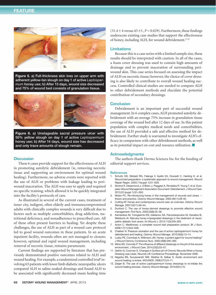
Case 5. Thirty-nine-year-old Mr. E, who had a history of hypertension, diabetes, chronic kidney disease, and myocar-dial infarction, had a fall and was found face down and un-responsive. MRI/MRA results confirmed he had suffered an acute stroke. On admission, he was extremely confused, com-bative, and agitated, requiring vest restraints for safety. He had recently immigrated to the US and had very little familial sup-port. He sustained linear full-thickness skin loss to the bilateral axillary area secondary to shear/friction damage from the vest restraint. The wound measured 1.0 cm x 12.0 cm with adher-ent yellow-tan slough (see Figure 5a). After wound cleansing, ALH and a secondary foam cover dressing were applied per the protocol described and changed every 3 days. At discharge after 2 weeks of using ALH, the wound showed marked im-provement, measuring 0.5 cm x 4.0 cm, with 25% slough and 75% epithelialized tissue (see Figure 5b). Mr. E had a total of 7 dressing changes before his discharge home.
Case 6. Ms. F is an 81-year-old woman with a history of schizophrenia, an implantable atrial defibrillator, and PEG feedings. She was admitted with C. difficile following 20 days of diarrhea. She was extremely cachectic. She had an unstageable sacral pressure ulcer measuring 8.0 cm x 4.0 cm with 50% yel-low slough and 50% red granulation tissue (see Figure 6a). Af-ter 1 week of treatment with topical antibiotic cream, ALH was started to aid in autolytic debridement of the adherent slough. After wound cleansing, ALH and a secondary foam cover dress-ing were applied per the protocol described and changed every 3 days. Ms. F was discharged after 2 weeks of ALH/cover dressing treatment (a total of 5 dressing changes); the pressure ulcer was classified Stage III and had decreased in size to 1.5 cm x 2.0 cm with only trace amounts of slough remaining (see Figure 6b).
Figure 2. a) Necrotic heel ulcer covered with eschar on day 1 of active Leptospermum honey use; b) After 15 days, eschar removal is almost complete and amount of granulation tissue has increased.
a. b.
Figure 4. a) Sacral pressure ulcer containing 40% thick slough on day 1 of active Leptospermum honey use; b) After 16 days wound debridement is complete and wound bed contains 100% granulation tissue.
a. b.
Figure 3. a) Foot ulcer with 100% adherent slough on day 1 of active Leptospermum honey use; b) After 20 days, wound debridement is complete and the wound is covered with 100% granulation tissue.
a. b.
66 OSTOMY WOUND MANAGEMENT® APRIL 2015 www.o-wm.com
FEATURE
Discussion
These 6 cases provide support for the effectiveness of ALH in promoting autolytic debridement (ie, removing necrotic tissue and supporting an environment for optimal wound healing). Furthermore, no adverse events were reported with the use of ALH or problems with leakage leading to peri-wound maceration. The ALH was easy to apply and required no specific training, which allowed it to be quickly integrated into the facility’s protocols of care.
As illustrated in several of the current cases, treatment of inner city, indigent, often elderly and immunocompromised adults with clinically complex wounds is very difficult due to factors such as multiple comorbidities, drug addiction, nu-tritional deficiency, and nonadherence to prescribed care. All of these often present barriers to healing. Yet despite these challenges, the use of ALH as part of a wound care protocol led to good wound outcomes in these patients. In an acute inpatient facility, wounds often are not managed to closure; however, optimal and rapid wound management, including removal of necrotic tissue, remains paramount.
Current findings are supported by literature that has pre-viously demonstrated positive outcomes related to ALH and wound healing. For example, a randomized controlled trial6 in-volving 63 patients with lower limb diabetic neuropathic ulcers compared ALH to saline-soaked dressings and found ALH to be associated with significantly decreased mean healing time
(31.4 ± 4 versus 43 ±3 , P = 0.029). Furthermore, these findings underscore existing case studies that support the effectiveness of honey, including ALH, for wound debridement.5,7,8
LimitationsBecause this is a case series with a limited sample size, these
results should be interpreted with caution. In all of the cases, a foam cover dressing was used to contain high amounts of drainage and to prevent maceration of surrounding peri-wound skin. This case series focused on assessing the impact of ALH on necrotic tissue; however, the choice of cover dress-ing is also likely to contribute to overall wound healing suc-cess. Controlled clinical studies are needed to compare ALH to other debridement methods and elucidate the potential contribution of secondary dressings.
ConclusionDebridement is an important part of successful wound
management. In 6 complex cases, ALH promoted autolytic de-bridement with an average 75% increase in granulation tissue coverage of the wound bed after 12 days of use. In this patient population with complex medical needs and comorbidities, the use of ALH provided a safe and effective method for de-bridement. Further study is warranted to investigate ALH’s ef-ficacy in comparison with other debridement methods, as well as its potential impact on cost and resource utilization. n
Acknowledgments The authors thank Derma Sciences Inc for the funding of
editorial support services.
References1. Schultz GS, Sibbald RG, Falanga V, Ayello EA, Dowsett C, Harding K, et al.
Wound bed preparation: a systematic approach to wound management. Wound Repair Regen. 2003;11(suppl 1):S1–S28.
2. Strohol R, Dissemond J, O’Brien J, Piaggesi A, Rimdeika R, Young T, et al. Euro-pean Wound Management Association Document: Debridement. J Wound Care. 2013;22 (suppl 1):S1–S52.
3. Molan PC. Re-introducing honey in the management of wounds and ulcers — theory and practice. Ostomy Wound Manage. 2002;48(11):28–40.
4. Cutting KF. Honey and contemporary wound care: an overview. Ostomy Wound Manage. 2007;53(11):49–54.
5. Dunford C. The use of honey-derived dressings to promote effective wound management. Prof Nurs. 2005;20(8):35–38.
6. Kamaratos AV, Tzirogiannis KN, Iraklianou SA, Panoutsopoulos GI, Kanellos IE, Melidonis AI. Manuka honey-impregnated dressings in the treatment of neuro-pathic diabetic foot ulcers. Int Wound J. 2012;11(3):259–263.
7. Acton C. Medihoney: a complete wound bed preparation product. Br J Nurs. 2008;17(11):S44–S48.
8. Chaiken N. Pressure ulceration and the use of active Leptospermum honey for debridement and healing. Ostomy Wound Manage. 2010;56(5):12–14.
9. Lusby PE, Coombes A, Wilkinson JM. Honey: a potent agent for wound healing? J Wound Ostomy Continence Nurs. 2002;29(6):295–300.
10. Milne SD, Connolly P. The influence of different dressings on the pH of the wound environment. J Wound Care. 2014;23(2):53–57.
11. Gethin G, Cowman S. Changes in Surface pH of Chronic Wounds When a Honey Dressing was Used. Wounds UK Conference Proceedings. November 2006.
12. Nagoba BS, Suryawanshi NM, Wadher B, Selkar S. Acidic environment and wound healing: a review. WOUNDS. 2006;27(1):5–11.
13. Zeiger B. The use of active Leptospermum honey dressings to re-initiate the wound healing process. Ostomy Wound Manage. 2010;65(1):10.
Figure 5. a) Full-thickness skin loss on upper arm with adherent yellow-tan slough on day 1 of active Leptosper-mum honey use; b) After 13 days, wound size decreased and 75% of wound bed consists of granulation tissue.
a. b.
Figure 6. a) Unstageable sacral pressure ulcer with 50% yellow slough on day 1 of active Leptospermum honey use; b) After 14 days, wound size has decreased and only trace amounts of slough remain.
a. b.