use of ultrasound in portal hypertension - bmus.org · and color/power doppler modules to assess...
TRANSCRIPT
Institute for Liver and Digestive Health
Use of Ultrasound in Portal Hypertension
Dr. Davide Roccarina Specialist in General Medicine Specialist Doctor in Clinical Ultrasound and non-invasive liver assessment Hepatology Department Division of Medicine Royal Free Hospital

Portal Hypertension: definition
«Portal hypertension (PH) is a frequent clinical syndrome haemodynamically defined by an increase in the portal pressure gradient (difference between portal vein pressure and inferior vena cava pressure) over the normal limit of 5 mmHg»
Berzigotti A, Piscaglia F. Ultraschall bei Pfortaderhochdruck… Ultraschall in Med 2011; 32: 548–571

OLTx DEATH
SBP
ASCITES
ESOPHAGEAL VARICES
RAPTURE
BLEEDING
HEPATO-RENAL SYNDROME
CSPH is an independent predictor of clinical decompensation

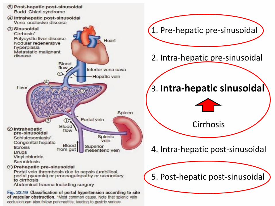
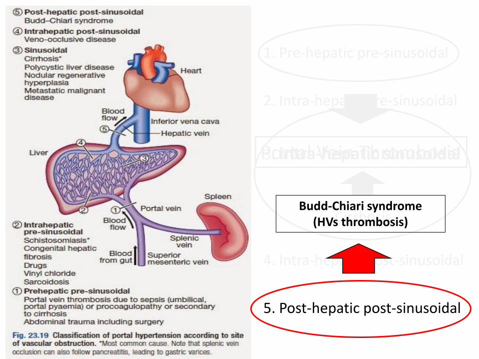
1. Pre-hepatic pre-sinusoidal
2. Intra-hepatic pre-sinusoidal
3. Intra-hepatic sinusoidal
4. Intra-hepatic post-sinusoidal
5. Post-hepatic post-sinusoidal
Cirrhosis

HVPG
Portal Hypertension: reference standard

1. First-line imaging technique used in patients with suspected PH, since it is noninvasive, repeatable and cheap.
2. US examiners should be able to detect and report correctly the most important signs of PH.
Role of Ultrasound
3. Most US signs of PH are independent of its underlying cause, and their interpretation should always be integrated with clinical information.
4. When patients already show overt clinical features of PH and no other data is available, US examination facilitates the classification of the mechanism which led to PH.
Instrument-based requirements
Convex transducers between 3.5 and 5MHz. Higher frequencies might be necessary in children. Moreover, linear transducers (7.5–10MHz) might be needed to properly assess the liver surface, since they significantly increase the performance of US in the detection of liver cirrhosis.
The US equipment should be provided with pulsed and color/power Doppler modules to assess the patency of the vessels and to characterize the haemodynamic features of portal and splanchnic circulations.
Berzigotti A, Piscaglia F. , Ultraschall in Med 2011; 32: 548–571
Examination procedure The liver, spleen and portal venous system should
always be examined in any patient with suspected PH.
Patient needs to be in a fasting state for at least 6 hours (food ingestion induces hemodynamic changes)
Examination should be started after 5-10 minutes of a
supine position (exercise and posture changes induce hemodynamic changes).
Quantitative Doppler measurements should be
performed in suspended normal respiration, avoiding deep inspiration or expiration.
Berzigotti A, Piscaglia F. , Ultraschall in Med 2011; 32: 548–571

LIVER and SPLEEN assessment: B-mode US
LIVER
SPLEEN
Profile/edge/surface: • normal • irregular • nodular
Echotexture: • homogeneous • heterogeneous • coarsened
Caudate lobe: • normal size • hypertrophy
Normal or enlarged (size < 12 cm; area < 45 cm2)
Right/left lobe: • normal size • hypotrophy • relative hypertrophy
Margin: • sharp • rounded
NEVER FORGET THE HEART!!!
ASCITES
Vascular assessment: B-mode, pulsed-Doppler, color/power Doppler
Berzigotti A, Piscaglia F. , Ultraschall in Med 2011; 32: 548–571
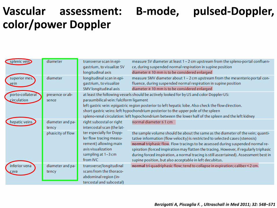
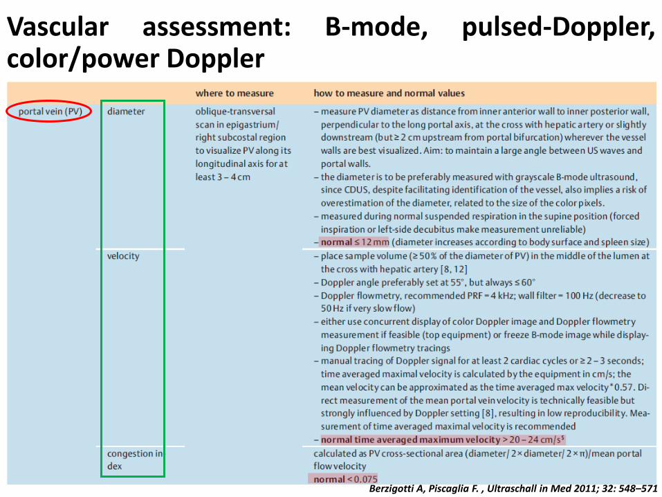
Vascular assessment: B-mode, pulsed-Doppler, color/power Doppler
Berzigotti A, Piscaglia F. , Ultraschall in Med 2011; 32: 548–571

Berzigotti A, Piscaglia F. , Ultraschall in Med 2011; 32: 548–571
1. Pre-hepatic pre-sinusoidal
2. Intra-hepatic pre-sinusoidal
3. Intra-hepatic sinusoidal
4. Intra-hepatic post-sinusoidal
5. Post-hepatic post-sinusoidal
Cirrhosis

1. Pre-hepatic pre-sinusoidal
2. Intra-hepatic pre-sinusoidal
3. Intra-hepatic sinusoidal
4. Intra-hepatic post-sinusoidal
5. Post-hepatic post-sinusoidal
Cirrhosis

Cirrhotic Portal Hypertension: pathogenesis
OHM’S LAW
ΔP = Q × R
Dynamic Component
Static Component

LIVER CIRRHOSIS
Eco-color Doppler: Portal Vein: Flow velocity (TAPV) < 20-24 cm/sec Hepatofugal flow
Hepatic Artery: RI > 0,70
Hepatic Veins: Monophasic/biphasic Doppler waveform
Other vessels: Umbilical vein recanalization Collateral vessels/varices

B-mode US:
Irregular/nodular liver profile Caudate hypertrophy/right hypotrophy/relative left
hypertrophy Heterogeneous/coarse echo-texture Rounded margin PV calibre > 13 mm No changes of PV calibre with respiration Absence of respiratory phasicity Enlarged spleen (size > 13mm, area > 45 cm2)
LIVER CIRRHOSIS

PHILIPS

PHILIPS

PHILIPS

PHILIPS
PHILIPS

PHILIPS

PHILIPS


PHILIPS
PHILIPS

PHILIPS


1. Pre-hepatic pre-sinusoidal
2. Intra-hepatic pre-sinusoidal
3. Intra-hepatic sinusoidal
4. Intra-hepatic post-sinusoidal
5. Post-hepatic post-sinusoidal
Cirrhosis
Portal Vein Thrombosis

Acute Longstanding
Portal Vein Thrombosis
Absence of flow in the PV (neither colour- nor pulsed-Doppler signal)
Benign Neoplastic
Few US findings

Acute Longstanding
Portal Vein Thrombosis
Absence of flow in the PV (neither colour- nor pulsed-Doppler signal)
Benign Neoplastic
Few US findings

Arterial colour- and pulsed-Doppler
signals inside the clot with high RI
Contrast enhancement
Disruption of vein wall
No arterial colour-Doppler signals inside the clot
No contrast enhancement
Well defined vein wall
Absence of flow
Echogenic material in the vein lumen and
vein dilatation
Portal cavernoma transformation
Thick-walled GB with peri-cholecistic varices
Peri-choledocic varices
Biliary ducts dilatation
Benign Neoplastic
Tumor
Portal Vein Thrombosis
Involvement of only one branch
Marked dilatation
Possible involvement of SMV and SV

B-mode US:
Normal or heterogeneous liver parenchyma Focal liver lesion A marked heterogenous hepatic area: infiltrating
carcinoma Hepatic artery hypertrophy Enlarged spleen (size > 13mm, area > 45 cm2)
Portal Vein Thrombosis
Eco-color Doppler:
Portal cavernoma transformation Hepatic Artery:
RI > 0,70
Collateral vessels/varices


PHILIPS


PHILIPS

1. Pre-hepatic pre-sinusoidal
2. Intra-hepatic pre-sinusoidal
3. Intra-hepatic sinusoidal
4. Intra-hepatic post-sinusoidal
5. Post-hepatic post-sinusoidal
Cirrhosis
Portal Vein Thrombosis 1. Chronic right heart failure with
cardiac liver cirrhosis
2. Tricuspid valve disease
3. Constrictive pericarditis

B-mode US:
Enlarged/congested liver US features of liver cirrhosis (cardiac cirrhosis) Enlarged spleen (size > 13mm, area > 45 cm2) US features of tricuspid regurgitation and/or heart
failure PV size > 13 mm No PV calibre changes with respiration IVC calibre > 2 cm HVs calibre > 1 cm No IVC inspiratory collapse or < 40%
PH related to Heart disease
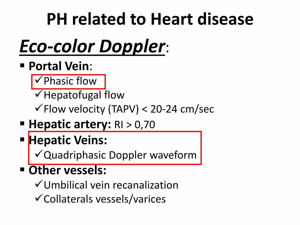
Eco-color Doppler: Portal Vein: Phasic flow Hepatofugal flow Flow velocity (TAPV) < 20-24 cm/sec
Hepatic artery: RI > 0,70
Hepatic Veins: Quadriphasic Doppler waveform
Other vessels: Umbilical vein recanalization Collaterals vessels/varices
PH related to Heart disease


INSP EXP


PHILIPS

PHILIPS
1. Pre-hepatic pre-sinusoidal
2. Intra-hepatic pre-sinusoidal
3. Intra-hepatic sinusoidal
4. Intra-hepatic post-sinusoidal
5. Post-hepatic post-sinusoidal
Cirrhosis
Portal Vein Thrombosis
Budd-Chiari syndrome (HVs thrombosis)

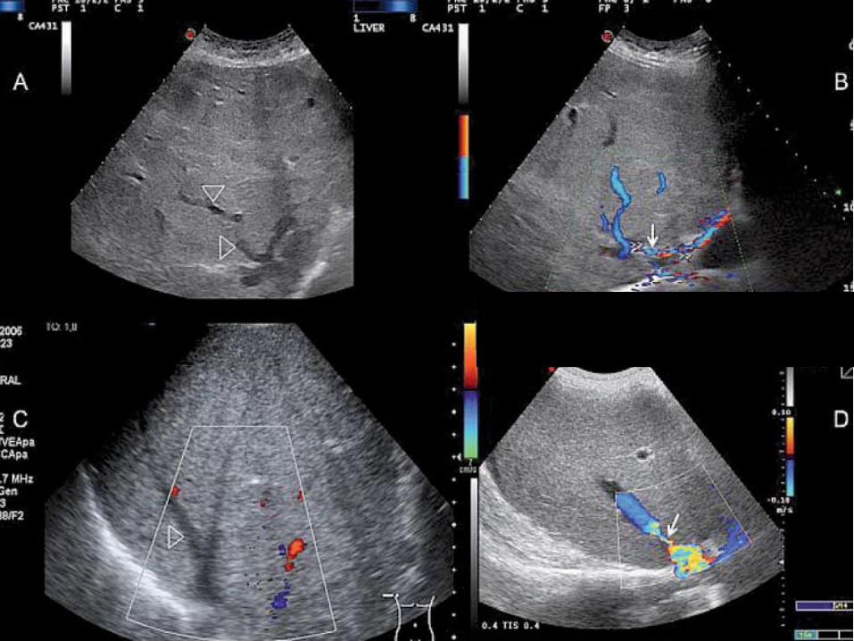
Budd-Chiari Syndrome
In the acute form of BCS
Eco-color Doppler: Lack of visualization of one or more hepatic veins at the color-Doppler
B-mode US: A thrombus filling the vein, vein stenosis, or a tumor invading or compressing the
veins.
In subacute and chronic forms of BCS
Eco-color Doppler: A fragmented vein with flow reversal, or new venous vessels that drain sub-capsular
circulation to another hepatic vein or directly to the inferior vena cava
B-mode US: A fibrous tract replacing the obstructed hepatic veins Caudate lobe and caudate vein hypertrophy Enlarged liver with grossly heterogeneous echo-texture


Limitations of US examination in this field Abdominal air interposition which may prevent correct and
complete visualization of the abdominal organs and vessels.
Massive ascites also impairs the imaging of the liver and abdominal vessels.
A well recognized limitation of quantitative Doppler measurements is the inter-equipment and inter-observer variability which reduces the comparability of this data among different centers.
Patients in follow-up should preferably be examined by the same operator and with the same equipment whenever possible.
Cooperative studies have shown that it is possible to reduce inter-observer variability by using a standardized protocol of examination

THANK YOU