use of repetitive transcranial magnetic stimulation (rtms
TRANSCRIPT

Use of Repetitive Transcranial Magnetic Stimulation (rTMS) to Augment Hypnotic Analgesia
Study Protocol and Statistical Analysis Plan
NCT02969707
February 14, 2019
Study Product Guidelines and Considerations 1 of 99 Version 1.5 14 FEB 2019
FULL PROTOCOL TITLE
Use of Repetitive Transcranial Magnetic Stimulation (rTMS) to Augment Hypnotic Analgesia:
A randomized, double-blind, sham-controlled, mechanistic clinical trial assessing the efficacy of rTMS in modulating the neural circuitry underlying hypnotizability, hypnosis, and hypnotic analgesia among participants with a diagnosis of Fibromyalgia Syndrome (FMS) and low-
moderate hypnotizability
Principal Investigators:
Nolan R. Williams, MD Stanford University
Department of Psychiatry 401 Quarry Rd Mail Code 5719
Stanford, CA 94305 Tel: (650) 723-1147 Fax:
Email: [email protected]
David Spiegel, M.D. Stanford University
Department of Psychiatry 401 Quarry Rd Mail Code 5718
Stanford, CA 94305 Tel: (650) 723-6421 Fax:
Email: [email protected]
Supported by:
The National Center for Complementary and Integrative Health
1R33AT009305-01

Study Product Guidelines and Considerations 2 of 99 Version 1.5 14 FEB 2019
Tool Revision History
Version Number: 1.1 Version Date: 09/18/2016 Summary of Revisions Made: per emailed request
Version Number: 1.2 Version Date: 10/18/16 Summary of Revisions Made: per emailed request
Version Number: 1.3 Version Date: 03/14/2018 Summary of Revisions Made:
The assessments were updated: Sense of Agency, Tellegen’s Absorption Scale and the Ishihara Vision Test were added. ATHF was changed to ATRQ and the McGill Pain Questionnaire Long Form was changed to the Short Form Version.
A targeting MRI scan was added as part of the screening process for the TMS administration.
The assessments table was updated to reflect the schedule of events: hematology was removed, HIP/HIS were separated and administration of the HIP time points were updated, SOARS was added to the HIP, the online screening consent was added
Vital signs were removed from the SOE. Urinalysis was updated to reflect that it is only assessed on TMS administration days. Due to the stability of trait hypnosis, enrollment time frame has been updated from 1-
month to be based upon individual subjects’ availability to schedule their study visits this is in response to subjects not being available to complete all required visits within a 1-month period.
Version Number: 1.4 Version Date: 10/10/2018 Summary of Revisions Made:
Clarification was made under Section: Schedule and Type of Evaluations, that the target scan and first study visit can be on the same day.
Under 4.2, it was clarified that the high risk participants will be excluded if they are already taking opioids.
Pain Numeric Rating Scale (PNRS) and Dissociative Experiences Scales were added to Section 6.2.1.
Needle-like sensations and pain was added to Section 7.4 as an expected AE.

Study Product Guidelines and Considerations 3 of 99 Version 1.5 14 FEB 2019
Version Number: 1.5 Version Date: 02/14/2019 Summary of Revisions Made:
Under 6.2.2, spectroscopy language was added to the Baseline MRI Session for TMS targeting
Under 6.2.5, optional MRI spectroscopy assessment is added. Under Secondary objectives, section F and G are added to include MRS secondary-
aims. Under 9.6.2, section F and G are added to include MRS secondary outcome measures
and analyses.

Study Product Guidelines and Considerations 4 of 99 Version 1.3 14 MAR 2018
TABLE OF CONTENTS Page
FULL PROTOCOL TITLE......................................................................................................1
Tool Revision History .............................................................................................................2
TABLE OF CONTENTS ..........................................................................................................4
STUDY TEAM ROSTER .......................................................................................................7
PARTICIPATING STUDY SITES..........................................................................................8
PRÉCIS ...................................................................................................................................9
1. STUDY OBJECTIVES ....................................................................................................... 11
1.1 Primary Objective ...................................................................................................... 11
1.2 Secondary Objectives ................................................................................................ 13
2. BACKGROUND AND RATIONALE ................................................................................ 14
2.1 Background on Condition, Disease, or Other Primary Study Focus ............................ 14
2.2 Study Rationale ......................................................................................................... 14
3. STUDY DESIGN ................................................................................................................. 19
4. SELECTION AND ENROLLMENT OF PARTICIPANTS ............................................. 27
4.1 Inclusion Criteria ....................................................................................................... 27
4.2 Exclusion Criteria ...................................................................................................... 28
4.3 Study Enrollment Procedures ..................................................................................... 29
5. STUDY INTERVENTIONS ............................................................................................... 31
5.1 Interventions, Administration, and Duration .............................................................. 31
5.2 Handling of Study Interventions ................................................................................ 33
5.3 Concomitant Interventions ......................................................................................... 34 5.3.1 Allowed Interventions ............................................................................................ 34
5.3.2 Required Interventions ........................................................................................... 34 5.3.3 Prohibited Interventions ......................................................................................... 34
5.4 Adherence Assessment .............................................................................................. 34
6. STUDY PROCEDURES ..................................................................................................... 34
6.1 Schedule of Evaluations ............................................................................................. 35
6.2 Description of Evaluations ......................................................................................... 36
6.2.1 Screening Evaluation ............................................................................................. 36 6.2.2 Enrollment, Baseline, and/or Randomization.......................................................... 38
Study Product Guidelines and Considerations 5 of 99 Version 1.3 14 MAR 2018
6.2.3 Blinding ................................................................................................................. 42 6.2.4 Followup Visits...................................................................................................... 43
6.2.5 Completion/Final Evaluation ................................................................................. 44
7. SAFETY ASSESSMENTS .................................................................................................. 44
7.1 Specification of Safety Parameters ............................................................................. 46
7.2 Methods and Timing for Assessing, Recording, and Analyzing Safety Parameters ..... 46
7.3 Adverse Events and Serious Adverse Events .............................................................. 47
7.4 Reporting Procedures ................................................................................................. 49
7.5 Followup for Adverse Events ..................................................................................... 52
7.6 Safety Monitoring ...................................................................................................... 52
8. INTERVENTION DISCONTINUATION ......................................................................... 54
9. STATISTICAL CONSIDERATIONS ................................................................................ 54
9.1 General Design Issues ................................................................................................ 55
9.2 Sample Size and Randomization ................................................................................ 59
9.3 Stimulation Group Assignment Procedures ................................................................. 61
9.4 Definition of Populations ........................................................................................... 64
9.5 Interim Analyses and Stopping Rules ......................................................................... 65
9.6 Outcomes .................................................................................................................. 65
9.6.1 Primary Outcome ................................................................................................... 59 9.6.2 Secondary Outcomes ............................................................................................. 60
9.7 Data Analyses ............................................................................................................ 73
10. DATA COLLECTION AND QUALITY ASSURANCE ................................................. 76
10.1 Data Collection Forms ............................................................................................... 76
10.2 Data Management ...................................................................................................... 76
10.3 Quality Assurance...................................................................................................... 78 10.3.1 Training ............................................................................................................. 78
10.3.2 Quality Control Committee ................................................................................ 78 10.3.3 Metrics ............................................................................................................... 78
10.3.4 Protocol Deviations ............................................................................................ 79 10.3.5 Monitoring ......................................................................................................... 80
11. PARTICIPANT RIGHTS AND CONFIDENTIALITY .................................................. 82
11.1 Institutional Review Board (IRB) Review .................................................................. 82
11.2 Informed Consent Forms ........................................................................................... 82
11.3 Participant Confidentiality ......................................................................................... 82

Study Product Guidelines and Considerations 6 of 99 Version 1.3 14 MAR 2018
11.4 Study Discontinuation................................................................................................ 83
12. COMMITTEES ................................................................................................................. 84
13. PUBLICATION OF RESEARCH FINDINGS ................................................................ 85
14. REFERENCES .................................................................................................................. 78
15. SUPPLEMENTAL MATERIAL ...................................................................................... 89
I. Stanford IRB-Approved Informed Consent Form
II. TMS Stimulation Manual III. TMS Blinding Manual
IV. Hypnosis and Hypnotic Analgesia Manual

Study Product Guidelines and Considerations 7 of 99 Version 1.3 14 MAR 2018
STUDY TEAM ROSTER
Nolan Williams, M.D., Principal Investigator
Stanford, CA 94305 Tel: (650) 723-1147 Fax: Email: [email protected] David Spiegel, M.D., Principal Investigator
Stanford, CA 94305 Tel: (650) 723-6421 Fax: Email: [email protected]
Leanne Williams, Ph.D., Co-Investigator
Palo Alto, CA 94305 Phone: Fax: N/A Email: Dave Yeomans, Ph.D., Co-Investigator
Stanford, CA 94305 Phone: Fax: Email: Booil Jo, Ph.D., Co-Investigator
Stanford, CA 94305 Phone: Fax: Email:

Study Product Guidelines and Considerations 8 of 99 Version 1.3 14 MAR 2018
PARTICIPATING STUDY SITES Nolan Williams, M.D. Principal Investigator Stanford University Department of Psychiatry
Stanford, CA 94305 Tel: (650) 723-1147 Fax: Email: [email protected]
David Spiegel, M.D. Principal Investigator Stanford University Department of Psychiatry
Stanford, CA 94305 Tel: (650) 723-6421 Fax: Email: [email protected]

Study Product Guidelines and Considerations 9 of 99 Version 1.3 14 MAR 2018
PRÉCIS
Title
Use of Repetitive Transcranial Magnetic Stimulation (rTMS) to Augment Hypnotic Analgesia
Objectives
Primary Mechanistic Objective: To determine the effect of active, inhibitory rTMS (continuous theta-burst stimulation-cTBS) over left dorsolateral prefrontal cortex (L-DLPFC) on modulating the neural network that underlies hypnotizability and hypnosis.
Relevant Clinical Objective: To determine the effect of active, inhibitory rTMS (cTBS) over L-DLPFC on enhancing hypnotic analgesia (HA) as measured by change in pain thresholds.
Secondary Objective A: To determine the effect of active, inhibitory rTMS (cTBS) over L-DLPFC on modulating the neural network that underlies hypnotic intensity.
Secondary Objective B: To determine the effect of active, inhibitory rTMS (cTBS) over L-DLPFC on enhancing hypnotizability (as measured by the Hypnotic Induction Profile-HIP and Stroop Task) and hypnotic intensity (as measured by the Hypnotic Intensity Scale-HIS).
Secondary Objective C: To determine the effect of active, inhibitory rTMS (cTBS) over L-DLPFC on the neural network underlying the conflict regulation system as a surrogate of effective modulation of the neural circuitry that underlies hypnotizability.
Secondary Objective D: To determine the effect of active, inhibitory rTMS (cTBS) over L-DLPFC on the neural network that underlies the post-hypnotic Stroop Effect.
Secondary Objective E: To determine the effect of active, inhibitory rTMS (cTBS) over L-DLPFC on modulating the neural network that underlies hypnotic analgesia (HA).
Design and Outcomes
Study Design: This study is a double blind, placebo-controlled, mechanistic trial assessing rTMS as a strategy for the modulation of the neural circuitry underlying hypnotizability and hypnotic analgesia in participants with a diagnosis of Fibromyalgia Syndrome (FMS).
Schedule and Type of Evaluations: There are four visits for this. The first visit is a screening visit where the participant will be consented to be screened for the study. The participant will then be screened to determine inclusion and exclusion criteria. If the participant meets criteria, the participant will be consented for the imaging and rTMS portions of the study. The participant will be assigned their randomization code and their counterbalanced imaging days. Up to three separate imaging visits will be conducted including a baseline MRI for TMS target identification (Visit #2) and two separate MRI-TMS sessions consisting of a hypnotic analgesia and hypnotic stroop procedures (Visits #3-4). Visit #2 and #3 can take place on the same day. During the TMS-

Study Product Guidelines and Considerations 10 of 99 Version 1.3 14 MAR 2018
MRI visits which include thermal pain and stroop MRI sessions, the participant will receive either sham or active rTMS (depending on randomization) in a non-crossover design.
Study Overview Diagram:
All participants will undergo the Hypnotic Stroop Scan Session Day. All participants will receive the identical pre-rTMS scan session. One-half (n=50) will receive active rTMS and one-half (n=50) will receive sham rTMS (two arm, double-blind, sham-controlled). All participants will receive the identical post-rTMS scan session. Changes in resting state functional connectivity between L-DLPFC and dACC in the active rTMS versus the sham rTMS conditions will be assessed. This primary endpoint is a between-group comparison. We will also measure the difference in brain activation during the hypnotic induction, and Stroop task (after post-hypnotic suggestion) between the active and sham rTMS. This will also be a between-groups comparison. Using the Hypnotic Induction Profile (HIP) and Hypnotic Intensity Scale (HIS), we will compare the change in hypnotizability and hypnotic intensity score between the active and sham rTMS groups. All participants will undergo the Hypnotic Analgesia Scan Session Day. All participants will complete identical pre- and post- rTMS scan session. One-half will receive active rTMS and one-half will receive sham rTMS. Changes in resting state functional connectivity between L-DLPFC and dACC in the active rTMS versus the sham rTMS conditions will be assessed. This primary endpoint is a between-group comparison. This scan-session also addresses a secondary aim of determining the difference in functional connectivity within the hypnotic analgesia network between the active and the sham rTMS groups. This will be a between-groups comparison. We will also compare the activation within the hypnotic analgesia network between the active and sham rTMS groups. This will be a between-groups comparison. We will also utilize a perturbation technique to probe the hypnotic induction in an alternative way. Finally, we will compare the difference in pain thresholds between the active and sham rTMS groups.
Interventions and Duration
Intervention: We will compare active versus sham rTMS as a technique for modulating the neural
Recruitment/Screening of Fibromyalgia Subjects (Visit #1)(n=100)
Active rTMS Group (n=50)
Randomization
(n=25)
Hypnotic Stroop Session (Visit #3)
Counterbalancing
Sham rTMS Group (n=50)
Counterbalancing
TMS Target MRI Visit (Visit #2)
Hypnotic Analgesia Session (Visit #4)
MRI #1 rTMS MRI #2
MRI #1 rTMS MRI #2
(n=25)
Hypnotic Analgesia Session (Visit #3)
Hypnotic Stroop Session (Visit #4)
MRI #1 rTMS MRI #2
MRI #1 rTMS MRI #2
(n=25)
Hypnotic Stroop Session (Visit #3)
Hypnotic Analgesia Session (Visit #4)
MRI #1 rTMS MRI #2
MRI #1 rTMS MRI #2
(n=25)
Hypnotic Analgesia Session (Visit #3)
Hypnotic Stroop Session (Visit #4)
MRI #1 rTMS MRI #2
MRI #1 rTMS MRI #2
Study Product Guidelines and Considerations 11 of 99 Version 1.3 14 MAR 2018
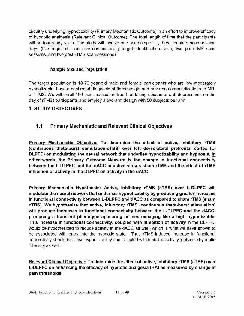
circuitry underlying hypnotizability (Primary Mechanistic Outcome) in an effort to improve efficacy of hypnotic analgesia (Relevant Clinical Outcome). The total length of time that the participants will be four study visits. The study will involve one screening visit, three required scan session days (five required scan sessions including target identification scan, two pre-rTMS scan sessions, and two post-rTMS scan sessions).
Sample Size and Population
The target population is 18-70 year-old male and female participants who are low-moderately hypnotizable, have a confirmed diagnosis of fibromyalgia and have no contraindications to MRI or rTMS. We will enroll 100 pain medication-free (not taking opiates or anti-depressants on the day of rTMS) participants and employ a two-arm design with 50 subjects per arm. 1. STUDY OBJECTIVES
1.1 Primary Mechanistic and Relevant Clinical Objectives
Primary Mechanistic Objective: To determine the effect of active, inhibitory rTMS (continuous theta-burst stimulation-cTBS) over left dorsolateral prefrontal cortex (L-DLPFC) on modulating the neural network that underlies hypnotizability and hypnosis. In other words, the Primary Outcome Measure is the change in functional connectivity between the L-DLPFC and the dACC in active versus sham rTMS and the effect of rTMS inhibition of activity in the DLPFC on activity in the dACC.
Primary Mechanistic Hypothesis: Active, inhibitory rTMS (cTBS) over L-DLPFC will modulate the neural network that underlies hypnotizability by producing greater increases in functional connectivity between L-DLPFC and dACC as compared to sham rTMS (sham cTBS). We hypothesize that active, inhibitory rTMS (continuous theta-burst stimulation) will produce increases in functional connectivity between the L-DLPFC and the dACC, producing a transient phenotype appearing on neuroimaging like a high hypnotizable. This increase in functional connectivity, coupled with inhibition of activity in the DLPFC, would be hypothesized to reduce activity in the dACC as well, which is what we have shown to be associated with entry into the hypnotic state. Thus rTMS-induced increase in functional connectivity should increase hypnotizability and, coupled with inhibited activity, enhance hypnotic intensity as well.
Relevant Clinical Objective: To determine the effect of active, inhibitory rTMS (cTBS) over L-DLPFC on enhancing the efficacy of hypnotic analgesia (HA) as measured by change in pain thresholds.

Study Product Guidelines and Considerations 12 of 99 Version 1.3 14 MAR 2018
Relevant Clinical Hypothesis: Active, inhibitory rTMS (cTBS) over L-DLPFC will enhance the efficacy of hypnotic analgesia (HA) as compared to sham rTMS (sham cTBS).

Study Product Guidelines and Considerations 13 of 99 Version 1.3 14 MAR 2018
1.2 Secondary Objectives Secondary Objective A: To determine the effect of active, inhibitory rTMS (cTBS) over L-DLPFC on modulating the neural network that underlies hypnotic intensity.
Secondary Hypothesis A: Active, inhibitory rTMS (cTBS) over L-DLPFC will modulate the neural network that underlies hypnotic intensity by decreasing activity in L-DLPFC and dACC as compared to sham rTMS (sham cTBS) as measured by BOLD fMRI and interleaved TMS-MRI.
Secondary Objective B: To determine the effect of active, inhibitory rTMS (cTBS) over L-DLPFC on enhancing hypnotizability (as measured by the Hypnotic Induction Profile-HIP and Stroop Task) and hypnotic intensity (as measured by the Hypnotic Intensity Scale-HIS).
Secondary Hypothesis B: Active, inhibitory rTMS (cTBS) over L-DLPFC will increase the HIP and HIS scores as well as hypnotic reduction of the Stroop Effect as compared to sham rTMS (sham cTBS).
Secondary Objective C: To determine the effect of active, inhibitory rTMS (cTBS) over L-DLPFC on the neural network underlying the conflict regulation system as a surrogate of effective modulation of the neural circuitry that underlies hypnotizability.
Secondary Hypothesis C: Active, inhibitory rTMS (cTBS) over L-DLPFC will modulate the neural network that underlies conflict regulation by producing greater increases in functional connectivity between the right inferior frontal gyrus (rIFG) and the default mode network (DMN) as compared to sham rTMS (sham cTBS).
Secondary Objective D: To determine the effect of active, inhibitory rTMS (cTBS) over L-DLPFC on the neural network that underlies the post-hypnotic Stroop Effect.
Secondary Hypothesis D: Post-hypnotic instruction of word blindness after active, inhibitory rTMS (cTBS) over L-DLPFC will reduce dACC activity during the Stroop task (similar to high hypnotizables) as compared to sham rTMS (sham cTBS).
Secondary Objective E: To determine the effect of active, inhibitory rTMS (cTBS) over L-DLPFC on modulating the neural network that underlies hypnotic analgesia (HA).
Secondary Hypothesis E: Active, inhibitory rTMS (cTBS) over L-DLPFC will modulate the neural network that underlies HA by producing a decrease in activity and an increase functional connectivity among the anterior cingulate, dorsolateral, insular, and somatosensory cortices (hypnotic analgesia network) as compared to sham rTMS (sham cTBS).

Study Product Guidelines and Considerations 14 of 99 Version 1.3 14 MAR 2018
2. BACKGROUND AND RATIONALE
2.1 Background on Fibromyalgia Syndrome (FMS)
Fibromyalgia syndrome (FMS) is a poorly understood disorder characterized clinically as widespread pain lasting three months or more which is not explainable by any other disease/disorder (1). Traditional treatments have limited benefit (2), and long-term use of opioid medications is problematic in these patients (2) due to demonstrated reduced opiate binding potential in FMS (3). Hypnotherapy as an intervention has been demonstrated to be quite successful in the treatment of FMS (4). It appears that the type of suggestion utilized is of primary importance to the efficacy of the hypnotherapy intervention for FMS (5). Hypnotic analgesia for FMS has been demonstrated to have effects on the orbitofrontal, cingulate cortices, and thalamus (6, 7). There have been numerous studies looking at rTMS for the modulation of pain syndromes (8, 9) as well as for the assessment of anti-nociception properties of rTMS through experimentally induced pain in normal healthy controls (10). L-DLPFC rTMS both have demonstrated efficacy in modulating the pain experienced in fibromyalgia (8, 11).
2.2 Study Rationale
Neural Circuitry Underlying Regulation of Cognition: Theories of cognitive regulation suggest that there is a central neural network with two necessary components, which regulate cognition and control conflict in the human brain. The first component is the dorsolateral prefrontal cortex (DLPFC), which initiates and implements a cascade of control (12). The second, the dorsal anterior cingulate cortex (dACC), monitors performance and signals when adjustments in control are needed(12, 13). The DLPFC and dACC predictably interact together during conflict tasks such as the Stroop task(13) and the flanker task(14). Transcranial Magnetic Stimulation: A Technique for Probing and Manipulating Neural Networks: Tools that selectively manipulate cognitive brain circuitry can effectively drive cognitive processes (15). Transcranial magnetic stimulation (TMS) noninvasively activates neuronal elements. In TMS, high-intensity magnetic field is produced by passing a brief electric current through a magnetic coil. The scalp and skull are crossed unimpeded, and a transient electric field is induced in underlying excitable neuronal tissue. Repetitive TMS produces periods of lasting activation or inhibition that persist after stimulation (16). In particular, inhibition generally results from stimulation at or below 1 Hz and excitation is produced from 5 Hz or higher.

Study Product Guidelines and Considerations 15 of 99 Version 1.3 14 MAR 2018
Theta-Burst Stimulation: In 2005, a new rTMS approach, termed theta-burst stimulation (TBS), was developed (17). This TBS approach was modeled from earlier work in animal models of hippocampal slice physiology and LTP induction (18, 19). This approach is not only more efficient than traditional rTMS(20), but also much safer (21-24) and the after-effects of TBS have a much longer duration of effects then traditional rTMS(25). This is important because the currently utilized traditional rTMS approaches have very limited duration of effect in comparison(20, 26). While single applications of theta-burst have greater duration of effect than traditional rTMS, these single stimulations have a limited duration of effect in comparison to multiple, spaced stimulation sessions(20, 27). In order to produce a prolonged after-effect of stimulation that is capable of maintaining the after-effects for the entire duration of the planned scanner sessions, the application of two theta-burst stimulations with spacing of 10-20 min is necessary(27-30). The Neural Circuitry of Hypnotizability and Manipulated by Hypnotic Techniques: Hypnotizability is a measurable behavioral phenotype capable of reflecting differences in functional connectivity between the L-DLPFC and the dACC(31, 32), the two central brain regions that process conflict (13, 14). Consistent with this is the functional neuroimaging finding of reduced dACC activity upon entering the hypnotic state (33, 34). On functional neuroimaging, it has been observed that there is increased connectivity between the left dorsolateral prefrontal cortex (L-DLPFC) and the dorsal anterior cingulate cortex (dACC) in high hypnotizables compared with lows during rest(31). During conflict tasks, specific post-hypnotic instruction-induced changes in response latency correlate with changes on functional imaging in the dACC(32, 35, 36). There is also evidence of less dACC activity among highs than lows during a flanker task (32). During hypnosis and only among high hypnotizables there is reduced activity in the dorsal anterior cingulate cortex increased functional connectivity between the L-DLPFC (a node in the Central Executive Network-CEN) and the insular cortex (a node in the Salience Network-SN), and reduced connectivity between the L-DLPFC (CEN), medial frontal and posterior cingulate cortices (a node in the default mode network-DMN) (33, 34).
Hypnotic Analgesia Neural Network: Hypnotic analgesia (HA) involves a neural network including the prefrontal cortex (L-DLPFC), the anterior cingulate cortex (ACC), the insular cortex, and the somatosensory cortex(37). A proposed mechanism for hypnotic analgesia involves activity that links L-DLPFC with dACC and insula, thereby modulating activity in S2 and dorsal posterior insula. Other studies have indicated involvement of dACC and the insular cortex (IC) in hypnotic analgesia (7, 37-40). These and other brain regions have been identified in mixed effects analysis of pain-reducing properties of leading analgesic drugs during pain versus placebo stimulation as measured by fMRI and PET pain reports (41, 42). Other brain regions are involved in hypnotic analgesia including the sensory cortex (S1) (38, 39, 43), and periaqueductal grey (39).
Hypnotic Analgesia Tool for the Modulation of Pain: Hypnotic analgesia utilizes targeted verbal instruction to affect a specific, targeted neural network(s) involved in the pain perception

Study Product Guidelines and Considerations 16 of 99 Version 1.3 14 MAR 2018
system within the human brain(38, 44). The specific hypnotic instruction modulates the neural network involved with that instruction. The degree of pain experienced under the hypnotic suggestion of induced pain is positively correlated with L-DLPFC, dACC, and insular activity(45). Hypnotic analgesia reduces activity in the dACC (44). The Neural Circuitry Modulated by DLPFC rTMS: Like hypnosis, rTMS delivered over the L-DLPFC is correlated with a reciprocal interaction of the L-DLPFC with the ACC. This reciprocal interaction is seen in normal healthy controls performing conflict tasks(15) as well as in individuals with depression(46). Over L-DLPFC, rTMS has been demonstrated to exert frequency dependent changes where inhibitory rTMS modulates the network in one direction and excitatory rTMS modulates the network in the opposite direction(47). This infers that changes in the targeted network occur predictably depending on the frequency selected(47). Hypnosis and rTMS as Tools for Modulating Brain Activity and Connectivity: Manipulations of brain circuitry can be measured through imaging techniques(47-49). BOLD fMRI is a technique that measures changes in blood flow are accompanied by lesser changes in oxygen consumption(50). Functional connectivity MRI (fcMRI) is a neuroimaging technique that measures connectivity between functionally connected brain regions(51). This technique captures both the brain processes at work in a variety of cognitive states(52) as well as the manipulation of these networks through targeted perturbations such as with rTMS(47) or hypnosis(31, 48). rTMS modulation of brain activity has behavioral outcomes that are correlated with change in functional connectivity(46). fcMRI and rTMS can be combined to image the resulting rTMS brain connectivity manipulations(53). One could consider rTMS as a tool for driving functional connectivity in a given direction for a predictable period of time. fcMRI has not only been demonstrated to underlie the propensity to experience hypnosis(31), but additionally to measure the effects of hypnosis as compared to the baseline scan(48). Therefore, fcMRI can measure the effect of rTMS-augmented hypnotherapy, as it has been demonstrated to have the sensitivity to capture the effects of each of these interventions in isolation(31, 48).
Change in Functional Connectivity and Activity Modulated by rTMS (Primary Outcome Measure): rTMS has been demonstrated to reliably change functional connectivity in a frequency dependent manner(54). There are numerous studies demonstrating rTMS-modulated increases in functional connectivity(46, 47, 53-58), suggesting that the rTMS modulation of L-DLPFC rTMS will cause a predictable increase in functional connectivity to the dACC(47). Hypnosis/hypnotizability will be increased with rTMS as has been demonstrated previously(26, 59) which we believe will have a beneficial effect on the treatment outcome as has been previously demonstrated on attention measures(60, 61) and clinical populations(62). This increase in functional connectivity, coupled with inhibition of activity in the L-DLPFC, would be hypothesized to reduce activity in the dACC as well, which is what we have shown to be associated with entry into the hypnotic state. Thus rTMS induced increase in functional connectivity should increase hypnotizability and, coupled with inhibited activity, enhance hypnotic intensity as well.
Neural Networks Affected By Fibromyalgia and the Effect of HA on the FMS Networks: In addition to abnormalities in pain processing, there is aberrant processing of non-painful
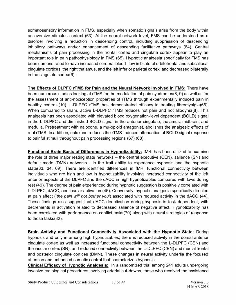
Study Product Guidelines and Considerations 17 of 99 Version 1.3 14 MAR 2018
somatosensory information in FMS, especially when somatic signals arise from the body within an aversive stimulus context (63). At the neural network level, FMS can be understood as a disorder involving a reduction in descending control, including suppression of descending inhibitory pathways and/or enhancement of descending facilitative pathways (64). Central mechanisms of pain processing in the frontal cortex and cingulate cortex appear to play an important role in pain pathophysiology in FMS (65). Hypnotic analgesia specifically for FMS has been demonstrated to have increased cerebral blood-flow in bilateral orbitofrontal and subcallosal cingulate cortices, the right thalamus, and the left inferior parietal cortex, and decreased bilaterally in the cingulate cortex(6).
The Effects of DLPFC rTMS for Pain and the Neural Network Involved in FMS: There have been numerous studies looking at rTMS for the modulation of pain syndromes(8, 9) as well as for the assessment of anti-nociception properties of rTMS through experimentally induced pain in healthy controls(10). L-DLPFC rTMS has demonstrated efficacy in treating fibromyalgia(66). When compared to sham, active L-DLPFC rTMS reduces hot pain and hot allodynia(8). This analgesia has been associated with elevated blood oxygenation-level dependent (BOLD) signal in the L-DLPFC and diminished BOLD signal in the anterior cingulate, thalamus, midbrain, and medulla. Pretreatment with naloxone, a mu-opioid antagonist, abolishes the analgesic effects of real rTMS. In addition, naloxone reduces the rTMS-induced attenuation of BOLD signal response to painful stimuli throughout pain processing regions (67) (68).
Functional Brain Basis of Differences in Hypnotizability: fMRI has been utilized to examine the role of three major resting state networks – the central executive (CEN), salience (SN) and default mode (DMN) networks - in the trait ability to experience hypnosis and the hypnotic state(33, 34, 69). There are identified differences in fMRI functional connectivity between individuals who are high and low in hypnotizability involving increased connectivity of the left anterior aspects of the DLPFC and the dACC in high hypnotizables compared with lows during rest (49). The degree of pain experienced during hypnotic suggestion is positively correlated with L-DLPFC, dACC, and insular activation (45). Conversely, hypnotic analgesia specifically directed at pain affect (‘the pain will not bother you’) associated with reduced activity in the dACC (44). These findings also suggest that dACC deactivation during hypnosis is task dependent, with decrements in activation related to decreased salience of negative affect. Hypnotizability has been correlated with performance on conflict tasks(70) along with neural strategies of response to those tasks(32).
Brain Activity and Functional Connectivity Associated with the Hypnotic State: During hypnosis and only in among high hypnotizables, there is reduced activity in the dorsal anterior cingulate cortex as well as increased functional connectivity between the L-DLPFC (CEN) and the insular cortex (SN), and reduced connectivity between the L-DLPFC (CEN) and medial frontal and posterior cingulate cortices (DMN). These changes in neural activity underlie the focused attention and enhanced somatic control that characterizes hypnosis. Clinical Efficacy of Hypnotic Analgesia: In a randomized trial among 241 adults undergoing invasive radiological procedures involving arterial cut-downs, those who received the assistance

Study Product Guidelines and Considerations 18 of 99 Version 1.3 14 MAR 2018
of hypnosis utilized less medication, reported less pain and anxiety, experienced fewer procedural complications, and were able to complete their procedures in an average of 17 minutes less time (71-73). Hypnosis made their experience less uncomfortable, less anxiety provoking, and shorter. A similar randomized clinical trial has been conducted to evaluate whether hypnotic relaxation, when compared to routine care, could decrease children’s distress and the ease and duration of performing a voiding cystourethrogram (VCUG) (74). In that study, forty-four children scheduled for an upcoming VCUG were randomized to receive hypnosis (n = 21) or routine care (n = 23) while undergoing the procedure. In 4 of 5 outcomes (parent report, observational rating of the child, medical staff rating of the procedure, and total procedure time), effect sizes were moderate to large (.56-.86). Hypnosis is effective in reducing chronic cancer pain (75, 76). A combination of weekly group psychotherapy and training in self-hypnosis reduced pain by 50% with the same low amount of analgesic medication among 86 women (75).
Role of Endogenous Opiates in Hypnotic Analgesia and DLPFC rTMS: The mechanism of hypnotic analgesia has been demonstrated to be independent from the endogenous opiate system. In this study, all 6 participants were first determined to be able to achieve hypnotic pain reduction. Then in double blind fashion, these participants were injected with either saline or 10 milligrams of naloxone. There was no difference in the extent of analgesia between the conditions, demonstrating that blockage of endogenous opiate receptors does not interfere with hypnotic analgesia (77). Conversely, excitatory L-DLPFC rTMS exerts its anti-nociception effects through endogenous opiate pain mechanisms. Administration of naloxone pretreatment has been demonstrated to abolish the efficacy of rTMS anti-nociception (67). Neuroimaging has been utilized to confirm that the administration of naloxone does in fact block the neural elements involved in the anti-nociception properties of active, excitatory L-DLPFC rTMS (68).
Modulation of Pain in Fibromyalgia with rTMS at dACC and DLPFC: Several groups have investigated the analgesic effect rTMS in FMS related pain (11). In a recent study, sixteen FMS patients received 20 rTMS sessions over a 4-week period. The primary outcome measure was the Brief Pain Inventory (BPI) item 5 rating (“average pain rating in the last 24 hours” expressed on a 11-point numeric rating scale with “0” being “no pain” and “10” being “worst pain imaginable”) measured 4 weeks after the last modulation session. It was demonstrated that there was up to an 84% decrease in pain ratings (average decrease 31%) after active TMS when stimulating at 10 Hz compared to a control group which was stimulated with 1 Hz. L-DLPFC rTMS can be utilized in the modulation of fibromyalgia(78) with improvements in measures of pain (66). In these studies, the stimulation parameters were 10 Hz with a pulse train duration (on time) 5 seconds, power (intensity) level 120% of resting motor threshold, and an inter-train interval (off time) 10 seconds (15 second cycle time). The primary outcome measure for this study was also the Brief Pain Inventory (BPI) item 5 rating. Excitatory L-DLPFC rTMS was shown to demonstrate a mean 29% (statistically significant) reduction in pain symptoms in comparison to the FMS patient’s baseline pain. Sham rTMS participants had a 4% non-significant change in daily pain from their baseline pain(66). Inhibitory rTMS over the dACC was demonstrated to not exert a clinically meaningful effect on pain, suggesting that outside of hypnosis, inhibitory rTMS alone should not exert an independent effect on pain(11).
Intervention Regimen: We will employ a spaced continuous theta-burst technique that is capable of providing a change in neural activity, which will last for the entirety of each of the participant’s

Study Product Guidelines and Considerations 19 of 99 Version 1.3 14 MAR 2018
post-rTMS scanner sessions(27-30, 79).
Name and Justification: This study will utilize two applications of 800 pulses of continuous theta-burst stimulation at 80% of the resting motor threshold as this has been demonstrated effective in producing a prolonged change in cortical excitability(30) as well as safe(17). The %rMT will be adjust based off of skull to cortex distance as previously reported. The spaced application of theta-burst stimulation (TBS) was chosen as the stimulation strategy because this study will require more than an hour of change in the targeted neural network node of interest and the spaced approach has been demonstrated to have a much longer duration of effect than a single 800 pulse cTBS stimulation session(27, 28, 30, 79). The fifteen minute spacing is essential as it has been demonstrated that prolonged stimulation approaches without spacing in between can cause a reversal of intended effect(80). The spacing has been utilized in animal models of LTP/LTD induction(19).
Route: The spaced TBS approach will be applied to the left dorsolateral prefrontal cortex (L-DLPFC) in the region of the L-DLPFC that has the highest functional connectivity with the dorsal anterior cingulate (dACC). This sub-region of the L-DLPFC will be identified by utilizing neuronavigation hardware and coupling this with the latest in cutting-edge rTMS targeting methodology(81).
Dosage: 80% of the resting motor threshold adjusted to target depth will be utilized as the dose of cTBS because this dose has been demonstrated effective in modulating the desired cortical target across several studies (27, 30). 800 pulses of continuous theta-burst with 30 Hz bursts at 6 Hz will be utilized as this is the optimal approach for producing inhibition(82). This study approach will utilize two stimulation trains of 800 pulses with a 15 minute spacing in between each train of continuous theta-burst as has been previously described(27, 30). This stimulation approach will be applied in between the pre- and post- rTMS scanner sessions for both scanner session days (total of two stimulation sessions for each participant). Known and Potential Risks: There is no known risk of seizure with the currently stated parameters(17, 24). In order to have a seizure from rTMS or theta-burst stimulation, one must receive stimulation that is at or greater than 100% of the participant’s motor threshold (24). The motor threshold is reflective of stimulation output necessary to cause a neuronal depolarization (83). Without neuronal depolarization, seizure from rTMS has not been demonstrated to occur. The traditional rTMS parameters have resulted in seizure because these require 120% of the motor threshold (84). Theta-burst stimulation has been demonstrated to modulate the brain with less than 100% motor threshold (17). Only one case of theta-burst stimulation has resulted in seizure and this case was related to using 100% resting motor threshold (24, 85). Theta-burst stimulation has been utilized safely in children with no incidence of seizure (21, 22) and has been suggested to be similar risk to single and paired pulse TMS (21), which has been rated as minimal risk for children (23). 3. STUDY DESIGN

Study Product Guidelines and Considerations 20 of 99 Version 1.3 14 MAR 2018
This study is a double-blind, placebo-controlled, mechanistic trial assessing active rTMS as a strategy for the modulation of the neural circuitry underlying hypnotizability and hypnotic analgesia in participants with FMS (31). We will utilize the gold standard for blinded rTMS studies which utilizes a double-sided active/sham rTMS coil that allows for double blinding of the treater and the participant (Magventure Cool-B65 A/P Butterfly Coil)(86). All 100 participants will be randomized to one of the two stimulation arms (sham or active rTMS) and will go through both of the hypnotic stroop and hypnotic analgesia scan-session days. The only difference between these two groups is the application of active versus sham rTMS. Otherwise, all hypnosis interventions and MRI scans include the same structural and functional acquisitions.
Figure 2: This is a double blind, sham-controlled, counterbalanced, mechanistic trial. There will be 100 participants with a confirmed diagnosis of FMS recruited, enrolled, and randomized to one of the two arms (active or sham rTMS). The scan sessions will be counterbalanced to control for order effects.
Primary Mechanistic Objective: To determine the effect of active, inhibitory rTMS (continuous theta-burst stimulation-cTBS) over left dorsolateral prefrontal cortex (L-DLPFC) on modulating the neural network that underlies hypnotizability and hypnosis. In other words, the Primary Mechanistic Outcome Measure is the change in functional connectivity between the L-DLPFC and the dACC in active versus sham rTMS (cTBS), and the associated effect of inhibition of activity in the L-DLPFC on activity in the dACC.
Primary Mechanistic Hypothesis: Active, inhibitory rTMS (cTBS) over L-DLPFC will modulate the neural network that underlies hypnotizability by producing greater increases in functional connectivity between L-DLPFC and dACC as compared to sham rTMS (sham cTBS). We hypothesize that active, inhibitory rTMS (continuous theta-burst stimulation) will produce increases in functional connectivity between the L-DLPFC and the dACC(47), producing a transient neural phenotype(30) which appears on neuroimaging like a high hypnotizable(31). This increase in functional connectivity, coupled with inhibition of activity in the L-DLPFC, would be hypothesized to reduce activity in the dACC as well, which is what we have shown to be associated with entry into the hypnotic state. Thus rTMS induced increase in functional
Recruitment/Screening of Fibromyalgia Subjects (Visit #1)(n=100)
Active rTMS Group (n=50)
Randomization
(n=25)
Hypnotic Stroop Session (Visit #3)
Counterbalancing
Sham rTMS Group (n=50)
Counterbalancing
TMS Target MRI Visit (Visit #2)
Hypnotic Analgesia Session (Visit #4)
MRI #1 rTMS MRI #2
MRI #1 rTMS MRI #2
(n=25)
Hypnotic Analgesia Session (Visit #3)
Hypnotic Stroop Session (Visit #4)
MRI #1 rTMS MRI #2
MRI #1 rTMS MRI #2
(n=25)
Hypnotic Stroop Session (Visit #3)
Hypnotic Analgesia Session (Visit #4)
MRI #1 rTMS MRI #2
MRI #1 rTMS MRI #2
(n=25)
Hypnotic Analgesia Session (Visit #3)
Hypnotic Stroop Session (Visit #4)
MRI #1 rTMS MRI #2
MRI #1 rTMS MRI #2

Study Product Guidelines and Considerations 21 of 99 Version 1.3 14 MAR 2018
connectivity should increase hypnotizability and, coupled with inhibited activity, enhance hypnotic intensity as well.
Relevant Clinical Objective: To determine the effect of active, inhibitory rTMS (cTBS) over L-DLPFC on enhancing HA.
Relevant Clinical Hypothesis: Active, inhibitory rTMS (cTBS) over L-DLPFC will enhance HA as compared to sham rTMS (sham cTBS).
Secondary Objectives
Secondary Objective A: To determine the effect of active, inhibitory rTMS (cTBS) over L-DLPFC on modulating the neural network that underlies hypnotic intensity.
Secondary Hypothesis A: Active, inhibitory rTMS (cTBS) over L-DLPFC will modulate the neural network that underlies hypnotic intensity by producing reduced activity in the dACC, increased functional connectivity between the L-DLPFC (CEN) and the insula (SN), and reduced connectivity between the L-DLPFC (CEN) and the posterior cingulate cortex (DMN) as compared to sham rTMS (sham cTBS) as measured by BOLD, interleaved TMS-BOLD, and functional connectivity MRI.
Secondary Objective B: To determine the effect of active, inhibitory rTMS (cTBS) over L-DLPFC on enhancing hypnotizability (as measured by the Hypnotic Induction Profile-HIP and Stroop Task) and hypnotic intensity (as measured by the Hypnotic Intensity Scale-HIS).
Secondary Hypothesis B: Active, inhibitory rTMS (cTBS) over L-DLPFC will increase the HIP and HIS scores as well as Stroop Effect as compared to sham rTMS (sham cTBS).
Secondary Objective C: To determine the effect of active, inhibitory rTMS (cTBS) over L-DLPFC on the neural network underlying the conflict regulation system as a reflection of effective modulation of the neural circuitry underlying hypnotizability.
Secondary Hypothesis C: Active, inhibitory rTMS (cTBS) over L-DLPFC will modulate the neural network that underlies conflict regulation by producing greater increases in functional connectivity between the right inferior frontal gyrus (rIFG) and the default mode network (DMN) as compared to sham rTMS (sham cTBS).
Secondary Objective D: To determine the effect of active, inhibitory rTMS (cTBS) over L-DLPFC on the neural network underlying post-hypnotic Stroop Effect.
Secondary Hypothesis D: Post-hypnotic instruction of word blindness after active, inhibitory rTMS (cTBS) over L-DLPFC will reduce dACC activity during the Stroop task (similar to high hypnotizables) as compared to sham rTMS (sham cTBS).
Secondary Objective E: To determine the effect of active, inhibitory rTMS (cTBS) over L-DLPFC on modulating the neural network that underlies hypnotic analgesia (HA).

Study Product Guidelines and Considerations 22 of 99 Version 1.3 14 MAR 2018
Secondary Hypothesis E: Active, inhibitory rTMS (cTBS) over L-DLPFC will modulate the neural network that underlies HA by producing a decrease in activity and an increase functional connectivity among the anterior cingulate, dorsolateral, insular, and somatosensory cortices (hypnotic analgesia network) as compared to sham rTMS (sham cTBS).
Study Population: This study will enroll female and male participants between 18-65 years-old with a confirmed diagnosis of FMS(1), low-moderate hypnotizability (HIP=0-8)(87), the ability to safely receive rTMS(88) and MRI(89), as well as the willingness to participate in two scanner session days and receive rTMS stimulation during both of those days.
Groups/Arms: There will be two arms, active versus sham rTMS (cTBS) over L-DLPFC. The participants, other interventions, and assessments are otherwise identical. In order to preserve the blinding, the active rTMS group will be receive active rTMS (cTBS) for both scanner session days and the sham rTMS group will receive sham rTMS (cTBS) on both the scanner session days(86).
Racial Categories
Not
Hispanic or Latino Hispanic or Latino
Female Male Female Male Total
American Indian/ Alaskan Native 0 0 0 0 0
Asian 10 2 0 0 12
Native Hawaiian or other Pacific Islander 0 0 0 0 0
Black or African American 5 1 0 0 6
White 60 7 4 1 67
More than one race 4 1 0 0 5
Total 74 11 4 1 100
Table 1: This table describes the breakdown of gender and race across the study.
Study Locations:

Study Product Guidelines and Considerations 23 of 99 Version 1.3 14 MAR 2018
1. Central Laboratory-The Center on Stress and Health: The center provides the office space and infrastructure for the personnel and effort necessary for this trial. The Department of Psychiatry and Behavioral Sciences has seminar and conference room space that will be utilized for the grant-related meetings, training, and consultations. The lab has desktop computers equipped with cloud backup and security access control. A laptop computer and a portable LCD projector will be made available for presentations and training purposes. The computers are also equipped with licensed software packages including SPSS, Antivirus, and Microsoft Office.
2. The Stanford Center for Cognitive and Neurobiological Imaging: The Stanford Center for Cognitive and Neurobiological Imaging (CNI) has been designed to reflect experimental needs in the social sciences disciplines. The core instrumentation provided by the CNI is a research-dedicated 3T MRI scanner, a GE Discovery MR750 that will be used with a Nova Medical 32-channel head coil. Improvements in fMRI technology are implemented from time to time at major academic research environments. Fidelity to sequences used at the beginning of the study will be maintained to ensure that changes in signal-to-noise ratio (SNR) do not take place during the course of the study.
Duration of Enrollment Period and Follow-up: Given that this is a mechanistic trial, there is no follow-up period beyond the two scan sessions. The period of enrollment will be dependent upon the availability of the participant and the facility scheduling availability to complete the screening visits and the scan sessions. The entire trial is estimated to take approximately 3 years of data collection with an additional year of analysis.
TMS System: The Magventure MagPro System is a computerized electromechanical instrument that produces and delivers brief duration, rapidly alternating (pulsed) magnetic fields to induce electrical currents in localized regions of the cerebral cortex. The Magventure MagPro System has been FDA-cleared for use in adult subjects. There is a clinical research option for the Magventure MagPro System that provides features necessary to conduct randomized sham-controlled trials and other TMS research.
The Magventure MagPro System consists of the following equipment and software:
System Software
TMS Data Management System software
Stimulation Coils included is a coil with two sides:
o A blinded sham side (acoustically matched to protect the integrity of the blind)
o A blinded active side.
TMS Administration: The TMS stimulator (MagPro, Medtronic Functional Diagnostics, Skovlunde, Denmark) will be used to generate repetitive biphasic magnetic pulses. Both

Study Product Guidelines and Considerations 24 of 99 Version 1.3 14 MAR 2018
stimulation groups (active and sham rTMS) will receive simulation from a specially designed coil, the Cool-B65 AP Butterfly coil (Magventure Magpro; Farum, Denmark), which is capable of delivering stimulation is a double-blind fashion. A manual of rTMS administration procedures is included with this protocol and is titled Appendix 2 and 3.
Motor Threshold (MT) Elicitation: Given that accurate elicitation of the motor threshold is crucial in establishing a safe and accurate dose of TBS stimulation, the MT will be elicited by two separate TMS operators. Each operator will stimulate the hand representation within the left motor cortex with single pulses to determine the individual motor threshold by corresponding muscle twitching of the subject’s relaxed abductor pollicis brevis (APB). We will utilize the PEST procedure for this part of the MT assessment (90). The lower of the two elicited MT numbers will be chosen as the MT for that participant. The first MT elicitation will be performed prior to the pre-TMS scan session and the second will be performed after the pre-TMS scan session and before the rTMS (cTBS) application. There will be a separate TMS operator for each of these elicitations. We will utilize an electromyography (EMG) instrument for the first MT elicitation and visual inspection for the second elicitation. These methods have been demonstrated to be very closely correlated with each other (91). The MT will be elicited using the same coil (C-B60 Butterfly Coil) for both measurements. The C-B60 coil is designed with the exact same windings as the TBS stimulation coil (Cool B70 A/P Butterfly coil).
Continuous TBS Session: The continuous TBS will be applied using the Cool-B65 A/P coil which has a built in sham system. The Cool-B65 A/P coil is capable of delivering either active or sham rTMS in a manner that is randomized by the system itself and therefore blinded to the treater. The sham setting on this coil looks and sounds similar to the active setting, but has a hidden aluminum plate blocking actual stimulation. The Magventure device holds a blinded key code that is kept by the unblinded CRC. The operator is instructed to flip the coil to correspond with the key code, but does not know the stimulation group (active versus sham). Stimulation Dose: This study utilizes two applications of 800 pulses of continuous TBS at 80% of the resting motor threshold as this has been demonstrated effective in producing a prolonged change in cortical excitability (30) as well as being demonstrated as safe(17). We will adjust the dose based off of the difference between motor cortex to skull and prefrontal cortex to skull measurements as has been previously reported(92). The spaced application of theta-burst stimulation (TBS) approach was chosen because this study will require more than an hour of modulation of the target in order to complete the scan sessions. The spaced approach has been demonstrated to have a much longer duration of effect than a single 800-pulse cTBS stimulation session(27, 28, 30, 79). The fifteen minute spacing between the two 800-pulse cTBS applications is essential because it has been demonstrated that prolonged stimulation approaches without spacing in between can cause a reversal of intended effect(80). Stimulation Site: The spaced cTBS approach utilized for this study will be applied to the left dorsolateral prefrontal cortex (L-DLPFC) in the sub-region that has the highest functional connectivity with the dorsal anterior cingulate (dACC). This will be done through the use of neuronavigation hardware coupled with the latest in cutting edge targeting(81). This study will

Study Product Guidelines and Considerations 25 of 99 Version 1.3 14 MAR 2018
utilize a combined structural and functional targeting approach because the area of the L-DLPFC that is being targeted to modulate is the area that has the highest connectivity with the dACC. It has been demonstrated previously that such an approach can be applied within the L-DLPFC(58). Once the pre-TMS scan session is complete, the images will be exported to the Localite Neuronavigation System (93) so that while the participant is undergoing their motor threshold (MT) elicitation and the neuronavigation targeting is occurring such that the participant’s target and dose will be completed in time for the rTMS (cTBS) session. Stimulation Parameters: 80% of the resting motor threshold (rMT) was chosen as the dose of cTBS as this dose has been demonstrated to be optimal in suppressing the MEP. We will adjust the skull-prefrontal cortex distance to account for any differences in differential volume loss. We will utilize 800 pulses of continuous theta-burst (cTBS) with 30 Hz bursts at 6Hz as this has been demonstrated to be optimal in suppressing the MEP(82). We will apply two stimulation trains of 800 pulses with a 15-minute space in between each train of cTBS as has been previously described(27, 30). This stimulation session will be performed in between the pre- and post- TMS scanner sessions for both scanner session days (total of two stimulation sessions for each participant). The participant will receive cTBS stimulation in the manner described in the manual that is attached to the appendix of this protocol. The handle of the coil will be pointed backwards (45° angle to the sagittal line) and will be fixated in an arm. In order to preserve the blind, the participant will be randomized to the same rTMS (cTBS) condition for both scanner days(86). Hypnosis Intervention: Hypnosis will be induced while the subject is in the scanner though the use of headphones and a pre-recorded induction script. Hypnotic instructions will be standardized, and will involve a simple induction instruction that has been used in prior research on the brain signature of the hypnotic state (48) as well as in clinical care (94). The instruction includes being asked to: “Look up and close your eyes, take a deep breath, let the breath out, let your eyes relax, and let your body float, as though you were in a bath, a lake, a hot tub, or floating in space. With your eyes closed and remaining in this state of concentration, enjoy this state of floating relaxation and allow yourself to feel it more and more intensely.” The ability to enter and maintain the hypnotic state through such an induction mechanism in the fMRI environment has been previously demonstrated in by Oakley, who compared induction using these means in the MRI environment with hypnotic induction “off-line” (95). An extended description of the hypnosis protocol is attached in Appendix 4 (describing hypnotic induction, Hypnotic Induction Profile (HIP), and hypnotic analgesia script). Randomization and Blinding: Both active and sham rTMS groups will receive simulation from a specially designed coil called the Cool-B65 AP Butterfly coil (Magventure Magpro; Farum, Denmark). This coil is capable of delivering either active or sham rTMS (cTBS) in a manner that is randomized by the system itself and therefore blinded to the treater. The sham setting on this coil looks and sounds similar to the active setting, but has a hidden aluminum plate blocking actual stimulation. The Magventure TMS device holds a blinded key code that is kept by the individual that holds the blind. During the rTMS setup, the operator is instructed to flip the coil to correspond with the key code, but is unclear as to the active versus sham stimulation group.

Study Product Guidelines and Considerations 26 of 99 Version 1.3 14 MAR 2018
Centralization: Evaluations will be centralized in the Center on Stress and Health, which is directed by David Spiegel, MD. The Center on Stress and Health has sufficient office space and infrastructure within the Department of Psychiatry and Behavioral Sciences to house the study staff for this study. The Department of Psychiatry and Behavioral Sciences has seminar and conference room space that would be utilized for the grant-related meetings, training, and consultations. The lab has desktop computers equipped with cloud backup and security access control. A laptop computer and a portable LCD projector are available for presentations and training purposes. The computers are also equipped with licensed software packages including SPSS, Antivirus, and Microsoft Office.

Study Product Guidelines and Considerations 27 of 99 Version 1.3 14 MAR 2018
4. SELECTION AND ENROLLMENT OF PARTICIPANTS
4.1 Inclusion Criteria
Participants must meet all of the inclusion criteria to participate in this study.
Inclusion Criteria Include: 1. Fulfill 2010 Fibromyalgia Diagnostic Criteria(1)
2. Age 18-70 years old
3. Right-handed(96)
4. Agree to and able to have at least three fMRI scan sessions as well as rTMS(97) sessions
5. Willingness to suspend use of analgesic drugs or cough suppressants for 24 hours prior to the scans
6. Willingness to suspend use of antidepressant drugs for 2 weeks prior to the scans (6 weeks for fluoxetine)
7. Proficiency in English sufficient to complete questionnaires / follow instructions during fMRI assessments
8. US Citizen or resident able to receive payment legally
9. Low-Moderate Hypnotizability in the Hypnotic Induction Profile (score of 0-8)(98)
10. Normal color vision
11. Not pregnant and if participant is of childbearing potential, must agree to use adequate contraception prior to study and for the duration of study participation.
We will enroll 18-70 year old right-handed(96)*, male and female participants with a confirmed diagnosis of Fibromyalgia Syndrome (FMS)(1), low-moderate hypnotizability (a HIP score from 0-8)(98), no contraindications to rTMS(97) or MRI(89) and no other disease causing pain.
*Right-hand dominant ambidextrous participants are included.
Required Diagnostics: Individuals will meet gold-standard criteria for FMS. The confirmation of diagnosis involves meeting the gold-standard criteria for the diagnosis of FMS, the American College of Rheumatology Preliminary Diagnostic Criteria for Fibromyalgia(1). Participants will meet criteria within 2 months prior to the date of first scanning session. Individuals will have a normal CBC and inflammatory panel. Participants will have to have labs drawn within 1 year.
Prior Therapy: Participants cannot be taking psychoactive medications (antidepressants, opiates) during the rTMS sessions due to increased risk of seizure(99) and/or manipulation of the participant’s cortical excitability(100). We will not require participants to have taken any prior medications/therapies for FMS to be included in this study. Participants will have to have the ability to understand study procedures and to comply with them for the entire length of the study.

Study Product Guidelines and Considerations 28 of 99 Version 1.3 14 MAR 2018
Participants cannot have been exposed to TMS in any way prior to enrolling in this study because it will affect blinding(86).
4.2 Exclusion Criteria
All candidates meeting any of the exclusion criteria at baseline will be excluded from study participation.
Exclusion Criteria Include:
1. A medical condition that would contraindicate the use of rTMS (101)
2. Metal implants that would contraindicate MRI (like ferromagnetic metal in their body) or contraindicate rTMS (metal in or near the head) (101)
3. Pregnancy (99)
4. Any significant neurologic disease, including dementia, Parkinson's or Huntington’s disease, brain tumor, seizure disorder, subdural hematoma, multiple sclerosis, history of significant head trauma (101)
5. Current use of an antidepressant medication for depression(102)
6. Previous exposure to any rTMS approach (86)
7. High Hypnotizability in the Hypnotic Induction Profile (score of 9-10)(98)
8. High risk for opiate withdrawal due to excessive use as determined on the Opiate Risk Tool if the participant is already on opiates (103).
Participant cannot be diagnosed with any condition expected to change their morbidity or mortality within 6 months of the start of the study such that appropriate diagnosis, treatment, or follow-up for the trial will be affected. Participants with known malignancy will be excluded from this study if the malignancy is causing pain that will interfere with the study or if there is any known/potential neurological involvement. All drug and alcohol dependence will be excluded from this study except nicotine dependence will be allowed. Inability or unwillingness of individual to give written informed consent will exclude that individual from participating.
The effects of rTMS on the developing human fetus are unknown(104). We will not be enrolling pregnant women to this study. Women of childbearing potential must agree to use adequate contraception (hormonal / barrier method of birth control or abstinence) prior to study entry and for the duration of study participation. Females of childbearing-age, will have a pregnancy test prior to receiving each rTMS stimulation session. Should a woman become pregnant or suspects she is pregnant while participating in this study, she should inform study staff.
Taking psychoactive medication during the rTMS portion of the study is contraindicated due to potential for increase of seizure risk(99) and change in cortical excitability(100). Participants taking antidepressants for depression will be excluded. Participants taking antidepressant medication for FMS will be offered the option of a wash-out (2 weeks prior to first scan session except fluoxetine is 5 weeks due to the half-lives of these medications). Patients taking

Study Product Guidelines and Considerations 29 of 99 Version 1.3 14 MAR 2018
antidepressants at the time of consent will be excluded from the study. No investigational treatments will be allowed during this study.
4.3 Study Enrollment Procedures This study will recruit 100 individuals assuming about 20% missing data due to unusable imaging data and dropouts as has been previously demonstrated (34). The ability to be hypnotized is a stable and measurable trait that can be pre-screened and quantified(94, 105, 106). A member of the study team that has been trained by Dr. Spiegel will select subjects prior to randomization according to their ability to be hypnotized prior to the MRI visits using the Hypnotic Induction Profile (HIP). Hypnotizability is correlated about 0.6 with the ability to experience hypnotic pain relief (107). Participants will be selected those with low-moderate hypnotizability (scores of 0-8 on the HIP from a range 0-10) (94) in order to prevent a ceiling effect (see Appendix IV for the manual describing the HIP). IRB approval has been obtained to request that treating doctors at Stanford Hospital and Clinics send an invitation to patients they are seeing with Fibromyalgia Syndrome to participate in the study. Recruitment efforts will concentrate on physicians at the Stanford Center for Integrative Medicine, the Stanford Pain Clinic, the Stanford General Neurology Clinic, the Stanford Immunology and Rheumatology Clinic and the Stanford Chronic Fatigue Clinic.
The NCCIH Site Screening and Enrollment Log will be utilized to record the consent and screening of all subjects and the outcome of each screening. This log will provide a comprehensive list of all subjects who were screened for eligibility if the information is not maintained electronically. Subjects will be recorded as they are consented, to ensure completeness and accuracy of the data. All subjects will be included who were consented and screened, including screen failures. This log will not contain identifying information. Subjects will be tracked separately on logs in a coded list with a key. Each page will be numbered and maintained in this log in the Essential Documents Binder, behind the Screening/Enrollment Log tab. Pages will be stored in reverse chronological order, with the newest pages of the log placed at the front of the section. At the conclusion of the study, the final page of the log will be identified by checking the box in the footer. The participant will be consented in the sequence defined by the NCCIH. The consent process will start with an introductory paragraph that describes the study. This will be followed by a description of the purpose of the research. Next, the procedures will be described and the time duration of these procedures and as well as the total study duration. The participant will be notified of the discomforts and risks along with the potential benefits of participation in this study. The participant will then receive a statement of confidentiality. The participant will be notified that there are no costs for participation in this study as well as being notified that there is compensation for participation in the study. The participant will be made aware of the research funding source. The participant will be given information regarding the fact that their participation is voluntary. The participant will then be given the contact information for questions or concerns. Finally, the participant will be asked for their signature in order to consent and give permission to be in the research study.

Study Product Guidelines and Considerations 30 of 99 Version 1.3 14 MAR 2018
The consent includes a statement that involves a description of the research. The consent includes an explanation of the purposes of the research. The expected duration of the individual’s participation is listed in the text of the consent. A description of the procedures to be followed is also included in the text of the consent. Identification of the experimental procedures is listed in the consent. A description of any reasonably foreseeable risks or discomforts to the participant is stated in the consent. A description of the benefits to the participant or to others, which may be reasonably expected from the research study is listed in the consent. A disclosure of appropriate alternative procedures or courses of treatment if any that might be advantageous to the patient is listed in the consent. A statement describing the extent, if any, to which confidentiality of records identifying the patient will be maintained is listed in the consent. An explanation of who to contact for answers to pertinent questions about the research and participant’s rights and whom to contact in the event of a research related injury to the participant is listed in the consent. A statement that the research is voluntary, refusal to participate will involve no penalty or loss of benefits to which the individual is otherwise entitled and the individual may discontinue participation at any time without penalty or loss of benefits, to which he/she is otherwise entitled is listed. A description of the clinical trial will be made available on www.ClinicalTrials.gov as required by US Law. The participants will be made aware of this and that this listing will not in any way identify them.

Study Product Guidelines and Considerations 31 of 99 Version 1.3 14 MAR 2018
5. STUDY INTERVENTIONS
5.1 Interventions, Administration, and Duration The transcranial magnetic stimulation (TMS) device (MagPro, Medtronic Functional Diagnostics, Skovlunde, Denmark) utilized is a computerized electromechanical instrument that produces and delivers brief duration, rapidly alternating (pulsed) magnetic fields to induce electrical currents in localized regions of the cerebral cortex. Since the TMS device produces a time varying magnetic field, its intended effect derives fundamentally from Faraday’s Law, which asserts that a time-varying magnetic field produces an electrical current in an adjacent conductive substance. During TMS application, the conductive substance of interest is the brain, in particular the region of the cortex that lies beneath the stimulation coil.
The electric current induced in the targeted region of the cortex travels in a path orthogonal to the direction of the alternating magnetic field with the point of maximum field strength and greatest current located directly beneath the center of the coil, which is the component that rests against the patient’s head and transmits magnetic pulses to the patient’s brain. The induced current is tangential to the scalp at the cortical surface, and diminishes in magnitude with increasing depth. In the area of the motor cortex targeted for motor threshold acquisition, where field strength achieves the stimulation threshold, it is postulated that neuronal depolarization occurs. The peak magnetic field strength achieved with each pulse is approximately 0.5 Tesla.
Although the mechanism of action is unknown, it is hypothesized that a TMS device causes direct neuronal modulation in brain regions immediately adjacent to the magnetic coil, and also results in changes in functional activity and connectivity in areas of the brain that are synaptically connected to the brain regions experiencing direct neuronal modulation. It is thought that these actions may cause various physiologic changes in the brain. TMS is a technique capable of modulating targeted cognitive processes (108, 109). TMS has previously been demonstrated to enhance hypnotizability (26). Traditional rTMS has limited duration of after-effects (26, 110). A new stimulation approach termed continuous theta-burst stimulation (17) has been demonstrated to have an extended duration of after-effects, particularly if administered in a patterned, spaced paradigm (27-30, 79). Administration: The TMS stimulator (MagPro, Medtronic Functional Diagnostics, Skovlunde, Denmark) will be used to generate repetitive biphasic magnetic pulses. Magnetic pulses will be delivered with a figure-eight-coil (Cool-B65 A/PCoil). The L-DLPFC will be localized according to previously described procedures where combined functional and structural imaging is utilized to find the area of greatest connectivity with dACC (81). The participant will receive stimulation over the left motor cortex with single pulses in order to determine that individual’s motor threshold through measurement of the corresponding muscle twitching of the subject’s relaxed abductor pollicis brevis as measured by electromyography (EMG) once and visual inspection once (91). The PEST procedure will be utilized during the MT acquisition portions of the study (90). Once the MT has been determined, the coil will be moved to the neuronavigated L-DLPFC target(81). The handle of the coil will be pointed backwards (45° angle to the sagittal line) and will be fixed to the stimulation arm (30).

Study Product Guidelines and Considerations 32 of 99 Version 1.3 14 MAR 2018
Dosing Schedule: A spaced theta-burst technique will be employed that will modulate the neural network node in targeted neural network for the entirety of the scanning session (27-30, 79). Theta burst TMS will be applied over the left dorsolateral prefrontal cortex (L-DLPFC). Two applications of 800 pulses of continuous theta-burst stimulation at 80% of the resting motor threshold will be utilized as this has been demonstrated effective in producing several hours of change in cortical excitability (30) and safe (17). The decision to choose a 15-minute interval is based on the results of the several human experiments using this spaced approach(29, 30) and on long-term depression (LTD) protocols used in animals, where it has been demonstrated that cTBS trains spaced in the order of 10-15 minute intervals generate more persistent LTD (111). Potential Adverse Events: Repetitive transcranial magnetic stimulation (rTMS) is generally regarded as safe and without any serious or lasting adverse effects (99, 112). A newer form of rTMS, termed continuous theta-burst stimulation (cTBS), is an even safer intervention and is suggested to be minimal risk in children (21-23). Inadvertent induction of a seizure is the most medically significant, potential safety concern. Seizure has only been observed once with cTBS and only in exceedingly high stimulation settings (24, 85). Furthermore, with the adoption and widespread use of recommendations delineating a safe margin for TMS dosing parameters as disseminated in the 1998 TMS consensus safety guideline from the National Institute of Neurological Disorders and Stroke (NINDS), the risk for seizures during rTMS in general is significantly mitigated. This study will comply with NINDS guideline standards for transcranial magnetic stimulation (TMS) (99, 112). It is important to note that there is no evidence in the literature to indicate that a single seizure during TMS makes subsequent seizures more likely in an otherwise non-seizure-prone individual. There are potential consequences of a seizure, regarding employment or insurability in the future. If a subject does experience a seizure related to this investigation, a letter from the PI will note that the seizure was produced under experimental conditions, and there is no reason to expect another seizure in the future. There is no known risk of seizure with the currently stated parameters (17, 24). In order to have a seizure from inhibitory rTMS (cTBS), one must receive stimulation that is at or greater than 100% of the participant’s motor threshold (MT) (24). The MT is reflective of stimulation output necessary to cause a neuronal depolarization (83). Without neuronal depolarization, seizure from rTMS has not been demonstrated to occur. The traditional rTMS parameters have resulted in seizure because these require 120% of the MT (84). cTBS has been demonstrated to modulate the brain with less than 100% motor threshold (17). Only one case of cTBS has resulted in seizure and this case was related to using 100% MT (24, 85). cTBS has been utilized safely in children with no incidence of seizure (21, 22) and has been suggested to be similar risk to single and paired pulse TMS (21), which has been rated as minimal risk for children (23). Inhibitory rTMS (cTBS) will be administered in the Stanford Center for Cognitive and Neurobiological Imaging and outside the scanner. Benzodiazepines will be on hand in the event of seizure and the participant will be immediately transferred to the Emergency Room. Because of the fixed nature of the stimulation dose, there will be no modification to the stimulation parameters. The dose of inhibitory rTMS (cTBS) is personalized for each participant through the use of resting motor threshold acquisition (90).

Study Product Guidelines and Considerations 33 of 99 Version 1.3 14 MAR 2018
Hearing Risk: There is the potential risk of alteration in auditory threshold secondary to exposure to rTMS. As a result of the rapid changes in the magnetic field during rTMS administration, the coil produces an audible, high-energy clicking sound, which may be associated with temporary increases in auditory threshold. During previous studies with the rTMS approaches, all subjects were required to use hearing protection at a protection rating of ≥30 db. No change in hearing was found with air conduction threshold testing in the two randomized clinical studies when this method of ear protection was used. All participants will be asked to use earplugs for this study.
Other side effects of TMS: Prophylactic use of acetaminophen or ibuprofen will be allowed for subjects reporting sensations at or near the stimulation site, which are uncomfortable or painful. Participants reporting headaches during or following study stimulation session will be encouraged to take acetaminophen or ibuprofen prior to the stimulation session. All subjects will be monitored, and appropriate treatment will be recommended including the possibility of discontinuing the next scanner day. Any other potential side effects will be managed symptomatically with treatment(s) deemed appropriate by the study site Principal Investigator. All symptomatic interventions will be recorded in the subject’s case file and, if applicable, adverse event CRF.
5.2 Handling of Study Interventions
Manual of Procedures for Theta-Burst Stimulation: Appendix 2
Mechanism for Blinding rTMS: Appendix 3
Manual of Procedures for Hypnotic Stroop and Hypnotic Analgesia: Appendix 4

Study Product Guidelines and Considerations 34 of 99 Version 1.3 14 MAR 2018
5.3 Concomitant Interventions
5.3.1 Allowed Interventions Zaleplon, zolpidem, or zopiclone (1 dose) as needed for significant insomnia during the night prior to the scanner sessions or lorazepam (up to 2 mg) for significant anxiety during the day and night prior to the scanner sessions. These medications may be administered up to the night prior to the stimulation session (until 8:00 PM), but not during the morning of the sessions or at any time during the scanning day (100). The use of alternative hypnotics or anxiolytic compounds requires prior approval from the PIs. Hormonal contraceptives are allowed if the subject has been on a stable dose for at least 3 months. Short-term treatments for headaches, allergies, colds, and flu symptoms will be allowed during the study provided the medications utilized have no established psychotropic effects that would be expected to confound interpretation of study outcome measures. These medications may include non-sedating, over-the-counter, or prescription antihistamines, analgesics and decongestants.
5.3.2 Required Interventions There are no required interventions.
5.3.3 Prohibited Interventions Any medication administered for the treatment of any psychiatric or neurologic disorder or any other known CNS active drugs, including herbal, over-the-counter, and homeopathic medications, MAOIs, other antidepressants, antipsychotics, stimulants, opiates, pain medications, and mood stabilizers are prohibited during the day prior to and day of the scanner sessions. Use of zaleplon, zolpidem, zopiclone or lorazepam too close to the time of stimulation or beginning a new regimen of hormonal contraception may lead to excluding the subject from the study.
5.4 Adherence Assessment
Because this is a mechanistic trial involving a screening visit and three mandatory scanner visits, adherence will be determined by whether or not the participant arrives to his/her scanner sessions. We will make every effort that every participant completes this protocol and given the limited time commitment, it is likely we will have good study adherence.

Study Product Guidelines and Considerations 35 of 99 Version 1.3 14 MAR 2018
6. STUDY PROCEDURES
6.1 Schedule of Evaluations
Assessment Redcap Screening:
Online
Baseline, Enrollment, Randomization: Visit
1 (Day 0)
TMS Target
Scan Day Hypnotic Stroop
Scan Day
Hypnotic Analgesia
Scan Day
Online Consent X
Informed Consent Form 1 (Screening) X
Demographics X
Screening Forms X X X X X
Medical History X X
Current Medications
X X X X
Informed Consent 2 (Full) X
Blood Chemistries X
Urine Analysis X X
Inclusion/Exclusion Criteria
X X
Enrollment/
Randomization X
Stimulation Log X X
Concomitant Medications X X X
Adverse Events X X X X
HIP/SOARS X X X
HIS X X
Pain Induction X
Stroop Task X
Table 2: The table includes the schedule for all of the assessments.

Study Product Guidelines and Considerations 36 of 99 Version 1.3 14 MAR 2018
6.2 Description of Evaluations
6.2.1 Screening Evaluation
Consenting Procedure
Consenting Processes: There will be three consent processes: one online screening process for absolute contraindications, one consent process for in-person screening, and the other for study procedures. For the first in-person consent process, the clinical research coordinator (CRC) will screen the participant’s eligibility for participation in the study. Dr. David Spiegel or Dr. Nolan Williams will consent the participants for the second in-person phase of the consenting process, which involves discussing the risk of the rTMS procedure and MRI scanning. The participant will be consented in the sequence that has been defined by the NCCIH.
Education and Informed Consent Process: The consent process will start with an introductory paragraph that describes the study. This statement will be followed by a description of the purpose of the research. Next, the procedures will be described and the time duration of the procedures and study. The participant will be notified of the discomforts and risks along with the potential benefits. The participant will receive a Statement of Confidentiality. The participant will be notified as to the costs for participation (none) as well as the compensation for participation. The participant will be made aware of the research funding source. The participant will be given information regarding the fact that their participation is voluntary. The participant will then be given the contact information for questions or concerns. Finally, the participant will be asked for their signature in order to consent and give permission to be in the research study.
Plan for Review of Consent Document: The signed consent document will be confirmed by Dr. Spiegel or Dr. Williams and will be rechecked by the CRC assigned to this study.
Documentation of Signed Consent: The NCCIH Site Screening and Enrollment Log will be utilized to record the consent and screening of all subjects and the outcome of each screening. This log will provide a comprehensive list of all subjects who were screened for eligibility and this information will be maintained electronically in REDCAP. Subjects will be recorded as they are consented in order to ensure completeness and accuracy of the data. All subjects who were consented and screened will be included in this long, including screen failures. This log will not contain identifying information. Subjects will be tracked separately on logs in a coded list with a key. Each page will be numbered and maintained in this log in the Essential Documents Binder, behind the Screening/Enrollment Log tab. Pages will be stored in reverse chronological order, with the newest pages of the log placed at the front of the section. At the conclusion of the study, the final page of the log will be identified by checking the box in the footer.

Study Product Guidelines and Considerations 37 of 99 Version 1.3 14 MAR 2018
Screening Visit
During screening, participants will be screened using the following:
Hypnotic Induction Profile (HIP): This is a clinician-administered instrument. Will allow for assessment of level of hypnotizability(113). Those individuals with a score of 0-8 (low-moderately hypnotizable) on the HIP will be selected in order to prevent a potential ceiling effect(26). HIP will be assessed twice during the in-person screening visit. 2010 Fibromyalgia Diagnostic Criteria: This is a clinician-administered instrument. Participants must fulfill diagnostic criteria for FMS(1). This gold-standard assessment will be utilized to ascertain that the participant does in fact meet standardized criteria for FMS. It is important to establish the formal FMS diagnosis to have a homogenous group of participants. Massachusetts General Hospital (MGH) Antidepressant Treatment Response Questionnaire (ATRQ): This is a clinician-administered instrument. Participants taking SSRIs will be asked if they are willing to discontinue(114). Participants actively taking antidepressants will be offered a wash-out or excluded for theoretical seizure risk(102). This form is an established tool that allows for the assessment of past and current antidepressant use in depression and pain(114). The Mini-International Neuropsychiatric Interview (M.I.N.I): The MINI is a short structured diagnostic interview, developed jointly by psychiatrists and clinicians for Diagnostic and Statistical Manual of Mental Disorders-IV (DSM-IV) and International Classification of Diseases (ICD) 10th revision psychiatric disorders. The M.I.N.I. was designed to meet the need for a short but accurate structured psychiatric interview for multicenter clinical trials and epidemiology studies and to be used as a first step in outcome tracking in non-research clinical settings(115). Transcranial Magnetic Stimulation Adult Safety Screen (TASS): This is a participant self-report instrument. Participants will fill out a TASS questionnaire to determine risk of seizure related to rTMS(88). Any identified seizure risk will disqualify the participant. This assessment is utilized in order to minimize the risk of seizure induction with rTMS. MRI Safety Screening Form: This is a participant self-report instrument. Participant will fill out a MRI Safety Screening Form to determine if the participant is safe to receive an MRI. We will utilize this screening tool to make sure that the participant has no contraindications for MRI that would exclude the participant from participating(89). Edinburgh Handedness Inventory: This is a clinician-administered instrument. Participants must be right-handed to participate in this study(96). It is standard to restrict rTMS studies to right-handed individuals only due to variability in hemispheric dominance of left-handed individuals. The Opioid Risk Tool (ORT): This is a brief, self-report screening tool designed for use with adult patients in primary care settings to assess risk for opioid abuse among individuals prescribed opioids for treatment of chronic pain. Patients categorized as high-risk are at increased likelihood of future abusive drug-related behavior (103).
The Brief Pain Inventory: The BPI measures both the intensity of pain (sensory dimension) and interference of pain in the patient's life (reactive dimension). It also queries the patient about pain

Study Product Guidelines and Considerations 38 of 99 Version 1.3 14 MAR 2018
relief, pain quality, and patient perception of the cause of pain(116). The BPI has been validated in FMS(117). Short-Form McGill Pain Questionnaire (SF-MPQ): The SF-MPQ is a shortened version of of the MPQ providing information pertaining to the sensory, affective, and evaluative dimensions of the pain experience. Ishihara Color Vision Test: The Ishihara color vision test will be administered during the screening visit to ensure normal color vision per inclusion criteria. Sense of Agency Rating Scale (SOARS): This scale is administered in conjuction with the HIP to assess the subjective experience of the participant during the HIP. Tellegens Absorption Scale (TAS): This scale is administered during the screening visit to assess the absorption experiences participants have throughout their lives. This scale is shown to be correlated with hypnotizability. Pain Numeric Rating Scale (PNRS): This scale is administered to assess subjective intensity of pain. Dissociative Experiences Scale (DES): This scale is administered to assess dissociative experiences participants have throughout their lives. Screening Complete Form: This checklist includes a list of all of the obtained screening questionnaires to verify participant eligibility and ensure the completion of all required documentation.
6.2.2 Enrollment, Baseline, and Randomization
Enrollment
All participants will be provided a careful consent discussion prior to enrollment.
At the time of the enrollment visit, potential subjects will be provided with a written copy of the current IRB-approved informed consent form.
Due to the nature of a sham-controlled trial, all subjects will need to understand that during the trial, they will be randomized to either the active stimulation condition or the sham stimulation condition.

Study Product Guidelines and Considerations 39 of 99 Version 1.3 14 MAR 2018
Case Report Forms: This study utilizes the NCCIH Case Report Forms (CRFs) for each subject enrolled into the study to ensure consistent data collection.
Data will be captured by qualified study staff who will perform primary data collection from source-document reviews to case report forms (CRF).
Data will be collected for this study utilizing the following methods:
1. Data will be transcribed from the Electronic Medical Record (EMR-an electronic source that will be available for review) onto the CRF. A copy of the EMR will be printed and placed in the subject’s case file as source documentation.
2. Data will be captured directly onto the CRF and transcribed into the EDC system by study staff and paper documentation will be retained and available for review. Data reported in the CRF will be consistent with the source documents and any the discrepancies will be explained.
Baseline MRI Session for TMS Targeting
TMS Target Baseline Scan Session: This scan session includes all of the scans necessary to identify the r-TMS target for subsequent hypnotic pain and hypnotic stroop experimental sessions.
MRI Acquisition
o Structural Acquisition (T1): This structural scan is necessary for several reasons: demonstration that the participant’s brain is structurally normal, imaging analysis, and rTMS targeting (81).
o BOLD fMRI Acquisitions
Resting State : Task independent BOLD fMRI will be acquired while the participant eyes are open. Individuals with high hypnotizability have been demonstrated to have higher functional connectivity between the dorsolateral prefrontal cortex (DLPFC) and the dorsal anterior cingulate (dACC) (31). Clusters in the L-DLPFC identified to have the greatest correlation with the dACC will be used for r-TMS targeting.

Study Product Guidelines and Considerations 40 of 99 Version 1.3 14 MAR 2018
Thumb Tapping: Alternating right and left thumb tapping will be used to identify the thumb motor area for each participant and may be implemented to make r-TMS intensity adjustments.
Spectroscopy: Non-invasive magnetic resonance spectroscopy (MRS) will be used to quantitatively measure brain metabolites, including both inhibitory and excitatory neurotransmitters as well as markers of inflammation.
Pre-rTMS Stroop Assessment
Hypnotic Stroop and Non-Hypnotic Stroop Pre-rTMS Scan Session: This session includes all of the pre-rTMS questionnaires, behavioral evaluations and scans necessary to compare to the post-rTMS.
Medication Washout, Drug Screen, Pregnancy Test Form: This form will be administered at the beginning of each pre-rTMS assessment to ensure medication washout compliancy (if applicable), document drug screen results, and screen for pregnancy (if applicable).
Hypnotic Induction Profile: This administration of the HIP will serve as the baseline measure for comparison after the rTMS stimulation session(87). HIP will be measured on three separate occasions throughout the stroop assessment visit - 1) immediately following the pre-rTMS stroop MRI session, 2) immediately following rTMS, and, 3) immediately following the post-rTMS stroop MRI session.
Stroop Task: This administration of the Stroop task will serve as the baseline measure for comparison to the post-rTMS session(32, 118).
Pre-TMS Hypnotizability Scan Session (1 hour duration)
o Structural Acquisition (T1): This structural scan is necessary for several reasons: demonstration that the participant’s brain is structurally normal, imaging analysis, and rTMS targeting(81).
o BOLD fMRI Acquisitions
Resting State: This is the baseline scan required for the primary endpoint. Individuals with high hypnotizability have been demonstrated to have higher functional connectivity between the dorsolateral prefrontal cortex and the dorsal anterior cingulate cortex (31).
Stroop Task: Brain activity while performing conflict tasks has been demonstrated to be different depending on a subject’s level of hypnotizability. This baseline would demonstrate the subject’s normal functional brain response to the Stroop Task

Study Product Guidelines and Considerations 41 of 99 Version 1.3 14 MAR 2018
(32). Functional brain response to the stroop task will be measured following hypnosis (Hypnotic Stroop) and non-hypnosis (Non-Hypnotic Stroop) audio instructions. These acquisitions will be randomized to prevent order effects. Highly hypnotizable individuals can have a loss of the Stroop Effect with specific post-hypnoitc instruction (119, 120) which correlates with the reduction in neural activity during conflict (35).
Pre-rTMS Analgesia Assessment
Pre-rTMS Analgesia Session: This hour-long scan session includes all of the pre-rTMS questionnaires, behavioral evaluations, and scans necessary to compare to the post-rTMS.
Medication Washout, Drug Screen, Pregnancy Test Form: This form will be administered at the beginning of each pre-rTMS assessment to ensure medication washout compliancy (if applicable), document drug screen results, and screen for pregnancy (if applicable).
Pain Thresholds: Baseline heat-pain thresholds and supra-pain thresholds will be
determined for each volunteer based on responses to computer-controlled thermal stimuli delivered to the left forearm with a 30 x 30 mm advanced thermal stimulator (ATS) thermode (Medoc Pathway Model ATS). Thresholds will be measured using a standard method of limits protocol. Specifically, thermal pain threshold and tolerance values will be assessed outside the MRI scanner to identify a moderate pain intensity value that will be applied during the hypnotic and non-hypnotic analgesia MRI scans. The moderate pain intensity value will be used for both pre- and post-rTMS analgesia MRI sessions.
Hypnotic Induction Profile: This administration of the HIP will serve as the baseline measure for comparison after the rTMS stimulation session(87). HIP will be measured on three separate occasions throughout the stroop assessment visit - 1) immediately following the pre-rTMS analgesia MRI session, 2) immediately following rTMS, and, 3) immediately following the post-rTMS analgesia MRI session.
Pre-TMS Scans
o Structural Acquisition (T1): This structural scan is necessary for several reasons: demonstration that the participant’s brain is structurally normal, imaging analysis, and rTMS targeting (81).
o BOLD fMRI Acquisitions
Resting State: This is the baseline scan required for the primary endpoint. Individuals with higher hynotizability have been demonstrated to have

Study Product Guidelines and Considerations 42 of 99 Version 1.3 14 MAR 2018
higher functional connectivity between the dorsolateral prefrontal cortex and the dorsal anterior cingulate cortex (31).
Pain Induction: Functional brain responses to acute, thermally, induced pain, will be measured using the moderate pain intensity value following hypnosis (Hypnotic Analgesia) and non-hypnosis (Non-Hypnotic Analgesia) audio instructions. These acquisitions will be randomized to prevent order effects. Hypnotic analgesia during pain induction has been demonstrated to be instruction specific (38, 44) and produce alterations in functional connectivity (123).
Randomization
The randomization occurs at the time immediately preceding the stimulation session. The stimulation session will be initiated immediately after randomization. We will perform randomization using permuted block to ensure balancing between arms. The operator is instructed to flip the coil to correspond with the key code, but is unclear as to the treatment group.
6.2.3 Blinding
Blinding and Unblinding methods: For stimulation sessions, this study utilizes the Cool-B65 A/P coil, which has a built-in position sensor used to ensure that the correct (active or sham) side of the coil faces towards the patient's head. If the coil position is wrong the operator will get a "Flip Coil" prompt on the MagPro screen. To ensure best possible blinding of patients, the current stimulation pads (provided with the Cool-B65 A/P coil) should be used to stimulate the patient's skin and simulate the sensation of active rTMS. When a stimulation session is completed, the session data is stored on both the Patient Key and the Operator Key. Dr. David Spiegel and Dr. Nolan Williams are authorized to break the blind.
Circumstances for Breaking the Blind: While the safety of the subject always comes first, it is important to seriously consider if unblinding the study randomization assignment is necessary to ensure a subject’s safety. In the event of a serious adverse device effect, the PIs will carefully assess whether breaking the blind will critically affect how a subject is treated in response to the adverse effect and whether this knowledge outweighs the implications to the scientific soundness of the study. In the case of most serious adverse effects, the study would be discontinued and symptoms treated symptomatically irrespective of the knowledge of whether the stimulation received was active or sham in nature. In these instances, having this information would not significantly alter the treatment of the adverse effect(s). As an additional safeguard against bias, the Independent Monitoring Committee (IMC) has been charged with making the final

Study Product Guidelines and Considerations 43 of 99 Version 1.3 14 MAR 2018
recommendations for breaking the study blind. If the IMC recommends unmasking the study, NCCIH will be contacted and the key to active or sham stimulation will be obtained. Notation regarding the nature of the type of stimulation that the subject had been receiving will be documented in the subject’s source document. If the decision to break the blind is made immediately upon learning of the adverse event, this information will be reported to the NCCIH and reviewing IRB at the time of initial adverse event reporting. If the unblinding occurs after the initial reporting, the NCCIH will be notified of the action within ten working days from the time of breaking the blind. The reviewing IRB will be notified according to their reporting guidelines if the decision is made to break the study blind after the initial reporting.
Procedure for Breaking the Blind at Study Completion: To minimize any source of bias, unblinding of the study will not be done until all subjects have completed all study phases. However, unblinding will occur if the Independent Monitoring Committee, consistent with their assigned charter and associated stopping rules, determines that it necessary to do so. At the end of the study Patient Keys are returned to the Principal Investigator for data analysis. The MagPro double-blinded research system ensures efficacy, accuracy and consistency. The system comes with a MagLink software program specifically developed for data collection in double-blinded studies. The program is used to define the stimulation protocol for each patient (real or sham stimulation).
6.2.4 rTMS Administration (Active and Sham)
Hypnotic Stroop and Hypnotic Analgesia Scanner Session Days (Same Protocol for Both Days)
o Evaluation of Adverse Events
o Motor Threshold Acquisition
PEST(90)
Visualization and MEP monitor (Electromyography)(132)
o Neuronavigation
Structural Targeting(81)
Functional Connectivity Targeting(81)
Continuous Theta-Burst Stimulation(17)800 pulses of continuous theta-burst stimulation at 80% rMT (30)
15 minute break(30)
800 pulses of continuous theta-burst stimulation at 80% rMT (30)

Study Product Guidelines and Considerations 44 of 99 Version 1.3 14 MAR 2018
o Evaluation of Adverse Events
6.2.5 Post-rTMS (Active and Sham) Evaluations
Post-rTMS Stroop Assessment
Hypnotic Stroop and Non-Hypnotic Stroop Pre-rTMS Scan Session: This session includes all of the post-rTMS behavioral evaluations and scans necessary to compare to the pre-rTMS.
Hypnotic Induction Profile: This administration of the HIP will serve as the post-rTMS measure for comparison to the pre-rTMS stimulation session.
Post-TMS Hypnotizability Scan Session (1 hour duration)
o Structural Acquisition (T1): This structural scan is necessary for several reasons: demonstration that the participant’s brain is structurally normal, imaging analysis, and rTMS targeting(81).
o BOLD fMRI Acquisitions
Resting State: This is the scan required for evaluation of the primary endpoint. Individuals with high hypnotizability have been demonstrated to have higher functional connectivity between the dorsolateral prefrontal cortex and the dorsal anterior cingulate cortex (31).
Stroop Task: Brain activity while performing conflict tasks has been demonstrated to be different depending on a subject’s level of hypnotizability. These scans will demonstrate the subject’s functional brain response to the Stroop Task post-rTMS. Functional brain response to the stroop task will be measured following hypnosis (Hypnotic Stroop) and non-hypnosis (Non-Hypnotic Stroop) audio instructions. Randomization of hypnosis and non-hypnosis scan order for each participant will be kept consistent between both pre- and post-rTMS scans. Highly hypnotizable individuals can have a loss of the Stroop Effect with specific post-hypnoitc instruction (119, 120) which correlates with the reduction in neural activity

Study Product Guidelines and Considerations 45 of 99 Version 1.3 14 MAR 2018
during conflict (35).
Post-rTMS Analgesia Assessment
Post-rTMS Analgesia Session: This hour-long scan session includes all of the post-rTMS behavioral evaluations and scans necessary to compare to the pre-rTMS.
Hypnotic Induction Profile: This administration of the HIP will serve as the baseline measure for comparison after the rTMS stimulation session(87).
Pre-TMS Scans
o Structural Acquisition (T1): This structural scan is necessary for several reasons: demonstration that the participant’s brain is structurally normal, imaging analysis, and rTMS targeting (81).
o BOLD fMRI Acquisitions
Resting State: This is the baseline scan required for the primary endpoint. Individuals with higher hynotizability have been demonstrated to have higher functional connectivity between the dorsolateral prefrontal cortex and the dorsal anterior cingulate cortex (31).
Pain Induction: Using the same moderate thermal pain values identified above, functional brain responses to acute pain, will be measured following hypnosis (Hypnotic Analgesia) and non-hypnosis (Non-Hypnotic Analgesia) audio instructions. Randomization of hypnosis and non-hypnosis scan order for each participant will be kept consistent between both pre- and post-rTMS scans. Hypnotic analgesia during pain induction has been demonstrated to be instruction specific (38, 44) and produce alterations in functional connectivity (123).
MR Spectroscopy Assessment (Optional) Imaging Procedure
o Structural Acquisition (T1/T2/DWI): A structural scan will be acquired for imaging analysis.
o 1H-MRS Spectroscropy: MEGA-PRESS (GABA/Glx) and NFL-PRESS (Broad spectra) spectroscopy sequences will be acquired bilaterally within the left and right DLPFC using a cutting-edge automated voxel placement procedure that precisely targets the identified connectivity coordinates.
o Resting State fMRI: A BOLD fMRI acquisition will be acquired while the participant is at rest to assess functional connectivity between the insula and DLPFC (bilaterally)

Study Product Guidelines and Considerations 46 of 99 Version 1.3 14 MAR 2018
7. SAFETY ASSESSMENTS
7.1 Specification of Safety Parameters
Subject Screening: The Transcranial Magnetic Stimulation Adult Safety Screen (TASS) will be utilized to confirm that the potential participant does not have any conditions or devices that would contraindicate rTMS (TBS) administration (101). We will use the MRI screening form to assure that there are no contraindications to MRI scanning (89).
MRI Practices: The CRCs will be trained to ensure that the participant does not enter into the MRI room with any ferromagnetic objects(89).
Motor Threshold Acquisition: The participant’s motor threshold will be determined to be accurate through the use of a combination of PEST software (90), visualization, EMG monitoring (91), and neuroimaging of the cortical target of the hand representation. The MT will be attained twice in order to assure accuracy. Each MT acquisition will be performed by a separate TMS operator and the lower of the two MT acquisition trials will be utilized.
Dose Calculation: The dose (80% of the rMT) will be assured to be calculated correctly. Two separate calculations will be done to assure that the dose was correctly calculated.
7.2 Methods and Timing for Assessing, Recording, and Analyzing Safety Parameters
Monitoring of Mental Status during rTMS: All personnel will be familiar with the procedures for subject screening for risk factors prior to treatment, individual risks and potential benefits for specific subjects, appropriate discussion of the risks and potential benefits of study participation as outlined in the informed consent document, the stimulation parameters to be used in this study, monitoring subjects for the potential development of seizures by continuous visual inspection during the course of each treatment session (especially the more subtle signs and symptoms of frontal lobe seizures), and first responder management in the event of a seizure (99). All TMS treaters will be certified in basic life support training(99).

Study Product Guidelines and Considerations 47 of 99 Version 1.3 14 MAR 2018
7.3 Adverse Events and Serious Adverse Events
An adverse event (AE) is any untoward, undesired, or unplanned event in the form of signs, symptoms, disease, or laboratory or physiological observations occurring in a person who has received stimulation with a TMS device or an MRI scan. The event need not be causally related to the TMS device or the MRI scan.
An AE includes, but is not limited to:
Any clinically significant worsening of a pre-existing condition; An AE occurring from overdose (i.e., a dose higher than that described in the protocol) of
a TMS device, whether accidental or intentional; An AE occurring from abuse (e.g., use for non-study reasons) of a TMS device; An AE that has been associated with a preexisting condition is a clinical condition
(including a condition being treated) that is diagnosed before an informed consent form is signed and is documented as part of the subject’s medical history.
The questions concerning whether the condition existed before the start of the active phase of the study and whether it has increased in severity and/or frequency will be used to determine whether an event is an intervention-emergent AE (IEAE).
An AE is considered to be intervention-emergent if [1] it was not present when the active phase of the study began and is not a chronic condition that is part of the subject’s medical history, or [2] it was present at the start of the active phase of the study or as part of the subject’s medical history, but the severity or frequency increased during the active phase. The stimulation phase of the study begins at the time of the first administration of the TMS stimulation (active or sham).
For this study, the treatment follow-up period for adverse events is defined as 30 days following the last study visit.
Follow up will be documented in the subjects study file.

Study Product Guidelines and Considerations 48 of 99 Version 1.3 14 MAR 2018
A serious adverse event (SAE) is defined as an AE that:
Results in death: Is life threatening (see below) Requires inpatient hospitalization or prolongation of an existing hospitalization (see below) Results in a substantial disruption of a person's ability to conduct normal life functions, i.e.,
the adverse event resulted in a significant, persistent or permanent change, impairment, damage or disruption in the patient's body function/structure, physical activities and/or quality of life
Necessitates medical or surgical intervention to preclude such impairment Results in a congenital anomaly or birth defect
Additionally, important medical events that may not result in death, be life-threatening, or require hospitalization, may be considered SAEs, based upon appropriate medical judgment.
Life threatening refers to immediate risk of death as the event occurred or use or continued use of the device or other medical product might have resulted in the death per the reporter. A life-threatening event does not include an event that, had it occurred in a more severe form, might have caused death, but as it actually occurred, did not create an immediate risk of death.
Hospitalization is to be considered only as an admission. Hospitalization or prolongation of a hospitalization constitutes an AE to be classified as serious.
Note: Hospitalizations planned before the start of the study, for a preexisting condition that has not worsened, do not constitute an SAE (e.g., elective hospitalization for a total knee replacement due to a preexisting condition of osteoarthritis of the knee that has not worsened during the study).
An adverse event of special interest is a device-specific adverse event designated by the PI for transmission in the same time frame as an SAE, even if it does not meet serious reporting criteria. For this protocol, seizure should be reported as an adverse event of special interest.
If there is any doubt whether the information constitutes an SAE, the information should be treated as an SAE for the purpose of this study.
Timing for Reporting of Serious Adverse Events: Any SAE, regardless of causal relationship, must be reported immediately to the Independent Monitoring Committee (within one business

Study Product Guidelines and Considerations 49 of 99 Version 1.3 14 MAR 2018
day) by faxing a completed serious adverse event form to the chair of the committee, Dr. Alan Schatzberg. Compliance with this time requirement is essential so that the Sponsor may comply with its regulatory obligations. Follow-up information relating to an SAE will be reported to the Independent Monitoring Committee and the NCCIH within one business day after the information is sent to the PI by faxing a completed serious adverse event form to the chair of the committee, Dr. Alan Schatzberg. The subject should be observed and monitored carefully until the condition resolves or stabilizes or its cause is identified. Any emergency will be reported to NCCIH and the Independent Monitoring Committee immediately (within one business day) by contacting the Dr. Alan Schatzberg.
Data Collected to Assess Safety During rTMS: Treaters will be monitoring subjects for the potential development of seizures by continuous visual inspection during the course of each stimulation session (especially the more subtle signs and symptoms of frontal lobe seizures), and first responder management in the event of a seizure. The study personnel have immediate (i.e., within minutes) availability of more sophisticated medical support, including access to an emergency room, in the event that a seizure is not a self-limited event, access to antiepileptic medications, and to life support equipment including oxygen, suction, blood pressure monitoring and cardiopulmonary (CPR) equipment.
Seizure and the following are AE’s that will be reported at solicited events:
Table 2: Adverse events with an incidence in active rTMS at a rate of > 5% and at least 2x sham in the safety exposure study population. These symptoms have occurred only during and not after rTMS administration.
7.4 Reporting Procedures

Study Product Guidelines and Considerations 50 of 99 Version 1.3 14 MAR 2018
Research staff will be trained to attend to signs of adverse events (AE) and to report any potential AE immediately to the PI regardless of whether the possible cause of the AE is related to the research study. If adverse events occur, appropriate medical and/or psychiatric care will be offered immediately to the research participant, and an official written report will be completed using the Stanford University Human Subjects Adverse Event form and submitted to the Institutional Review Board at Stanford University and NIH within 7 days. The NCCIH Program Officer will be informed of any actions taken by the IRB as a result of any adverse events within 7 days of notification by the IRB (e.g. study modifications imposed by the IRB).
Adverse events will be monitored continuously by the clinical research coordinators and Principal Investigator who will be responsible for ensuring that unanticipated problems, including adverse events, are reported to the IRB in compliance with their requirements for reporting serious and unexpected adverse events. Reporting will be conducted in compliance with guidelines specified by the Stanford University Research Compliance Office. All subjects will have telephone and email contact information to reach the Principal Investigator in case of any distress or adverse response to the hypnosis or other components of the study. Dr. Spiegel has 40 years of experience as a psychiatrist in clinical and research psychiatry involving hypnosis, stress management, and psychotherapy, and he will respond to all inquiries immediately. Dr. Yeomans has over 25 years of experience in the field of analgesia experimentation, and he will also be on hand to address any concerns if they arise. Dr. Nolan Williams is a neurologist and psychiatrist who has 10 years of rTMS experience and will also be on hand to address any concerns if they arise.
An AE or SAE can occur from the time that the subject signs the informed consent form to 7 days from the subject’s last study visit regardless of relationship to the protocol or TMS device. This includes events that emerge during the pre-study screening phase. All AEs and SAEs will be recorded on source documents and recorded on the subject’s case report forms (CRFs). All AEs and SAEs that occur after the pre-study screening period will be recorded on the subject’s CRFs, which will be provided to the NCCIH. The Independent Monitoring Committee (IMC) will instruct the Principal Investigator to follow all AEs, SAEs, and other reportable events until the event has subsided or values have returned to baseline, or in case of permanent impairment, until the condition stabilizes. The Investigator will provide all relevant documentation pertaining to an SAE (e.g., additional laboratory tests, consultation reports, discharge summaries, postmortem reports, etc.) to the IMC and NCCIH in a timely manner. Reports relative to the subject’s subsequent course will be submitted to the IMC and NCCIH until the event has subsided or, in case of permanent impairment, until the condition stabilizes. Other information reportable to the IMC and NCCIH, while not meeting the definition of an AE, is reportable to NCCIH and the IMC with the timeliness of an SAE.
This includes:
Seizure
Study Product Guidelines and Considerations 51 of 99 Version 1.3 14 MAR 2018
Pregnancy occurring during the study period in which the subject was exposed to the TMS device;
Overdose (e.g., a dose higher than that prescribed by a healthcare professional for clinical reasons) with or without AEs;
Abuse (e.g., use for non-clinical reasons) with or without an AE; Inadvertent or accidental exposure with or without an AE; Device malfunction that would likely result in death, serious injury or other significant
adverse event.
At each required study visit, all AEs that have occurred since the previous visit will be recorded in the adverse event record of the subject’s CRF. The information recorded should be based on the signs or symptoms detected during the physical examination and clinical evaluation of the subject.
In addition to the information obtained from those sources, the subject should be asked the following nonspecific question: "How have you been feeling since your last visit?" Signs and symptoms will be recorded using standard medical terminology.
The following AE information must be included (when applicable): the specific condition or event and direction of change; whether the condition was preexisting (i.e., an acute condition present at the start of the study or history of a chronic condition) and, if so, whether it has worsened (e.g., in severity and/or frequency); the dates and times of occurrence; severity; causal relationship to the TMS device; action taken; and outcome.
Causal relationship options and definitions are as follows:
Definitely related: Event can be fully attributable to administration of the TMS stimulation or the MRI device.
Probably related: Event is most likely to be explained by administration of the TMS device or MRI device, rather than the subject’s clinical state or other agents/therapies.
Possibly related: Event is as likely explained by administration of the TMS stimulation or MRI device, as by the subject’s clinical state or other agents/therapies.
Probably not related: Event is most likely to be explained by the subject’s clinical state or other agents/therapies, rather than the TMS stimulation or MRI device.
Definitely not related: Event can be fully explained by the subject’s clinical state or other agents/therapies, rather than the TMS stimulation or MRI device.
When assessing the relationship between an investigational product/protocol and an AE, the following parameters are considered:

Study Product Guidelines and Considerations 52 of 99 Version 1.3 14 MAR 2018
Temporal relationship between the TMS device/protocol and the AE Biologic plausibility of relationship Subjects’ underlying clinical state or concomitant agents/therapies Where applicable, whether the AE abates on discontinuation of the TMS device or MRI
device (de-challenge) Where applicable, whether the AE reappears on repeat exposure to the TMS device or
MRI device (re-challenge)
Sensations of pain (needle-like sensation, hurting) during the ~45 second TMS sessions will be an expected AE.
SAEs that are not TMS/MRI device-related may nevertheless be considered by the Principal Investigator or the IMC to be related to the conduct of the study, i.e., to a subject's participation in the study.
7.5 Follow-up for Adverse Events All clinical personnel involved in the motor threshold determinations and the TMS stimulation sessions will be familiar with the ISTS Consensus Statement on Managing the Risks of Repetitive Transcranial Magnetic Stimulation, with specific attention to the requirements of medical supervision and first-responder capability in the event of a seizure as outlined in that document. Specifically, all personnel will be familiar with the procedures for subject screening for risk factors prior to treatment, individual risks and potential benefits for specific subjects, appropriate discussion of the risks and potential benefits of study participation as outlined in the informed consent document, the stimulation parameters to be used in this study, monitoring subjects for the potential development of seizures by continuous visual inspection during the course of each treatment session (especially the more subtle signs and symptoms of frontal lobe seizures), and first responder management in the event of a seizure. The Stanford Center for Cognitive and Neurobiological Imaging will have immediate (i.e., within minutes) availability of more sophisticated medical support, including access to an emergency room, in the event that a seizure is not a self-limited event, access to antiepileptic medications, and to life support equipment including oxygen, suction, blood pressure monitoring and cardiopulmonary (CPR) equipment. For this study, the treatment follow-up period for adverse events is defined as 30 days following the last study visit.
7.6 Safety Monitoring
Study Product Guidelines and Considerations 53 of 99 Version 1.3 14 MAR 2018
The Independent Monitoring Committee for this study is comprised of Drs. , and . Drs. , and are not associated with this research
project and work independently of the PI, Dr. David Spiegel. Dr. is a physician and has previously served on a monitoring committee. Dr. is a neuro-anesthesiologist and an expert in pain. Dr. is a biostatistician and will serve the role of evaluation of the data. They are not part of the key personnel involved in this grant. No member of the Committee has collaborated or co-published with the PI within the past three years. They are qualified to review the patient safety data generated by this study because of their unique expertise.

Study Product Guidelines and Considerations 54 of 99 Version 1.3 14 MAR 2018
8. INTERVENTION DISCONTINUATION Subjects may withdraw voluntarily from the study at any time. Subjects may be withdrawn from the study by the Principal Investigator if a subject:
Experiences a seizure
Is non-compliant with study procedures
The randomization code is broken for this subject The Principal Investigator may also withdraw a subject if he/she believes that for safety reasons it is in the best interest of the subject to be withdrawn. Discontinuation information [e.g., date and the reason(s) for discontinuation] must be recorded in the subject’s CRF (i.e., Study Completion Form). Subjects withdrawn from the study due to an AE will be followed up for 30 days or until resolution. Subjects withdrawn from the study will be replaced if withdrawal occurs prior to scanner session. An effort will be made to determine why a subject does not return for the required visits or is dropped from the study. This information will subsequently be recorded on the subject’s CRF. Subjects will be encouraged to remain compliant with all expected study visits. Non-adherence to expected study visits will be documented and may result in removal from the study. This will be clearly discussed during the consent/assent process and reinforced throughout the study through regular screening for issues with compliance.

Study Product Guidelines and Considerations 55 of 99 Version 1.3 14 MAR 2018
9. STATISTICAL CONSIDERATIONS
9.1 General Design Issues
Statistical Hypotheses: In the Primary Mechanistic Hypothesis (previously functional connectivity portion of Hypothesis 1a-in grant), we will examine the effect of active rTMS on the functional connectivity between L-DLPFC and dACC. In the Relevant Clinical Hypothesis (previously Hypothesis 2b), we will examine the effect of active rTMS on hypnotic analgesia. In Secondary Hypothesis B (previously Hypothesis 1b-in grant), we will examine the effect of rTMS on hypnotizability and hypnotic intensity using the same analytical strategy. In Secondary Hypotheses E (previously Hypotheses 2a in grant), we will repeat our investigation with various secondary outcomes including activation and connectivity among anterior cingulate, dorsolateral, insular, and somatosensory cortices. Finally, we will explore whether the effect of rTMS on hypnotizability is mediated by the change in functional connectivity in DLPFC and dACC. Rationale for Study Design: The randomized, double-blind, counterbalanced, mechanistic clinical trial design allows for the most definitive assessment of the feasibility of modulating the neural circuitry underlying hypnotizability with rTMS along with the measuring the effects of this neuromodulation strategy on the underlying neural circuitry. The same subjects are utilized for both scan session days, that is, for the hypnotic stroop scan session (to address the hypnosis stroop interference aims) and for the hypnotic analgesia scan session (to address hypnotic analgesia aims). The rationale for using the same sample twice is to achieve sufficient statistical power with the moderate sample size. The order will be counterbalanced and the two groups (rTMS versus sham) will be cross-sectionally compared treating the contrast (baseline vs post rTMS) as one univariate outcome, which is customary in imaging studies.
Washout Period: Due to risk of seizure (97), we will implement a washout period for all psychoactive medications (99, 102) that will be implemented prior to the first scan session and continued through to the second scan session day. For all antidepressant medications other than fluoxetine, there will be a 2-week washout. For fluoxetine, there will be a 5-week washout period(102). The reason for 2 weeks for all antidepressants except fluoxetine (5 weeks) has to do with the half-life of the antidepressants(102). We will only washout those individuals who are taking antidepressants for pain. We will not washout antidepressant prescribed for depression due to the risk of exacerbating depression. If participants find this portion of the study intolerable from a mood/pain standpoint, we will exclude the participant prior to the scanner portion of the study and this participant will be considered a screen failure and replaced.
The safety monitoring protocol for the medication wash-out has been successfully employed in two former randomized controlled trials for suicide prevention in which the Chair of the Independent Monitoring Committee was a mentor (DOD/MSRC [W81XWH-10-2-0178] and NIH

Study Product Guidelines and Considerations 56 of 99 Version 1.3 14 MAR 2018
[K23MH093490]). All procedures will be closely supervised by the study PIs and on-site Independent Monitoring Committee, Dr. Schatzberg (Mood Disorders Expert). Suicide risk assessment will be conducted using empirically-established risk categorizations (minimal, mild, moderate, severe, imminent) to routinize clinical decision-making and emergency referral procedures for suicidal behaviors. This study will utilize a comprehensive, in-built infrastructure and set of standard operating procedures that support safe conduct of the current trial. Based on evidence-based best practices in standardized risk assessment and management, subjects at imminent risk will be referred for immediate hospitalization. Proposed procedures have been used in previous IRB-approved proposals consistent with DSMP protocols employed by Drs. Schatzberg.
Best practices in suicide risk assessment and management procedures will be reviewed at the outset of the anti-depressant washout with all individuals requiring this intervention, as a central component to the informed consent process. Notification of next of kin procedures, and permission to contact this individual in the event of a no-show or elevation in risk, will be reviewed prior to study enrollment, in addition to all confidential and non-confidential referral resources available to the participant in the event of an elevation in symptoms. Participants will be referred to a supervising clinician if a participant experiences distress at any point during the medication wash-out. This will prompt standardized suicide risk assessment according to established frameworks. All safety and risk assessment interactions will be closely supervised by a licensed clinician, and thoroughly documented. 24-hr on-call clinical coverage teams will be assembled and in place throughout the wash out period. The PIs will oversee on-call (via pager) 24-hour clinical coverage to assist in evaluating suicide risk and need for mental health services using an in-use protocol for an MSRC-funded and IRB-approved clinical trial (NCT01958541).
The SSI-C will be administered on the phone weekly as well as if the individual calls in with distress. A score > 6 on the SSI-C will prompt standardized suicide risk assessment and administration of The Suicide Checklist and Suicide Assessment Decision Tree (See Below).
a. If risk is elevated but not imminent, established behavioral methods will be used to effectively manage risk on an outpatient basis. The PI will closely monitor decision-making and assessment, and action taken will be clearly documented.
b. If risk is imminent, participants will be referred for immediate hospitalization and emergency mental health services. The PI/Co-Is will closely monitor decision-making and assessment, and action taken will be documented.

Study Product Guidelines and Considerations 57 of 99 Version 1.3 14 MAR 2018
Primary Mechanistic Objective and Relevant Clinical Objective
Primary Mechanistic Objective: To determine the effect of active, inhibitory rTMS (continuous theta-burst stimulation-cTBS) over left dorsolateral prefrontal cortex (L-DLPFC) on modulating the neural network that underlies hypnotizability and hypnosis. In other words, the Primary Mechanistic Outcome Measure is the change in functional connectivity between the L-DLPFC and the dACC and associated reduction in activity in the L-DLPFC and dACC in active versus sham rTMS.
Primary Mechanistic Hypothesis: Active, inhibitory rTMS (cTBS) over L-DLPFC will modulate the neural network that underlies hypnotizability and hypnosis by producing greater increases in functional connectivity between L-DLPFC and dACC as compared to sham rTMS (sham cTBS). We hypothesize that active, inhibitory rTMS (continuous theta-burst stimulation) will produce increases in functional connectivity between the L-DLPFC and the dACC (47), producing a transient phenotype (30) appearing on neuroimaging like a high hypnotizable (31). This increase in functional connectivity, coupled with inhibition of activity in the DLPFC, would be hypothesized to reduce activity in the dACC as well, which is what we have shown to be associated with entry into the hypnotic state. Thus rTMS induced increase in functional connectivity should increase hypnotizability and, coupled with inhibited activity, enhance hypnotic intensity as well.
Validity of the Approach: Resting-state fMRI has survived the (appropriate) initial skepticism with which it was met (134) and has been buttressed by multimodal imaging in humans (135-137) supporting a neural origin of these BOLD signal fluctuations. Advances in artifact reduction (138-140) and analysis (141) have proceeded apace and the reproducibility of this method has been established (142). Resting-state fMRI has emerged as among the most rapidly growing sub-fields of functional imaging. Functional connectivity MRI has been proposed to be the optimal method for tracking rTMS-induced changes in the human brain (53).
Reliability of the Approach: It has been demonstrated across multiple studies that both traditional rTMS as well as theta-burst stimulation (56, 57) is capable of modulating functional connectivity (54). This approach has been assessed not only in the motor system, but also across numerous cortical nodes, including the left dorsolateral prefrontal cortex (46, 47, 143).
Relevant Clinical Objective: To determine the effect of active, inhibitory rTMS (cTBS) over L-DLPFC on enhancing HA-related reduction in nociception.
Relevant Clinical Hypothesis: Active, inhibitory rTMS (cTBS) over L-DLPFC will enhance HA as compared to sham rTMS (sham cTBS).
Validity of the Approach: HA has been demonstrated to be effective in reducing subjective pain sensation in both normal controls (144) as well as participants with FMS (7). Thermal pain has been demonstrated to be fiber specific (145).
Reliability of the Approach: Thermal pain as an experimental pain paradigm is a highly validated approach for activating pain fibers and the neural structures that are connected to these pain fibers (146-149). Hypnotic analgesia has been demonstrated to be a physiological phenomenon that is reliable in highly hypnotizable individuals (150, 151).

Study Product Guidelines and Considerations 58 of 99 Version 1.3 14 MAR 2018
Secondary Objectives
Secondary Objective A: To determine the effect of active, inhibitory rTMS (cTBS) over L-DLPFC on modulating the neural network that underlies hypnotic intensity.
Secondary Hypothesis A: Active, inhibitory rTMS (cTBS) over L-DLPFC will modulate the neural network that underlies hypnotic intensity by decreasing activity in L-DLPFC and dACC as compared to sham rTMS (sham cTBS) as measured by BOLD and interleaved TMS-BOLD.
Validity of the Approach: Hypnotic intensity is strongly correlated with measured hypnotizability (133). rTMS has been demonstrated to affect hypnotizability and it is suspected that rTMS (TBS) (26) will therefore affect the neural networks that underlie hypnotic intensity (133).
Reliability of the Approach: It has been demonstrated that both the BOLD and functional connectivity measures are statistically, significantly different in the hypnotic state as in comparison to the resting, waking state (133, 152).
Secondary Objective B: To determine the effect of active, inhibitory rTMS (cTBS) over L-DLPFC on enhancing hypnotizability (as measured by the Hypnotic Induction Profile-HIP and Stroop Task) and hypnotic intensity (as measured by the Hypnotic Intensity Scale-HIS).
Secondary Hypothesis B: Active, inhibitory rTMS (cTBS) over L-DLPFC will increase the HIP and HIS scores as well as Stroop Effect as compared to sham rTMS (sham cTBS).
Validity of the Approach: It has been previously demonstrated that inhibitory rTMS over the L-DLPFC can modulate the level of hypnotizability of the subject (26). Because rTMS has been demonstrated to affect hypnotizability and it is suspected that rTMS (26) will therefore affect hypnotic intensity (133). It has been demonstrated that rTMS can manipulate the Stroop effect bi-directionally, in a frequency-dependent manner (118, 153, 154). It has also been demonstrated that performance on conflict tasks such as the Stroop task and the flanker task are correlated with hypnotizability (32).
Reliability of the Approach: It has been demonstrated across numerous studies that rTMS is capable of modulating cognitive tasks in a frequency-dependent manner (155-157).
Secondary Objective C: To determine the effect of active, inhibitory rTMS (cTBS) over L-DLPFC on the neural network underlying the conflict regulation system as a reflection of effective modulation of the neural circuitry underlying hypnotizability.
Secondary Hypothesis C: Active, inhibitory rTMS (cTBS) over L-DLPFC will modulate the neural network that underlies conflict regulation system by decreasing activity in right inferior frontal gyrus (rIFG) and the connectivity of the rIFG to the default mode network (DMN).
Validity of the Approach: Difference in hypnotizability has been correlated with difference in neural strategy for conflict tasks such as the Stroop task and the flanker task (32).
Reliability of the Approach: It has been demonstrated that both the BOLD and functional connectivity measures are reliable across numerous studies (51).

Study Product Guidelines and Considerations 59 of 99 Version 1.3 14 MAR 2018
Secondary Objective D: To determine the effect of active, inhibitory rTMS (cTBS) over L-DLPFC on the neural network underlying post-hypnotic Stroop Effect.
Secondary Hypothesis D: Post-hypnotic instruction of word blindness after active, inhibitory rTMS (cTBS) over L-DLPFC will reduce dACC activity during the Stroop task (similar to high hypnotizables) as compared to sham rTMS (sham cTBS).
Validity of the Approach: It has been demonstrated that a post-hypnotic instruction of word blindness will produce a loss of the Stroop Effect in highly hypnotizable individuals (119, 120). It has also been demonstrated that the loss of the Stroop Effect is correlated with reduction in conflict signaling in the dorsal anterior cingulate (35).
Reliability of the Approach: It has been demonstrated across numerous studies that the post-hypnotic instruction of word blindness is capable of eliminating the neural response to the Stroop task (35, 36).
Secondary Objective E: To determine the effect of active, inhibitory rTMS (cTBS) over L-DLPFC on modulating the neural network that underlies hypnotic analgesia (HA).
Secondary Hypothesis E: Active, inhibitory rTMS (cTBS) over L-DLPFC will modulate the neural network that underlies HA by producing a decrease in activity and an increase functional connectivity among the anterior cingulate, dorsolateral, insular, and somatosensory cortices (hypnotic analgesia network) as compared to sham rTMS (sham cTBS).
Validity of the Approach: It has been demonstrated that high hypnotizables have reduced activation of the pain system when thermal pain is applied and these subjects are under hypnotic analgesia(144). It has been demonstrated that there is a differential brain response between high and low hypnotizable individuals with FMS (7).
Reliability of the Approach: Thermal pain has been demonstrated to be effective in eliciting fiber-specific pain and therefore particular brain activation across numerous studies (146-149). It has been demonstrated that there are circuit-specific changes that can be observed with hypnotic analgesia is high hypnotizables (144).
Secondary Objective F: To determine the association between metabolic concentrations and clinical pain measures.
Secondary Hypothesis F: L-DLPFC GABA levels will be negatively associated with clinical pain measures in participants with FMS.
Validity of the Approach No study has utilized a functional connectivity defined approach to investigate L-DLPFC neurochemistry – a hub for pain modulation and a potential target to increase hypnotizability. To overcome arbitrary MRS voxel placement, especially challenging for DLPFC spectroscopy, we will first identify the L-DLPFC cluster coordinates representing the greatest resting state functional connectivity to the dACC and then use a novel automated coordinate-based voxel placement strategy.

Study Product Guidelines and Considerations 60 of 99 Version 1.3 14 MAR 2018
Reliability of the Approach: In FMS, changes in MRS signatures and functional connectivity have been identified in separate populations [175], however, an association between MRS metrics and functional connectivity has not been reported, but could illuminate mechanisms of central network dysfunction.
Secondary Objective G: Determine the relationship between the metabolic alterations pre and post-TMS and the association with hypnotic analgesia.
Secondary Hypothesis F: cTBS neuromodulation of L-DLPFC-dACC connectivity is mediated by inhibitory inter-neuronal cortical pathways measured by GABA MRS in the L-DLPFC. Neuromodulation induced by TMS has been shown to increase L-DLPFC-dACC connectivity, but the neurochemistry underlying these changes is unknown.
Validity of the Approach: MRS has been used to characterize associations between these neurotransmitters in regions of altered functional connectivity in response to acupuncture, intermittent TMS (excitatory), or cTBS (inhibitory) 175, 176, 177].
Reliability of the Approach: Implementation of novel image-guided-approach for MRS ensures the reliability of voxel placement that is superior to manual voxel placement.
9.2 Sample Size and Randomization
For the power estimation, this study will use the significance level of .05 with two-tailed test. This study will recruit 100 individuals assuming about 20% missing data due to unusable imaging data and dropouts. In the Primary Mechanistic Hypothesis (formerly connectivity portion of Hypothesis 1a), power was estimated by focusing on the functional connectivity between DLPFC and dACC as the Primary Mechanistic Outcome. According to one prior study (47), the change in connectivity between DLPFC and dACC from pre to post rTMS is change in the quite large (d=0.69). We have augmented this one finding with numerous findings from other rTMS connectivity and functional activity studies, which demonstrated quite large effect sizes (see table 3 and table 4 below). Therefore, a somewhat conservative effect size of d=0.8 is being utilized for this study. The change from baseline to post-stimulation will be negligible in the sham rTMS group. Given this scenario, with the usable sample of 80 (40 rTMS, 40 sham), the estimated power is 0.8 (alpha=.05, two-sided). For the Relevant Clinical Hypothesis (formerly Hypothesis 2b) and Secondary Hypotheses E (formerly Hypothesis 2a) as well as Secondary Hypotheses A, C, and D, both effect sizes and p-values will be monitored, with more emphasis on gathering information on effect sizes.
Manuscript Site of Stimulation Functional Connectivity Pair Effect Size
Eldaief et al L Posterior Intraparietal Lobule rpIPL<->mPFC 0.72 Eldaief et al L Posterior Intraparietal Lobule rpIPL<->PCC 0.62 Nettekoven Primary motor cortex Primary Motor Cortex<->Premotor Cortex 0.9 Halko et al Lateral cerebellum DMN<->Posterior cingulate 1.26 Valchev et al Primary somatosensory Premotor 0.79

Study Product Guidelines and Considerations 61 of 99 Version 1.3 14 MAR 2018
Vercammen Temporal-Parietal-Junction Temporal-Parietal-Junction<->Right Insula 1.33
Watanabe et al Left Motor Cortex Left Motor Cortex<-> Right Motor Cortex 1.79 Watanabe et al Left Motor Cortex Left Motor Cortex<-> Right Motor Cortex 2
Table 3: Effect sizes for rTMS induced changes in functional connectivity as a surrogate for hypnotizability (Hoeft 2012)(31) modulated by rTMS(54, 56, 57, 158-160).
Manuscript Stimulation Site MRI/PET Effect Size Hubl Frontal Eye Field BOLD 0.4242
Bestmann Motor/Somatosensory cortex BOLD 2.36686405 Bestmann Motor/Somatosensory cortex BOLD 2.30589176
Cho Right Dorsolateral prefrontal cortex PET (CBF) 1.70333333 Valchev Motor/Somatosensory cortex BOLD 2.86192037
Valchev Motor/Somatosensory cortex BOLD 1.23857467 Table 4: Effect sizes for rTMS induced changes in brain activity as a surrogate for hypnosis (Jiang 2016)(34) modulated by rTMS(159, 161-163). As our study is a randomized controlled study, in line with the intention to treat principle, our primary analysis will be a straightforward comparison of the active versus sham groups using a linear model. Following the convention in imaging studies, we will conduct our primary analyses treating the contrast (baseline vs post) as one univariate outcome. The univariate model described below can be estimated either using linear regression or analysis of variance procedures. A continuous outcome Y for individual i (i=1,2,3…., N) can be expressed as Yi = α + γ Zi + εi, where α is the mean of the sham group, Zi is the randomized treatment assignment status (0=sham, 1=active), γ is the treatment effect, and ε i ~ N(0, σ2). According to random assignment, the estimate of γ will be interpreted as causal effect of treatment assignment.
We do not expect much variation across our narrowly defined subjects, although some variation is still possible. As a way of sensitivity analysis, we will also analyze the data in the linear mixed effects modeling framework allowing for random intercepts, although this is not customary in imaging studies. The linear mixed effects model described below will be estimated using the maximum likelihood estimation method implemented in SPSS or in Mplus. The outcome Y for individual i at time point t (t = 1,2 for pre and post) is now expressed as Yit = η0i + ηliWt + εit, (2) η0i = η0 + γ0Zi + ζ0i (3) ηli = ηl + γlZi + ζLi (4)

Study Product Guidelines and Considerations 62 of 99 Version 1.3 14 MAR 2018
where η0i is the initial status and ηli is the linear growth. The set of time scores W t reflects the linear growth (0,1) for the pre and post assessments. The residual ε it is allowed to vary across time, and is assumed to be normally distributed in out parametric estimation approach. The intercepts in (3)-(4) can be interpreted as the main initial status (ηl) and linear growth (η2) for the sham group. The random effect residual ζ0i is assumed to be normally distributed, whereas ζLi is fixed at zero as we only have two time point data. The effect of treatment on initial status (γ0) will be fixed at zero in line with the random assignment. The estimate of γl will be interpreted as causal effect of treatment assignment on the pre and post change in the outcome.

Study Product Guidelines and Considerations 63 of 99 Version 1.3 14 MAR 2018
9.3 Stimulation Group Assignment Procedures Stimulation Group Assignment Procedures: Random assignment is a procedure used in experiments to create study groups with similar characteristics so that the groups are equivalent at the beginning of the study. We perform randomization using permuted block to ensure balancing between arms.
Randomization Rationale and Procedure: The MagLink software program is used by the principal investigator to define the stimulation protocol for each patient enrolled in the study. This includes defining whether a given patient is to receive real or sham stimulation. When the protocol has been defined it is downloaded to a Patient Key (USB memory device).
Maintenance of Randomization Codes: In order to use Active/Placebo (A/P) for different groups, an Excel spreadsheet with number series for operators and subjects (active stimulation as well as placebo stimulation) is stored by a separate clinical research coordinator who is trained to maintain the blind. Subjects codes (Randomization Stimulation Numbers) are followed by 0 or 1, where “0” is placebo stimulation and “1” is real stimulation.
Maintaining Appropriate Masking: All site personnel will be masked to the stimulation group assignment (active versus sham) for each subject. Specific aspects of the trial design are intended to optimize the integrity of the masking and all staff will be trained to the procedures for masking during the site initiation visit. In addition to those procedures, the treater will be provided with individually sealed and numbered envelopes, corresponding to subject randomization numbers.
Procedures for Planned and Unplanned Breaking of Randomization Codes: In the case of an emergency, an envelope may be opened to identify the TMS stimulation group assignment for a particular subject. In all circumstances, the Principal Investigator (PI) will notify NCCIH prior to unmasking the TMS stimulation group assignment for any subject, if possible. In the event that the emergency circumstances preclude first notifying NCCIH immediately, the PI will contact NCCIH within one business day and provide the reason(s) for unmasking and date of opening of the envelope must be documented in the subject’s files. The unmasking must also be documented on the Adverse Event page of the CRF, and in the subject’s source documents. Additionally, the PI will submit a written explanation describing the unmasking within 5 working days to NCCIH.
Blinding and Unblinding methods: For stimulation sessions, MagLink will require use of the Cool-B65 A/P coil that has a built-in position sensor used to ensure that the correct (active or sham) side of the coil faces towards the patient's head. If the coil position is wrong the operator will get a "Flip Coil" prompt on the MagPro screen. To ensure best possible blinding of patients the current stimulation provided with the Cool-B65 A/P coil should be used to stimulate the patient's skin. When a stimulation session is completed, the session data are stored on both the

Study Product Guidelines and Considerations 64 of 99 Version 1.3 14 MAR 2018
Patient Key and the Operator Key. Dr. David Spiegel and Dr. Nolan Williams are authorized to break the blind.
Circumstances for Breaking the Blind: In the event of a serious adverse device effect, the Principal Investigator will carefully assess whether breaking the blind will critically affect how a subject is treated in response to the adverse effect and whether this knowledge outweighs the implications to the scientific soundness of the study. In the case of most serious adverse effects, the study would be discontinued and symptoms treated symptomatically irrespective of the knowledge of whether the stimulation received was active or sham in nature. In these instances, having this information would not significantly alter the treatment of the adverse effect(s). As an additional safeguard against bias, the IMC has been charged with making the final recommendations for breaking the study blind. If the IMC recommends unmasking the study, the key to active or sham stimulation will be obtained from the unblended CRC. Notation regarding the nature of the type of stimulation that the subject had been receiving will be documented in the subject’s source document. If the decision to break the blind is made immediately upon learning of the adverse event, this information will be reported to the NCCIH and reviewing IRB at the time of initial adverse event reporting. If the unblinding occurs after the initial reporting, the NCCIH will be notified of the action within ten working days from the time of breaking the blind. The reviewing IRB will be notified according to their reporting guidelines if the decision is made to break the study blind after the initial reporting.
Procedure for Breaking the Blind at Study Completion: To minimize any source of bias, unblinding of the study will not be done until all subjects have completed all study phases. However, unblinding will occur if the Independent Monitoring Committee, consistent with their assigned charter and associated stopping rules, determines that it necessary to do so. At the end of the study Patient Keys are returned to the principal investigator for data analysis. The MagPro double-blinded research system ensures efficacy, accuracy and consistency. The system comes with a MagLink software program specifically developed for data collection in double-blinded studies. The program is used to define the stimulation protocol for each patient (real or sham stimulation).
9.4 Definition of Populations
We will compare individuals assigned to the active rTMS condition and individuals assigned to the sham condition as randomized in line with the ITT principle. Noncompliance with rTMS (or sham) is not expected, although in that case, we will compare groups as randomized regardless of the compliance status in line with ITT. Missing assessment due to unusable imaging data or dropout will be handled as missing at random conditional on observed information.

Study Product Guidelines and Considerations 65 of 99 Version 1.3 14 MAR 2018
9.5 Interim Analyses and Stopping Rules
Because of the anticipated low level of adverse events of TMS and MRI, the IMC will be charged with reviewing adverse events at least every six months. Serious adverse events will be reviewed on a monthly basis, unless a more urgent review is requested. Only under extreme circumstances or if it were determined that a high level of side effects was due to TMS and/or MRI, would the IMC be charged with breaking the study mask. This study will be stopped prior to its completion if: [1] the intervention is associated with adverse effects that call into question the safety of the intervention; [2] difficulty in study recruitment or retention will significantly impact the ability to evaluate the study endpoints; [3] any new information becomes available during the trial that necessitates stopping the study; or [4] other situations occur that might warrant stopping the study.
9.6 Outcomes
9.6.1 Primary Outcomes
Primary Mechanistic Outcome Measure: To determine the effect of active, inhibitory rTMS (cTBS) over L-DLPFC on modulating the connectivity of the neural network that underlies hypnotizability.
This study will test an augmentation approach where one modality (rTMS) (30) is being used to modulate the neural circuitry underlying another technique (hypnotizability) in an effort that this combinatory approach may transiently produce the high hypnotizable phenotype on formal testing(31). It will be determined if modulation of the neural circuitry that underlies hypnotizability with rTMS can potentially produce a brain connectivity pattern that resembles a highly hypnotizable individual(31). This study proposes to measure the change in hypnotizability by contrasting the functional connectivity between the L-DLPFC and dACC at pre- and post-rTMS for both rTMS conditions (active versus sham cTBS) (31).
Hypnotizability Scans for Primary Outcome Measure: All participants will receive the same identical pre-rTMS resting state scan(31, 51). After randomization (to either active DLPFC rTMS or sham rTMS) where one half (n=45) will receive active DLPFC rTMS (cTBS), and one-half sham DLPFC rTMS (cTBS) (n=45)(30), all participants have their hypnotizability post-rTMS scan session(31, 51).

Study Product Guidelines and Considerations 66 of 99 Version 1.3 14 MAR 2018
fMRI Preprocessing and Analysis: All fMRI data will be preprocessed and analyzed using FSL (FMRIB Software Library, version 5.0) (88). Physiological signals will first be removed using RETROICOR and RVHRCOR (97, 98). The first 6 volumes will be discarded to allow for signal stabilization. The following standard preprocessing steps will then be applied: motion correction using least square minimization (99), removal of non-brain tissues (100), resampling to 2 x 2 x 2 mm3 voxels, spatial smoothing with a 6 mm full-width at half-maximum (FWHM) Gaussian kernel to improve functional alignment between participants, and mean-based intensity normalization of all volumes by the same factor and high pass temporal filtering with a Gaussian-weighted least-squares straight line fitting, and linear registration using the FMRIB’s Linear Registration Tool (FLIRT, 6 degrees of freedom (DOF)) to T1-weighted anatomical images and Montreal Neurological Institute (MNI) standard space.
For each run, a first-level general linear model (GLM) will be conducted using FMRIB’s improved linear model prewhitening convolved using a boxcar regressor model (101). Across runs and for each contrast, a within-subject fixed effects analysis will be performed. For group analysis, FMRIB’s local analysis of mixed effects will be used within a mask of our a priori ROIs dACC and L-DLPFC, thresholded using family-wise error (FWE) corrected Z>2.3; cluster p<0.05. Average contrast of parameter estimate values will be extracted from our ROIs and Spearman’s correlations will be calculated between these values and differences in active versus sham rTMS.
Primary Mechanistic Hypothesis: Active, inhibitory rTMS (cTBS) over L-DLPFC will modulate the neural network that underlies hypnotizability by producing greater increases in functional connectivity between DLPFC and dACC and reduced activity in both L-DLPFC and dACC.
Connectivity between L-DLPFC and dACC will be assessed with resting state functional connectivity MRI(31, 51) at the baseline time-point and as well as at the post-rTMS time-point for both rTMS conditions (active and sham). It is hypothesized that inhibitory rTMS (cTBS) to DLPFC will result in increased fc between the DLPFC and dACC. Analyses will be restricted to the dACC and L-DLPFC for the Primary Outcome Measure. To determine if the active rTMS condition is specifically associated with change in dACC and DLPFC functional connectivity, a mask restricted to dACC and DLPFC will be utilized to extract the fMRI connectivity data. It is expected that the correlation in active rTMS group over L-DLPFC to be stronger than the correlation obtained in the sham rTMS group. Regression analysis will be used to investigate whether greater connectivity will be present between L-DLPFC and dACC for active versus sham rTMS (cTBS). Activity in both regions will be measured using the fractional amplitude of low-frequency fluctuation (fALFF) of the fMRI signal to measure the amplitude of regional spontaneous activity throughout the brain. (164) It is a ratio of the power spectrum of low-frequency (0.01-0.08 Hz) to that of the entire frequency range, thereby controlling for overall physiological noise.
Relevant Clinical Outcome: To determine the effect of active, inhibitory rTMS over DLPFC on enhancing hypnotic analgesia.
Study Product Guidelines and Considerations 67 of 99 Version 1.3 14 MAR 2018
This aim explores if active rTMS + HA will have produce greater anti-nociceptive effects on pain thresholds than sham rTMS. Inhibitory rTMS (cTBS) will be applied to L-DLPFC and it is expected that the stimulation-induced augmentation of hypnotic analgesia will be greater in the active rTMS condition over the sham rTMS condition. It is expected that the strength of this correlation will be greater than in the active rTMS + HA condition versus sham rTMS + HA given that the hypothesis is that the rTMS will increase fc between L-DLPFC and dACC which is believed to transiently modulate these participants to have similar response to hypnotic analgesia as do high hypnotizables(31) given the 0.6 correlation between measured hypnotizability and the ability to achieve hypnotic analgesia. HA network activation/connectivity to painful thermal stimulation will be assessed at baseline, during pre-rTMS hypnotic analgesia, after rTMS, and during post-rTMS HA. Changes in mean pain ratings between baseline pain and active rTMS + HA (TBS) and sham rTMS + HA conditions will be compared using ANOVA. It is expected that there will be a significantly greater mean decrease in pain ratings with the active rTMS + HA than the sham rTMS + HA condition at the p<0.05, level.
Relevant Clinical Hypothesis: Active, inhibitory rTMS over DLPFC will enhance hypnotic analgesia as compared to sham rTMS.
It is hypothesized that the rTMS modulation in hypnotizability(26) will result in greater hypnotic analgesia than sham rTMS. It is expected that a significantly greater mean increase across measures active rTMS than with the sham rTMS condition at the p<0.05, level. This hypothesis will be tested with linear mixed modeling. It is hypothesized that the rTMS modulation will result in greater analgesia than sham rTMS. This study will measure the response to thermal pain stimuli at baseline and after active rTMS versus sham rTMS. In particular, it is hypothesized that the temperature necessary to produce threshold level heat pain during the pre-TMS period will not produce pain following active rTMS + HA. The temperature for evoking threshold and moderate pain will be assessed prior to and at 5-minute intervals for 30 minutes following the application of rTMS + HA. It is expected that the active rTMS stimulation + HA will temporarily eliminate pain following thermal stimulation at an intensity, which produced threshold level pain prior to rTMS + HA. It is also hypothesized that temperatures that produced moderate pain prior to rTMS will be significantly reduced following active rTMS versus sham. This hypothesis will be tested with linear mixed modeling and changes p<.05 are considered statistically significant.

Study Product Guidelines and Considerations 68 of 99 Version 1.3 14 MAR 2018
9.6.2 Secondary Outcomes
Secondary Outcome Measure A: To determine the effect of active, inhibitory rTMS (cTBS) over L-DLPFC on modulating the activity and connectivity of the neural network that underlies the hypnotic state.
This study is designed to measure the change in hypnotic intensity by contrasting the BOLD activity(133), TMS-BOLD activity(130), and functional connectivity(133) between the L-DLPFC and dACC at pre- and post-rTMS time-points for both rTMS conditions (active L-DLPFC rTMS (cTBS) and sham L-DLPFC rTMS (cTBS))(31).
Hypnosis Scans for Secondary Outcome Measure A: All participants will receive the same identical pre-rTMS resting state scan(31, 51). After randomization (to either active DLPFC rTMS or sham rTMS) where one half (n=45) will receive active DLPFC rTMS (spaced cTBS), and one-half sham DLPFC rTMS (spaced cTBS) (n=45)(30), all participants have their hypnotizability post-rTMS scan session(31, 51).
fMRI Preprocessing and Analysis: All fMRI data will be preprocessed and analyzed using FSL (FMRIB Software Library, version 5.0) (88) and in the manner described in the Primary Outcome. For group analysis, FMRIB’s local analysis of mixed effects will be used within a mask of our a priori ROIs ACC and DLPFC, thresholded using family-wise error (FWE) corrected Z>2.3; cluster p<0.05. Average contrast of parameter estimate values will be extracted from our ROIs and Spearman’s correlations will be calculated between these values and differences in active versus sham rTMS.
Secondary Hypothesis A: Active, inhibitory rTMS (cTBS) over L-DLPFC will modulate the neural network that underlies hypnosis by producing greater increases in activity and functional connectivity between L-DLPFC and dACC.
Connectivity between L-DLPFC and dACC will be assessed with resting state functional connectivity MRI(31, 51) at the baseline time-point and as well as at the post-rTMS time-point for both rTMS conditions (active and sham cTBS). Analyses will be restricted to the dACC and L-DLPFC for this Secondary Outcome Measure. To determine if rTMS condition during the hypnotic state is specifically associated with change in dACC and DLPFC activity and functional connectivity, a mask restricted to dACC and DLPFC will be utilized to extract the fMRI activity and connectivity data. It is expected that the correlation in active rTMS group over L-DLPFC to be stronger than the correlation obtained in the sham rTMS group. Regression analysis will determine the strength of the relationship between the reductions of fMRI activity between dACC and L-DLPFC during the active versus sham rTMS conditions. Regression analysis will be used to investigate whether greater connectivity will be present between L-DLPFC and dACC for active rTMS versus sham rTMS.

Study Product Guidelines and Considerations 69 of 99 Version 1.3 14 MAR 2018
Secondary Outcome Measure B: To determine the effect of active, inhibitory rTMS (cTBS) over L-DLPFC on enhancing hypnotizability (as measured by the Hypnotic Induction Profile-HIP and Stroop Task) and hypnotic intensity (as measured by the Hypnotic Intensity Scale-HIS).
Augmenting hypnosis with active rTMS may potentially produce an intervention that works better (i.e. deeper hypnotic state) and/or extends to a larger part of the population (makes more of the population hypnotizable)(26). Hypnosis Related Measures: All participants will receive a baseline Stroop task, HIP, and HIS. After randomization (to either active DLPFC rTMS or sham rTMS) where one half (n=45) will receive active rTMS (cTBS), and one-half sham rTMS (cTBS) (n=45)(30), participant will be placed back in scanner and their HIS will be reassessed. After the post-rTMS scans, all participants will be reassessed with the Stroop task and the HIP.
Analysis of Measures: Changes in HIP, Stroop Effect, and HIS between active DLPFC rTMS and sham rTMS conditions will be compared using ANOVA. It is expected that there will be a significantly greater mean increase in HIP, HIS, and Stroop Effect with active rTMS condition than with the sham rTMS condition at the p<0.05, level.
Secondary Hypothesis B: Active, inhibitory rTMS over DLPFC will increase hypnotizability and hypnotic intensity as compared to sham rTMS.
There are four decades of research on the effects of psychopharmacology on hypnotizability suggesting that while hypnotizability is a trait, it can be modified for the duration of the intervention(70). It is hypothesized that inhibitory left DLPFC rTMS (cTBS) will increase hypnotizability. It is expected that there will be a significant change in the pre-rTMS to post-rTMS HIP scores. It is hypothesized that the change in hypnotic depth (pre-rTMS to post-rTMS) achieved will be greater for the active rTMS group over the sham rTMS group given the early pilot work that has already been performed(26). It is expected that there will be a significantly greater mean increase in hypnotizability ratings with active rTMS than with the sham rTMS condition at the p<0.05, level.
Secondary Objective C: To determine the effect of active, inhibitory rTMS (cTBS) over L-DLPFC on the neural network underlying the conflict regulation system as a reflection of effective modulation of the neural circuitry underlying hypnotizability.
It has been demonstrated that high hypnotizables recruit the right inferior frontal gyrus (rIFG) during conflict tasks while lows recruit the attentional network(32). Inhibitory rTMS has been demonstrated to not only modulate the hypnosis circuitry, but also change the manner in which the brain attends to stimuli(61).

Study Product Guidelines and Considerations 70 of 99 Version 1.3 14 MAR 2018
Hypnosis Scans for Secondary Outcome Measure C: All participants will receive the same identical pre-rTMS Stroop task scan(32). After randomization (to either active DLPFC rTMS or sham rTMS) where one half (n=45) will receive active DLPFC rTMS (cTBS), and one-half sham DLPFC rTMS (cTBS) (n=45)(30), all participants have their post-rTMS Stroop task scan session(32).
fMRI Preprocessing and Analysis: All fMRI data will be preprocessed and analyzed using FSL (FMRIB Software Library, version 5.0) (88) and in the manner described in the Primary Outcome Measure. For group analysis, FMRIB’s local analysis of mixed effects will be used within a mask of our a priori ROIs rIFG, DMN nodes, and attentional network nodes, thresholded using family-wise error (FWE) corrected Z>2.3; cluster p<0.05. Average contrast of parameter estimate values will be extracted from our ROIs and Spearman’s correlations will be calculated between these values and differences in active versus sham rTMS.
Secondary Hypothesis C: Active, inhibitory rTMS (cTBS) over L-DLPFC will modulate the neural network that underlies conflict regulation by producing greater activity in rIFG as well as increases in functional connectivity between the right inferior frontal gyrus (rIFG) and the default mode network (DMN) as compared to sham rTMS (sham cTBS).
Connectivity between rIFG and DMN will be assessed with BOLD and resting state functional connectivity MRI at the baseline time-point and as well as at the post-rTMS time-point for both rTMS conditions (active and sham)(32). It is expected that the stimulation-induced functional connectivity between these the rIFG and the DMN would be increased compared to change in the connectivity in those receiving sham rTMS. It is hypothesized that inhibitory rTMS (cTBS) to L-DLPFC will result in increased fc between the rIFG and the DMN as well as increased activity in rIFG(32).
Analyses will be restricted to the rIFG and the nodes of the DMN for this Secondary Outcome Measure(32). To determine if rTMS condition during the hypnotic state is specifically associated with change in rIFG and DMN activity and functional connectivity, a mask restricted to rIFG and DMN will be utilized to extract the fMRI activity and connectivity data(32). It is expected that the correlation in active rTMS group over L-DLPFC to be stronger than the correlation obtained in the sham rTMS group. Regression analysis will determine the strength of the relationship between the reductions of fMRI activity between dACC and L-DLPFC during the active versus sham rTMS conditions. Regression analysis will be used to investigate whether greater connectivity will be present between rIFG and DMN for active rTMS versus sham rTMS.
Secondary Objective D: To determine the effect of active, inhibitory rTMS (cTBS) over L-DLPFC on the neural network underlying the post-hypnotic suggestion Stroop Effect.
Post-hypnotic suggestion of word-blindness eliminates the Stroop effect in high hypnotizables(119). High hypnotizables demonstrate reduced conflict signaling during the Stroop task after the post-hypnotic suggestion of word-blindness(35).

Study Product Guidelines and Considerations 71 of 99 Version 1.3 14 MAR 2018
Hypnosis Scans for Secondary Outcome Measure D: All participants will receive the same identical pre-rTMS post-hypnotic suggestion Stroop task scan(35). After randomization (to either active DLPFC rTMS or sham rTMS) where one half (n=45) will receive active DLPFC rTMS (cTBS), and one-half sham DLPFC rTMS (cTBS) (n=45)(30), all participants have their post-rTMS post-hypnotic suggestion Stroop task scan session(35).
fMRI Preprocessing and Analysis: All fMRI data will be preprocessed and analyzed using FSL (FMRIB Software Library, version 5.0) and in the manner described in the Primary Outcome Measure. For group analysis, FMRIB’s local analysis of mixed effects will be used within a mask of our a priori ROIs ACC and DLPFC, thresholded using family-wise error (FWE) corrected Z>2.3; cluster p<0.05. Average contrast of parameter estimate values will be extracted from the ROIs and Spearman’s correlations will be calculated between these values and differences in active versus sham rTMS.
Secondary Hypothesis D: Post-hypnotic instruction of word blindness after active, inhibitory rTMS (cTBS) over L-DLPFC will reduce dACC activity during the Stroop task (similar to high hypnotizables) as compared to sham rTMS (sham cTBS).
Activity in dACC will be assessed with fMRI(35) at the baseline time-point and as well as at the post-rTMS time-point for both rTMS conditions (active and sham). Long-lasting (~1 hour) inhibitory rTMS (spaced continuous theta-burst stimulation approach(30)) will be applied to L-DLPFC and expect that the stimulation-induced functional connectivity between these two nodes would be increased as a surrogate of increased hypnotizability and therefore increased post-hypnotic Stroop Effect.
Analyses will be restricted to the dACC and DLPFC for this Secondary Outcome Measure. To determine if rTMS condition during the hypnotic state is specifically associated with change in dACC and DLPFC activity, a mask restricted to dACC and DLPFC will be utilized to extract the fMRI activity. The correlation in active rTMS group over L-DLPFC is expected to be stronger than the correlation obtained in the sham rTMS group. Regression analysis will determine the strength of the relationship between the reductions of fMRI activity between dACC and L-DLPFC with active versus sham rTMS conditions.
Secondary Outcome E: To determine the effect of active, inhibitory rTMS over DLPFC on modulating the neural network that underlies hypnotic analgesia.
This aim explores if L-DLPFC rTMS modulated increase in hypnotizability will have downstream effects on the hypnotic analgesia modulation of the rest of the hypnotic analgesia network (ACC, insula and/or the somatosensory cortex) by strengthening the activations/deactivations induced by HA as well as strengthening the functional connectivity induced by HA.

Study Product Guidelines and Considerations 72 of 99 Version 1.3 14 MAR 2018
Hypnosis Scans for Secondary Outcome Measure D: The participants will have a baseline scan with baseline pain induction in the scanner. For this scanning session, task fMRI data will be collected while participants are randomized to receive and rate painful thermal stimulations after either active rTMS (cTBS) + hypnotic analgesia (HA) or sham rTMS + hypnotic analgesia. The differences in pain ratings and regional brain activity of the networks related to hypnotic analgesia (restricted to masks of the insula, somatosensory, L-DLPFC, and anterior cingulate cortices) as well as analyzing the functional connectivity changes between dACC and the L-DLPFC, insula, somatosensory cortices as well as between L-DLPFC and the dACC, insula, and somatosensory cortices will be analyzed. Given the relative mechanistic independence of these two putative interventions (rTMS and HA)(37, 68), it is possible that active rTMS would also directly affect analgesia, although unlikely given stimulation frequency. Certainly the downstream neural network effects of both interventions have overlapping and non-overlapping brain regions that are specific to the intervention(37, 68). rTMS, however, has been demonstrated to reach what were previously thought to be non-connected regions of a given neural network in a given condition or state(46). It is possible that rTMS alone may activate certain network nodes in one state, but in a different mental state activate other network nodes(125). This study strives to determine if rTMS modulation of hypnotizability can produce the downstream effect of increasing functional connectivity and decreasing activation of anterior cingulate, insular, and somatosensory cortices, which are areas involved in the HA network(37). fMRI Preprocessing and Analysis: All fMRI data will be preprocessed and analyzed using FSL (FMRIB Software Library, version 5.0) and in the manner described in the analysis description for the Primary Outcome Measure. For group analysis, FMRIB’s local analysis of mixed effects will be used within a mask of our a priori ROIs (L-DLPFC, dACC, insula, and somatosensory cortices), thresholded using family-wise error (FWE) corrected Z>2.3; cluster p<0.05. Average contrast of parameter estimate values will be extracted from our ROIs and Spearman’s correlations will be calculated between these values and differences in active versus sham rTMS.
Secondary Hypothesis E: Active, inhibitory rTMS over DLPFC will modulate the neural network that underlies hypnotic analgesia by producing a decrease in activity and an increase functional connectivity among the anterior cingulate, dorsolateral, insular, and somatosensory cortices (hypnotic analgesia network) as compared to sham rTMS.
The neural network involved with HA (anterior cingulate, dorsolateral prefrontal, insular, and somatosensory cortices) will be assessed at baseline (after pre-TMS hypnotic analgesia instruction) hypnosis and after rTMS (again after hypnotic analgesia instruction). Activation/deactivation as well as connectivity between baseline and post-TMS will be contrasted to determine if only the active rTMS condition results in greater reductions of activity within the network involved in hypnotic analgesia(6, 37) in comparison to sham rTMS. Changes in activity and connectivity between baseline scan and HA + active DLPFC rTMS and HA + sham rTMS conditions will be compared using ANOVA. We expect a significantly greater mean decrease in

Study Product Guidelines and Considerations 73 of 99 Version 1.3 14 MAR 2018
activity and increase in connectivity with the HA + active rTMS conditions than the HA + sham rTMS condition at the p<0.05, level.
Analyses will be restricted to the anterior cingulate, L-DLPFC, insular, and somatosensory cortices. The correlation in the active rTMS + HA condition is expected to be stronger than the correlation obtained in the sham rTMS + HA. To determine if a given rTMS condition is specifically associated with HA induced change in that stimulated L-DLPFC activity, a mask restricted to dACC, DLPFC, insula and somatosensory cortex will be utilized to extract the fMRI activity data. Regression analysis will determine the strength of the relationship between the reductions of fMRI activity within the hypnotic analgesia network(4, 19) during the active versus sham rTMS conditions. Regression analysis will be used to investigate whether greater activity and connectivity will be present in the HA network (anterior cingulate, L-DLPFC, insular, and somatosensory cortices) for active rTMS-augmented hypnotic analgesia versus hypnotic analgesia alone (sham rTMS).
Secondary Outcome Measure F: F.1.To evaluate cortical excitatory and inhibitory neurotransmitters, 1H-MRS will be acquired to determine the concentrations of GABA and glutamate/glutamine (Glx). F.2. We will evaluate the associations between neurotransmitter levels and functional connectivity as well as behavioral measures of clinical pain level and duration. F.3. As an exploratory aim, we will acquire a broad spectra MRS scan to determine dysregulation in other neurometabolites, particularly myo-inositol - a marker of neuroinflammation linked to glial reactivity.
Secondary Hypothesis F: L-DLPFC GABA levels will be negatively associated with clinical pain measures in participants with FMS.
MRS Analysis: GABA and Glx Spectra Analysis GABA and Glx MRS will be analyzed using Gannet software implemented within MATLAB. Analysis will be conducted in two principal stages: 1) processing of raw time-domain data from the scanner into GABA-edited frequency-domain difference (DIFF) spectra with the following steps – line broadening, zerofill, fast Fourier transform, and extraction of water frequency; 2) Spectra fitting to quantify the edited GABA signals. To perform GABA J-difference editing two acquisitions will be acquired differing in the manipulation of the GABA spin system. Subsequent subtraction of the manipulated acquisitions will reveal GABA signals under the more prominent creatine (Cr) signal at 3 ppm. Next difference editing will be applied by subtracting the larger Cr signal to identify the smaller GABA signal. Next, Cr signal frequency-domain fitting will occur in the editing-OFF spectra. This will be conducted because the small GABA signal is susceptible to magnet instabilities including scanner drift and experimental quandaries such as subject movement. Quantification of GABA spectra will be obtained by applying a Gaussian model with linear baseline to the GABA signals in the DIFF spectra constrained within frequencies from 2.8 to 3.6 ppm, a Lorentzian model for the Cr signals in the OFF spectra constrained between 2.7 and 3.1 ppm, and a GuassianLorentzian model for the unsuppressed water spectrum constrained between 2.8 and 3.6 ppm. The resulting outputs include the integral ratio of GABA relative to Cr, GABA concentration relative

Study Product Guidelines and Considerations 74 of 99 Version 1.3 14 MAR 2018
to water in institutional units (i.u.). Glx ratio will be derived from LCModel version 6.3-1 software using a simulated basis set, the jMRUI Amares algorim in a similar method descried by O’Gorman and colleagues [178]. Integration of the co-edited Glx signal at 3.75 ppm will carried out using a linear fit to the baseline and the sum of two Lorentzians to fit the doublet peak. A regression and/or correlation analysis will be used to determine the relationship between changes in connectivity, MRS and behavioral metrics (clinical pain measures and hypnotizability). We will include regressors of non-interest for time between study visits, sex, and age. Secondary Outcome Measure G: G.1. Using MRS, we will evaluate the L-DLPFC GABA concentrations before and after cTBS in a double-blinded sham-controlled design. The cartesian coordinate of the L-DLPFC cTBS target will be used for MRS voxel placement. As an internal control, right DLPFC MRS will be acquired for each subject to distinguish the laterality of the hypnosis effect. Aim G.2. We will test whether changes in functional connectivity and behavioral measures of hypnotizability and hypnotic analgesia are associated with alterations in GABA MRS concentration.
Secondary Hypothesis G: cTBS neuromodulation of L-DLPFC-dACC connectivity is mediated by inhibitory inter-neuronal cortical pathways measured by GABA MRS in the L-DLPFC. Neuromodulation induced by TMS has been shown to increase L-DLPFC-dACC connectivity, but the neurochemistry underlying these changes is unknown.
MRS Analysis:
MRS analysis will be conducted identical to the Secondary Outcome Measure F. A subtraction of metabolic concentrations between pre- and post-TMS sessions will be used for subsequent correlation between changes in hypnotic analgesia and clinical pain measures. After completion of the study, unblinded data will be used to determine active versus sham changes in metabolic concentrations.
9.7 Data Analyses
The Primary Mechanistic Outcome Measure for this study is increased functional connectivity between the L-DLPFC and dACC. The Relevant Clinical Outcome Measure is change in hypnotic analgesia as measured by change in pain thresholds. For secondary aims, the emphasis is on identifying the magnitude of effects (clinical significance, effect size) instead of statistical significance. We will test whether the change in connectivity from baseline to post-treatment is greater in the active rTMS group as compared to the sham group. Following the convention in imaging studies, we will conduct our primary analyses treating the contrast (baseline versus post) as one univariate outcome. We do not expect much variation across our narrowly defined subjects, although some variation is still possible. Given that, we will also analyze the data in the linear mixed effects modeling framework (165, 166) allowing for random intercepts (although this is not customary in imaging studies) as a way of sensitivity analysis. The same analytical strategy will be used to test the effect of rTMS on the DLPFC and dACC activity, which is Secondary

Study Product Guidelines and Considerations 75 of 99 Version 1.3 14 MAR 2018
Hypothesis A (previously activity portion of Hypothesis 1a-in grant), activity during conflict task (Secondary Hypothesis C), and during post-hypnotic instruction conflict task (Secondary Hypothesis D).
We are using thermal pain stimuli percept-matched to the individual receiving the intervention. Examining the treatment-effect moderating the role of the pain level is not a primary aim of this project.
Moderator/mediator investigation: We will explore various baseline variables as potential moderators of rTMS effect. For this investigation, we will employ the McArthur approach (167, 168) for moderator analysis. We will also examine potential mediators of rTMS effect using the McArthur approach (167, 168) as well as contemporary causal mediation approaches (169-174), which we believe will provide valuable insights regarding the neuromodulation mechanism for the next phase of investigation.
Handling of missing data: We use the same sample twice to achieve enough statistical power with our moderate sample size, not to model the change between scanning sessions. With no longitudinal components, the two groups (rTMS versus sham) will be cross-sectionally compared in our primary aims. The impact of missing data due to attrition is minimal. Further, time between assessments is so narrow that the probability of having cases with missing data (dropout, attrition) is very low. As a way of assessing the impact of missing scan sessions, we will repeat our main analyses treating outcomes measured in the hypnosis scan and in the hypnotic analgesia scan as multivariate outcomes. This will allow us to include all participants in the analysis as long as one of the two scans is available. In this analysis framework, missing data will be handled assuming that it is missing at random conditional on observed scan session data (maximum likelihood estimation). Analyzing the data using both univariate and multivariate analysis approaches will also serve as sensitivity analyses. Additionally, we will include the order of scan sessions in the model to account for the carryover and order effects.

Study Product Guidelines and Considerations 76 of 99 Version 1.3 14 MAR 2018
10. DATA COLLECTION AND QUALITY ASSURANCE
10.1 Data Collection Forms Participants entering the study will be assigned a unique and random participant ID. All research material will be identified by ID number, which will be unrelated to the participant’s identity. There will be a locked file that links the ID number to identity, but only the PIs will have access. Research staff analyzing the data will only have ID numbers to use. Written records will be kept confidential, locked in file cabinets in private offices in our research laboratory. The Principal Investigator is responsible for ensuring the accuracy, completeness, legibility, and timeliness of the data reported. All source documents should be completed in a neat, legible manner to ensure accurate interpretation of data. The investigators will maintain adequate case histories of study subjects, including accurate case report forms (CRFs), and source documentation. Data collection and accurate documentation are the responsibility of the study staff under the supervision of the Principal Investigator. All source documents and laboratory reports will be reviewed by the study team and data entry staff, which will ensure that they are accurate and complete. Unanticipated problems and adverse events will be reviewed by the Principal Investigators.
10.2 Data Management
The study staff will enter data from the CRFs into an electronic data capture (EDC) system that is compliant with 21 CFR (Code of Federal Regulations) Part 11 FDA (Food and Drug Administration) requirements. Edit checks, electronic queries, and audit trails are built into the system to ensure accurate and complete data collection and security. Details of the study stimulation sessions including stimulation parameters, such as motor threshold measurements (MT) for each subject, will be retained within the TMS device and REDCAP system. In addition, a printed report of the scan sessions will be stored as part of the subject’s source documents. Details from the study sessions will be entered into EDC. Case files will be created for each subject where completed CRFs will be stored.
The database will be centralized on a dedicated file server located on a secure rack in the Stanford University Forsythe Hall data center. The data center is maintained by Stanford University IT Services and includes secured entry, 24/7 monitoring, environmental control systems, fire detection and suppression, and an Uninterruptible Power Supply (UPS) for protection against power anomalies. A Backup and Recovery Service (BaRS) is available. The file server is only accessible through the Stanford University network (SUNET), however off campus access to the

Study Product Guidelines and Considerations 77 of 99 Version 1.3 14 MAR 2018
file server through SUNET is sometimes necessary and a secure connection is available through the Stanford Virtual Private Network (VPN) service. This file server has been utilized in the past to store databases for previous studies and have also use the VPN service without incident. All raw data forms received will be promptly stamped, entered or scanned, and filed in a locked filing cabinet. No information will be released to persons other than the patient without permission. Confidentiality is assured. Results of all studies are published in a manner that does not reveal the identity of individual subjects.
Database and Web servers will be secured through controlled physical access. For security reasons, and in compliance with regulatory guidelines, EDC system access is granted to the user who owns the sign on identification and password in use. Access codes are non-transferrable. Personnel who have not undergone training will not access the study eCRF’s until appropriate training is completed and documented. The eCRF data elements do not reside on the users workstation; they are transmitted to a secure database as forms are completed or updated. Protocol-specified source documents (e.g. hospital discharge summaries, operative/procedural reports) will be retrieved as necessary. Copies of all study-related documentation will be retained within the central laboratory. Case files will be located in a secured area at each study site. All completed CRFs will be de-identified and subjects will be referred to using only their assigned study subject identifier and initials. Information stored in the source documents will be safeguarded according to institutional guidelines.
Access to the database would be restricted and only the database manager can alter the complete database. The research assistants will be able to modify parts of the database to enter data or make necessary corrections to the data. Access to specific parts of the database can be granted to other project members with permission obtained through the Principal Investigator. All personnel will be required to successfully complete HIPAA training. Access to the database on the file server is monitored and controlled through SUNET and separate SUNET IDs and passwords are required. Secure email alternatives will be used to safely communicate information or send data between project members. Stanford IT Services provides a Secure Email service, which is very easy and seamless to use. To achieve email encryption, only the word “SECURE:” needs to be added to the subject line in an email message. Stanford School of Medicine Information. Resources and Technology (IRT) group also provides MedSecureSend (MSS) service for large files up to 20GB. Secure Email is only available to project member and collaborators with SUNET IDs. For collaborators outside of campus, a MedSecureSend email message can include an option to invite a recipient to create a MedSecure account before the content of the email can be opened or read.

Study Product Guidelines and Considerations 78 of 99 Version 1.3 14 MAR 2018
10.3 Quality Assurance
10.3.1 Training
Staff training:
rTMS Training: Every TMS operator on this study will be trained in a basic knowledge of brain physiology, of basic mechanisms of TMS, of the potential risks of the rTMS procedure, of the physiological changes induced by rTMS (99). Nolan Williams (PI) or a certified TMS technician will train all TMS operators and provide a letter for completion of training. Training will also include the ability and certification to deal with potential acute complications of TMS. This training will be uploaded into REDCAP database. In order to operate the rTMS device, the operator must have a verified rTMS training completion form loaded into REDCAP.
Thermode Training: All thermode operators will be trained on the safe use of the Medoc Pathway Model ATS and its computer software by Dr. Yeomans or a certified Medoc technician. They will receive a letter certifying completion for safety training. This training will be uploaded into REDCAP database. In order to operate the thermode device, the operator must have a completed thermode safety completion form loaded into REDCAP.
10.3.2 Quality Control Committee
The Independent Monitoring Committee for this study is comprised of Drs. Alan Schatzberg, Jane Kim, and Boris Heifets. These investigators are not associated with this research project and work independently of the PI, David Spiegel. Dr. Schatzberg is a physician and has previously served on a monitoring committee. Dr. Heifets is a neuro-anesthesiologist and an expert in pain. Dr. Kim is a biostatistician and will serve the role of evaluation of the data. They review source documents, regulatory documents, data collection instruments, and study data.
10.3.3 Metrics
The EDC database will have consistency checks programmed into the system to inform the investigators of potential data issues as the data entry progresses. The exception log for entries will be reviewed by the IMC to identify potential training and/or data integrity issues. NCCIH will perform site monitoring, including review of the CRFs with verification to the source documentation to verify accuracy of CRF data. During monitoring visits, the study staff will make their computer and/or high-speed internet access available to the NCCIH study monitor so that he or she may verify the data entries with the source documentation as needed. If data integrity issues are suspected, they will be reported to the NCCIH immediately.
Study Product Guidelines and Considerations 79 of 99 Version 1.3 14 MAR 2018
10.3.4 Protocol Deviations
Protocol deviations that constitute unanticipated problems involving risks require prompt reporting to the Stanford IRB.
A protocol deviation that constitutes an unanticipated problem involving risks to subjects or to others will be reported promptly to the IRB, as follows:
1. Emergency deviations: When a deviation occurs in an emergency situation, such as when a departure from the protocol is required to protect the life or physical well being of a participant. The NCCIH and Stanford IRB will be notified as soon as possible and no later than 5 days after the emergency situation occurred. The PI will submit a report to the Stanford IRB in eIRB under the Further Study Action activity, and use the Problem/Event Report.
2. Major, non-emergent deviations without prior approval: A planned deviation that is non-emergent and represents a major change in the protocol as approved by the Stanford IRB. The Stanford IRB must approve the request before the proposed change is implemented. The PI must submit non-emergent deviations to the IRB for review in eIRB under the Further Study Action activity, and use the Change in Research activity. If a major, non-emergent deviation occurs without prior IRB approval the event is considered non-compliance. Non-compliance will be reported to the IRB promptly, in eIRB under the Further Study Action activity, and use the Problem/Event Report. The PI’s failure to report promptly any major, non-emergent deviation for which the PI did not obtain prior approval is itself an incident of non-compliance.
Protocol deviations that are only minor or administrative: At Stanford, minor or administrative protocol deviations are defined as those, which do not affect the scientific soundness of the research plan or the rights, safety, or welfare of human subjects. If a protocol deviation occurs which meets this definition, the deviation will be reported to the Stanford IRB at the time the continuing review application is submitted. Examples of minor or administrative deviations could include: follow up visits that occurred outside the protocol required time frame because of the participant’s schedule, or blood samples obtained at times close to but not precisely at the time points specified in the protocol.

Study Product Guidelines and Considerations 80 of 99 Version 1.3 14 MAR 2018
10.3.5 Monitoring
An Independent Monitoring Committee (IMC) will be assembled to oversee the safety of the study, subjects, and the scientific validity and integrity of data collected as part of the study. This IMC will include members from Stanford and will consist of one non-study, board-certified psychiatrist, one pain expert, and one biostatistician. The responsibilities and decision points for the IMC will be captured in the IMC charter. The IMC has oversight for the overall safety of the study. This safety monitoring will include careful assessment and appropriate reporting of adverse events as noted above, as well as the construction and implementation of a data and safety-monitoring plan. Medical monitoring will include a regular assessment of the number and type of serious adverse events.
Following written standard operating procedures, the monitors will verify that the clinical trial is conducted and data are generated, documented (recorded), and reported in compliance with the protocol, Good Clinical Practice (GCP), and the applicable regulatory requirements. The investigational site will provide direct access source data/ documents, and reports for the purpose of monitoring and auditing by the NCCIH, and inspection by local and regulatory authorities. Quality control procedures will be implemented beginning with the data entry system and data quality control checks that will be run on the database will be generated. Any missing data or data anomalies will be communicated to the IMC for clarification/resolution.
Ongoing quality control will include regular data verification and protocol compliance checks to be performed by the PI (Nolan Williams, MD) and his research team. An ongoing review of study procedures will be done to ensure that the privacy of participants and confidentiality of data is not violated. There will also be adequate provisions for monitoring the collected data to ensure the safety of participants and to maintain the confidentiality of the research data. This second CRC will be responsible for addressing quality assurance issues (correcting procedures that are not in compliance with protocol) and quality control issues (correcting errors in data entry).
Source data comprise all information, original records of clinical findings, observations, or other activities in a clinical trial necessary for the reconstruction and evaluation of the trial. Source data are contained in source documents. Examples of these original documents and data records include: hospital records, clinical and office charts, laboratory notes, memoranda, subjects’ diaries or evaluation checklists, pharmacy dispensing records, recorded data from automated instruments, copies or transcriptions certified after verification as being accurate and complete, microfiches, photographic negatives, microfilm or magnetic media, x-rays, subject files, and records kept at the pharmacy, at the laboratories, and at medico-technical departments involved in the clinical trial. When applicable, information recorded on the CRF shall match the source data recorded on the source documents.

Study Product Guidelines and Considerations 81 of 99 Version 1.3 14 MAR 2018
Data will be entered by one clinical research coordinator and will be re-checked by a separate clinical research coordinator (monitor) to ensure accuracy. Following written standard operating procedures, this monitor will verify that this mechanistic clinical trial is conducted and data are generated, documented (recorded), and reported in compliance with the protocol, GCP, and the applicable regulatory requirements.
Documents to be reviewed:
CRFs: Reviewed by CRC and PI (N. Williams), reviewed every 3 months.
Clinic Notes: Reviewed by CRC and PI (N. Williams), reviewed every 3 months.
Questionnaires: Reviewed by CRC and PI (N. Williams), reviewed every 3 months.
Neuroimaging: Reviewed by post-doc and PI (N. Williams), reviewed every 3 months.
The PI (Nolan Williams) is the individual responsible for addressing quality assurance issues (correcting procedures that are not in compliance with protocol) and quality control issues (correcting errors in data entry). The frequency of internal QA review will be weekly. Measures to be taken for corrective action include closer monitoring of the CRC responsible for the data entry on first error and reassignment of this individual if pattern of errors continues. A statement reflecting the results of the ongoing data review will be incorporated into the Annual Report for the Independent Monitoring Committee.

Study Product Guidelines and Considerations 82 of 99 Version 1.3 14 MAR 2018
11. PARTICIPANT RIGHTS AND CONFIDENTIALITY
11.1 Institutional Review Board (IRB) Review
This protocol and the informed consent document (Appendix 1) and any subsequent modifications have been reviewed and approved by the Stanford Institutional Review Board, which is responsible for oversight of this study.
11.2 Informed Consent Forms A signed consent form will be obtained from each participant.
For participants who cannot consent for themselves, such as those with a legal guardian (e.g., person with power of attorney), this individual must sign the consent form. The consent form will describe the purpose of the study, the procedures to be followed, and the risks and benefits of participation.
A copy will be given to each participant or legal guardian and this fact will be documented in the participant’s record.
11.3 Participant Confidentiality Information about study subjects will be kept confidential and managed according to the requirements of the Health Insurance Portability and Accountability Act of 1996 (HIPAA).
Those regulations require a signed subject authorization informing the subject of the following:
What protected health information (PHI) will be collected from subjects in this study Who will have access to that information and why
Who will use or disclose that information
The rights of a research subject to revoke their authorization for use of their PHI.

Study Product Guidelines and Considerations 83 of 99 Version 1.3 14 MAR 2018
In the event that a subject revokes authorization to collect or use PHI, the Principal Investigator, by regulation, retains the ability to use all information collected prior to the revocation of subject authorization. For subjects who have revoked authorization to collect or use PHI, attempts should be made to obtain permission to collect at least vital status (long term survival status that the subject is alive) at the end of their scheduled study period. Subject confidentiality is strictly held in trust by the investigators, study staff, and the NCCIH. This confidentiality is extended to cover testing of biological samples and genetic tests in addition to any study information relating to subjects.
The study protocol, documentation, data, and all other information generated will be held in strict confidence. No information concerning the study or the data will be released to any unauthorized third party without prior written approval of the sponsor. The NCCIH may inspect all study documents and records required to be maintained by the Principal Investigator, including but not limited to, medical records (office, clinic, or hospital) for the study subjects. The Stanford study site will permit access to such records.
Any data, specimens, forms, reports, video recordings, and other records that leave the Stanford study site will be identified only by a participant identification number (Participant ID, PID) to maintain confidentiality. All records will be kept in a locked file cabinet. All computer entry and networking programs will be done using PIDs only. Information will not be released without written permission of the participant, except as necessary for monitoring by the IRB, NCCIH, and OHRP.
11.4 Study Discontinuation
The study may be discontinued at any time by the IRB, the NCCIH, the OHRP, the FDA, or other government agencies as part of their duties to ensure that research participants are protected.

Study Product Guidelines and Considerations 84 of 99 Version 1.3 14 MAR 2018
12. COMMITTEES
List of the Committees:
Steering Committee: David Spiegel M.D., Nolan Williams M.D., David Yeomans Ph.D., Leanne Williams Ph.D., and Booil Jo Ph.D.
Roles: David Spiegel, Chair. Nolan Williams, Co-Chair.
Publication Committee: David Spiegel M.D., Nolan Williams M.D., David Yeomans Ph.D., Leanne Williams Ph.D., and Booil Jo Ph.D.
Roles: David Spiegel, Chair. Nolan Williams, Co-Chair.

Study Product Guidelines and Considerations 85 of 99 Version 1.3 14 MAR 2018
13. PUBLICATION OF RESEARCH FINDINGS
Publication of the results of this trial will be governed by the policies and procedures developed by the Steering Committee. Any presentation, abstract, or manuscript will be made available for review by the NCCIH prior to submission.

Study Product Guidelines and Considerations 86 of 99 Version 1.3 14 MAR 2018
14. REFERENCES 1. Wolfe F, Clauw DJ, Fitzcharles MA, Goldenberg DL, Katz RS, Mease P, Russell AS, Russell IJ, Winfield JB, Yunus MB. The American College of Rheumatology preliminary diagnostic criteria for fibromyalgia and measurement of symptom severity. Arthritis care & research. 2010;62(5):600-10. doi: 10.1002/acr.20140. PubMed PMID: 20461783. 2. Bellato E, Marini E, Castoldi F, Barbasetti N, Mattei L, Bonasia DE, Blonna D. Fibromyalgia syndrome: etiology, pathogenesis, diagnosis, and treatment. Pain Res Treat. 2012;2012:426130. doi: 10.1155/2012/426130. PubMed PMID: 23213512; PMCID: PMC3503476. 3. Harris AC, Rothwell PE, Gewirtz JC. Effects of the NMDA receptor antagonist memantine on the expression and development of acute opiate dependence as assessed by withdrawal-potentiated startle and hyperalgesia. Psychopharmacology (Berl). 2008;196(4):649-60. doi: 10.1007/s00213-007-0998-2. PubMed PMID: 18026718. 4. Picard P, Jusseaume C, Boutet M, Duale C, Mulliez A, Aublet-Cuvellier B. Hypnosis for management of fibromyalgia. The International journal of clinical and experimental hypnosis. 2013;61(1):111-23. doi: 10.1080/00207144.2013.729441. PubMed PMID: 23153388. 5. Castel A, Perez M, Sala J, Padrol A, Rull M. Effect of hypnotic suggestion on fibromyalgic pain: comparison between hypnosis and relaxation. European journal of pain. 2007;11(4):463-8. doi: 10.1016/j.ejpain.2006.06.006. PubMed PMID: 16889999. 6. Wik G, Fischer H, Bragee B, Finer B, Fredrikson M. Functional anatomy of hypnotic analgesia: a PET study of patients with fibromyalgia. European journal of pain. 1999;3(1):7-12. doi: 10.1053/eujp.1998.0093. PubMed PMID: 10700332. 7. Derbyshire SW, Whalley MG, Oakley DA. Fibromyalgia pain and its modulation by hypnotic and non-hypnotic suggestion: an fMRI analysis. European journal of pain. 2009;13(5):542-50. doi: 10.1016/j.ejpain.2008.06.010. PubMed PMID: 18653363. 8. Brighina F, De Tommaso M, Giglia F, Scalia S, Cosentino G, Puma A, Panetta M, Giglia G, Fierro B. Modulation of pain perception by transcranial magnetic stimulation of left prefrontal cortex. The journal of headache and pain. 2011;12(2):185-91. doi: 10.1007/s10194-011-0322-8. PubMed PMID: 21350791; PMCID: 3072504. 9. Mhalla A, Baudic S, Ciampi de Andrade D, Gautron M, Perrot S, Teixeira MJ, Attal N, Bouhassira D. Long-term maintenance of the analgesic effects of transcranial magnetic stimulation in fibromyalgia. Pain. 2011;152(7):1478-85. doi: 10.1016/j.pain.2011.01.034. PubMed PMID: 21397400. 10. Borckardt JJ, Smith AR, Reeves ST, Weinstein M, Kozel FA, Nahas Z, Shelley N, Branham RK, Thomas KJ, George MS. Fifteen minutes of left prefrontal repetitive transcranial magnetic stimulation acutely increases thermal pain thresholds in healthy adults. Pain research & management : the journal of the Canadian Pain Society = journal de la societe canadienne pour le traitement de la douleur. 2007;12(4):287-90. PubMed PMID: 18080048; PMCID: 2670740. 11. Tzabazis A, Aparici CM, Rowbotham MC, Schneider MB, Etkin A, Yeomans DC. Shaped magnetic field pulses by multi-coil repetitive transcranial magnetic stimulation (rTMS) differentially modulate anterior cingulate cortex responses and pain in volunteers and fibromyalgia patients. Molecular pain. 2013;9:33. doi: 10.1186/1744-8069-9-33. PubMed PMID: 23819466; PMCID: 3750766. 12. Silton RL, Heller W, Towers DN, Engels AS, Spielberg JM, Edgar JC, Sass SM, Stewart JL, Sutton BP, Banich MT, Miller GA. The time course of activity in dorsolateral prefrontal cortex and anterior cingulate cortex during top-down attentional control. NeuroImage. 2010;50(3):1292-302. doi: 10.1016/j.neuroimage.2009.12.061. PubMed PMID: 20035885.
Study Product Guidelines and Considerations 87 of 99 Version 1.3 14 MAR 2018
13. MacDonald AW, 3rd, Cohen JD, Stenger VA, Carter CS. Dissociating the role of the dorsolateral prefrontal and anterior cingulate cortex in cognitive control. Science. 2000;288(5472):1835-8. PubMed PMID: 10846167. 14. Casey BJ, Thomas KM, Welsh TF, Badgaiyan RD, Eccard CH, Jennings JR, Crone EA. Dissociation of response conflict, attentional selection, and expectancy with functional magnetic resonance imaging. Proceedings of the National Academy of Sciences of the United States of America. 2000;97(15):8728-33. PubMed PMID: 10900023; PMCID: 27016. 15. Vanderhasselt MA, De Raedt R, Baeken C, Leyman L, D'Haenen H. The influence of rTMS over the left dorsolateral prefrontal cortex on Stroop task performance. Experimental brain research. 2006;169(2):279-82. doi: 10.1007/s00221-005-0344-z. PubMed PMID: 16418843. 16. Bestmann S, Ruff CC, Blankenburg F, Weiskopf N, Driver J, Rothwell JC. Mapping causal interregional influences with concurrent TMS-fMRI. Experimental brain research. 2008;191(4):383-402. doi: 10.1007/s00221-008-1601-8. PubMed PMID: 18936922. 17. Huang YZ, Edwards MJ, Rounis E, Bhatia KP, Rothwell JC. Theta burst stimulation of the human motor cortex. Neuron. 2005;45(2):201-6. doi: 10.1016/j.neuron.2004.12.033. PubMed PMID: 15664172. 18. Nguyen PV, Kandel ER. Brief theta-burst stimulation induces a transcription-dependent late phase of LTP requiring cAMP in area CA1 of the mouse hippocampus. Learning & memory. 1997;4(2):230-43. PubMed PMID: 10456066. 19. Larson J, Munkacsy E. Theta-burst LTP. Brain research. 2015;1621:38-50. doi: 10.1016/j.brainres.2014.10.034. PubMed PMID: 25452022; PMCID: PMC4411212. 20. Nyffeler T, Wurtz P, Luscher HR, Hess CW, Senn W, Pflugshaupt T, von Wartburg R, Luthi M, Muri RM. Repetitive TMS over the human oculomotor cortex: comparison of 1-Hz and theta burst stimulation. Neuroscience letters. 2006;409(1):57-60. doi: 10.1016/j.neulet.2006.09.011. PubMed PMID: 17049743. 21. Hong YH, Wu SW, Pedapati EV, Horn PS, Huddleston DA, Laue CS, Gilbert DL. Safety and tolerability of theta burst stimulation vs. single and paired pulse transcranial magnetic stimulation: a comparative study of 165 pediatric subjects. Frontiers in human neuroscience. 2015;9:29. doi: 10.3389/fnhum.2015.00029. PubMed PMID: 25698958; PMCID: 4316715. 22. Wu SW, Shahana N, Huddleston DA, Lewis AN, Gilbert DL. Safety and tolerability of theta-burst transcranial magnetic stimulation in children. Developmental medicine and child neurology. 2012;54(7):636-9. doi: 10.1111/j.1469-8749.2012.04300.x. PubMed PMID: 22515662. 23. Gilbert DL, Garvey MA, Bansal AS, Lipps T, Zhang J, Wassermann EM. Should transcranial magnetic stimulation research in children be considered minimal risk? Clinical neurophysiology : official journal of the International Federation of Clinical Neurophysiology. 2004;115(8):1730-9. doi: 10.1016/j.clinph.2003.10.037. PubMed PMID: 15261851. 24. Oberman L, Edwards D, Eldaief M, Pascual-Leone A. Safety of theta burst transcranial magnetic stimulation: a systematic review of the literature. J Clin Neurophysiol. 2011;28(1):67-74. doi: 10.1097/WNP.0b013e318205135f. PubMed PMID: 21221011; PMCID: PMC3260517. 25. Wischnewski M, Schutter DJ. Efficacy and Time Course of Theta Burst Stimulation in Healthy Humans. Brain stimulation. 2015;8(4):685-92. doi: 10.1016/j.brs.2015.03.004. PubMed PMID: 26014214. 26. Dienes Z, Hutton S. Understanding hypnosis metacognitively: rTMS applied to left DLPFC increases hypnotic suggestibility. Cortex; a journal devoted to the study of the nervous system and behavior. 2013;49(2):386-92. doi: 10.1016/j.cortex.2012.07.009. PubMed PMID: 23083914. 27. Goldsworthy MR, Pitcher JB, Ridding MC. The application of spaced theta burst protocols induces long-lasting neuroplastic changes in the human motor cortex. Eur J Neurosci. 2012;35(1):125-34. doi: 10.1111/j.1460-9568.2011.07924.x. PubMed PMID: 22118241.

Study Product Guidelines and Considerations 88 of 99 Version 1.3 14 MAR 2018
28. Goldsworthy MR, Pitcher JB, Ridding MC. Neuroplastic modulation of inhibitory motor cortical networks by spaced theta burst stimulation protocols. Brain Stimul. 2013;6(3):340-5. doi: 10.1016/j.brs.2012.06.005. PubMed PMID: 22835528. 29. Nyffeler T, Cazzoli D, Hess CW, Muri RM. One session of repeated parietal theta burst stimulation trains induces long-lasting improvement of visual neglect. Stroke; a journal of cerebral circulation. 2009;40(8):2791-6. doi: 10.1161/STROKEAHA.109.552323. PubMed PMID: 19520986. 30. Nyffeler T, Wurtz P, Luscher HR, Hess CW, Senn W, Pflugshaupt T, von Wartburg R, Luthi M, Muri RM. Extending lifetime of plastic changes in the human brain. The European journal of neuroscience. 2006;24(10):2961-6. doi: 10.1111/j.1460-9568.2006.05154.x. PubMed PMID: 17156218. 31. Hoeft F, Gabrieli JD, Whitfield-Gabrieli S, Haas BW, Bammer R, Menon V, Spiegel D. Functional brain basis of hypnotizability. Archives of general psychiatry. 2012;69(10):1064-72. doi: 10.1001/archgenpsychiatry.2011.2190. PubMed PMID: 23026956; PMCID: 4365296. 32. Cojan Y, Piguet C, Vuilleumier P. What makes your brain suggestible? Hypnotizability is associated with differential brain activity during attention outside hypnosis. NeuroImage. 2015;117:367-74. doi: 10.1016/j.neuroimage.2015.05.076. PubMed PMID: 26049149. 33. Jiang H, White M, Greicius MD, Waelde L, Spiegel D, editors. Brain Activity and Connectivity Underlying Hypnosis. American College of Neuropsychopharmacology; 2014; Phoenix, AZ. 34. Jiang H, White MP, Greicius MD, Waelde LC, Spiegel D. Brain Activity and Functional Connectivity Associated with Hypnosis. Cerebral cortex. 2016. doi: 10.1093/cercor/bhw220. PubMed PMID: 27469596. 35. Raz A, Fan J, Posner MI. Hypnotic suggestion reduces conflict in the human brain. Proceedings of the National Academy of Sciences of the United States of America. 2005;102(28):9978-83. doi: 10.1073/pnas.0503064102. PubMed PMID: 15994228; PMCID: PMC1174993. 36. Egner T, Jamieson G, Gruzelier J. Hypnosis decouples cognitive control from conflict monitoring processes of the frontal lobe. NeuroImage. 2005;27(4):969-78. doi: 10.1016/j.neuroimage.2005.05.002. PubMed PMID: 15964211. 37. Del Casale A, Ferracuti S, Rapinesi C, Serata D, Caltagirone SS, Savoja V, Piacentino D, Callovini G, Manfredi G, Sani G, Kotzalidis GD, Girardi P. Pain perception and hypnosis: findings from recent functional neuroimaging studies. The International journal of clinical and experimental hypnosis. 2015;63(2):144-70. doi: 10.1080/00207144.2015.1002371. PubMed PMID: 25719519. 38. Rainville P, Carrier B, Hofbauer RK, Bushnell MC, Duncan GH. Dissociation of sensory and affective dimensions of pain using hypnotic modulation. Pain. 1999;82(2):159-71. PubMed PMID: 10467921. 39. Fairhurst M, Fairhurst K, Berna C, Tracey I. An fMRI study exploring the overlap and differences between neural representations of physical and recalled pain. PloS one. 2012;7(10):e48711. Epub 2012/11/03. doi: 10.1371/journal.pone.0048711. PubMed PMID: 23119093; PMCID: PMC3485317. 40. Faymonville ME, Laureys S, Degueldre C, DelFiore G, Luxen A, Franck G, Lamy M, Maquet P. Neural mechanisms of antinociceptive effects of hypnosis. Anesthesiology. 2000;92(5):1257-67. PubMed PMID: 10781270. 41. Duff EP, Vennart W, Wise RG, Howard MA, Harris RE, Lee M, Wartolowska K, Wanigasekera V, Wilson FJ, Whitlock M, Tracey I, Woolrich MW, Smith SM. Learning to identify CNS drug action and efficacy using multistudy fMRI data. Science translational medicine. 2015;7(274):274ra16. doi: 10.1126/scitranslmed.3008438. PubMed PMID: 25673761. 42. Wagner KJ, Sprenger T, Kochs EF, Tolle TR, Valet M, Willoch F. Imaging human cerebral pain modulation by dose-dependent opioid analgesia: a positron emission tomography

Study Product Guidelines and Considerations 89 of 99 Version 1.3 14 MAR 2018
activation study using remifentanil. Anesthesiology. 2007;106(3):548-56. PubMed PMID: 17325514. 43. Jensen KB, Berna C, Loggia ML, Wasan AD, Edwards RR, Gollub RL. The use of functional neuroimaging to evaluate psychological and other non-pharmacological treatments for clinical pain. Neurosci Lett. 2012;520(2):156-64. doi: 10.1016/j.neulet.2012.03.010. PubMed PMID: 22445888; PMCID: 3810294. 44. Rainville P, Duncan GH, Price DD, Carrier B, Bushnell MC. Pain affect encoded in human anterior cingulate but not somatosensory cortex. Science. 1997;277(5328):968-71. PubMed PMID: 9252330. 45. Raij TT, Numminen J, Narvanen S, Hiltunen J, Hari R. Strength of prefrontal activation predicts intensity of suggestion-induced pain. Hum Brain Mapp. 2009;30(9):2890-7. Epub 2009/02/03. doi: 10.1002/hbm.20716. PubMed PMID: 19184995. 46. Liston C, Chen AC, Zebley BD, Drysdale AT, Gordon R, Leuchter B, Voss HU, Casey BJ, Etkin A, Dubin MJ. Default mode network mechanisms of transcranial magnetic stimulation in depression. Biological psychiatry. 2014;76(7):517-26. doi: 10.1016/j.biopsych.2014.01.023. PubMed PMID: 24629537; PMCID: 4209727. 47. Gratton C, Lee TG, Nomura EM, D'Esposito M. The effect of theta-burst TMS on cognitive control networks measured with resting state fMRI. Frontiers in systems neuroscience. 2013;7:124. doi: 10.3389/fnsys.2013.00124. PubMed PMID: 24416003; PMCID: 3874542. 48. Jiang H, White M, Greicius MD, Waelde L, Spiegel D. Brain Activity and Functional Connectivity Associated with Hypnosis. Cerebral Cortex. 2016;in press. 49. Hoeft F, Gabrieli JD, Whitfield-Gabrieli S, Haas BW, Bammer R, Menon V, Spiegel D. Functional brain basis of hypnotizability. Archives of general psychiatry. 2012;69(10):1064-72. Epub 2012/10/03. doi: 10.1001/archgenpsychiatry.2011.2190. PubMed PMID: 23026956. 50. Raichle ME. Behind the scenes of functional brain imaging: a historical and physiological perspective. Proceedings of the National Academy of Sciences of the United States of America. 1998;95(3):765-72. PubMed PMID: 9448239; PMCID: 33796. 51. Greicius MD, Krasnow B, Reiss AL, Menon V. Functional connectivity in the resting brain: a network analysis of the default mode hypothesis. Proceedings of the National Academy of Sciences of the United States of America. 2003;100(1):253-8. doi: 10.1073/pnas.0135058100. PubMed PMID: 12506194; PMCID: 140943. 52. Shirer WR, Ryali S, Rykhlevskaia E, Menon V, Greicius MD. Decoding subject-driven cognitive states with whole-brain connectivity patterns. Cereb Cortex. 2012;22(1):158-65. doi: 10.1093/cercor/bhr099. PubMed PMID: 21616982; PMCID: 3236795. 53. Fox MD, Halko MA, Eldaief MC, Pascual-Leone A. Measuring and manipulating brain connectivity with resting state functional connectivity magnetic resonance imaging (fcMRI) and transcranial magnetic stimulation (TMS). NeuroImage. 2012;62(4):2232-43. doi: 10.1016/j.neuroimage.2012.03.035. PubMed PMID: 22465297; PMCID: PMC3518426. 54. Eldaief MC, Halko MA, Buckner RL, Pascual-Leone A. Transcranial magnetic stimulation modulates the brain's intrinsic activity in a frequency-dependent manner. Proceedings of the National Academy of Sciences of the United States of America. 2011;108(52):21229-34. doi: 10.1073/pnas.1113103109. PubMed PMID: 22160708; PMCID: 3248528. 55. Baeken C, Marinazzo D, Wu GR, Van Schuerbeek P, De Mey J, Marchetti I, Vanderhasselt MA, Remue J, Luypaert R, De Raedt R. Accelerated HF-rTMS in treatment-resistant unipolar depression: Insights from subgenual anterior cingulate functional connectivity. The world journal of biological psychiatry : the official journal of the World Federation of Societies of Biological Psychiatry. 2014;15(4):286-97. doi: 10.3109/15622975.2013.872295. PubMed PMID: 24447053. 56. Halko MA, Farzan F, Eldaief MC, Schmahmann JD, Pascual-Leone A. Intermittent theta-burst stimulation of the lateral cerebellum increases functional connectivity of the default network. The Journal of neuroscience : the official journal of the Society for Neuroscience.

Study Product Guidelines and Considerations 90 of 99 Version 1.3 14 MAR 2018
2014;34(36):12049-56. doi: 10.1523/JNEUROSCI.1776-14.2014. PubMed PMID: 25186750; PMCID: PMC4152606. 57. Nettekoven C, Volz LJ, Kutscha M, Pool EM, Rehme AK, Eickhoff SB, Fink GR, Grefkes C. Dose-dependent effects of theta burst rTMS on cortical excitability and resting-state connectivity of the human motor system. J Neurosci. 2014;34(20):6849-59. doi: 10.1523/JNEUROSCI.4993-13.2014. PubMed PMID: 24828639; PMCID: PMC4019799. 58. Fox MD, Buckner RL, White MP, Greicius MD, Pascual-Leone A. Efficacy of transcranial magnetic stimulation targets for depression is related to intrinsic functional connectivity with the subgenual cingulate. Biological psychiatry. 2012;72(7):595-603. doi: 10.1016/j.biopsych.2012.04.028. PubMed PMID: 22658708; PMCID: 4120275. 59. Parris BA. The Prefrontal Cortex and Suggestion: Hypnosis vs. Placebo Effects. Frontiers in psychology. 2016;7:415. doi: 10.3389/fpsyg.2016.00415. PubMed PMID: 27065297; PMCID: PMC4812013. 60. Johnson JA, Strafella AP, Zatorre RJ. The role of the dorsolateral prefrontal cortex in bimodal divided attention: two transcranial magnetic stimulation studies. Journal of cognitive neuroscience. 2007;19(6):907-20. doi: 10.1162/jocn.2007.19.6.907. PubMed PMID: 17536962. 61. Wagner M, Rihs TA, Mosimann UP, Fisch HU, Schlaepfer TE. Repetitive transcranial magnetic stimulation of the dorsolateral prefrontal cortex affects divided attention immediately after cessation of stimulation. Journal of psychiatric research. 2006;40(4):315-21. doi: 10.1016/j.jpsychires.2005.06.001. PubMed PMID: 16055155. 62. Kreuzer PM, Poeppl TB, Bulla J, Schlee W, Lehner A, Langguth B, Schecklmann M. A proof-of-concept study on the combination of repetitive transcranial magnetic stimulation and relaxation techniques in chronic tinnitus. Journal of neural transmission. 2016. doi: 10.1007/s00702-016-1588-4. PubMed PMID: 27315823. 63. Montoya P, Sitges C, Garcia-Herrera M, Izquierdo R, Truyols M, Blay N, Collado D. Abnormal affective modulation of somatosensory brain processing among patients with fibromyalgia. Psychosomatic medicine. 2005;67(6):957-63. doi: 10.1097/01.psy.0000188401.55394.18. PubMed PMID: 16314601. 64. Petersel DL, Dror V, Cheung R. Central amplification and fibromyalgia: disorder of pain processing. Journal of neuroscience research. 2011;89(1):29-34. doi: 10.1002/jnr.22512. PubMed PMID: 20936697. 65. Burgmer M, Pogatzki-Zahn E, Gaubitz M, Stuber C, Wessoleck E, Heuft G, Pfleiderer B. Fibromyalgia unique temporal brain activation during experimental pain: a controlled fMRI Study. J Neural Transm. 2010;117(1):123-31. doi: 10.1007/s00702-009-0339-1. PubMed PMID: 19937376. 66. Short EB, Borckardt JJ, Anderson BS, Frohman H, Beam W, Reeves ST, George MS. Ten sessions of adjunctive left prefrontal rTMS significantly reduces fibromyalgia pain: a randomized, controlled pilot study. Pain. 2011;152(11):2477-84. doi: 10.1016/j.pain.2011.05.033. PubMed PMID: 21764215; PMCID: 3199360. 67. Taylor JJ, Borckardt JJ, George MS. Endogenous opioids mediate left dorsolateral prefrontal cortex rTMS-induced analgesia. Pain. 2012;153(6):1219-25. doi: 10.1016/j.pain.2012.02.030. PubMed PMID: 22444187; PMCID: 3530383. 68. Taylor JJ, Borckardt JJ, Canterberry M, Li X, Hanlon CA, Brown TR, George MS. Naloxone-reversible modulation of pain circuitry by left prefrontal rTMS. Neuropsychopharmacology : official publication of the American College of Neuropsychopharmacology. 2013;38(7):1189-97. doi: 10.1038/npp.2013.13. PubMed PMID: 23314221; PMCID: 3656361. 69. Seeley WW, Menon V, Schatzberg AF, Keller J, Glover GH, Kenna H, Reiss AL, Greicius MD. Dissociable intrinsic connectivity networks for salience processing and executive control. J Neurosci. 2007;27(9):2349-56. doi: 10.1523/JNEUROSCI.5587-06.2007. PubMed PMID: 17329432; PMCID: PMC2680293.

Study Product Guidelines and Considerations 91 of 99 Version 1.3 14 MAR 2018
70. Dixon M, Brunet A, Laurence JR. Hypnotizability and automaticity: toward a parallel distributed processing model of hypnotic responding. Journal of abnormal psychology. 1990;99(4):336-43. PubMed PMID: 2266206. 71. Lang EV, Benotsch EG, Fick LJ, Lutgendord S, Berbaum ML, Berbaum KS, Logan H, Spiegel D. Adjunctive non-pharmacological analgesia for invasive medical procedures: a randomised trial. The Lancet. 2000;355:1486-90. 72. Lang EV, Joyce JS, Spiegel D, Hamilton D, Lee KK. Self-hypnotic relaxation during interventional radiological procedures: effects on pain perception and intravenous drug use. The International journal of clinical and experimental hypnosis. 1996;44(2):106-19. Epub 1996/04/01. PubMed PMID: 8871338. 73. Spiegel D. Wedding hypnosis to the radiology suite. Pain. 2006;126(1-3):3-4. doi: 10.1016/j.pain.2006.09.009. PubMed PMID: 17055649. 74. Butler LD, Symons BK, Henderson SL, Shortliffe LD, Spiegel D. Hypnosis reduces distress and duration of an invasive medical procedure for children. Pediatrics. 2005;115(1):e77-85. doi: 10.1542/peds.2004-0818. PubMed PMID: 15629969. 75. Spiegel D, Bloom JR. Group therapy and hypnosis reduce metastatic breast carcinoma pain. Psychosomatic medicine. 1983;45(4):333-9. PubMed PMID: 6622622. 76. Butler LD, Koopman C, Neri E, Giese-Davis J, Palesh O, Thorne-Yocam KA, Dimiceli S, Chen XH, Fobair P, Kraemer HC, Spiegel D. Effects of supportive-expressive group therapy on pain in women with metastatic breast cancer. Health Psychol. 2009;28(5):579-87. doi: 10.1037/a0016124. PubMed PMID: 19751084. 77. Spiegel D, Albert LH. Naloxone fails to reverse hypnotic alleviation of chronic pain. Psychopharmacology. 1983;81(2):140-3. PubMed PMID: 6415744. 78. Short B, Borckardt JJ, George M, Beam W, Reeves ST. Non-invasive brain stimulation approaches to fibromyalgia pain. Journal of pain management. 2009;2(3):259-76. PubMed PMID: 21841959; PMCID: 3153879. 79. Goldsworthy MR, Pitcher JB, Ridding MC. Spaced Noninvasive Brain Stimulation: Prospects for Inducing Long-Lasting Human Cortical Plasticity. Neurorehabilitation and neural repair. 2015;29(8):714-21. doi: 10.1177/1545968314562649. PubMed PMID: 25505220. 80. Gamboa OL, Antal A, Moliadze V, Paulus W. Simply longer is not better: reversal of theta burst after-effect with prolonged stimulation. Experimental brain research. 2010;204(2):181-7. doi: 10.1007/s00221-010-2293-4. PubMed PMID: 20567808; PMCID: PMC2892066. 81. Opitz A, Fox MD, Craddock RC, Colcombe S, Milham MP. An integrated framework for targeting functional networks via transcranial magnetic stimulation. NeuroImage. 2016;127:86-96. doi: 10.1016/j.neuroimage.2015.11.040. PubMed PMID: 26608241; PMCID: PMC4836057. 82. Goldsworthy MR, Pitcher JB, Ridding MC. A comparison of two different continuous theta burst stimulation paradigms applied to the human primary motor cortex. Clinical neurophysiology : official journal of the International Federation of Clinical Neurophysiology. 2012;123(11):2256-63. doi: 10.1016/j.clinph.2012.05.001. PubMed PMID: 22633917. 83. Di Lazzaro V, Restuccia D, Oliviero A, Profice P, Ferrara L, Insola A, Mazzone P, Tonali P, Rothwell JC. Magnetic transcranial stimulation at intensities below active motor threshold activates intracortical inhibitory circuits. Experimental brain research. 1998;119(2):265-8. PubMed PMID: 9535577. 84. George MS, Taylor JJ, Short EB. The expanding evidence base for rTMS treatment of depression. Current opinion in psychiatry. 2013;26(1):13-8. doi: 10.1097/YCO.0b013e32835ab46d. PubMed PMID: 23154644; PMCID: 4214363. 85. Oberman LM, Pascual-Leone A. Report of seizure induced by continuous theta burst stimulation. Brain stimulation. 2009;2(4):246-7. doi: 10.1016/j.brs.2009.03.003. PubMed PMID: 20160904; PMCID: PMC2769021.

Study Product Guidelines and Considerations 92 of 99 Version 1.3 14 MAR 2018
86. George MS, Lisanby SH, Avery D, McDonald WM, Durkalski V, Pavlicova M, Anderson B, Nahas Z, Bulow P, Zarkowski P, Holtzheimer PE, 3rd, Schwartz T, Sackeim HA. Daily left prefrontal transcranial magnetic stimulation therapy for major depressive disorder: a sham-controlled randomized trial. Archives of general psychiatry. 2010;67(5):507-16. doi: 10.1001/archgenpsychiatry.2010.46. PubMed PMID: 20439832. 87. Frischholz EJ, Tryon WW, Spiegel H, Fisher S. The relationship between the hypnotic induction profile and the stanford hypnotic susceptibility scale, form C: revisited. The American journal of clinical hypnosis. 2015;57(2):129-36. doi: 10.1080/00029157.2015.967069. PubMed PMID: 25928598. 88. Keel JC, Smith MJ, Wassermann EM. A safety screening questionnaire for transcranial magnetic stimulation. Clinical neurophysiology : official journal of the International Federation of Clinical Neurophysiology. 2001;112(4):720. PubMed PMID: 11332408. 89. Expert Panel on MRS, Kanal E, Barkovich AJ, Bell C, Borgstede JP, Bradley WG, Jr., Froelich JW, Gimbel JR, Gosbee JW, Kuhni-Kaminski E, Larson PA, Lester JW, Jr., Nyenhuis J, Schaefer DJ, Sebek EA, Weinreb J, Wilkoff BL, Woods TO, Lucey L, Hernandez D. ACR guidance document on MR safe practices: 2013. Journal of magnetic resonance imaging : JMRI. 2013;37(3):501-30. doi: 10.1002/jmri.24011. PubMed PMID: 23345200. 90. Borckardt JJ, Nahas Z, Koola J, George MS. Estimating resting motor thresholds in transcranial magnetic stimulation research and practice: a computer simulation evaluation of best methods. The journal of ECT. 2006;22(3):169-75. doi: 10.1097/01.yct.0000235923.52741.72. PubMed PMID: 16957531. 91. Pridmore S, Fernandes Filho JA, Nahas Z, Liberatos C, George MS. Motor threshold in transcranial magnetic stimulation: a comparison of a neurophysiological method and a visualization of movement method. The journal of ECT. 1998;14(1):25-7. PubMed PMID: 9661090. 92. Nahas Z, Li X, Kozel FA, Mirzki D, Memon M, Miller K, Yamanaka K, Anderson B, Chae JH, Bohning DE, Mintzer J, George MS. Safety and benefits of distance-adjusted prefrontal transcranial magnetic stimulation in depressed patients 55-75 years of age: a pilot study. Depression and anxiety. 2004;19(4):249-56. doi: 10.1002/da.20015. PubMed PMID: 15274174. 93. Sparing R, Buelte D, Meister IG, Paus T, Fink GR. Transcranial magnetic stimulation and the challenge of coil placement: a comparison of conventional and stereotaxic neuronavigational strategies. Human brain mapping. 2008;29(1):82-96. doi: 10.1002/hbm.20360. PubMed PMID: 17318831. 94. Spiegel H, Spiegel D. Trance and Treatment: Clinical Uses of Hypnosis. Washington, D.C.: American Psychiatric Publishing; 2004. 95. Oakley DA, Deeley Q, Halligan PW. Hypnotic depth and response to suggestion under standardized conditions and during FMRI scanning. The International journal of clinical and experimental hypnosis. 2007;55(1):32-58. Epub 2006/12/01. doi: 10.1080/00207140600995844. PubMed PMID: 17135062. 96. Oldfield RC. The assessment and analysis of handedness: the Edinburgh inventory. Neuropsychologia. 1971;9(1):97-113. PubMed PMID: 5146491. 97. Dobek CE, Blumberger DM, Downar J, Daskalakis ZJ, Vila-Rodriguez F. Risk of seizures in transcranial magnetic stimulation: a clinical review to inform consent process focused on bupropion. Neuropsychiatric disease and treatment. 2015;11:2975-87. doi: 10.2147/NDT.S91126. PubMed PMID: 26664122; PMCID: 4670017. 98. Frischholz EJ, Spiegel D, Trentalange MJ, Spiegel H. The hypnotic induction profile and absorption. The American journal of clinical hypnosis. 2015;57(2):122-8. doi: 10.1080/00029157.2015.967065. PubMed PMID: 25928597. 99. Rossi S, Hallett M, Rossini PM, Pascual-Leone A, Safety of TMSCG. Safety, ethical considerations, and application guidelines for the use of transcranial magnetic stimulation in clinical practice and research. Clinical neurophysiology : official journal of the International

Study Product Guidelines and Considerations 93 of 99 Version 1.3 14 MAR 2018
Federation of Clinical Neurophysiology. 2009;120(12):2008-39. doi: 10.1016/j.clinph.2009.08.016. PubMed PMID: 19833552; PMCID: 3260536. 100. Fitzgerald PB, Benitez J, Oxley T, Daskalakis JZ, de Castella AR, Kulkarni J. A study of the effects of lorazepam and dextromethorphan on the response to cortical 1 Hz repetitive transcranial magnetic stimulation. Neuroreport. 2005;16(13):1525-8. PubMed PMID: 16110283. 101. Rumi DO, Gattaz WF, Rigonatti SP, Rosa MA, Fregni F, Rosa MO, Mansur C, Myczkowski ML, Moreno RA, Marcolin MA. Transcranial magnetic stimulation accelerates the antidepressant effect of amitriptyline in severe depression: a double-blind placebo-controlled study. Biological psychiatry. 2005;57(2):162-6. doi: 10.1016/j.biopsych.2004.10.029. PubMed PMID: 15652875. 102. Hoffman DA, Schiller M, Greenblatt JM, Iosifescu DV. Polypharmacy or medication washout: an old tool revisited. Neuropsychiatric disease and treatment. 2011;7:639-48. doi: 10.2147/NDT.S24375. PubMed PMID: 22090799; PMCID: PMC3215520. 103. Webster LR, Webster RM. Predicting aberrant behaviors in opioid-treated patients: preliminary validation of the Opioid Risk Tool. Pain medicine. 2005;6(6):432-42. doi: 10.1111/j.1526-4637.2005.00072.x. PubMed PMID: 16336480. 104. Kim DR, Epperson N, Pare E, Gonzalez JM, Parry S, Thase ME, Cristancho P, Sammel MD, O'Reardon JP. An open label pilot study of transcranial magnetic stimulation for pregnant women with major depressive disorder. Journal of women's health. 2011;20(2):255-61. doi: 10.1089/jwh.2010.2353. PubMed PMID: 21314450. 105. Hilgard E. Hypnotic Susceptibility. New York: Harcourt, Brace & World; 1965. 106. Piccione C, Hilgard ER, Zimbardo PG. On the degree of stability of measured hypnotizability over a 25-year period. Journal of personality and social psychology. 1989;56(2):289-95. PubMed PMID: 2926631. 107. Hilgard ER, Hilgard JR. Hypnosis in the Relief of Pain. Los Altos: William Kauffman; 1975. 108. Mark S George EBS, Suzanne Kerns, Xingbao Li, Colleen Hanlon, Christopher Pelic, Joseph J Taylor, Bashar W Badran, Jeffrey J Borckardt, Nolan Williams, James Fox. Repetitive Transcranial Magnetic Stimulation (rTMS) for Depression and Other Indications. In: Rosch P, editor. Bioelectromagnetic and Subtle Energy Medicine, Second Edition. Boca Raton, Fl: Taylor and Francis Group; 2015. 109. Mark S George EBS, Suzanne E Kerns, Xingbao Li, Colleen Hanlon, Christopher Pelic, Joseph J Taylor, Bashar W Badran, Jeffrey J Borckardt, Nolan Williams, James Fox. Therapeutic Applications of rTMS for Psychiatric and Neurological Conditions In: I R, editor. Brain Stimulation: Methodologies and Interventions: Wiley-Blackwell; 2015. 110. Brighina F, Bisiach E, Oliveri M, Piazza A, La Bua V, Daniele O, Fierro B. 1 Hz repetitive transcranial magnetic stimulation of the unaffected hemisphere ameliorates contralesional visuospatial neglect in humans. Neuroscience letters. 2003;336(2):131-3. PubMed PMID: 12499057. 111. Abraham WC. How long will long-term potentiation last? Philosophical transactions of the Royal Society of London Series B, Biological sciences. 2003;358(1432):735-44. doi: 10.1098/rstb.2002.1222. PubMed PMID: 12740120; PMCID: PMC1693170. 112. Wassermann EM. Risk and safety of repetitive transcranial magnetic stimulation: report and suggested guidelines from the International Workshop on the Safety of Repetitive Transcranial Magnetic Stimulation, June 5-7, 1996. Electroencephalography and clinical neurophysiology. 1998;108(1):1-16. PubMed PMID: 9474057. 113. Spiegel H. The hypnotic induction profile (HIP): a review of its development. Annals of the New York Academy of Sciences. 1977;296:129-42. PubMed PMID: 279239. 114. Sackeim HA. The definition and meaning of treatment-resistant depression. The Journal of clinical psychiatry. 2001;62 Suppl 16:10-7. PubMed PMID: 11480879.

Study Product Guidelines and Considerations 94 of 99 Version 1.3 14 MAR 2018
115. Sheehan DV, Lecrubier Y, Sheehan KH, Amorim P, Janavs J, Weiller E, Hergueta T, Baker R, Dunbar GC. The Mini-International Neuropsychiatric Interview (M.I.N.I.): the development and validation of a structured diagnostic psychiatric interview for DSM-IV and ICD-10. The Journal of clinical psychiatry. 1998;59 Suppl 20:22-33;quiz 4-57. PubMed PMID: 9881538. 116. Cleeland CS, Ryan KM. Pain assessment: global use of the Brief Pain Inventory. Ann Acad Med Singapore. 1994;23(2):129-38. PubMed PMID: 8080219. 117. Williams DA, Arnold LM. Measures of fibromyalgia: Fibromyalgia Impact Questionnaire (FIQ), Brief Pain Inventory (BPI), Multidimensional Fatigue Inventory (MFI-20), Medical Outcomes Study (MOS) Sleep Scale, and Multiple Ability Self-Report Questionnaire (MASQ). Arthritis care & research. 2011;63 Suppl 11:S86-97. doi: 10.1002/acr.20531. PubMed PMID: 22588773; PMCID: PMC3527080. 118. Lowe CJ, Hall PA, Staines WR. The effects of continuous theta burst stimulation to the left dorsolateral prefrontal cortex on executive function, food cravings, and snack food consumption. Psychosomatic medicine. 2014;76(7):503-11. doi: 10.1097/PSY.0000000000000090. PubMed PMID: 25215552. 119. Raz A, Shapiro T, Fan J, Posner MI. Hypnotic suggestion and the modulation of Stroop interference. Archives of general psychiatry. 2002;59(12):1155-61. PubMed PMID: 12470132. 120. Nordby H, Hugdahl K, Jasiukaitis P, Spiegel D. Effects of hypnotizability on performance of a Stroop task and event-related potentials. Perceptual and motor skills. 1999;88(3 Pt 1):819-30. doi: 10.2466/pms.1999.88.3.819. PubMed PMID: 10407889. 121. Chizh BA, Greenspan JD, Casey KL, Nemenov MI, Treede RD. Identifying biological markers of activity in human nociceptive pathways to facilitate analgesic drug development. Pain. 2008;140(2):249-53. doi: 10.1016/j.pain.2008.09.024. PubMed PMID: 18950938; PMCID: PMC4711771. 122. Jiang Y, Oathes D, Hush J, Darnall B, Charvat M, Mackey S, Etkin A. Perturbed connectivity of the amygdala and its subregions with the central executive and default mode networks in chronic pain. Pain. 2016. doi: 10.1097/j.pain.0000000000000606. PubMed PMID: 27168362. 123. Faymonville ME, Roediger L, Del Fiore G, Delgueldre C, Phillips C, Lamy M, Luxen A, Maquet P, Laureys S. Increased cerebral functional connectivity underlying the antinociceptive effects of hypnosis. Brain research Cognitive brain research. 2003;17(2):255-62. PubMed PMID: 12880897. 124. Mobascher A, Brinkmeyer J, Warbrick T, Musso F, Wittsack HJ, Saleh A, Schnitzler A, Winterer G. Laser-evoked potential P2 single-trial amplitudes covary with the fMRI BOLD response in the medial pain system and interconnected subcortical structures. NeuroImage. 2009;45(3):917-26. doi: 10.1016/j.neuroimage.2008.12.051. PubMed PMID: 19166948. 125. Massimini M, Boly M, Casali A, Rosanova M, Tononi G. A perturbational approach for evaluating the brain's capacity for consciousness. Progress in brain research. 2009;177:201-14. doi: 10.1016/S0079-6123(09)17714-2. PubMed PMID: 19818903. 126. Nahas Z, Lomarev M, Roberts DR, Shastri A, Lorberbaum JP, Teneback C, McConnell K, Vincent DJ, Li X, George MS, Bohning DE. Unilateral left prefrontal transcranial magnetic stimulation (TMS) produces intensity-dependent bilateral effects as measured by interleaved BOLD fMRI. Biological psychiatry. 2001;50(9):712-20. PubMed PMID: 11704079. 127. Bohning DE, Denslow S, Bohning PA, Walker JA, George MS. A TMS coil positioning/holding system for MR image-guided TMS interleaved with fMRI. Clinical neurophysiology : official journal of the International Federation of Clinical Neurophysiology. 2003;114(11):2210-9. PubMed PMID: 14580621. 128. Bohning DE, Shastri A, Nahas Z, Lorberbaum JP, Andersen SW, Dannels WR, Haxthausen EU, Vincent DJ, George MS. Echoplanar BOLD fMRI of brain activation induced by

Study Product Guidelines and Considerations 95 of 99 Version 1.3 14 MAR 2018
concurrent transcranial magnetic stimulation. Investigative radiology. 1998;33(6):336-40. PubMed PMID: 9647445. 129. Hanlon CA, Canterberry M, Taylor JJ, DeVries W, Li X, Brown TR, George MS. Probing the frontostriatal loops involved in executive and limbic processing via interleaved TMS and functional MRI at two prefrontal locations: a pilot study. PloS one. 2013;8(7):e67917. doi: 10.1371/journal.pone.0067917. PubMed PMID: 23874466; PMCID: 3706588. 130. Hanlon CA, Dowdle LT, Austelle CW, DeVries W, Mithoefer O, Badran BW, George MS. What goes up, can come down: Novel brain stimulation paradigms may attenuate craving and craving-related neural circuitry in substance dependent individuals. Brain research. 2015;1628(Pt A):199-209. doi: 10.1016/j.brainres.2015.02.053. PubMed PMID: 25770818. 131. Li X, Teneback CC, Nahas Z, Kozel FA, Large C, Cohn J, Bohning DE, George MS. Interleaved transcranial magnetic stimulation/functional MRI confirms that lamotrigine inhibits cortical excitability in healthy young men. Neuropsychopharmacology : official publication of the American College of Neuropsychopharmacology. 2004;29(7):1395-407. doi: 10.1038/sj.npp.1300452. PubMed PMID: 15100699. 132. Conforto AB, Z'Graggen WJ, Kohl AS, Rosler KM, Kaelin-Lang A. Impact of coil position and electrophysiological monitoring on determination of motor thresholds to transcranial magnetic stimulation. Clinical neurophysiology : official journal of the International Federation of Clinical Neurophysiology. 2004;115(4):812-9. doi: 10.1016/j.clinph.2003.11.010. PubMed PMID: 15003761. 133. H Jiang MW, M Greicius, L Waelde, D Spiegel. Brain Activity and Functional Connectivity Associated with Hypnosis. Cerebral cortex. 2016;In Press. 134. Maldjian JA. Functional connectivity MR imaging: fact or artifact? AJNR Am J Neuroradiol. 2001;22(2):239-40. PubMed PMID: 11156761. 135. Greicius MD, Supekar K, Menon V, Dougherty RF. Resting-State Functional Connectivity Reflects Structural Connectivity in the Default Mode Network. Cereb Cortex. 2008. PubMed PMID: 18403396. 136. Hagmann P, Cammoun L, Gigandet X, Meuli R, Honey CJ, Wedeen VJ, Sporns O. Mapping the structural core of human cerebral cortex. PLoS Biol. 2008;6(7):e159. doi: 10.1371/journal.pbio.0060159. PubMed PMID: Hagmann2008. 137. Laufs H, Krakow K, Sterzer P, Eger E, Beyerle A, Salek-Haddadi A, Kleinschmidt A. Electroencephalographic signatures of attentional and cognitive default modes in spontaneous brain activity fluctuations at rest. Proceedings of the National Academy of Sciences of the United States of America. 2003;100(19):11053-8. PubMed PMID: 12958209. 138. Birn RM, Diamond JB, Smith MA, Bandettini PA. Separating respiratory-variation-related fluctuations from neuronal-activity-related fluctuations in fMRI. NeuroImage. 2006;31(4):1536-48. PubMed PMID: 16632379. 139. Chang C, Glover GH. Effects of model-based physiological noise correction on default mode network anti-correlations and correlations. NeuroImage. 2009;47(4):1448-59. doi: 10.1016/j.neuroimage.2009.05.012. PubMed PMID: 19446646; PMCID: 2995588. 140. Glover GH, Li TQ, Ress D. Image-based method for retrospective correction of physiological motion effects in fMRI: RETROICOR. Magn Reson Med. 2000;44(1):162-7. PubMed PMID: 10893535. 141. Supekar K, Menon V, Rubin D, Musen M, Greicius MD. Network analysis of intrinsic functional brain connectivity in Alzheimer's disease. PLoS computational biology. 2008;4(6):e1000100. doi: 10.1371/journal.pcbi.1000100. PubMed PMID: 18584043; PMCID: 2435273. 142. Damoiseaux JS, Rombouts SA, Barkhof F, Scheltens P, Stam CJ, Smith SM, Beckmann CF. Consistent resting-state networks across healthy subjects. Proceedings of the National Academy of Sciences of the United States of America. 2006;103(37):13848-53. Epub 2006/09/02. doi: 10.1073/pnas.0601417103. PubMed PMID: 16945915; PMCID: 1564249.
Study Product Guidelines and Considerations 96 of 99 Version 1.3 14 MAR 2018
143. Downar JD, K; Schultze, L; Mansouri, F; Vila-Rodriguez, F; Giacobbe, P; Kennedy, S; Daskalakis, Z; Blumberger, D; Fabbri, D. Resting-state functional connectivity to subgenual cingulate cortex differentially predicts treatment response for 10 Hz versus intermittent theta-burst rTMS in major depression. Brain stimulation. 2015;8(2):396. 144. Valentini E, Betti V, Hu L, Aglioti SM. Hypnotic modulation of pain perception and of brain activity triggered by nociceptive laser stimuli. Cortex; a journal devoted to the study of the nervous system and behavior. 2013;49(2):446-62. doi: 10.1016/j.cortex.2012.02.005. PubMed PMID: 22464451. 145. Arendt-Nielsen L, Bjerring P. Sensory and pain threshold characteristics to laser stimuli. Journal of neurology, neurosurgery, and psychiatry. 1988;51(1):35-42. PubMed PMID: 3351529; PMCID: PMC1032711. 146. Bingel U, Quante M, Knab R, Bromm B, Weiller C, Buchel C. Single trial fMRI reveals significant contralateral bias in responses to laser pain within thalamus and somatosensory cortices. NeuroImage. 2003;18(3):740-8. PubMed PMID: 12667851. 147. Bingel U, Quante M, Knab R, Bromm B, Weiller C, Buchel C. Subcortical structures involved in pain processing: evidence from single-trial fMRI. Pain. 2002;99(1-2):313-21. PubMed PMID: 12237210. 148. Bornhovd K, Quante M, Glauche V, Bromm B, Weiller C, Buchel C. Painful stimuli evoke different stimulus-response functions in the amygdala, prefrontal, insula and somatosensory cortex: a single-trial fMRI study. Brain : a journal of neurology. 2002;125(Pt 6):1326-36. PubMed PMID: 12023321. 149. Bingel U, Lorenz J, Glauche V, Knab R, Glascher J, Weiller C, Buchel C. Somatotopic organization of human somatosensory cortices for pain: a single trial fMRI study. NeuroImage. 2004;23(1):224-32. doi: 10.1016/j.neuroimage.2004.05.021. PubMed PMID: 15325369. 150. Spiegel D, Bierre P, Rootenberg J. Hypnotic alteration of somatosensory perception. The American journal of psychiatry. 1989;146(6):749-54. doi: 10.1176/ajp.146.6.749. PubMed PMID: 2729425. 151. De Pascalis V, Carboni G. P300 event-related-potential amplitudes and evoked cardiac responses during hypnotic alteration of somatosensory perception. The International journal of neuroscience. 1997;92(3-4):187-207. PubMed PMID: 9522266. 152. McGeown WJ, Mazzoni G, Venneri A, Kirsch I. Hypnotic induction decreases anterior default mode activity. Consciousness and cognition. 2009;18(4):848-55. doi: 10.1016/j.concog.2009.09.001. PubMed PMID: 19782614. 153. Kim SH, Han HJ, Ahn HM, Kim SA, Kim SE. Effects of five daily high-frequency rTMS on Stroop task performance in aging individuals. Neuroscience research. 2012;74(3-4):256-60. doi: 10.1016/j.neures.2012.08.008. PubMed PMID: 22974554. 154. Vanderhasselt MA, De Raedt R, Baeken C, Leyman L, Clerinx P, D'Haenen H. The influence of rTMS over the right dorsolateral prefrontal cortex on top-down attentional processes. Brain research. 2007;1137(1):111-6. doi: 10.1016/j.brainres.2006.12.050. PubMed PMID: 17229406. 155. Bagherzadeh Y, Khorrami A, Zarrindast MR, Shariat SV, Pantazis D. Repetitive transcranial magnetic stimulation of the dorsolateral prefrontal cortex enhances working memory. Experimental brain research. 2016. doi: 10.1007/s00221-016-4580-1. PubMed PMID: 26884132. 156. Bogdanov M, Schwabe L. Transcranial Stimulation of the Dorsolateral Prefrontal Cortex Prevents Stress-Induced Working Memory Deficits. The Journal of neuroscience : the official journal of the Society for Neuroscience. 2016;36(4):1429-37. doi: 10.1523/JNEUROSCI.3687-15.2016. PubMed PMID: 26818528. 157. Schicktanz N, Fastenrath M, Milnik A, Spalek K, Auschra B, Nyffeler T, Papassotiropoulos A, de Quervain DJ, Schwegler K. Continuous theta burst stimulation over the left dorsolateral prefrontal cortex decreases medium load working memory performance in

Study Product Guidelines and Considerations 97 of 99 Version 1.3 14 MAR 2018
healthy humans. PloS one. 2015;10(3):e0120640. doi: 10.1371/journal.pone.0120640. PubMed PMID: 25781012; PMCID: 4364010. 158. Watanabe T, Hanajima R, Shirota Y, Ohminami S, Tsutsumi R, Terao Y, Ugawa Y, Hirose S, Miyashita Y, Konishi S, Kunimatsu A, Ohtomo K. Bidirectional effects on interhemispheric resting-state functional connectivity induced by excitatory and inhibitory repetitive transcranial magnetic stimulation. Human brain mapping. 2014;35(5):1896-905. doi: 10.1002/hbm.22300. PubMed PMID: 23897535. 159. Valchev N, Curcic-Blake B, Renken RJ, Avenanti A, Keysers C, Gazzola V, Maurits NM. cTBS delivered to the left somatosensory cortex changes its functional connectivity during rest. NeuroImage. 2015;114:386-97. doi: 10.1016/j.neuroimage.2015.04.017. PubMed PMID: 25882754; PMCID: PMC4968652. 160. Vercammen A, Knegtering H, Liemburg EJ, den Boer JA, Aleman A. Functional connectivity of the temporo-parietal region in schizophrenia: effects of rTMS treatment of auditory hallucinations. Journal of psychiatric research. 2010;44(11):725-31. doi: 10.1016/j.jpsychires.2009.12.011. PubMed PMID: 20189190. 161. Hubl D, Nyffeler T, Wurtz P, Chaves S, Pflugshaupt T, Luthi M, von Wartburg R, Wiest R, Dierks T, Strik WK, Hess CW, Muri RM. Time course of blood oxygenation level-dependent signal response after theta burst transcranial magnetic stimulation of the frontal eye field. Neuroscience. 2008;151(3):921-8. doi: 10.1016/j.neuroscience.2007.10.049. PubMed PMID: 18160225. 162. Bestmann S, Baudewig J, Siebner HR, Rothwell JC, Frahm J. Functional MRI of the immediate impact of transcranial magnetic stimulation on cortical and subcortical motor circuits. The European journal of neuroscience. 2004;19(7):1950-62. doi: 10.1111/j.1460-9568.2004.03277.x. PubMed PMID: 15078569. 163. Cho SS, Pellecchia G, Ko JH, Ray N, Obeso I, Houle S, Strafella AP. Effect of continuous theta burst stimulation of the right dorsolateral prefrontal cortex on cerebral blood flow changes during decision making. Brain stimulation. 2012;5(2):116-23. doi: 10.1016/j.brs.2012.03.007. PubMed PMID: 22494829; PMCID: 3707841. 164. Zou QH, Zhu CZ, Yang Y, Zuo XN, Long XY, Cao QJ, Wang YF, Zang YF. An improved approach to detection of amplitude of low-frequency fluctuation (ALFF) for resting-state fMRI: fractional ALFF. J Neurosci Methods. 2008;172(1):137-41. doi: 10.1016/j.jneumeth.2008.04.012. PubMed PMID: 18501969; PMCID: PMC3902859. 165. Raudenbush SW BA. Hierarchical linear models : applications and data analysis methods. Thousand Oaks, Calif: Sage; 2002. 166. Singer JD WJ. Applied longitudinal data analysis : modeling change and event occurrence. New York: Oxford University Press; 2003. 167. Kraemer HC, Wilson GT, Fairburn CG, Agras WS. Mediators and moderators of treatment effects in randomized clinical trials. Archives of general psychiatry. 2002;59(10):877-83. PubMed PMID: 12365874. 168. Kraemer HC KM, Essex M, Kupfer DJ. How and why criteria defining moderators and mediators differ between the Baron & Kenny and MacArthur approaches. Health Psychol Off J Div Health Psychol Am Psychol Assoc. 2008;27(2 Suppl):S101-8. 169. Imai K, Keele L, Tingley D. A general approach to causal mediation analysis. Psychological methods. 2010;15(4):309-34. doi: 10.1037/a0020761. PubMed PMID: 20954780. 170. Imai K JB, Stuart EA. Using Potential Outcomes to Understand Causal Mediation Analysis: Comment on. Multivar Behav Res. 2011;46(5):861-73. 171. Jo B. Causal inference in randomized experiments with mediational processes. Psychological methods. 2008;13:314-6. 172. Jo B SE, Mackinnon DP, Vinokur AD. The Use of Propensity Scores in Mediation Analysis. Multivar Behav Res. 2011;46(3):425-252.
Study Product Guidelines and Considerations 98 of 99 Version 1.3 14 MAR 2018
173. TJ V. Simple relations between principal stratification and direct and indirect effects. Stat Probab Lett. 2008;78(17):2957-62. 174. Vanderweele TJ VS. Conceptual issues concerning mediation, interventions and composition. Stat Interface. 2009;2(4):457-68. 175. Napadow, V. and R.E. Harris, What has functional connectivity and chemical neuroimaging in fibromyalgia taught us about the mechanisms and management of 'centralized' pain? Arthritis Res Ther, 2014. 16(5): p. 425. 176. Iwabuchi, S.J., et al., Targeted transcranial theta-burst stimulation alters fronto-insular network and prefrontal GABA. Neuroimage, 2017. 146: p. 395-403. 177. Stagg, C.J., et al., Neurochemical effects of theta burst stimulation as assessed by magnetic resonance spectroscopy. J Neurophysiol, 2009. 101(6): p. 2872-7. 178. O'Gorman, R.L., et al., In vivo detection of GABA and glutamate with MEGA-PRESS: reproducibility and gender effects. J Magn Reson Imaging, 2011. 33(5): p. 1262-7.

Study Product Guidelines and Considerations 99 of 99 Version 1.3 14 MAR 2018
15. SUPPLEMENTS/APPENDICES