use of national guidelines in management of...
TRANSCRIPT

i
USE OF NATIONAL GUIDELINES IN MANAGEMENT OF SEVERE PRE-
ECLAMPSIA/ECLAMPSIA AT GARISSA PROVINCIAL GENERAL
HOSPITAL
A COHORT STUDY
A RESEARCH DISSERTATION SUBMITTED FOR MASTER OF MEDICINE IN
OBSTETRICS AND GYNAECOLOGY
PRINCIPAL INVESTIGATOR
Dr. John Omweri Omboga, M.B.Ch.B,
Post graduate student, department of Obstetrics and Gynecology, School of Medicine, College of
Health Sciences, University of Nairobi.
Reg.no. H58/63998/2010

ii
SUPERVISORS 1. Prof. James N. Kiarie, M.B.Ch.B, (M.Med.Obs/Gyn), M.P.H
Associate Professor, Department of Obstetrics and Gynecology, School of Medicine, College of
Health Sciences, University of Nairobi and
Consultant Obstetrician and Gynecologist, Kenyatta National Hospital.
2. Dr.Weston Khisa, M.B.Ch.B, M.Med. (Obs/Gyn).
Honorary lecturer, consultant, department of reproductive health and VVF project manager,
Kenyatta National Hospital

iii
TABLE OF CONTENTS
SUPERVISORS ......................................................................................................................... ii
CERTIFICATE OF SUPERVISION ......................................................................................... vi
PRINCIPAL RESEARCHER .................................................................................................... vi
DECLARATION ..................................................................................................................... vii
DEDICATION ........................................................................................................................ viii
ACKNOWLEDGEMENT ......................................................................................................... ix
LIST OF ABBREVIATIONS......................................................................................................x
LIST OF FIGURES ................................................................................................................. xii
LIST OF TABLES .................................................................................................................. xiii
ABSTRACT ............................................................................................................................xiv
CHAPTER ONE .........................................................................................................................1
INTRODUCTION ...............................................................................................1
BROAD OBJECTIVE .........................................................................................2
SPECIFIC OBJECTIVES ....................................................................................2
CHAPTER TWO ........................................................................................................................3
LITERATURE REVIEW ............................................................................................................3
EPIDEMIOLOGY ................................................................................................3
PATHOGENESIS .................................................................................................5
DIFFERENTIAL DIAGNOSIS .............................................................................6
GUIDELINES .................................................................................................... 12
CONCEPTUAL FRAMEWORK .............................................................................................. 22
CHAPTER THREE ................................................................................................................... 24
METHODOLOGY.................................................................................................................... 24
STUDY AREA .................................................................................................. 24
STUDY DESIGN............................................................................................... 24
STUDY POPULATION ............................................................. 25
Inclusion criteria ......................................................................... 25
Exclusion criteria ........................................................................ 25
SAMPLE SIZE DETERMINATION ................................................................. 25
STUDY PROCEDURES .................................................................................... 26

iv
STUDY INSTRUMENT .................................................................................... 26
DATA COLLECTION ....................................................................................... 26
QUALITY CONTROL ...................................................................................... 27
DATA ENTRY, CLEANING AND ANALYSIS ............................................... 27
ETHICAL CONSIDERATIONS ........................................................................ 28
CHAPTER FOUR ..................................................................................................................... 29
RESULTS ................................................................................................................................. 29
Clinical data on eclampsia and preeclampsia management ................................. 30
Management of severe preeclampsia .................................................................. 30
Guideline adherence and newborn outcomes ...................................................... 31
Identification of signs for severe preeclampsia/eclampsia by health workers ...... 37
Organ risk during eclampsia ............................................................................... 38
Pharmacologic management of eclampsia and preeclampsia .............................. 40
Guideline adherence ........................................................................................... 40
Availability of guidelines as perceived by health workers .................................. 41
Drug inventory record ........................................................................................ 43
STUDY LIMITATIONS .................................................................................... 44
CHAPTER FIVE ...................................................................................................................... 45
DISCUSSIONS ......................................................................................................................... 45
CHAPTER SIX ......................................................................................................................... 48
CONCLUSION AND RECOMMENDATIONS ....................................................................... 48
RECOMMENDATIONS .......................................................................................................... 48
REFERENCES ......................................................................................................................... 49
APPENDICES .......................................................................................................................... 52
APPENDIX 1: INFORMED CONSENT ............................................................ 52
APPENDIX 2: CONSENT FORM ..................................................................... 54
APPENDIX 3: QUESTIONNAIRE ................................................................... 55
For healthcare workers ...................................................................................... 55
APPENDIX 4: DATA ABSTRACTION TOOL FOR MANAGEMENT OF SEVERE PRE-ECLAMPSIA/ECLAMPSIA ...................................................... 58
APPENDIX 5: ................................................................................................... 60

v
NATIONAL GUIDELINES FOR QUALITY OBSTETRICS AND PERINATAL CARE ................................................................................................................ 60
APPENDIX 6: DRUG INVENTORY RECORD ................................................ 67

vi
CERTIFICATE OF SUPERVISION This is to certify that this Thesis was developed under my guidance.
1. Prof. James N. Kiarie, M.B.Ch.B, M.Med. (Obs/Gyn), M.P.H;
Associate Professor, Department of Obstetrics and Gynecology,
School of Medicine, College of Health Sciences, University of Nairobi and
Consultant obstetrician and gynecologist, Kenyatta National hospital.
Signature…………………………………Date…………………………….
2. Dr.Weston Khisa, M.B.Ch.B, M.Med (Obs/Gyn);
Honorary lecturer, consultant, department of reproductive health and VVF project manager, Kenyatta National Hospital.
Signature……………………………………Date…………………………….
PRINCIPAL RESEARCHER Dr. John O. Omboga, M.B.Ch.B,
Post graduate student, Department of Obstetrics and Gynecology, School of Medicine, College of Health Sciences, University Of Nairobi.
Reg.No.H58/63998/2010
Signature………………………………………Date……………………………………

vii
DECLARATION I, Dr. John O. Omboga, the principal researcher declare that this is my original work and that this
dissertation has never been presented to any university for award of a degree.

viii
DEDICATION This book is dedicated to my daughter Krister, son Kyle and dear wife Melody for their
unconditional love and support.
To my late parents for their sincere sacrifice and continued support to ensure that I got the best
education.

ix
ACKNOWLEDGEMENT I am grateful to GOD for giving me the opportunity to do this postgraduate program. I thank the
Government of Kenya through the Ministry Of Health for sponsorship in this training.
I give my sincere thanks to my supervisors Prof. James Kiarie and Dr. Weston Khisa for
providing me with invaluable mentorship and guidance in developing and writing this
dissertation.
My gratitude goes to PRIME-K for funding this study.
“The funding is from the Linked-Strengthening Maternal, Newborn and Child Health (MNCH)
Research Training in Kenya. The grant is linked to partnership of innovative Medical Education
in Kenya (PRIME-K). The project was supported by Award Number 5R24TW008907 from the
US National institutes of Health. The content is solely the responsibility of the authors and does
not necessarily represent the official views of the US National Institutes of Health”.
I thank the Medical superintended of Garissa Provincial General Hospital, Dr. Amos Ayunga and
the consultant obstetrician and gynecologist Dr. Musalia who helped me collect data and Mr.
Philip Ayieko, the biostatistician, who carried out the data analysis.
I would like to thank all the consultants and senior registrars in the department of Obstetrics and
Gynecology, University Of Nairobi and Kenyatta National Hospital for their invaluable guidance
during training. To my fellow students, thank you for the advice and support you accorded me.
A special thank you to Neema Hospital, Mbagathi District Hospital and Mama Lucy Hospital for
allowing me to rotate in their institutions during my elective term from February 2012 to January
2013.
Last but not least, I thank my dear wife, Melody and children Krister and Kyle for encouraging
me and tolerating my absence from home during this training.

x
LIST OF ABBREVIATIONS ACOG ………………………..American College of Obstetricians and Gynecologists
ALT…………………………...Alanine aminotransferase
ACE…………………………...Angiotensin Converting Enzyme
AST……………………………Aspartate aminotransferase
AFI…………………………….Amniotic Fluid Index
ARM…………………………..Artificial Rapture of Membranes
BP ……………………………...Blood Pressure
BPP …………………………….Biophysical Profiles
CCF ……………………………Congestive Cardiac Failure
C/S……………………………... Caesarean Section
CTG …………………………….Cardio Tocograph
EBM……………………………..Evidence-Based Medicine
FBC………………………………Full Blood Count
GDP……………………………...Gross Domestic Product
GP ………………………………..General Practitioner
GPGH ……………………………Garissa Provincial General Hospital
HELLP…………………………... Hemolysis Elevated Liver enzymes, Low Platelets
ICU ……………………………….Intensive Care Unit
I.M …………………………………….Intramuscular
I.V ……………………………………..Intravenous
xi
IOL …………………………………….Induction of Labor
IUGR …………………………………..Intra Uterine Growth Restriction
KDHS…………………………………..Kenya Demographic Health Survey
KNH ……………………………………Kenyatta National Hospital
LFTs ……………………………………Liver Function Tests
MgSO4 …………………………………..Magnesium sulphate
MOH-…………………………………….Ministry of Health
NST ……………………………………...Non-Stress Test
RCOG ……………………………………Royal College of Obstetricians and Gynecologists
RI …………………………………………Resistance Index
RFTs ……………………………………...Renal Function Tests
WHO ……………………………………..World Health Organization
IFN-Y……………………………………..Interferon gamma
PIGF……………………………………….Platelet Inhibitory Growth Factor
sEng………………………………………..Soluble Endoglin
Th1………………………………………..T helper 1 cells
VEGF……………………………………...Vasculoendothelial Growth Factor

xii
LIST OF FIGURES Figure 1: Number of guideline recommended tasks performed for eclampsia patients admitted at
Garissa PGH………………………………………………………………………………………………..32
Figure 2: Number of guideline treatment tasks completed versus development of complications33 Figure 3: Health worker recognition of alerts for severe pre-eclampsia at Garissa PGH .......... 38 Figure 4: Drugs identified for management of eclampsia and preeclampsia by health workers at Garissa PGH ............................................................................................................................ 40 Figure 5: Health worker awareness of guideline availability in GPGH……………………….......41
Figure 6: health workers’ reported barriers to utilization of MoH guidelines…………………….42

xiii
LIST OF TABLES Table 1: Patient characteristics ………………………………………………………………..29
Table 2: Basic characteristics of health workers providing maternity care at Garissa PGH...30
Table 3: Performance of guideline recommended management practices for eclampsia and
preeclampsia at Garissa PGH……………………………………………………………………31
Table 4: Use of guidelines, mode of delivery and neonatal apgar scores at 5 min. …………32
Table 5: Newborn outcomes and guidelines…………………………………………………...34
Table 6: Documented management of eclampsia……………………………………………...35
Table 7: Health worker assessment of knowledge and treatment of preeclampsia/eclampsia
……………………………………………………………………………………………………36
Table 8: Body organs identified as at risk during eclampsia by health workers at Garissa PGH
……………………………………………………………………………………………………38
Table 9: Assessment of guideline use………………………………………………………….39
Table 10: Drug inventory record………………………………………………………………..43

xiv
ABSTRACT Background: Research shows that there is an improved outcome when standardized guidelines
are used in the management of mothers with severe preeclampsia/eclampsia1, 2.
Unavailability and non-use of these guidelines could contribute to deaths and poor outcomes
reported in many District and Provincial Hospitals in Kenya. Lack of resources for guideline
implementation and lack of continuous knowledge appraisal for healthcare workers regarding the
current recommendations in the management of severe preeclampsia/eclampsia could be
contributing to non-use of guidelines.
According to Kenya Demographic Health Survey (KDHS) 2008/9, maternal mortality rate is
higher in Garissa Provincial General Hospital (GPGH) standing at 1000-1300 per 100,000 live
births, compared to the national average of 488 per 100,000 live births.
The World Health Organization (WHO) has made progress in formulating evidence based
policies. The Kenyan ministry of health guidelines for management of severe pre-
eclampsia/eclampsia uses WHO guidelines as the reference with modification to fit in with the
local situations. The WHO has shown and recommended the use of magnesium sulphate (Mgo4)
in the management of severe pre-eclampsia/eclampsia as it improves maternal outcome and
minimizes morbidities and mortalities. Despite this policy recommendation and the eclampsia
trial which showed efficacy of MgSO4 for management of severe preeclampsia/eclampsia having
been published over ten years ago and despite it being a drug of choice in WHO policy, this is
not widely practiced in most hospitals in Kenya. Garissa Provincial General Hospital (GPGH) is
a case in point. So, clearly, shifting policy is one thing and changing behavior among health
workers is another.
This study helped to identify the barriers health care workers faced in application of guidelines
and helped fill the gaps between policy and practice.
Objective: To assess barriers to using severe preeclampsia/eclampsia guidelines at Garissa
PGH.
Design: A cohort study where antenatal, intrapartum and postpartum treatment given to women
with severe pre-eclampsia/eclampsia were analyzed. An interviewer administered questionnaire

xv
was used to assess health workers’ knowledge, attitude and practices. A drug inventory chart was
used to assess drug stocking in the hospital. Women were classified into those in whose
management guidelines were adhered to and those where they were not. Their subsequent
outcomes were documented. The target population was antenatal women visiting Garissa PGH
and a sample size of 81 cases was used to estimate the proportion in whom guidelines were
followed with a 10% precision. Recruitment was done by convenient sampling. Women were
included if they developed severe pre-eclampsia/eclampsia from the 20th week of gestation or in
the puerperium. Data was analyzed using SPSS 16.
Outcome measures: maternal morbidity was assessed based on postnatal hospital stay,
occurrence of eclampsia in a patient with severe pre-eclampsia, presence of organ damage and
maternal death.
Fetal outcome was assessed based on the need for admission to nursery, Apgar score at 5
minutes, birth weight and gestational age at which pregnancy was terminated.
Setting: Garissa Provincial General Hospital.
Materials and methods: data abstraction tool used to determine whether treatment given to
women with preeclampsia/eclampsia was according to guideline recommendations. An
interviewer administered questionnaire was used to assess knowledge, attitude and practices of
healthcare workers.
Ethical considerations: permission to carry out this research was sought from the Kenyatta
National Hospital (KNH)/UoN ethics and research committee (appendix 7). Permission was
obtained from the medical superintended at Garissa PGH.
Health workers in this study were required to give a written informed consent (appendix 2) prior
to their participation. The information gathered from research participants was treated
confidentially.
Results: The study showed that more nurses (61.19%) and clinical officers (23.43%) were the
majority of healthcare professionals handling women with severe preeclampsia/eclampsia than
trained doctors (15.38%). It also showed that doctors were generally aware of guideline
recommendations than were nurses and clinical officers (p value=1.000). Though a majority of

xvi
health care workers alluded to the existence of guidelines in the Hospital, medical records of
patients managed with severe preeclampsia/eclampsia examined were short of the guideline
recommendations.
Although most of healthcare workers were in agreement that guidelines for management of
severe preeclampsia/eclampsia existed, they were rarely followed, if at all and thus the high
mortality and morbidity noted can be attributed to this.
Conclusion: guidelines for management of severe preeclampsia/eclampsia were available in
Garissa PGH but management of women with these conditions did not always adhere to
guideline recommendations. Most of the staff managing these women had little or no knowledge
on what to do hence the high mortality and morbidity reported.
Recommendations: There is need to consider continuing medical education for nurses, clinical
officers and medical officers to shore up their knowledge on management of women with severe
preeclampsia/eclampsia as per the guideline recommendations. The relevant authorities charged
to ensure that quality healthcare is offered should intensify supervision to ensure the
recommended management is practiced. Prospective and cohort studies will be needed to
validate the findings and confirm whether our findings are due to failures in recording tasks that
are actually performed or whether some tasks that are recorded are actually not performed.

1
CHAPTER ONE
INTRODUCTION
Severe preeclampsia/eclampsia remains one of the leading causes of maternal mortality and
morbidity and poor neonatal outcomes worldwide, affecting 5-9% of all pregnancies. The WHO
has developed evidence-based guidelines that recommend a raft of measures to reduce problems
associated with this condition. One of them is the landmark preeclampsia/eclampsia trial which
showed that MgSo4 significantly improves outcome. The Kenyan MOH borrows heavily from
the WHO guidelines for management of preeclampsia/eclampsia. These guidelines are compiled
in a booklet called ‘National Guidelines for quality obstetrics and perinatal care, 2012’
(appendix 5). However, in many Kenyan Hospitals, mortality and morbidity from this condition
still remains high. This study set out to use Garissa PGH to assess and document availability and
use of these proven guidelines in such settings.
STATEMENT OF THE PROBLEM
Are guidelines for preeclampsia/eclampsia management existent in GPGH; are they referred to
by healthcare workers and what are the consequences of use and/or non-use in Garissa PGH.
RESEARCH QUESTION
Are guidelines for management of severe preeclampsia/eclampsia used by healthcare workers in
Garissa PGH and is failure to use guidelines associated with poor maternal and neonatal
outcomes?
NULL HYPOTHESIS (HO)
Among women with severe preeclampsia/eclampsia, pregnancy outcomes are not different in
those where evidence based guidelines are used from those in whom these guidelines are not
used.

2
ALTERNATIVE HYPOTHESIS (H1)
Use of evidence based guidelines in the management of women with severe pre-
eclampsia/eclampsia is associated with improved maternal and neonatal outcomes as compared
to non-use of these guidelines.
BROAD OBJECTIVE
To assess the barriers that healthcare workers faced in guideline use in the management of severe
pre-eclampsia/eclampsia in Garissa PGH and to document the outcomes in terms of maternal and
neonatal morbidities and mortalities due to non adherence to guidelines.
SPECIFIC OBJECTIVES
1. To describe current practices in peri-natal healthcare for women with severe pre-
eclampsia/eclampsia in Garissa PGH and determine whether they were aligned with MOH
guideline recommendations.
2. To determine barriers to the utilization of MOH guidelines in management of severe
preeclampsia/eclampsia.
3. To assess knowledge, attitude and practices of different cadres of healthcare professionals in
the management of severe pre-eclampsia/eclampsia

3
CHAPTER TWO
LITERATURE REVIEW Pre-eclampsia is a multisystem disorder unique to pregnancy, which is usually associated with
raised blood pressure and with or without significant proteinuria. It can present before 20 weeks
gestation. Worldwide it affects 5-9% of all pregnancies5.
Eclampsia is when one or more generalized seizures occurs in association with the syndrome of
preeclampsia.
EPIDEMIOLOGY
Pre-eclampsia affects 5-9% of all pregnancies worldwide7, with onset of symptoms in the second
or third trimester, most commonly after the 32nd week. Some women will experience pre-
eclampsia as early as late first trimester or early second trimester, though this is rare. It is much
more common in women who are pregnant for the first time8, and its frequency drops
significantly in subsequent pregnancies.
Pre-eclampsia is also more common in women who have preexisting hypertension, diabetes,
autoimmune disease such as lupus, various inherited thrombophilias such as factor V Leiden,
renal disease, women with a family history of pre-eclampsia, obese women and women with
multiple gestation. The single most significant risk for developing pre-eclampsia is having had
pre-eclampsia in a previous pregnancy.
Pre-eclampsia may also occur in the immediate postpartum period, the most common period
being 24-48 hours postpartum and careful attention should be given to pre-eclampsia signs and
symptoms within the period.
CAUSES
Pre-eclampsia is thought in many cases to be caused by shallowly implanted placenta which
become hypoxic leading to an immune reaction characterized by secretion of up regulated
inflammatory mediators from the placenta and acting on the vascular endothelium. The shallow
implantation is thought to stem from the maternal immune systems’ response to the placenta.
This theory emphasizes the role of maternal immune system and refers to evidence suggesting a
lack of established immunological tolerance in pregnancy resulting in an immune reaction

4
against paternal antigens from the fetus and its placenta12. In some cases of pre-eclampsia it is
thought the mother lacks the receptors for the proteins the placenta is using to down regulate the
maternal immune systems’ response to it13. This view is also consistent with evidence showing
many miscarriages to be an immunological disorder where the mother’s immune system
unleashes a destructive attack on tissues of the developing child14.
Studies have shown that the initiating event in preeclampsia is reduced uteroplacental perfusion
as a result of abnormal cytotrophoblast invasion of spiral arterioles. Placental ischemia is thought
to lead to widespread activation/dysfunction of the maternal vascular endothelium that results in
enhanced formation of endothelin and thromboxane, increased vascular sensitivity to angiotensin
II, and decreased formation of vasodilators such as nitric oxide (NO) and prostacyclin. These
endothelial abnormalities, in turn, cause hypertension by impairing renal-pressure natriuresis and
increasing total peripheral resistance34.
In many cases of the pre-eclampsia syndrome, however, the maternal response to the placenta
appears to have allowed for normal implantation. It is possible that women with higher baseline
levels of inflammation stemming from underlying conditions such as chronic hypertension or
autoimmune disease may have less tolerance for the inflammatory burden of pregnancy. Severe
pre-eclampsia can progresses to fulminant pre-eclampsia with headaches, visual disturbance and
epigastric pain, and further to Hemolysis, Elevated Liver Enzymes and Low Platelets (HELLP)
syndrome and eclampsia. Placental abruption is associated with hypertensive pregnancies. These
are life threatening conditions both for the developing fetus and its mother. Many theories have
attempted to explain why pre-eclampsia arises, and have linked the syndrome to the presence of
the following:
Endothelial cell injury
Immune rejection of the placenta
Compromised placental perfusion
Altered vascular reactivity
Imbalance between prostacyclin and
thromboxane
Decreased glomerular filtration rate
with retention of salt and water
Decreased intravascular volume
Increased central nervous system
irritability
Disseminated intravascular
coagulation

5
Uterine muscle stretch(ischemia)
Dietary factors including vitamin
deficiency
Hughes syndrome
Genetic factor 15
Air pollution16
Obesity 17
Unfamiliar sperm theory18
Thyroid dysfunction: subclinical hypothyroidism in early pregnancy compared with normal
thyroid function has been extruded to increase the risk of pre-eclampsia with odds ratio of 1.7 19
The current understanding of the syndrome is a two stage process, with highly variable first stage
which predisposes the placenta to hypoxia followed by release of soluble factors which are in
many of the other observed phenomena. Many of the older theories can be subsumed under this
umbrella, as the soluble factors like sFlt-1, a vascular endothelial growth factor and soluble
endoglin (sEng) have been shown to cause, for, example endothelial cell injury, altered vascular
reactivity, the classic lesion of glomerular endotheliosis, decreased intravascular volume,
inflammation, etc.
PATHOGENESIS
The pathogenesis and mechanism of pre-eclampsia remain uncertain despite much research
work. Some studies support notions of inadequate blood supply to the placenta making it release
particular hormones or chemical agents that in mothers predisposed to the condition, leads to
damage of the endothelium, alterations in metabolism, inflammation and other possible factors 6.
Abnormalities in maternal immune system and insufficiency of gestational immune tolerance
seem to play major roles in pre-eclampsia. One of the main differences found in pre-eclampsia is
a shift towards T-helper 1(Th1) response and the production of IFN-Y. The origin of IFN-Y is
not clearly identified and it could be the natural killer cells of the uterus, the placental dendritic
cells modulating the response of T helper cells, alteration in the synthesis of or response to
regulatory molecules, or changes in the function of regulatory T cells in pregnancy 20. Aberrant
immune responses promoting pre-eclampsia may also be due to an altered fetal allorecognition
or to inflammatory triggers20. It has been documented that fetal cells such as fetal erythroblasts as
well as cell-free fetal deoxyribonucleic acid (DNA) are increased in the maternal circulation in
women who develop pre-eclampsia. These findings have given rise to the hypothesis that pre-
eclampsia is a disease process by which a placental lesion such as hypoxia allows increased fetal

6
material into maternal circulation that leads to an immune response and endothelial damage
ultimately resulting in pre-eclampsia/eclampsia.
Some studies suggest that hypoxia resulting from hypoperfusion up regulates sFlt-1, a VEGF and
PIGF antagonist leading to damaged maternal endothelium and restriction of placental growth 21.
In addition, endoglin, a TGF-beta antagonist, is elevated in pregnant women who develop pre-
eclampsia 22. Soluble endoglin is likely up regulated by the placenta in response to upregulation
of cell surface endoglin produced by the maternal immune system; although there is also the
potential that sEng is produced by the maternal endothelium.
Levels of both sFlt-1 and sEng increase as severity of disease increases, with levels of sEng
surpassing levels of sFlt-1 in HELLP syndrome cases. Recent data indicate that Gaddasa stress
signaling regulates elevated sFlt-1 expression in pre-eclampsia23.
Both sFlt-1 and sEng are elevated in all pregnant women to some extent, supporting the idea that
hypertensive disease in pregnancy is a normal pregnancy adaptation gone awry. Its natural killer
cells are intimately in placentation and as placentation involves a degree of maternal immune
tolerance for a foreign placenta which requires maternal resources for its support, it is not
surprising that the maternal immune system might respond more negatively to the arrival of
some placenta under certain circumstances such as a placenta which is more invasive than
normal. Initial maternal rejection of the placental cytotrophoblasts may be the cause of the
inadequately remodeled spiral arteries in those cases of pre-eclampsia associated with shallow
implantation, leading to downstream hypoxia and the appearance of maternal symptoms in
response to up regulated sFlt-1 and sEng.
DIFFERENTIAL DIAGNOSIS
Pre-eclampsia can mimic and be confused with many other diseases including chronic
hypertension, chronic renal disease, primary seizure disorders, gallbladder and pancreatic
disease, immune or thrombotic thrombocytopenic purpura, antiphospholipid syndrome and
hemolytic uremic syndrome. It must always be considered a possibility in any pregnant woman
beyond 20 weeks of gestation with elevated blood pressure and proteinuria in urine dipstick.
In clinical practice, taking of maternal BP is paramount. The woman should be resting and sitting
at an angle greater than 45 degrees with her feet supported. The BP cuff should be of appropriate

7
size and should be placed at a level of the heart. Standard cuff for <33cm circumference. Large
cuff (15x33cm bladder) for larger arms. Inflate cuff to 20-30mmHg above palpated systolic
pressure. Deflate slowly. Read and record BP to nearest 2mmHg. Systolic BP is the first sound
heard (korotkoff phase 1). Korotcoff phase 5 sound (sound disappearance) is the appropriate
measurement of diastolic BP. Where this does not occur, korotcoff sound 4(muffing) is
acceptable. Multiple readings should be taken over several hours to confirm the diagnosis of
preeclampsia due to natural variation. BP readings must be manually recorded during the
titration (of antihypertensive medicine) phase.
Complications associated with severe pre-eclampsia in the mother include placental abruption,
disseminated intravascular coagulopathy, HELLP syndrome, pulmonary edema, acute renal
failure, acute fatty liver of pregnancy, liver rupture, intracerebral hemorrhage and eclampsia.
Fetal complications include fetal growth restriction and in utero fetal death. Neonatal
complications include those associated with preterm birth, hypoxic and neurologic injury and
perinatal death.
In diagnosis, severe pre-eclampsia is defined as systolic BP 160-170mmhg and/or diastolic BP
110mmhg or higher measured on at least two occasions over several hours, combined with
proteinuria >300mg total protein in a 24hr urine collection, or ratio of protein to creatinine
>30mg/mmol, or usually accompanied by other hematological, neurological, hepatic or renal
derangements. Additional symptoms of pre-eclampsia include onset of edema of face, head or
feet, headache or visual disturbance or both, epigastric pain or vomiting or both and reduced fetal
movements.
Signs of severe pre-eclampsia include increased signs of clonus, pitting edema, papilloedema,
liver tenderness.
Biochemical changes include serum creatinine >0.09 units and or oliguria, raised transaminases
(ALT or AST rising to above 70i.u/l), platelets less than 100x109, DIC, hemolysis and raised
serum uric acid levels.

8
In-patient care should be provided for women with severe pre-eclampsia. Women with mild pre-
eclampsia, pre-existing or pregnancy induced hypertension, monitoring may be undertaken on an
outpatient basis.
Management of severe pre-eclampsia requires a multidisciplinary approach involving an
obstetrician, midwife, anesthetist, physician, hematologist and a pediatrician. Blood should be
sent to the laboratory for grouping and cross match, FBC, U/E/C, LFTS and coagulation profile.
Patients may continue to take oral antenatal medicine, usually methyldopa if BP>140/90mmHg.
For BP >170/110 mmHg, prompt treatment is required. Prophylaxis with MgSO4 should be
implemented where there are premonitory signs of eclampsia (increased reflexes associated with
clonus and or severe headache), visual changes or following diagnosis of severe pre-eclampsia
(diastolic BP >110mmHg, proteinuria>300mg/24hr, abnormal LFTS, thrombocytopenia).
MgSO4 is commenced and continued as a maintenance infusion. Serum MgSO4 concentrations
should be checked every 6 hrs in the ante partum and intrapartum phase to achieve a therapeutic
range of 1.7-3.5 mmol/l. Reflexes and signs of clonus should be assessed at 2 hrly intervals. BP
must stabilize following administration of MgSO4 before considering other anti hypertensive
agents. The goal is to maintain a diastolic BP of 90-100mmHg.
Hydralazine is the drug of choice for women with asthma or CCF. Accurate assessment of fluid
in-put/output is essential. Iatrogenic fluid overload is a main cause of maternal death in the pre-
eclampsia/eclampsia sequelae.
Protein excretion should be monitored by a full analysis of urine 4 hourly. Establish an
indwelling urinary catheter for urine output measurement hourly. A urine output of less than
30mls/hr is considered inadequate during MgSO4 administration. Management of oliguria during
MgSO4 administration should be multidisciplinary. Consider giving Hartman’s 250mls stat. if no
improvement, consider repeating bolus infusion. Persistent oliguria may be an indication for
diuretic use following obstetric/anesthetic consultation.
Ongoing monitoring and observation include ½ hourly BP, pulse, respiratory rate taking, 1hr
patella reflexes, 1hourly urine output measurement + 4 hourly testing of urinary protein, 2hourly
temperature chart, continuous electronic fetal monitoring(ante partum and intrapartum) of fetus
9
from 26wks until clinical review, discussion by medical staff. Between 24-26wks gestation,
individualized management in regard to fetal monitoring will be considered. During labor, an
epidural may be considered for pain management as it has additional benefit of lowering the
women’s BP in the absence of contraindications and platelets must be more than 100X109/l.
Fetal monitoring involves continuous electronic fetal monitoring in labor (IUGR fetus will have
less tolerance of labor than well-grown fetus). Continuous electronic fetal monitoring during
administration of MgSO4 is recommended.
Delivery is indicated in the setting of the following: severe pre-eclampsia/eclampsia (once
stable), uncontrollable BP despite treatment, deterioration in LFT and RFT, progressive decrease
in platelets, neurological symptoms/eclampsia, abruption and non reassuring fetal status (NRFS).
A fetus of gestation age greater than 37weeks should be delivered without further delay. An
attempt may be made to defer delivery at very early gestation around limits of viability for
steroid administration.
Mode of delivery will depend on maternal and fetal factors (gestation, presentation). If induction
of labor (IOL) is undertaken with oxytocin (syntocinon)/ARM, an oxytocin (syntocinon) infusion
must be delivered in a concentrated dose via a syringe driver pump. Operative birth is not
routinely required for the 2nd stage but may be necessary if the BP is poorly controlled, woman
has symptoms of severe cerebral irritability, or progress is inadequate. 3rd stage should be
actively managed with 10 i.u oxytocin bolus i.v. Do not give ergometrine or syntometrine. This
is because ergometrine has been shown to constrict coronary arteries and other peripheral blood
vessels raising blood pressure to dangerous levels. This is especially fatal for women with
already elevated blood pressures.32
In postpartum, most women show signs of recovery within the first 24hours of delivery, however
a minority remain unstable or deteriorate after birth. Eclampsia may occur after birth; therefore
close monitoring should continue until BP is stable-diuresis has occurred and urine output
normalized and FBC, LFTS, RFT, serum uric acid levels are stable/normalized /improving.
MgSO4 can be continued until 24 hours after delivery or after the last eclamptic fit. Postpartum

10
MgSO4 levels may be adequately clinically assessed (reflexes, respiratory rate) unless there is
renal impairment/oliguria when serum levels should be performed 6 hourly. Continue to check
hourly patellar reflexes until infusion has ceased.
Indications for inpatients admission include BP >150/100 mmHg on 2 occasions, maternal
symptoms and concern for fetal wellbeing. Inpatient surveillance include 4 hourly BP chart,
daily urinalysis, FBC, U/E/C, uric acid, LFTS (alternate days), AFI, Doppler on admission and
repeated as indicated by fetal condition, CTG 2-3 times per week, BPP weekly as required.
Antihypertensive therapy if BP > 160/100 mmHg (maintain BP at 130-140/80-90mmHg).
Medications of choice are labetalol (caution in asthmatics), methyldopa and nifedipine. There is
insufficient evidence to recommend one antihypertensive over the other. Drug of choice will
therefore depend on the clinician’s experience and familiarity with a particular drug and on what
is known about its side effect profile for the woman and her baby.
There should be proper postnatal follow up for women who have had pre-eclampsia/eclampsia.
Persistent high BP after 6 weeks postpartum should warrant proper physician review for other
causes of hypertension and treatment administered accordingly.
PREDICTING PREECLAMPSIA
In November 2011, doctors working at the Mayo clinic announced that a new test had been
discovered which involves checking patients’ urine for specific cells called podocytes. Out of the
300 women tested, all women who went on to have pre-eclampsia were found to have podocytes
in their urine, whilst none of the women who had a normal pregnancy; or who suffered from
pregnancy induced hypertension tested positive for podocytes. Doctors said that this test appears
to be extremely accurate and should become a recognized medical test soon. Podocyturia, the
appearance of kidney podocyte cells in urine, appears to be an early and specific predictor of pre-
eclampsia. Podocyturia results when epithelial podocyte cells from within the glomerulus detach
and are shed in the urine. In this study, urine was collected from pregnant women, and cells in
urine were cultured overnight and then prepared and stained for podocin, a specific protein to
podocytes. The researchers followed 300 women with singleton pregnancies and successful
deliveries from their first prenatal visits to delivery. Urine specimen for podocyte analysis was
collected at midterm (gestational weeks 25-28) and just before delivery.

11
They found out that all the women who went on to have pre-eclampsia/eclampsia had
podocyturia in their urine before any of the classical symptoms of these syndromes developed;
While those who had uncomplicated deliveries or just pregnancy induced hypertension did not
have podocytes in their urine.
The researchers therefore suggested that this test can help physicians identify women at risk of
developing pre-eclampsia in their pregnancy, and it will give us the opportunity to implement
early treatment of severe high blood pressure, which may improve maternal and fetal outcomes.
Since severe pre-eclampsia/eclampsia is a disease that if not well treated has deleterious effects
to the mother and her unborn child, several investigators have devoted valuable time and money
to find ways of preventing it. For instance, the British National Institute for health and clinical
excellence (NICE) has recommended women at increased risk of developing pre-eclampsia
should consider taking low dose aspirin at 75mg tablet daily from 12 weeks gestation of
pregnancy until birth. This has been shown to reduce the risk of developing pre-eclampsia and
delivering a low birth weight child. The NICE has also recommended that calcium supplements
during pregnancy are a safe way of reducing the risk of pre-eclampsia in women at increased
risk. The benefit of calcium supplements translate to less likelihood of death or serious problems
due to pre-eclampsia/eclampsia. Babies are also less likely to be born preterm. However, these
are research recommendations from some researchers and other researchers have failed to show a
direct benefit of calcium supplementations and outcomes in women with pre-eclampsia. For
instance, a Cochrane data base of systematic reviews (2002) (1):CD001059, 33 showed that
calcium supplementation had minimal effect on the development of preeclampsia. Therefore,
there still exists a large knowledge gap on what can be done to prevent pre-eclampsia. As such,
aspirin and calcium do not seem to be standard routine guideline recommendations for the
pregnant woman but should be individualized as per clinician’s judgment.
In order to optimize good neonatal outcome the ACOG recommends weekly NST and /or BPP,
twice weekly testing if oligohydramnios or fetal growth restriction is suspected, and ultrasound
examination every three weeks. ACOG also recommends daily assessment of fetal movements.
ACOG does not recommend routine cesarean section (C/S) as a preferred mode of delivery for
severe pre-eclampsia/eclampsia. Vaginal delivery at term is cited as the best mode of delivery

12
and C/S should be individualized. For the management and prevention of eclampsia, ACOG
recommends use of MgSO4, which should be given intravenously or intramuscularly to control
convulsions and prevent recurrence. 4g-6g loading dose diluted in 100ml of fluid is given i.v for
15-20 minutes then a continuous i.v infusion is administered at a rate of 1-2g/hr. A one American
research showed that antioxidant therapy (vit.C 1000mg/day, vit.E 400mg/day) may have
promise in preventing pre-eclampsia/eclampsia. Improved prenatal care, including early
detection of signs and symptoms of pre-eclampsia and prophylactic use of MgSO4 generally
leads to a reduction in the incidence of eclampsia5.
GUIDELINES
The government of Kenya through the Ministry of Health has formulated the following
guidelines to be used by healthcare workers when managing women with severe preeclampsia
and eclampsia (appendix 5). These guidelines were developed and rolled out in 2012. The
participants were drawn from various organizations including nongovernmental organizations
and training institutions. Among them included the Dr. Njoroge Waithaka, Dr. Guyo Jaldesa and
Prof. Z. Qureshi (KOGS/UoN).
Diagnosis of preeclampsia/eclampsia
History- many cases are detected through routine prenatal screening.
CNS
Headache
Visual disturbances- blurred vision, scintillating scotomata
Altered mental status
Cortical or retinal blindness
Dyspnoea
Edema-this exists in many pregnant women but sudden increase in edema or facial edema
is more concerning for preeclampsia
Epigastric or right upper quadrant (RUQ) abdominal pain: hepatic involvement occurs in
10% of women with severe preeclampsia
Weakness or malaise

13
Physical examination
Findings on physical examination may include the following:
Increased BP compared with the patient’s baseline or > 140/90 mmHg
Altered mental status
Decreased vision or scotomas
Papilloedema
Epigastric or RUQ abdominal pain
Sudden increase in edema or facial edema
Hyperreflexia or clonus: although deep tendon reflexes are more useful in assessing
magnesium toxicity, the presence of clonus may indicate an increased risk of convulsions
seizures
Focal neurologic deficit
Investigations:
Laboratory studies
CBC count and peripheral smear
Microangiopathic hemolytic anemia
Thrombocytopenia <100,000
Hemoconcetration may occur in severe preeclampsia
Schistocytes on peripheral smear
Liver function tests: transaminase levels are elevated from hepatocellular injury and in
HELLP syndrome
Serum creatinine levels elevated due to decreased intravascular volume and decreased
glomerular filtrate rate (GFR)
Urinalysis: more than 300mg or +1 proteinuria in a 24 hour urine sample
Abnormal coagulation profile: PT and aPTT are elevated
Disseminated intravascular coagulopathy testing will show fibrin split products and
decreased fibrinogen levels
Hyperuricemia

14
Ultrasonography:
This is used to assess the status of the fetus as well as to evaluate for growth restriction (typically
asymmetrical IUGR). Aside from transabdominal ultrasonography, umbilical artery Doppler
ultrasonography should be performed to assess blood flow
Management of patients with preeclampsia/eclampsia
Control BP
Goal is to prevent cerebrovascular and cardiac complications while maintaining
uteroplacental blood flow
Control of mildly elevated BP does not appear to improve perinatal morbidity and
mortality and , in fact, it may reduce birth weight
Antihypertensive treatment is indicated for BP >160/105 mmHg. Goal is to maintain
diastolic BP 90-100mmHg and systolic BP 140-155 mmHg
First-line medications are labetalol given orally or I.V, nifedipine given orally or I.V, or
hydralazine I.V. (Atenolol, ACE inhibitors, ARBs, and diuretics should be avoided)
Control of seizures
Follow basic principle airway, breathing and circulation (ABC)
Treat active seizure with intravenous magnesium sulphate as a first-line agent
Prophylactic treatment with magnesium sulphate is indicated for all patients with severe
preeclampsia
Magnesium levels, respiratory rate, reflexes and urine output must be monitored to detect
magnesium toxicity. Magnesium sulphate is mostly excreted in urine and therefore urine
output needs to be closely monitored. If urine output falls below 20mls/hr, the
magnesium infusion should be stopped
For seizure refractory to magnesium sulphate, benzodiazepines and /or phenytoin should
be considered

15
Fluid management
These patients are intravascularly volume depleted with high peripheral vascular
resistance. Diuretics should be avoided
Aggressive volume resuscitation may lead to pulmonary edema, which occurs mostly 48-
72 hours postpartum, probably due to mobilization of extravascular fluid
Total fluids should be limited to 80mls/hr or 1ml/kg/hr
Careful measurement of fluid input and output especially in the immediate postpartum
period is advisable
If fluids are required, preferably use Ringers Lactate or Normal saline
Delivery
Patients with mild preeclampsia are induced after 37 weeks gestation. Give steroids prior
to this
Induction of labor should be considered in patients with severe preeclampsia after 34
weeks gestation
Eclampsia is common after delivery up to 6 weeks
Medication
Magnesium sulphate:
Antagonizes calcium channels of smooth muscle. Administer IV/IM for seizure prophylaxis in
preeclampsia. Use IV for quicker onset of action in eclampsia
Schedule
Loading
20% solution or 4g IV over 5 minutes
Follow promptly with 10g of 50% solution, 5g in each buttock as deep IM injection with 1ml of
2% lignocaine in the same syringe

16
If convulsions occur after 15 minutes, give 2g magnesium sulphate (50% solution) IV over 5
minutes
Maintenance dose
Give magnesium sulphate (50%) solution + 1ml lignocaine 2% IM every 4 hours into alternate
buttocks. Continue treatment for 24 hrs after delivery or the last convulsion, whichever occurs
last. If 50% solution is not available, give 1 g of 20% solution IV every hour by continuous
infusion.
Before repeat administration, ensure that:
Respiratory rate is at least 16 per minute
Patellar reflexes are present
Urinary output is at least 30mls/hr over preceding 4 hours
If the above are absent, withhold or delay drug
Keep antidote ready:
In case of respiratory arrest, assist ventilation with mask and bag, intubate. Give 1g (10mls) of
calcium gluconate IV slowly until respiration begins
Phenytoin
Cardiac monitoring required because of associated bradycardia and hypotension. It is given as
10mg/kg loading dose infused IV no faster than 50mg/min, followed by a maintenance dose
started 2 hrs later at 5mg/kg
In absence of magnesium sulphate, diazepam is used following the regime below:
Intravenous
Loading
20mg IV slowly over 2 minutes

17
If convulsions occur, repeat loading dose
Maintenance dose
Diazepam 40mg in 500ml IV fluids titrated to keep the woman sedated but can be aroused
Do not give more than 100mg in 24 hours
Rectal administration
Give diazepam rectally when IV access is not possible. The loading dose is 20mg in 10 mls
syringe. If convulsions are not controlled within 10 minutes, administer an additional 10mg per
hour or more, depending on the size of the woman and her clinical response
Hydralazine (Apresoline)
First line therapy against preeclamptic hypertension. It is given 5mg IV slowly over 10minutes.
Repeat 5mg q20min to a maximum of 20mg
Labetalol
Recommended 2nd line therapy. It’s onset of action is more rapid than hydralazine. It’s given at
20mg bolus; subsequently give doses of 40mg followed by 80mg IV at 10-20 min intervals to
achieve BP control to a maximum of 300mg. it may also be administered by continuous infusion
at 1mg/kg/hr
Nifedipine
Given at 10mg orally. May be repeated after 30minutes as needed
Definitive management
Severe pre-eclampsia….diastolic BP>110mmhg
Admit in labor ward in a quiet room
Administer parenteral antihypertensive (hydralazine 5mg iv slowly initially then 5-10mg
I.V 20-30minutes PRN) or sublingual nifedipine until BP is reasonably controlled
Monitor vital signs every 15-30 minutes

18
Consider timing and mode of delivery
Start MgSO4 regime and deliver within 24 hours
Fluid regime management (input/output)
Do blood chemistry-LFTS, U/E/C, FBC
If at health center, refer to a comprehensive centre accompanied by a trained nurse
Management of eclampsia
Maintain open airway
Control fits
Control BP and monitor quarter hourly
Maintain fluid balance
Management of the fitting patient
Patient should be put in semi prone position so that mucus and saliva can flow out
Tight fitting dresses around the neck should be loosened or removed
Clean mouth and nostrils gently and remove secretions
No attempt should be made to insert any instrument into the mouth
Give oxygen if available continuously during fit and for 5 minutes after each fit
Fitting should be allowed to complete its course without physically attempting to hold the
patient down
Privacy and dignity of patient must be observed –pull screens around her
Administer magnesium sulphate as per regime to control fits.
The confidential enquiries into maternal deaths in The United Kingdom revealed substantial
reduction of deaths due to preeclampsia/eclampsia from 11.9/ million to 7/million when
standardized guidelines are used. Of these deaths, nine women died from cerebral causes with
substandard care in 50% of cases. Therefore, there is room for improvement. In particular control
of hypertension and fluid management were highlighted. The Yorkshire series had no deaths in
over 1000 cases of severe preeclampsia and eclampsia and supports the view that a standardized
care package for preeclampsia over delivery with proven interventions may reduce the rate of
eclampsia.

19
The Royal College of Obstetricians and Gynecologists in collaboration with the world health
organization, setting standards to improve women’s health; Guideline No. 10(A) 25 recommends
the following:
The RCOG recommends senior obstetric and anesthetic staff and experienced midwives to be
involved in the assessment and management of women with severe preeclampsia and eclampsia.
It also recommends BP to be checked every 15 minutes until the woman is stabilized and then
every 30 minute in the initial phase of assessment. The BP should then be checked 4 hourly if a
conservative management plan is envisaged and the woman is stable and asymptomatic. Further,
the woman requires FBC, LFTs and RFTs done. These should be repeated at least daily when the
results are normal but more often if the clinical condition changes or if there are abnormalities.
Clotting studies are not required if platelet count is 100x106/L. A close fluid balance with
charting of input and output is essential. A catheter with an hourly urometer is advisable in the
acute situation, especially in the immediate post-partum period. An AST or ALT level above 70
i.u/l is seen as significant and a level above 150 i.u/l is associated with increased morbidity to the
mother.
The RCOG recommends that the fetus should be assessed in acute setting by CTG. This gives
information about fetal wellbeing at that time but does not give any predictive information.
Women in labor with severe preeclampsia should have continuous electronic fetal monitoring. If
conservative management is planned, then further assessment of the fetus with ultrasound
measurements of fetal size, umbilical artery Doppler and liquor volume should be undertaken.
Then serial assessment will allow timing of delivery to be optimized.
For control of BP, RCOG recommends starting antihypertensive treatment in women with BP >
160/110mmHg. In women with other markers of potentially severe disease, treatment can be
considered at lower degrees of hypertension. Labetalol, given orally or I.V, Nifedipine given
orally or I.V hydralazine can be used for the acute management of severe hypertension. In
moderate hypertension, treatment may assist prolongation of the pregnancy. Nifedipine should
be given orally and not sublingually. Labetalol should be avoided in women with known asthma.
Methyldopa is good for long-term preeclampsia treatment. Atenolol is associated with IUGR and
should not be used. ACE inhibitors and angiotensin receptor blocking agents are contraindicated

20
because of unacceptable fetal renal system adverse effects. Diuretics should only be used if there
is pulmonary edema.
Prophylactic MgSO4 should be considered for women with preeclampsia for whom there is
concern about the risk of eclampsia. This is usually in the context of severe preeclampsia once a
delivery decision has been made and in the immediate post-partum period. In women with less
severe disease the decision is less clear and will depend on individual case assessment. MgSO4
should be continued for 24 hours following delivery or 24 hours after the last seizure. When
magnesium sulfate is given, regular assessment of the urine output, maternal reflexes, respiratory
rate and oxygen saturation is important. Seizures should be controlled following the basic
principles of airway, breathing and circulation. Magnesium sulfate is the therapy of choice. A
loading dose of 4g should be given by infusion pump over 5-10 minutes followed by a further
infusion of 1g/hr maintained for 24 hours after the last seizure. Recurrent seizures should be
treated with either a further bolus of 2g magnesium sulfate or an increase in the infusion rate to
1.5 or 2.0g /hr. Once stabilized, plans can then be made to deliver the woman but there is no
particular hurry and a delay of several hours to make sure the correct care is in hand, is
acceptable assuming that there is no acute fetal concern such as fetal bradycardia. The woman’s
condition always comes first. Magnesium sulfate is the therapy of choice and diazepam and
phenytoin should no longer be used as first line drugs. Urine output should be closely monitored
and if it becomes reduced below 20 mls/hr, the magnesium sulfate infusion should be halted.
Further, if there is loss of deep tendon reflexes or respiratory depression, magnesium sulfate
should be halted. Calcium gluconate 1g [10mls] over 10 minutes can be given if there is concern
over respiratory depression. If seizures are not controlled, magnesium sulfate can be given a
further 2g/hr. If this does not prevent the seizures, alternative agents such as diazepam or
thiopental may be used, but only as single doses since prolonged use is associated with an
increase in maternal death. If convulsions persist, intubation is likely to be necessary to protect
the airway and maintain oxygenation. Transfer to ICU with intermittent positive pressure
ventilation is appropriate in these circumstances.
Fluid restriction is advisable to reduce the risk of fluid overload in the intra-partum and post-
partum periods. In usual circumstances, total fluids should be limited to 80mls/hr or 1ml/kg/hr.
This should be maintained until there is post-partum diuresis. The decision to deliver should be

21
made once the woman is stable and with appropriate senior personnel present. If the fetus is less
than 34 weeks of gestation and delivery can be deferred, corticosteroids should be given
although after 24 hours the benefits of conservative management should be reassessed.
Conservative management at very early gestations may improve the post-natal outcome but must
be carefully balanced with maternal wellbeing. The mode of delivery should be determined after
considering the presentation of the fetus and fetal condition, together with the likelihood of
success of induction of labor after assessment of the cervix. The 3rd stage should be managed
with 5 units I.M syntocinon or 5 units infusion synticinon slowly. Ergometrine or syntometrine
should not be given as these can further increase BP.
Vaginal delivery is generally preferable but if gestation is below 32 weeks, C/S is more likely as
the success of induction is reduced. Women should then have a careful review before discharge
from hospital. Antihypertensive medication should be continued after delivery as dictated by the
BP. Women with persisting hypertension and proteinuria at 6 weeks may have renal disease and
should be considered for further evaluation.
Clinicians should be aware that up to 44% of eclampsia occurs post-partum, especially at term,
so women with signs or symptoms of preeclampsia should be carefully assessed. Steroids are
used in HELLP syndrome for rapid resolution of biochemical and hematological abnormalities.
An assessment of BP and proteinuria by GP at 6 weeks post-partum is recommended. If
hypertension and proteinuria persists then further investigation is recommended25.
The Royal Cornwall Hospital NHS Trust clinical Guidelines for the management of a woman
with eclampsia and/ or severe preeclampsia recommends that for the control of BP, nifedipine
5mg orally stat then repeat at 20 minute intervals until BP is controlled to a maximum of 4 doses.
mgso4 for the control of fits is given loading dose of 8mls (4g) of mgso4 (50%) diluted with 12
mls of N/S (0.9%) =total 20mls given I.V over 20 minutes using syringe driver at rate of 60
mls/hr. Maintenance dose 1g/hr-20 mls MgSO4 (10gms) diluted with 30 mls of N/S (0.9%)
=total 50mls given I.V using a syringe driver at a rate of 5mls/hr. In recurrent seizures while on
MgSO4, give a further bolus of 4mls MgSO4 (2g) diluted with 6mls of N/S (0.9%) given over
five minutes.

22
If platelet count is less than 50X109, a platelet transfusion should be considered and if for C/S,
this should be in consultation with the consultant hematologist.
RATIONALE
The burden of mortality and morbidity related to pregnancy and childbirth remains concentrated
in developing countries. The world health statistics indicate that the majority of the deaths are
occurring in Africa, more specifically sub-Saharan Africa. Poorly equipped rural health facilities
that lack basic essential equipment, drugs and are understaffed make the few available health
workers to be overworked and therefore compromise on the quality of healthcare given. This
leads to continued reliance of the institutional norms of managing patients rather than practice
evidence based medicine. A cohort study under the context was the most appropriate to assess
the actual practice given to patients while minimizing potential biases. Guidelines have a unique
role to simplify and standardize care.
CONCEPTUAL FRAMEWORK
The management of pre-eclampsia is influenced by many factors including disease severity,
gestational age, and fetal condition. Optimal management requires an appreciation of the
complexity of the disease process and familiarity with its manifestation in multiple organ
systems. Maternal and fetal risks and benefits must be assessed thoroughly. As evidence based
medicine has shown, use of standardized guidelines has led to a significant decline in maternal
and neonatal morbidities and mortalities:-prior to adoption of these guidelines, hypertensive
disorders of pregnancy (read pre-eclampsia/eclampsia) were the number one cause of maternal
mortalities in developed and developing world.
Individualized treatment plans should be formulated and discussed with the patient, and she
should be encouraged to participate in major decisions regarding her care. In atypical cases,
alternative diagnoses must be considered.

23
DIAGRAMATIC REPRESENTATION OF THE CONCEPTUAL FRAMEWORK
Maternal and perinatal morbidity and mortality due to pre eclampsia/ eclampsia remains high due to low use of EBM
Are guidelines for management of severe pre eclampsia/ eclampsia available in GPGH?, are they followed?, what are the outcomes due to failure to use EBM?
Objectives
1. To determine availability of guidelines in Garissa PGH 2. To describe barriers that hinder utilization of MOH guidelines in management of severe
pre eclampsia/ eclampsia 3. KAP assessment of health care workers involved in the management of women with
severe pre eclampsia/ eclampsia 4. To describe current practices in perinatal care for women with severe pre eclampsia/
eclampsia
Methods
A descriptive cohort study
Audit medical records of women with severe pre eclampsia/ eclampsia
Health care givers interviews
Severe pre-eclampsia/ eclampsia
Poor neo-natal outcomes
Other direct factors
High maternal and neonatal morbidity and mortality
Lack of EBM
Use of EBM
Use of guidelines
Determinants
Availability Resources Training Supervision
Research
Audit of medical records Interviews
Knowledge

24
CHAPTER THREE
METHODOLOGY STUDY AREA
Garissa PGH is the main referral hospital for North-Eastern counties of Wajir, Mandera and
neighboring counties of Isiolo, Kitui in Eastern and Tana River to the coast. It is also a main
referral facility for parts of Somalia. North Eastern counties have a population of 2,345,000(70%
are nomads) that is dispersed in a vast region within an area of 126,000km2. This constitutes
about 20% of the total land in Kenya.
It has an in-patient capacity of 248 beds distributed as paediatrics-54, surgery-67, obstetrics and
gynaecology-42, and medicine 85. The average bed occupancy is 90% per year.
The obstetric and gynecological services offered by GPGH include antenatal clinics, special high
risk obstetric clinics, labor ward, theatre services for emergency and elective caesarean sections
and postnatal and gynecological lie in wards. On average, GPGH attends to about 480 women
annually. At the time of study, there was one consultant obstetrician and gynecologist, six
medical officers, ten clinical officers and forty eight nurses. The burden of
preeclampsia/eclampsia is higher in GPGH than the national average standing at 1000-
1300/100000; national average being 488/100000.
STUDY DESIGN
A retrospective cohort study was used in which medical records of Women with severe pre-
eclampsia/eclampsia seen in the maternity ward, postnatal ward and those attending antenatal
clinics were analyzed for the care given and pregnancy outcomes were documented.
A cross sectional survey of healthcare workers who were involved in the management of women
with severe preeclampsia/eclampsia at the time of the study was done to assess their knowledge,
attitude and practice using an interviewer administered questionnaire.
The information gathered from medical records was carefully analyzed against the MoH standard
guideline recommendations to ascertain whether management was in accordance to the
recommendations. The healthcare workers’ knowledge, attitude and practices with regard to
management of women with severe preeclampsia/eclampsia was marched against guideline

25
recommendations and inconsistencies discovered were compared with the shortfalls depicted
from medical records.
A drug inventory chart was used to assess the hospital’s essential drug stocking status.
STUDY POPULATION
The study population was of medical records of women with severe pre-eclampsia/eclampsia
seen in maternity and postnatal wards of Garissa PGH as well as those who attended ante-natal
clinic and health care workers who attended to the women with these conditions.
Inclusion criteria
Women with severe pre-eclampsia/eclampsia admitted to GPGH at gestation >20 weeks
and <6 weeks postpartum.
Documented maternal and fetal outcome in study station.
All healthcare workers involved in the management of women with severe pre-
eclampsia/eclampsia.
Exclusion criteria
Patients received on referral after delivery.
Health workers providing only supportive services such as physiotherapy, nutrition and
counseling.
SAMPLE SIZE DETERMINATION
Women treated for severe preeclampsia/eclampsia
Fisher’s formula for estimating means and proportions was used to determine the sample size.
n=z2 (p (1-p)/e2
Where;
n=sample size
p=percentage of pregnant women with severe pre-eclampsia/eclampsia attending or admitted to
Garissa PGH managed according to MoH guidelines.
z=0.96 z value at 95% confidence
e=margin of error
Substituting a P-value of 0.05 with a precision of 10%, this gives a sample size of 81.

26
Healthcare workers who managed women with severe preeclampsia/eclampsia
All healthcare workers involved in the management of women with severe pre-
eclampsia/eclampsia in the study period were interviewed. These included a consultant
obstetrician and gynecologist, medical officers, clinical officers and nurses.
STUDY PROCEDURES
Study participants were identified as follows: From registers at the MCH and labor ward, we
identified all women seen with severe preeclampsia/eclampsia during the period. Files of all
patients admitted or seen in ante natal clinic with severe preeclampsia/eclampsia were extracted.
Review of medical records for the period June 2011 to June 2012 identified participants meeting
inclusion/exclusion criteria. A data abstraction tool was used to document the quality of
management given. The healthcare workers involved in management of women with severe
preeclampsia/eclampsia were interviewed after giving a written consent. Interviews and
completion of questionnaires by healthcare workers was done confidentially.
STUDY INSTRUMENT
The data abstraction tool used on medical records documented information on proper diagnosis,
laboratory & imaging studies, medical management, mode of delivery and postnatal follow-
up.The healthcare worker questionnaire was an interviewer administered with both open-ended
and closed-ended questions.
DATA COLLECTION
Data was collected at Garissa PGH by the principal investigator and trained assistants using the
study instruments. From medical records of women seen in Garissa PGH with severe
preeclampsia/eclampsia, we extracted files meeting the inclusion/exclusion criteria. We then
used coded data abstraction tool in which we documented the actual treatment administered as it
was recorded. We identified health workers who worked in labor ward, antenatal and postnatal
clinics and postnatal wards for interview. We explained the purpose of the study to them and had
them sign informed consent. We then gave them coded questionnaires which they filled and
returned to us. We went to the pharmacy section and carried out a drug stock of the essential
drugs for preeclampsia/eclampsia management from June 2011 to June 2012. We collected data
for two months.

27
QUALITY CONTROL
The questionnaire was piloted by administering it to at least 10 healthcare workers in Kenyatta
National Hospital. The piloting was done by the principal investigator and a trained research
assistant for standardization. The questionnaires were analyzed and flaws in the design of the
questionnaire were corrected. In order to avoid double recruitment, the participants’ file numbers
were entered in a register upon recruitment for serialization. This register was counter checked
on a regular basis for any double entries and if any were discovered, one of the questionnaires
was withdrawn and discarded and the serialization rectified before recruitment continued.
DATA ENTRY, CLEANING AND ANALYSIS
Data collected was entered into an SPSS version 15.0 database by the principal investigator.
Each record was assigned a unique identifier and names were dropped so as to maintain
participants’ confidentiality. Quality of data was assessed by conducting consistency checks.
Data was stored in a password protected computer.
All continuous data had their measures of central tendency determined and presented as means
together with their standard deviations. All data that was not Normally distributed was presented
in frequency tables, bar and pie charts. Comparisons of continuous variables were done using the
student t-test for Normally distributed variables and the Mann-Whitney test was applied for
skewed continuous variables.
All categorical data was presented in frequency tables and graphs where applicable. Associations
between these categorical variables were tested using the Pearson’s Chi-square or the Fisher’s
exact test. A p-value of less than 0.05 was considered statistically significant.
Patient characteristics such as age, level of education, employment, marital status, and religion
were summarized in a table.
Health professionals’ characteristics such as age, years of service, number using guidelines and
number not using was summarized in a table.

28
ETHICAL CONSIDERATIONS
Approval was sought from KNH/UoN Ethics and Research Committee. Informed consent was
obtained from all study participants. Records were coded and patients’/clinicians’ names were
not used. Information collected remained confidential and was used for purposes of the study
only. No incentives were given to study participants.
LIMITATIONS
Not all files reviewed had adequate documentation as per the National guidelines.

29
CHAPTER FOUR
RESULTS A total of 67 health workers at Garissa PGH involved in management of maternity patients were
interviewed. Data were extracted from medical notes of 45 maternity patients seen at the hospital
with a diagnosis of either severe pre-eclampsia or eclampsia.
Table 1: Patient characteristics
Frequency Percent Age in years 15-25 17 37.8 26-35 10 22.2 >36 18 40 Religion Christian 21 46.7 Muslim 24 53.3 Marital status Married 39 86.7 Single 6 13.3 Employment status Employed 5 11.1 Self-employed 40 88.9 Level of education Primary 28 62.2 Secondary 11 24.4 Tertiary 6 13.3
About 60% of the patients seen were below 35 years old while 40% were above 35 years. About
equal numbers were either Muslims or Christians. Majority (86.7%) of the patients were married.
Only 11.1% of the patients were gainfully employed.

30
Health workers interviews
Health worker characteristics
Table 2: Basic characteristics of health workers providing maternity care at Garissa PGH
The mean age of the health workers was 26.9 years (± 6.9). The age range was 29 years (19 to 48
years). Thirty-two (48.5%) health workers were between the ages of 19 and 24 years (Table 2).
There were 41 (61.2%) female health workers in the study. Most of the health workers providing
maternity care were either Nursing Officers, 48(73.9%) or Clinical Officers, 10 (15.4%).
Clinical data on eclampsia and preeclampsia management
A total of 45 medical records of patients admitted to Garissa Provincial Hospital with severe
preeclampsia were obtained. [For the study period, 36 cases sampled had elevated blood
pressure and proteinuria in urine and were diagnosed as preeclampsia. However, these
represented mild forms of the condition and therefore, as such, were not considered in the study].
Five (11.1%) out of these 45 patients developed eclampsia and were also managed for eclampsia.
Management of severe preeclampsia
The mean treatment duration was 3.6 days (SD = 1.6), range 1 to 9 days. Over one-half of all
severe preeclampsia patients were on treatment for durations of 3 to 4 days.
Frequency(n = 67) Percent
Gender
Male 26 38.8
Female 41 61.2
Age in years
19-24 years 32 48.5
25-34 years 27 40.9
35 years and above 7 10.6
Level of training
Consultant Obs/Gyn 1 1.5
Medical officer 6 9.2
Clinical officer 10 15.4
Nursing officer 48 73.9

31
Guideline recommended management practices were performed for most of the patients’ records
reviewed in this study. Importantly, the basic management namely admission to a quiet room
and vital sign monitoring was adhered to for all the patients (Table 3). However, drug
administration practices did not consistently adhere to MoH guideline recommendations.
Table 3: Performance of guideline recommended management practices for eclampsia and
preeclampsia at Garissa PGH
Guideline recommendation
Number managed as
per recommendation Percent
Admitted in quiet room 45 100
Given IV hydralazine 5mg slowly or sublingual adalat 15 37.5
Vital signs monitored every 15-30 minutes 45 100
Mgso4 started and delivered within 24 hours 18 40.9
Blood work up 24 54.6
Guideline adherence and newborn outcomes
Most patients had at least three out of the five guideline recommended tasks presented in table 3
above documented to have been done within their clinical records (Figure 1).

32
Guideline tasks performed
Figure 1: Number of guideline recommended tasks performed for eclampsia patients admitted at
Garissa PGH
Ten patients had two out of the five guideline recommended tasks performed.
Eighteen patients had three tasks out of the five guideline recommended tasks performed.
Twelve patients had four and five patients had five out of the five guideline
recommended tasks performed.
Table 4: Use of guidelines, mode of delivery and neonatal outcomes
Guidelines Some aspects followed Not followed
Apgar score at 5 min. Alive Died Alive Died
Vaginal, n=28 18 2 3 5
Cesarean section, n=17 6 4 1 6
The table above shows proportion of women in whom some aspects of guidelines were followed
and those where these were not followed, their mode of delivery and the neonatal outcome at 5
minutes. In total, 30 women had some aspects of guidelines followed and 24 neonates survived
at 5 minutes with only 6 dying. In those where minimal or no aspects of guidelines were
followed, [n=15], only 4 neonates survived at 5 minutes with 11 mortalities.
02468
1012141618
1 2 3 4 5
0
10 (22.2%)
18 (40%)
12 (26.7%)
5(11.1%)
Num
ber o
f pat
ient
s wit
h se
vere
pr
eecl
amps
ia
Number of tasks performed

33
Guideline treatment tasks completed versus development of complications
Figure 2: Number of guideline treatment tasks completed versus development of complications
Five patients developed eclampsia, 2 within the first two days and 3 on either the third or fourth
day of treatment. The level of adherence to the guideline treatment task did not show a
significant association with development of eclampsia (p = 0.28).
The remaining 40 mothers with preeclampsia were all judged to be clinically stable. The most
common mode of delivery was spontaneous vertex delivery 29(72.5%) and emergency C/S was
conducted in 11 (27.5%) cases. The pregnancy outcomes are shown in Table 5 below, 30% had
good outcomes and 10% of the infants had died
10
16
9
5
0
23
00
2
4
6
8
10
12
14
16
18
2 3 4 5
Num
bber
of p
atie
nts
Number of tasks performed
Did not develop eclampsia (n = 40)
Developed eclampsia (n = 5)
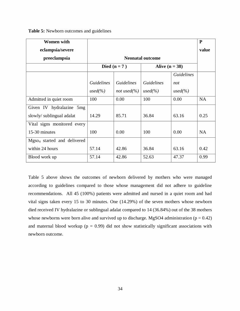
34
Table 5: Newborn outcomes and guidelines
Table 5 above shows the outcomes of newborn delivered by mothers who were managed
according to guidelines compared to those whose management did not adhere to guideline
recommendations. All 45 (100%) patients were admitted and nursed in a quiet room and had
vital signs taken every 15 to 30 minutes. One (14.29%) of the seven mothers whose newborn
died received IV hydralazine or sublingual adalat compared to 14 (36.84%) out of the 38 mothers
whose newborns were born alive and survived up to discharge. MgSO4 administration (p = 0.42)
and maternal blood workup (p = 0.99) did not show statistically significant associations with
newborn outcome.
Women with
eclampsia/severe
preeclampsia Neonatal outcome
P
value
Died (n = 7 ) Alive (n = 38)
Guidelines
used(%)
Guidelines
not used(%)
Guidelines
used(%)
Guidelines
not
used(%)
Admitted in quiet room 100 0.00 100 0.00 NA
Given IV hydralazine 5mg
slowly/ sublingual adalat 14.29 85.71 36.84 63.16 0.25
Vital signs monitored every
15-30 minutes 100 0.00 100 0.00 NA
Mgso4 started and delivered
within 24 hours 57.14 42.86 36.84 63.16 0.42
Blood work up 57.14 42.86 52.63 47.37 0.99

35
Management of eclampsia
Table 6 : Documented management of eclampsia
In general documentation of the management tasks for eclampsia was poor. Out of the five
patients with eclampsia none had documentation of support to maintain airway patency. Control
of blood pressure and convulsion was not documented in two of the five patients and details of
fluid maintenance were documented for two patients only. Treatment duration for eclampsia
ranged from 2 to 4 days. All the five deliveries were conducted through SVD resulting in 3 fetal
or neonatal deaths and one maternal death.
Task performed N = 5 Percent Guideline recommendations Airway maintained open 0 0 Maintain open air way
Fits controlled 3 60 Control fits
Blood pressure controlled and
monitored quarter hourly
3 60 Control blood pressure and monitor
quarter hourly
Fluid balance maintained
2 40 Start I.V line but restrict fluid intake
to avoid pulmonary and cerebral
edema
Treatment duration for eclampsia
Catheterise and closely monitor fluid
intake and urine output
2 days 2 40
3 days 2 40
4 days 1 20
Delivery management and
outcome
Normal SVD 5 100
Neonatal death/ still birth 3 60
Maternal outcome
Dead 1 20
Alive 4 80

36
Table 7: Health worker assessment of knowledge and treatment of preeclampsia/eclampsia
The recognition of headache as an alert sign for pre-eclampsia or eclampsia differed by type of
health worker training with clinicians being more likely to be aware of this sign compared to
nurses (p = 0.024). All seven (100%) doctors and eight (80%) clinical officers- were aware of
this sign compared to 50% of nurses.
Recognition of preeclampsia
and type of training
Doctors
(n = 7)
Clinical
officers (n=10)
Nurses
(n=48)
P value
n/N % n/N % n/N %
High blood pressure > 160/110 7/7 100.0 10/10 100.0 43/48 89.6 0.764
Generalized edema 6/7 85.7 8/10 80.0 38/48 79.2 1.000
Proteinuria in urine sample 6/7 85.7 10/10 100.0 35/48 72.9 0.116
Headache 7/7 100.0 7/10 70.0 24/48 50.0 0.024
Urinary frequency 0/7 0.0 2/10 20.0 6/48 12.5 0.684
Other 3/7 42.9 2/10 20.0 9/48 18.8 0.369
Organs at risk
Brain 7/7 100.0 9/10 90.0 30/48 62.5 0.051
Kidney 7/7 100.0 9/10 90.0 44/48 91.7 1.000
Stomach 0/7 0.0 1/10 10.0 3/48 6.25 0.713
Liver 6/7 85.7 8/10 80.0 14/48 29.2 <0.001
Blood 4/7 57.1 5/10 50.0 15/48 31.3 0.247
Other 0/7 0.0 1/10 10.0 0/48 0.0 0.262
Fetal assessment for possible
compromise
Weekly NST 3/7 42.9 5/10 50.0 21/48 43.8 0.92
Two weekly BPP 6/7 85.7 2/10 20.0 14/48 29.2 0.009
Amniocentesis 2/7 28.6 4/10 40.0 20/48 41.7 0.915
Urine dip stick analysis 2/7 28.6 4/10 40.0 15/48 31.3 0.906
Emergency C/S 0/7 0.0 5/10 50.0 10/48 20.8 0.044
Other 0/7 0.0 2/10 20.0 0/48 0.0 0.032

37
Recognition of the remaining signs of impeding preeclampsia or eclampsia including high blood
pressure (p = 0.764), edema (p = 1.000) and proteinuria (p = 0.116) did not show statistically
significant associations with type of training.
Recognition of risk to the remaining organs was not significantly associated with the type of
training of the health workers.
All the 67 (100%) health workers recognized that unborn fetuses of mothers with preeclampsia
or eclampsia were at risk. Only doctors (85.7%) were able to correctly identify two weekly BBP
as method of fetal surveillance (p = 0.009). Less than 50% of doctors correctly identified weekly
NST as a method of fetal surveillance compared to 50 % of clinical officers, and only 43.8 % of
the nurses, p = 0.92 (Table 7). On the other end, a few doctors, nurses and clinical officers
erroneously identified amniocentesis, urine dipstick analysis and emergency C/S as ways of fetal
surveillance.
Identification of signs for severe preeclampsia/eclampsia by health workers
The common physical presentations of eclampsia outlined in the MoH Guidelines were well
known by most of the health workers (Figure 3). At least 90% of health workers reported that
blood pressure measurements greater than 160/100 mmHg was a major criteria for alerting
caregivers about the possibility of pre-eclampsia/ eclampsia. Generalized edema and proteinuria
in urine were also well recognized signs of pre-eclampsia and eclampsia. However, headaches
were recognized as a sign of pre-eclampsia or eclampsia by only 59.7% of health workers.

38
Health worker recognition of alerts for severe pre-eclampsia at Garissa PGH
Figure 3: Health worker recognition of alerts for severe pre-eclampsia at Garissa PGH
In addition staff commonly mentioned other alerts of eclampsia or pre-eclampsia (Figure 3)
including convulsions or fits (n = 5), and blurred vision (n = 2).
Organ risk during eclampsia
Table 8: Body organs identified as at risk during eclampsia by health workers at Garissa PGH
Frequency (n = 67) Percent
Brain 47 70.15
Kidney 62 92.54
Stomach 4 5.97
Liver 30 44.78
Blood 25 37.31
Other 1 1.49
Only 11 health workers (16.4%) correctly identified all the three organs –brain, liver and kidney-
at risk during preeclampsia or eclampsia. The percentage of health workers correctly identifying
the individual body organs at risk during eclampsia or preeclampsia varied from 44.78% for
liver, to 70.15% for brain and 92.54% for kidneys (Table 8).
62 (92.5%)
54 (80.6%) 53 (79.1%)
40 (59.7%)
8 (11.9%)
15 (22.4%)
0
10
20
30
40
50
60
70
Blood pressure >
160/110mmhg
Generalized edema
Proteinuria in urine sample
Headache Urinary frequency
Other
Num
ber o
f hea
lth
wor
kers

39
A statistically significant association was noted between type of health worker training and the
recognition of at risk organs (p < 0.001) Nurses (29.2%) were less likely to recognize the risk
posed to the liver by preeclampsia or eclampsia compared to the doctors or clinical officers,
85.7% and 80% respectively (Table 8).
Table 9: Assessment of guideline use
Intervention for preeclampsia/eclampsia
Frequency
(n = 67) Percent
Guideline
Recommendations
Induced 53 79.1
Vaginal delivery.
C/S for standard
indications
All for emergency C/S 21 31.3
Catheterized for input-output monitoring 54 80.6
Closely monitor
fluid intake and
urine output
Blood for U/E/C, FHG and LFTS monitoring 42 62.7
Blood chemistry
alternate days
Given magnesium sulphate infusion 53 79.1
Start MgSO4
regime
Other 3 4.5
Benefits of guidelines
Reduces maternal morbidities and
mortalities 59 88.1
Improves neonatal outcome 43 64.2
Leads to unnecessary extra costs to the
woman and her relatives 17 25.4
Saves time 23 34.3
Enables early detection of complications and
therefore timely intervention 48 71.6
Other 1 1.5

40
The majority of health workers correctly understood the preferred management for women with
severe pre-eclampsia/eclampsia. Only 31.3% of the healthcare workers erroneously indicated ‘all
for emergency C/S’ as a preferred management option (Table 9).
Generally, majority of the healthcare workers were in agreement that the benefits of use of
guidelines in the management of severe pre-eclampsia/eclampsia far outweigh the perceived
disadvantages.
Pharmacologic management of eclampsia and preeclampsia
The majority of healthcare workers correctly identified methyl-dopa, nifedipine and magnesium
sulphate as drugs commonly used in managing severe pre-eclampsia/eclampsia (Figure 4).
Labetalol, a less frequently used drug for management of severe PET/eclampsia, was correctly
identified by 16.4 % of health workers.
Figure 4: Drugs identified for management of eclampsia and preeclampsia by health workers at
Garissa PGH
Guideline adherence
Based on health worker self report, guidelines were mostly used when managing eclampsia and
severe pre-eclampsia at Garissa PGH. Among the 67 participants, only one clinical officer and
five nurses reported that guidelines were not followed in the institution when managing
eclampsia or severe pre-eclampsia (p = 1.00).
51 (76.1%)
11 (16.4%)
2 (3.0%)
47 (70.2%)
63 (94.0%)
7 (10.5%)
0
10
20
30
40
50
60
70
Metyl-dopa Labetalol Zinc sulphate Nifedipine Magnesium sulphate
Other drugs

41
Availability of guidelines as perceived by health workers
All doctors and clinical officers were aware of the existence of standardized guidelines for
management of severe preeclampsia and eclampsia. Among the nurses, four (8.0%) nurses
reported that they were unaware about the availability of these guidelines. The level of guideline
availability awareness for the different health worker cadres is shown in figure 5 and reported
availability did not differ significantly when it was compared among doctors (100%), clinical
officers (100%) and nurses (91.7%), p-value = 0.997.
Figure 5: Health worker awareness of guideline availability in GPGH
7(100%)10(100%)
44(92.0%)
4(8.%)
0
10
20
30
40
50
60
Doctors Clinical officers Nurses
Num
ber o
f hea
lth
wor
kers
Unaware of guideline availability
Aware of guideline availability

42
Barriers to Utilization of MOH Guidelines for Management of Severe
preeclampsia/eclampsia at Garissa GPH
Figure 6: health workers’ reported barriers to utilization of MoH guidelines
Health workers reported the main impediments to utilization of MoH guidelines as: lack of
essential drugs (97.0%), lack of continuous knowledge appraisal (92.5%), lack of an experienced
sonographer (89.6%) lack of a supportive laboratory (77.6%). Other reported hindrances include
lack of supervision (56.7%), management based on individual experience (71.6%), lack of
ultrasound machine (22.4%) and unavailability of guidelines (17.9%).
0 10 20 30 40 50 60 70
Lack of supportive laboratory
Lack of ultrasound machine
Lack of experienced Sonographer
Lack of CME
Lack of essential drugs
Guidelines not available
Manage according to individual experience
Lack of MOH enforcement

43
Drug inventory record
Table 10: Drug inventory record
Drug Stock in Stock out/cleared Remarks
Magnesium sulphate -23/5/11-received 100
doses
-3/3/2012-received
150 doses
All doses cleared on
20/11/11
All doses cleared on
2/6/2012
November 20th, 2011
to march 2012-The
hospital was without
MgSO4
Methyl-Dopa -10/7/2011-received 2
cartons
-5/1/12-received 3
cartons
-6/4/2012-received 3
cartons
Cleared December
2011
Cleared April 2012
Stock fairly well
maintained
Nifedipine -15/9/11-received 2
cartons
-7/5/12-received 2
cartons
Cleared in April 2012 Stock fairly well
maintained
Hydralazine -13/5/2012-received 3
cartons
-2/4/12- received 3
cartons
Cleared February
2012
Hospital was without
this drug for 2 months
A drug inventory conducted in the hospital pharmacy section showed that essential drugs for
management of severe preeclampsia/eclampsia were not in a constant supply. For instance,
magnesium sulphate was stocked in low quantities and there was a long out of stock period
before new stock was brought in. similarly, inventory for nifedipine and hydralazine showed that
there was a considerable out of stock interval before new stock was replenished.
44
STUDY LIMITATIONS
We were only able to extract 45 files of women managed for severe preeclampsia/eclampsia out
of our sample size of 81 covering the study period. Medical records of bad outcomes may have
been kept away from us and therefore their data not reflected in this study and reducing our
sample size. A number of medical records were mild forms of preeclampsia which we did not
consider in our study. We did the study at the time when insecurity in Garissa was at its highest,
hence did not get adequate time to trace all the medical records of women seen with severe
preeclampsia/eclampsia thus further reducing our sample size.
Since there was no direct observation, some healthcare workers may have reported practices that
were different from what they actually do e.g. reporting adherence to guidelines yet data
extracted from medical records showed they were rarely, if ever used.

45
CHAPTER FIVE
DISCUSSIONS The objective of this study was to find out if the MOH approved guidelines for management of
severe preeclampsia/eclampsia existed in GPGH and if they were followed by healthcare
workers while managing women with these life threatening conditions. The study also sought to
establish whether healthcare workers had a general understanding of how women with these
conditions should be managed. The main findings of the study are that: - The impact of severe
preeclampsia was not fully understood by healthcare workers; Knowledge of effect of severe
preeclampsia varied by type of health worker training; Knowledge of guideline recommendation
varied by type of health worker training; Adherence to guideline was high and non adherence
was associated with increased adverse effects. Although knowledge of guidelines was high, some
aspects were lower for nurses and clinical officers. However, adherence to guidelines was poor
except for two aspects and this non-adherence was associated with poor pregnancy outcomes.
From our study, we established that most of the healthcare givers were nurses and clinical
officers. These cadres of health professionals are mostly inexperienced and could be contributing
to the high morbidity and mortality rate. This is in contrast to the Royal College of Obstetricians’
recommendation that women with severe preeclampsia/eclampsia be handled by obstetricians,
obstetric registrars and experienced midwives25. In a study by Ahmad Reda and his colleagues
titled ‘updating Nurses’ knowledge about preeclampsia patients’2008, the researchers concluded
that experience is an important source of knowledge but education is a neglected area that impact
on how nurses update their knowledge. They recommended that an ongoing education should be
adopted to improve the quality of nursing care for preeclampsia patients27.
All healthcare workers were aware of the availability of guidelines except 4(8.0%) nurses. Most
reported that guidelines were followed in management of women with severe
preeclampsia/eclampsia but data extracted from medical records did not show that these
guidelines were followed. Further, awareness of the preferred drug management for severe
preeclampsia/eclampsia was generally high but the actual practice did not always adhere to
guideline recommendations. This could mean that there is inadequate or no supervision,
healthcare giver practice based on their individual experience or there is a chronic shortage of the
essential drugs and equipment necessary to offer proper care. In a study by USAID and SPS

46
(strengthening pharmaceutical systems in Kenya), 2012, it was revealed that severe
preeclampsia/eclampsia is one of the leading maternal mortality in Kenya, contributing to about
19%. It established that information on the availability and management of
preeclampsia/eclampsia (especially MgSO4) is limited hence the high mortality due to these
conditions. The study also established that there were guideline inconsistencies in the
management of severe preeclampsia/eclampsia in majority of Kenyan health institutions both
public and faith-based. It also showed significant discrepancies between inventory records and
physical stock. For example, 78 facilities had inventory records of availability of MgSO4 but
only 36 facilities had records matching stock. In 65% of the time MgSO4 was unavailable in 84%
of the facilities observed. It also showed that MOH staff had limited or no knowledge of and
access of the essential guideline drugs26. In a study by Abdalah K Ziraba and colleagues, March
2009, titled ‘the state of emergency obstetric care services in Nairobi informal settlements and
environs: Results from a maternity health facility survey’ showed that many health facilities do
not meet a criteria for comprehensive emergency obstetric care, while the rest provided less than
basic care. Lack of obstetric skills, equipment and supplies were cited as hampering facilities
from providing lifesaving emergency obstetric care. They concluded that specific areas that
require attention include supervision, regulation of maternity facilities and ensuring that basic
equipment, supplies and trained personnel are available in order to handle obstetric
complications in both public and private facilities28. This is in agreement with our study findings
that Garissa PGH lacks basic equipment and has inconsistent essential drugs stocking.
In our study, we established that signs of preeclampsia such as elevated BP, and proteinuria were
well identified by majority of healthcare workers. Headache which is a sign of severe
preeclampsia/eclampsia was not well identified (40.7%). A small number of health workers, 5
(7.5%) and 2(3.0%), identified convulsions and blurred vision as signs. Doctors (100%) and
clinical officers (70%) were able to recognize signs of impending eclampsia as compared to
nurses (50%) p=0.024. only 11(16.4%) of health workers were able to identify all the body
organs at risk during severe preeclampsia/eclampsia. Type of training had positive correlation
with identification of organs at risk, more in doctors than other cadres, p<0.001. All health
workers reported that the fetus is at risk but most did not know how to assess for the risks. Only
85.7% of doctors had some idea on one or two assessment methods. Understanding of the
preferred management steps was high (60%), but these were not reflected in the medical records.

47
In cases where guideline recommended steps were followed, neonatal outcomes were good
compared to cases where these were not followed. These observations mean that majority of
Kenyan healthcare workers are ill equipped with the necessary knowledge and skills to combat
the syndrome of preeclampsia/eclampsia. In a study by Frank Kagema(2010) and colleagues
titled ‘quality of care for prevention and management of common maternal and newborn
complications: Findings from a national health facility survey in Kenya- Are services provided
according to international standards’, they found that the quality of much of the maternal and
newborn care provided was below the internationally accepted standards. Guidelines were
lacking for the provision of evidence-based practices. The study also showed that although most
of the health providers correctly diagnosed severe preeclampsia (83%) and knew appropriate
initial management (77%); only about 1/3 knew the correct treatment for convulsions and the
ongoing management steps29. In another study by Josephine Kibaru(2009) and colleagues titled
‘influence of provider training on quality of emergency obstetric care in Kenya’, they found out
that less than 20% of maternal health workers had received training in focused antenatal and
postnatal care and few (18%) had training on lifesaving skills30. So clearly, there is need for
health workers’ capacity building if women with the syndrome of preeclampsia/eclampsia are to
be given the treatment they deserve.
Severe preeclampsia/eclampsia poses a significant and fatal risk to the unborn fetus if it is not
well monitored and timely intervention administered. From the study findings, nearly all
healthcare workers admitted that the unborn child to a mother with severe
preeclampsia/eclampsia is at significant risk. However, doctors were able to identify the various
modes of surveillance and fetal assessment compared to few clinical officers and even fewer
nurses. In their study titled ‘Biophysical profile scores and resistance indices of the umbilical
artery as seen in patients with pregnancy-induced hypertension at Kenyatta National Hospital
and Marter Hospital, Nairobi24’,2006; Nguku SW, Wanyoike-Gichuhi and S Aywak AA
showed that RI of umbilical artery was positively related to the duration of illness. RI is an
earlier indicator of fetal compromise before any fetal distress becomes apparent. Thus, regular
obstetric ultrasound fetal surveillance in severe preeclampsia/eclampsia is important for fetal
well being.

48
CHAPTER SIX
CONCLUSION AND RECOMMENDATIONS 1. MOH guidelines for management of severe preeclampsia/eclampsia do exist in GPGH.
2. Majority of healthcare workers at GPGH were aware of the existence of MOH guidelines.
3. Doctors and some clinical officers, in some instances, were the health workers who
frequently referred to guidelines in management of women with severe
preeclampsia/eclampsia at GPGH.
4. The type of training (doctor, clinical officer or nurse) had a big impact in the assessment,
diagnosis and overall management of women with severe preeclampsia/eclampsia.
RECOMMENDATIONS 1. Given the low application of guidelines and the poor understanding of the recognizable
signs, risks and management of severe preeclampsia among nurses and these form the
bulky of health workers giving care to women with severe preeclampsia/eclampsia, there
is a need to consider continuing medical education such as seminars, grand rounds and
case discussions in the nursing group to shore up their skills. It is important that such
trainings should be extended to clinical officers and medical officers to improve further
on their professional understanding.
2. There is need for increased health sector funding by the government to address chronic
essential drug and equipment shortages.
3. The relevant authorities charged with ensuring that quality healthcare is offered should
intensify supervision to ensure the recommended management is practiced.
4. Further studies will need to be done in order to evaluate the use of guidelines
countrywide.
5. Rolling out of guidelines to all health facilities to improve standards and optimize care of
women with severe preeclampsia/eclampsia.

49
REFERENCES 1. Lowe S A, Brown M A, Dekker; Society of obstetric medicine of Australia and New
Zealand, 2008, Sydney: Guidelines for management of hypertensive disorders of
pregnancy. http://www.somanz.org/pdfs/somanz_guidelines_2008.pdf(accessed Dec.
2008).
2. Metin A., Souza J P, Mathews Mathai, Pena-Rosas J P, Olufemi T, Oladapo, et al.
Evidence-base (2011),WHO recommendations for treatment of pre-eclampsia and
eclampsia: Geneva, 2011.ISBN: 97892 4 1548335.
3. Hall D R, Odendaal H J and Smith M. The use of magnesium sulphate for treatment of
severe preeclampsia/eclampsia, Usmanu Danfodiyo University Teaching Hospital.
Annals of African medicine 2009: vol. 8 no.2; 76-80. Doi: 10.4103/1596-3519.56232.
PMID: 19805935.http://www.annalsafrmed.org.
4. Alderson P, Laura G, Sue L, Elaine C, Victoria T, Jayne R. NICE. Royal College of
Obstetricians and Gynecologists; The management of severe preeclampsia/eclampsia.
Viewed March 2011. http:/www.nice.org.uk.
5. Al-safi et al.delayed postpartum pre-eclampsia/eclampsia: demographics, clinical course
and complications. November 2011-volume18.issue5.p111 0-1107.
6. Drite Jo, magowan (Ed.). Clinical obstetrics & gynecology, chap.39 pg.367-370. ISBN
0-7020-1775-2.
7. WHO. World health report 2005: make every mother and child count. Geneva. Pg 63.
8. Christopher P. Crum. Robins and Cotran, pathologic basis of disease, 7th Ed: 1082-84.
The female genital tract: Philadelphia, W.B Saunders Company; 1999.
9. Hjartardottir S, Leifsson BG, Geirsson RT, steinthors dottir V (2004). Paternity change
and the recurrence risk in familial hypertensive disorder in pregnancy. Hypertension in
pregnancy 23(2):219-25.doi.10.1081/PRG-120037889. PMID 15364654.
10. Zhang J. Partner change, birth interval and risk of pre-eclampsia: a paradoxical triangle:
pediatric and perinatal epidemiology, 2007: 21(suppl 1): 31-5.doi.10.1111/j.1365-
3016.2007.00835x. PMID 17593195.
11. Munjuhili N, Lipman M, Valentine A, Hardiman P, Madean AB. Postpartum eclampsia
of late onset. bmj (Nov.2005) 331(7524):1070-1.doi.10.1136/bmj.331.7524.1070. PMC
1283194. PMID16269495.

50
12. Burne, Jeome (2006-01-30): give sperm a fighting chance. The times. Retrieved 2007-
11-16.(dead link)
13. Mottett A, Hiby SE. How does the maternal immune system contribute to the
development of pre-eclampsia? Placenta 28(suppl A),2007:S51-
6.doi.10.1016/j.placenta.2006.11.008 PMID 17292469.
14. BBC news 2000-01-20: Immune system causes miscarriage. Retrieved 2007-11-26.
15. Courtney Reynold, MD, William C. Mabie MD & Baha M. Sibai MD.pre-eclampsia.
Pregnancy-hypertensive disorders. Armenian medical network, 2006: Retrieved 2006-
11-23.
16. Jun Wu, Cizao Ren, Ralph J. deltino, Judith Chung, mitchelle Wilhelm, Beate Ritz.
Association between local traffic-generated air pollution and pre-eclampsia and preterm
delivery in the south coast air basin of California. Environmental health perspectives,
2009: Retrieved 20009-07-05. 17. Madline plus: Excess weight raises pregnancy risks, 2010. Retrieved 2010-02-15.(dead link)
18. The view from the cuckhold. Ep journal.20007. Retrieved 2011-07-20.
19. Van den Boogaard, Vissenberg E, land J.A, van wely, Van M, Der post et al, (2011).
Significance of (sub) clinical thyroid dysfunction and thyroid autoimmunity before
conception and in early pregnancy. A systematic review. Human reproduction updates
17(5):605-619.doi.10.1093/humupd/dmr024. PMID 21622978.edit.
20. Laresgoti-servitje t, Gomez-Lopez N, Olson DM. An immunological insight into the
origins of pre-eclampsia. Hum reprods update, 2010:
16(5):51024.doi.10.093/humupd/dmq007. PMID 20388637.
21. Maynard SE, Mingy, Merchan J, et al. Excess placental soluble trans-like tyrosine kinase
(sFlt-1) may contribute to endothelial dysfunction, hypertension and proteinuria in pre-
eclampsia. The journal of clinical investigation, 2003: 111(5). 649-
58.doi.10.1172/jc117189. PMC 151901. PMID 12618519.
22. Vankatesha S, Torposian M, Lamc, et al. Soluble endoglin contributes to the
pathogenesis of pre-eclampsia. Nature medicine, 2006: 12(6): 642-
9.doi.10.1038/nm1429. PMID 16751767.

51
23. Xiong Y, Liberman DA, Tront Js, et al. Gaddasa stress signaling regulates sFlt-1
expression in pre-eclampsia: journal of cellular physiology, 2009: 220 (3): 632-
9.doi.1002/jcp.21800. PMID 19452502.
24. Nguku SW, Wanyoike-Gichuhi, Aywak AA et al. Biophysical profile scores and
resistance indices of the umbilical artery as seen in patients with pregnancy-induced
hypertension: eamj, Mar 2006; 83(3): 96-101.
25. Royal College of Obstetricians and Gynecologists: setting standards to improve
women’s health; March 2006, Guideline No.10 (A).
26. The Royal Cornwall Hospital, NHS Trust 2012 (August): clinical guidelines for the
management of a woman with preeclampsia and/ or eclampsia.
27. Ahamad Reda, Adawy EL, Ola M. I, Mousa and Hala Abdel Fatah August 2008;-
updating Nurses’ knowledge about preeclamptic patient.
28. Abdalah K Ziraba, Samuel Mills, Nyovani Madise, Teresa Saliku and Jean-Christophe
Fotso, March 2009,“The state of emergency obstetric care services in Nairobi
informal settlements and environs: Results from a maternity health facility survey”.
29. Frank Kagema, Jim Ricca, Barbara Rawlins, Heather Rosen, Pamela Hynam and Nancy
Kidala:- 2010: quality of care for prevention and management of common maternal and
newborn complications: Findings from a national Health facility survey in Kenya- Are
services provided according to international standards?
30. Joyce Olenja, Pamela Godia, Josephine Kibaru and Thaddaeus Egondi; January 2009,
“influence of provider training on quality of emergency obstetric care in Kenya”.
31. www.msh.org/SPS, 2012- strengthening pharmaceutical systems in Kenya.
32. T. Dennis; management of preeclampsia: issues for anesthetists, bmj; 26 JUN 2012.
Doi:10.1111/j.1365-2044.2012.07195.
33. Atallah A. N, Hofmeyr GJ, Amley L,: the Cochrane database of systematic reviews
(2002)(1):CD001059. Pubmed central. Calcium supplementation in pregnancy for
preventing hypertensive disorders and related problems.(PMID:1869587).
Doi:10.1002/14651858.CD001059.
34. Joey P; Granger, PhD, Department of physiology and Biophysics, University of
Mississippi Medical center, 2500 N state st. Jackson, Ms 39216-4505. E-mail.
jgranger@physiology. umsmed.edu.

52
APPENDICES
APPENDIX 1: INFORMED CONSENT I am Doctor John Omboga, a postgraduate student registered for masters in medicine-M.med
obstetrics and gynecology in the University of Nairobi. I am carrying out a study as part of the
requirement for M.med qualification. My objectives are to determine the availability of National
guidelines and their application in the management of severe pre-eclampsia/eclampsia in GPGH.
Study approval has been given by the Kenyatta National Hospital/University Of Nairobi ethics
committee {KNH/UON-ERC}. The committee can be contacted through;
Prof. A. N. Guantai,
Secretary, KNH/UON-ERC,
P.o box 20723,
Nairobi.
E-mail:[email protected]
My supervisors, both of whom are based at the department of obstetrics and gynecology at the
University of Nairobi are:
1. Prof. James N. Kiarie
2. Dr. Weston khisa
I am requesting your participation in this study as a health worker working at GPGH. I would
like to bring to your attention the following ethical considerations which will guide your
participation.
1. Participation in this study is purely voluntary.
2. You may withdraw from the study at any time and there are no consequences for your
decision to withdraw.
3. After you read through the explanations, please feel free to ask any questions that will
allow you to understand the nature of the study.

53
4. Any information you provide including details on your demographic characteristics will
be treated as confidential.
5. The study protocol has been reviewed by the ethics committee. The protocol can be
accessible to you should you choose to know the details.
I will be available to answer any questions that will help you understand the nature of the study.
If you wish to seek any clarification, kindly contact me on 0720258887.
PROCEDURE: A questionnaire will be provided. It should take approximately 10-15 minutes
to complete. I will be available to guide you through the questions.
BENEFITS: There are no direct personal benefits for participating in this study.
It is expected that study findings will help improve the management of women with severe pre-
eclampsia/eclampsia in GPGH.

54
APPENDIX 2: CONSENT FORM I, the undersigned, do hereby consent to participate in this study whose nature, purpose and
objectives have been fully explained to me. I am aware that participation is voluntary and that
there are no consequences to withdrawal from the study. I have been informed that all data
provided will be used for the purposes of study only.
Signed…………………………………………………date………………………………………..
I,…………………………………………………………………………..declare that I have adequately
explained to participant the purpose of the of the study, procedures, risks and benefits. I have
given the participant time to ask questions and seek clarification regarding the study.
Signed………………………………………………date…………………………………………..

55
APPENDIX 3: QUESTIONNAIRE
For healthcare workers Demographic data.
Indicate your gender…………………………………………………..M/F
Indicate your age………………………………………………………….
Indicate your type of training……………………………………..consultant/medical
officer/clinical officer/nursing officer
For each question, there are numbered possible responses. Please indicate T for true or F for
false against each of the responses. A space is provided for your other possible response not
covered in the numbered options.
1. What are the main physical findings that alert you about the possibility of developing
severe pre-eclampsia/eclampsia?
a) High blood pressure above 160/110mmhg
b) Generalized edema
c) Proteinuria in urine sample
d) Headache
e) Urinary frequency
f) Other……………………………………………………………………………….
2. Which organ systems are mainly affected by severe pre-eclampsia/eclampsia?
a) Brain
b) Kidney
c) Stomach
d) Liver
e) Blood
f) Other………………………………………………………... …………………………
3. In your opinion, is the unborn fetus to a mother suffering from severe pre-
eclampsia/eclampsia at any risk?
a) Yes
b) No

56
c) Other…………………………………………………………………………………
4. If your response to question 3 is yes, how can the fetus be assessed for any possible signs
of compromise?
a) Weekly NST
b) Two weekly BPP
c) Amniocentesis
d) Urine dipstick analysis
e) Emergency C/S
f) Other…………………………………………………………………………………
5. Are you aware of the existence of standardized guidelines for management severe pre-
eclampsia/eclampsia?
a) Yes
b) No
c) Other……………………………………………………………………………………
6. In your institution, are these guidelines followed when managing severe pre-
eclampsia/eclampsia? If not, which alternative is used?
a) Yes
b) No
Other…………………………………………………………………………………..
7. If your answer to question 5 is yes, list the drugs that are essential for management of
severe pre-eclampsia/eclampsia.
a) Methyldopa
b) Labetalol
c) Zinc sulphate
d) Nifedipine
e) Magnesium sulphate
f) Other………………………………………………………………………………….
8. In your opinion, what are the advantages and disadvantages of strict use of guidelines in
management of severe pre-eclampsia/eclampsia?
a) Reduces maternal morbidities and mortalities
b) Improves neonatal outcome

57
c) Leads to unnecessary extra costs to the woman and her relatives
d) Saves time
e) Enables early detection of complications and therefore timely intervention
f) Other………………………………………………………………………………….
9. In your institution, how are pregnant women with severe pre-eclampsia/eclampsia
managed?
a) Induced
b) All for emergency C/S
c) Catheterized for input-output monitoring
d) Blood for U/E/C, FHG and LFTS monitoring
e) Given magnesium sulphate infusion
f) Other……………………………………………………………………………………
10. Would you recommend the use of guidelines for management of severe pre-
eclampsia/eclampsia?
a) Yes
b) No
c) Other…………………………………………………………………
11. In your opinion, what are the factors that hinder optimal utilization of MOH guidelines
for management of severe preeclampsia/eclampsia in Garisa PGH?
………………………………………………………………………………………………
………………………………………………………………………………………………
………………………………………………………………………………………………
………………………………………………………………………………………………
………………………………………………………………………………………………
………………………………………………………………………………………………
………………………………………………………………………………………………
………………………………………………………………………………………………

58
APPENDIX 4: DATA ABSTRACTION TOOL FOR MANAGEMENT OF SEVERE PRE-ECLAMPSIA/ECLAMPSIA
Hospital no……………………………..serial no…………………………………….
Mild pre-eclampsia…BP 140/90mmhg
a) Admitted
Not admitted
b) Oral aldomet 250mg given to maintain diastolic BP at 90-100mmhg…………
Not given………………………………………………………………………
c) Maternal and fetal condition monitored weekly……………………………….
Not monitored………………………………………………………………….
d) Advised to take diet rich in proteins, fiber and vitamins and low in
carbohydrate and salt…………………………………………………………..
Not advised…………………………………………………………………….
e) Duration of treatment……………………………………………………….
f) Complications…………………………………………………………………
…………………………………………………………………………………
g) Mode of delivery……………………………………………………………….
h) Pregnancy outcome……………………………………………………………
i) Condition of
mother…………………………………………..baby…………………………
j) Apgar score at 5 minutes………………………………………………………
Severe pre-eclampsia……Diastolic BP >110mmhg
a) Admitted in quiet room…………………………………………….
Not admitted……………………………………………………….
b) Given IV hydralazine 5mg slowly or sublingual adalat…………………….
Not given………………………………………………………………………
c) Vital signs monitored every 15-30 minutes…………………………………...
Not monitored…………………………………………………………………
d) Mgso4 started and delivered within 24 hours…………………………………
Not started…………………………………………………………………….
e) Bloodworkup...U/E/C, LFTS, FHG done…………......................................

59
Not done………………………………………………………………………
f) Duration of treatment………………………………………………………….
g) Complications…………………………………………………………………
h) Mode of delivery………………………………………………………………
i) Pregnancy outcome……………………………………………………………
j) Condition of mother……………………………..baby……………………
k) Apgar score at 5 minutes………………………………………………….
Management of eclampsia
a) Airway maintained open…………………………………………………..
Not maintained……………………………………………………………..
b) Fits controlled………………………………………………………………
Not controlled…………………………………………………………………
c) Blood pressure controlled and monitored quarter hourly………………………
Not controlled and not monitored………………………………………………
d) Fluid balance maintained……………………………………………………….
Not maintained………………………………………………………………….
e) Duration of treatment…………………………………………………………...
f) Complications…………………………………………………………………..
g) Mode of delivery……………………………………………………………….
h) Pregnancy outcome…………………………………………………………….
i) Condition of mother……………………………baby………………………….
j) Apgar score at 5 minutes…………………………

60
APPENDIX 5: REPUBLIC OF KENYA, MINISTRY OF HEALTH
NATIONAL GUIDELINES FOR QUALITY OBSTETRICS AND PERINATAL CARE For Health Service Providers in Kenya FOURTH EDITION………November 2012.
PRE-ECLAMPSIA AND ECLAMPSIA
Diagnosis of preeclampsia/eclampsia
History- many cases are detected through routine prenatal screening.
CNS
Headache
Visual disturbances- blurred vision, scintillating scotomata
Altered mental status
Cortical or retinal blindness
Dyspnoea
Edema-this exists in many pregnant women but sudden increase in edema or facial edema
is more concerning for preeclampsia
Epigastric or right upper quadrant (RUQ) abdominal pain: hepatic involvement occurs in
10% of women with severe preeclampsia
Weakness or malaise
Physical examination
Findings on physical examination may include the following:
Increased BP compared with the patient’s baseline or > 140/90 mmHg
Altered mental status
Decreased vision or scotomas
Papilloedema
Epigastric or RUQ abdominal pain
Sudden increase in edema or facial edema

61
Hyperreflexia or clonus: although deep tendon reflexes are more useful in assessing
magnesium toxicity, the presence of clonus may indicate an increased risk of convulsions
seizures
Focal neurologic deficit
Investigations:
Laboratory studies
CBC count and peripheral smear
Microangiopathic hemolytic anemia
Thrombocytopenia <100,000
Hemoconcetration may occur in severe preeclampsia
Schistocytes on peripheral smear
Liver function tests: transaminase levels are elevated from hepatocellular injury and in
HELLP syndrome
Serum creatinine levels elevated due to decreased intravascular volume and decreased
glomerular filtrate rate (GFR)
Urinalysis: more than 300mg or +1 proteinuria in a 24 hour urine sample
Abnormal coagulation profile: PT and aPTT are elevated
Disseminated intravascular coagulopathy testing will show fibrin split products and
decreased fibrinogen levels
Hyperuricemia
Ultrasonography:
This is used to assess the status of the fetus as well as to evaluate for growth restriction (typically
asymmetrical IUGR). Aside from transabdominal ultrasonography, umbilical artery Doppler
ultrasonography should be performed to assess blood flow

62
Management of patients with preeclampsia/eclampsia
Control BP
Goal is to prevent cerebrovascular and cardiac complications while maintaining
uteroplacental blood flow
Control of mildly elevated BP does not appear to improve perinatal morbidity and
mortality and , in fact, it may reduce birth weight
Antihypertensive treatment is indicated for BP >160/105 mmHg. Goal is to maintain
diastolic BP 90-100mmHg and systolic BP 140-155 mmHg
First-line medications are labetalol given orally or I.V, nifedipine given orally or I.V, or
hydralazine I.V. (Atenolol, ACE inhibitors, ARBs, and diuretics should be avoided)
Control of seizures
Follow basic principle airway, breathing and circulation (ABC)
Treat active seizure with intravenous magnesium sulphate as a first-line agent
Prophylactic treatment with magnesium sulphate is indicated for all patients with severe
preeclampsia
Magnesium levels, respiratory rate, reflexes and urine output must be monitored to detect
magnesium toxicity. Magnesium sulphate is mostly excreted in urine and therefore urine
output needs to be closely monitored. If urine output falls below 20mls/hr, the
magnesium infusion should be stopped
For seizure refractory to magnesium sulphate, benzodiazepines and /or phenytoin should
be considered
Fluid management
These patients are intravascularly volume depleted with high peripheral vascular
resistance. Diuretics should be avoided
Aggressive volume resuscitation may lead to pulmonary edema, which occurs mostly 48-
72 hours postpartum, probably due to mobilization of extravascular fluid
Total fluids should be limited to 80mls/hr or 1ml/kg/hr

63
Careful measurement of fluid input and output especially in the immediate postpartum
period is advisable
If fluids are required, preferably use Ringers Lactate or Normal saline
Delivery
Patients with mild preeclampsia are induced after 37 weeks gestation. Give steroids prior
to this
Induction of labor should be considered in patients with severe preeclampsia after 34
weeks gestation
Eclampsia is common after delivery up to 6 weeks
Medication
Magnesium sulphate:
Antagonizes calcium channels of smooth muscle. Administer IV/IM for seizure prophylaxis in
preeclampsia. Use IV for quicker onset of action in eclampsia
Schedule
Loading
20% solution or 4g IV over 5 minutes
Follow promptly with 10g of 50% solution, 5g in each buttock as deep IM injection with 1ml of
2% lignocaine in the same syringe
If convulsions occur after 15 minutes, give 2g magnesium sulphate (50% solution) IV over 5
minutes
Maintenance dose
Give magnesium sulphate (50%) solution + 1ml lignocaine 2% IM every 4 hours into alternate
buttocks. Continue treatment for 24 hrs after delivery or the last convulsion, whichever occurs
last. If 50% solution is not available, give 1 g of 20% solution IV every hour by continuous
infusion.
64
Before repeat administration, ensure that:
Respiratory rate is at least 16 per minute
Patellar reflexes are present
Urinary output is at least 30mls/hr over preceding 4 hours
If the above are absent, withhold or delay drug
Keep antidote ready:
In case of respiratory arrest, assist ventilation with mask and bag, intubate. Give 1g (10mls) of
calcium gluconate IV slowly until respiration begins
Phenytoin
Cardiac monitoring required because of associated bradycardia and hypotension. It is given as
10mg/kg loading dose infused IV no faster than 50mg/min, followed by a maintenance dose
started 2 hrs later at 5mg/kg
In absence of magnesium sulphate, diazepam is used following the regime below:
Intravenous
Loading
20mg IV slowly over 2 minutes
If convulsions occur, repeat loading dose
Maintenance dose
Diazepam 40mg in 500ml IV fluids titrated to keep the woman sedated but can be aroused
Do not give more than 100mg in 24 hours

65
Rectal administration
Give diazepam rectally when IV access is not possible. The loading dose is 20mg in 10 mls
syringe. If convulsions are not controlled within 10 minutes, administer an additional 10mg per
hour or more, depending on the size of the woman and her clinical response
Hydralazine (Apresoline)
First line therapy against preeclamptic hypertension. It is given 5mg IV slowly over 10minutes.
Repeat 5mg q20min to a maximum of 20mg
Labetalol
Recommended 2nd line therapy. It’s onset of action is more rapid than hydralazine. It’s given at
20mg bolus; subsequently give doses of 40mg followed by 80mg IV at 10-20 min intervals to
achieve BP control to a maximum of 300mg. it may also be administered by continuous infusion
at 1mg/kg/hr
Nifedipine
Given at 10mg orally. May be repeated after 30minutes as needed
Management of patients with pre-eclampsia
Mild pre-eclampsia…BP 140/90mmhg
Establish if mother can rest at home
Advice patient and relatives on importance of bed rest
Give oral antihypertensive (aldomet 250mg tds) to maintain diastolic BP at 90-100mmhg
Monitor maternal and fetal condition weekly
Admit if coming too far away from hospital-advice on the worsening signs of the
conditions and should report if any be present
Advice mother to take diet rich in proteins, fiber, vitamins and low in carbohydrate and
salt
If no improvement, refer to hospital if at health center

66
Severe pre-eclampsia….diastolic BP>110mmhg
Admit in labor ward in a quiet room
Administer parenteral antihypertensive (hydralazine 5mg iv slowly) or sublingual
nifedipine
Monitor vital signs every 15-30 minutes
Consider timing and mode of delivery
Start MgSO4 regime and deliver within 24 hours
Fluid regime management (input/output)
Do blood chemistry-LFTS, U/E/C, FBC
If at health center, refer to a comprehensive centre accompanied by a trained nurse
Management of eclampsia
Maintain open airway
Control fits
Control BP and monitor quarter hourly
Maintain fluid balance
Management of the fitting patient
Patient should be put in semi prone position so that mucus and saliva can flow out
Tight fitting dresses around the neck should be loosened or removed
Clean mouth and nostrils gently and remove secretions
No attempt should be made to insert any instrument into the mouth
Give oxygen if available continuously during fit and for 5 minutes after each fit
Fitting should be allowed to complete its course without physically attempting to hold the
patient down
Privacy and dignity of patient must be observed –pull screens around her
Administer magnesium sulphate as per regime to control fits

67
APPENDIX 6: DRUG INVENTORY RECORD
Drug Stock in/replenished Stock out/cleared Remarks
Magnesium Sulphate
Methyl-Dopa
Nifedipine
Hydralazine