use and misuse of endoscopy in ulcerative colitis gary r lichtenstein, md director, center for ibd...
TRANSCRIPT
Use and Misuse of Endoscopy in Ulcerative Colitis
Gary R Lichtenstein, MD
Director, Center for IBD
University of Pennsylvania School of Medicine
Hospital of the University of PA
Philadelphia, PA

Gary Lichtenstein, MDDisclosures
Research, Advisory, and/or Honorarium
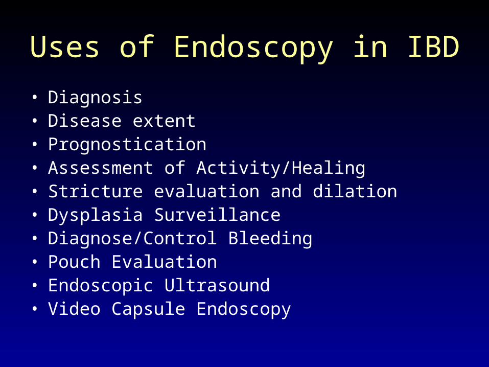
Uses of Endoscopy in IBD
• Diagnosis• Disease extent• Prognostication• Assessment of Activity/Healing• Stricture evaluation and dilation• Dysplasia Surveillance• Diagnose/Control Bleeding• Pouch Evaluation• Endoscopic Ultrasound• Video Capsule Endoscopy
I. Diagnosis
• The Gold Standard for Diagnosis of UC (also Crohn’s colitis and ileocolitis)
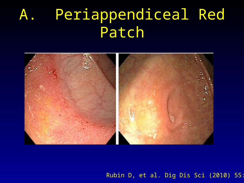
A. Periappendiceal Red Patch
Rubin D, et al. Dig Dis Sci (2010) 55:3495–3501

• PARP is PARP is NOTNOT Crohn’s Disease Crohn’s Disease
• Present in 29/367 (7.9%) patientsPresent in 29/367 (7.9%) patients
•
•
A. Periappendiceal Red Patch
Rubin D, et al. Dig Dis Sci (2010) 55:3495–3501PARP- Periappendiceal Red Patch
I. Diagnosis
• Mimickers of Crohn’s ileitis and colitis
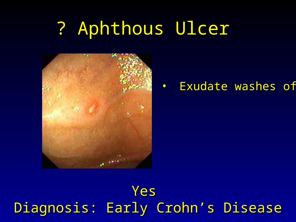
? Aphthous Ulcer ? Aphthous Ulcer
Yes Yes Diagnosis: Early Crohn’s DiseaseDiagnosis: Early Crohn’s Disease
• Exudate washes offExudate washes off
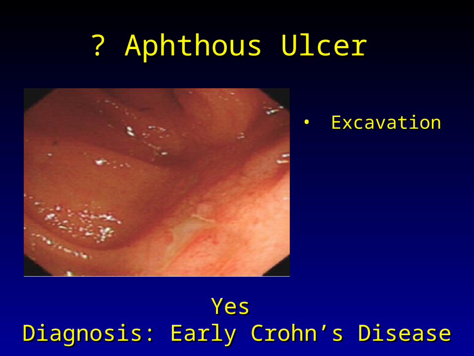
? Aphthous Ulcer ? Aphthous Ulcer
Yes Yes Diagnosis: Early Crohn’s DiseaseDiagnosis: Early Crohn’s Disease
• ExcavationExcavation
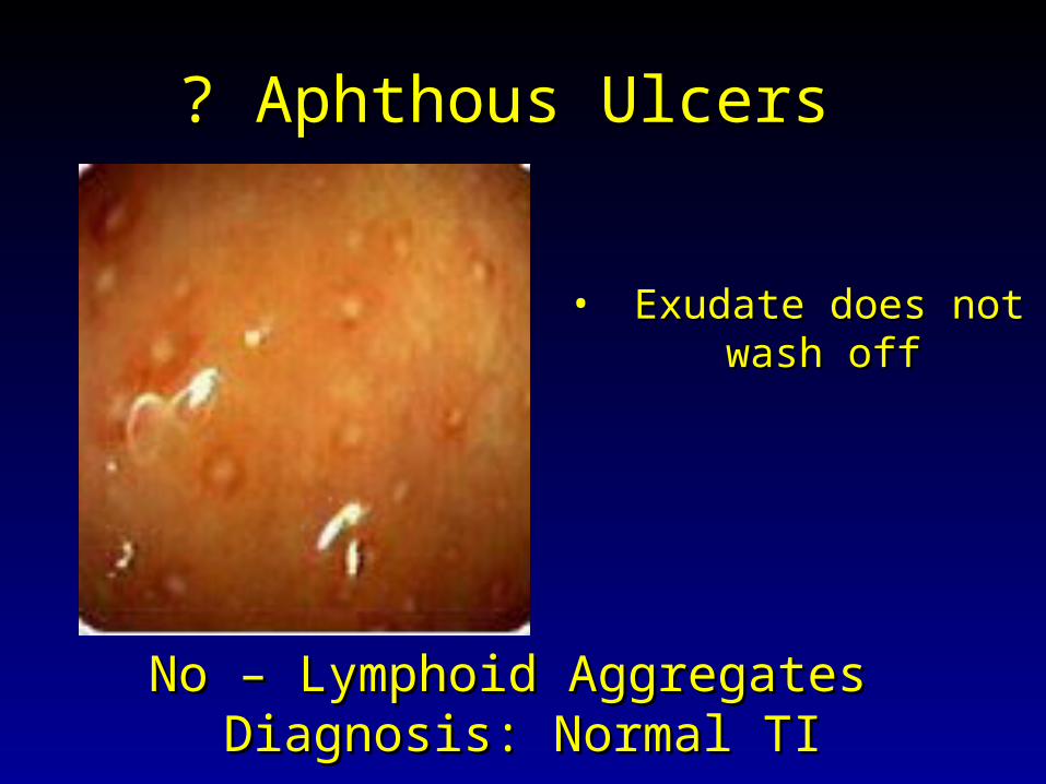
? Aphthous Ulcers ? Aphthous Ulcers
No – Lymphoid Aggregates No – Lymphoid Aggregates Diagnosis: Normal TIDiagnosis: Normal TI
• Exudate does not Exudate does not wash offwash off
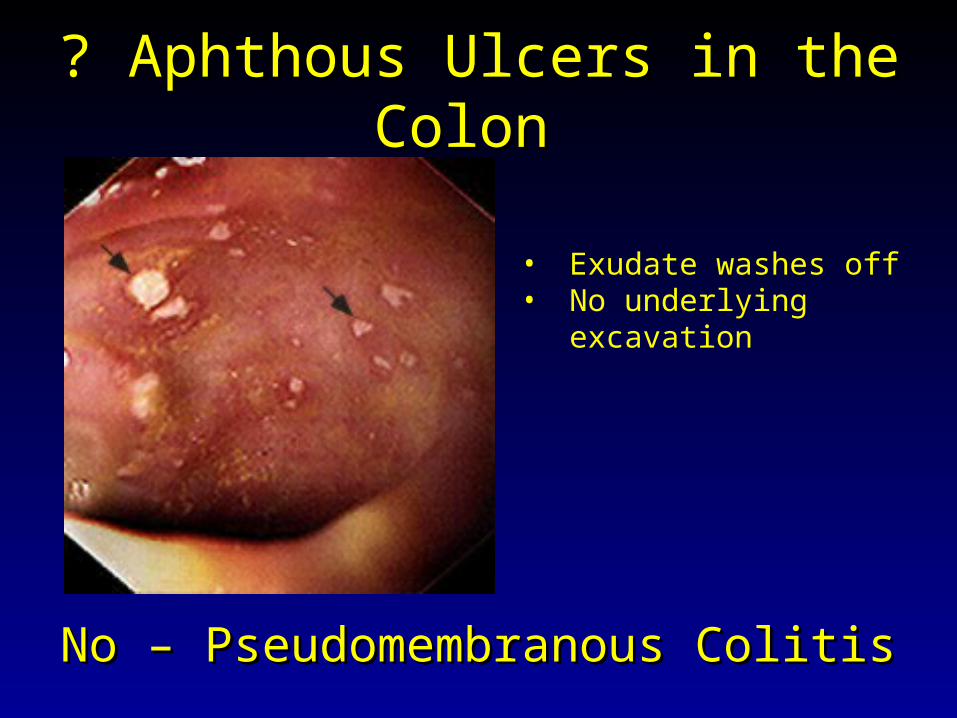
? Aphthous Ulcers in the Colon ? Aphthous Ulcers in the Colon
No – Pseudomembranous ColitisNo – Pseudomembranous Colitis
• Exudate washes off• No underlying
excavation
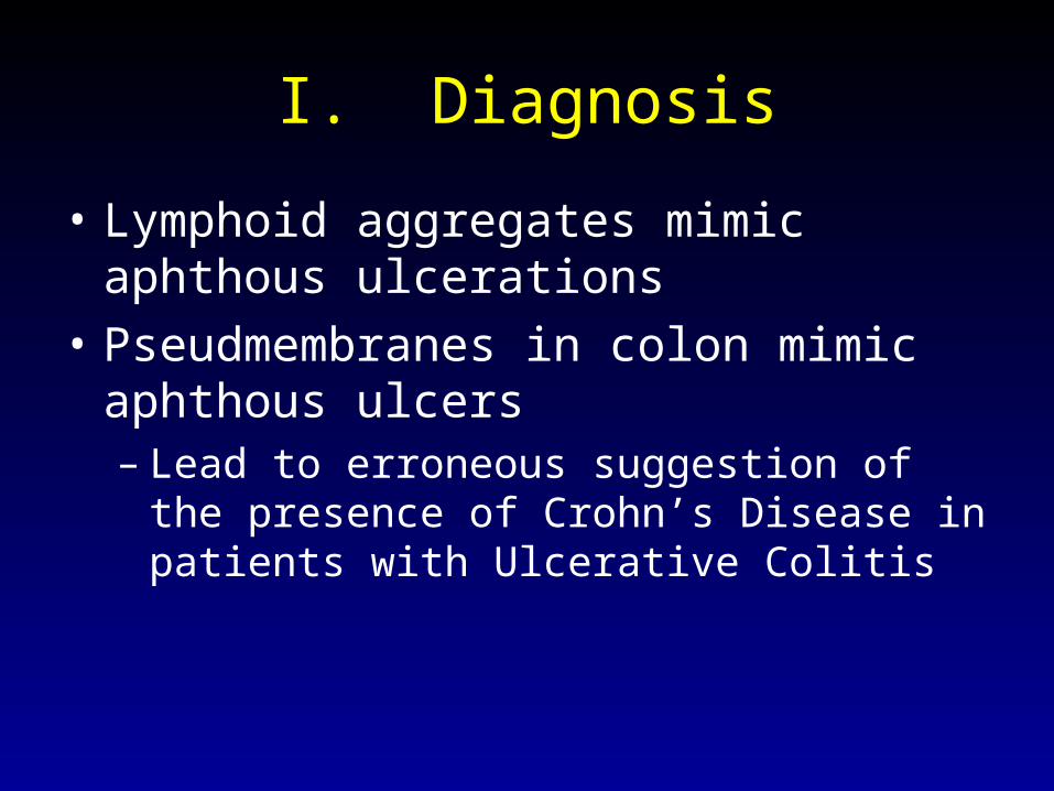
I. Diagnosis
• Lymphoid aggregates mimic aphthous ulcerations
• Pseudmembranes in colon mimic aphthous ulcers– Lead to erroneous suggestion of the presence
of Crohn’s Disease in patients with Ulcerative Colitis
II. Need to BiopsyII. Need to Biopsy
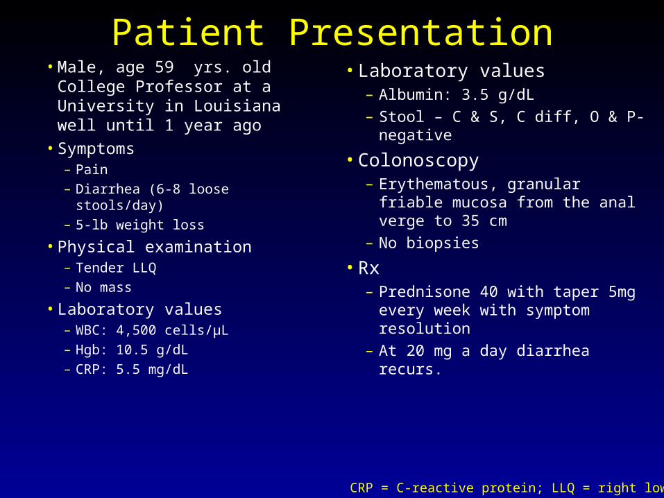
Patient Presentation• Male, age 59 yrs. old College
Professor at a University in Louisianawell until 1 year ago
• Symptoms– Pain– Diarrhea (6-8 loose stools/day)– 5-lb weight loss
• Physical examination– Tender LLQ– No mass
• Laboratory values– WBC: 4,500 cells/µL– Hgb: 10.5 g/dL– CRP: 5.5 mg/dL
• Laboratory values– Albumin: 3.5 g/dL– Stool – C & S, C diff, O & P-
negative
• Colonoscopy– Erythematous, granular friable
mucosa from the anal verge to 35 cm
– No biopsies
• Rx– Prednisone 40 with taper 5mg
every week with symptom resolution
– At 20 mg a day diarrhea recurs.
CRP = C-reactive protein; LLQ = right lower quadrant.
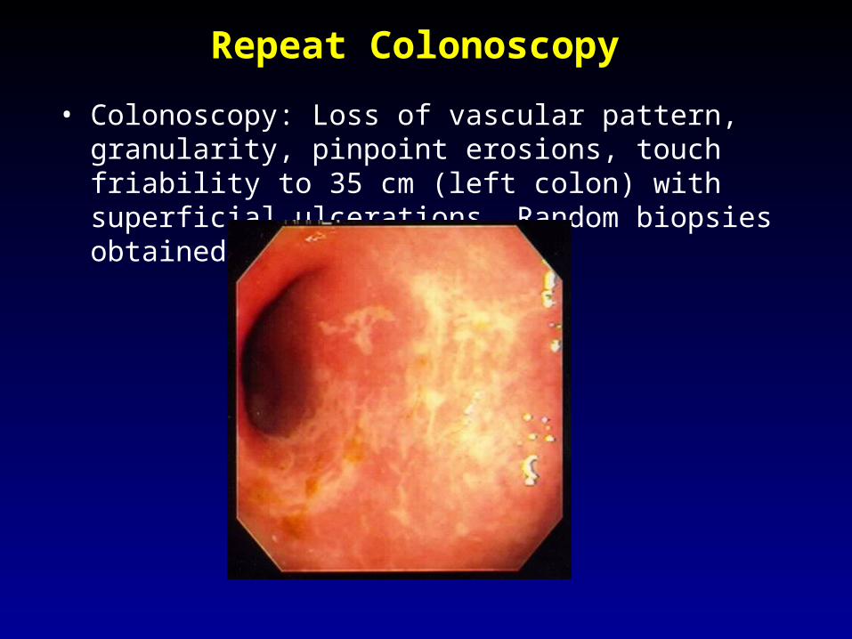
Repeat Colonoscopy
• Colonoscopy: Loss of vascular pattern, granularity, pinpoint erosions, touch friability to 35 cm (left colon) with superficial ulcerations. Random biopsies obtained.
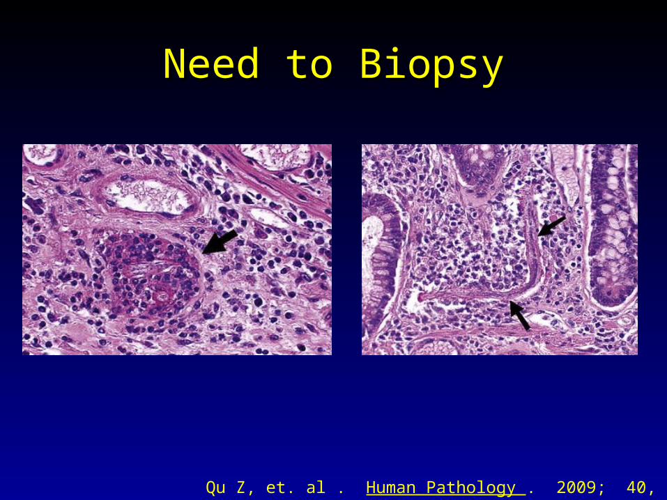
Need to BiopsyNeed to Biopsy
Qu Z, et. al . Human Pathology . 2009; 40, 572–577
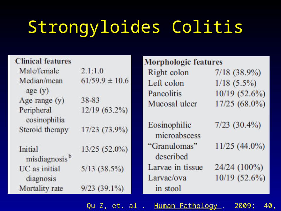
Strongyloides Colitis Strongyloides Colitis
Endemic areas : - Appalachian region and Louisiana in the United States- Regions with large influx of tourists and emigrants from these endemic areas, southeastern Asia, and southern, eastern, and central Europe also have high incidence and prevalence of the disease .
Who Gets Disease: - The infection may remain clinically indolent.- When the host is immune-compromised, hyperinfection syndrome (i.e., larvae overload in
the lung and involvement of the rest of the gastrointestinal system) and
disseminated strongyloidiasis (i.e., involvement of other organs) occur with a mortality rate near 90% Qu Z, et. al . Human Pathology . 2009; 40, 572–577
Strongyloides Colitis Strongyloides Colitis
Qu Z, et. al . Human Pathology . 2009; 40, 572–577

Infectious Colitis that Mimics UC Infectious Colitis that Mimics UC
Rameshshanker R., et. al . World J Gastrointest Endosc 2012 June 16; 4(6): 201-211
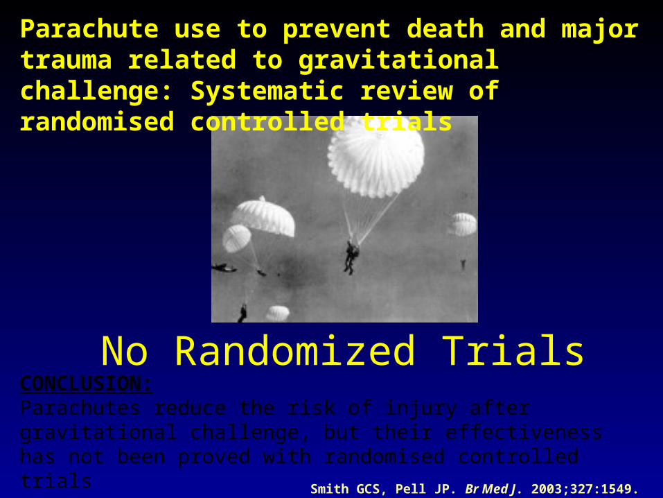
No Randomized Trials
Smith GCS, Pell JP. Br Med J. 2003;327:1549.
Parachute use to prevent death and major trauma related to gravitational challenge: Systematic review of randomised controlled trials
CONCLUSION:Parachutes reduce the risk of injury after gravitational challenge, but their effectiveness has not been proved with randomised controlled trials
III. Disease Extent
• Assess extent of disease– when having active symptoms
• Mucosa may have complete endoscopic healing or remission with when in remission.
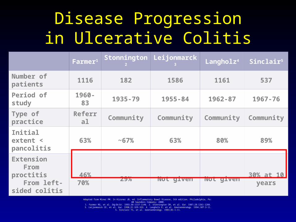
Disease Progressionin Ulcerative Colitis
Adapted from Miner PM. In Kirsner JB, ed. Inflammatory Bowel Disease, 5th edition. Philadelphia, Pa: WB Saunders Company; 2000.
1. Farmer RG, et al. Dig Dis Sci. 1993;38:1137-1146. 2. Stonnington CM, et al. Gut. 1987;28:1261-1266.3. Leijonmarck CE, et al. Gut. 1990;31:329-333. 4. Langholz E, et al. Gastroenterology. 1994;107:3-11.
5. Sinclair TS, et al. Gastroenterology. 1983;85:1-11.
Farmer1 Stonnington2 Leijonmarck3 Langholz4 Sinclair5
Number of patients
1116 182 1586 1161 537
Period of study 1960-83 1935-79 1955-84 1962-87 1967-76
Type of practice Referral Community Community Community Community
Initial extent < pancolitis
63% ~67% 63% 80% 89%
Extension From proctitis From left-sided colitis
46%70%
29% Not given Not given30% at 10
years
IV. Prognostication
• Assess histology to predict future probability of flare
• Mucosal healing predicts– lower colectomy rate– less steroid use in the future
• Treat individuals with potential for aggressive behavior with appropriately aggressive therapy.
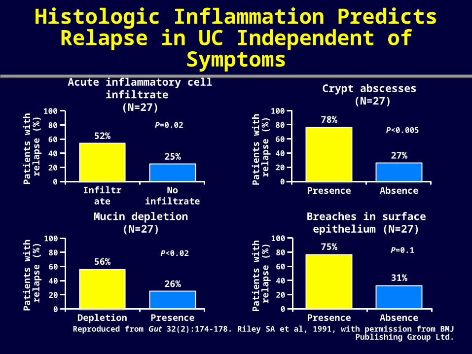
Histologic Inflammation Predicts Relapse in UC Independent of Symptoms
Reproduced from Gut 32(2):174-178. Riley SA et al, 1991, with permission from BMJ Publishing Group Ltd.
Acute inflammatory cell infiltrate (N=27)
Crypt abscesses (N=27)
Mucin depletion (N=27)
Breaches in surface epithelium (N=27)
52%
25%
0
100
Infiltrate No infiltrate
56%
26%
Depletion Presence
78%
27%
Presence Absence
75%
31%
Presence Absence
Pat
ien
ts w
ith
re
lap
se
(%)
Pat
ien
ts w
ith
re
lap
se
(%)
P<0.02
P=0.02
P=0.1
P<0.005
Pat
ien
ts w
ith
re
lap
se
(%)
Pat
ien
ts w
ith
re
lap
se
(%)
80
60
40
20
0
100
80
60
40
20
0
100
80
60
40
20
0
100
80
60
40
20
Reprinted from Gastroenterology 120, Bitton A et al, Clinical, biological, and histologic parameters as predictors of relapse in ulcerative colitis, 13-20. Copyright
(2001), with permission from the American Gastroenterological Association.
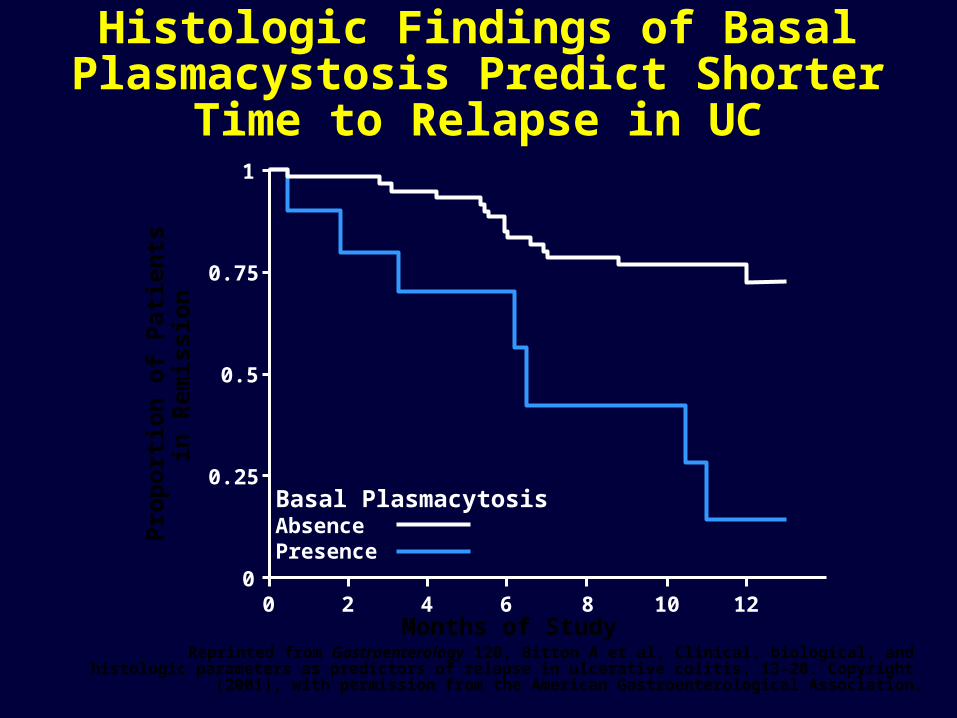
Histologic Findings of Basal Plasmacystosis Predict Shorter Time to
Relapse in UCP
rop
ort
ion
of
Pat
ien
ts
in R
emis
sio
n
0
0.25
0.5
0.75
1
0 2 4 6 8 10 12Months of Study
Basal PlasmacytosisAbsencePresence
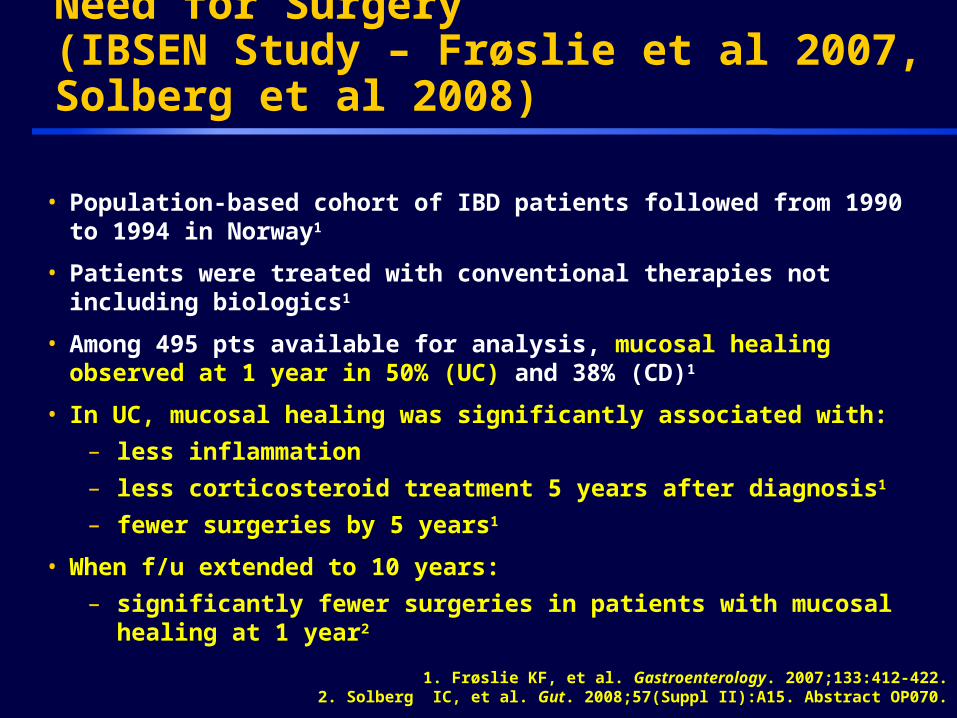
• Population-based cohort of IBD patients followed from 1990 to 1994 in Norway1
• Patients were treated with conventional therapies not including biologics1
• Among 495 pts available for analysis, mucosal healing observed at 1 year in 50% (UC) and 38% (CD)1
• In UC, mucosal healing was significantly associated with:
– less inflammation
– less corticosteroid treatment 5 years after diagnosis1
– fewer surgeries by 5 years1
• When f/u extended to 10 years:
– significantly fewer surgeries in patients with mucosal healing at 1 year2
Mucosal Healing Can Impact the Need for Surgery(IBSEN Study – Frøslie et al 2007, Solberg et al 2008)
25RGU11005
1. Frøslie KF, et al. Gastroenterology. 2007;133:412-422.2. Solberg IC, et al. Gut. 2008;57(Suppl II):A15. Abstract OP070.
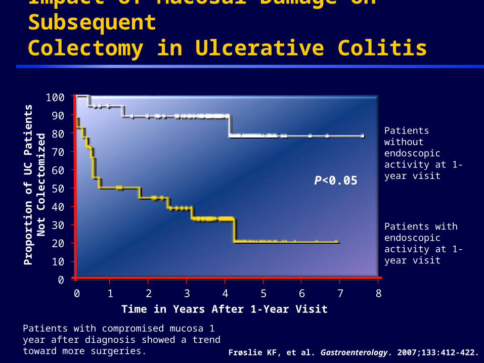
Time in Years After 1-Year Visit
Pro
port
ion
of
UC
Pati
en
tsN
ot
Cole
cto
miz
ed
Patients with compromised mucosa 1 year after diagnosis showed a trend toward more surgeries. Frøslie KF, et al. Gastroenterology. 2007;133:412-422.
Patients without endoscopic activity at 1-year visit
Patients with endoscopic activity at 1-year visit
100
0 1 4 5 6 72 3 8
60
90
80
70
50
0
10
40
20
30
Impact of Mucosal Damage on Subsequent Colectomy in Ulcerative Colitis
P<0.05

Week 8 Mayo Endoscopy Subscore Predicts Corticosteroid-Free Symptomatic Remission at Week 30 During Anti-TNF Antibody Therapy- Results from ACT I and ACT II
Week 8 Mayo endoscopy
Subscore
Corticosteroid-free symptomatic
Remission, n/n (%)P value
0 30/65 (46) <.0001
1 35/102 (34)
2 8/71 (11)
3 2/31 (6.5)
Colombel JF, et al. Gastroenterology. 2011;141:1194-1201.

Mucosal Healing Correlates to Rate of Colectomy: Results from ACT 1 (Infliximab)
1 = MILD 2 = MODERATE 3 = SEVERE0 = NORMAL
Colombel JF, et al. Gastroenterology. 2011;141:1194-1201.
1.00
0.75
0.500 10 20 30 40 50
Time to colectomy or commercial infliximab use (weeks)
Pro
po
rtio
n
wit
ho
ut
cole
cto
my
or
com
mer
cial
in
flix
imab
use
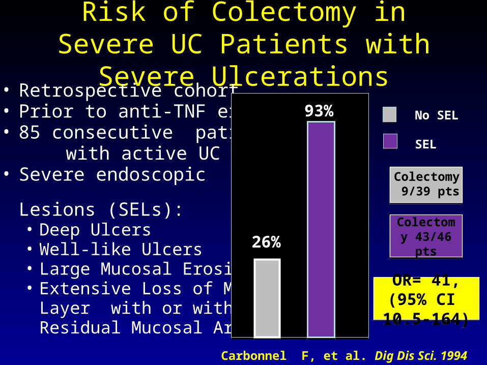
Risk of Colectomy in Severe UC Patients with Severe Ulcerations
• Retrospective cohort• Prior to anti-TNF era • 85 consecutive patients with active UC• Severe endoscopic
Lesions (SELs):• Deep Ulcers• Well-like Ulcers• Large Mucosal Erosions• Extensive Loss of Mucosal
Layer with or withoutResidual Mucosal Areas
Carbonnel F, et al. Dig Dis Sci. 1994 Jul;39(7):1550-7.
93%
26%
No SEL
SEL
Colectomy 43/46 ptsColectomy 43/46 pts
Colectomy 9/39 pts
OR= 41, (95% CI 10.5-164)OR= 41, (95% CI 10.5-164)
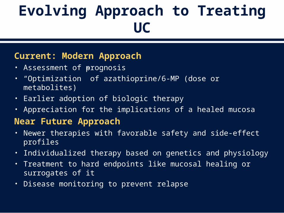
Evolving Approach to Treating UC
Current: Modern Approach• Assessment of prognosis• “Optimization” of azathioprine/6-MP (dose or metabolites)• Earlier adoption of biologic therapy• Appreciation for the implications of a healed mucosa
Near Future Approach• Newer therapies with favorable safety and side-effect profiles • Individualized therapy based on genetics and physiology• Treatment to hard endpoints like mucosal healing or surrogates
of it• Disease monitoring to prevent relapse
V. Dysplasia Surveillance
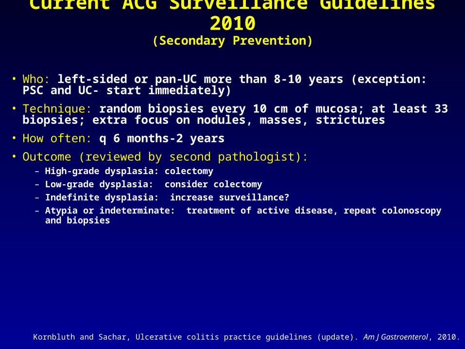
Current ACG Surveillance Guidelines 2010Current ACG Surveillance Guidelines 2010(Secondary Prevention)(Secondary Prevention)
• Who:Who: left-sided or pan-UC more than 8-10 years (exception: PSC and UC- start immediately)
• Technique:Technique: random biopsies every 10 cm of mucosa; at least 33 biopsies; extra focus on nodules, masses, strictures
• How often: How often: q 6 months-2 years
• Outcome (reviewed by second pathologist): Outcome (reviewed by second pathologist): – High-grade dysplasia: colectomy
– Low-grade dysplasia: consider colectomy
– Indefinite dysplasia: increase surveillance?
– Atypia or indeterminate: treatment of active disease, repeat colonoscopy and biopsies
Kornbluth and Sachar, Ulcerative colitis practice guidelines (update). Am J Gastroenterol, 2010.
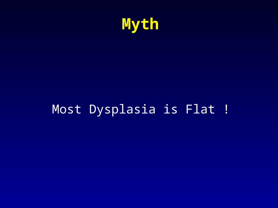
Myth
Most Dysplasia is Flat !
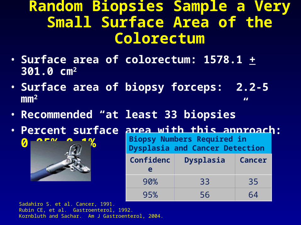
Random Biopsies Sample a Very Small Surface Area of the
Colorectum• Surface area of colorectum: 1578.1 + 301.0 cm2
• Surface area of biopsy forceps: 2.2-5 mm2
• Recommended “at least 33 biopsies”
• Percent surface area with this approach: 0.05%-0.1%
Sadahiro S. et al. Cancer, 1991.Rubin CE, et al. Gastroenterol, 1992.Kornbluth and Sachar. Am J Gastroenterol, 2004.
Biopsy Numbers Required in Dysplasia and Cancer Detection
Confidence Dysplasia Cancer
90% 33 35
95% 56 64
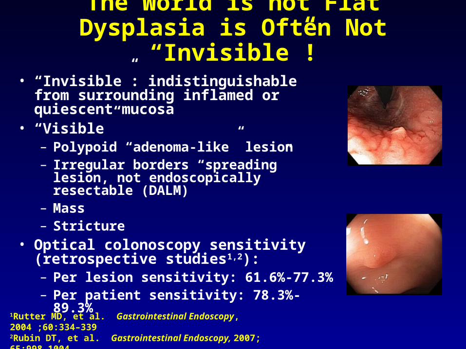
The World is not FlatDysplasia is Often Not “Invisible”!
• “Invisible”: indistinguishable from surrounding inflamed or quiescent mucosa
• “Visible”– Polypoid “adenoma-like” lesion– Irregular borders “spreading” lesion, not
endoscopically resectable (DALM)– Mass– Stricture
• Optical colonoscopy sensitivity (retrospective studies1,2): – Per lesion sensitivity: 61.6%-77.3%– Per patient sensitivity: 78.3%-89.3%
1Rutter MD, et al. Gastrointestinal Endoscopy, 2004 ;60:334–3392Rubin DT, et al. Gastrointestinal Endoscopy, 2007; 65:998-1004.3 Blonski W et al. Scand J. Gastronterol 2008;43(6):698-703.
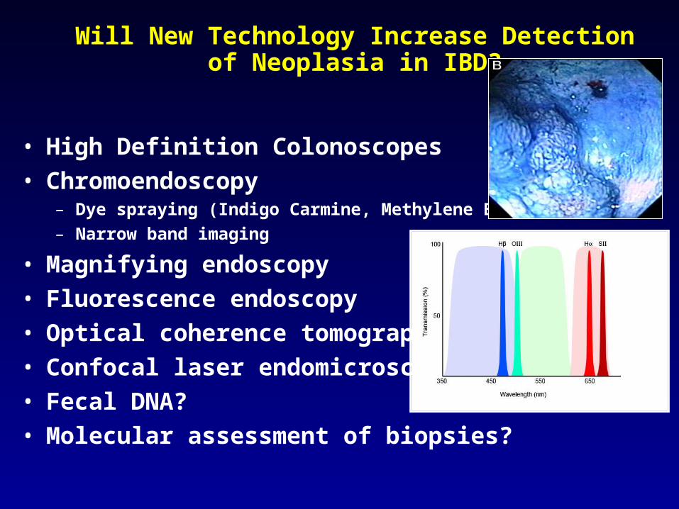
Will New Technology Increase Detection of Neoplasia in IBD?
• High Definition Colonoscopes
• Chromoendoscopy– Dye spraying (Indigo Carmine, Methylene Blue)
– Narrow band imaging
• Magnifying endoscopy
• Fluorescence endoscopy
• Optical coherence tomography
• Confocal laser endomicroscopy
• Fecal DNA?
• Molecular assessment of biopsies?
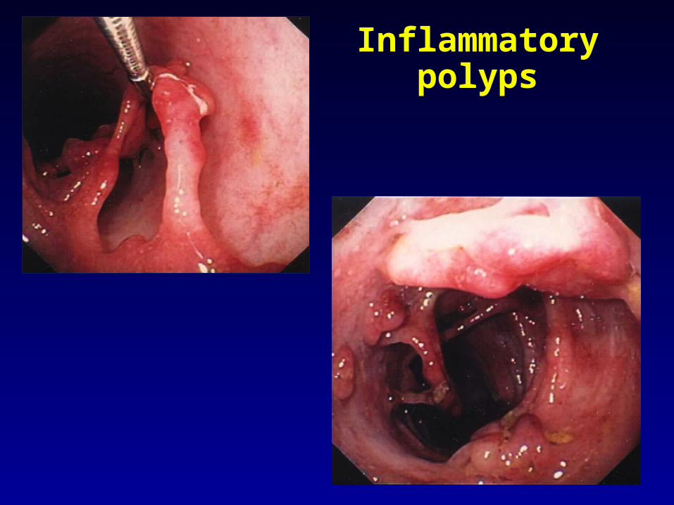
Inflammatory polyps

Dysplasia Associated Lesion or Mass
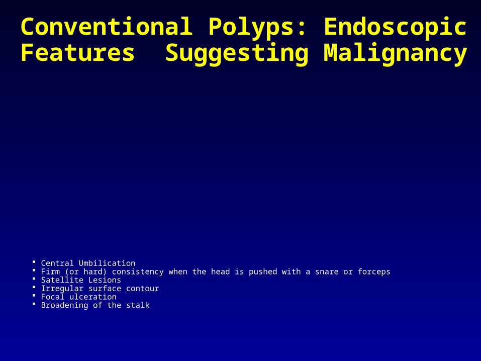
Conventional Polyps: Endoscopic Features Suggesting Malignancy
Central Umbilication Firm (or hard) consistency when the head is pushed with a snare or forceps Satellite Lesions Irregular surface contour Focal ulceration Broadening of the stalk
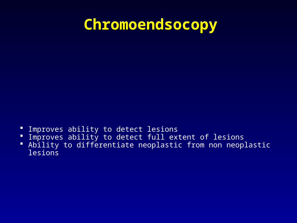
Chromoendsocopy
Improves ability to detect lesions Improves ability to detect full extent of lesions Ability to differentiate neoplastic from non neoplastic lesions
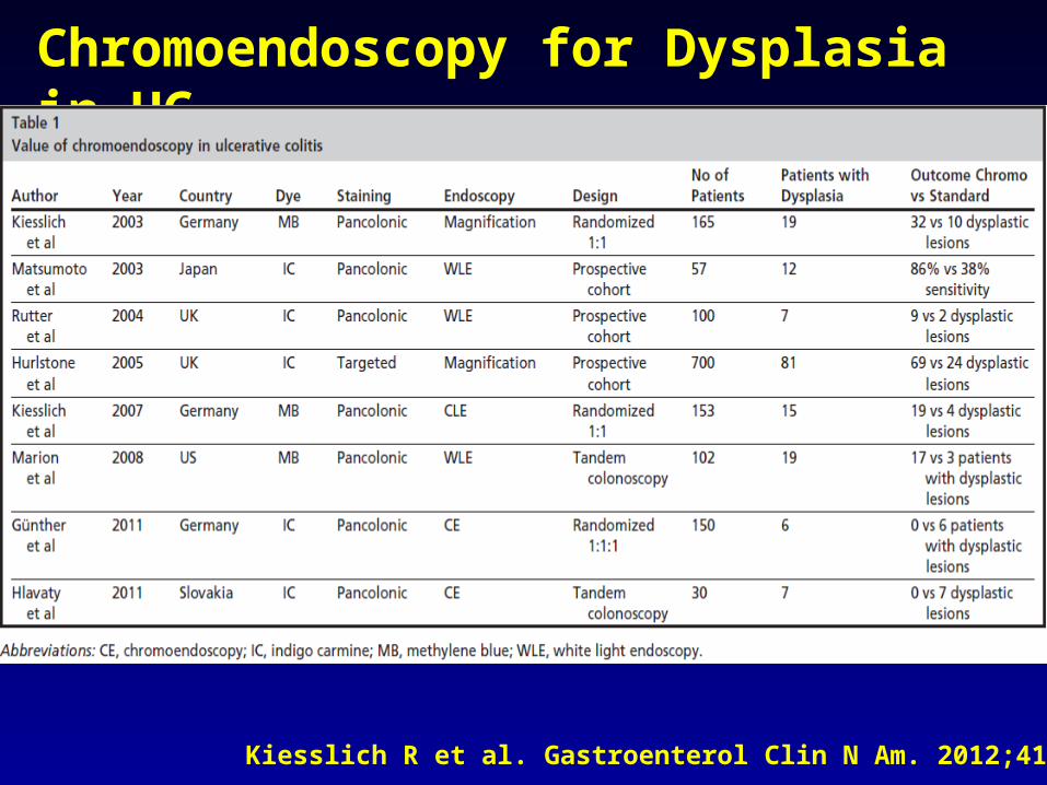
Chromoendoscopy for Dysplasia in UC
Kiesslich R et al. Gastroenterol Clin N Am. 2012;41: 291–302
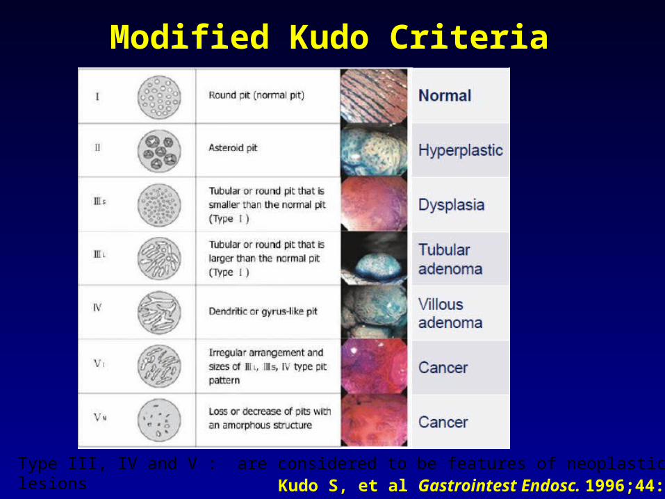
Type III, IV and V : are considered to be features of neoplastic lesionsKudo S, et al Gastrointest Endosc. 1996;44:95–96.
Modified Kudo Criteria
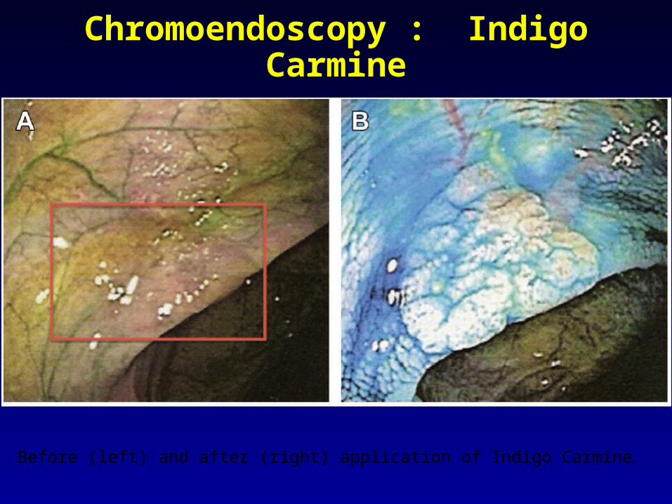
Chromoendoscopy : Indigo Carmine
Before (left) and after (right) application of Indigo Carmine.

Pit Patterns with Chromoendoscopy
Conclusion
Conclusion
Most dysplasia is raised- not flat Chromoendoscopy
Improves ability to detect lesions Improves ability to detect full extent of lesions Ability to differentiate neoplastic from non neoplastic lesions