usaf study of toxicity of nano-particles of aluminum
TRANSCRIPT

8/2/2019 USAF Study of Toxicity of Nano-particles of Aluminum
http://slidepdf.com/reader/full/usaf-study-of-toxicity-of-nano-particles-of-aluminum 1/10
AFRL-HE-WP-TP-2006-0022
Air Force Research Laboratory
In Vitro Toxicity of Aluminum Nanoparticles
in Rat Alveolar Macrophages
Andrew Wagner
Charles Bleckmann
E England
Air Force Institute of Technology
Wright-Patterson AFB OH 45433
Krista Hess
Geo-Centers, Inc.
Dayton OH
Saber HussainJohn J. Schlager
Air Force Research LaboratoryHuman Effectiveness Directorate
Applied Biotechnology Branch
Wright-Patterson AFB, OH 45433-5707
March 2001
FINAL REPORT FOR TH E PERIOD JUNE 1993 TO MARCH 2001
20060403509
Approved for public release; Air Force Research Laboratory
distribution unlimited Human Effectiveness Directorate
Biosciences and Protection Division
Applied Biotechnology Branch
Wright-Patterson AFB, OH 45433-5707
8/2/2019 USAF Study of Toxicity of Nano-particles of Aluminum
http://slidepdf.com/reader/full/usaf-study-of-toxicity-of-nano-particles-of-aluminum 2/10
R R DFormApproved
REPORT DOCUMENTATION PAGE OMB No. 0704-0188
Public reporting burden for this collection of information is estimated to average I hour per response, including the time for reviewing instructions searching existing data sources, gathering and maintaining the
data needed, and completing and reviewing this collection of information. Send comments regarding this burden estimate or any other aspect of this collection of information, including suggestions for reducing
this burden to Department of Defense, Washington Headquarters Services, Directorate for Information Operations and Reports (0704-0188), 1215 Jefferson Davis Highway Suite 1204, Arlington, VA 22202-
4302. Respondents should be aware that notwithstanding any other provision of law, no person shall be subject to any penalty for failing to comply with a collection of information if it does not display a currentl
valid OM B control number PLEASE DO NOT RETURN YOUR FORM TO THE ABOVE ADDRESS.
1. REPORT DATE (DD-MM-YYYY) 2. REPORT TYPE 3. DATES COVERED (From - To)
Technical Paper
4. TITLE AND SUBTITLE Sa. CONTRACT NUMBER
In Vitro Toxicity of Aluminum Nanoparticles in Rat Alveolar N/A
Macrophages 5b. GRANT NUMBER
N/A
5c. PROGRAM ELEMENT NUMBER
61102F
6. AUTHOR(S) 5d. PROJECT NUMBER
Andrew Wagner, Charles Bleckmann, E. England (AFIT, WPAFB OH) 2312
Krista Hess (Geo-Centers, Inc, Dayton OH),. 5e . TASK NUMBERA2
John J. Schlager, Saber M. Hussain (AFRL/HEPB) 5f. WORK UNIT NUMBER
2312A214
7. PERFORMING ORGANIZATION NAME(S) AND ADDRESS(ES) 8. PERFORMING ORGANIZATION REPORT
AND ADDRESS(ES) NUMBER
Air Force M ateriel Command A ir Force Institute of Technology
Air Force Research Laboratory WPAFB OH
Human Effectiveness Directorate Geo-Centers, Inc, Dayton OH AFRL-HE-WP-TP-2006-0022
Biosciences and Protection Division, Applied Biotechnology Branch
W right-Patterson AF B OH 45433
9. SPONSORING I MONITORING AGENCY NAME(S) AND ADDRESS(ES) 10. SPONSOR/MONITOR'S ACRONYM(S)AFRL/HEPB
11. SPONSOR/MONITOR'S REPORT
NUMBER(S)
12. DISTRIBUTION IAVAILABILITY STATEMENT
Approved fo r public release; distribution is unlimited. Cleared as AFRL/WS-06-0492, 21 Feb 06
13. SUPPLEMENTARY NOTES
Poster Presentation at th e Society of Toxicology, San Diego CA, March 2006
14 . ABSTRACT
The purpose of this research was to investigate and characterize the in vitro cellular effects of exposing rat lung
macrophages to aluminum oxide nanoparticles (30 and 40nm average size) compared to aluminum metal
nanoparticles (50, 80, and 120nm). This study used toxicity endpoints involving cell viability, mitochondrial
function, phagocytotic ability, and inflammatory response. Results indicated none to minimal toxicological effects
occurred with exposure of macrophages as high as 500 .tg/ml for 24 hours with aluminum oxide nanoparticles.
However, there was significant delayed toxicity that occurred at 96 and 144 h post exposure. Exposure to aluminum
metal nanoparticles indicated slight to moderate toxicity after 24 hours exposure at 100 and 250 pg/ml. The
phagocytic ability of these cells was significantly hindered by exposure to all tested aluminum nanoparticles at 25
Vtg/ml for 24 hours, but no t by the aluminum oxide nanoparticles. A series of cytokine and nitric oxide assays
performed showed aluminum nanoparticles did not induce an inflammatory response.
15. SUBJECT TERMS
In Vitro cellular effects, aluminum oxide nanoparticles, macrophages, phagocytic, nitric oxide, inflammatory response
16. SECURITY CLASSIFICATION OF: 17. LIMITATION 18. NUMBER 19a. NAME OF RESPONSIBLE PERSON
OF ABSTRACT OFPAGES Saber M. Hussain
a. REPORT b. ABSTRACT c. THIS PAGE 19b. TELEPHONE NUMBER (include are
U U U 8 code)
Standard Form 298 (Rev. 8-98)Prescribed by ANSI Std. 239.18

8/2/2019 USAF Study of Toxicity of Nano-particles of Aluminum
http://slidepdf.com/reader/full/usaf-study-of-toxicity-of-nano-particles-of-aluminum 3/10
In Vitro Toxicity of Aluminum Nanoparticles In Rat Alveolar Macrophages
A Wagner', C Bleckmann', E England', K Hess3, J J Schlager 2, S M Hussain2
'Air Force Institute of Technology, Wright Patterson AFB OH; 2Applied
Biotechnology Branch, Human Effectiveness Directorate, Wright Patterson AFB
OH ; 3Geo-Centers, Inc., Dayton, OH
Abstract:
The purpose of this research was to investigate and characterize the in vitro cellular
effects of exposing rat lung macrophages to aluminum oxide nanoparticles (30 and 40nm
average size) compared to aluminum metal nanoparticles (50, 80, and 120nm). This study
used toxicity endpoints involving cell viability, mitochondrial function, phagocytotic
ability, and inflammatory response. Results indicated none to minimal toxicological
effects occurred with exposure of macrophages as high as 500 [tg/ml for 24 hours with
aluminum oxide nanoparticles. However, there was significant delayed toxicity that
occurred at 96 and 144 h post exposure. Exposure to aluminum metal nanoparticles
indicated slight to moderate toxicity after 24 hours exposure at 10 0 and 250 Ptg/ml. The
phagocytic ability of these cells was significantly hindered by exposure to al l tested
aluminum nanoparticles at 25 p.g/ml for 24 hours, but not by the aluminum oxide
nanoparticles. A series of cytokine and nitric oxide assays performed showed aluminum
nanoparticles did not induce an inflammatory response.
Introduction:" The recent revolution in nanotechnology brought advantages in areas of our lives
as diverse advancements in engineering, information technology, and diagnostics
fields.
" NASA is currently investigating oxide coated Al particles ranging in size from 20to 100 nrn. These particles allow for increases in fuel density, safety, and exhaust
velocity while reducing fuel slosh, leakage, and the overall size of the vehicle
(Palaszewski, 2003).
"* The US Army Research Lab is investigating metallic nanopowders e.g.,
Aluminum nanoparticles in explosives (Miziolek, 2002).
"* U.S. Naval Air Warfare Center is investigating aluminum nanocomposites as"green" bullet primers (Loney, 2004).
" Currently the Navy is using a nanocomposite of alumina-titania as wear resistant
coatings on propeller shafts (DoD, 2005).
"* Existing minimal data suggests that nanoparticles may be able to have adverse
effects at their portal of entry, for example, the lungs, as well as gain entry into
deep tissue sites.
"* Alveolar macrophages are the first line of capture and immunological defense
from inhaled particles. They serve as a good model to understand how inhaled
particles can adversely affect health (Kleinman at al., 2003).
"* In view of importance application of nanoparticles, the current study was
undertaken to study toxicity of Al nanoparticles in alveolar macrophages.

8/2/2019 USAF Study of Toxicity of Nano-particles of Aluminum
http://slidepdf.com/reader/full/usaf-study-of-toxicity-of-nano-particles-of-aluminum 4/10
Objective:
"* To assess and compare the relative toxicity of various states of
commonly used aluminum nanoparticles in systems
"* To investigate any functional changes in phagocytosis and
inflammatory response due to exposure.
Method:
Cell Culture:Alveolar macrophage cells obtained from ATCC (CRL-2192). Cells were
cultured in rat tail collagen coated flasks, with Ham's F 12K medium (Sigma) containing
20% FBS (fetal bovine serum) and penicillin/streptomycin; incubated in a 5% C02
incubator at 37°C. In preparation for in vitro experiments, macrophages were seeded in
coated 24-well plated for mitochondrial function loss (MTT) or 6-well plates for
cytokine, nitric oxide (NO) assays or on 2 chambered slides for phagocytosis assay.
Nanoparticles:All nanoparticles were obtained from Nanotechnologies Inc. Austin Tx .
(Aluminum oxide nanoparticles 30 and 40 nm average size and pure aluminumnanoparticles 50, 80, and 120 nm). Dry particles were suspended in deionized water to a
concentration of 1Omg/mL (stock solution). The stock solution prior to each use was
sonicated for 20 seconds to reduce agglomeration of particles. Media/nanoparticle
suspensions were then pipetted into 6 and 24 well plates or slides at an established
concentration ranging from 5 to 500 jig/ml.
ViabilityAssay: Mitochondrial function was used to establish how viable the alveolar
macrophages were after and exposure of Al nanoparticles. Results were determined
spectrophotometrically by measuring the reduction of the tetrazolium salt MTT to
formazan by succinate dehydrogenase (Carmichael et al., 1987).
Phagocytosis:Phagocytic function was measured by the uptake of 2 pm latex beads and
was observed on an Olympus IX71 inverted fluorescent microscope and CytoViva.
Phagocytosis index is described by Paine et. al., 2004.
Cytokine andNitricOxideAssays: Nitric Oxide, MIP-2, and TNF-a assays were used to
characterize what effects Al nanoparticles might have on an inflammatory response.
Performed as directed by the manufacturer (Promega) and (Biosource).
Lipopolysacharide (LPS) was used as a positive control.

8/2/2019 USAF Study of Toxicity of Nano-particles of Aluminum
http://slidepdf.com/reader/full/usaf-study-of-toxicity-of-nano-particles-of-aluminum 5/10
Results:
U1 l Oxide 30nm U Al Oxide 40 nm
150-
100-
S'Iota ililli~o
0 25 50 100 250 500
Aluminum Oxide Nanoparticles (pglml)
Figure 1: Percent MTT reduced by Alveolar Macrophages after 24 hoursof exposure to aluminum oxide nanoparticles. MTT values observed intriplicate and data reproduced in 3 separate experiments. Resultsconfirm that A10 3 particles do not have a large impact on the viability of
these cells even at concentrations as high as 500 pig/ml. A103 40 nm atdoses between 250 and 500 gtg/ml were the only data points thatindicate a statistical significant difference between the cells not exposed
to any aluminum particles (control) with a p value < .05. ( *asteriskindicates doses that are significantly different than the zero control)

8/2/2019 USAF Study of Toxicity of Nano-particles of Aluminum
http://slidepdf.com/reader/full/usaf-study-of-toxicity-of-nano-particles-of-aluminum 6/10
Ai
Figure 2: Percent MU" educed by Alveolar Macrophages after A) 48hours, B) 96 hours, and C) 144 hours of exposure to A103
nanoparticles. MTU values observed in triplicate and data reproducedin3 separate experiments. Results illustrate the amount of delayedtoxicity of A10 3 nanoparticles on these cells. MTT reduction after 96hrs in both A120 3 30 and 40 nm were significant at a dose of 250jg/ml. MTT reduction after 144 hrs in both A10 3 30 and 40 nm were
significant at 250 gig/ml with a decrease in reduction to approximately35% for both and at 100 lag/ml where it was reduced 43% and 45%respectfully. ( *asterisk indicates doses that are significantly differentthan the zero control)
I Al 50 nm a A]80nOAI120n
121
I 00 . . .80 T
4020
0 25 100 250
Aluminum Nanop roles (algml)
Figure 3: Percent MTT reduced by Alveolar Macrophages after 24
hours of exposure to aluminum nanoparticles. MTT values observedin triplicate and data reproduced in3 separate experiments. Resultsindicate that aluminum nanoparticles have a more drastic effect oncell viability. Al 50 and 120 nm created a significant reduction in MTT
production at 100 and 250 gg/ml. Al 50 nm reduced MTT reduction to
54 and 40% respectfully and Al 120 nm reduced it to 61 and 39%respectfully. Al 80 created a significant reduction of MTT at all threedosing points at 25 mg/ml MTT reduction was reduced to 79%, at 100mg/ml to 63%, and at 250 mg/ml to 49%. ( * asterisk indicates doses
that are significantly different than the zero control)

8/2/2019 USAF Study of Toxicity of Nano-particles of Aluminum
http://slidepdf.com/reader/full/usaf-study-of-toxicity-of-nano-particles-of-aluminum 7/10
A B
Figure 4: A) Phagocytosis Index of Alveolar Macrophages exposed to
various Al nanoparticles at 25 pig/ml for 24 hours (100 cells counted foreach exposure on 3 separate experiments, 300 total cells counted foreach exposure). B) Phagocytosis Index of Alveolar Macrophagesexposed to various Al nanoparticles at 5 lag/ml for 24 hours (100 cellscounted for each exposure on 4 separate experiments, 400 total cellscounted for each exposure). Results indicate that cells exposed to 25pg/ml of various aluminum nanoparticles will have varied results.A120 3 30 and 40 nm show a slight, but no significant decrease inphagocytosis ability (p value > .05 when comparing to the control). Al
50, 80, and 120nm all show a significant reduction in phagocytosiscompared to the control (p value < .05). Cells exposed to 5 gg/ml of Al50, 80, and 120 nm will again have a slightly reduced phagocytosisindex, but only Al 50 nm is significant (p value < .05). ( *asteriskindicates doses that are sionificantlv different than the zero control)
8/2/2019 USAF Study of Toxicity of Nano-particles of Aluminum
http://slidepdf.com/reader/full/usaf-study-of-toxicity-of-nano-particles-of-aluminum 8/10
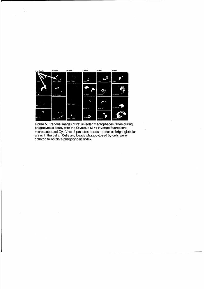
2 Ipmbeads 25 lighil 25 Ipghl 5 pghinl 5 J'arnl 5 pgftnl
ErniEmm.Figure 5: Various images of rat alveolar macrophages taken duringphagocytosis assay with the Olympus IX71 inverted fluorescent
microscope and CytoViva. 2 lam latex beads appear as bright globularareas in the cells. Cells and beads phagocytosed by cells werecounted to obtain a phagocytosis Index.
8/2/2019 USAF Study of Toxicity of Nano-particles of Aluminum
http://slidepdf.com/reader/full/usaf-study-of-toxicity-of-nano-particles-of-aluminum 9/10
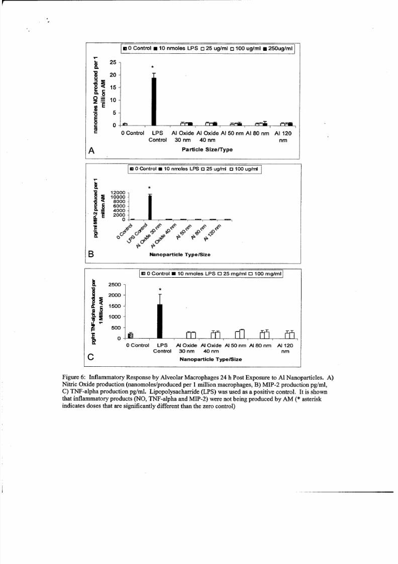
ma 0 Control u 10 nmoles LPS o725 ug/ml 0 10 0 ug/m= 250ug/ml
25
S20
e 15-
0710
6 5EC 0 _ _ _ _ _ _ _ _ _ _ _cc
0 Control LP S Al Oxide Al Oxide Al 50 nm Al 80 nm Al 120
Control 30 nm 40 nm nm
A Particle SizelType
m 0 Control w 10 nmoles LPS 03 25 ug/mI D 100 ug/mlr
S 12000-10000
•. 8000-
6000-2 4000
20001 _
B Nanoparticle Type/Size
M 0 Control 0 10 nmoles LPS 0 25 mg/mI 0 100 mg/ml
•. 2500
2000
1500
. 1000
500 Conro_
0 Control LPS AI Oxide Al Oxide Al 50 nm Al 80 nm A 120
Control 30 nm 40 nm nm
C Nanoparticle TypelSize
Figure 6: Inflammatory Response by Alveolar Macrophages 24 h Post Exposure to Al Nanoparticles. A)Nitric Oxide production (nanomoles/produced pe r 1million macrophages, B) MIP-2 production pg/ml,C) TNF-alpha production pg/ml. Lipopolysacharride (LPS) was used as a positive control. It is shownthat inflammatory products (NO, TNF-alpha and MIP-2) were not being produced by AM (* asteriskindicates doses that are significantly different than the zero control)

8/2/2019 USAF Study of Toxicity of Nano-particles of Aluminum
http://slidepdf.com/reader/full/usaf-study-of-toxicity-of-nano-particles-of-aluminum 10/10
Conclusion:
Aluminum oxide nanoparticles displayed significant toxicity after 96 and 144 hours post
exposure at high doses (100 and 250 jtg/ml). Aluminum nanoparticles also showed slight
toxicity after 24 hours at high doses (100 and 250 pgg/ml). When these cells were dosed
at lower non toxic levels (25 gig/ml) Al 50, 80,120 nm caused a significant reduction in
phagocytosis. Even at a dose as low as 5 .g/ml Al 50 nm still caused a significantreduction. None of these nanoparticles caused the induction of nitric oxide, TNF-alpha,or MIP-2, important components in inflammatory responses. In summary, based on
viability, aluminum nanoparticles appear to be slightly toxic to rat alveolar macrophages.However, there was a significant reduction in phagocytic function of macrophages
References:
Palaszewski B. (2002) Nanotechnology Investigated for Future Gelled and
Metallized Gell Fuels. Acquired fromhttp://www.grc.nasa.gov/WWW/RT2002/5000/5830palaszewskil.html on 10-10-2005
Department of Defense Director, Defense Research and Engineering, 2005, Defense NanotechnologyResearch and D evelopment. Acquired from http://www.nano.gov/html/res/DefenseNano2005.pdf on 1-4-
2006Loney, Dennis, 2004, Weapons that Tread Lightly. American Chemical Society. Acquired fromhttp://www.chemistry.org/portal/aJc/s/ 1/feature ent.html?DOC=enthusiasts%5Cent green
explosives.html on 1/10/2006
Miziolek A. (2002) Nanoenergetics: An Emerging Technology Area of National Importance. AMPTIAC
Quarterly6: 43-48.
Kleinman M.T.; Sioutas C.; Chang M.C.; Boere A.J.F; Cassee F.R. (2003) Ambient Fine and coarseparticles suppression of alveolar macrophage functions. Toxicology Letters 137: 151-158.
Carmichael J, Degraff W.G., Gazdar A.F., Minna J.D., Mitchell J.B. (1987) Evaluation of a tetrazolium-based semi automated colorimetric assay: Assessment of chemo sensitivity testing. CancerRes. 47,
936-942.
Paine R.; Morris S.B.; Hong J.; Wilcoxen S.E.;Phare S.M.; Moore B.B.; Coffey M.J.; Toews G.B. (2001)
Impaired functional activity of alveolar macrophages from GM-CSF-deficient mice. AmericanJournal
ofPhysiologyLung CellMolecularPhysiology 281: 1210-18