urinary tract cytology - asl5. · pdf filenormal urinary tract cytology ... •catheterized...
TRANSCRIPT

Urine Cytology
Diagnostic Categories and Atypia
Tarik M. Elsheikh, MD
Professor and Medical Director
Anatomic Pathology
Cleveland Clinic
Outline
• Indications and diagnostic accuracy of urine cytology
• Atypia in urine cytology
• Diagnostic categories
• The Paris system classification 2015

Introduction
• Majority of UT malignancies are UC
– Urothelial carcinoma, 80-90%
– Mixed Carcinoma- UC (5%)
– Squamous cell carcinoma (5%)
– Adenocarcinoma (2%)
– Small Cell Carcinoma (1%)
• The main function of urine cytology is to diagnose urothelial carcinoma (UC)

Indications
1. Establish Dx in symptomatic patients-hematuria
– Most common, low yield (5-10% malignancy)
2. Screen high risk patients (exposure to industrial chemicals, metals, etc.)
3. Follow-up patients with Hx of UC
4. Complementary to cystoscopy and biopsy: detect small and hidden lesions (diverticuli, ureters, renal pelvis)
• Urine cytology is the most reliable method for detecting urothelial CIS (> biopsies)

Diagnostic Accuracy of Urine Cytology
• Number of Specimens
-Voided urine on 3 consecutive days
- 50% accuracy (1 specimen)
- 75-90% accuracy (3 specimens)
• Patient Population
- High risk and history of CA
• Tumor Grade
• HGUC: > 90 %
• LGUC: <50 %

Atypia in Urine Cytology

Diagnostic Categories
• JH created a template
similar to Gyn TBS:
1. Negative
2. AUC-US
3. AUC-H
4. LG neoplasm
5. HG neoplasm
6. Non-diagnostic
Rosenthal, cancer cytopath 2013

Diagnostic Categories
• JH created a template
similar to Gyn TBS:
1. Negative
2. AUC-US (26%)
3. AUC-H (5%)
4. LG neoplasm
5. HG neoplasm
6. Non-diagnostic
Rosenthal, cancer cytopath 2013
• Cleveland Clinic
1. Negative
2. Atypical (14%)
3. Suspicious for HG UC
(2%)
5. Positive

Diagnostic Categories
Preferred by Urologists
1. Negative for HGUC
2. Suspicious for HGUC
3. Positive for HGUC

Should We Eliminate the
“Atypical” Category?
• Approx 10-20% of urines classified as
“atypical”
• Considerable inter-observer variability among
pathologists as to what constitutes atypia
• Currently, most urologists interpret “atypia” as
negative or unhelpful

Arguments for Not Eliminating “Atypia”
• Significant proportion of malignant cases
would be missed if “atypia” was eliminated
– Malignant rate on FU: 23-68%
• Ancillary studies such as FISH can be helpful
in those cases

Variations in Atypical rate
• Inter-institutional:
– Reported wide variation: 2-31%
• Intra-departmental:
• ≈ 23,000 urine cases signed out by 12
cytopathologists at CC, over 3 yr period
- All were cytopathology board certified
• Variable experience ranging from 2-26 yrs
Reynolds, Elsheikh, 2015

Reynolds, Elsheikh 2015
• Wide variation in Atypical rate: 8-28% (avg 14%)
• Higher rates are not related to level of experience
• More dependent on individual threshold for atypia
0,0%
5,0%
10,0%
15,0%
20,0%
25,0%
30,0%
Group 1 Group 1 Group 1 Group 1 Group 2 Group 2 Group 2 Group 2 Group 3 Group 3 Group 3 Group 3
Atypical rate
Group 1
Group 1
Group 1
Group 1
Group 2
Group 2
Group 2
Group 2
Group 3
Group 3
Group 3
Group 3
< 5 yrs 6-15 yrs > 15 yrs

Urine Dx’s Categorized by Pathologist BMH, Indiana 2009

In Need of Standardization!
• Standard classification and terminology system
• Well defined and reproducible diagnostic criteria
• Uniform inter- and intra-departmental
communications
• Consistent prognostic and management
information leading to optimal patient care

The Paris System for Reporting
Urinary Tract Cytology


TPS Diagnostic Categories
• Negative for HGUC
• Atypical Urothelial Cells
• Suspicious for HGUC
• High Grade Urothelial Carcinoma
• Low Grade Urothelial Neoplasm
• Other malignancies, both primary and
secondary

Urinary Tract Histology
• Superficial cell layer
one cell thick, superficial
squames-size or larger,
multinucleated
• Intermediate cell layer
approximately 5 cell layers,
parabasal-size cells
• Basal cell layer
one cell thick, cuboidal-
columnar
Bladder

Ureter and Renal
Pelvis • Lining cells are
larger and more
pleomorphic than
bladder (decreased
cell turnover &
exfoliation)
Upper Urinary Tract Histology 2

Normal Urinary Tract
Cytology
• Superficial urothelial cells
– Marked variation in size and
shape (10-150 u), low N/C
– Often polygonal
– Abundant pale cytoplasm, well
defined borders
– Occasional vacuolization
– Round-oval nuclei, often
multinucleated

Normal Urinary Tract Cytology 2
• Deep urothelial cells
– Uniform in shape and
size (10-20 u)
– Scant to moderate dense
cytoplasm, distinct
borders, fine
vacuolization
– Central nuclei, finely
granular chromatin,
small nucleoli

Normal Urinary Tract Cytology 3
• Voided urine is sparsely cellular, single cells-
degeneration

• Catheterization or washings specimens have many clusters
Normal Urinary Tract Cytology 4
TPS Diagnostic Categories
• Negative for HGUC
• Atypical Urothelial Cells
• Suspicious for HGUC
• High Grade Urothelial Carcinoma
• Low Grade Urothelial Neoplasm
• Other malignancies, both primary and
secondary

High Grade Urothelial CA
Often invasive, 70% mortality
90% of pts dying of disease
present initially with HGUC
• Cytology cannot reliably
separate CIS from invasive CA
• High diagnostic accuracy of
cytology
– Sensitivity 80-90 %
– Specificity > 95%

HGUC- TPS Criteria
Non-superficial and non-degenerated (viable) urothelial cells
• High N/C ratio > 0.5-0.7 (required)
• Hyperchromasia, moderate-severe (required)
–and one of the following:
• Irregular clumpy chromatin
• Irregular nuclear membranes
At least 5-10 abnormal cells
– Based on pathologist’s level of comfort
– Voided vs. upper tract instrumented specimen

• Single cells and/or disorganized
clusters
• Irregular, hyperchromatic nuclei
HGUC

- Pleomorphic bizarre cells, enlarged eccentric
nuclei, coarse dark chromatin

TPS Diagnostic Categories
• Negative for HGUC
• Atypical Urothelial Cells
• Suspicious for HGUC
• High Grade Urothelial Carcinoma
• Low Grade Urothelial Neoplasm
• Other malignancies, both primary and
secondary
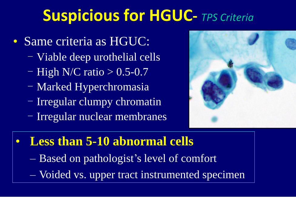
Suspicious for HGUC- TPS Criteria
• Same criteria as HGUC:
- Viable deep urothelial cells
- High N/C ratio > 0.5-0.7
- Marked Hyperchromasia
- Irregular clumpy chromatin
- Irregular nuclear membranes
• Less than 5-10 abnormal cells
– Based on pathologist’s level of comfort
– Voided vs. upper tract instrumented specimen


Coy Cells
• Suspicious for HGUC
• Opposite of “decoy cells”
– Often sparse in number
– Been compared to
“litigation cells” on Paps
– Small cells with
hyperchromatic irregular
nuclei
– India ink/coal black nuclei

Differential Diagnosis of HGUC
• Human polyoma viral infection
• Therapy effect
• Stones and reactive changes
• Other malignancies
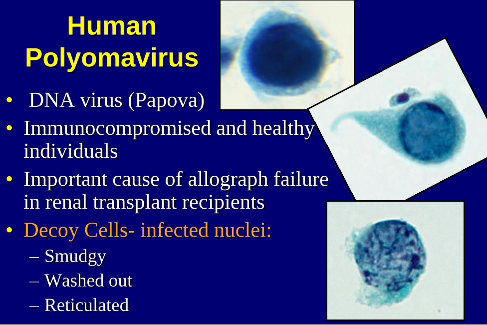
• DNA virus (Papova)
• Immunocompromised and healthy individuals
• Important cause of allograph failure in renal transplant recipients
• Decoy Cells- infected nuclei:
– Smudgy
– Washed out
– Reticulated
Human
Polyomavirus

Polyoma Virus
• Diff DX is degenerated HG UC
Polyoma virus HG UC
Architecture Single cells Single cells &
clusters
Nuclear
membrane
Smooth, round Marked irregularity
Chromatin Uniform, smudgy,
reticulated
Coarsely granular,
clumped

HGUC Polyoma

Therapy Effect
• Cytoxan & Busulfan – Systemic treatment of non
urothelial malignancies
– Hemorrhagic cystitis
– Severe cytologic atypia may be indistinguishable from CA
– Atypia more bizarre than usual HGUC
– Atypia often has degenerative features
Photo from Modern Cytopathology.
Elsevier Science, 2004

Therapy Effect 2
Thiotepa & Mitomycin C
Intravesical Rx of sup UC
Repair-like changes
BCG Vaccine
Treatment of CIS
Granulomas, mild atypia
Radiation Change
Extreme cytomegaly,
multinucleation, but low N/C ratio
Photo from Murphy WM. Urinary
Cytopatholgy. ASCP Press,2000

Lithiasis
• Papillary clusters common
• Smooth bordered clusters
• Centrally placed nuclei,
smooth nuclear membranes,
finely granular chromatin
• Hyperchromatic smudgy
nuclei (degenerative
changes)

Lithiasis 2
• Occasionally marked cytologic atypia, including
nuclear pleomorphism, coarsely granular chromatin,
mitotic figures false-positive diagnosis of HGUC
• Inflammation & debris
in background may be
misinterpreted as tumor
diathesis
• May be impossible
to distinguish from
LGUC

Lithiasis 3
• Important source of false positive Dx for
LGUC and HGUC
• Clinical history not reliable: filling defect in
upper UT stone vs. neoplasm
• Persistent atypical features (weeks)
aggressively worked up for neoplasia

TPS Diagnostic Categories
• Negative for HGUC
• Atypical Urothelial Cells
• Suspicious for HGUC
• High Grade Urothelial Carcinoma
• Low Grade Urothelial Neoplasm
• Other malignancies, both primary and
secondary

Mild Nuclear Atypia
• Single cells with enlarged and irregular nuclei
(no significant hyperchromasia)
• Most common and most frustrating

Atypical Urothelial Cells (AUC)-TPS
Definition:
1.Atypia that falls short of “Suspicious” or
“HGUC”
2.Degenerative changes where nature and degree
of atypia cannot be explained

AUC- TPS Criteria
• High N/C ratio > 0.5 (required)
and one of the following:
• Hyperchromasia, mild-moderate
- Compared to benign urothelial or
squamous cell nuclei
• Nuclear Irregularity, significant
• Irregular clumpy chromatin, mild
1. Non-degenerated, Non-superficial urothelial cells

• Nuclear irregularity, high NC ratio, but no significant
hyperchromasia, compared to benign urothelial cells
AUC

High NC ratio, mild hyperchromasia
AUC
High NC ratio, nuclear irregularity

AUC- TPS Criteria
• High N/C ratio and
hyperchromasia
• Extensive degeneration of nuclei
and/or incomplete cytoplasm
2. Degenerated non-superficial urothelial cells

AUC- Exclusions
• Mere presence of
degeneration does not
equate to AUC
• Excludes atypia secondary
to known conditions:
• Polyoma virus, stones,
reactive/repair, therapy,
instrumentation, etc.

AUC Negative
Lost nuclear membrane Intact nuclear membrane,
hyperchromatic

TPS Diagnostic Categories
• Negative for HGUC
• Low Grade Urothelial Neoplasm
• Other malignancies, both primary and
secondary

Low Grade Urothelial
CA
• Predominately papillary
• Capacity to invade (<20%)
• Rarely metastasizes
• Progression < 15%

Low Grade Urothelial CA 2
• Cytologic diagnosis of LGUC is problematic
– Minimal shedding of neoplastic cells
– Subtle cytologic alterations, difficult to distinguish
from reactive changes, i.e. stones, instrumentation
– No discriminating cytologic features between
PUNLMP and LGUC
– Wide range of sensitivities 0-73% (Avg 25-40%)
Whisnant, 2003

Mcroskey 2015
• Compared biopsy proven LGUC cytologies (98
cases) to negative cytologies (53 cases)
• Instrumented urine specimens
• Evaluated 17 published cytologic features
• All cases were examined blinded to histology
• No single cytologic feature was found to be
helpful in DDX, except for papillary clusters
with fibrovascular cores (2/98 cases)

LGUC Benign
Few cells with enlarged slightly irregular nuclei.

Clusters in voided urine
• Papillary clusters (without fibrovascular core are not associated with increased risk of neoplasia
• Should place less reliance on presence or shape of clusters
• More emphasis on nuclear features
(Deshpande & Mckee, Cancer Cytopathol, 2005)

Low Grade Urothelial Neoplasm- TPS Criteria
• 3D papillary clusters (extreme nuclear overlapping) with fibrovascular cores- very rare
Cell Block
• Diagnosis should also be qualified as Neg for HGUC
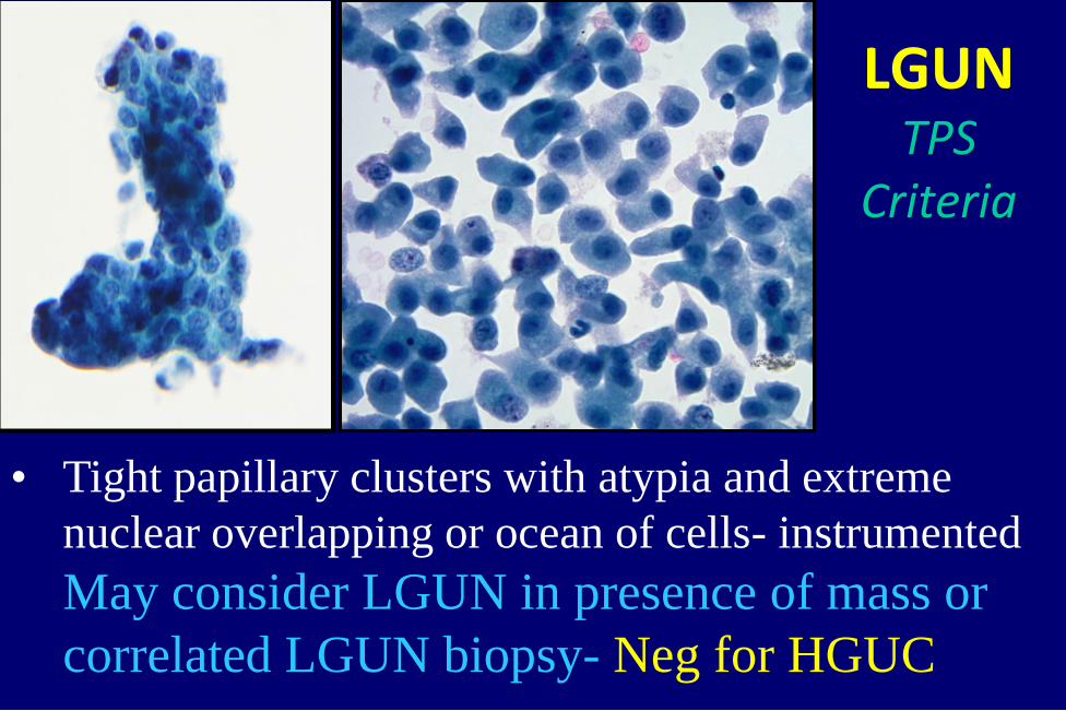
• Tight papillary clusters with atypia and extreme
nuclear overlapping or ocean of cells- instrumented
May consider LGUN in presence of mass or
correlated LGUN biopsy- Neg for HGUC
LGUN TPS
Criteria

Differential Diagnosis of
LG Urothelial CA
• Reactive/reparative changes
• Upper urinary tract sampling
• Instrumentation effect
• Lithiasis

• Reactive changes
and repair

Upper Urinary Tract specimens
• Direct sampling of upper UT is effective in
detecting HGUC, but poor for low grade lesions
• Sensitivity: LGUC 37% vs. HGUC 80% Barkan 2015
• Normal upper UT epithelium shows more atypia
than lower UT and occasionally more than LGUC
N/C ratio, nuclear irregularities, papillary clusters
• Almost impossible to distinguish low grade UC
from upper tract benign changes

• Negative cystoscopy, biopsy & followup
Renal pelvis/ureter brushing

Instrumentation Effect
• Catheterized urine and bladder wash specimens
• Large pseudopapillary groups and 3D clusters
• Nuclear overlap and crowding
• Low N/C ratio
• Finely granular chromatin with even distribution
• Well defined cytoplasmic borders
• Nuclear palisading at periphery of clusters with
abundant cytoplasm (cytoplasmic collar)

Instrumentation effect
ThinPrep

How Long is Cytology Abnormal
after Cystoscopy?
• Evaluated 48 patients
• Examined urine before, immediately after, 1,
2, 7, 14 and 28 days
• Instrumentation effect was transient, mostly
disappearing within 1 day after cystoscopy
McVey et al. BJU INT, 2004

TPS Diagnostic Categories
• Negative for HGUC
• Atypical Urothelial Cells
• Suspicious for HGUC
• High Grade Urothelial Carcinoma
• Low Grade Urothelial Neoplasm
• Other malignancies, both primary and
secondary

Negative for HGUC-TPS Criteria
• If there is a known cause for “atypia”- it’s
Negative
– Reactive urothelial cells
– Instrumentation effect
– Upper urinary tract specimens
– Changes associated with lithiasis
– Polyoma viral cytopathic effect
– Post-therapy effects
– Clusters without fibrovascular cores or atypia

NEGATIVE
Instrumentation
Reactive Repair
Therapy
Polyoma
Upper tract
Stones
Clusters

Negative for HGUC-TPS Criteria
• This category also
includes “LGUN”
Sample Dx:
- Negative for HGUC
- Changes consistent/suggestive
of LGUN

Nu
cle
ar
/ cyto
log
ic a
typ
ia
Probability of high grade UC
low moderate/high certain
AUC-Suspicious
8%-30%
HGUC NFM
Slide courtesy of
D. Rosenthal, MD

Ancillary Tests for Detecting & Monitoring UC
Test Sensitivity
% (range)
Specificity
% (range)
Lab Comment
Urine cytology 54 (35-68) 95 (83-100)
DNA ploidy 62 (45-86) 89 (76-100) IA, FCM
BTA 60 (32-100) 77 (40-96) POC, Ref ⇈ False +
NMP22 67 (47-81) 72 (60-86) Ref
ImmunoCyt 50-100 69-79 ⇈ False +
Telomerase 74 (62-93) 79 (60-99) Ref ⇈ False +
Microsatellite
Analysis
83-95 83-100 Ref
FISH 69-87 85-97 Ref

FISH (UroVysion)
• A 4-probe set that targets the common chromosomal abnormalities in UC
• FISH+ results:
– Polysomy 3, 7, 17
• Gain of 2 or more chromosomes
• Seen mostly in HGUC, but not LGUC
– Deletion 9p21
• Seen in LGUC

FISH 2
• FDA approved for surveillance of patients with
hematuria and history of UC
• Recommended in hematuria pts with other risk
factors such as smoking hx and age > 45
• High sensitivity 69-93%, esp. for
HGUC, lower for LG UC
• ? FISH positive AUC treated as Susp/ HGUC

FISH 3
Impressive Sensitivity results:
• Surveillance UC patients: – FISH +/ cystoscopy-/ cytology- 65%
recurrent CA (within 29 months)
– FISH- 13% recurrent CA
• Hematuria surveillance: – FISH+/cytology- (30%) 60% UC
• Post BCG therapy – FISH+ approx 10 times more likely to develop
invasive cancer

False-Positive FISH
• Be careful about significance of FISH+ in upper
tract cytlogy
– Limited value for upper tract tumor surveillance
– High false + (Johannes, J Urol. 2010)
• Polyoma virus can cause false + FISH (approx
15%)
– Usually in pts with high viral titers (renal transplant)

FISH vs. Cytology
• FISH more sensitive but less specific than urine cytology
• PPV of urine cytology in HGUC > 90% – PPV of FISH: as low as 50%
– Cytology= 7-10 times cheaper (Murphy 2009)
• Combined FISH & Cytology 98% sensitivity and > 95% specificity
• FISH-neg patients (low risk) may be allowed extended time intervals between cystoscopies

Summary
• Urine cytology is best applied to HGUC
• Cytology less helpful for detecting and monitoring LG neoplasms
– Not major limitation
– LG neoplasms rarely aggressive and can be readily detected by cystoscopy
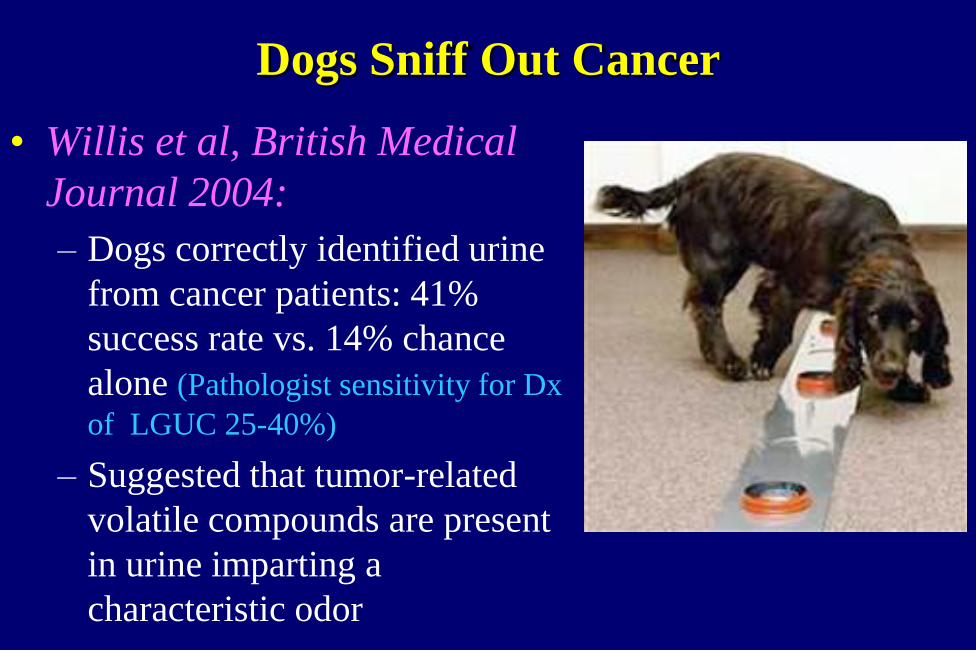
Dogs Sniff Out Cancer
• Willis et al, British Medical
Journal 2004:
– Dogs correctly identified urine
from cancer patients: 41%
success rate vs. 14% chance
alone (Pathologist sensitivity for Dx
of LGUC 25-40%)
– Suggested that tumor-related
volatile compounds are present
in urine imparting a
characteristic odor

Summary 2
• “Atypical” diagnoses should not be used for reactive/reparative changes Negative for HGUC
• TPS provides strict cytologic criteria and aims to establish a standardized practical approach to urinary cytology classification

Is there a pathologist-or dog- in the house?

Thank You!