unusual new signs of pneumothorax at lung ultrasound · in hydropneumothorax, the air/fluid border...
TRANSCRIPT

Unusual new signs of pneumothoraxat lung ultrasoundVolpicelli et al.
Volpicelli et al. Critical Ultrasound Journal 2013, 5:10http://www.criticalultrasoundjournal.com/content/5/1/10

Volpicelli et al. Critical Ultrasound Journal 2013, 5:10http://www.criticalultrasoundjournal.com/content/5/1/10
SHORT COMMUNICATION Open Access
Unusual new signs of pneumothoraxat lung ultrasoundGiovanni Volpicelli1*, Enrico Boero1, Valerio Stefanone1 and Enrico Storti2
Abstract
Background: The diagnosis of pneumothorax with a bedside lung ultrasound is a powerful methodology. Theconventional lung ultrasound examination consists of a step-by-step procedure targeted towards the detection offour classic ultrasound signs, the lung sliding, the B lines, the lung point and the lung pulse. In most cases, acombination of these signs allows a safe diagnosis of pneumothorax. However, the widespread application ofsonographic methodology in clinical practice has brought out unusual cases which raise new sonographic signs.The purpose of this article was to introduce some of these new signs that are described after the analysis ofunusual and complex cases encountered during the clinical daily practice in an emergency department.
Findings: The double lung point consists of the alternating patterns of sliding and non-sliding lung intermittentlyappearing at the two opposite sides of the scan. The septate pneumothorax allows B lines and lung pulse to be stillvisible in a condition of pneumothorax with absent sliding. In hydropneumothorax, the air/fluid border is imagedby lung ultrasound as the interposition between an anechoic space and a non-sliding A-pattern, a sign that may benamed hydro-point.
Conclusions: In bedside lung ultrasound, the operator should be aware and interpret double lung point, septatepneumothorax and hydro-point. The conventional diagnostic protocol of bedside lung ultrasound forpneumothorax should be occasionally adapted to such complex cases.
Keywords: Emergency ultrasound; Critical ultrasound; Lung ultrasound; Pneumothorax
FindingsIntroductionLung ultrasound is nowadays acknowledged as a usefulmethodology for the diagnosis of pneumothorax at bedside[1]. The diagnostic value of the sonographic signs ofpneumothorax is similar to the well-known and largelyused radiologic signs of the same condition in chest radiog-raphy. Visualization of a gap between the parietal andvisceral pleura in a chest film is largely used, and intuitivelydiagnostic of pneumothorax, even though, to our know-ledge there are no published studies specifically designed toevaluate its specificity in comparison with a gold standard.Nevertheless, there is not any doubt about the power andhigh specificity of this radiologic sign of pneumothorax.Similarly, the sonographic signs of pneumothorax arelargely intuitive of the condition and share the same veryhigh specificity with the radiologic signs [2,3]. Absence of
* Correspondence: [email protected] Medicine, San Luigi Gonzaga University Hospital, Turin 10043, ItalyFull list of author information is available at the end of the article
© 2013 Volpicelli et al.; licensee Springer. This iAttribution License (http://creativecommons.orin any medium, provided the original work is p
lung sliding or pulse and absence of B lines combined witha lung point during a lung ultrasound examination have thesame diagnostic meaning of the visualization of a pleuralgap in a chest radiography. However, whereas chest radiog-raphy is a conventional method, lung ultrasound needed tobreak the wall of scientific prejudices before it was acknow-ledged as a viable method for a neumothorax diagnosis.This is the reason why many years and clinical studies wereneeded to change clinical guidelines and protocols andfinally introduce bedside lung ultrasound in clinical prac-tice. We cannot say anything unless this process has beencompleted. The need for a widespread use of lung ultra-sound is compelling. If it is true, as it has been proven, thatthe specificity of a chest radiography is as high as a lungultrasound, it is also true that many studies showed thatsensitivity of lung ultrasound is far higher [1,4-6]. Thecategory of radio-occult pneumothorax is part of the dailyexperience of any physician who faces trauma patients,post-procedural thoracic complications and spontaneouspneumothoraces.
s an open access article distributed under the terms of the Creative Commonsg/licenses/by/2.0), which permits unrestricted use, distribution, and reproductionroperly cited.
Volpicelli et al. Critical Ultrasound Journal 2013, 5:10 Page 2 of 4http://www.criticalultrasoundjournal.com/content/5/1/10
Sonographic diagnosis of pneumothoraxThe bedside lung ultrasound diagnostic methodology forpneumothorax has been described in literature. A recentconsensus process of the main experts in the field hasled to the approval of a step-by-step methodology thatconsists of combining the four main sonographic signsof pneumothorax: the lung sliding, the lung pulse, the Blines and the lung point [1]. While the first three signsare strongly predictive of the absence of the condition,the lung point is the only sign that confirms a pneumo-thorax. However, the value of this general rule varies ifwe consider different settings and scenarios.In the stable patient, the conventional methodology con-
sists of scanning the most superior aspect of the chest withrespect to gravity, bilaterally. In the supine patient, this areacorresponds to the anterior-inferior chest. The probeshould be applied on this area, on both sides. Detection oflung sliding and/or B lines is enough to conclude the exam-ination and rule-out pneumothorax. Absence of sliding andB lines need to be confirmed by moving the probe towardsthe lateral chest to visualize the lung point. If the latter isdetected, then the examination is concluded and thepneumothorax is diagnosed with an extremely high specifi-city. When the lung point cannot be detected, then absenceof a lung pulse should also be checked. In this condition,absence of even the slightest pulsation of the subpleurallung image allows the diagnosis of a pneumothorax charac-terized by a complete pulmonary collapse. Conversely, evenin the absence of sliding and B lines, visualization of a lungpulse rules out pneumothorax.In the unstable patient or in a scenario of cardiac arrest,
the methodology varies slightly. The most superior area ofthe chest is scanned for any sliding, pulse and B lines
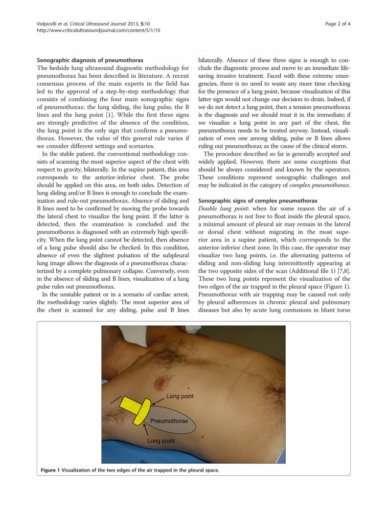
Figure 1 Visualization of the two edges of the air trapped in the pleu
bilaterally. Absence of these three signs is enough to con-clude the diagnostic process and move to an immediate life-saving invasive treatment. Faced with these extreme emer-gencies, there is no need to waste any more time checkingfor the presence of a lung point, because visualization of thislatter sign would not change our decision to drain. Indeed, ifwe do not detect a lung point, then a tension pneumothoraxis the diagnosis and we should treat it in the immediate; ifwe visualize a lung point in any part of the chest, thepneumothorax needs to be treated anyway. Instead, visuali-zation of even one among sliding, pulse or B lines allowsruling out pneumothorax as the cause of the clinical storm.The procedure described so far is generally accepted and
widely applied. However, there are some exceptions thatshould be always considered and known by the operators.These conditions represent sonographic challenges andmay be indicated in the category of complex pneumothorax.
Sonographic signs of complex pneumothoraxDouble lung point: when for some reason the air of apneumothorax is not free to float inside the pleural space,a minimal amount of pleural air may remain in the lateralor dorsal chest without migrating in the most supe-rior area in a supine patient, which corresponds to theanterior-inferior chest zone. In this case, the operator mayvisualize two lung points, i.e. the alternating patterns ofsliding and non-sliding lung intermittently appearing atthe two opposite sides of the scan (Additional file 1) [7,8].These two lung points represent the visualization of thetwo edges of the air trapped in the pleural space (Figure 1).Pneumothorax with air trapping may be caused not onlyby pleural adherences in chronic pleural and pulmonarydiseases but also by acute lung contusions in blunt torso
ral space.

Volpicelli et al. Critical Ultrasound Journal 2013, 5:10 Page 3 of 4http://www.criticalultrasoundjournal.com/content/5/1/10
trauma [9]. Even without abnormal pleural adherences,very small spontaneous pneumothoraces may not haveenough pressure to allow complete detachment of thepleural layers and the floating of air towards the most su-perior chest areas [7]. Being aware of this condition or incase of strong suspicion, the operator should alwayscomplete the scan of the lateral chest in the supine patientto confirm lung siding even when this latter is first visual-ized in the parasternal anterior-inferior chest. In the un-stable patient, this extension of the technique is lessimportant. Presence of lung sliding in the anterior-inferiorchest may conclude the ultrasound examination, unlessthe patient is intubated for pressure ventilation or is goingto be transported by helicopter [10]. In these two lattercases, the lateral chest should always be scanned to ruleout even the smallest pneumothorax that may need to bemonitored or warrant prophylactic drainage.Septate pneumothorax: recurrent pneumothoraces after
invasive therapeutic procedures are often characterized byabnormal ultrasound findings. In patients with failed pleur-odesis, it is quite common to observe the typical ultrasoundpattern of septate pneumothorax [11]. In this case, the ab-sence of sliding may be combined with the persistence of Blines and lung pulse in the same scan (Additional file 2).While, in the majority of patients, visualization of B lines andlung pulse rules out pneumothorax, there are rare caseswhere the negative predictive power of B lines and lung pulsemay be misleading. In the context of absent lung sliding, thesmall areas showing B lines and lung pulse correspond tosmall lung regions where the parietal and visceral pleura arestill touching due to the presence of septa (Figure 2).
Figure 2 The small areas showing B lines and lung pulsecorrespond to small pleural adherences.
Demonstration of a lung point in other areas of the chest is adecisive step to conclude the examination and diagnosepneumothorax. A sonographic pattern that combines an ab-sence of lung sliding but presence of B lines and/or lungpulse with presence of a lung point is diagnostic of septatepneumothorax.Hydropneumothorax: iatrogenic pneumothorax follow-
ing procedures of thoracentesis in pleural effusion is awell known complication. While interposition between thenormally aerated lung and pneumothorax (air/air interface)is demonstrated in a lung ultrasound by the lung point sign,air/fluid interface in the pleural space gives a different sono-graphic pattern. In hydropneumothorax, the pleural effu-sion is demonstrated by the visualization of space, usuallyanechoic, between the two pleural layers while pneumo-thorax gives the well-known A pattern, i.e. the reverber-ation of the chest wall image below the pleural line with Alines, absence of sliding or pulse and absence of B lines(Additional file 3). Opposition between these two patternsis the hydro-point (Figure 3). This recently described sono-graphic sign shares the same diagnostic power with thelung point for the diagnosis of pneumothorax [12].
ConclusionLung ultrasound is rapidly spreading as a safe bedsidemethodology for the diagnosis of pneumothorax in differ-ent settings. Because of its increasing use in the clinicalpractice, observations of some unusual and complicatedcases are also emerging. The conventional step-by-stepsonographic technique and the four conventional ultra-sound signs of pneumothorax should be slightly modified
Figure 3 Opposition between the air/fluid patterns isthe hydro-point.
Volpicelli et al. Critical Ultrasound Journal 2013, 5:10 Page 4 of 4http://www.criticalultrasoundjournal.com/content/5/1/10
to consider the possibility of facing complex cases. Com-plicated pneumothorax may be encountered in many dif-ferent settings, such as trauma patients, spontaneouspneumothorax, recurrent pneumothorax after pleurodesisand post-procedural pneumothorax. The operator shouldbe aware and know how to interpret unusual sonographicsigns and patterns, such as the double lung point, the sept-ate pneumothorax and the hydro-point.
Additional files
Additional file 1: The double lung point: video shows thealternating patterns of sliding and non-sliding lung intermittentlyappearing at the two opposite sides of the scan. This patternrepresents the visualization of the two edges of the air trapped in thepleural space.
Additional file 2: Septate pneumothorax: video shows presence of Blines and lung pulse in a condition of absence of lung sliding. Theareas where B lines and pulse are still visible correspond to small lungregions where the parietal and visceral pleura are still touching due tothe presence of septa.
Additional file 3: Hydropneumothorax: video shows the interpositionbetween a fluid filled anechoic space and the lung non-slidingA-pattern. This sign is the hydro-point and has the same meaning ofthe lung point.
Competing interestsThe authors declare that they have no competing interests.
Authors’ contributionsGV observed the cases and collected the videos, conceived and wrote themanuscript. EB, VS and ES contributed to collect the videos and to write themanuscript. All authors read and approved the final manuscript.
Author details1Emergency Medicine, San Luigi Gonzaga University Hospital, Turin 10043, Italy.2Intensive Care Unit, Niguarda Ca’ Granda Hospital, Milan 20162, Italy.
Received: 11 June 2013 Accepted: 10 December 2013Published: 19 December 2013
References1. Volpicelli G, Elbarbary M, Blaivas M, Lichtenstein DA, Mathis G, Kirkpatrick
AW, Melniker L, Gargani L, Noble VE, Via G, Dean A, Tsung JW, Soldati G,Copetti R, Bouhemad B, Reissig A, Agricola E, Rouby JJ, Arbelot C, Liteplo A,Sargsyan A, Silva F, Hoppmann R, Breitkreutz R, Seibel A, Neri L, Storti E,Petrovic T, International Liaison Committee on Lung Ultrasound forInternational Consensus Conference on Lung Ultrasound (2012)International evidence-based recommendations for point-of-care lung ultra-sound. Intensive Care Med 38(4):577–591
2. Lichtenstein DA (2007) Ultrasound in the management of thoracic disease.Crit Care Med 35(5 Suppl):S250–S261
3. Volpicelli G (2011) Sonographic diagnosis of pneumothorax. Intensive CareMed 37(2):224–232
4. Lichtenstein DA, Meziere G, Lascols N, Biderman P, Courret JP, Gepner A,Goldstein I, Tenoudji-Cohen M (2005) Ultrasound diagnosis of occultpneumothorax. Crit Care Med 33(6):1231–1238
5. Reissig A, Kroegel C (2005) Accuracy of transthoracic sonography inexcluding post-interventional pneumothorax and hydropneumothorax.Comparison to chest radiography. Eur J Radiol 53(3):463–470
6. Soldati G, Testa A, Sher S, Pignataro G, La Sala M, Silveri NG (2008) Occulttraumatic pneumothorax: diagnostic accuracy of lung ultrasonography inthe emergency department. Chest 133(1):204–211
7. Volpicelli G (2011) The double lung point. Am J Emerg Med 29(7):832–833
8. Volpicelli G, Audino B (2011) The double lung point: an unusualsonographic sign of juvenile spontaneous pneumothorax. Am J Emerg Med29(3):355. e1-2
9. Soldati G, Sher S, Copetti R (2010) If you see the contusion, there is nopneumothorax. Am J Emerg Med 28(1):106–107
10. Kirkpatrick AW, Rizoli S, Ouellet JF, Roberts DJ, Sirois M, Ball CG, Xiao ZJ,Tiruta C, Meade M, Trottier V, Zhu G, Chagnon F, Tien H, Canadian TraumaTrials Collaborative and the Research Committee of the Trauma Associationof Canada (2013) Occult pneumothoraces in critical care: a prospectivemulticenter randomized controlled trial of pleural drainage for mechanicallyventilated trauma patients with occult pneumothoraces. J Trauma AcuteCare Surg 74(3):747–754
11. Volpicelli G, Garofalo G, Lamorte A, Frascisco MF (2012) Images inemergency medicine. Young man with left thoracic pain. Recurrentpneumothorax after failed pleurodesis. Ann Emerg Med 60(2):e3–e4
12. Volpicelli G, Lamorte A, Tullio M, Boero E, Stefanone V (2013) Worseningdyspnea and cough following thoracentesis. Chest 144(2):e1–e3
doi:10.1186/2036-7902-5-10Cite this article as: Volpicelli et al.: Unusual new signs of pneumothoraxat lung ultrasound. Critical Ultrasound Journal 2013 5:10.
Submit your manuscript to a journal and benefi t from:
7 Convenient online submission
7 Rigorous peer review
7 Immediate publication on acceptance
7 Open access: articles freely available online
7 High visibility within the fi eld
7 Retaining the copyright to your article
Submit your next manuscript at 7 springeropen.com