unraveling the structure and function of g protein … · unraveling the structure and function of...
TRANSCRIPT

Unraveling the Structure and Function of G Protein-CoupledReceptors Through NMR Spectroscopy
Tikhonova, I. G., & Costanzi, S. (2009). Unraveling the Structure and Function of G Protein-Coupled ReceptorsThrough NMR Spectroscopy. Current Pharmaceutical Design, 15(35), 4003-4016.
Published in:Current Pharmaceutical Design
Document Version:Peer reviewed version
Queen's University Belfast - Research Portal:Link to publication record in Queen's University Belfast Research Portal
General rightsCopyright for the publications made accessible via the Queen's University Belfast Research Portal is retained by the author(s) and / or othercopyright owners and it is a condition of accessing these publications that users recognise and abide by the legal requirements associatedwith these rights.
Take down policyThe Research Portal is Queen's institutional repository that provides access to Queen's research output. Every effort has been made toensure that content in the Research Portal does not infringe any person's rights, or applicable UK laws. If you discover content in theResearch Portal that you believe breaches copyright or violates any law, please contact [email protected].
Download date:31. May. 2018

Unraveling the structure and function of G protein-coupledreceptors through NMR spectroscopy
Irina G. Tikhonova and Stefano Costanzi*Laboratory of Biological Modeling, National Institute of Diabetes and Digestive and KidneyDiseases, National Institutes of Health, DHHS, Bethesda, MD 20892, USA
AbstractG protein-coupled receptors (GPCRs) are a large superfamily of signaling proteins expressed on theplasma membrane. They are involved in a wide range of physiological processes and, therefore, areexploited as drug targets in a multitude of therapeutic areas. In this extent, knowledge of structuraland functional properties of GPCRs may greatly facilitate rational design of modulator compounds.Solution and solid-state nuclear magnetic resonance (NMR) spectroscopy represents a powerfulmethod to gather atomistic insights into protein structure and dynamics. In spite of the difficultiesinherent the solution of the structure of membrane proteins through NMR, these methods have beensuccessfully applied, sometimes in combination with molecular modeling, to the determination ofthe structure of GPCR fragments, the mapping of receptor-ligand interactions, and the study of theconformational changes associated with the activation of the receptors. In this review, we provide asummary of the NMR contributions to the study of the structure and function of GPCRs, also in lightof the published crystal structures.
KeywordsG protein-coupled receptors (GPCRs); NMR; three-dimensional structures; ligand recognition;receptor activation
G protein-coupled receptors (GPCRs), also known as seven transmembrane-spanningreceptors (7TMRs), are a large superfamily of signaling proteins expressed on the plasmamembrane that function as receivers for extracellular stimuli [1]. About 1000 GPCRs havebeen identified in the human genome [2]. Since their signaling is involved in numerousphysiological functions and pathological conditions, GPCRs constitute the molecular target ofa significant percentage of the currently marketed drugs. Moreover, many additional membersof the superfamily have been identified as potential targets for the treatment of a variety ofdiseases and are the object of substantial drug discovery efforts. From the molecular point ofview, all GPCRs share a common molecular architecture, being composed of a singlepolypeptide chain folded into a bundle of seven α-helical transmembrane domains (TMs)connected by three extracellular and three intracellular loops (ELs and ILs). The N-terminusis located in the extracellular space, while the C-terminus is in the cytosol (Fig. (1)).
Given that structure-based drug discovery is an efficient method to rationally design noveldrugs and improve the properties of old drugs, the scientific community has been striving fora long time to shed light onto the elusive structure-function relationships of GPCRs employinga variety of direct biophysical and indirect biochemical methods [3]. The most direct methodthat has been used is X-ray crystallography. However, up until now, this technique has been
*address for correspondence: [email protected].
NIH Public AccessAuthor ManuscriptCurr Pharm Des. Author manuscript; available in PMC 2010 January 5.
Published in final edited form as:Curr Pharm Des. 2009 ; 15(35): 4003–4016.
NIH
-PA Author Manuscript
NIH
-PA Author Manuscript
NIH
-PA Author Manuscript

applied successfully only to a limited number of receptors, namely rhodopsin, the unligandedopsin, the beta-adrenergic receptors (β-ARs), and the adenosine A2A receptor [4–13]. Inparticular, the low resolution projection map of bovine rhodopsin published by Schertler in1993 [14] and the subsequent 2.8 Å resolution structure published by Paczewski in 2000 [15]provided the first static three-dimensional (3D) pictures of a GPCR, and have been widely usedas templates for the construction of homology models. The subsequent recent publication ofthe X-ray structure of the β-ARs and the A2A receptor [6;7;10;11] proved conclusively thatGPCRs share a common arrangement of the TMs, while suggesting a greater variability forthe extracellular and intracellular regions. This includes the second extracellular loop (EL2),which, together with the portions of the TMs facing the extracellular side, is thought to linethe ligand binding pocket for the great majority of GPCRs.
On the basis of the available crystallographic information, various studies reported insightsinto the structure and functions of GPCRs obtained through indirect methods such as site-directed mutagenesis, zinc crosslinking of histidines, photoaffinity labeling and site-specificchemical labeling (see as examples [16–22]). In this context, in silico molecular modelingconducted in an iterative manner with the gathering of experimental data became a widespreadtool to infer the 3D structure of the receptors [23–25]. Like other researchers, we havesuccessfully applied this approach to the identification of modulators of several GPCRs (seeas examples [26–29]). Despite the relatively low sequence identity between rhodopsin andmost other GPCRs, which instilled many doubts on the reliability and the scope of rhodopsin-based modeling, a recent comparison between in silico models and X-ray structure of theβ2-AR firmly supported the applicability of homology modeling and molecular docking to thestudy of GPCR/ligand complexes [30].
Besides crystallography, other direct methods of investigation aimed at gathering atomic-resolution information have been also applied, among which electron crystallography, electronparamagnetic resonance, UV absorbance and fluorescence spectroscopy, and, as we willdiscuss at length in this review, nuclear magnetic resonance (NMR) spectroscopy (see asexamples [14;31–38]).
NMR spectroscopy, conducted in solution or in solid-state, is a powerful method to studyprotein structure, protein dynamics, and protein-ligand interactions. NMR techniques areroutinely used in drug discovery for soluble protein targets [39;40]. Although their applicationto GPCRs is hindered by difficulties related to the preparation of receptor samples and theinterpretation of the complex spectra, many studies directed towards a structuralcharacterization of GPCRs through NMR have been reported, providing useful informationfor structure-based drug discovery.
The methods and technical challenges underlying the application of NMR spectroscopy tomembrane proteins and GPCRs have been covered in recently published reviews [41–43].Here, we will focus on the advances in the understanding of the structure and function of GPCRsthat have been made through NMR studies. In particular, the review is divided in three sections:in the first one, we will discuss attempts to determine 3D structures of GPCRs; in the secondone, we will illustrate studies on receptor-ligand interactions conducted by labeling ligandsand/or selected residues in the binding pocket; in the third section we will review studiesintended to elucidate the motions and the structural changes consequent to the activation of thereceptor, also conducted by labeling selected receptor residues.
1. Determination of the 3D structure of GPCRsMany obstacles interfere with the determination of the complete 3D structure of a GPCR.Structural studies by NMR require the assignment of all resonance picks in the spectra.Since 1H is the only NMR active isotope abundantly present in proteins, a complete assignment
Tikhonova and Costanzi Page 2
Curr Pharm Des. Author manuscript; available in PMC 2010 January 5.
NIH
-PA Author Manuscript
NIH
-PA Author Manuscript
NIH
-PA Author Manuscript

of the complex NMR spectra for a whole protein the size of a GPCR is contingent to thepreparation of protein samples uniformly labeled with NMR active C and/or N isotopes.
NMR spectra are complicated by anisotropic nuclear spin interactions, such as dipole-dipolecoupling, chemical shielding anisotropy, and quadrupole coupling. Solution-state NMR, whichtakes advantage of the averaging effect on anisotropic interactions due to the fast tumbling ofthe samples, is routinely applied to the analysis of proteins with a molecular weight between30 and 50 kDa. Solid-state NMR spectra, ideal for membrane proteins such as GPCRs, arecomplicated by the restricted molecular movements that prevent the averaging of theanisotropic effects, inducing a peak-broadening that lowers significantly the resolution of thespectra [42;43].
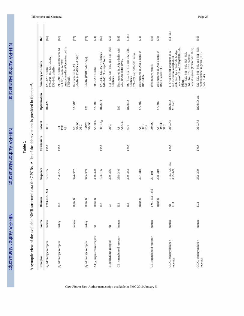
The introduction of the magic-angle spinning and oriented-sample techniques substantiallyaddressed these problems. However, the complexity of the solid-state NMR spectra anddifficulties related to sample preparation have prevented, so far, the solution of the structureof whole GPCRs through NMR. For these reasons, most of the studies have been directedtoward the solution of individual portions of the receptor structure either in aqueous solutionor in different lipid-mimetic solvents [41]. In Table 1, we report a synoptic view of the available3D structural data. Additionally, for the NMR-derived GPCR fragments with 3D coordinatesavailable in the Protein Data Bank, we provide, in Fig. (2) and (3), a structural alignment withthe X-ray structure of rhodopsin and the β-ARs.
Transmembrane domainsWe now know from crystallographic data that some of the TMs present kinks in their α-helicalstructures, particularly pronounces in TM6 and TM7. These data emerged clear from NMRstudies, even before the publication of GPCR X-ray structures. In particular, kinks weredetected studying portions of TM6 of the Saccharomyces cerevisiae α-factor receptor (Ste2pR)[44] and TM7 of the tachykinin receptor NK1 [45] (Table 1). Solution and solid-state NMRstudies of the TMs of the Ste2pR continued after the disclosure of the X-ray structure ofrhodopsin and, among other findings, confirmed the existence of the helical kink of TM6previously detected through solution NMR with the a more sophisticated solid-state NMR ofthe 15N labeled TM6 peptide in 1,2-dimyristoyl-sn-glycero-phosphocholine bilayers [46–49].As mentioned, the kinks of TM6 and TM7 postulated on the basis of the NMR results areconsistent with the conformation of the homologous regions of the receptors that have nowbeen crystallized (Fig. (2), panel A) [12;15], supporting the idea that even structural datagathered for isolated domains can be very informative. Probably, even greater insights couldbe obtained from the analyses of larger substructures or even whole receptors. At this purpose,Zheng and coworkers biosynthesized a quite large segment of the cannabinoid CB2 receptor,including TM1, IL1 and TM2, using a fusion protein overexpression strategy [50]. Thepreliminary results gathered by the authors for the 13C/15N labeled peptide argue in favor ofthe applicability of this strategy to the synthesis of GPCR helical bundles for NMR studies.
Extracellular loopsWhile homology modeling based on the available GPCR X-ray structures allows theconstruction of reliable models of the TMs, modeling the 3D structure of extracellular andintracellular regions remains a difficult task [30]. Low sequence similarity as well as numerousinsertions and deletions, hamper an effective application of homology modeling to theseregions. NMR spectroscopy could prove particularly useful in solving this problem: thestructures of loops and termini of the receptor of interest could be derived through NMR andsubsequently combined with homology models to build complete 3D structures.
Tikhonova and Costanzi Page 3
Curr Pharm Des. Author manuscript; available in PMC 2010 January 5.
NIH
-PA Author Manuscript
NIH
-PA Author Manuscript
NIH
-PA Author Manuscript

To obtain structures of ILs and ELs close to the naturally occurring ones, many experimentaliststurned to the application of structural constraints meant to keep the termini at a distancecompatible with their role of connecting two TMs. In some cases, portions of the adjacent TMshave been added to act as anchors for shackling loops or termini to the lipid environment.Alternatively, the loops have been constrained with disulfide bridges between cysteines [51]or S-carboxymethylcysteines [52] added to the peptide termini, with or without methylenelinkers [53].
Since for most of the GPCRs activated by peptides ligand binding is thought to occur at thelevel of the extracellular domains, NMR analyses have been extensively applied to studyreceptor-peptide interactions. For example, the contact interactions of CCK8–the naturalcholecystokinin peptide, composed of eight amino acids–with the N-terminus and EL3 of theCCK1 and with EL3 of the CCK2 subtypes of the cholecystokinin receptors have been studiedby NMR by monitoring chemical shift perturbations and intermolecular NOEs [54–56] (Table1). For the CCK2 the analysis has been complemented by integrating NMR-derived structuralinformation with rhodopsin-based homology modeling. With a similar strategy, theinteractions between the CCK1 and synthetic agonists have also been studied [57]. Theintegration of homology modeling and NMR-derived information provided also a structuralhypothesis for the binding of substance P to the NK1 receptor, confirming and rationalizingthe results previously obtained through a variety of indirect biochemical methods [58]. Asimilar approach also corroborated the postulated existence of a disulfide bridge between EL1and EL2 in the thromboxane A2 receptor (TP), highlighting also the involvement in ligandbinding of the WCF motif of EL2 [59;60].
Lastly, Pham and coworkers developed an alternative methodology for studying GPCR loopsbased on the computational design of a peptide containing segments that mimic the selfassembling of the ends of two TMs connected by the loop under examination [61]. Whenapplying their strategy to the analyses of EL1 of the sphingosine 1-phosphate receptor S1P4,the authors obtained the best results with the coiled-coil strategy, i.e. by substituting the TM2and TM3 segments with sequences from structurally characterized water-soluble proteins witha pair of antiparallel helices oriented in a way consistent with the S1P4 homology model. Theaddition of a disulfide bridge appeared also necessary to keep the helices together. The abilityof the peptide to bind an analog of the agonist’s headgroup suggested that EL1 adopted abiologically relevant conformation. Here, we superimposed this NMR-derived structure to thehomologous regions in the crystal structure of the β2 receptor (Fig. (2), panel A) and, inagreement with the conclusions of the authors, revealed a sensible conformation and orientationfor the TM2 and TM3 mimetics.
Intracellular loopsSince the ILs, and IL2 in particular, are directly involved in the coupling of the receptors withthe G proteins, [62–64] the experimental elucidation of the structure of these binding interfaceswould provide insights into the molecular mechanisms of the selectivity toward the various Gprotein subtypes.
NMR studies of IL2 in the adrenergic α2A receptor, rhodopsin, the bradykinin B2 receptor, andthe vaspressin V1a receptor have been performed (Table 1) [52;65;66]. In particular, NMRstudies of the peptide corresponding to the IL2 of theα2A receptor revealed the presence of acytoplasmic helix not detected in the analogous rhodopsin peptides [65] or in the crystalstructure of rhodopsin. X-ray crystallography revealed that this helix, although not detected inthe β2, is indeed present in the β1 subtype [7;11].
Conversely, the NMR-derived structure of IL2 of the B2 receptor revealed a “U” shape [66]similar to that detected in rhodopsin. Also in the case of the vasopressin V1A receptor, the
Tikhonova and Costanzi Page 4
Curr Pharm Des. Author manuscript; available in PMC 2010 January 5.
NIH
-PA Author Manuscript
NIH
-PA Author Manuscript
NIH
-PA Author Manuscript

NMR analysis of a linear and a cyclic IL2 peptide revealed a conformation in TFE/H2O solventsimilar to that shown in rhodopsin, particularly with the cyclic peptide [52].
The structure of IL3 has also been determined for several receptors, including the cannabinoidCB1 receptor, the β2 receptor, and the parathyroid hormone PTH1 receptor (Table 1) [53;67;68]. Of note, the IL3 peptide of the CB1 receptor (Fig. (2), panel A) assumed a helicalconformation in aqueous solution when Gαi1 was added, while a mutated IL3 formed just asingle helixturn [68]. The authors proposed the importance of this portion of IL3 in theformation of the G protein binding interface.
Also for the ILs, hybrid solutions composed of molecular modeling and NMR studies havebeen applied. In particular, Wu and coworkers derived by NMR the structure of all three ILsof the thromboxane A2 receptor and subsequently incorporated them into a rhodopsin-basedhomology model [51]. They consequently proposed a list of amino acid residues potentiallyinvolved in the formation of the G protein-binding interface that proved consistent withavailable mutagenesis data.
N-/C- terminiIn addition to the mentioned NMR structure of the CCK1 N-terminus, the structure of the N-terminus of PTH1 receptor has also been solved [69], revealing to be different from that ofrhodopsin (Fig. (2), panel A). These data confirm the idea that each GPCR may be characterizedby unique extracellular regions that contribute to the selectivity of the receptors for specificligands.
All the available GPCR X-ray structures are characterized by the presence of aα-helix, knownas helix 8 (H8), in the portion of the C-terminus proximal to TM7. NMR in conjunction withCD spectroscopy confirmed the presence of an H8 for the CB1 andCB2 receptors, the Ste2preceptor, the β1and β2 receptors, the angiotensin AT1A receptor, and the bradykinin B2 receptorin detergent [70–75]. In Fig. (2), panel B, we superimposed the NMR-derived the β1 receptorH8 to the corresponding segment of the crystal structure of the same receptor, revealing a verygood agreement between crystallographic and NMR data. Conversely, a random coilconformation was detected in water for this region [71–73]. These conformational changes ofH8 have been speculated to be indicative of possible structural rearrangements consequent theactivation of the receptors. For instance, partial unfolding of H8 upon light activation has beendetected in rhodopsin by Fourier transform infrared (FTIR) and fluorescence spectroscopy[76].
Several NMR studies of the rhodopsin C-terminus have been published [33;77;78]. BeyondH8, this region resulted unstructured in aqueous solution, both in unphosphorylated orphosphorylated form, while has adopted a defined 3D structure when phosphorylated and inthe presence of arrestin (Fig. (2), panel C) [78]. Here we could only compare this NMR-derivedstructure (pdb code: 1nzs) with the crystal structures of lumirhodopsin and batorhodopsin (pdbcode: 2hpy and 2g87), for which this distal portion of the C-terminus has been determined.The detected fairly high root mean square deviations of 5.5–6 Å suggest that the C-terminusof rhodopsin undergoes a conformational change when bound to arrestin.
Yeagle’s approach to derive the structure of rhodopsin through NMRBefore the obtainment of high resolution X-ray structures of GPCRs, Yeagle and coworkersdevised a methodology intended to solve the 3D structure of portions of the receptors andsubsequently assemble them together. Through two dimensional homonuclear 1H NMRanalyses in solution, the authors determined the structure of overlapping peptides spanning theentire sequence of the rhodopsin and subsequently computationally assembled the fragments
Tikhonova and Costanzi Page 5
Curr Pharm Des. Author manuscript; available in PMC 2010 January 5.
NIH
-PA Author Manuscript
NIH
-PA Author Manuscript
NIH
-PA Author Manuscript

into a single structure [79–81]. The information about the packing of the helical bundle,necessary to put the pieces together, was gathered from electron paramagnetic spectroscopy(EPS) [36;82;83], zinc crosslinking of histidines [16], and electron cryo-microscopy data[84]. In the form of distance constraints, this was then applied to the NMR-derived fragmentsthrough simulated annealing. Using different sets of experimental data collected for the ground-state and activated (META II) rhodopsin, the authors intended to generate models for bothstates (PDB codes: 1jfp and 1ln6). After the publication of the crystal structure of rhodopsin,the authors discovered that the largest TM peptides yielded the best superimposition with thecorresponding segments of the crystal structure [85].
Here we superimposed Yeagle’s ground state rhodopsin to the crystal structures of the groundstate rhodopsin (PDB code: gzm), detecting a RMSD of the backbone of about 6 Å (Fig. (3),panel A). The packing of the TM helical bundle is similar in the two structures, however,Yeagle’s model predicts the presence of several non helical portions, in particular in TM7, notconfirmed by the crystallography. The intracellular and extracellular regions appearsignificantly divergent in the Yeagle’s model and the crystal structure. We also superimposedYeagle’s Meta II rhodopsin to the recently solved crystal structure of opsin in its G proteininteracting conformation [8], detecting, also in this case, an RMSD of the backbone of about6 Å (Fig. (3), panel B).
Whole GPCRsDispite the difficulties, several attempts to solve the structure of whole GPCRs by NMR arecurrently being undertaken. Solution NMR spectroscopy experiments, conducted for theuniformly 15N-labelled vasopressin V2 receptor, solubilized with lyso-myristoylphosphatidylcholine, indicated the potential suitability of the technique for structuralstudies of GPCRs. Notably, the authors observed over 250 amide peaks out of the expected349 in their 1H,15N-TROSY spectrum [86]. Additionally, solid-state NMR studies of auniformly 15N-labeled and selectively 15N-Ile-labeled chemokine CXCR1 receptor inmagnetically aligned bicelles provided spectra that suggested the potential applicability ofNMR to structural determinations and the elucidation of structure-activity relationships (videinfra) [32].
2. Mapping receptor-ligand interactionsDirect atomic resolution information on the interactions of GPCR with their ligands is availablethrough X-ray crystallography only for a limited number of receptors, while, for the greatmajority of them, indirect methods of analysis have been used. A synergistic application ofmutagenesis studies, photoaffinity labeling, chemical modifications of the ligands, andcomputational modeling has been the most common way of studying the determinants of ligandrecognition. Taken together, these results have contributed to the general definition of acommon binding pocket for GPCRs located towards the extracellular opening of the helicalbundle [87;88]. Also for receptors naturally stimulated by ligands that bind to their N-terminalectodomains, such as the calcium sensing or the glycoprotein hormone receptors, the possibilityof modulation through ligands that bind within their helical bundle has been demonstrated[89–93].
In this context, structure-activity relationships (SAR) analyses and drug-discovery studieswould greatly benefit from direct experimental proofs of receptor-ligand interactions, such asthose that NMR could provide. Solid-state NMR spectroscopy has been applied successfullyto membrane proteins to detect ligand binding and to analyze protein-ligand interactions,especially when coupled to selective isotopic labeling of specific residues located in the bindingpocket and/or of the ligand [94]. Through this expedient, significantly simplified spectra witha manageable amount of signals have been obtained, thus rendering NMR applicable to the
Tikhonova and Costanzi Page 6
Curr Pharm Des. Author manuscript; available in PMC 2010 January 5.
NIH
-PA Author Manuscript
NIH
-PA Author Manuscript
NIH
-PA Author Manuscript

analysis of bioactive ligand conformations, protonation states, receptor/ligand interactions andresidue/residue interactions. Moreover, although not yet for GPCRs, NMR coupled to selectiveisotopic labeling has successfully been applied to rational fragment-based drug discovery. Inparticular, ligands or fragments that bind to specific pockets of a protein have been identifiedby monitoring the changes in the chemical shifts of 15N or 1H-amide atoms in 15N-labelledprotein, an approach known as SAR by NMR [95;96].
Rhodopsin has been extensively studied by solid-state NMR coupled to selective labeling.Before any high-resolution crystal structures were released, selective 15N-labeling of Lysresidues led to the conclusion that the distance between the protonated Schiff base by whichthe retinylidene chromophore is covalently bound to K296(7.43 according to the Ballesterosand Weinstein residue indexing [97]) and the E113(3.28) counterion (Fig. (4)) was greater than4 Å [98]. The subsequent publication of crystal structures, revealed that the distance is, in fact,about 3.5 Å.
The conformation of retinal and its interactions with rhodopsin have also been studiedextensively through NMR coupled to selective labeling. For example, high-resolution solidstate deuterium (2H) NMR experiments provided detailed information on the orientation ofretinal in the binding pocket and on the conformational changes that occur with the transitionfrom the ground state to the Meta I intermediate of the activation cycle. The study applied threeretinal analogues labeled with deuterium at three different pairs of adjacent carbons [99]. Ultrahigh field solid-state magic angle spinning 2D homonuclear and 2D heteronuclear NMRspectroscopy has also been used to study rhodopsin reconstituted with a uniformly 13C-labeled11-cis-retinal. Complete assignment of the 13C and 1H chemical shifts for retinal highlightednonbonding interactions between the protons of the methyl groups of its ionone ring and thenearby aromatic acid residues F208(5.43), F212(5.47), and W265(6.48) (Fig. (4)).Furthermore, it was shown that binding of retinal involves a chiral selection of the ringconformation, resulting in equatorial and axial positions for CH3-16 and CH3-17 [100].Additional evidences of the interactions between retinal and rhodopsin in the ground and MetaI states have also been obtained through NMR experiments conducted variously labeling theligand with 2H [101–104]. These data support the hypothesis that a strain in the polyen aroundthe cis bond assists the photoisomerization of retinal. A 13C-labelled 9-cis retinal isomer,typical of isorhodopsin, has also been investigated, leading to similar conclusions [105].
Application to rhodopsin of an NMR technique named by the authors “selective interfacedetection spectroscopy” (SIDY), based on the detection of the correlations between 13C atomsof labeled ligands and 1H atoms of unlabelled receptors, led to the detection of several contactsbetween the aliphatic carbons of the 11-cis-retinal ionone ringand residues in the bindingpocket. Although, SIDY data do not provide sequence-specific assignments of the contacts,these could be identified by means of additional data, and, in the case of rhodopsin, resultedin good agreement with the available crystallographic structures [106].
NMR spectroscopy provides also a very effective way of studying the conformational changesthat a ligand undergoes upon binding to a receptor. In this context, the analysis of the unboundand receptor-bound 13C,15N-labelled neurotensin, a 13-residue peptide, revealed that the ligandis in a disordered state in the absence of the receptor, while adopts a beta-strand conformationwhen bound to the NTS1 receptor [107]. Similarly, the structure of the bradykinin (BK) peptidebound to the bradykinin B2 receptor has been studied through solid-state NMR, revealing adouble S-shape structure [108]. These type of studies can be also conducted to rationalize thebiological activities of compounds on the basis of their bioactive conformations, as it has beendone in the case of two natural peptides active at the neuropeptide NPR-1 receptor one [109].
Tikhonova and Costanzi Page 7
Curr Pharm Des. Author manuscript; available in PMC 2010 January 5.
NIH
-PA Author Manuscript
NIH
-PA Author Manuscript
NIH
-PA Author Manuscript

In addition to conformational changes, NMR spectroscopy can also detect changes in theprotonation state of a ligand as a consequence of binding to a receptor. In this context, solidstate NMR experiments conducted with uniformly labeled 13C,15N-histamine bound to thehuman histamine H1 receptor revealed that the ligand can bind either in a monocationic or adicationic form. On the basis of their data, the authors hypothesized that, in analogy withrhodopsin, also in the case of the H1 receptor a protonation switch might be part of the activationmechanism [110].
3. Detection of motions and conformational changes associated withreceptor activation
Upon binding of agonists, which typically occurs in proximity of the extracellular opening ofthe helical bundle, GPCRs undergo a series of structural changes that cascade from theextracellular to the intracellular part of the receptor and ultimately lead to G protein activation.Detailed structural knowledge of the ground and activated state of the receptor and mechanisticinsights into the activation process would significantly assist drug discovery, allowing a morerational design of compounds capable of stimulating or blocking the receptor.
A wealth of information has been derived by the analysis of mutations that affect the basalactivity of GPCR, either naturally occurring or generated through site-directed mutagenesis.Mutations that cause an increase of the basal activity are likely to stabilize the activated stateof the receptor and are referred to as constitutively active mutants. Those that prevent theactivation of the receptor are, instead, called uncoupling mutants and are likely to stabilize itsinactive state or to disrupt the cascade of conformational changes that lead to signaling.Through such mutational analyses, various molecular switches for GPCR activation have beenproposed [111;112]. Alternatively, the structural changes that occur upon receptor activationhave been monitored through electron paramagnetic resonance spectroscopy (EPR) [34;36;83;113], UV absorbance spectroscopy [35], engineering of metal-ion-binding sites [16], aswell as site-specific chemical labeling coupled to fluorescence spectroscopy [37], and, as wewill see in the coming paragraphs, NMR spectroscopy. These data shed light onto a numberof intramolecular distances that are characteristic of the activated receptors. We recently usedthese biophysically measured distances to construct a computational structure of the activatedrhodopsin based on coarse-grained and all atom simulations, and subsequently study thedynamics of the activation process [114]. The substantial conformational changes predictedby these indirect methods were not detected in the crystal structure of a photoactivateddeprotonated intermediate (PDI) of rhodopsin that shows absorption maxima consistent withthe META II state [13]. The crystal lattice may have prevented large-scale structuralrearrangements. However, a significant opening of the intracellular surface of the receptor anda furthering of the intracellular ends of TM3 and TM6 have been captured in the crystalstructure of opsin with a fragment of the C-terminus of transducin. At least relatively to thecytosolic half, this may represent the first detailed structure of an activated GPCR [8].
Compared to crystallography, NMR spectroscopy offers the possibility of conducting structuralanalyses in a less constrained membrane-like environment. In this context, NMR is well suitedto the study of receptor activation through isotopic labeling of specific residues located in areasaffected by the structural changes. In this context, the specific labeling of rhodopsin led toobservance of the transition between its inactive and activated states.
2D-dipolar-assisted rotational resonance NMR measurements between 13C-labels on the C14,C15, C19, C20 atoms of retinal and 13C-labeled G114(3.29), T118(3.33), G121(3.36), Y178(4.68), G188(EL2), Y191(EL2), S196(EL2), and Y268(6.51), all located in the retinal bindingpocket, was performed for the ground state and META II rhodopsin (Fig. (5), panel A, purpleand green spheres indicate retinal and rhodopsin atoms, respectively) [115]. The results
Tikhonova and Costanzi Page 8
Curr Pharm Des. Author manuscript; available in PMC 2010 January 5.
NIH
-PA Author Manuscript
NIH
-PA Author Manuscript
NIH
-PA Author Manuscript

highlighted that the extracellular portion of the receptor undergoes conformational changesupon activation. In particular, a large rotation of the C20 methyl group of retinal toward EL2and also a translation of about 4–5 Å of the retinal chromophore toward TM5 were observed.This displacement of retinal has been associated with the motions of the TM5, TM6, and TM7predicted by different biophysical and biochemical methods [115]. Subsequent measurementsbetween the C19 and C20-methyl groups of retinal and 13C-labeled W265(6.48) confirmedthat, in agreement with what suggested by atomic microscopy [38], the movement of the beta-ionon ring causes a reorientation of the side chain of W265(6.48) upon activation (Fig. (5),panel A, a blue sphere indicates W265). In agreement with what seen in the crystal structureof opsin [8], the study also suggested that TM6, but not TM3, undergoes a significantrearrangement [116]. Most likely, the local movements triggered by the isomerization of retinalcause a perturbation of the complex network of hydrogen bonds that stabilizes the helicalbundle of the ground state rhodopsin. This is suggested also by the comparison of the chemicalshifts of 15N and 13C-labeled wild type and mutant rhodopsin in the ground and META II states[117], which, among other observations, led to the detection of the disruption of the stronghydrogen bond between Glu 122 in TM3 and H211 in TM5 (Fig. (5), pink spheres indicateGlu122 and H211), thus indicating a motion of TM5. A more recent study demonstrated thatthe transition to META II is accompanied also by a displacement of EL2 from the retinalbinding site, and suggested that this displacement is coupled to the rotation of TM5 and thebreakage of the ionic lock connecting TM3 and TM6 [118]. As mentioned, all theseinterconnected conformational changes caused by the photoisomerization of retinal eventuallycascade down towards the cytosolic portion of the TMs, thus leading to G protein activation.To specifically monitor the changes that occur in this region, NMR spectroscopy of 19F labeledrhodopsin has been applied after mutation of specific residues to Cys and subsequentattachment of trifluoroethylthio groups via disulfide bridges (C65, C139, C140, C251, C316,indicated by yellow spheres in Fig. (5), panel A). The results support the idea that the tertiarystructure of the cytoplasmic face of the receptor changes significantly upon light activation[119;120].
Beyond the activation process, NMR analyses of 15N labeled rhodopsin have been applied alsoto the study of the dynamics of the dark adapted receptor. In particular, a study on α-15N-Lys-labeled rhodopsin revealed that, while the single Lys residue located in the C-terminus isendowed with nanosecond scale movements, those located in different regions of the receptorspresent micro- to millisecond timescale motions (Fig. (5), panel B). In contrast with Lysresidues, which in rhodopsin are located mostly in the cytosolic domains, Trp residues are, forthe most part, present in the TMs (Fig. (5), panel B). A subsequent study of α and ε-15N-Trp-labeled rhodopsin, confirmed that there are micro- to millisecond timescale backbone motionsin the inactive dark state, while suggesting a substantial restriction of the Trp side chains to asingle specific conformation [121;122].
ConclusionDespite the numerous technical challenges, NMR spectroscopy has the potential of becominga leading technique in the study of the structure-function relationships of GPCRs and theirinteractions with ligands. In contrast to crystallographic methods, which provide high-resolution but static molecular snapshots, NMR techniques can provide dynamic pictures ofreceptor structures and receptor-ligand interactions, thus offering insights into the molecularmechanisms of ligand recognition and receptor activation.
Through this article, we reviewed many of the contributions that NMR studies have given tothe understanding of the molecular structure and functioning of GPCRs. In particular, NMRspectroscopy has been extensively applied to the study of individual portions of GPCRs, alsoin combination with homology modeling. Through selective labeling of ligands and/or specific
Tikhonova and Costanzi Page 9
Curr Pharm Des. Author manuscript; available in PMC 2010 January 5.
NIH
-PA Author Manuscript
NIH
-PA Author Manuscript
NIH
-PA Author Manuscript
receptor residues, NMR studies have also been applied to the study of GPCR-ligandinteractions and to the investigations of the molecular changes coupled to activation of thereceptor.
Recently, dynamic nuclear polarization (DNP), a technique intended to increase the NMRsensitivity by transferring the polarization of electron spins to nuclei, has been successfullyapplied to the study the photocycle of bacteriorhodopsin, a 7TM light-driven ion pump withsome structural analogies to rhodopsin [123]. Due to the enhanced sensitivity of theexperiments, DNP allows the analysis of low concentration samples in a lower timeframe, andthe detection of low populated conformations.
The introduction of DNP, as well as other advances in solution and solid-state NMRtechnology, together with progresses in the preparation of GPCR samples, are expected to leadin the future to the determination of the 3D structure of whole GPCRs, and to provide furthermechanistic insights into the complex cascade of motions and rearrangements characteristicof the activation process. Furthermore, these methodological advances are also likely to fosterthe application of SAR by NMR techniques to the discovery of lead compounds for GPCRsthrough NMR-based high throughput screenings and their subsequent structure-basedoptimization.
AcknowledgmentsThis research was supported by the Intramural Research Program of the NIH, National Institute of Diabetes andDigestive and Kidney Diseases.
References1. Pierce KL, Premont RT, Lefkowitz RJ. Seven-transmembrane receptors. Nature Reviews Molecular
Cell Biology 2002;3:639–650.2. Takeda S, Kadowaki S, Haga T, Takaesu H, Mitaku S. Identification of G protein-coupled receptor
genes from the human genome sequence. Febs Letters 2002;520:97–101. [PubMed: 12044878]3. Yeagle PL, Albert AD. G-protein coupled receptor structure. Biochimica et Biophysica Acta-
Biomembranes 2007;1768:808–824.4. Li J, Edwards PC, Burghammer M, Villa C, Schertler GF. Structure of bovine rhodopsin in a trigonal
crystal form. J Mol Biol 2004;343:1409–1438. [PubMed: 15491621]5. Okada T, Sugihara M, Bondar AN, Elstner M, Entel P, Buss V. The retinal conformation and its
environment in rhodopsin in light of a new 2.2 Å crystal structure. J Mol Biol 2004;342:571–583.[PubMed: 15327956]
6. Jaakola VP, Griffith MT, Hanson MA, Cherezov V, Chien EY, Lane JR, et al. The 2.6 AngstromCrystal Structure of a Human A2A Adenosine Receptor Bound to an Antagonist. Science2008;322:1211–7. [PubMed: 18832607]
7. Warne T, Serrano-Vega MJ, Baker JG, Moukhametzianov R, Edwards PC, Henderson R, et al.Structure of a beta(1)-adrenergic G-protein-coupled receptor. Nature 2008;454:486–4U2. [PubMed:18594507]
8. Scheerer P, Park JH, Hildebrand PW, Kim YJ, Krauss N, Choe HW, et al. Crystal structure of opsinin its G-protein-interacting conformation. Nature 2008;455:497–U30. [PubMed: 18818650]
9. Park JH, Scheerer P, Hofmann KP, Choe HW, Ernst OP. Crystal structure of the ligand-free G-protein-coupled receptor opsin. Nature 2008;454:183–U33. [PubMed: 18563085]
10. Cherezov V, Rosenbaum DM, Hanson MA, Rasmussen SG, Thian FS, Kobilka TS, et al. High-resolution crystal structure of an engineered human beta2-adrenergic G protein-coupled receptor.Science 2007;318:1258–1265. [PubMed: 17962520]
11. Rosenbaum DM, Cherezov V, Hanson MA, Rasmussen SG, Thian FS, Kobilka TS, et al. GPCRengineering yields high-resolution structural insights into beta2-adrenergic receptor function.Science 2007;318:1266–1273. [PubMed: 17962519]
Tikhonova and Costanzi Page 10
Curr Pharm Des. Author manuscript; available in PMC 2010 January 5.
NIH
-PA Author Manuscript
NIH
-PA Author Manuscript
NIH
-PA Author Manuscript

12. Rasmussen SG, Choi HJ, Rosenbaum DM, Kobilka TS, Thian FS, Edwards PC, et al. Crystal structureof the human beta2 adrenergic G-protein-coupled receptor. Nature 2007;450:383–387. [PubMed:17952055]
13. Salom D, Lodowski DT, Stenkamp RE, Le TI, Golczak M, Jastrzebska B, et al. Crystal structure ofa photoactivated deprotonated intermediate of rhodopsin. Proc Natl Acad Sci U S A 2006;103:16123–16128. [PubMed: 17060607]
14. Schertler GFX, Villa C, Henderson R. Projection Structure of Rhodopsin. Nature 1993;362:770–772.[PubMed: 8469290]
15. Palczewski K, Kumasaka T, Hori T, Behnke CA, Motoshima H, Fox BA, et al. Crystal structure ofrhodopsin: A G protein-coupled receptor. Science 2000;289:739–745. [PubMed: 10926528]
16. Sheikh SP, Zvyaga TA, Lichtarge O, Sakmar TP, Bourne HR. Rhodopsin activation blocked by metal-ion-binding sites linking transmembrane helices C and F. Nature 1996;383:347–350. [PubMed:8848049]
17. Marco E, Foucaud M, Langer I, Escrieut C, Tikhonova IG, Fourmy D. Mechanism of activation of aG protein-coupled receptor, the human cholecystokinin-2 receptor. J Biol Chem 2007;282:28779–28790. [PubMed: 17599907]
18. Tan YV, Couvineau A, Murail S, Ceraudo E, Neumann JM, Lacapere JJ, et al. Peptide agonist dockingin the N-terminal ectodomain of a class II G protein-coupled receptor, the VPAC1 receptor -Photoaffinity, NMR, and molecular modeling. Journal of Biological Chemistry 2006;281:12792–12798. [PubMed: 16520374]
19. Dawson ES, Henne RM, Miller LJ, Lybrand TP. Molecular models for cholecystokinin-A receptor.Pharmacology & Toxicology 2002;91:290–296. [PubMed: 12688371]
20. Turek JW, Halmos T, Sullivan NL, Antonakis K, Le Breton GC. Mapping of a ligand-binding sitefor the human thromboxane A(2) receptor protein. Journal of Biological Chemistry 2002;277:16791–16797. [PubMed: 11877412]
21. Hoffmann C, Gaietta G, Bunemann M, Adams SR, Oberdorff-Maass S, Behr B, et al. A FlAsH-basedFRET approach to determine G protein - coupled receptor activation in living cells. Nature Methods2005;2:171–176. [PubMed: 15782185]
22. Kristiansen K. Molecular mechanisms of ligand binding, signaling, and regulation within thesuperfamily of G-protein-coupled receptors: molecular modeling and mutagenesis approaches toreceptor structure and function. Pharmacology & Therapeutics 2004;103:21–80. [PubMed:15251227]
23. Moro S, Spalluto G, Jacobson KA. Techniques: Recent developments in computer-aided engineeringof GPCR ligands using the human adenosine A(3) receptor as an example. Trends in PharmacologicalSciences 2005;26:44–51. [PubMed: 15629204]
24. Moro S, Deflorian F, Bacilieri M, Spalluto G. Ligand-based homology modeling as attractive tool toinspect GPCR structural plasticity. Current Pharmaceutical Design 2006;12:2175–2185. [PubMed:16796562]
25. Patny A, Desai PV, Avery MA. Homology modeling of G-protein-coupled receptors and implicationsin drug design. Current Medicinal Chemistry 2006;13:1667–1691. [PubMed: 16787212]
26. Tikhonova IG, Sum CS, Neumann S, Thomas CJ, Raaka BM, Costanzi S, et al. Bidirectional, IterativeApproach to the Structural Delineation of the Functional “Chemoprint” in GPR40 for AgonistRecognition. J Med Chem 2007;50:2981–2989. [PubMed: 17552505]
27. Engel S, Skoumbourdis AP, Childress J, Neumann S, Deschamps JR, Thomas CJ, et al. A virtualscreen for diverse ligands: discovery of selective g protein-coupled receptor antagonists. Journal ofthe American Chemical Society 2008;130:5115–5123. [PubMed: 18357984]
28. Kellenberger E, Springael JY, Parmentier M, Hachet-Haas M, Galzi JL, Rognan D. Identification ofnonpeptide CCR5 receptor agonists by structure-based virtual screening. Journal of MedicinalChemistry 2007;50:1294–1303. [PubMed: 17311371]
29. Tikhonova IG, Sum CS, Neumann S, Engel S, Raaka BM, Costanzi S, et al. Discovery of novelAgonists and antagonists of the free fatty acid receptor 1 (FFAR1) using virtual screening. Journalof Medicinal Chemistry 2008;51:625–633. [PubMed: 18193825]
Tikhonova and Costanzi Page 11
Curr Pharm Des. Author manuscript; available in PMC 2010 January 5.
NIH
-PA Author Manuscript
NIH
-PA Author Manuscript
NIH
-PA Author Manuscript

30. Costanzi S. On the Applicability of GPCR Homology Models to Computer-Aided Drug Discovery:A Comparison between In Silico and Crystal Structures of the beta2-Adrenergic Receptor. J MedChem 2008;51:2907–2914. [PubMed: 18442228]
31. Peleg G, Ghanouni P, Kobilka BK, Zare RN. Single-molecule spectroscopy of the beta(2) adrenergicreceptor: Observation of conformational substates in a membrane protein. Proceedings of theNational Academy of Sciences of the United States of America 2001;98:8469–8474. [PubMed:11438704]
32. Park SH, Prytulla S, De Angelis AA, Brown JM, Kiefer H, Opella SJ. High-resolution NMRspectroscopy of a GPCR in aligned bicelles. Journal of the American Chemical Society2006;128:7402–7403. [PubMed: 16756269]
33. Werner K, Richter C, Klein-Seetharaman J, Schwalbe H. Isotope labeling of mammalian GPCRs inHEK293 cells and characterization of the C-terminus of bovine rhodopsin by high resolution liquidNMR spectroscopy. Journal of Biomolecular Nmr 2008;40:49–53. [PubMed: 17999150]
34. Altenbach C, Cai KW, Klein-Seetharaman J, Khorana FG, Hubbell WL. Structure and function inrhodopsin: Mapping light-dependent changes in distance between residue 65 in helix TM1 andresidues in the sequence 306–319 at the cytoplasmic end of helix TM7 and in helix H8. Biochemistry2001;40:15483–15492. [PubMed: 11747423]
35. Lin SW, Sakmar TP. Specific tryptophan UV-absorbance changes are probes of the transition ofrhodopsin to its active state. Biochemistry 1996;35:11149–11159. [PubMed: 8780519]
36. Farrens DL, Altenbach C, Yang K, Hubbell WL, Khorana HG. Requirement of rigid-body motion oftransmembrane helices for light activation of rhodopsin. Science 1996;274:768–770. [PubMed:8864113]
37. Dunham TD, Farrens DL. Conformational changes in rhodopsin. Movement of helix f detected bysite-specific chemical labeling and fluorescence spectroscopy. J Biol Chem 1999;274:1683–1690.[PubMed: 9880548]
38. Ruprecht JJ, Mielke T, Vogel R, Villa C, Schertler GF. Electron crystallography reveals the structureof metarhodopsin I. EMBO J 2004;23:3609–3620. [PubMed: 15329674]
39. Vogtherr, M.; Fiebig, K. Modern Methods of Drug Discovery. In: Hillish, A.; Hilgenfeld, R., editors.Birkhauser. 2003. p. 183-202.
40. Homans SW. NMR spectroscopy tools for structure-aided drug design. Angewandte Chemie-International Edition 2004;43:290–300.
41. Ratnala VRP. New tools for G-protein coupled receptor (GPCR) drug discovery: combination ofbaculoviral expression system and solid state NMR. Biotechnology Letters 2006;28:767–778.[PubMed: 16786240]
42. Nielsen NC, Malmendal A, Vosegaard T. Techniques and applications of NMR to membrane proteins(Review). Molecular Membrane Biology 2004;21:129–141. [PubMed: 15204621]
43. Sanders CR, Sonnichsen F. Solution NMR of membrane proteins: practice and challenges. MagneticResonance in Chemistry 2006;44:S24–S40. [PubMed: 16826539]
44. Arshava B, Liu SF, Jiang HL, Breslav M, Becker JM, Naider F. Structure of segments of a G protein-coupled receptor: CD and NMR analysis of the Saccharomyces cerevisiae tridecapeptide pheromonereceptor. Biopolymers 1998;46:343–357. [PubMed: 9798427]
45. Berlose JP, Convert O, Brunissen A, Chassaing G, Lavielle S. 3-Dimensional Structure of the HighlyConserved 7Th Transmembrane Domain of G-Protein-Coupled Receptors. European Journal ofBiochemistry 1994;225:827–843. [PubMed: 7957220]
46. Arevalo E, Estephan R, Madeo J, Arshava B, Dumont M, Becker JM, et al. Biosynthesis andbiophysical analysis of domains of a yeast G protein-coupled receptor. Biopolymers 2003;71:516–531. [PubMed: 14517901]
47. Naider F, Arshava B, Ding FX, Arevalo E, Becker JM. Peptide fragments as models to study thestructure of a G-protein coupled receptor: The alpha-factor receptor of Saccharomyces cerevisiae.Biopolymers 2001;60:334–350. [PubMed: 12115145]
48. Estephan R, Englander J, Arshava B, Samples KL, Becker JM, Naider F. Biosynthesis and NMRanalysis of a 73-residue domain of a Saccharomyces cerevisiae G protein-coupled receptor.Biochemistry 2005;44:11795–11810. [PubMed: 16128581]
Tikhonova and Costanzi Page 12
Curr Pharm Des. Author manuscript; available in PMC 2010 January 5.
NIH
-PA Author Manuscript
NIH
-PA Author Manuscript
NIH
-PA Author Manuscript

49. Valentine KG, Liu SF, Marassi FM, Veglia G, Opella SJ, Ding FX, et al. Structure and topology ofa peptide segment of the 6th transmembrane domain of the Saccharomyces cerevisae alpha-factorreceptor in phospholipid bilayers. Biopolymers 2001;59:243–256. [PubMed: 11473349]
50. Zheng HA, Zhao J, Sheng WY, Xie XQ. A transmembrane helix-bundle from G-protein coupledreceptor CB2: Biosynthesis, purification, and NMR characterization. Biopolymers 2006;83:46–61.[PubMed: 16634087]
51. Wu JX, Feng M, Ruan KH. Assembling NMR structures for the intracellular loops of the humanthromboxane A(2) receptor: Implication of the G protein-coupling pocket. Archives of Biochemistryand Biophysics 2008;470:73–82. [PubMed: 18073117]
52. Demene H, Granier S, Muller D, Guillon G, Dufour MN, Delsuc MA, et al. Active peptidic mimicsof the second intracellular loop of the V-1A vasopressin receptor are structurally related to the secondintracellular rhodopsin loop: A combined H-1 NMR and biochemical study. Biochemistry2003;42:8204–8213. [PubMed: 12846569]
53. Mierke DF, Royo M, Pellegrini M, Sun HM, Chorev M. Peptide mimetic of the third cytoplasmicloop of the PTH/PTHrP receptor. Journal of the American Chemical Society 1996;118:8998–9004.
54. Pellegrini M, Mierke DF. Molecular complex of cholecystokinin-8 and N-terminus of thecholecystokinin A receptor by NMR spectroscopy. Biochemistry 1999;38:14775–14783. [PubMed:10555959]
55. Giragossian C, Mierke DF. Intermolecular interactions between cholecystokinin-8 and the thirdextracellular loop of the cholecystokinin A receptor. Biochemistry 2001;40:3804–3809. [PubMed:11300760]
56. Giragossian C, Mierke DF. Intermolecular interactions between cholecystokinin-8 and the thirdextracellular loop of the cholecystokinin-2 receptor. Biochemistry 2002;41:4560–4566. [PubMed:11926817]
57. Giragossian C, Sugg EE, Szewczyk JR, Mierke DF. Intermolecular interactions between peptidic andnonpeptidic agonists and the third extracellular loop of the cholecystokinin 1 receptor. Journal ofMedicinal Chemistry 2003;46:3476–3482. [PubMed: 12877585]
58. Ulfers AL, Piserchio A, Mierke DF. Extracellular domains of the neurokinin-1 receptor: Structuralcharacterization and interactions with substance P. Biopolymers 2002;66:339–349. [PubMed:12539262]
59. Ruan KH, Wu JX, So SP, Jenkins LA, Ruan CH. NMR structure of the thromboxane A(2) receptorligand recognition pocket. European Journal of Biochemistry 2004;271:3006–3016. [PubMed:15233797]
60. Ruan KH, So SP, Wu JX, Li DW, Huang AM, Kung J. Solution structure of the second extracellularloop of human thromboxane A(2) receptor. Biochemistry 2001;40:275–280. [PubMed: 11141080]
61. Pham TCT, Kriwacki RW, Parrill AL. Peptide design and structural characterization of a GPCR loopmimetic. Biopolymers 2007;86:298–310. [PubMed: 17443712]
62. Kushwaha N, Harwood SC, Wilson AM, Berger M, Tecott LH, Roth BL, et al. Molecular determinantsin the second intracellular loop of the 5-hydroxytryptamine-1A receptor for G-protein coupling.Molecular Pharmacology 2006;69:1518–1526. [PubMed: 16410407]
63. Neumann S, Krause G, Claus M, Paschke R. Structural determinants for G protein activation andselectivity in the second intracellular loop of the thyrotropin receptor. Endocrinology 2005;146:477–485. [PubMed: 15498884]
64. Havlickova M, Blahos J, Brabet I, Liu JF, Hruskova B, Prezeau L, et al. The second intracellular loopof metabotropic glutamate receptors recognizes C termini of G-protein alpha-subunits. Journal ofBiological Chemistry 2003;278:35063–35070. [PubMed: 12829705]
65. Chung DA, Zuiderweg ERP, Fowler CB, Soyer OS, Mosberg HI, Neubig RR. NMR structure of thesecond intracellular loop of the alpha 2A adrenergic receptor: Evidence for a novel cytoplasmic helix.Biochemistry 2002;41:3596–3604. [PubMed: 11888275]
66. Piserchio A, Prado GN, Zhang R, Yu J, Taylor L, Polgar P, et al. Structural insight into the role ofthe second intracellular loop of the bradykinin 2 receptor in signaling and internalization.Biopolymers 2002;63:239–246. [PubMed: 11807751]
Tikhonova and Costanzi Page 13
Curr Pharm Des. Author manuscript; available in PMC 2010 January 5.
NIH
-PA Author Manuscript
NIH
-PA Author Manuscript
NIH
-PA Author Manuscript

67. Jung H, Windhaber R, Palm D, Schnackerz KD. Nmr and Circular-Dichroism Studies of SyntheticPeptides Derived from the 3Rd Intracellular Loop of the Beta-Adrenoceptor. Febs Letters1995;358:133–136. [PubMed: 7828722]
68. Ulfers AL, McMurry JL, Miller A, Wang LG, Kendall DA, Mierke DF. Cannabinoid receptor-Gprotein interactions: G(alpha i1)-bound structures of IC3 and a mutant with altered G proteinspecificity. Protein Science 2002;11:2526–2531. [PubMed: 12237474]
69. Pellegrini M, Bisello A, Rosenblatt M, Chorev M, Mierke DF. Binding domain of human parathyroidhormone receptor: From conformation to function. Biochemistry 1998;37:12737–12743. [PubMed:9737850]
70. Choi G, Landin J, Xie XQ. The cytoplasmic helix of cannabinoid receptor CB2, a conformationalstudy by circular dichroism and H-1 NMR spectroscopy in aqueous and membrane-likeenvironments. Journal of Peptide Research 2002;60:169–177. [PubMed: 12213126]
71. Choi G, Guo JX, Makriyannis A. The conformation of the cytoplasmic helix 8 of the CB1 cannabinoidreceptor using NMR and circular dichroism. Biochimica et Biophysica Acta-Biomembranes2005;1668:1–9.
72. Katragadda M, Maciejewski MW, Yeagle PL. Structural studies of the putative helix 8 in the humanbeta(2) adrenergic receptor: an NMR study. Biochimica et Biophysica Acta-Biomembranes2004;1663:74–81.
73. Jung H, Windhaber R, Palm D, Schnackerz KD. Conformation of a beta-adrenoceptor-derived signaltransducing peptide as inferred by circular dichroism and H-1 NMR spectroscopy. Biochemistry1996;35:6399–6405. [PubMed: 8639586]
74. Franzoni L, Nicastro G, Pertinhez TA, Tato M, Nakaie CR, Paiva ACM, et al. Structure of the C-terminal fragment 300–320 of the rat angiotensin II AT(1A) receptor and its relevance with respectto G-protein coupling. Journal of Biological Chemistry 1997;272:9734–9741. [PubMed: 9092505]
75. Piserchio A, Zelesky V, Yu J, Taylor L, Polgar P, Mierke DF. Bradykinin B2 receptor signaling:Structural and functional characterization of the C-terminus. Biopolymers 2005;80:367–373.[PubMed: 15682437]
76. Lehmann N, Alexiev U, Fahmy K. Linkage between the intramembrane H-bond network aroundaspartic acid 83 and the cytosolic environment of helix 8 in photoactivated rhodopsin. Journal ofMolecular Biology 2007;366:1129–1141. [PubMed: 17196983]
77. Getmanova E, Patel AB, Klein-Seetharaman J, Loewen MC, Reeves PJ, Friedman N, et al. NMRspectroscopy of phosphorylated wild-type rhodopsin: Mobility of the phosphorylated C-terminus ofrhodopsin in the dark and upon light activation. Biochemistry 2004;43:1126–1133. [PubMed:14744159]
78. Kisselev OG, McDowell JH, Hargrave PA. The arrestin-bound conformation and dynamics of thephosphorylated carboxy-terminal region of rhodopsin. Febs Letters 2004;564:307–311. [PubMed:15111114]
79. Choi G, Landin J, Galan JF, Birge RR, Albert AD, Yeagle PL. Structural studies of metarhodopsinII, the activated form of the G-protein coupled receptor, rhodopsin. Biochemistry 2002;41:7318–7324. [PubMed: 12044163]
80. Yeagle PL, Choi G, Albert AD. Studies on the structure of the G-protein-coupled receptor rhodopsinincluding the putative G-protein binding site in unactivated and activated forms. Biochemistry2001;40:11932–11937. [PubMed: 11570894]
81. Yeagle PL, Alderfer JL, Albert AD. Structure of the third cytoplasmic loop of bovine rhodopsin.Biochemistry 1995;34:14621–14625. [PubMed: 7578070]
82. Cai K, Itoh Y, Khorana HG. Mapping of contact sites in complex formation between transducin andlight-activated rhodopsin by covalent crosslinking: use of a photoactivatable reagent. Proc Natl AcadSci U S A 2001;98:4877–4882. [PubMed: 11320237]
83. Cai K, Klein-Seetharaman J, Hwa J, Hubbell WL, Khorana HG. Structure and function in rhodopsin:Effects of disulfide cross-links in the cytoplasmic face of rhodopsin on transducin activation andphosphorylation by rhodopsin kinase. Biochemistry 1999;38:12893–12898. [PubMed: 10504260]
84. Unger VM, Schertler GFX. Low-Resolution Structure of Bovine Rhodopsin Determined by ElectronCryomicroscopy. Biophysical Journal 1995;68:1776–1786. [PubMed: 7612819]
Tikhonova and Costanzi Page 14
Curr Pharm Des. Author manuscript; available in PMC 2010 January 5.
NIH
-PA Author Manuscript
NIH
-PA Author Manuscript
NIH
-PA Author Manuscript

85. Katragadda M, Chopra A, Bennett M, Alderfer JL, Yeagle PL, Albert AD. Structures of thetransmembrane helices of the G-protein coupled receptor, rhodopsin. Journal of Peptide Research2001;58:79–89. [PubMed: 11454172]
86. Tian CL, Breyer RM, Kim HJ, Karra MD, Friedman DB, Karpay A, et al. Solution NMR spectroscopyof the human vasopressin V2 receptor, a G protein-coupled receptor. Journal of the AmericanChemical Society 2005;127:8010–8011. [PubMed: 15926814]
87. Klabunde T, Hessler G. Drug design strategies for targeting G-protein-coupled receptors.Chembiochem 2002;3:929–944.
88. Surgand JS, Rodrigo J, Kellenberger E, Rognan D. A chemogenomic analysis of the transmembranebinding cavity of human G-protein-coupled receptors. Proteins 2006;62:509–538. [PubMed:16294340]
89. Jaschke H, Neumann S, Moore S, Thomas CJ, Colson AO, Costanzi S, et al. A low molecular weightagonist signals by binding to the transmembrane domain of thyroid-stimulating hormone receptor(TSHR) and luteinizing hormone/chorionic gonadotropin receptor (LHCGR). Journal of BiologicalChemistry 2006;281:9841–9844. [PubMed: 16488885]
90. Moore S, Jaeschke H, Kleinau G, Neumann S, Costanzi S, Jiang JK, et al. Evaluation of small-molecule modulators of the luteinizing hormone/choriogonadotropin and thyroid stimulatinghormone receptors: Structure-activity relationships and selective binding patterns. Journal ofMedicinal Chemistry 2006;49:3888–3896. [PubMed: 16789744]
91. Hu JX, Jiang JK, Costanzi S, Thomas C, Yang W, Feyen JHM, et al. A missense mutation in theseven-transmembrane domain of the human Ca2+ receptor converts a negative allosteric modulatorinto a positive allosteric modulator. Journal of Biological Chemistry 2006;281:21558–21565.[PubMed: 16735501]
92. Heitman LH, Oosterom J, Bonger KM, Timmers CM, Wiegerinck PHG, Ijzerman AP. [H-3]Org43553, the first low-molecular-weight agonistic and allosteric Radioligand for the human luteinizinghormone receptor. Molecular Pharmacology 2008;73:518–524. [PubMed: 17989351]
93. Neumann S, Kleinau G, Costanzi S, Moore S, Jiang JK, Raaka BM, et al. A Low Molecular WeightAntagonist for the Human Thyrotropin Receptor with Therapeutic Potential for Hyperthyroidism.Endocrinology 2008;149:5945–5950. [PubMed: 18669595]
94. Middleton DA. Solid-state NMR spectroscopy as a tool for drug design: from membrane-embeddedtargets to amyloid fibrils. Biochemical Society Transactions 2007;35:985–990. [PubMed: 17956260]
95. Shuker SB, Hajduk PJ, Meadows RP, Fesik SW. Discovering high-affinity ligands for proteins: SARby NMR. Science 1996;274:1531–1534. [PubMed: 8929414]
96. Hajduk PJ. SAR by NMR: Putting the pieces together. Molecular Interventions 2006;6:266–272.[PubMed: 17035667]
97. Ballesteros JA, Weinstein H. Integrated methods for the construction of three-dimensional modelsand computational probing of structure-function relations in G-protein coupled receptors. MethodsNeurosci 1995;25:366–428.
98. Eilers M, Reeves PJ, Ying WW, Khorana HG, Smith SO. Magic angle spinning NMR of the protonatedretinylidene Schiff base nitrogen in rhodopsin: Expression of N-15-lysine-and C-13-glycine-labeledopsin in a stable cell line. Proceedings of the National Academy of Sciences of the United States ofAmerica 1999;96:487–492. [PubMed: 9892660]
99. Grobner G, Burnett IJ, Glaubitz C, Choi G, Mason AJ, Watts A. Observations of light-inducedstructural changes of retinal within rhodopsin. Nature 2000;405:810–813. [PubMed: 10866205]
100. Creemers AFL, Kiihne S, Bovee-Geurts PHM, Degrip WJ, Lugtenburg J, de Groot HJM. H-1 andC-13 MAS NMR evidence for pronounced ligand-protein interactions involving the ionone ring ofthe retinylidene chromophore in rhodopsin. Proceedings of the National Academy of Sciences ofthe United States of America 2002;99:9101–9106. [PubMed: 12093898]
101. Brown MF, Heyn MP, Job C, Kim S, Moltke S, Nakanishi K, et al. Solid-State H-2 NMRspectroscopy of retinal proteins in aligned membranes. Biochimica et Biophysica Acta-Biomembranes 2007;1768:2979–3000.
102. Salgado GFJ, Struts AV, Tanaka K, Fujioka N, Nakanishi K, Brown MF. Deuterium NMR structureof retinal in the ground state of rhodopsin. Biochemistry 2004;43:12819–12828. [PubMed:15461454]
Tikhonova and Costanzi Page 15
Curr Pharm Des. Author manuscript; available in PMC 2010 January 5.
NIH
-PA Author Manuscript
NIH
-PA Author Manuscript
NIH
-PA Author Manuscript

103. Salgado GFJ, Struts AV, Tanaka K, Krane S, Nakanishi K, Brown MF. Solid-state H-2 NMRstructure of retinal in metarhodopsin I. Journal of the American Chemical Society 2006;128:11067–11071. [PubMed: 16925423]
104. Struts AV, Salgado GFJ, Tanaka K, Krane S, Nakanishi K, Brown MF. Structural analysis anddynamics of retinal chromophore in dark and metal states of rhodopsin from H-2 NMR of alignedmembranes. J Mol Biol 2007;372:50–66. [PubMed: 17640664]
105. Creemers AFL, Bovee-Geurts PHM, Degrip WJ, Lugtenburg J, de Groot HJM. Solid-state NMRanalysis of ligand-receptor interactions reveals an induced misfit in the binding site of isorhodopsin.Biochemistry 2004;43:16011–16018. [PubMed: 15609995]
106. Kiihne SR, Creemers AFL, de Grip WJ, Bovee-Geurts PHM, Lugtenburg J, de Groot HJM. Selectiveinterface detection: Mapping binding site contacts in membrane proteins by NMR spectroscopy.Journal of the American Chemical Society 2005;127:5734–5735. [PubMed: 15839640]
107. Luca S, White JF, Sohal AK, Filippov DV, van Boom JH, Grisshammer R, et al. The conformationof neurotensin bound to its G protein-coupled receptor. Proceedings of the National Academy ofSciences of the United States of America 2003;100:10706–10711. [PubMed: 12960362]
108. Lopez JJ, Shukla AK, Reinhart C, Schwalbe H, Michel H, Glaubitz C. The structure of theneuropeptide bradykinin bound to the human G-protein coupled receptor bradykinin B2 asdetermined by solid-state NMR spectroscopy. Angewandte Chemie-International Edition2008;47:1668–1671.
109. Dossey AT, Reale V, Chatwin H, Zachariah C, Debono M, Evans PD, et al. NMR analysis ofCaenorhabditis elegans FLP-18 neuropeptides: Implications for NPR-1 activation. Biochemistry2006;45:7586–7597. [PubMed: 16768454]
110. Ratnala VRP, Kiihne SR, Buda F, Leurs R, de Groot HJM, Degrip WJ. Solid-state NMR evidencefor a protonation switch in the binding pocket of the H1 receptor upon binding of the agonisthistamine. Journal of the American Chemical Society 2007;129:867–872. [PubMed: 17243823]
111. Ballesteros JA, Jensen AD, Liapakis G, Rasmussen SGF, Shi L, Gether U, et al. Activation of thebeta(2)-adrenergic receptor involves disruption of an ionic lock between the cytoplasmic ends oftransmembrane segments 3 and 6. Journal of Biological Chemistry 2001;276:29171–29177.[PubMed: 11375997]
112. Shi L, Liapakis G, Xu R, Guarnieri F, Ballesteros JA, Javitch JA. beta2 Adrenergic receptoractivation. Modulation of the proline kink in transmembrane 6 by a rotamer toggle switch Journalof Biological Chemistry 2002;277:40989–40996.
113. Altenbach C, Klein-Seetharaman J, Cai K, Khorana HG, Hubbell WL. Structure and function inrhodopsin: mapping light-dependent changes in distance between residue 316 in helix 8 and residuesin the sequence 60–75, covering the cytoplasmic end of helices TM1 and TM2 and their connectionloop CL1. Biochemistry 2001;40:15493–15500. [PubMed: 11747424]
114. Tikhonova IG, Best RB, Engel S, Gershengorn MC, Hummer G, Costanzi S. Atomistic insights intorhodopsin activation from a dynamic model. Journal of the American Chemical Society2008;130:10141–10149. [PubMed: 18620390]
115. Patel AB, Crocker E, Eilers M, Hirshfeld A, Sheves M, Smith SO. Coupling of retinal isomerizationto the activation of rhodopsin. Proceedings of the National Academy of Sciences of the UnitedStates of America 2004;101:10048–10053. [PubMed: 15220479]
116. Crocker E, Eilers M, Ahuja S, Hornak V, Hirshfeld A, Sheves M, et al. Location of Trp265 inmetarhodopsin II: Implications for the activation mechanism of the visual receptor rhodopsin.Journal of Molecular Biology 2006;357:163–172. [PubMed: 16414074]
117. Patel AB, Crocker E, Reeves PJ, Getmanova EV, Eilers M, Khorana HG, et al. Changes in interhelicalhydrogen bonding upon rhodopsin activation. J Mol Biol 2005;347:803–812. [PubMed: 15769471]
118. Ahuja S, Hornak V, Yan ECY, Syrett N, Goncalves JA, Hirshfeld A, et al. Helix movement iscoupled to displacement of the second extracellular loop in rhodopsin activation. Nat Struct MolBiol 2009;16:168–175. [PubMed: 19182802]
119. Loewen MC, Klein-Seetharaman J, Getmanova EV, Reeves PJ, Schwalbe H, Khorana HG. SolutionF-19 nuclear Overhauser effects in structural studies of the cytoplasmic domain of mammalianrhodopsin. Proceedings of the National Academy of Sciences of the United States of America2001;98:4888–4892. [PubMed: 11320239]
Tikhonova and Costanzi Page 16
Curr Pharm Des. Author manuscript; available in PMC 2010 January 5.
NIH
-PA Author Manuscript
NIH
-PA Author Manuscript
NIH
-PA Author Manuscript

120. Klein-Seetharaman J, Getmanova EV, Loewen MC, Reeves PJ, Khorana HG. NMR spectroscopyin studies of light-induced structural changes in mammalian rhodopsin: Applicability of solutionF-19 NMR. Proceedings of the National Academy of Sciences of the United States of America1999;96:13744–13749. [PubMed: 10570143]
121. Klein-Seetharaman J, Yanamala NVK, Javeed F, Reeves PJ, Getmanova EV, Loewen MC, et al.Differential dynamics in the G protein-coupled receptor rhodopsin revealed by solution NMR.Proceedings of the National Academy of Sciences of the United States of America 2004;101:3409–3413. [PubMed: 14990789]
122. Klein-Seetharaman J, Reeves PJ, Loewen MC, Getmanova EV, Chung L, Schwalbe H, et al. SolutionNMR spectroscopy of [alpha-N-15]lysine-labeled rhodopsin: The single peak observed in bothconventional and TROSY-type HSQC spectra is ascribed to Lys-339 in the carboxyl-terminalpeptide sequence. Proceedings of the National Academy of Sciences of the United States of America2002;99:3452–3457. [PubMed: 11904408]
123. Mak-Jurkauskas ML, Bajaj VS, Hornstein MK, Belenky M, Griffin RG, Herzfeld J. Energytransformations early in the bacteriorhodopsin photocycle revealed by DNP-enhanced solid-stateNMR. Proceedings of the National Academy of Sciences of the United States of America2008;105:883–888. [PubMed: 18195364]
124. Ulfers AL, McMurry JL, Kendall DA, Mierke DF. Structure of the third intracellular loop of thehuman cannabinoid 1 receptor. Biochemistry 2002;41:11344–11350. [PubMed: 12234176]
125. Macdonald D, Mierke DF, Li HZ, Pellegrini M, Sachais B, Krause JE, et al. Photoaffinity labelingof mutant neurokinin-1 receptors reveals additional structural features of the substance P/NK-1receptor complex. Biochemistry 2001;40:2530–2539. [PubMed: 11327875]
126. Piserchio A, Bisello A, Rosenblatt M, Chorev M, Mierke DF. Characterization of parathyroidhormone/receptor interactions: Structure of the first extracellular loop. Biochemistry2000;39:8153–8160. [PubMed: 10889021]
127. Dorey M, Hargrave PA, McDowell JH, Arendt A, Vogt T, Bhawsar N, et al. Effects ofphosphorylation on the structure of the G-protein receptor rhodopsin. Biochimica et BiophysicaActa-Biomembranes 1999;1416:217–224.
Tikhonova and Costanzi Page 17
Curr Pharm Des. Author manuscript; available in PMC 2010 January 5.
NIH
-PA Author Manuscript
NIH
-PA Author Manuscript
NIH
-PA Author Manuscript

Figure 1.The topology of G protein coupled receptors.
Tikhonova and Costanzi Page 18
Curr Pharm Des. Author manuscript; available in PMC 2010 January 5.
NIH
-PA Author Manuscript
NIH
-PA Author Manuscript
NIH
-PA Author Manuscript

Figure 2.Structural alignment of the NMR coordinates of GPCR portions deposited in the PDB and thecrystal structures of the and bovine rhodopsin. Panel A: superimposition of the β2-AR crystalstructure (2RH1, red) with the NMR-derived structures of the N-termini of PTH1 (1BL1–white)and CCK1 (1D6G, orange), EL1 of S1P4, (2DCO, aquamarine), IL3 of CB1 (1LVQ, light blue),TM6 of Ste2pR (1PJD, yellow), and EL3 of CCK1 (1HZN, pink) and CCK2 (1L4T, dark blue).Panel B: superimposition of the crystal structure of the β1-AR (2VT4, plum) with the NMR-derived structure of H8 of the same receptor (1DEP, green). Panel C: superimposition of thecrystal structure of rhodopsin (2HPY, white) and the NMR-derived structure of the C-terminusof the same receptor (1NZS, blue purple). Pictures prepared with Maestro 8.0.308, Schrodinger.
Tikhonova and Costanzi Page 19
Curr Pharm Des. Author manuscript; available in PMC 2010 January 5.
NIH
-PA Author Manuscript
NIH
-PA Author Manuscript
NIH
-PA Author Manuscript

Figure 3.Superimposition of the crystal structures and the NMR-based three-dimensional models ofbovine rhodopsin. Panel A: Superimposition of the X-ray structure (1GZM, white) and theNMR-based model of the ground state (1LN6, purple) receptor. Panel B: Superimposition ofthe X-ray structure of opsin in its G-protein-interacting conformation (3DQB, orange) andNMR-based model of Meta II rhodopsin (1JFP, green). Picture prepared with Maestro 8.0.308,Schrodinger.
Tikhonova and Costanzi Page 20
Curr Pharm Des. Author manuscript; available in PMC 2010 January 5.
NIH
-PA Author Manuscript
NIH
-PA Author Manuscript
NIH
-PA Author Manuscript

Figure 4.Two dimensional schematic representation of the retinal binding site in ground state rhodopsin.Picture prepared with MOE 2008.10, Chemical Computing Group.
Tikhonova and Costanzi Page 21
Curr Pharm Des. Author manuscript; available in PMC 2010 January 5.
NIH
-PA Author Manuscript
NIH
-PA Author Manuscript
NIH
-PA Author Manuscript
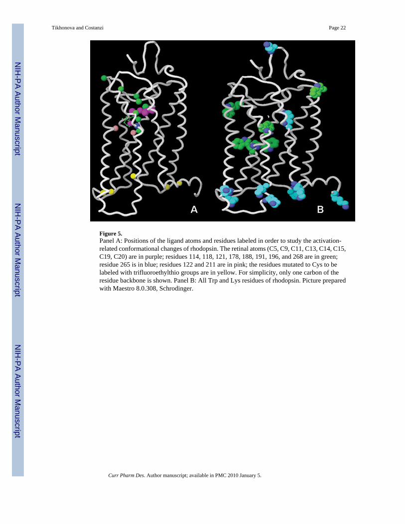
Figure 5.Panel A: Positions of the ligand atoms and residues labeled in order to study the activation-related conformational changes of rhodopsin. The retinal atoms (C5, C9, C11, C13, C14, C15,C19, C20) are in purple; residues 114, 118, 121, 178, 188, 191, 196, and 268 are in green;residue 265 is in blue; residues 122 and 211 are in pink; the residues mutated to Cys to belabeled with trifluoroethylthio groups are in yellow. For simplicity, only one carbon of theresidue backbone is shown. Panel B: All Trp and Lys residues of rhodopsin. Picture preparedwith Maestro 8.0.308, Schrodinger.
Tikhonova and Costanzi Page 22
Curr Pharm Des. Author manuscript; available in PMC 2010 January 5.
NIH
-PA Author Manuscript
NIH
-PA Author Manuscript
NIH
-PA Author Manuscript
NIH
-PA Author Manuscript
NIH
-PA Author Manuscript
NIH
-PA Author Manuscript
Tikhonova and Costanzi Page 23
Tabl
e 1
A sy
nopt
ic v
iew
of t
he a
vaila
ble
NM
R st
ruct
ural
dat
a fo
r GPC
Rs.
A li
st o
f the
abb
revi
atio
ns is
pro
vide
d in
foot
note
a .
Rec
epto
rSo
urce
Dom
ain
Sequ
ence
Con
stra
ints
Solv
ent
Opt
imiz
atio
nSu
mm
ary
of R
esul
tsR
ef.
α 2 a
dren
ergi
c re
cept
orhu
man
TM3-
IL2-
TM4
121–
155
TMA
DPC
DG
/EM
120–
126:
α-h
elix
.12
7–13
1: fl
exib
le α
-hel
ix.
132–
143:
α-h
elix
.
[65]
β 2 a
dren
ergi
c re
cept
ortu
rkey
IL3
284–
295
TMA
LPC
TFE
AS
290–
294:
α-h
elix
and
flex
ible
Nt
in L
PC; α
-hel
ix in
TFE
;un
stru
ctur
ed in
AS;
rand
om co
il in
TFE/
AS.
[67]
hum
anH
elix
832
4–35
7A
SD
MSO
DPC
SA/M
DU
nstru
ctur
ed in
AS.
α-he
lix in
DM
SO a
nd D
PC.
[72]
β 1 a
dren
ergi
c re
cept
ortu
rkey
Hel
ix 8
345–
359
LMPC
DM
PCEM
α-he
lix (P
DB
cod
e:1d
ep).
[73]
AT 1
A a
ngio
tens
in re
cept
orra
tH
elix
830
0–32
0A
S/TF
ESA
/MD
306–
320:
α-h
elix
.[7
4]
IL2
123–
156
TMA
DPC
-d38
DG
/MD
128–
139,
147
–156
: α-h
elic
es.
141–
145:
“U
-sha
pe”
form
.[6
6]
B2 b
rady
kini
n re
cept
orra
tC
t30
9–36
6D
PC31
1–32
6, 3
33–3
45, a
nd 3
48–3
63:
α-he
lices
.[7
5]
CB
1 can
nabi
noid
rece
ptor
hum
anIL
333
8–34
6A
SA
S/Gα i
1
DG
Uns
truct
ured
in A
S; α
-hel
ix w
ithGα i
1 (PD
B c
ode:
1lv
q).
[68]
IL3
300–
343
TMA
SDS
DG
/MD
300–
310,
312
–319
and
332
–346
:α-
helic
es.
321–
327
and
329–
331:
turn
s.
[124
]
Hel
ix 8
397–
418
AS
DPC
SDS
SA/M
DU
nstru
ctur
ed in
AS;
α-h
elix
inD
PC/S
DS.
[71]
CB
2 can
nabi
noid
rece
ptor
hum
anTM
1-IL
1-TM
227
–101
DM
SOPr
elim
inar
y re
sults
[50]
Hel
ix 8
298–
319
AS
DM
SOD
PC
SA/M
DU
nstru
ctur
ed in
AS;
α-h
elix
inD
MSO
and
DPC
.[7
0]
CC
K1 c
hole
cyst
okin
i nre
cept
orhu
man
Nt
EL3
1–47
, 329
–357
352–
379
TMA
DPC
/AS
DG
/MD
MD
-wd
1–47
: α-h
elic
al st
ruct
ures
at N
-an
d C
-term
ini a
nd β
-she
etst
abili
zed
by a
dis
ulfid
e br
idge
betw
een
C18
and
C29
(PD
B co
de:
1d6g
).33
3–33
7, 3
41–3
45, 3
53–3
56,
364–
367,
372
–378
: con
tain
helic
al re
gion
s (PD
B c
ode:
1hz
n).
[54–
56]
CC
K2 c
hole
cyst
okin
i nre
cept
orhu
man
EL3
352–
379
TMA
DPC
/AS
DG
/MD
-wd
332–
339,
341
–346
, and
351
–356
:co
ntai
n he
lical
regi
ons (
PDB
code
: 1l4
t).
[56]
Curr Pharm Des. Author manuscript; available in PMC 2010 January 5.
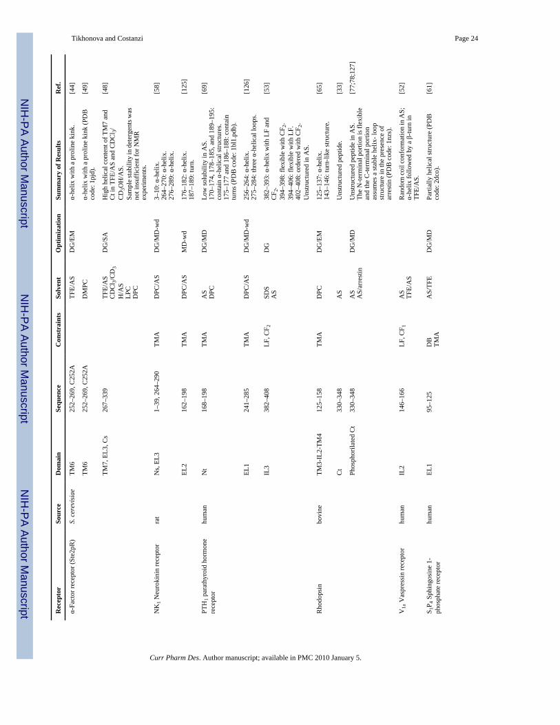
NIH
-PA Author Manuscript
NIH
-PA Author Manuscript
NIH
-PA Author Manuscript
Tikhonova and Costanzi Page 24
Rec
epto
rSo
urce
Dom
ain
Sequ
ence
Con
stra
ints
Solv
ent
Opt
imiz
atio
nSu
mm
ary
of R
esul
tsR
ef.
α-Fa
ctor
rece
ptor
(Ste
2pR
)S.
cer
evis
iae
TM6
252–
269,
C25
2ATF
E/A
SD
G/E
Mα-
helix
with
a p
rolin
e ki
nk.
[44]
TM6
252–
269,
C25
2AD
MPC
α-he
lix w
ith a
pro
line
kink
(PD
Bco
de: 1
pjd)
.[4
9]
TM7,
EL3
, Cs
267–
339
TFE/
AS
CD
Cl 3/
CD
3H
/AS
LPC
DPC
DG
/SA
Hig
h he
lical
con
tent
of T
M7
and
Ct i
n TF
E/A
S an
d C
DC
l 3/C
D3O
H/A
S.Sa
mpl
e sta
bilit
y in
det
erge
nts w
asno
t ins
uffic
ient
for N
MR
expe
rimen
ts.
[48]
NK
1 Neu
roki
nin
rece
ptor
rat
Ns,
EL3
1–39
, 264
–290
TMA
DPC
/AS
DG
/MD
-wd
3–10
: α-h
elix
.26
4–27
0: α
-hel
ix.
276–
289:
α-h
elix
.
[58]
EL2
162–
198
TMA
DPC
/AS
MD
-wd
176–
182:
α-h
elix
.18
7–18
9: tu
rn.
[125
]
PTH
1 par
athy
roid
hor
mon
ere
cept
orhu
man
Nt
168–
198
TMA
AS
DPC
DG
/MD
Low
solu
bilit
y in
AS.
170–
174,
178
–185
, and
189
–195
:co
ntai
n α-
helic
al st
ruct
ures
.17
5–17
7 an
d 18
6–18
8: c
onta
intu
rns (
PDB
cod
e: 1
bl1.
pdb)
.
[69]
EL1
241–
285
TMA
DPC
/AS
DG
/MD
-wd
256–
264:
α-h
elix
.27
5–28
4: th
ree α-
helic
al lo
ops.
[126
]
IL3
382–
408
LF, C
F 2SD
SA
SD
G38
2–39
3: α
-hel
ix w
ith L
F an
dC
F 2.
394–
398:
flex
ible
with
CF 2
.39
4–40
6: fl
exib
le w
ith L
F.40
2–40
8: o
rder
ed w
ith C
F 2.
Uns
truct
ured
in A
S.
[53]
Rho
dops
inbo
vine
TM3-
IL2-
TM4
125–
158
TMA
DPC
DG
/EM
125–
137:
α-h
elix
.14
3–14
6: tu
rn-li
ke st
ruct
ure.
[65]
Ct
330–
348
AS
Uns
truct
ured
pep
tide.
[33]
Phos
phor
ilate
d C
t33
0–34
8A
SA
S/ar
rest
inD
G/M
DU
nstru
ctur
ed p
eptid
e in
AS.
The
N-te
rmin
al p
ortio
n is
flex
ible
and
the
C-te
rmin
al p
ortio
nas
sum
es a
stab
le h
elix
- loo
pst
ruct
ure
in th
e pr
esen
ce o
far
rest
in (P
DB
cod
e: 1
nzs)
.
[77;
78;1
27]
V1a
Vas
pres
sin
rece
ptor
hum
anIL
214
6–16
6LF
, CF 1
AS
TFE/
AS
Ran
dom
coil
conf
orm
atio
n in
AS;
α-he
lix fo
llow
ed b
y a β-
turn
inTF
E/A
S.
[52]
S 1P 4
Sph
ingo
sine
1-
phos
phat
e re
cept
orhu
man
EL1
95–1
25D
BTM
AA
S/TF
ED
G/M
DPa
rtial
ly h
elic
al st
ruct
ure
(PD
Bco
de: 2
dco)
.[6
1]
Curr Pharm Des. Author manuscript; available in PMC 2010 January 5.

NIH
-PA Author Manuscript
NIH
-PA Author Manuscript
NIH
-PA Author Manuscript
Tikhonova and Costanzi Page 25
Rec
epto
rSo
urce
Dom
ain
Sequ
ence
Con
stra
ints
Solv
ent
Opt
imiz
atio
nSu
mm
ary
of R
esul
tsR
ef.
NK
1 tac
hyki
nin
rece
ptor
TM7
AM
SSTM
YN
PIIS
SL
DM
SOD
MSO
/CD
Cl 3
PFTB
/CD
3O
D
EM/M
DK
inke
d α-
helix
as a
min
orco
nfor
mer
in D
MSO
; inc
reas
edfo
lded
con
tent
in D
MSO
/CD
Cl 3;
twoα
-hel
ical
regi
ons l
inke
d by
ahi
nge
arou
nd th
e N
P m
otif
inPF
TB/C
D3O
D.
[45]
A2 t
rom
boxa
ne re
cept
orhu
man
EL1
88–1
04D
BA
SD
G/M
Dβ-
turn
s.[5
9]
EL2
173–
193
DG
/MD
A tw
o-tu
rns l
oop
inco
rres
pond
ence
of 1
83 a
nd 1
88.
[60]
IL2–
312
9–14
9, 2
20–2
46D
BA
SEM
/MD
IL2:
larg
e tu
rn.
IL3:
hel
ical
-like
con
form
atio
n.[5
1]
a AS–
aque
ous s
olut
ion;
BS–
bios
ynth
esis
in E
sche
richi
a col
i; C
D–c
ircul
ar d
ichr
oism
; CF1
: cyc
lized
form
of p
eptid
e with
term
ini s
ubst
itute
d by
S-c
arbo
xym
ethy
lcys
tein
e; C
F2: c
ycliz
ed fo
rm o
f the
pep
tide w
itha l
inke
r of 8
met
hyle
nes;
Ct–
C-te
rmin
us; D
B d
isul
fide b
ridge
; DG
–dis
tanc
e geo
met
ry; D
MPC
–deu
tera
ted
dim
yris
toyl
- pho
spha
tidyl
chol
ine;
DM
SO–d
imet
hyl s
ulfo
xide
; DPC
dod
ecyl
phos
phoc
holin
e mic
elle
s;D
QF-
CO
SY–d
oubl
e-qu
antu
m fi
ltere
d co
rrel
atio
n sp
ectro
scop
y; E
L–ex
trace
llula
r loo
p EM
–ene
rgy
min
imiz
atio
n; H
SQC
–Het
eron
ucle
ar si
ngle
qua
ntum
coh
eren
ce; I
L–in
trace
llula
r loo
p; L
F–lin
ear f
orm
of t
hepe
ptid
e; L
MPC
–deu
tera
ted
lyso
myr
isto
yl-p
hosp
hatid
ylch
olin
e; L
PC–1
-pal
mito
yl-2
-hyd
roxy
-sn-
glyc
ero-
3-ph
osph
ocho
line;
MD
–mol
ecul
ar d
ynam
ics;
MD
-wd-
mol
ecul
ar d
ynam
ics i
n w
ater
-dec
ane
envi
ronm
ent;
MS–
man
ual s
ynth
esis
; NO
ESY
–nuc
lear
ove
rhau
ser e
nhan
cem
ent s
pect
rosc
opy;
Nt–
N-te
rmin
us; P
FTB
–per
fluor
o-te
rt-b
utan
ol; P
C–P
eptid
e C
onst
rain
s; S
A–s
imul
ated
ann
ealin
g; S
DS
sodi
umdo
decy
l sul
fate
; SF–
solid
fase
aut
omat
ic sy
nthe
sis;
ssN
MR
–sol
id-s
tate
NM
R; T
FE–t
riflu
oroe
than
ol; T
M–t
rans
mem
bran
e he
lix; T
MA
–tra
nsm
embr
ane
helix
anc
hor;
TOC
SY–t
otal
cor
rela
tion
spec
trosc
opy;
TRO
SY–t
rans
vers
e re
laxa
tion
optim
ized
spec
trosc
opy.
Curr Pharm Des. Author manuscript; available in PMC 2010 January 5.