unraveling the mystery of ms differential diagnosis
TRANSCRIPT

Unraveling the Mystery of MS Differential Diagnosis
Aliza Ben-Zacharia, ANPAnn Marie Rooney-Crino, ANP
The Corinne Goldsmith DickinsonCenter for Multiple Sclerosis
The Mount Sinai Medical Center

MS or Not???• Clinical Presentation• Comorbidities• Family History• Imaging on MRIs• Spinal Fluid• Blood Work-up• Follow up

Coyle P. 7th Annual Review of Multiple Sclerosis. May 2004.
Clinical Features That May Suggest Misdiagnosis
• Normal neurological examination• No dissemination over time and space• Onset of symptoms before age 10 or after
age 55• Progressive course before age 35• Localized disease

Coyle P. 7th Annual Review of Multiple Sclerosis. May 2004.
Clinical Features That May Suggest Misdiagnosis
• Atypical presentation– Fever– Headache– Abrupt hemiparesis– Abrupt hearing loss– Prominent pain– Normal optic exam– Normal sensory exam

Coyle P. 7th Annual Review of Multiple Sclerosis. May 2004.
Clinical Features That May Suggest Misdiagnosis
• Normal bladder/bowel function• Progressive myelopathy• Impaired level of consciousness• Prominent uveitis• Peripheral neuropathy• Gray matter features
• Early dementia, seizures, aphasia, extrapyramidal features

Red Flags RE labs
• Normal or atypical MRI• Normal CSF• Abnormal blood tests • Antibodies serum levels
– ANA– EBV
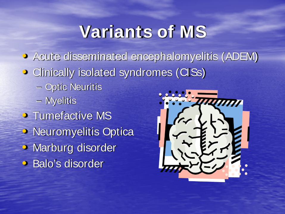
Variants of MS• Acute disseminated encephalomyelitis (ADEM)• Clinically isolated syndromes (CISs)
– Optic Neuritis– Myelitis
• Tumefactive MS• Neuromyelitis Optica• Marburg disorder• Balo’s disorder

Monophasic Disorders

Acute Disseminated Encephalomyelitis (ADEM)
• DefinitionA syndrome caused by autoimmune response to a viral infectionPost vaccines (measles, smallpox, mumps, rubella, varicella)One episode but may be recurrentRapid onsetReversible or irreversibleDifficult to distinguish between ADEM and MS
• Mimics of MS• Characteristics
gait abnormalities, confusion, disorientation, coma, problems with bladder or bowel control, muscle weakness

ADEM• MRI
Large lesions on MRI Multiple Gadolinium lesionsLesions in the brain tend to be bilateral and symmetric which distinguish it from MSPeriventricular lesions are not common VS MS
• Spinal FluidIncreased white cell number (high number as opposed to MS)Oligoclonal bands and IgG not present in ADEM as opposed to MS


ADEM Management
• Intravenous steroids• Plasma exchange • IV Immunoglobulins • Follow-up
Neurological symptomsChanges on the MRI Critical in determining the diagnosis of MS versus ADEM
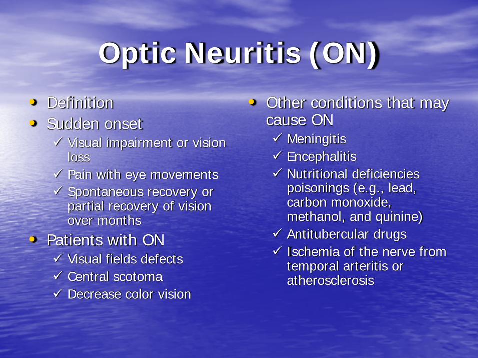
Optic Neuritis (ON)• Definition• Sudden onset
Visual impairment or vision loss Pain with eye movements Spontaneous recovery or partial recovery of vision over months
• Patients with ON Visual fields defectsCentral scotomaDecrease color vision
• Other conditions that may cause ON
MeningitisEncephalitisNutritional deficiencies poisonings (e.g., lead, carbon monoxide, methanol, and quinine)Antitubercular drugsIschemia of the nerve from temporal arteritis or atherosclerosis

Optic Neuritis – Mimics of MS
• Visual evoked potential (VEP)
• Neuro-imaging studies• Brain MRI, orbital MRI• Cervical spine MRI - baseline • CT/MRI may be ordered to rule
out the presence of foreign bodies, hemorrhage, fractures, or orbital damage from trauma
• 10 year optic neuritis trial, even one demyelinating brain lesion can increase the risk for MS
• Neuro-ophthalmologic testing– Visual defects– Optic disc impairment– Other visual screening tests– The fundoscopic exam may be
normal during the acute phase • Lumbar Puncture
– Spinal fluid should be tested to rule out infectious or inflammatory diseases
• Chest CT or radiographic testing– This test should be done to
rule out sarcoidosis

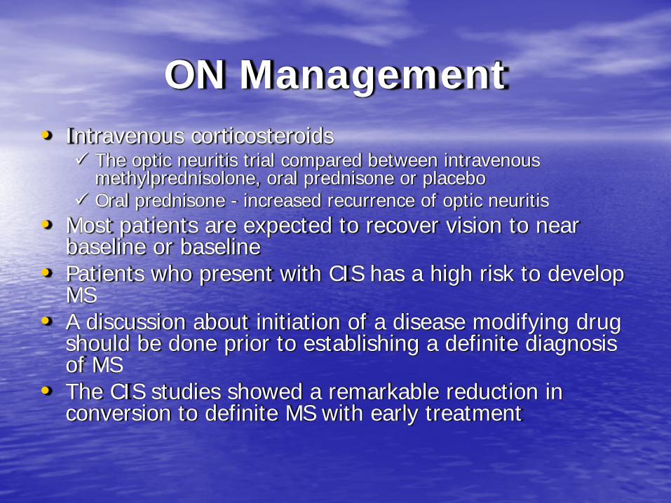
ON Management• Intravenous corticosteroids
The optic neuritis trial compared between intravenous methylprednisolone, oral prednisone or placeboOral prednisone - increased recurrence of optic neuritis
• Most patients are expected to recover vision to near baseline or baseline
• Patients who present with CIS has a high risk to develop MS
• A discussion about initiation of a disease modifying drug should be done prior to establishing a definite diagnosis of MS
• The CIS studies showed a remarkable reduction in conversion to definite MS with early treatment

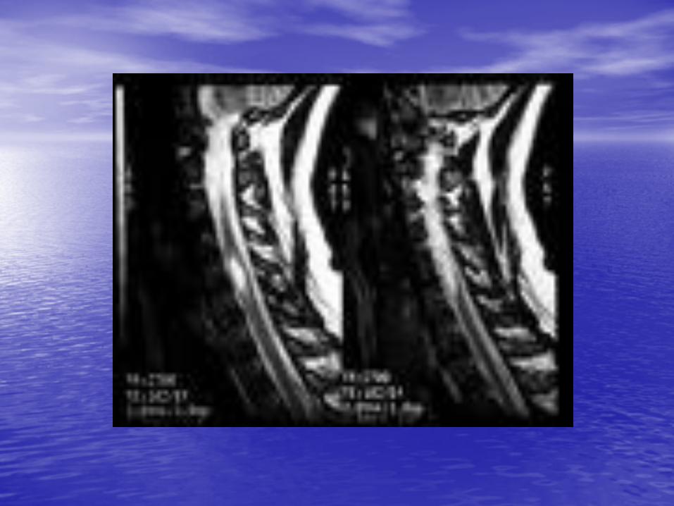
Transverse Myelitis• Definition
Inflammation of the spinal cord“myelitis" refers to an inflammation of the fatty insulating material that covers nerve cell fibers (myelin)“transverse" describes the location of the inflammation — across the width of spinal cordTransverse myelitis often develops following a viral infection
• Mimics of MSCharacteristics (spinal symptoms)MRI – spinal lesion
• TreatmentIV steroids & Plasma exchange

Neuromyelitis Optica (NMO)• Definition
Inflammation of the optic nerve & spinal cordSevere diseaseAA, Japanese decent & Caucasians
• Mimics of MSCharacteristics (optic s/s, spinal symptoms)MRI findings (long segmental lesions)Spinal fluid (Tests to differentiate (NMO titer, disease progression)


Tumefactive Lesion
• DefinitionLarge lesion on MRI similar to MSAttack of MS versus tumorCharacteristics
• ManagementIV steroidsMonitoringCT chest, abdomen, pelvic

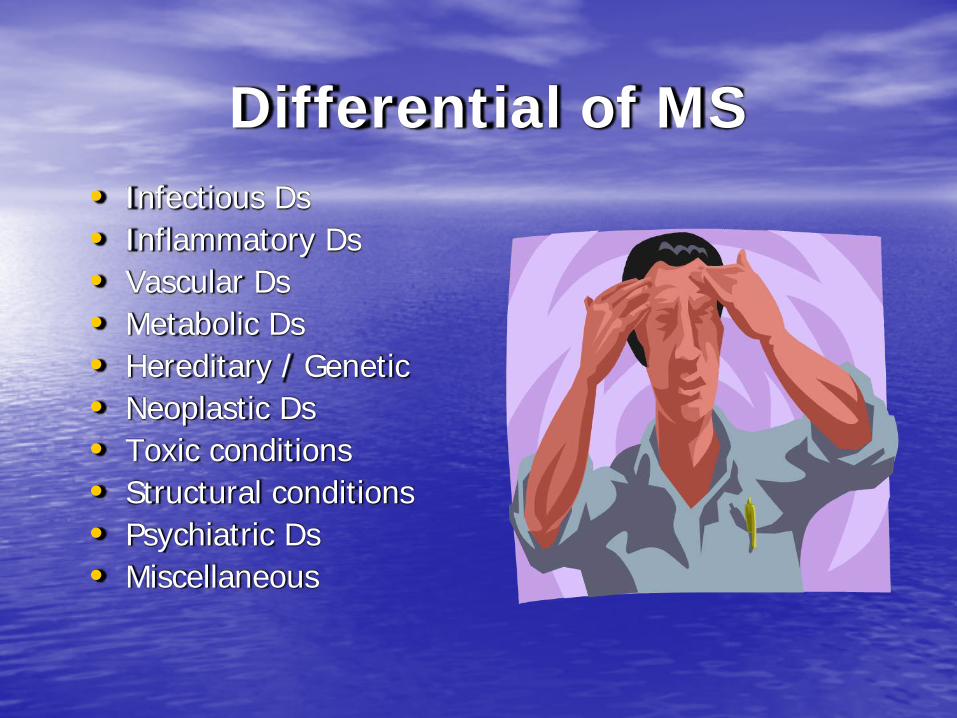
Differential of MS• Infectious Ds• Inflammatory Ds• Vascular Ds• Metabolic Ds• Hereditary / Genetic• Neoplastic Ds• Toxic conditions• Structural conditions• Psychiatric Ds• Miscellaneous

Infectious Diseases

Infections
• Definition– Infections that affect the nervous system
• Mimics MS– Lesions on MRIs

Viral infections that mimic MS
• Herpes virus type 6 – demyelinating lesions in the CNS
• Varicella Zoster virus – encephalitis in immune-compromised
• Measles – neurological symptoms, abnormal MRI & CSF
• Retrovirus – HTLV-1, HIV• JC virus (PML)

Tropical Spastic Paraparesis
• Tropical spastic paraparesis is a retroviral disease caused by HTLV-1 virus
• It is uncommon in the continental United States, but may be seen infrequently in patients who resided for some time around the Caribbean Sea
• The major clinical manifestations are progressive spastic paraparesis or generalized white matter disease

Progressive Multifocal Leukoencephalopathy (PML)
• PML is a rare, progressive disease • PML mostly affects people who have
a weakened immune system• It is caused by activation of a virus
called - JC virus • MRI – Large lesions – Non-enhancing• CSF - + JC virus

Bacterial Infections
• Brucella species• Chlamydia Pneumoniae• Spirochetes
– Lyme Disease– Syphilis
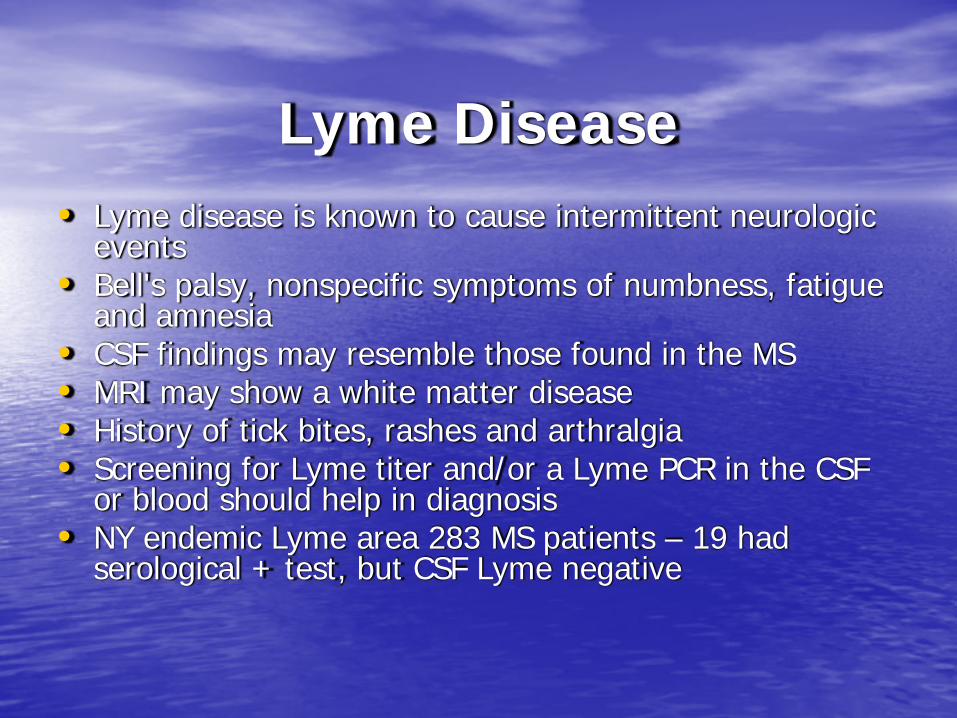
Lyme Disease• Lyme disease is known to cause intermittent neurologic
events• Bell's palsy, nonspecific symptoms of numbness, fatigue
and amnesia• CSF findings may resemble those found in the MS• MRI may show a white matter disease• History of tick bites, rashes and arthralgia • Screening for Lyme titer and/or a Lyme PCR in the CSF
or blood should help in diagnosis • NY endemic Lyme area 283 MS patients – 19 had
serological + test, but CSF Lyme negative

Tertiary Syphilis
• Neurosyphilis is a slowly progressive and destructive infection of the brain or spinal cord
• It occurs many years after the primary infection• Syphilis may result in dorsal column (sensory
deficits) abnormalities and dementia• CSF-VDRL test positive• Pleocytosis (CSF WBC count >10/mm3 ) • CSF protein elevated (> 0.50 g/l)• Treatment: IV Penicillin (pregnant)

Inflammatory / Collagen vascular diseases

Behçet Syndrome
• Behçet syndrome can cause MRI findings that are very similar to MS
• The main distinguishing features of this condition are oral and genital ulcers, and uveitis
• Involvement of lungs, joints, intestines, and heart
• This group of patients may present with either quadriparesis, pseudobulbar palsy, cranial neuropathy, cerebellar ataxia or cerebral venous thrombosis

Systemic Lupus Erythematosus
• Systemic lupus erythematosus may cause multiple neurologic pathology
• Optic abnormalities, encephalopathy, transverse myelitis, strokes
• Systemic abnormalities, such as elevated antinuclear antibody, leukopenia, hematuria, elevated erythrocyte sedimentation rate
• On some occasions lupus erythematosus and MS may be found in the same patient
Sjogren's Syndrome
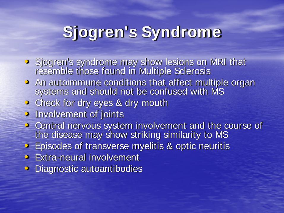
• Sjogren's syndrome may show lesions on MRI that resemble those found in Multiple Sclerosis
• An autoimmune conditions that affect multiple organ systems and should not be confused with MS
• Check for dry eyes & dry mouth• Involvement of joints• Central nervous system involvement and the course of
the disease may show striking similarity to MS• Episodes of transverse myelitis & optic neuritis• Extra-neural involvement• Diagnostic autoantibodies
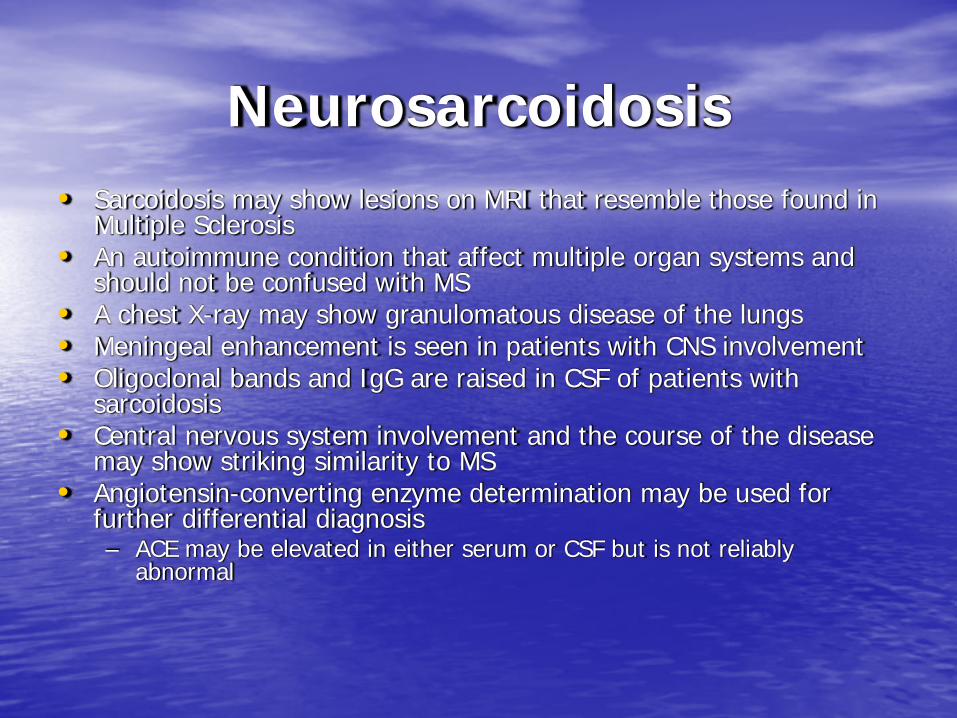
Neurosarcoidosis• Sarcoidosis may show lesions on MRI that resemble those found in
Multiple Sclerosis• An autoimmune condition that affect multiple organ systems and
should not be confused with MS• A chest X-ray may show granulomatous disease of the lungs• Meningeal enhancement is seen in patients with CNS involvement• Oligoclonal bands and IgG are raised in CSF of patients with
sarcoidosis• Central nervous system involvement and the course of the disease
may show striking similarity to MS• Angiotensin-converting enzyme determination may be used for
further differential diagnosis– ACE may be elevated in either serum or CSF but is not reliably
abnormal

Myasthenia Gravis
• Young women (18-25yrs) (men: 60-80yrs)• Waxing & waning abnormalities• Attacks are short and tend to appear with
effort and late in the day• Check Acetylcholine receptor antibodies
(serum test)
Neoplastic disorders (Tumors)

Paraneoplastic disorders
• Underlying cancer destroy or damage portions of the CNS
• Small cell lung cancer – Anti-Hu antibody• Ovarian or breast cancer – Anti-Yo
antibody• Testicular cancer – Anti-Ta antibody• Several cancers – lung, breast, parotid
gland, colon – Anti-Ma antibody

Paraneoplastic Syndrome
• Paraneoplastic syndromes affecting CNS• Inflammatory infiltrates of T cells &
plasma cells are found in the CNS & in the cancer
• IgG that normally do not present in the CNS are found in neurons
• Cancer is more indolent in paraneoplastic syndromes which influence therapy type

Primary CNS lymphomas
• Large edema around the area seen in MRI• Enhancement on MRI

Metabolic Disorders

Deficiencies • Folate deficiency
– Degeneration • Vitamin E deficiency
– Intestinal fat malabsorption– Polyneuropathy
• Cobalamin Deficiency– Pernicious anemia– Vitamin B12 deficiency

Vitamin B-12 Deficiency
• Vitamin B-12 deficiency may result in dorsal column abnormalities (Myelopathy) and dementia
• This condition need to be ruled out when patients present with the above mentioned symptoms as their chief complaints
• Dorsal column symptoms: sensory deficits

Hereditary/Genetic Disorders

Leukodystrophies of Adulthood
• Hereditary Disease• Leukodystrophies of adulthood • Large areas of involvement on the MRI
scan where no normal white matter can be found
• Males versus females• Long chain fatty acids

Hereditary Degenerative Disorders
• Hereditary degenerative disorders (pontocerebellar degeneration, spinocerebellar degeneration, etc.)
• May resemble chronic-progressive MS• Characteristic white matter lesions on the
MRI scan are usually absent • The CSF is normal in these patients

Vascular Conditions

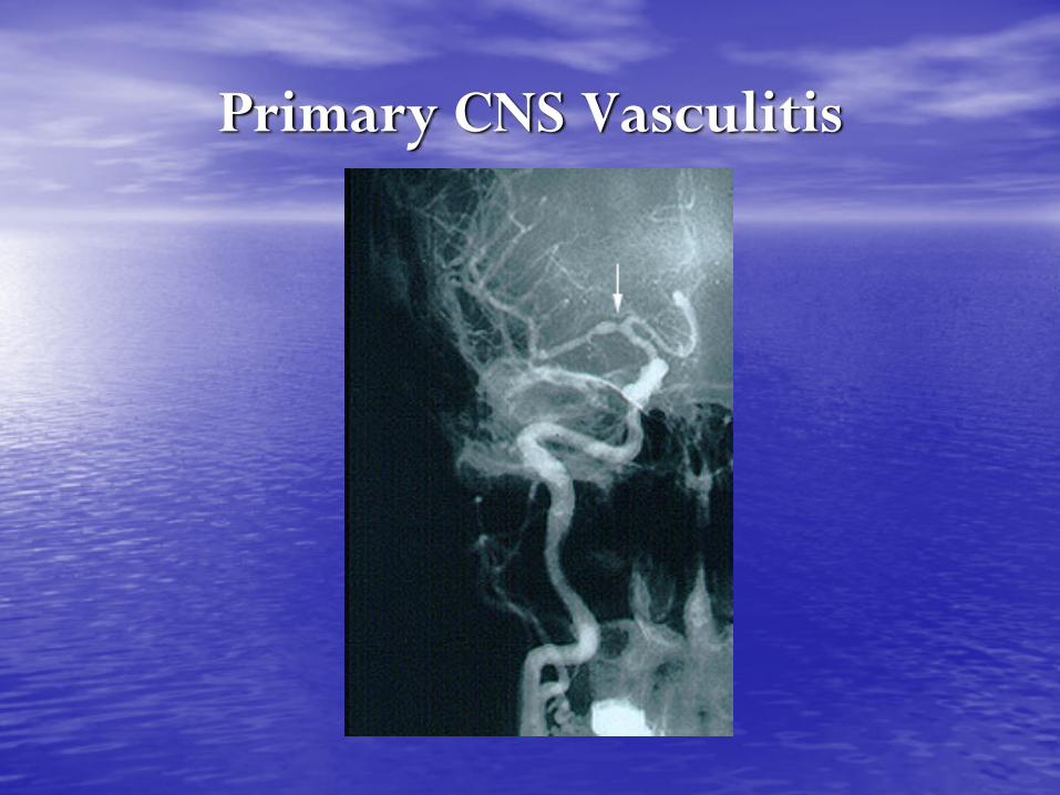
Primary CNS Vasculitis
Defined:Inflammation of blood vessels within the central nervous system
Clinical mimics of MS:
Recurrent focal CNS deficits
Cognitive changes
Occurs in young persons
20% have oligoclonal bands in CSF

Primary CNS Vasculitis
Diagnostic clues to distinguish from MS :Severe headaches
Sudden stroke-like episodes
Seizures
Abnormal angiogram of cerebral vessels
Positive antinuclear or anti phospholipid antibodies
Primary CNS Vasculitis

Susac Syndrome
Defined:microinfarcts of the retina, brain and inner ear
Clinical mimics of MS:RelapsesCognitive changesMean age of onset 28 (range 18-59)Female predominance (> 4:1)

Susac Syndrome
Diagnostic clues to distinguish from MS :Bilateral hearing loss/tinnitus
Headache
Branch retinal occlusions
No IgG abnormalities with CSF analysis
MRI reveals callosal lesions involving the central fibers sparing the periphery
Brain biopsy would reveal multifocal microinfarcts

Susac Syndrome

CADASIL
Defined:Cerebral autosomal dominant arteriopathy with subcortical infarcts and leukoencephalopathy
Clinical mimics of MS:Presents between age 30-50May present with cognitive impairmentGait and bladder dysfunction are common

CADASIL
Diagnostic clues to distinguish from MS:Family history of stroke (without risk factors) and dementiaTypically presents with migraine headaches and recurrent subcortical strokes. MRI can demonstrate bilateral subcortical white matter lesions, with an affinity for the anterior temporal lobes and external capsuleNormal CSF DNA mutation in the notch-3 gene and demonstration of the characteristic small vessel microangiopathy by skin biopsy

CADASIL

Toxic Conditions

Nitrous Oxide Toxicity
Defined:Myeloneuropathy is caused by the inactivation of vitamin B12 by nitrous oxide
Clinical mimics of MS:Sensory changesWeaknessImbalanceMRI may reveal increased signal within the posterior columns bilaterally
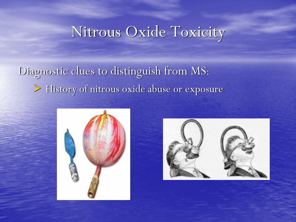
Nitrous Oxide Toxicity
Diagnostic clues to distinguish from MS:
History of nitrous oxide abuse or exposure

Nitrous Oxide Toxicity


Somatization Disorder
Defined:Psychiatric disorder characterized by a chronic pattern of physical expression of psychological stress
Clinical mimics of MS:
Patients have multiple recurring and relapsing neurologic symptoms which can include weakness, numbness, dizziness, and similar complaints.

Somatization Disorder
Diagnostic clues to distinguish from MS:Functional exam
MRI, CSF, evoked potentials will be normal

Conversion Disorder
Defined:Acute onset of motor or sensory loss unexplained by physical findings, not intentionally produced
Clinical mimics of MS:
Patients may present with neurologic symptoms very similar to those seen in MS

Conversion Disorder
Diagnostic clues to distinguish from MS:Functional exam
Normal MRI, CSF, and evoked potentials
Genetic Disorders
Adrenoleukodystrophy
Defined: An X-linked genetic disorder of myelin, usually pediatric but can present in adulthoodInvolves mutation in the gene encoding ALD protein (Xq28)
Clinical mimics of MS:Adults show slowly progressive weakness of the LEs, ataxia, cognitive dysfunction and visual disturbancesFemale carriers may present with mild disease

Adrenoleukodystrophy
Diagnostic clues to distinguish from MS:Serum testing will show high levels of very long chain fatty acids
ACTH stimulation test usually shows impaired adrenal function
Confluent white matter
changes on MRI

Fabry’s Disease
Defined:An X-linked deficiency of alpha-galactosidase
Clinical mimics of MS:
Pain and abnormal feelings in extremities
MRI signal abnormalities
Young males are usually the most severely affected

Fabry’s Disease
Diagnostic clues to distinguish from MS:Recurrent strokesSkin lesions on skin and within the mouth (red, raised lesions)Diminished ability to sweat Corneal opacities Renal diseaseImpairment of heart function DNA testing can confirm diagnosisLow serum alpha-galactosidase levels
Leber’s Hereditary Optic Neuropathy
Defined:Mitochondrial mutation causing sub-acute bilateral optic neuropathy
Clinical mimics of MS:Optic neuropathyMyelopathyAtaxiaAbnormal MRI signal changes

Leber’s Hereditary Optic Neuropathy
Diagnostic clues to distinguish from MS:CSF is normal
Optic neuropathy is
bilateral
Positive mitochondrial
genetic testing

CNS Structural Conditions

Arteriovenous Malformations of the Spinal Cord
Defined: An abnormal tangle of blood vessels on, in or near the spinal cord
Clinical mimics of MS:Relapsing or progressive spinal cord symptoms Occurs in young personsMRI of spinal cord can show intrinsic signal abnormalities that could be easily mistaken for MS

Arteriovenous malformations of the spinal cord
Diagnostic clues to distinguish from MS:MRI of brain will be normal
CSF will be normal
Normal visual evoked responses

Arnold-Chiari Malformation
Defined:Descent of cerebellar tonsils below the foramen magnum causing brainstem and spinal cord compression
Clinical mimics of MS:Nystagmus
Ataxia
Sensory impairment
Paralysis of the extraocular muscles

Arnold-Chiari Malformation
Clinical clues to distinguish from MS:Sagittal images on MRI will detect the malformation
CSF is normal
Severe headaches, made
worse by straining

Cervical Spondylosis
Defined: Spinal cord compression due to chronic degeneration or injury
Clinical mimics of MS:Progressive onset of neurologic signs and symptomsWhite matter abnormalities at the level of the discElevated protein in CSF

Cervical Spondylosis
Clinical clues to differentiate from MS:Neck pain (radiates to arm or shoulder)Localized muscle tightnessA previous neck injury or advance ageMRI of cervical cord will usually showcompressionNormal IgG synthesis in CSFVisual evoked responses normal

Summary
Key points:Thorough initial assessment is needed to ensure accurate diagnosis
A thoughtful analysis will exclude other conditions
If diagnosis remains uncertain, patients should be reassessed at a later time point