eprints.soton.ac.uk · university of southampton abstract faculty of engineering, science &...
TRANSCRIPT

University of Southampton Research Repository
ePrints Soton
Copyright © and Moral Rights for this thesis are retained by the author and/or other copyright owners. A copy can be downloaded for personal non-commercial research or study, without prior permission or charge. This thesis cannot be reproduced or quoted extensively from without first obtaining permission in writing from the copyright holder/s. The content must not be changed in any way or sold commercially in any format or medium without the formal permission of the copyright holders.
When referring to this work, full bibliographic details including the author, title, awarding institution and date of the thesis must be given e.g.
AUTHOR (year of submission) "Full thesis title", University of Southampton, name of the University School or Department, PhD Thesis, pagination
http://eprints.soton.ac.uk

UNIVERSITY OF SOUTHAMPTON
Faculty of Engineering, Science and Mathematics
School of Chemistry
Novel Nucleotide Analogues for Forming Stable DNA
Triple Helices
by
Simon Richard Gerrard
A thesis submitted for the degree of Doctor of Philosophy.
June 2009

UNIVERSITY OF SOUTHAMPTON
ABSTRACT
FACULTY OF ENGINEERING, SCIENCE & MATHEMATICS
SCHOOL OF CHEMISTRY
Doctor of Philosophy
NOVEL NUCLEOTIDE ANALOGUES FOR FORMING STABLE DNA TRIPLE
HELICES
by Simon Richard Gerrard
DNA triple helices are an important tool in a variety of medicinal and biotechnological
applications, such as gene therapy and chemotherapeutics. DNA triple helices are
formed by binding of a triplex-forming oligonucleotide (TFO) to a DNA duplex, via
specific recognition of the individual base pairs in the target sequence.
Mixed-sequence recognition of duplex DNA by TFOs is therefore an essential
requirement for successful targeting. However, achieving strong, yet specific binding to
the pyrimidine.purine (Py.Pu) base pairs CG and TA, by TFOs is a greater challenge
than to the purine.pyrimidine (Pu.Py) base pairs (GC, AT), as fewer hydrogen bonds are
presented for binding in the major groove of the double helix.
Selective recognition of CG, could be achieved by utilising additional interactions
across the CG base pair, via amino-modified nucleosides, to form more stable, selective
triplets than those which can be formed by the natural base T. Four modified
phosphoramidite monomers, meta-aminophenyl-modified analogues of the bicyclic
nucleosides, (2,3H)-furano[2,3-d]pyrimidin-2(7H)-one and N-methyl-(2,3H)-pyrrolo-
[2,3-d]pyrimidin-2(7H)-one, were synthesised to address this potential hydrogen-
bonding motif.
Biophysical studies demonstrate selective recognition of the CG base pair. Results
indicate selectivity for CG and binding affinity are much improved on previous
modifications. Their fluorescence properties and general oligonucleotide deprotection
conditions were also studied.
In addition, the synthesis of a bis-amine modified 6-oxocytidine phosphoramidite
monomer for GC recognition was re-investigated.
This research shows significant advances in the field of triplexes for therapeutic use.

Contents
Abstract i
Contents ii
Declaration vii
Acknowledgements viii
List of Abbreviations ix
List of Monomers Used xiv
Chapter 1. Base Pair Recognition within DNA Triple Helices: Introduction
1.1 Structure of DNA 2
1.1.1. Primary Structure 2
1.1.2 Secondary Structure 4
1.2 DNA Triplexes 6
1.2.1 Purine Recognition 10
1.2.1.1 AT Base Pair Recognition 10
1.2.1.2 GC Base Pair Recognition using Pyrimidine Bases 12
1.2.1.3 6-Oxocytosine in GC recognition 14
1.2.1.4 GC Base Pair Recognition using Purine Bases 16
1.2.1.5 Other Novel Base designs 17
1.2.2 Pyrimidine Recognition 18
1.2.2.1 TA Base Pair Recognition 18
1.2.2.2 CG Base Pair Recognition using modified Pyrimidine
bases 19
1.2.2.3 Novel Pyrimidine-derived Monomers for CG Base Pair
Recognition 24
1.2.2.4 Novel Non-natural Heterocyclic Monomers for CG
Base Pair Recognition 25
1.3 Conclusion 28
Chapter 2. Modified 6-Oxocytidine Nucleosides for GC Recognition
2.1. Introduction 30
2.2. Bis-amino-6-oxocytidine: Synthesis 31
2.2.1 Synthesis of 5-Halo-6-oxocytidine nucleosides 32
2.2.1.1 Synthesis and Reaction of Benzyltriethylammonium
dichloroiodate, 71 33
ii
2.2.1.2 Other Methods of Halogenation of 6-Oxocytidine
derivatives 34
2.2.2 Synthesis of 2’-O-Phthalimidoethyl-modified Ribofuranose
Sugar, 65 35
2.2.3 6-Oxocytidine Synthesis 37
2.2.4 Sonogashira Cross-Coupling 38
2.3 Alternative Modifications at 5-position 41
2.3.1 Halide-Amine Exchange 42
2.3.2 Halide-Thiol Exchange 47
2.4 Alternative Sugar Modification at the 2’-Position 49
2.4.1 2’-Modification via Tricyclic Anhydronucleosides 50
2.5 Conclusion 52
Chapter 3. Amine-Modified Furano-dT Nucleosides for CG Recognition
3.1 Introduction 55
3.1.1 Monomer Nomenclature 56
3.2 Synthesis of Furano-dT Monomers for CG Recognition 57
3.2.1 Synthesis of 6-(3-Trifluoroacetamidophenyl)-furano-dT
monomer, 145 58
3.2.2 Synthesis of 6-(3-Ureidophenyl)-furano-dT Monomer, 146 60
3.2.3 Synthesis of 6-(3-Acetamidophenyl)-furano-dT Monomer, 147 62
3.3 Post-Synthetic Conversion of Furano-dT to N-Methylpyrrolo-dC 63
3.4 Synthesis of 5-(3-aminoprop-1-ynyl)-dU (pdU) monomer, 164 66
3.5 Conclusion 66
Chapter 4. Biophysical Studies of Furano-dT Modified TFOs for CG
Recognition 69
4.1 UV Melting Studies 69
4.2. Fluorescence Melting Studies 71
4.2.1 Binding Affinity/Selectivity study 72
4.2.2 Sequence study 74
4.3 Fluorescence Melting Mismatch Experiment 76
4.4 Conclusion 78
iii

Chapter 5. Amine-Modified N-Methylpyrrolo-dC Nucleosides for CG
Recognition
5.1 Introduction 81
5.1.1 Synthesis of 6-(3-Ureidophenyl)-N-methylpyrrolo-dC
monomer, 179 82
5.1.1.1 Synthesis via untritylated furano-dT derivative 83
5.1.1.2 Synthesis via tritylated furano-dT derivative 84
5.1.1.3 Synthesis via derivatisation of 5-iodo-4N-methyl-
dC, 185 85
5.1.2 Synthesis of 6-(3-Acetamidophenyl)-N-methylpyrrolo-dC
monomer, 191 88
5.1.3 Synthesis of 6-(3-Trifluoroacetamidophenyl)-N-methylpyrrolo-dC
monomer, 178 89
5.1.4 Synthesis of 6-(3-Guanidinylphenyl)-N-methylpyrrolo-dC
monomer, 199 92
5.1.4.1 Improved Synthesis of Guanidinylating Reagent, 200 92
5.1.4.2 Final Steps towards GPPp Monomer, 199 95
5.1.5 Synthesis of 6-(3-Thioureidophenyl)-N-methylpyrrolo-dC
monomer, 158 97
5.2 Future Work 100
5.3 Conclusion 101
Chapter 6. Biophysical Studies of N-Methylpyrrolo-dC Modified TFOs for CG
Recognition 104
6.1 Initial Fluorescence Melting Study 104
6.2 Primary UV Melting Study 105
6.3. Primary Fluorescence Melting Study 109
6.4. Melting Study Comparison 113
6.5. Fluorescence Properties of CG Recognition Monomers 114
6.6 Conclusions & Future Work 117
Chapter 7. Oligonucleotide Deprotection Study
7.1 Introduction 120
7.2 Deprotection Conditions for Oligonucleotides 121
7.3 Oligonucleotide Deprotection Study 122
7.3.1 Background 122
iv

7.3.2 Deprotection Study 125
7.3.2.1 Control T12 Oligonucleotide 127
7.3.2.2 Test Oligonucleotides – 5-(3-Aminopropynyl)-dU (pdU) 127
7.3.2.3 Test Oligonucleotides – Bis-amino-U (BAU) 131
7.3.2.4 Test Oligonucleotides – 6-(3-Acetamidophenyl)-N-methyl-
pyrrolo-dC (AcPPp) 132
7.3.2.5 Test Oligonucleotides – S base (TA recognition monomer,
S) 134
7.3.2.6 Test Oligonucleotides – 5-(3-aminopropynyl)-2’-O-
methoxyethyl-U (MOEpU) 134
7.3.2.7 Test Oligonucleotides – Other AT recognition monomers 135
7.4 Conclusion 136
Chapter 8. Conclusion
8.1. Modified 6-Oxocytidine Nucleosides for GC Recognition 139
8.2 Amino-Modified Bicyclic Nucleosides for CG Recognition 139
8.3 Oligonucleotide Deprotection Study 141
Chapter 9. Experimental
9.1 Synthesis 143
9.1.1 General 143
9.1.2 Experimental Procedure 146
9.2 Biophysical Studies 216
9.2.1 Synthesis of Oligonucleotides
216
9.2.2. Deprotection of Oligonucleotides 216
9.2.3 Purification of Oligonucleotides 217
9.2.4 Analysis of Oligonucleotides 217
9.2.5 UV Melting Studies 218
9.2.5.1 Set-Up Procedure 218
9.2.5.2 UV Melt Programmes 219
9.2.5.3 Data Analysis 219
9.2.6 Fluorescence Melting Studies 220
9.2.6.1 Set-Up Procedure 220
9.2.6.2 LC Melt Programmes 220
9.2.6.3 Data Analysis 222
v

9.2.7 Determining UV/Fluorescence Properties of Fluorescent
Nucleosides 222
9.2.7.1 Deprotection of Nucleosides 222
9.2.7.2 UV Absorption Measurements 222
9.2.7.3 Fluorescence Measurements 223
References 224
Appendices
A: List of Synthesised Compounds 238
B: Oligonucleotide List 246
C: Conferences Attended, Posters, Presentations and Publications 248
D: Paper
vi

Declaration
I, Simon Richard Gerrard declare that this thesis entitled “Novel Nucleotide Analogues
for Forming Stable DNA Triple Helices” and the work presented within are both my
own and have been generated by myself as the result of my own original research.
I confirm that:
• this work was done wholly or mainly in candidature for a research degree at this
University;
• where I have consulted/quoted the published work of others, this is always
clearly attributed/referenced;
• I have acknowledged all main sources of help;
• I have made clear, where the thesis is based on work done in conjunction with
another party, what my contribution to that work is;
• work described in Chapters 3 and 4 has been published in the paper below:
− Gerrard, Simon R.; Srinivasan, Natarajan; Fox, Keith R.; Brown, Tom; “CG
Base Pair Recognition Within DNA Triple Helices Using N-Methyl-3H-
pyrrolo[2,3-d]pyrimidin-2(7H)-one Nucleoside Analogues”, Nucleosides,
Nucleotides and Nucleic Acids, 26(10), 1363-1367.
DOI: 10.1080/15257770701533958
Signed:………………………………..
Date: 21st June 2009
vii

Acknowledgements
I have many people to thank for making my life at Southampton University so exciting
and rewarding. Firstly, thank you to my supervisor Professor Tom Brown for imparting
so much knowledge, guidance and enthusiasm, and for his endless project ideas,
patience and understanding - this project would have been impossible without him.
Many thanks also to Dr. Dorcas Brown and all at ATDBio Ltd. (including Dr. Imenne
Bouamaied), for their invaluable work in synthesising and purifying all my many
oligonucleotides. I would also like to thank my co-supervisor Professor Keith Fox for
his most useful feedback on my reports, posters and publication, and ideas throughout.
Working in the Brown Group, with such a large, welcoming, varied group of people
from around the world was fantastic. My thanks go out to all of them for putting up
with so many questions from me, and for the useful phrases in many different
languages! In particular, I would like to say a big thank you to past members, Dr. Li
Hong, Dr. Rohan Ranasinghe, Dr. Vicki Powers, Dr. Wang Yang, Prof. Dr. Xiao Qiang,
Chaturong Suparpprom, Dr. Sunil Vadhia, Martina Banchelli, Jan Michels, Jenny Hale,
Dr. Lavinia Brennan and Dr. Naomi Hammond for so much useful advice and support
throughout, and being genuinely great fun to work with. I must thank both present and
recently left members - especially Adeline Durand, Dr. Afaf El-Sagheer, Dr. Peng Guo-
mei, Dr. John Zhao, Dr. Imenne Bouamaied, James Richardson, Dr. Nittaya Gale, Dr.
Noha Ben-Gaïed and Dr. Petr Kočalka. The PhD wouldn’t have been the same without
their great sense of humour, and support, and putting up with inane chatter and a little
insanity from yours truly from time to time. A massive thank you for invaluable
support also goes to Dr. Mastoura Edrees-Abdou, with whom it was a pleasure to work
whilst finishing my final melting studies. Lucy, Radha, Chenguang (Harvey) and Paul
Douglas (Roach Group) – thanks also!
Many thanks to the Mass Spectrometry and NMR people: John Langley and Julie
Herniman, and Neil Wells and Joan Street, for providing such a quality service and
answering my many queries. I am also immensely grateful to Dr. Eugen Stulz for his
advice and allowing me extra time to finish my thesis.
Finally, I can’t thank my girlfriend Sarah enough, for supporting me throughout my
PhD, and for just being there!
viii

List of Abbreviations
1-deazaC 1-deazacytosine2-AES 2'-aminoethoxy-SAET 2’-O-aminoethyl-T2-AP 2-aminopyridine
3-EA 3-ethynylaniline, 3-aminophenyl acetylene
3-HPN 3-hydroxypropionitrile, 2-cyanoethanol4HT 5-methylpyrimidin-2(1H)-one
5'-dTMP 2'-deoxythymidine-5'-monophosphate5-MeC 5-methylcytosine5-MedC, M 5-methyl-2'-deoxycytidine6-oxoC 6-oxocytosine / 6-oxocytidine8-oxoA 8-oxoadenine
A adenine / adenosineAcPP * 6-(3-acetamidophenyl)-N-methylpyrrolo-dC
aegPNA aminoethylglycyl-PNAAEP 6-(aminoethyl)-N-methylpyrrolo-dC
aepPNA aminoethylprolyl-PNA
AIBN azo-bisisobutyronitrile
AMA conc. aq ammonia:conc. aq methylamine (1:1 v/v)
Anal. elemental (CHN) thermal combustion analysisAPP 6-(aminopropyl)-N-methylpyrrolo-dC
app apparentAPP * 6-(3-aminophenyl)-N-methylpyrrolo-dCapyC 4N-6-aminopyrid-2-yl-dC
aq aqueousBAoxC 2’-O-(2-aminoethyl)-5-(3-aminoprop-1-ynyl)-6-oxocytidineBAU 2’-O-aminoethyl-5-(3-aminoprop-1-ynyl)-uridine (bis-amino-U)
BER borohydride exchange resin (resin–NMe3+ BH4
–)
BNA bridged nucleic acid
BOC tert-butyloxycarbonyl
bp base pair
br broad (IR/NMR)
BTEA benzyltriethylammonium
ix

BTMA benzyltrimethylammonium
C cytosine / cytidine
cAMP adenosine 3',5'-cyclic phosphate
Cbz benzyloxycarbonyl
CE Capillary Electrophoresis
CEOC 2-cyanoethoxycarbonyl
COSY correlation spectroscopy
CPG controlled pore glass (solid support)
d 2'-deoxy, doublet (NMR)
δ chemical shift
D3 4-benzamido-1,3-imidazole
dA 2'-deoxyadenosine
dC 2'-deoxycytidine
DCA dichloroacetic acid
DCE 1,2-dichloroethane
DCM dichloromethane
DEA diethylamine
DEAD diethyl azodicarboxylate
DEPT distortionless enhancement through polarization transfer
dF* 2’-deoxy-β-D-ribofuranosyl-(2,3H)-furano[2,3-d]pyrimidin-2(7H)-one,
furano-dT
dG 2'-deoxyguanosine
DIHT diisopropylamine hydrotetrazolide
DIPEA diisopropylethylamine, Hünig's base
DMA N,N-dimethylacetamide
DMAP 4-dimethylamino pyridine
DMF dimethylformamide
dmf dimethylformamidine
DMS dimethyl sulfate
DMSO dimethyl sulfoxide
DMT 4,4'-dimethoxytrityl
dN 2’-deoxynebularine
DNA deoxyribonucleic acid
dP 2’-deoxy-β-D-ribofuranosyl- (2,3H)-pyrrolo[2,3-d]pyrimidin-2(7H)-one,
pyrrolo-dC
x
dR 2'-deoxyribofuranose
DSC disuccinimidyl carbonate
dT 2'-deoxythymidine
dU 2'-deoxyuridine
EDC ethyl (3-dimethylaminopropyl) carbodiimide
EDTA ethylenediamine tetra acetate/acetic acid
eq equivalents
ES ethylene sulfide
FAM carboxyfluorescein
FRET Förster Resonance Energy Transfer
FT Fourier Transform
G guanine / guanosine
GC gas chromatographyGP, GpdU 5-(3-guanidinylprop-1-ynyl)-dUGPP * 6-(3-guanidinylphenyl)-N-methylpyrrolo-dC
GR guanidinylating reagent, N,N’-bis-[(2-cyanoethoxy)carbonyl]-S-methyl-
isothiourea
H, HEG hexaethylene glycol
HMBC heteromultinuclear bond correlation, long-range 1H–13C COSY
HMDS hexamethyldisilazane
HMPA hexamethylphosphoramide
HMQC heteromultinuclear quantum correlation, 1H–13C COSY
HPLC high-performance liquid chromatography
HRMS high resolution MS
IONEX ion-exchange
IR infra-red
J coupling constant (Hz)
Kassoc association constant
LC LightCycler®
LRMS low resolution MS
m medium (IR), multiplet (NMR)
m/z mass to charge ratio
MALDI-TOFMatrix Assisted Laser Desorption/Ionisation Time-of-FlightMAP 3-methyl-2-aminopyridineMFP (2,3H)-furano[2,3-d]pyrimidin-2(7H)-one
xi

MMT 4-monomethoxytritylMNHP 6-methyl-(2,3H)-pyrrolo[2,3-d]pyrimidin-2(7H)-one, 6-methyl-pyrrolo-
dCMOEpU 5-(3-aminopropynyl)-2’-O-methoxyethyl-UmoxC 5-methyl-6-oxoC
Mp melting pointmPB 5-methyl-2-pyridoneMP * 6-methyl-N-methylpyrrolo-dC
mRNA messenger RNA
MS mass spectrometry
n.d. not determined
N7-G N7-guanine
N7-I N7-inosine
NBS N-bromosuccinimide
NCS N-chlorosuccinimide
NHS N-hydroxysuccinimide
NIS N-iodosuccinimide
NMI N-methylimidazole
NMR nuclear magnetic resonance
NPhth phthalimide
OD optical density (units)
OL oligonucleotide
P, pdU 5-(3-aminopropynyl)-2'-deoxyuridine
p-TsOH para-toluenesulfonic acid, p-tosic acid
P1 8-aza-9-deaza-9-methyl-G
PB 2-pyridone
PNA peptide nucleic acid
ppm parts per million
Pu purine base
Py pyrimidine base
pyDDA 6-aminopyrazin-2(1H)-one
pyr pyridine
q quartet (NMR)
QB 1-isoquinolone
Rf retention factor
xii

RNA ribonucleic acid
rt room temperature
S N-[3-(4-acetamidothiazol-2-yl)phenyl]-acetamide, S base
s strong (IR), singlet (NMR)
SNP single nucleotide polymorphism
SPE solid phase extraction
T thymine / thymidine
t triplet (NMR)
TAEA tris(2-aminoethyl)amine
TBAF tetrabutylammonium fluoride
TBDMS tert-butyldimethylsilyl
tC 1,3-diaza-2-oxophenothiazine
tCO 1,3-diaza-2-oxophenoxazine
TCA trichloroacetic acid
TFA trifluoroacetic acid
TFO triplex-forming oligonucleotide
THF tetrahydrofuran
TIPDS tetra-isopropylsiloxane-1,3-diyl
TLC thin layer chromatography
Tm DNA melting temperature
TMS trimethylsilylTUPP * 6-(3-thioureidophenyl)-N-methylpyrrolo-dC
U uracilUNI butylureido-naphthimidazoleUPP * 6-(3-ureidophenyl)-N-methylpyrrolo-dC
UV ultra-violet
vs very strong (IR)
w weak (IR)
WNA W-shape bicyclic nucleic acidXPPf (2,3H)-furano[2,3-d]pyrimidin-2(7H)-one (furano-dT) phosphoramidite
monomerXPPp (2,3H)-N-methylpyrrolo[2,3-d]pyrimidin-2(7H)-one (N-methylpyrrolo-
dC) phosphoramidite monomer
ψisoC pseudoisocytosine
* Monomers for CG recognition.
xiii
List of Monomers Used
Modified/non-natural monomers, labels (FAM, DABCYL) and spacer (HEG):
O
OH
HO N
N
O
N
H2N
APP
O
OH
HO N
N
O
N
MP
O
OH
HO N
N
O
N
HN
AcPP
O
O
OH
HO N
N
O
N
HN
UPP
H2NO
O
OH
HO N
N
O
N
HN
GPP
H2NNH2
O
OH
HO N
NH
O
pdU
OH3N
O OHO
HO2CHN
O
OH N
NN
NH
O
OHHO
OH
6
O
OH
HO N
NH
O
AET
O
ONH3
O
OH
HO N
NH
O
BAU
OH3N
ONH3
O
OH
HO N
NH
O
GpdU
ONH
H2N
NH2
O
OH
HO N
NH
O
MOEpU
OH3N
OOMe
O
OH
HO
O
NH N
S
HNO
S
FAM
DABCYL HEG
xiv

Chapter 1
Base Pair Recognition within DNA
Triple Helices – Introduction
1

1. Base Pair Recognition within DNA Triple Helices: Introduction
1.1 Structure of DNA1-3
1.1.1 Primary Structure
Deoxyribonucleic acid (DNA) and ribonucleic acid (RNA) are macromolecular
structures comprised of nucleotides (monomeric subunits). Each nucleotide is
composed of a heterocyclic nucleobase, a furanose sugar and a phosphate moiety.
Human genomic DNA is approximately 3.9×10⁹ nucleotides, or 1.33 m in length, and
each cell carries two copies.
In DNA, there are two purine bases, adenine 1 (A) and guanine 2 (G); and two
pyrimidine bases, cytosine 3 (C) and thymine 4 (T). Nucleobases combine with a 2’-
deoxy-ribofuranose sugar (dR) to give the nucleosides, 2’-deoxyadenosine 5 (dA), 2’-
deoxyguanosine 6 (dG), 2’-deoxycytidine 7 (dC) and 2’-deoxythymidine 8 (dT)
respectively. The bases are covalently bonded to C1’ of the sugar moiety by a glycosidic
bond; for pyrimidine bases via the N1-atom, and for purine bases, via the N9-atom
(Figure 1.1).
N
NN
N
NH2
O
HO HO
N
NH2
ONO
NH
N
N
O
NH2N
O
NH
O
ONO
HO HO
NH
N
N
O
NH2NH
N
NNH
N
NH2
N
NH2
ONH
NH
O
ONH
cytosine
thymineguanine
adenine
N
N
N
NH
N
N1
2
34
5
6
1
2
34
567
8
9
IUPAC purine/pyrimidine numbering
OHOH
OHOH
2'-deoxycytidine2'-deoxyadenosine
2'-deoxythymidine2'-deoxyguanosine
1'2'3'
4'
5'
35
6
2'
5'
7
93
1
31
42
75
862'2'
7 5
26
Figure 1.1. DNA nucleobases and corresponding 2’-deoxyribonucleosides.
RNA differs from DNA in two ways. The sugar moiety is ribofuranose, namely it has a
hydroxyl group in the 2’-position, and thymine is replaced by uracil 9, which lacks a
methyl group at the 5-position (Figure 1.2).
2

NH
O
ONO
HONH
O
ONH
uracil
OH
uridine
OH2'
5'
5
5
6
910
Figure 1.2. Uracil 9 and corresponding ribonucleoside, uridine 10 (U or rU).
Phosphodiesters link the nucleosides together to form the nucleic acid polymer. The
sugar-phosphate backbone of this polymer is directional and chiral, by nature of the
furanose ring structure. Nucleic acid sequences are therefore by definition, quoted as 5’
to 3’; the tetramer ACGT therefore is not the same as TGCA (Figure 1.3).
N
NN
N
N
O
O
PO
O
O
O N
N
ONO
O
PO
O
O N
N
N
O
NN
O
O
PO
O
O N
O
ONO
O
PO
O
O
5'
3'
5'
5'
5'
3'
3'
3' 2'
A
C
G
T
H H
H H
H
H
H
5'-end
3'-end
R
R
R
R
R = H (DNA)
OH (RNA)
O
PO
O
ONH
O
ONO
OH
5'
3'
deoxythymidine-5'-monophosphate
(5'-dTMP)
N
NN
N
NH2
OO
OHO
P
O
O
5'
adenosine 3',5'-cyclic phosphate
(cAMP)
H
11
12
Figure 1.3. Nucleic acid primary structure and examples of simple nucleotides 11,12.
Geometry of nucleotides within the macromolecule or polymer, and of the nucleosides,
is governed by sugar and sugar-base conformations. The furanose sugar ring can adopt
two energy-minimised conformations or puckers, the C2’-endo (S-type) and C3’-endo (N-
type) (Figure 1.4).
3

O
O3'
C5'R B
O
R
BO3'C5'
C2'-endo C3'-endo
B = nucleobase
R = H, OH
Figure 1.4. C2’-endo and C3’-endo sugar puckers.
The nucleobase lies almost perpendicular to the plane of the sugar allowing for different
conformations. The nucleoside may adopt one of two principal orientations, either syn-
or anti-conformations, by rotation around the C1’-N glycosidic bond (Figure 1.5).
O
HO
HO N
N
NH2
O O
HO
HO N
HN
O
O
HN
NN
O
H2N
N
N
NN
N
NH2
O
HO
HOO
HO
HO
anti-dC syn-dT anti-dA syn-dG
5 67 8
Figure 1.5. Syn- and anti- conformations of pyrimidine/purine nucleosides.
Pyrimidine nucleobases always occupy the anti-conformation due to repulsion between
the base carbonyl oxygen and furanose ether oxygen. Purine nucleobases, however, are
also able to occupy the syn-conformation. Guanine occupies this orientation
preferentially in mono-nucleotides, Z-DNA and some other oligomers, where
favourable interactions are possible between the NH2 and 5’-phosphate.
1.1.2 Secondary Structure
The secondary structure of B-DNA was published in 1953 by Watson and Crick,4
following extensive X-ray diffraction studies by their associates, Franklin and
Gosling,5,6 and Wilkins et al.7
Two DNA strands are bound in an anti-parallel, double-helical structure by
intramolecular hydrogen-bonding between bases. The four bases form Watson-Crick
base pairs;8 G pairs with C via three intramolecular hydrogen bonds, and A pairs with T
via two hydrogen-bonds.
4

Figure 1.6. B-DNA double helix (schematic/model) and Watson-Crick base pairing (red arrow
– major groove, blue arrow – minor groove).
Due to the antiparallel orientation of the two strands within the DNA duplex and non-
symmetrical structure of deoxyribofuranose, GC is non-equivalent to CG, and AT is
non-equivalent to TA. Therefore four orientation-specific Watson-Crick base pairs GC,
CG, AT and TA can be described.
In the DNA double-helix or duplex, the sugar-phosphate backbones form the highly
charged (poly-anionic), hydrophilic exterior of the structure, while the base pairs stack
on top of each other within the hydrophobic core of the duplex, via π-π interactions.
There are also two grooves running the entire length of the duplex, a major and a minor
groove (Figure 1.6).
RNA exists in both single-stranded (within cell nucleii) and double-stranded forms
(viral genome). It can form DNA.RNA hybrids, for example during transcription of
genetic code from an unwound, single-strand of DNA to mRNA.
There are two principle conformations for the DNA double helix, A and B, both of
which have been characterised by X-ray crystallography. B-DNA, which occurs in
conditions of high humidity/low salt (e.g. in solution, intracellular), is a right-handed
duplex. It has a periodicity of 10 base pairs; a pitch of 34 Å; a major groove 12 Å wide
and 9 Å deep; and a minor groove 6 Å wide and 8 Å deep. The sugar pucker is C2’-
endo and nucleosides adopt the anti-conformation.
6 Å
12 Å
22–24 Å
3’
5’
NN
N O
N
N
HH
HN
N
N
O
HH
R
R
R
R
NN
N
N N
NN
O
O
CH3
H
HH
H
(R = sugar-phosphate backbone)
G C
A T
5

A-DNA occurs under conditions of low humidity/high salt. This form is also adopted
by RNA duplexes and DNA.RNA hybrid duplexes. It is a right-handed duplex like B-
DNA but is wider; it has a periodicity of 11 base pairs; and the base pairs are tilted 20°
from the helical axis. The major groove is now much narrower (3 Å) but very deep
(14 Å), and the reverse is true for the minor groove. The sugars adopt the C3’-endo
pucker and the glycosidic bond has the anti conformation.
There are also a number of variants on A- and B-forms, some with synthetically-
modified bases (methylated, brominated); all are right-handed, with different
periodicities, dimensions and sugar conformations.
Z-DNA, a left-handed duplex, was first discovered by Rich et al. in 1979 during studies
on the DNA hexamer d(CGCGCG).9 Other sequences of alternating pyrimidine-purine
such as d(CG)n have also been found to adopt this form. Z-DNA has a periodicity of 12
base pairs, a very narrow, deep minor groove, lacks a formal major groove, and is stable
in conditions of high salt. Interestingly, for the left-handed duplex d(CG)n, although all
nucleosides should now adopt the syn-conformation, the pyrimidine nucleoside dC is
unable to, hence adopts the anti. The sugar pucker is C2’-endo for dC and C3’-endo for
dG. Due to these conformations, the backbone zigzags around the helical axis.
1.2 DNA Triplexes1,2,10-13
The major and minor grooves of the DNA duplex are lined with potential hydrogen-
bonding acceptors and donors. The major groove of B-DNA is accessible to DNA
binding regions of various proteins and large drug molecules. The minor groove,
although smaller, may also host a number of small molecules such as the pyrrole-
amidine antibiotics distamycin and netropsin, which have a preference for AT-rich
regions.1,14 Importantly, it is possible for a third strand of DNA (or RNA) to bind inside
the major groove to form a local triple-helix or triplex (Figure 1.7).
DNA triple helices were discovered by Felsenfeld and Rich in 1957.15 In experiments
using polyuridylic acid (polyU) and polyadenylic acid (polyA), they found a relatively
weak complex (compared with the duplex An.Un) was formed in a 2:1 ratio when these
were mixed in aqueous solution. Stabilised by Na+/Mg2+ cations, binding was specific
6

to polyU. No complexation was observed with polyC or other oligonucleotides. A
decade later, the same observation was made for d(CT)n binding to d(GA)n.d(CT)n, and
dGn binding to dGn.dCn.1
Figure 1.7. Schematic model (TFO in blue) and space-filling molecular model (TFO in red) of
a parallel triple helix. Molecular model derived from NMR data on a triplex-duplex
junction.16
It is only in recent years, however, that the potential applications have been recognised.
Triplexes could have important uses in gene therapy (antigene strategy),17-22 site-directed
cleavage17,23-25 and repair,17,26-28 or as a tool in molecular biology and biotechnology
applications.17,29,30 Binding of the third strand, or triplex-forming oligonucleotide
(TFO), to a specific region of the genome blocks unwinding of the duplex by helicases.
Importantly, this prevents transcription thereby inhibiting gene expression (gene
knockout), and prevents DNA replication hence interrupts cell division
(chemotherapeutics). TFOs for human therapeutic use should statistically be ca. 16-17
nucleotides long for the recognised duplex sequence to be unique, thus preventing
undesired inhibition. In genomic DNA however, the base pairs are not statistically-
distributed, hence TFOs may vary in length from 18 to 30 nucleotides or more, to
account for local variation and repeating motifs.31
In a DNA triplex, the TFO binds in a helical manor around the duplex via “Hoogsteen”
hydrogen-bonds32 to exposed hydrogen-bond donors and acceptors in the major groove
(Figures 1.7, 1.8).
← TFO →
FRONT BACK
5’5’
3’
5’
7

NN
N O
N
N
HH
HN
N
N
O
HH
O
RO
RO
O
OR
OR
GC
NN
N
O
HH
NN
NO
N
N
HH
HO
RO
RO
O
OR
OR
CG
ORO
RO
O
OR
OR
O
OR
OR
O
RO
RO
NN
N
N N
N
N
O
O
CH3
H
H
H
AT TA
NN
O
O
H3C
H NN
N
NNH
H
H H
aa dd dd aa
ad a a da
( = non-bonding methyl group, R = sugar-phosphate backbone)
Figure 1.8. Four Watson-Crick base pairs and hydrogen-bond acceptors (a) and donors (d)
exposed in major groove.
If the H-bond donors and acceptors on a nucleotide are correctly placed, it can bind to a
base pair forming a triplet. Such triplets, written in the form X.YZ, can be formed from
the natural bases.
There are two possible triplex configurations, which differ in the orientation of the third
strand (Figure 1.9). In the parallel triplex, the third strand (TFO) is orientated in
parallel with the strand to which it binds. In antiparallel triplexes, the reverse is true.
Antiparallel triplexes are inherently less stable than parallel triplexes, hence most work
in this area has involved parallel triple helices. DNA triplexes are structurally-
characterised predominantly by NMR studies and circular dichroism (CD). Although
the structure of DNA duplexes can be studied by X-ray crystallography (Section 1.1.2),
DNA triplexes remain very difficult to crystallise.
5'
5'
5'
5' 5'
5'
parallel antiparallel
Figure 1.9. TFO orientation in a Parallel and Antiparallel Triplexes.
The most stable, natural parallel triplets are C+.GC, T.AT, T.CG and G.TA.33 With all
natural triplets, binding can only occur to the nearest base in the pair, hence GC and AT,
the purine-pyrimidine base pairs with two available purine H-bond acceptors/donors
form the strongest triplets (Figure 1.10).
8

NN
N O
N
N
HH
HN
N
N
O
HH
dR
C+.GC
NN
N
O
HH
NN
NO
N
N
HH
HdR
dR
T.CG
dR
dRN
N
N
N N
NN
O
O
CH3
H
HH
G.TA
NN
O
O
H3C
H NN
N
NNH
HH H
N
N+ NOH
H
H
H
dRN
NO
O
CH3
H
dR
dR
N
N OO
CH3
H
dR
NN
N
O
N
N
HH H
dR
dR
dR
T.AT
Figure 1.10. Structures of natural parallel triplets C+.GC, T.CG, T.AT and G.TA (dR =
deoxyribofuranose).
Antiparallel triplets include G.GC, T.CG, A.AT and T.AT (Figure 1.11). No natural
base is capable of specifically recognising a TA base pair in an antiparallel triplex,
although C.TA is probably the least unfavourable.34
NN
N O
N
N
HH
HN
N
N
O
HH
dR
G.GC
NN
N
O
HH
NN
NO
N
N
HH
HdR
dR
T.CG
dR
NN
N
N N
NN
O
O
CH3
H
HH
A.AT
H H
dRdR
T.AT
N
NN
ON
N
H
H
H
dR
N
NO
OH3C
H
dR
N
NO O
H3C
H
dR
dR
NN
N
N N
NN
O
O
CH3
H
HH
dR
N
N
N
N
N
H
dR
H
Figure 1.11. Structures of natural “reverse Hoogsteen” antiparallel triplets (dR =
deoxyribofuranose).
The strongest and most selective natural base triplets, C+.GC and T.AT (parallel), have
been the subject of much research. However, there are intrinsic problems with both of
these when used in triplex base pair recognition.
Triplexes are inherently less stable than DNA duplexes due to electrostatic repulsion
associated with the close proximity of the three polyanionic strands; Hoogsteen
hydrogen-bonds are therefore longer and weaker. Protonation of cytosine at the N3-
position is required for the C+.GC triplet to form. However, due to the low pKa of this
protonated N3 atom (pKaH 4.5, as free nucleobase, cytosine), a low pH (<6.0) is required
for partial protonation. At low pH, the triplet is very stable as the positive charge aids
stabilisation of the negative charges on the phosphate groups, and there may be extra
stabilisation from favourable π-stacking of the positively-charged cytosine ring in the
complex. At physiological pH, however, few cytosines are protonated. Triplex stability
9

is then greatly reduced, such that TFOs containing many C bases or contiguous C bases
will not form triplexes above pH 6.0. C.GC triplets are much less stable, lacking a
second hydrogen-bond and favourable positive charge. The charge is the dominating
factor governing stability. The triplet T.AT is less stable than C+.GC as it lacks a
stabilising positive charge, but the stability is not dependent on pH as protonation is not
required for its formation.
1.2.1 Purine Recognition
Recognition of purine-pyrimidine base pairs, AT and GC, is potentially easiest because
two hydrogen-bonds are possible between the third base and the purine, giving greater
selectivity over other base pairs. Pyrimidine bases are more difficult to recognise
selectively as only one formal H-bond is possible. Indeed, both purine and pyrimidine
recognition have been subject to much research. DNA sequence recognition is still,
however, mainly restricted to tracts of contiguous purine-pyrimidine
(homopurine.homopyrimidine, Pu.Py) base pairs (AT, GC), as these form the most
stable and selective triplexes. Achieving four-base mixed-sequence recognition,
however, is essential to be able to target any gene or genetic code of choice. The
number of target Pu.Py genomic sequences is limited, and many contain one or several
pyrimidine interruptions, or Py.Pu inversions, which must be accommodated.
1.2.1.1 AT Base Pair Recognition
Recognition of the AT base pair was achieved by Sollogoub and Osborne et al.,35-37 with
a bis-amino-modified analogue of T, 2’-O-aminoethyl-5-(3-aminoprop-1-ynyl)-uridine
or bis-amino-U (BAU) 15 (Figure 1.12).
HO
NH
O
ONO
OOH
+H3N
NH3+
HO
NH
O
ONO
OH
+H3N
HO
NH
O
ONO
OOHNH3
+13 14 15
Figure 1.12. Analogues of T and nucleosides for AT recognition; 2’-O-aminoethyl-dT (AET)
13,38 5-(3-aminoprop-1-ynyl)-dU (pdU) 1439 and bis-amino-U (BAU) 15.37
10

Cuenoud et al.38 demonstrated that addition of a 2’-aminoethoxy group to dT enhances
triplex stability. The amine group is protonated at physiological pH (pH 7.0) hence
imparts partial charge-stabilisation of the anionic sugar-phosphate backbone. The
replacement of the methyl group of dT, with an aminopropynyl group at the 5-position,
significantly enhances stability of the triplex via partial charge-stabilisation39 as
described above, and through increased π-π stacking interactions.40 Combining these
modifications afforded BAU, which whilst retaining the hydrogen-bonding pattern of T,
binds to AT more strongly and with much better selectivity.
Guanidinium-based groups have also been assessed as moieties with potential for
charge-stabilisation. The guanidinium moiety remains protonated over a large pH range
(pKa ~12.5) and can form multiple hydrogen-bonds. Prakash et al.41 reported that a
2’-O-guanidinylethyl group had significant benefits over a 2’-O-aminoethyl group.
TFOs containing this modification showed high affinity for duplex DNA and RNA, and
when in isolated positions, increased the triplex/third strand melting temperature (Tm) by
~3.2 °C per modification. A novel protecting group, N-(2-cyanoethoxycarbonyl)-
(CEOC) was also used, which is compatible with oligonucleotide synthesis conditions.42
Sensitivity to base, however, even aqueous sodium bicarbonate or triethylamine, is an
issue during synthesis. The guanidinyl group is often added post-synthetically, for
example by heating the oligonucleotide with O-methylisourea chloride in aqueous
ammonia.43 This is not selective, however, as all exposed amines will be
guanidinylated.
Roig et al.44 also reported triplex formation using 2’-deoxyuridine with modification at
the 5-position by a range of alkynyl linkers terminating in one or two guanidinium
groups. These linkers gave an increase in triplex stability through charge-stabilisation.
Incorporation of two non-contiguous monomers gave a small increase in Tm over just
one (1.5-2.0 °C). The bis-guanidinyl version of BAU (synthesised in-house) was
discovered to be as effective as the mono-guanidinylated version. This was thought to
be due to steric hindrance factors. Both guanidinyl monomers were an improvement on BAU and these studies are continuing.
11

1.2.1.2 GC Base Pair Recognition using Pyrimidine Bases
Many modifications could be made to cytosine to give potential nucleotides for GC
recognition, and there have also been a variety of other strategies. The first logical
analogue was 5-methylcytosine45-47 (5-MeC) (Figure 1.13). Combining properties of T
and C, N3 has a slightly increased pKaH and triplexes containing a 5-MeC+.GC triplet were
stable at a higher pH than C, although still not at physiological pH. Thermodynamic
studies indicated the enhanced stability was entropic in origin (methyl groups displacing
water molecules from hydrophobic core), however, the methyl groups may also improve
base-stacking.46,48
Stable triplex formation has also been achieved using 5-MedC modified at the 4N-position
with spermine49-51 or tetraethyleneoxyamine.51 4-Guanidino analogues of C52 were also
synthesised and assessed against GC.53,54 However, although the predicted alignment
was good, triplexes could not be formed (Figure 1.13).
NN N
OH
R
H
CH35
3
NN N
O
H
H
R
H
CH3
N
NN
O
H
HR
H
N
NN
O
H
HR
H
NN
N
O
O
R
H
H
H
5-MeC+ ψisoC
6-oxoCpyDDA
N+N
H
HR
H
2-AP
NN
O
H
HR
H
1-deazaC
2
6
13 3
5
3
33
NN
H
HR
H
MAP
2
1
CH31
4-spermino/tetraethyleneoxyamino-5-MeC+
NN N
O
HR
CH3
4-guanidino-5-MeC
5
3N
NHH
H
H-(CH2)3NH(CH2)4NH(CH2)3NH2
-(CH2CH2O)3(CH2)2NH2
Figure 1.13. Cytosine analogues for GC recognition. R = furanose sugar
Pseudoisocytosine (ψisoC)55,56 was successfully incorporated into TFOs for recognition
of GC. The 2’-O-methyl-derivative of ψisoC formed pH-independent triplets, via two
permanent N–H bonds. It could also be protonated at the N1-position thereby increasing
stability of ψisoC.GC triplets by charge-stabilisation. These triplets were also more
stable than C+.GC, at sites containing several contiguous GC pairs. This was
presumably due to reduced charge repulsion, as is present between contiguous C+.GC
triplets.
Recently, ψisoC was incorporated into a peptidic analogue of DNA, aminoethylglycyl
peptide nucleic acid (aegPNA) for use in PNA/DNA triplex systems.57 In PNA58,59, the
12

DNA sugar-phosphate backbone is replaced by a flexible, pseudo-polypeptide, to which
the nucleobases (B) are attached (Figure 1.14).
NH
NNH
OO
B
N
OO
B
N
ONH
B
N
B
aepPNA
NH O
aegPNA
Figure 1.14. Example PNA structures, aegPNA and aepPNA.
1-Deazacytosine60,61 (1-deazaC), similar in structure to ψisoC, was investigated by
Sollogoub, Powers et al. as a non-protonated, C-nucleoside analogue of C.
Unfortunately, due to the challenging synthesis of the monomer although finally
successful, and problems in deprotection of subsequent 1-deazaC containing
oligonucleotides, biophysical studies were abandoned. There were also potential
problems with GC selectivity due to tautomeric ambiguity of the endocyclic amide
(Figure 1.15).61
N
N
O
H
H
R
H
N
N
HO
H
H
R
Figure 1.15. Tautomerism in 1-deazacytosine.61
2-Aminopyridine62-66 (2-AP) was successfully employed for recognition of GC. Triplexes
containing this C analogue, as the 2’-deoxy-derivative, were found to be far more stable
than C or 5-MeC at high pH. This is due to the much higher basicity of N1 (pKaH 5.93),
although it was still pH-dependent. This stability was demonstrated when a triplex
containing six contiguous 2-AP.GC triplets formed at pH 7.0. In addition, it had greater
stability at lower pH as well. Surprisingly, Bates et al.62 found the α-anomer more
effective in stabilisation of the parallel triplex than the β-anomer, although Cassidy et
al.63 report otherwise. Cassidy does report, however, that the α-anomer can bind in an
otherwise β-anomeric triplex with minimal structural perturbation.
3-Methyl-2-aminopyridine (MAP)61,65 also exhibits excellent triplex stability to higher pH
than 5-MeC. The methyl group, however, does not appear to exact a noticeable difference
in properties, hence the hydrophobic/entropic effects associated with5-MeC cannot be a factor in this circumstance. MAP also demonstrated excellent
13

selectivity for GC in a mixed four-base sequence,67 and forms very stable triplexes,
together with BAU, for alternating GC/AT base pairs.61
A lesser known pyrazine-based C analogue (pyDDA)68, demonstrated pH-independent
binding between pH 6.3 and 8.0, to GC base pairs. PyDDA was also analysed recently
in epimerisation studies as a new base for incorporation into duplex DNA69.
1.2.1.3 6-Oxocytosine in GC recognition
6-Oxocytosine (6-oxoC) and derivatives were used successfully as non-protonated C-
analogues, for pH-independent GC recognition (Figure 1.16).
NH
NH2
ONO
OH
HO O
NH
NH2
ONO
OCH3OH
HO O
NH
NH2
ONO
OCH3OH
HO O
NH
NH2
ONO
OH
HO O
OCH3
NH
NH2
ONO
OH
HO O
OH
2'-O-methyl-6-oxoC 2'-O-methyl-moxC 2'-deoxy-6-oxoC 2'-O-methyl-5-allyl-6-oxoC 5-allyl-6-oxoC
16 17 18 19 20
Figure 1.16. Example 6-oxocytidine (6-oxoC) derivatives for GC recognition
Berressem and Engels70 examined 6-oxoC and 5-methyl-6-oxo-C (moxC), as the 2’-O-
methyl derivatives (16,17), for efficacy in triplex formation. Both bases formed stable
triplets with a GC inversion in a tract of contiguous AT base pairs. This binding was
nearly pH independent with a small loss of stability in basic conditions around pH 8.0;
the 5’-methyl group proving slightly destabilising. They also reported that at conditions
of low pH, C+.GC formed the more stable triplet. This was proposed to be due to
enhanced base-stacking by the positively-charged cytosine ring, although 6-oxoC forms
stronger hydrogen bonds to guanine, due to the electron-withdrawing C6-carbonyl
group. Cytosine therefore has a greater binding affinity at low pH due to the positive
charge, but forms a much less stable triplet than 6-oxoC and moxC from pH 6.0-8.0. Parsch
and Engels71 later reported results of a number of 6-oxoC and 5-allyl-6-oxo-C derivatives
designed for guanine recognition. UV melting studies showed that
5-allyl-6-oxoC derivatives (inc. 19) destabilised the triplex. They gave a lower triplex
melting temperature (Tm), than 6-oxoC; hence, favourable hydrophobic/entropic properties
that the 5-methyl group gave to C, also could not play a role in this case (c.f. MAP). An
14

RNA TFO containing RNA monomers of 6-oxoC (inc. 20), however, did not form
triplexes with duplex DNA.
Xiang et al.72,73 reported that 2’-deoxy-moxC (moxdC) had the highest Tm of all analogues
tried, and demonstrated pH-independent binding from pH 6.4 to 8.5. However, triplex-
formation was weaker over five contiguous GC base pairs. If this base was used
alternatively with 5-MeC or in presence of the DNA binding agent, spermine, several
contiguous GC pairs could be recognised.
Following this work, Xiang and McLaughlin74 reported use of a simple acyclic
carbohydrate linker in place of 2’-deoxyribofuranose (dR) on moxC. The Tm was
increased for this compound at an isolated GC base pair, the linker flexibility aiding
orientation for hydrogen-bonding, which may be compromised due to steric repulsions.
Over several contiguous GC pairs, however, the Tm was reduced. The triplex may be
entropically destabilised by several contiguous flexible linkers.
6-Oxocytosine was even reported to be effective as a sequence-specific HIV-1 integrase
competitive inhibitor in vitro, in duplex DNA.75 Integrase is responsible for integrating
viral DNA into DNA of the host, thus enabling virus replication.
Studies conducted by Wang, Powers and Osborne11,37,76 on 2’-O-aminoethyl derivatives
of 6-oxoC and moxC indicated this base proved ineffective at GC recognition, because
triplexes were weak or did not form; the 2’-O-aminoethyl group proving non-effective
in triplex stabilisation. It was rationalised that a steric clash between the furanose
oxygen and C6-carbonyl oxygen caused the base to twist around the glycosidic bond
(Figure 1.17). This makes the triplet non-planar, thereby misaligning the Hoogsteen
hydrogen-bonding motif. This repulsion may also have induced a change in sugar
pucker.
Figure 1.17. Space-filling representation of steric clash and repulsion in 2’-O-aminoethyl-6-
oxocytidine.
15

1.2.1.4 GC Base Pair Recognition using Purine Bases
The natural purine base G forms triplets with GC, in both parallel (anti-conformer) and
antiparallel (syn-conformer) triplexes.33 Another logical approach therefore to analogue
development, involved the use of purines as bases for guanine recognition (Figure 1.18).
N
NN
N
O
NH
H
R
H
NN
N N
O
NH
H
R
H N
NN
N
O
NH
H
H
RN
NN
N
O
NH
H
H
R
CH3
NN
NN
N
O
CH3
HH
R
G (antiparallel, syn) G (parallel) N7-G
N6-methyl-8-oxoA (syn)
P1
N7-I
N
NN
N
O H
R81
7
9
77
7
2
9 9
9
6
9
NN
NN
N
O
H
HH
R
8-oxoA (syn)
86
Figure 1.18. Purines as C analogues for GC recognition.
The G-related compounds, N7-G77-80 and P1,81-83 form triplets with GC, independent of
pH. Their stability as isolated X.GC triplets is similar to 5-MeC+.GC, but have much
greater stability over 5-MeC, in triplexes with contiguous GC base pairs. They also
demonstrate selectivity for GC over other base pairs, as was detected by DNase I
footprinting studies. D’Costa et al.84 have also incorporated N7-G into two types of
PNA (aegPNA, aepPNA) as a C+ analogue for PNA/DNA triplex formation.
Marfurt et al.85 researched N7-inosine (N7-I) as a base for guanine recognition. This
structure would form only one hydrogen bond, most likely between N1H of N7-inosine
and N7 of guanine, but obtain selectivity over other base pairs by disfavoured steric
interactions. The α-anomer was shown to significantly reduce binding affinity relative
to a triplex containing a 5-MeC base at this position. The 2’-O-methyl- β-anomer slightly
destabilised the triplex but the 2’-deoxy- β-anomer gave a ~10-fold increase in binding
affinity on 5-MeC as determined by quantitative DNAse I footprinting analysis. Very
high selectivity was also observed over the other base pairs. Further molecular
modelling studies also suggested that H-bonding between N1H and C2H, with O6 of
guanine cannot be ruled out, as these favourable electrostatic interactions may help
stabilise the triplet.
16

8-Oxoadenine (8-oxoA) and N6-methyl-8-oxoadenine86-90 were synthesised for GC
recognition and again formed pH-independent triplets (syn-conformer). These triplets
were almost as stable at a range of pHs, as C+.GC is at low pH, and could form triplets
in DNA tracts containing several contiguous GC pairs. Selectivity for GC was achieved
by a combination of steric and electronic factors.
1.2.1.5 Other Novel Base designs
Other heterocyclic bases have been designed, which aim to mimic cytosine in binding to
the GC base pair. Christensen et al.57 used a novel 1,8-naphthyridine-2,7(1,8H)-dione
moiety (as a replacement for C+) in Hoogsteen-like triplex formation, in a
PNA.DNA/PNA system for recognition of a single-strand of DNA (Figure 1.19).
N
N
N N+
N
S
R
O
O
H
H R
O
H
H
N N+
N
O
R
O
H
HN
N
NS
R O
H
NN
N
NO
NHH
HR
tCGtCOtC
Figure 1.19. Bi- and tricyclic analogues of C; 1,8-naphthyridine-2,7(1,8H)-dione, 1,3-diaza-2-
oxophenothiazine (tC) and 1,3-diaza-2-oxophenoxazine (tCO), and the tCG base pair.
Capable of binding with two permanent NH bonds (major tautomer), the larger aromatic
system was proposed to improve base-stacking in the triplex, hence impart extra
stability. In isolated positions on the PNA it performed comparably with C+ at pH 5.0,
and better by 6.7 °C/modification with C+ and by 1.5 °C with ψisoC, at pH 7.0. In
adjacent positions, although both C and ψisoC displayed slightly reduced stability over
isolated positions, the naphthyridindione performed significantly better than in isolated
positions at pH 7 and 9. This was attributed to the increase in base-stacking over C and
ψisoC. Adjacent C+ and protonated ψisoC suffer some electrostatic repulsion due to
positive charges. This modified base has been used in triplex recognition studies.91,92
Related structures, 1,3-diaza-2-oxo-phenothiazine (tC) and –phenoxazine (tCO), have
also been used in various DNA systems (DNA and DNA.PNA duplexes), as their non-
protonated form, as C-substitutes in base pairing,93-95 Tricyclic base tC has also been
studied for its fluorescence properties.96,97
17

1.2.2 Pyrimidine Recognition
Pyrimidine.purine base pairs TA and CG are more difficult to target than
purine.pyrimidine base pairs GC and AT, as only one formal hydrogen-bonding residue
is offered for binding in the major groove. It may be possible to achieve either
selectivity or strong binding affinity, but achieving both in combination is a challenge.
1.2.2.1 TA Base Pair Recognition
The natural base G is capable of recognising TA with only limited affinity and
selectivity, hence alternative monomers have been designed to utilise the full hydrogen-
bonding pattern of the TA base pair. The most selective nucleoside for recognition of
TA base pairs is the S nucleoside 21 (Figure 1.20).98,99 This non-natural, novel
monomer was partly derived from the TA/CG recognition monomer D3 22,100 which was
actually demonstrated to intercalate selectively between a TA-AT step.101,102
O
OH
HO
O
NH N
S
HNO
O
OH
HO
ONH3
+
O
OH
HO N
NHN
O
dR
dRN
N
O
O
H3C
H NN
N
NNH
H
R
O
NN
S
N
O
H H
D3 S 2-AES
O
NH N
S
HNO
22 21 23
S.TA
Figure 1.20. TA recognition monomers; D3 22,100 S 2198,99 and 2’-aminoethoxy-S 23 (2-AES),76
and the S.TA triplet.
2’-O-Aminoethyl-S (2-AES) 23, evaluated by Wang et al.,76 showed increased binding
affinity to a TA interruption over S. The increase in triplex stability by introduction of a
2’-O-aminoethyl group is well-studied,37,38,103 and clustering of several 2’-O-aminoethyl-
modified nucleosides affords significant triplex stabilisation and faster rates of triplex
formation in vivo.104 The drawback with D3, S and 2-AES is a significant affinity for CG,
although selectivity for TA is increased for S and 2-AES at higher pH.76 Despite
selectivity issues, a TFO containing the recognition monomers, BAU, MAP, APP (Section
1.2.2.2) and S, was used successfully to target mixed-sequence duplex DNA at pH 7.67
18

TA recognition has also been achieved using 2’-O-aminoethyl-derivatives of G and 2-
aminopurine,105 although the 2’-O-aminoethyl group in this case proved detrimental to
selectivity and binding affinity. This may be due to the C3’-endo sugar pucker of these
RNA-derived nucleosides.
1.2.2.2 CG Base Pair Recognition using modified Pyrimidine bases
The natural base T can recognise CG with a moderate affinity, but also forms triplets
with AT. Thymine therefore cannot be used for CG recognition, but was a useful
starting point for a series of modified pyrimidine-derived nucleosides, which aimed to
utilise the full hydrogen-bonding pattern of the CG base pair.
The C2-carbonyl group is important in recognition of CG within a parallel triplex. This
is demonstrated by the moderate affinity of C, at lower pH, for CG.106
PB mPB
N O
R
RN
N
N
O
HH
NN
NO
N
N
HH
H
H
N
O
R
mPB.CG
OHO
OHO
OHO
OOH
N OO
HO
OOH
BO
HO
OOH
B
QB
N OO
HO
OOH24 25 26
Figure 1.21. 2’,4’-BNA-pyridinone nucleosides for CG recognition; 2-pyridone 24 (PB),107,108 5-
methyl-2-pyridone 25 (mPB)108 and 1-isoquinolone 26 (QB).109
The simple, non-natural bases 2-pyridone 24 (PB)107,108 and 5-methyl-2-pyridone 25
(mPB)108 efficiently recognise CG, as the conformationally-locked 2’-O,4’-C-methylene
bridged nucleic acid (2’,4’-BNA) nucleosides (Figure 1.21). The sugar is locked in the
C3’-endo conformation, which also affords a generalised increase in triplex stability,
presumably through favourable alignment of the third strand base.110
Hari et al.109 investigated a benzo-derivative of PB, the 1-isoquinolone-2’,4’-BNA
nucleoside, QB 26. PB was shown to have a slight affinity for AT, thereby reducing
selectivity for CG. The analogue QB demonstrated a marked reduction in affinity for
AT at pH 7.0, appearing no more selective for AT as an abasic site (a sugar with no
19

base at the 1’-position). They proposed that a steric clash between the isoquinolone
C7H and 5-methyl group of T, when placed against an AT base pair, inhibited binding.
Prévot-Halter and Leumann111 realised the importance of the N3-atom in addition to the
C2-carbonyl, in recognition of cytosine. Comparing 5-MeC, 2-AP, and 4HT 27 (4-oxo-
deletion mutant of T), the 4-amino group was shown to play no critical role in CG
recognition. In UV melting experiments, 4HT in fact showed much greater selectivity
for CG over GC and AT, although binding affinity was reduced.
Later studies, which tested recognition of a homopurine strand with a single C
interruption, suggested a weak C–H…O interaction between H5 of C and O=C2 of 4HT
was key to the recognition properties.112 This was partly based on observation that the 7H.GC triplet is as stable as the C+.GC triplet at pH 7.0, despite forming only one formal
H-bond; the two weak C–H…O interactions providing additional stability (Figure 1.22).
R
RN
N
N
O
HH
NN
NO
N
N
HH
H
H
4HT.CG
N
N
OO
HO
OH
4HT
NN
O
R
5
2N
N
NNR
HO
7H
NN
N O
N
N
HH
HN
N
N
O
HH
7H.GC
R
R
H
H
H
27
Figure 1.22. Pyrimidin-2-one nucleoside for CG recognition, 5-methylpyrimidin-2(1H)-one 27
(4HT),105,111-114 and putative 4HT.CG and 7H.GC triplets.112
Although the proposed interaction (pictured above) has not been confirmed by NMR
studies, replacement of C (of the CG base pair) for 5-fluorocytosine destabilised the
triplex by 8.3 °C, suggesting this interaction is highly significant.
Buchini and Leumann105,113,114 subsequently attempted to improve the binding affinity of 4HT for CG and triplex stability, by replacing the deoxyribofuranose (dR) sugar with that
of 2’-O-aminoethyl-ribofuranose. Targeting a homopurine.homopyrimidine duplex
containing a single CG inversion, the 2’-O-aminoethyl (2’-AE) group increased the
triplex stability by 1.3 °C at pH 7.0, whilst retaining selectivity against the other base
pairs. Triplexes were formed using a 15mer TFO, (using AET 13 and 2’-O-aminoethyl- 5-MeC), with up to five CG interruptions in the duplex; selectivity dramatically increasing
20

with increasing number of CG interruptions, as the Tm of the other triplexes fell sharply.
When using dT and 5-MedC in the TFO under the same conditions, however, general
triplex stability was poor, and with five interruptions, the Tm could not be determined.
Recognition of CG inversions using a fully 2’-AE-modified TFO produced interesting
results. Three and five consecutive CG inversions were tolerated using 2’-AE-4HT (x),
and position did not affect triplex stability. Neighbouring nucleosides, however, played
an important role; 2’-O-aminoethyl-5-MeC (c) proving especially destabilising compared
to AET 13 (t). Changing part of the TFO, from ‘ttxtxtxtc’ to ‘cxccxccxc’, (and the
corresponding duplex sequence accordingly), reduced the triplex Tm from 67 °C to
20 °C. This is likely to be due to electrostatic repulsion between neighbouring
protonated cytosine nucleobases. Once the binding motif of 4HT was established,
alternative nucleosides were sought, which would increase triplex stability, and binding
affinity and selectivity for CG.
The cyclisation reaction, (occurring via a 5-endo-dig mechanism),115 which leads to the
bicyclic nucleoside 3H-furano[2,3-d]pyrimidin-2(7H)-one (furano-dT) was first
published by Robins,116 then Cruickshank et al.,117 as a minor fluorescent side-product of
the Sonogashira reaction on 5-iodo-2’-deoxyuridine nucleosides. The 6-unsubstituted
furano-dT nucleoside, dF* 28 found use by Woo et al.118 as a fluorescent C analogue,
when converted to the 3H-pyrrolo[2,3-d]pyrimidin-2(7H)-one (pyrrolo-dC) nucleoside
dP 29, during oligonucleotide deprotection/resin cleavage, using conc. aqueous
ammonia. Although dP could not be directly incorporated into oligonucleotides due to
instability during the oxidation step, the conversion of dF* to dP was quantitative.
Nucleoside dP retains the hydrogen-bonding pattern of C and forms a stable base pair
with G, also acting as a fluorescent marker (Figure 1.23).
N
N
OO
HO
OH
dF*
O
N
N
OO
HO
OH
dP
NH
N
N
OO
HO
OH
O
N
N
OO
HO
OH
MNHP
NH
MFP
28 29 30 31
Figure 1.23. Bicyclic furano-dT/pyrrolo-dC nucleosides; dF* 28 and dP 29,118
6-methyl-furano-dT 30 (MFP) and 6-methyl-pyrrolo-dC 31 (MNHP).119
21

The 6-methyl- (MNHP, 31) and 6-phenyl analogues also found use as fluorescent markers;
the fluorescence being sensitive to base-pairing with G.119,120 These could be directly
incorporated into oligonucleotides as the pyrrolo-dC derivatives, or via post-synthetic
conversion of the furano-dT nucleoside with conc. aqueous ammonia, as previously
described. MNHP and other 6-alkyl derivatives were also tested by McGuigan et al. for
anti-viral activity.121
This core bicyclic structure was modified by Ranasinghe et al.,122 for use as a CG
recognition monomer (Figure 1.24). Post-synthetic conversion of the furano-dT
nucleotide to the N-methyl-pyrrolo-dC derivative using aqueous methylamine, instead
of ammonia, alters the hydrogen-bonding pattern. The N-methyl group prevents the
nucleoside acting as a C-analogue, blocking any recognition of the GC base pair whilst
retaining the C2 carbonyl and N3, essential for selective binding to CG.111
N
N
OO
HO
OH
MP
N
N
N
OO
HO
OH
N
N
N
OO
HO
OH
APP
N
AEP
+H3N+H3N
32 33 34
Figure 1.24. N-Methylpyrrolo-dC nucleosides for CG recognition; 6-methyl-N-methyl-pyrrolo-
dC 32 (MP), 6-(aminoethyl)-N-methylpyrrolo-dC 33 (AEP) and 6-(aminopropyl)-N-
methylpyrrolo-dC 34 (APP).122
Utilising the Sonogashira cross-coupling123 conditions of Hobbs Jr.124 and the
documented cyclisation reaction under reaction conditions published by McGuigan et
al.,121 two 6-aminoalkyl-furano-dT nucleosides were synthesised. After incorporation
into oligonucleotides, these, and the commercially-available 6-methyl-furano-dT 30
(MFP), were post-synthetically converted to the corresponding N-methylpyrrolo-dC
analogues 32-34 during deprotection with aque ous methylamine.
22

R
RN
N
N
O
HH
NN
NO
N
N
HH
H
H
MP.CG
N
NO
R
R
RN
N
N
O
HH
NN
NO
N
N
HH
H
H
APP.CG
N
NO
RN N N
HH
H
Figure 1.25. Proposed triplet motifs; MP.CG and APP.CG.
The purpose of using this structure was three-fold; firstly, the bicyclic-nucleobase
should enhance triplex stability by additional base-stacking; secondly, the amino group
would be protonated at pH 7 thus providing stability through charge-stabilisation, and
thirdly, it could form extra hydrogen-bonds to guanine. Using the extended hydrogen-
bonding motif of the CG base pair could enhance selectivity and binding affinity
(Figure 1.25).
Melting studies (fluorescence and UV) compared binding affinity and selectivity of the
core structure MP 32 and nucleosides AEP 33 and APP 34 with the natural base T, against a
single CG inversion. At pH 5.5, an increase in Tm for MP, AEP and APP, of 1.9 °C,
1.1 °C and 2.6 °C respectively, confirmed the aminopropyl linker performed best,
although the results are close in value. Quantitative DNase I footprinting studies
confirmed selectivity for the duplex target for all three monomers.
In order to compare with 4HT, UV melting was conducted using the same sequence and
conditions as used by Buchini and Leumann.113 At pH 7.0, MP and APP were shown to be
comparable with 4HT, and AEP comparable with T. Therefore, although an improvement
on T, and whilst selective for CG, the aminoalkyl moieties did not significantly increase
triplex stability. The pendant amino groups most likely did not form hydrogen-bonds
with G as originally hoped but contributed some charge-stabilisation. However, this did
set precedent for future analogues, which will be discussed in Section 3, and APP was
used successfully as a CG recognition monomer in a modified TFO against a mixed-
sequence DNA target.67
23

1.2.2.3 Novel Pyrimidine-derived Monomers for CG Base Pair Recognition
Modification of cytosine and use of non-natural heterocyclic compounds have been
assessed in CG recognition, in parallel and antiparallel triplexes, and in NMR studies in
organic solution. This significantly widens the scope for achieving CG recognition,
despite very variable binding affinities. Reduction in predicted stability was observed
when moving from NMR studies of isolated triplets to melting studies on triplex
systems.
R
RN
N
N
O
HH
NN
NO
N
N
HH
H
apyC.CG
N
NO
R
N N N
H
H
N
N
OO
HO
OH
H
HN N NH2NH2 NH
O
NH2
N
NH2
O
NH
O
NH2
O
O
H2N4N-modified-dC
N
N
O
O
O
O
NH
(R = Me)
R
R = H (35-42)
35
36
37
40
42
41
38 39
43
Figure 1.26. Modified C analogues 35-43 for CG recognition in parallel triplex,125-131 and
proposed 4N-6-aminopyrid-2-yl-dC.CG (apyC.CG) triplet.131
Modification with pendant groups at the 4N-position of dC (Figure 1.26) was used to
take advantage of the extended hydrogen-bonding motif of CG, and to afford extra
stability through increased base-stacking. The 4N-carbamoyl- 35 and 4N-
ureidocarbamoyl-dC 36 nucleosides of Guzzo-Pernell et al.128 were designed to form
extra hydrogen-bonds with CG, and could be fitted into reasonable triplets with CG.
However, no triplex could be observed, perhaps due to hydrophilicity of the pendant
groups preventing binding within the hydrophobic groove. An anthraniloyl-derivative
37 was also evaluated, introducing a hydrophobic benzene ring, but with no triplex
formation. A 4N-phenyl-carbamoyl derivative did form triplexes at pH 6.0, albeit very
weakly, with CG and GC base pairs, but the triplex was too unstable to be of practical
use.132
4N-Aminopropyl- 38 and 4N-acetamidopropyl-dC 39 nucleosides of Huang et al.,130
demonstrated selectivity for CG, but relatively weak binding affinity in UV melting
studies against a single CG inversion. It was a surprise to the author that the flexible
chain could support triplex formation, and more rigid modifications were considered.
24
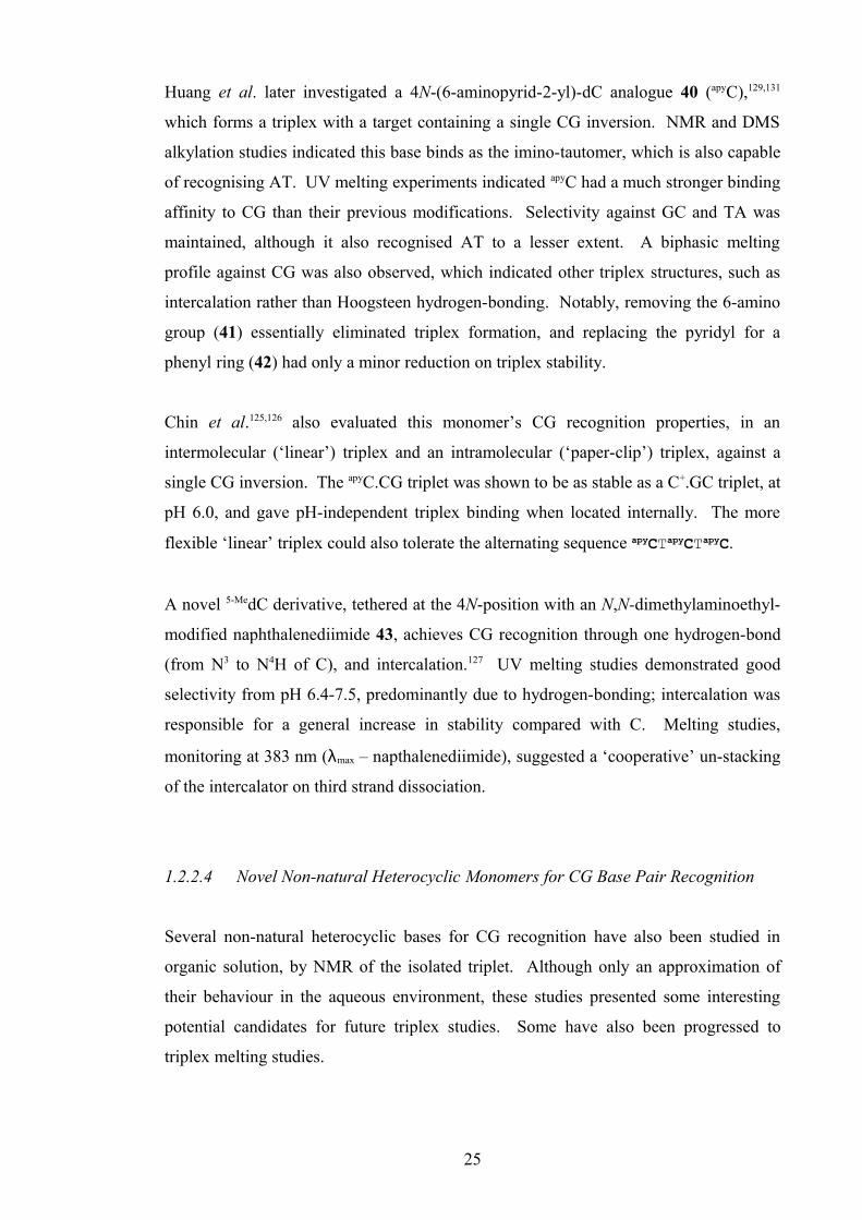
Huang et al. later investigated a 4N-(6-aminopyrid-2-yl)-dC analogue 40 (apyC),129,131
which forms a triplex with a target containing a single CG inversion. NMR and DMS
alkylation studies indicated this base binds as the imino-tautomer, which is also capable
of recognising AT. UV melting experiments indicated apyC had a much stronger binding
affinity to CG than their previous modifications. Selectivity against GC and TA was
maintained, although it also recognised AT to a lesser extent. A biphasic melting
profile against CG was also observed, which indicated other triplex structures, such as
intercalation rather than Hoogsteen hydrogen-bonding. Notably, removing the 6-amino
group (41) essentially eliminated triplex formation, and replacing the pyridyl for a
phenyl ring (42) had only a minor reduction on triplex stability.
Chin et al.125,126 also evaluated this monomer’s CG recognition properties, in an
intermolecular (‘linear’) triplex and an intramolecular (‘paper-clip’) triplex, against a
single CG inversion. The apyC.CG triplet was shown to be as stable as a C+.GC triplet, at
pH 6.0, and gave pH-independent triplex binding when located internally. The more
flexible ‘linear’ triplex could also tolerate the alternating sequence apyCTapyCTapyC.
A novel 5-MedC derivative, tethered at the 4N-position with an N,N-dimethylaminoethyl-
modified naphthalenediimide 43, achieves CG recognition through one hydrogen-bond
(from N3 to N4H of C), and intercalation.127 UV melting studies demonstrated good
selectivity from pH 6.4-7.5, predominantly due to hydrogen-bonding; intercalation was
responsible for a general increase in stability compared with C. Melting studies,
monitoring at 383 nm (λmax – napthalenediimide), suggested a ‘cooperative’ un-stacking
of the intercalator on third strand dissociation.
1.2.2.4 Novel Non-natural Heterocyclic Monomers for CG Base Pair Recognition
Several non-natural heterocyclic bases for CG recognition have also been studied in
organic solution, by NMR of the isolated triplet. Although only an approximation of
their behaviour in the aqueous environment, these studies presented some interesting
potential candidates for future triplex studies. Some have also been progressed to
triplex melting studies.
25

R
RN
N
N
O
HH
NN
NO
N
N
HH
H
UNI.CG
NN
H3C
R NH
O
NH N
NH3C
RN
O
NH H
N
ONH
R NH
OnHex, Et
NHN
NR
O
H,nPr
NH2NN
R
O
NH2NN
R
44 51,52
53,54
55
56
UNI
R1 = H 45
CONH2 48
CONHnBu 49
COCH3 46
CONHnHex 50
COPh 47N
O
O
R
NHR1
N
O
ONHR2RR2 =
Figure 1.27. Novel heterocyclic CG recognition monomers; ureido-naphthimidazole 44 (UNI),133
amino- 45, amido- 46,47, and ureido-phthalimides 49-50,134-136 ureido-isoindolin-1-one
51,52,135,137 urocanamide 53,54 and imidazole derivatives 55,56.138-140
A butylureido-naphthimidazole 44 (UNI)133 was assessed in NMR binding studies in
forming a triplet with CG, with hydrogen bonds to 4-NH of C and C=O6 and N7 of G.
Although incorporated into DNA, biophysical studies were not published.
Several modified phthalimide structures were also evaluated in NMR binding studies
with CG.134-136 Whereas amino- 45, acetamido- 46 and benzamido- 47 modifications
gave relatively weak binding, ureido- 48 and N-alkylureido- groups 49,50 demonstrated
strong binding via three hydrogen-bonds from the phthalimide C=O to 4-NH of C, and
from the urea to C=O6 and N7 of G.
A hexylureido-isoindolin-1-one structure 51,135 analogous to the above monomer, also
effectively bound to CG with an association constant (Kassoc) ten-times that of
Zimmerman’s naphthimidazole 44.133 This was also progressed into triplex melting
studies as the ethylureido-isoindolin-1-one-methylene monomer 52,137 however, it
proved no more effective at CG recognition than an abasic site, with a stability of
approximately 1.8-5.0 kcal/mol lower than the natural T.AT and C+.GC triplets.
Derivatives of urocanamide 53,54 and related imidazole structures 55,56 were evaluated
by Purwanto et al.138-140 in NMR studies and UV melting experiments. NOE
experiments indicated binding to CG occurred through two hydrogen-bonds, one to C7
and one to C=O6 of G. NMR binding studies of the primary amide 53, N-propylamide
54, alkyl- 55 and phenyl- 56 derivatives indicated strongest binding from the primary
amide (urocanamide). Reduced affinity for the other three were attributed to rotation
around the amide bond hindering binding for the N-ethylamide, too much flexibility for
alkyl derivatives, and too little flexibility for phenyl derivatives.
26

The N-propyl urocanamide 54 was progressed to triplex melting studies, targeting a
single CG inversion in an AT rich duplex. Despite showing a sharp third strand melting
transition, a broad melt transition against TA of a similar Tm was also detected, possibly
due to binding to A through protonation of the imidazole ring. An increase in
selectivity for CG over TA was observed on increasing pH to 6.5, confirming this pH
dependence of TA binding. Triplex stability, however, was approximately 10 °C lower
than for the C+.GC triplet, at pH 6.0, due in part to lack of π-π stacking area and
hydrophilicity of the amide moiety. These observations once again highlighted the
importance of base-stacking, hydrophobic interactions and conformation within the
triplex, which cannot be well approximated in NMR studies of isolated triplets.
R
RNN
N
O
HH
NN
NO
N
N
HH
H
H
NO
HO
OH
NN
N
dR
N
N NN
dR
N
NN
dR
N
NN
N
O
NHO
OOH H
carbohydrate.CG
R
O
5758 59
60
R
RN
N
N
O
HH
NN
NO
N
N
HH
H
H
WNA-βC.CG
OHO
OH 61 dN
OO
R''O
N
N
NO H
H
R'O
H
Figure 1.28. Novel carbohydrate and antiparallel CG recognition monomers/triplets.
Recognition of the CG base pair within antiparallel triplexes has also been employed
despite issues of stability. The azole monomers 57-60 of Durland et al.141 generally
bound to two or more base pairs with moderate affinity, and the purine nucleoside 2’-
deoxynebularine 61 (dN) demonstrated equal binding affinity to CG and AT at pH 7.4
via one hydrogen bond. The conformationally-locked WNA-βC nucleoside of Sasaki et
al.142 effectively recognised CG, whilst the G and T β-anomers, WNA-βG and WNA-
βT, bound selectively to TA within an antiparallel triplex, as determined by gel-shift
assays. A novel abasic carbohydrate was also assessed in NMR studies as a CG binding
agent.143 Recognition occurs via two hydrogen-bonds, although it is doubtful this could
be in useful in a triplex context.
Although some success has been afforded within the antiparallel triplex, CG recognition
is still most likely to be achieved through the parallel motif. Despite some degree of
pH-dependence parallel triplexes are more stable and easier to work with, but
antiparallel recognition is still of interest.
27

1.3 Conclusion
Genomic DNA is a promising target for many small-molecule and oligonucleotide-
based therapeutics, aimed at treating a wide variety of genetic diseases and cancers.
The sequence-specificity of DNA allows for oligotherapeutics to be synthesised that
target only specific sequences, to block a particular gene function, target cancer cells,
and for a range of site-specific DNA strand modifications. It is for these reasons that
extensive research has been carried out over a number of years, to achieve selective
recognition of each of the four base pairs, whilst understanding the effect of interaction
of neighbouring modified nucleosides on the efficacy of triplex formation, and
enhancing resistance to in vivo degradation.
The therapeutic use of DNA triplexes is an expanding, exciting field, which continues to
produce promising results in biophysical studies and against real targets in some
biological systems.
28

Chapter 2
Modified 6-Oxocytidine Nucleosides
for GC Recognition
29

2. Modified 6-Oxocytidine Nucleosides for GC Recognition
2.1. Introduction
Recognition of GC base pairs using the natural base C is both selective and strong
below pH 6.0. Due to the low pKaH of N3 of cytosine, protonation of which is required
for Hoogsteen hydrogen-bonding, the C+.GC or C.GC triplet is not stable, however, at
physiological pH (7.0). This severely limits the number of biological applications
where this can be used, especially against targets containing contiguous GC base pairs,
where repulsion between positive charges destabilises the triplex further.
A variety of pyrimidine- and purine-based monomers for GC recognition have been
evaluated as described in Section 1.2.1.2. These include the pyrimidine nucleosides, 5-
methylcytosine (5-MeC)45-47 and the C-nucleoside 3-methyl-2-aminopyridine (MAP),61,65
providing stability through a higher pKaH and hydrophobic methyl group; and N6-
methyl-8-oxoA87 and pyDDA,68 which form pH-independent triplets with permanent
hydrogen-bonds.
The non-natural base 6-oxocytosine (6-oxoC, 62)70-74 also found use as a GC recognition
monomer, which forms triplets independent of pH. Unlike some other GC recognition
monomers, 6-oxoC could potentially be modified at the 5-position on the nucleobase (and
2’-position of the sugar), with pendant amino groups, such as for bis-amino-U (BAU).
O
O
OO
ON
N
N
OO
N
N
H
H
P
O
O
O
H
P O -O
H
H
H
H H
H
N
N
N
O
N
N
OO
OP
-O O
O
P
O-
O
O
HH
H
O
N
NN
O
H
H
OO
charge-stabilisation
G
CHoogsteen hydrogen-bondingWatson-Crickbase pair
charge-stabilisation
6
74
3
NH
NH2
ONO
OOH
HO O
63
NH
NH2
ONH
O
NH3
H3N
62
BAoxC.GC
BAoxC
Figure 2.1. Structures of 6-oxocytosine 62, 2’-O-(2-aminoethyl)-5-(3-aminoprop-1-ynyl)-6-
oxocytidine 63 (BAoxC), and proposed hydrogen-bonding and electrostatic interactions within the
putative BAoxC.GC triplet.
30

In the above diagram (Figure 2.1), charge-stabilising interactions are shown between the
protonated 2’-amine moiety and guanidine 5’-phosphate, and also between the
protonated 5’-amine moiety and BAoxC phosphates. Hoogsteen hydrogen-bonds are also
shown between BAoxC and G.
The initial project aim therefore, was synthesis of a direct analogue of BAU, a bis-amino-
modified derivative of 6-oxocytidine, 2’-O-(2-aminoethyl)-5-(3-aminoprop-1-ynyl)-6-
oxocytidine 63 (BAoxC) (Figure 2.1). This would combine the triplex-stabilising effect of
the amine groups as exemplified by BAU and potentially enhanced π-stacking
interactions from the alkynyl moiety. GC recognition would be achieved, as previously
described, via two permanent hydrogen bonds between N3H and N4H of the base and N7
and C6=O of guanine, giving pH-independent triplex binding. Although poor binding
affinity was observed when using 2’-O-aminoethyl-modified 6-oxoC and moxC compared
to 2’-O-methyl- or 2’-deoxy- derivatives (Section 1.2.1.3), combining both amine
modifications was proposed to enhance alignment with the GC base pair in addition to
the properties described above.
2.2. Bis-amino-6-oxocytidine: Synthesis
The key steps in synthesis of a functionalised 6-oxocytidine monomer are coupling of
the persilylated 6-oxocytosine (6-aminouracil) base to the modified sugar; sugar
deprotection and reprotection of the 5’-position; halogenation at the 5-position;
protection of the 4-NH2 group; and subsequent base modification. The order in which
these are to be carried is open to variation. The final compound is then converted into a
3’-O-phosphoramidite144 for incorporation into triplex-forming oligonucleotides (TFOs).
Dr. Osborne conducted much work in house, into synthesis of a BAoxC monomer 64, for
incorporation into TFOs. After the sugar-base coupling using the 2’-O-
phthalimidoethyl-modified sugar 65, and three protection/deprotection steps, problems
were encountered in two key steps in the synthesis (Scheme 2.1).
Iodination under the above conditions required excess NIS and several days to effect the
transformation. The subsequent Sonogashira reaction under Hobbs’ conditions124 was
31
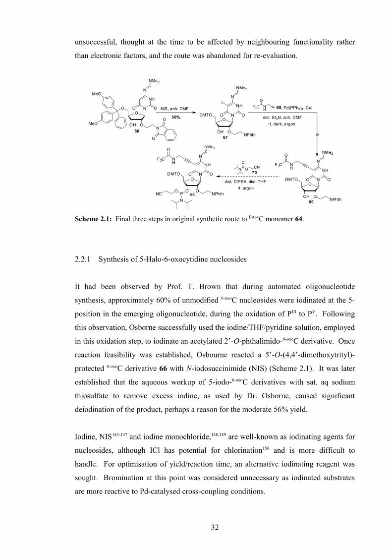
unsuccessful, thought at the time to be affected by neighbouring functionality rather
than electronic factors, and the route was abandoned for re-evaluation.
NIS, anh. DMF
56%
NH
F3C
O
68, Pd(PPh3)4, CuI
dist. Et3N, anh. DMF
P ON CNCl
70
dist. DIPEA, dist. THF
rt, argon
O
OOH
O N
NH
N
OO
NMe2
66
MeO
MeON
O
O
O
OOH
DMTO N
NH
N
OO
NMe2
67NPhth
I
rt, dark, argon
O
OOH
DMTO N
NH
N
OO
NMe2
69NPhth
NH
F3C
O
O
OO
DMTO N
NH
N
OO
NMe2
64 NPhth
NH
F3C
O
PO
N
NC
X
Scheme 2.1: Final three steps in original synthetic route to BAoxC monomer 64.
2.2.1 Synthesis of 5-Halo-6-oxocytidine nucleosides
It had been observed by Prof. T. Brown that during automated oligonucleotide
synthesis, approximately 60% of unmodified 6-oxoC nucleosides were iodinated at the 5-
position in the emerging oligonucleotide, during the oxidation of PIII to PV. Following
this observation, Osborne successfully used the iodine/THF/pyridine solution, employed
in this oxidation step, to iodinate an acetylated 2’-O-phthalimido-6-oxoC derivative. Once
reaction feasibility was established, Osbourne reacted a 5’-O-(4,4’-dimethoxytrityl)-
protected 6-oxoC derivative 66 with N-iodosuccinimide (NIS) (Scheme 2.1). It was later
established that the aqueous workup of 5-iodo-6-oxoC derivatives with sat. aq sodium
thiosulfate to remove excess iodine, as used by Dr. Osborne, caused significant
deiodination of the product, perhaps a reason for the moderate 56% yield.
Iodine, NIS145-147 and iodine monochloride,148,149 are well-known as iodinating agents for
nucleosides, although ICl has potential for chlorination150 and is more difficult to
handle. For optimisation of yield/reaction time, an alternative iodinating reagent was
sought. Bromination at this point was considered unnecessary as iodinated substrates
are more reactive to Pd-catalysed cross-coupling conditions.
32

Benzyltriethylammonium dichloroiodate (BTEA-ICl2) 71 (Figure 2.2), was used by
Harrowven et al.151 to iodinate a substituted anisole intermediate in synthesis of a target
macrocycle, in 99% yield over 5 hours. A co-catalyst ZnCl2 was used, which also
helped to solubilise BTEA-ICl2 in the solvent acetic acid.
N Cl I Cl
71
Figure 2.2. Benzyltriethylammonium dichloroiodate (BTEA-ICl2) 71.
Most ammonium polyhalide-derived reactions such as this, in the literature, have
utilised the benzyltrimethylammonium (BTMA) salt, which is commercially available,
for halogenation reactions; for example, the dichloroiodate, tetrachloroiodate and
tribromide or chlorobromate for iodination, chlorination and bromination respectively.
The preferred solvents for halogenation, using BTMA dichloroiodate (BTMA-ICl2),
were methanol or CH2Cl2/methanol and several inorganic co-catalysts, CaCO3,152,153
NaHCO3,152,154 and ZnCl2151,155 have been used. Although the BTEA ion has been
exploited as a range of salts, it has rarely been used as the dichloroiodate salt. Mitra et
al.156-159 however, published several papers discussing halogenation using solid-
supported BTEA-polyhalide salts. The BTEA-based reagent was used in preference to
BTMA-ICl2 by the Harrowven group, as it was believed to afford improved yield and
selectivity.
2.2.1.1 Synthesis and Reaction of Benzyltriethylammonium dichloroiodate
Literature methods for synthesising BTMA-ICl2, include reacting BTMA-chloride with
iodine monochloride in 86% yield,152 or with iodine and silver trifluoroacetate.160 The
first of these methods was attempted using BTEA-Cl 72 instead, however, only a gum
containing a mixture of salts was isolated. Attention was subsequently turned to a
cleaner route, published by Kosynkin and Tour,154 which used standard-grade reagents
(bleach, NaI, HCl) in aqueous medium. This was tested on a 10 g scale, affording
BTEA-ICl2 71 in 96% yield.
The solution phase iodination conditions of Kosynkin et al.154 and the solvent-free
conditions of Hajipour et al.161 (in which the reagent Me4N+ ICl2– was ground to a fine
33
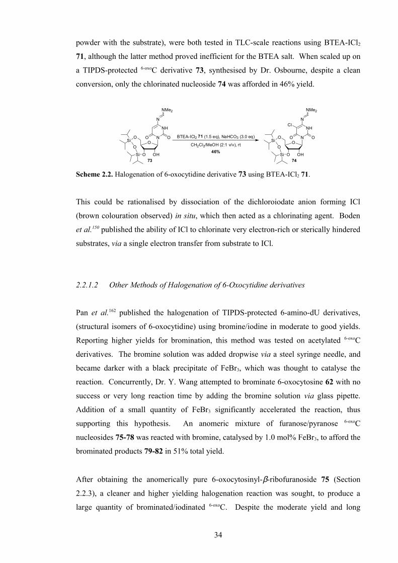
powder with the substrate), were both tested in TLC-scale reactions using BTEA-ICl2
71, although the latter method proved inefficient for the BTEA salt. When scaled up on
a TIPDS-protected 6-oxoC derivative 73, synthesised by Dr. Osbourne, despite a clean
conversion, only the chlorinated nucleoside 74 was afforded in 46% yield.
BTEA-ICl2 71 (1.5 eq), NaHCO3 (3.0 eq)
46%
O
OHO
O N
NH
N
OO
NMe2
73
Si
O
Si
CH2Cl2/MeOH (2:1 v/v), rt O
OHO
O N
NH
N
OO
NMe2
74
Si
O
Si
Cl
Scheme 2.2. Halogenation of 6-oxocytidine derivative 73 using BTEA-ICl2 71.
This could be rationalised by dissociation of the dichloroiodate anion forming ICl
(brown colouration observed) in situ, which then acted as a chlorinating agent. Boden
et al.150 published the ability of ICl to chlorinate very electron-rich or sterically hindered
substrates, via a single electron transfer from substrate to ICl.
2.2.1.2 Other Methods of Halogenation of 6-Oxocytidine derivatives
Pan et al.162 published the halogenation of TIPDS-protected 6-amino-dU derivatives,
(structural isomers of 6-oxocytidine) using bromine/iodine in moderate to good yields.
Reporting higher yields for bromination, this method was tested on acetylated 6-oxoC
derivatives. The bromine solution was added dropwise via a steel syringe needle, and
became darker with a black precipitate of FeBr3, which was thought to catalyse the
reaction. Concurrently, Dr. Y. Wang attempted to brominate 6-oxocytosine 62 with no
success or very long reaction time by adding the bromine solution via glass pipette.
Addition of a small quantity of FeBr3 significantly accelerated the reaction, thus
supporting this hypothesis. An anomeric mixture of furanose/pyranose 6-oxoC
nucleosides 75-78 was reacted with bromine, catalysed by 1.0 mol% FeBr3, to afford the
brominated products 79-82 in 51% total yield.
After obtaining the anomerically pure 6-oxocytosinyl-β-ribofuranoside 75 (Section
2.2.3), a cleaner and higher yielding halogenation reaction was sought, to produce a
large quantity of brominated/iodinated 6-oxoC. Despite the moderate yield and long
34

reaction time necessary for iodination of 6-oxoC using NIS, attention was turned back to
N-halosuccinimides using the alternative conditions of Hirota et al..163
Hirota et al. brominated several 4N-alkyl-1,3-dimethyl-6-oxocytosine derivatives
(Figure 2.3) using N-bromosuccinimide (NBS), with the radical initiator AIBN,
suspended in THF, in 80-95% yield. 6-Oxocytidine derivative 75 was transformed
cleanly under these conditions, using only 2.0 mol% AIBN in 30 minutes, to afford the
product 79 in 95% yield.
NH
NH2
ONO
OAcOAc
AcO O
5
75
NH
NH2
ONO
OAcOAc
AcO O
XN
NHR
ONO
R = Me, Et, Pr, iPr, BnCHex, CF3CH2-,
X = Br 79I 83
Figure 2.3. 4N-Alkyl-1,3-dimethyl-6-oxocytosine derivatives, acetylated 6-oxoC 75 and
halogenated 6-oxoC nucleosides 79,83.
Iodination using N-iodosuccinimide (NIS) under the same conditions resulted in a
brown colouration. Presumed to be due to decomposition of NIS in storage, a large
quantity was recrystallised (dioxane/chloroform)164 and the reaction was repeated.
Despite the same brown colouration whether conducted at rt or 0 °C, on a 5 g scale the
reaction was complete within 15 minutes. Partial or almost complete deiodination of
product occurred after aqueous workup, using dilute aq Na2S2O3 or Na2S2O5
respectively, as previously observed (Section 2.2.1). The sodium thiosulfate (Na2S2O3)
workup, however, was used successfully on a 0.5 g quantity of the reaction mixture,
separating the layers as soon as decolourisation occurred. Suspension of the remainder
in ethyl acetate and soaking over 2 days, however, removed all excess iodine and
succinimide by-product, affording the product 83 in 82% combined yield.
2.2.2 Synthesis of 2’-O-Phthalimidoethyl-modified Ribofuranose Sugar
The 2’-O-phthalimidoethyl-modified ribofuranose sugar 65, synthesised and used by
Osborne et al.37 in preparation of the AT-recognition monomer BAU, was prepared for
coupling to 6-oxocytosine 62 (Scheme 2.2), as part of the synthetic route to the BAoxC
monomer 64.
35

O
OHOH
HOOMeAcCl, MeOH
r.t., 3 h
(TIPSCl)2O, py
r.t., 6 h anh. DMF, -5oC to r.t.
BrCH2CO2Me, NaH
LiBH4
36% (86%)Phthalimide, PPh3
DEAD, dist. THF
r.t., 30 mins
AcOH, Ac2O, H2SO4
r.t., 2 h
quant. (72%) 60% (61%)
94% (91%)82% (79%)
OHO
OH OH
OH
O
OHO
OSi
Si
OOMe
86
O
OO
OSi
Si
OOMe
87
O
OMe8584
O
OO
OSi
Si
OOMe
88 OH
O
OO
OSi
Si
O
89
1α
N
O
O
OMe
O
OAcO
AcO
65 N
O
O
OAc r.t., 30 mins
dist. THF
Scheme 2.2. Synthesis of 2’-modified ribofuranose sugar 65 (lit.37 yields in parentheses).
The sugar synthesis starts with formation of the 1’-O-methyl acetal of D-ribose 85,
followed by protection of the 3’- and 5’-OH groups with the bidentate tetra-
isopropylsiloxane-1,3-diyl (TIPDS) group, using Markiewicz Reagent.165 The reaction
proceeded well over two steps, with minor impurities and solvent traces, which were
removed during the following steps (Scheme 2.3).
Alkylation of the 2’-OH with methyl bromoacetate gave the product 2’-O-
methylethanoyl sugar 87 in a moderate yield, containing a significant methyl
bromoacetate impurity which co-eluted with the product. Subsequent reduction of the
ester moiety using LiBH4 therefore, afforded a lower yield of 88 than in the literature.
Phthalimide substitution via Mitsunobu conditions166 gave the TIPDS-protected sugar
89 (α-anomer only) and subsequent acetolysis gave the final 2’-modified ribofuranose
sugar 65 in good yield.
This would then be coupled with 6-oxocytosine 62 (Scheme 2.3), once halogenation and
Sonogashira reactions were optimised.
62
N
NH
NH2
OO
O
OAcO
AcO
N
O
O
NH
NH
NH2
OO HMDS (10 eq)
(NH4)2SO4 (0.2 eq)
N
N
NHSiMe3
OSiMe3Me3SiO
O
OAcO
AcO
NPhth65 (1.0 eq)
OAc
TMSOTf (1.0 eq), anh. DCE
rt, argon, 48 hrsreflux, argon, 24 hrs 90 75quant . 87%
Scheme 2.3. Sugar-base coupling under Vorbrüggen’s167 conditions (in house).
36
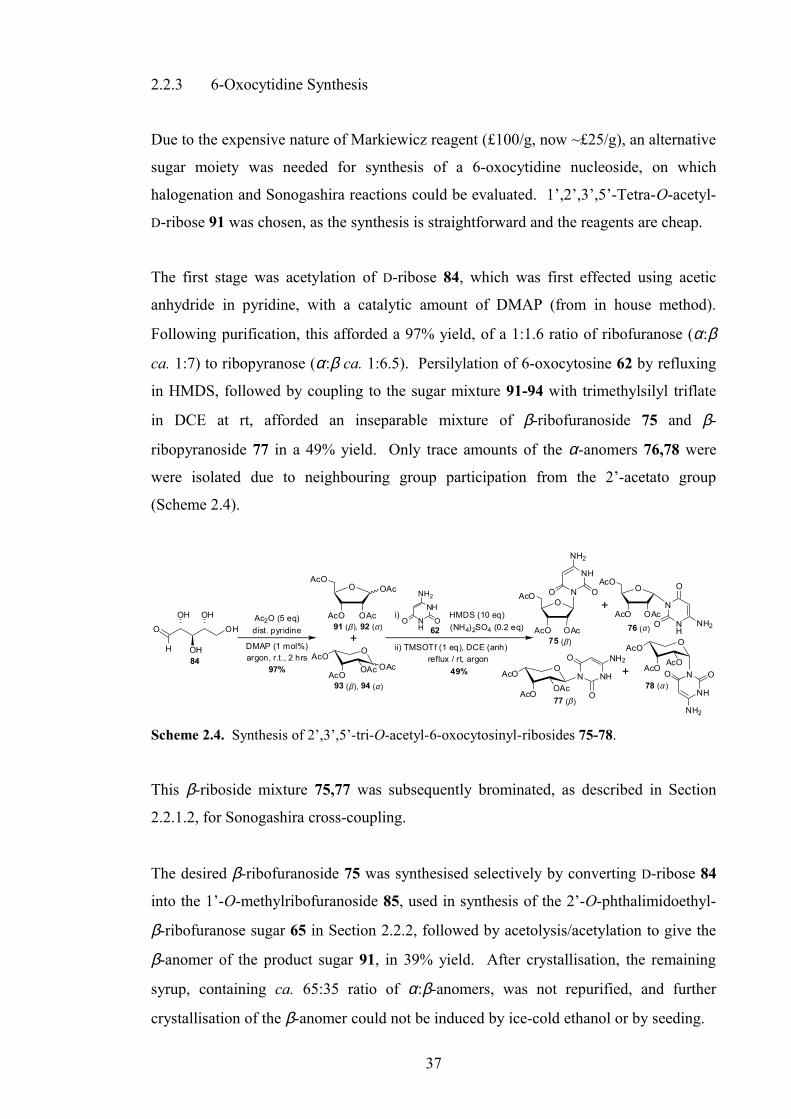
2.2.3 6-Oxocytidine Synthesis
Due to the expensive nature of Markiewicz reagent (£100/g, now ~£25/g), an alternative
sugar moiety was needed for synthesis of a 6-oxocytidine nucleoside, on which
halogenation and Sonogashira reactions could be evaluated. 1’,2’,3’,5’-Tetra-O-acetyl-
D-ribose 91 was chosen, as the synthesis is straightforward and the reagents are cheap.
The first stage was acetylation of D-ribose 84, which was first effected using acetic
anhydride in pyridine, with a catalytic amount of DMAP (from in house method).
Following purification, this afforded a 97% yield, of a 1:1.6 ratio of ribofuranose (α:β
ca. 1:7) to ribopyranose (α:β ca. 1:6.5). Persilylation of 6-oxocytosine 62 by refluxing
in HMDS, followed by coupling to the sugar mixture 91-94 with trimethylsilyl triflate
in DCE at rt, afforded an inseparable mixture of β-ribofuranoside 75 and β-
ribopyranoside 77 in a 49% yield. Only trace amounts of the α-anomers 76,78 were
were isolated due to neighbouring group participation from the 2’-acetato group
(Scheme 2.4).
i) HMDS (10 eq)
(NH4)2SO4 (0.2 eq)
ii) TMSOTf (1 eq), DCE (anh)
reflux / rt, argon
NH
NH
NH2
OO62 AcO OAc
OAcO N
NH
NH2
OO
+
Ac2O (5 eq)
dist. pyridine
DMAP (1 mol%)argon, r.t., 2 hrs
97% 49%
AcO OAc
OAcO
OAc
O OH
OH OH
OH84
91 (β ), 92 (α)
OAcO
AcOOAc OAc
93 (β ), 94 (α)
AcO OAc
OAcO
N
NH
NH2O
O
+
OAcO
AcOOAc
NH
NH2
O
O
OAcO
AcOAcO
NH
NH2
OON N+
76 (α)
75 (β )
78 (α )
77 (β )
H
Scheme 2.4. Synthesis of 2’,3’,5’-tri-O-acetyl-6-oxocytosinyl-ribosides 75-78.
This β-riboside mixture 75,77 was subsequently brominated, as described in Section
2.2.1.2, for Sonogashira cross-coupling.
The desired β-ribofuranoside 75 was synthesised selectively by converting D-ribose 84
into the 1’-O-methylribofuranoside 85, used in synthesis of the 2’-O-phthalimidoethyl-
β-ribofuranose sugar 65 in Section 2.2.2, followed by acetolysis/acetylation to give the
β-anomer of the product sugar 91, in 39% yield. After crystallisation, the remaining
syrup, containing ca. 65:35 ratio of α:β-anomers, was not repurified, and further
crystallisation of the β-anomer could not be induced by ice-cold ethanol or by seeding.
37
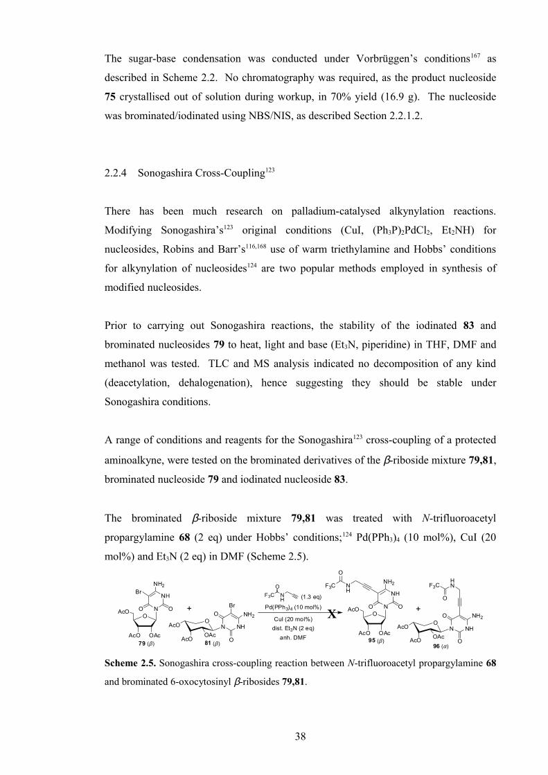
The sugar-base condensation was conducted under Vorbrüggen’s conditions167 as
described in Scheme 2.2. No chromatography was required, as the product nucleoside
75 crystallised out of solution during workup, in 70% yield (16.9 g). The nucleoside
was brominated/iodinated using NBS/NIS, as described Section 2.2.1.2.
2.2.4 Sonogashira Cross-Coupling123
There has been much research on palladium-catalysed alkynylation reactions.
Modifying Sonogashira’s123 original conditions (CuI, (Ph3P)2PdCl2, Et2NH) for
nucleosides, Robins and Barr’s116,168 use of warm triethylamine and Hobbs’ conditions
for alkynylation of nucleosides124 are two popular methods employed in synthesis of
modified nucleosides.
Prior to carrying out Sonogashira reactions, the stability of the iodinated 83 and
brominated nucleosides 79 to heat, light and base (Et3N, piperidine) in THF, DMF and
methanol was tested. TLC and MS analysis indicated no decomposition of any kind
(deacetylation, dehalogenation), hence suggesting they should be stable under
Sonogashira conditions.
A range of conditions and reagents for the Sonogashira123 cross-coupling of a protected
aminoalkyne, were tested on the brominated derivatives of the β-riboside mixture 79,81,
brominated nucleoside 79 and iodinated nucleoside 83.
The brominated β-riboside mixture 79,81 was treated with N-trifluoroacetyl
propargylamine 68 (2 eq) under Hobbs’ conditions;124 Pd(PPh3)4 (10 mol%), CuI (20
mol%) and Et3N (2 eq) in DMF (Scheme 2.5).
AcO OAc
OAcO N
NH
NH2
OO +O
AcO
AcOOAc
NH
NH2
O
O
N
79 (β ) 81 (β )
Br
Br
AcO OAc
OAcO N
NH
NH2
OO +
OAcO
AcOOAc
NH
NH2
O
O
N
95 (β )96 (α)
NH
HN
O
F3CF3C
O
Pd(PPh3)4 (10 mol%)
dist. Et3N (2 eq)
CuI (20 mol%)
anh. DMF
X
F3C NH
O
(1.3 eq)
Scheme 2.5. Sonogashira cross-coupling reaction between N-trifluoroacetyl propargylamine 68
and brominated 6-oxocytosinyl β-ribosides 79,81.
38

TLC analysis indicated a complex mixture and only starting material and debrominated
starting material were isolated after purification (analysed by MS). It is unusual, yet
possible that debromination occurred via hydride-insertion after Pd-insertion into the C-
Br bond.
Triethylamine was the base of choice, because it is non-nucleophilic and there is no
possibility of substitution of bromine. It was shown by Wang et al.,169 that
triethylamine also inhibits alkyne dimerisation/homo-coupling and promotes the desired
cross-coupling reaction. Diethylamine, as used by Kenkichi Sonogashira, was shown to
promote homo-coupling under the right conditions.
Using a large excess of Et3N (20 eq) or as a solvent, on 50 mg and 500 mg scale,
resulted only in partial debromination, deacetylation, and triphenylphosphine oxide. A
similar observation was made for the iodinated nucleoside 83.
The last reactions on the brominated β-riboside mixture 79,81 were carried out under
microwave heating. The cross-coupling reactions were conducted in DMF or THF at 60
°C for 10 mins, and in DMF at 100 °C for 10 mins. Once again, only complex mixtures
were formed.
A fresh quantity of N-trifluoroacetyl propargylamine117,170 68 (Scheme 2.6) was
synthesised for subsequent Sonogashira reactions; the quality of which was confirmed
by standard characterisation methods and in a test Sonogashira reaction using 5’-O-
(4,4’-dimethoxytrityl)-5-iodo-2’-deoxyuridine (DMT-IdU) 97 under Hobbs’
conditions.124 However, subsequent test reactions using the brominated 79 and
iodinated 83 6-oxoC derivatives, were unsuccessful. Only partial de-iodination of starting
material 83, and formation of triphenylphosphine oxide (in both cases) were observed.
O
NH
F3CH2N
O
OF3C(1.3 eq)
dry MeOH, argon, 0oC - rt98
6857% (74%)
Scheme 2.6. Synthesis of N-trifluoroacetyl propargylamine 68, (lit.117 yield in parentheses).
It was proposed that the above problems might be exacerbated by the presence of acetyl
groups hence a suitable method for deacetylation was sought. The brominated 79 and
iodinated 83 6-oxoC nucleosides were deacetylated by heating in 0.4 M K2CO3 in
39
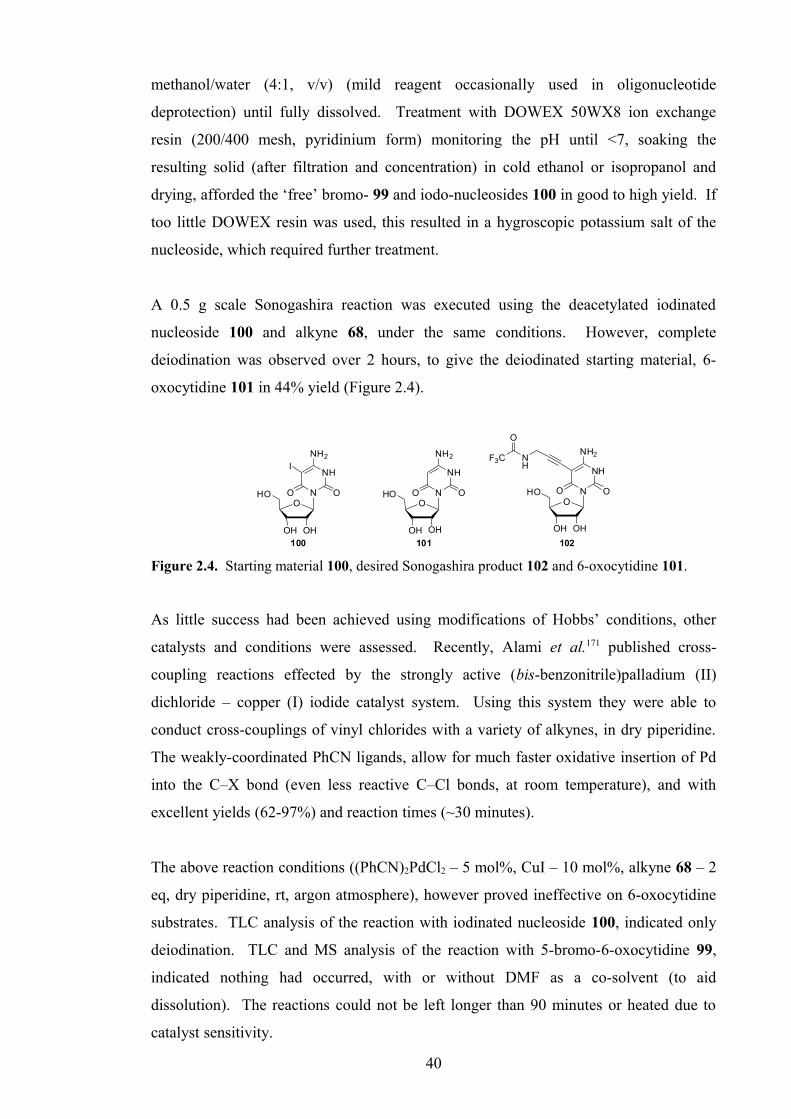
methanol/water (4:1, v/v) (mild reagent occasionally used in oligonucleotide
deprotection) until fully dissolved. Treatment with DOWEX 50WX8 ion exchange
resin (200/400 mesh, pyridinium form) monitoring the pH until <7, soaking the
resulting solid (after filtration and concentration) in cold ethanol or isopropanol and
drying, afforded the ‘free’ bromo- 99 and iodo-nucleosides 100 in good to high yield. If
too little DOWEX resin was used, this resulted in a hygroscopic potassium salt of the
nucleoside, which required further treatment.
A 0.5 g scale Sonogashira reaction was executed using the deacetylated iodinated
nucleoside 100 and alkyne 68, under the same conditions. However, complete
deiodination was observed over 2 hours, to give the deiodinated starting material, 6-
oxocytidine 101 in 44% yield (Figure 2.4).
NH
NH2
ONO
OH
HO O
OH
NH
NH2
ONO
OH
HO O
OH
NH
F3C
O
102101
NH
NH2
ONO
OH
HO O
OH
100
I
Figure 2.4. Starting material 100, desired Sonogashira product 102 and 6-oxocytidine 101.
As little success had been achieved using modifications of Hobbs’ conditions, other
catalysts and conditions were assessed. Recently, Alami et al.171 published cross-
coupling reactions effected by the strongly active (bis-benzonitrile)palladium (II)
dichloride – copper (I) iodide catalyst system. Using this system they were able to
conduct cross-couplings of vinyl chlorides with a variety of alkynes, in dry piperidine.
The weakly-coordinated PhCN ligands, allow for much faster oxidative insertion of Pd
into the C–X bond (even less reactive C–Cl bonds, at room temperature), and with
excellent yields (62-97%) and reaction times (~30 minutes).
The above reaction conditions ((PhCN)2PdCl2 – 5 mol%, CuI – 10 mol%, alkyne 68 – 2
eq, dry piperidine, rt, argon atmosphere), however proved ineffective on 6-oxocytidine
substrates. TLC analysis of the reaction with iodinated nucleoside 100, indicated only
deiodination. TLC and MS analysis of the reaction with 5-bromo-6-oxocytidine 99,
indicated nothing had occurred, with or without DMF as a co-solvent (to aid
dissolution). The reactions could not be left longer than 90 minutes or heated due to
catalyst sensitivity.
40

The bis-(triphenylphosphine)palladium (II) dichloride (10 mol%) – copper (I) iodide
(30 mol%) catalyst system, as used by Robins and Barr116,168 for cross-coupling reactions
with 5-iodo-2’-deoxyuridines in warm triethylamine (50 °C) was also assessed. Even
when using 4 equivalents of alkyne, only deiodination and alkyne dimerisation were
detected for substrate 100 and nothing was observed for the brominated substrate 99.
Alternatively, instead of conducting cross-couplings in absence of water, the reaction
was conducted in hot water, using 0.5 mol% (Ph3P)4Pd, as published by Bhattacharya
and Sengupta.172 The authors’ principal idea was to utilise the hydrophobic effect, and
although the 6-oxocytidine substrates are (partially) water-soluble, it was hoped these
conditions might be beneficial. However, again only deiodination of 100 was observed.
These reactions, using various catalysts, solvents, temperatures and ratios of reagents
were all unsuccessful. Brominated 6-oxoC nucleosides (β-anomers) did not react, and
iodinated 6-oxoC nucleosides were deiodinated. This illustrates the surprising strength of
the C–Br bond and susceptibility of the R–Pd(PPh3)2–I intermediate to decomposition.
There may also be other steric and electronic factors preventing this transformation,
such as the neighbouring C6-carbonyl and C4-amino groups. Although other alkynes
were not tested, N-trifluoroacetyl propargylamine 68 couples to protected and
unprotected 5-iodo-2’-deoxyuridine nucleosides in excellent yield.117,170
Sonogashira-like palladium-catalysed cross-coupling reactions were subsequently
abandoned. It is still possible, however, that results could be obtained using a triflato-
nucleoside,59 alkynylzinc reagents173,174 or even Stille coupling,175 which have been
successfully used to alkynylate other nucleosides.
2.3 Alternative Modifications at 5-position
Attempts to enhance base-stacking by attachment of an alkynyl group failed as
described above. It was also found that the 5-allyl-group of 6-oxodC caused
destabilisation of the triplex, relative to 6-oxodC alone. An alternative method was sought
therefore for attachment of a pendant amine group at the 5-position of 6-oxoC, with or
without additional base-stacking enhancement, which would increase triplex-stability by
charge-stabilisation.
41

2.3.1 Halide-Amine Exchange
Halide-amine exchange was proposed as an alternative method for introduction of
amine functionality to the base. There is much literature precedent for this
transformation on aromatic compounds, uridine nucleosides, uracils and aminouracils,
using primary and secondary amines and even ammonia. In 1951, Arthur Phillips
published a series of substitution reactions on 5-bromouracil, using amines such as
methylamine, butylamine, cyclohexylamine, piperidine and benzylmethylamine in
typically good to quantitative yields.176 Benzylmethylamine was used for introduction
of a methylamino group (following hydrogenolysis of the benzyl group), as reaction of
similar heterocycles with methylamine and ammonia often requires elevated
temperatures177 or high pressure,178,179 with long reaction times.
5-Bromo-6-oxocytidine 99 was heated at 85 °C in a sealed tube with four different
amines (piperidine, benzylamine, propargylamine, TAEA 103) for 3.5 hours (Figure
2.5). TLC analysis indicated loss of starting material, most likely through degradation,
and no positive identification of desired products was made by MS analysis. A tan-
coloured solid was isolated from the last reaction with the polyamine tris(2-
aminoethyl)amine, TAEA 103, which was confirmed by NMR, however, to be a salt of
TAEA.
O
OHOH
HO N
NH
NH2
OO
N
O
OHOH
HO N
NH
NH2
OO
HN
O
OHOH
HO N
NH
NH2
OO
HN
O
OHOH
HO N
NH
NH2
OO
HN
N
NH2
H2N
104 105 106 107
O
OHOH
HO N
NH
NH2
OO
Br
99
Figure 2.5. 5-Bromo-6-oxocytidine 99 and amine-halide exchange products 104-107.
Azam et al.180 attached the polyamine TAEA 103 to an anthraquinone intercalator-
modified chimeric TFO, by a simple chloride-amine exchange reaction. The polyamine
moiety demonstrated increased triplex stability without altering selectivity, and has
since been used, in house, by Dr. Z. Zhao, for synthesising simple polyamine-
anthraquinone intercalators for non-specific triplex stabilisation.181
42

Initially, test reactions were performed on 2’,3’,5’-O-triacetyl-5-bromo-6-oxocytidine
79, heating the nucleoside at 80 °C in a sealed tube, for 2.5 hours in piperidine or
benzylamine. Following treatment with DOWEX 50WX8-400 (pyridinium form) and
diethyl ether, a white gum containing some deacetylated piperidinyl compound 104, and
deacetylated starting material 101 were obtained respectively. Following this, to avoid
complex mixtures due to partial deacetylation, the free nucleoside 5-bromo-6-
oxocytidine 99 was used. This was treated with propargylamine, benzylamine and
TAEA 103.
Varying the reaction conditions for bromide-TAEA exchange was unsuccessful.
Heating 5-bromo-6-oxocytidine 98 with TAEA 103 at 180 °C for 20 minutes, heating at
75 °C in a 30% aq solution of the amine overnight, and refluxing in distilled pyridine
with 5 eq of TAEA 103 produced similar results as obtained previously and isolation of
amine salts.
In order to confirm that exchange had occurred successfully originally, when using
piperidine, nucleoside 99 was heated in the amine alone, or with pyridine, water or
DMF as co-solvent at 70 °C for 17 hours. This proved partially successful, with an
estimated 20-30% conversion (TLC analysis) for all reactions, except in water. These
results were also confirmed by MS analysis.
The amine-halide exchange was proposed to occur via an SNAr mechanism, which if
true would suggest a faster, more efficient reaction with chloro- and fluoro-, rather than
bromo- or iodo-groups. To take advantage of this, and to avoid problems due to
cleavage of acetyl protecting groups, a large quantity of unprotected 5-chloro-6-
oxocytidine 108 was synthesised for comparison with the other 5-halo-6-oxoC derivatives.
2’,3’,5’-Tri-O-acetyl-6-oxocytidine 75 was treated with N-chloro-succinimide (NCS)
based on the method published by Hirota et al.163 as described in Section 2.2.1.2
(Scheme 2.7). The product was purified by column chromatography, not soaked in
ethyl acetate to remove the succinimide by-product, as it is partially soluble in ths
solvent.
43

NH
NH2
ON
O
AcO
AcOO
Cl
OAc
NH
NH2
ON
O
AcO
AcOO
OAc
NCS (1.1 eq)
AIBN (2 mol%)
dist. THF, dark, argon
NH
NH2
ON
O
OH
HO O
OH
0.4M K2CO3
heat, 5 minutes
Cl
MeOH/H2O (4:1, v/v)
75 109 108
rt, 18 hrs
90% 90%
Scheme 2.7. Synthesis of 5-chloro-6-oxocytidine 108.
The product 109 was deacetylated as previously described to afford the desired free
nucleoside 108 in 81% yield over two steps, containing 1.8 mol % succinimide from the
previous step and 35.6 mol % KOAc, not observed for previous deacetylations by this
method.
In the next series of test exchange reactions, chlorinated 74,108 and iodinated 100 6-oxoC
nucleosides were treated with piperidine, and brominated nucleoside 99 with five 1,n-
diaminoalkanes (n = 2, 6, 8, 10, 12), at 70 °C for 18-20 hours (Figure 2.6).
O
OHO
O N
NH
N
OO
Cl
O
OHOH
HO N
NH
NH2
OO
Br
O
OHOH
HO N
NH
NH2
OO
I
Si
Si
O
N
O
OHOH
HO N
NH
NH2
OO
Cl
74 99 100 108
Figure 2.6. Substrates for amine-halide substitution reactions 74,99,100,108.
5-Iodo-6-oxocytidine 100 reacted partially with piperidine but less cleanly than 5-
bromo-6-oxocytidine 99, suggesting an SNAr mechanism might apply. The protected
chlorinated 6-oxoC derivative 74, however, underwent deprotection of the 4N-
dimethylformamidine protecting group, with no desired reaction, and none of the 1,n-
diaminoalkanes reacted with 5-bromo-6-oxocytidine 99.
Chloro-6-oxoC derivative 74 was treated with ethylene diamine (42 eq) and TAEA 103
(18 eq) at 75 °C for 90 minutes and also for 18-20 hours. Only formamidine
deprotection and amine degradation were observed, and trifluoroacetylation of the latter
reaction mixture yielded only trifluoroacetylated amine.
So far, successful amine-halide exchange had been limited to the secondary amine
piperidine, which is probably not beneficial to triplex formation. Attempts to couple
44

with primary amines, and in particular, polyamines, although unsuccessful thus far,
were continued using 5-chloro-6-oxocytidine 108. It was thought, that protection of
two of the primary amine moieties of tris(2-aminoethyl)amine 103, could potentially aid
the reaction by presenting only one centre for reaction, although the bis-protected
product was finally not used. Asseline et al.182 published the synthesis of the bis-
trifluoroacetylated polyamine 110 via alkylation of the trifluoroacetic acid salt of bis-
trifluoroacetylated diethylene triamine 111 with bromoethanol, followed by Mitsunobu
reaction with LiN3 and hydrogenation over Pd/C, in 23% overall yield.
NNH2H2N
NH2
NNH2H2N
NHDMT(p-MeOPh)2PhCCl 112
(0.6-1.1 eq)
distilled pyridine
argon, rt
O
OF3C (2.5-3.0 eq)
dry MeOH / TEAargon, 0oC-rt N
NHDMTNH
NH
F3C
O
O
F3C
NNH2N
H
NH
F3C
O
O
F3CAcOH / MeOH
rt
103 113 114 110
X
Scheme 2.8. First route to N-(2-aminoethyl)-bis-N,N-[2-(trifluoroacetamido)ethyl]amine 110.
The first synthetic procedure attempted (Scheme 2.8) began with mono-protection with
a 4,4’-dimethoxytrityl (DMT) group, by addition of a solution of DMTCl 112 in
distilled pyridine, dropwise over 20 minutes, to a stirred, ice-cold solution of TAEA 103
(6 eq) in distilled CH2Cl2. After aqueous workup, an off-white foam was afforded,
which unfortunately contained only bis- and tris-tritylated products.
i) TAEA 103 (7.0 eq)
ii) CF3CO2Et (10 eq)
CH2Cl2/Et3N (1:1 v/v), argon, rt
AcOH (1.5 eq)
MeOH, rt - 40 oC
NNHMMTN
H
NH
F3C
O
O
F3C
NNH2N
H
NH
F3C
O
O
F3C
116
110
NNH2H2N
NH2
103
CF3CO2Et (2.0 eq)
dist. Et3N, dist. MeOH
-5 oC, argon
37%
20%
Cl
H3CO
115
pyridine, CH2Cl2, argon, 0 oC
Scheme 2.9. Other routes to N-(2-aminoethyl)-bis-N,N-[2-(trifluoroacetamido)ethyl]amine 110.
The reaction was repeated using MMTCl 115 due to issues of stability with DMT, using
7 eq of polyamine 103 and dropwise addition of the MMTCl/pyridine solution over a
5½ hour period. This afforded, after workup, a green syrup/resin. Trifluoroacetylation
using ethyl trifluoroacetate (10 eq), with excess Et3N to prevent detritylation, afforded
the fully-protected compound 116 in a low 20% overall yield. The detritylation step
was carried out on a test scale, using acetic acid (30 eq) in methanol with heating to
afford a white oil, which could not be identified by MS analysis (Scheme 2.9).
45

Free amine 103 was also trifluoroacetylated directly using only 2 equivalents of ethyl
trifluoroacetate, which selectively protected only two amines in 37% yield. Indeed, the
selectivity obtained using ethyl trifluoroacetate is known, as published by Yang et al.,183
who described the selective protection of two and three chemically-equivalent amines in
1,4,7-triazacyclonane and cyclen respectively, despite using excess reagent.
Finally, after testing the substrate’s stability to DMF and pyridine, at 100 °C overnight,
a number of test amine-substitution reactions on 5-chloro-6-oxocytidine 108 were
carried out. The substrate was heated, under argon as described previously, at 100 °C in
DMF, with TAEA 103 (10 eq), diethylene triamine (10 eq) and piperidine (60 eq) for 21
hours; and with piperidine (30 eq), piperidine (30 eq) with KF (2.5 eq), TAEA 103 (30
eq) and 6-aminocaproic acid (30 eq) for up to 2½ days. Once again, only piperidine
proved suitably nucleophilic to react. It was difficult to determine if KF had any effect
on this reaction.
These studies indicated only the strongly nucleophilic amine piperidine was able to
displace a halogen at the 5-position of 6-oxocytidine.
There is literature precedent for reaction of primary amines with 5-bromo-6-
oxocytosine (5-bromo-6-aminouracil) derivatives (e.g. 5-bromo-1,3-dimethyl-6-
aminouracil, 117). Brederick et al.184 achieved efficient reaction with benzylamine, n-
butylamine and ethanolamine and Searle & Co.185 with 2-(N,N-
dimethylamino)ethylamine, by heating in ethanolic solution. Rybar et al.186 report
yields of 50-80%, using simple amines such as propylamine, allylamine and
benzylamine, but encountered further reaction with the product to form bis-6-
oxocytosinylamine compounds, even when using vast excess of the amine. Zhang and
Zhang187 also reported efficient reaction at 120 °C, under microwave irradiation, to
afford yields of 70-95% for several arylethylamines (Scheme 2.10).
N
N
NH2
OO
Br
N
N
NH2
OO
RHNRNH2
EtOH, ∆
N
N
NH2
OO
N
R
N
N
NH2
O O
+
R = Et, nPr, nBu, Bn, allyl, -CH2CH2OH, -CH2CH2N(CH3)2
117
Scheme 2.10. Bromide-amine exchange reaction on 5-bromo-1,3-dimethyl-6-oxocytosine
117.184-187
46

Based on the literature evidence, it was thought possible to attach a polyamine or
similar amine moiety to the base then couple to the sugar to form the modified
nucleoside. However, reaction with the desired amine TAEA 103 may give multiple
additions, hence a protecting group strategy or methylated-analogue of TAEA 118
would be needed, synthesis of which could be envisaged to proceed via a tetramethyl
diethylene triamine 119.188
NN
N
NH2
118N
NN
NPhth
122
N
HN
N119
Cl
HN
Cl120
(CH3)2NH, H2O
NOH
121
PPh3, DEAD
THF
O
O
NH2NH2 or MeNH2
EtOH
(li t. 21%)
Scheme 2.11. Proposed synthetic route to N,N,N’,N’-tetramethyl-tris-(2-aminoethyl)amine 118.
It is also unknown whether such a group would affect the sugar-base coupling and this
route would require much optimisation. It may also be possible therefore to exchange
Br with NH3, for further base-modification (4N,5N-cyclisation reactions to increase
base-stacking), however, the products (5,6-diaminouracils or 5-amino-6-oxocytosines)
are unstable due to oxidative self-condensation.189
2.3.2 Halide-Thiol Exchange
Amine-modified alkanethiols were chosen as the next candidate for nucleophilic
substitution of chloride. Being more nucleophilic than amines and with evidence of
chloride-thiol exchange in the literature,190 this was considered the logical next route.
Attachment of a mercaptocarboxylic acid was considered for coupling of various
amines, including TAEA 103 or its bis-trifluoroacetyl derivative 110. The amine could
be coupled to the acid, before or after halide-thiol exchange (Scheme 2.12).
NH
NNH2F3C
O
NH
NNH
HN
O
F3C
O
O
CF3
O
OHOH
HO N
NH
NH2
OO
Cl
O
OHOH
HO N
NH
NH2
OO
SRHN
O
110 123108
SH
EDC, DIPEA
THF
thiol 122, DMF, 100 oC
i) HSCH2CO2H, DMF, 100 oC
ii) amine 109, EDC, DIPEA, DMF
R = CH2CH2N(CH2CH2NHCOCF3)2 124
NH
O
F3CHSCH2CO2H
OR
Scheme 2.12. Proposed synthesis of thiol-modified amine 123 and coupling with 5-chloro-6-
oxocytidine 108.
47

5-Chloro-6-oxocytidine 108 was treated with mercaptoacetic acid (10 eq) in DMF at
100 °C for 18 hours, resulting in formation of a new product and near complete
consumption of starting material. A hygroscopic powder was obtained on precipitation
with methanol and diethyl ether, and a second powder was obtained on ether-induced
precipitation from the filtrate, the coupled product could not be detected by MS.
H2N
HN
NH2
NH
H2+
NNH
CF3
O
F3C
O
NH
NNH
SAc
CF3
O
F3C
O
127
111
130
NH
NNH
SH
CF3
O
F3C
O
125NH
HN
NH
CF3
O
F3C
O
128
CF3CO2-
SBr
S
O
129H2N
NNH2
SH
126
CF3CO2Et (3.5 eq), H2O (1.2 eq)
CH3CN, reflux, 93% (95%)
CF3CO2Et (2.3 eq), Et3N (2.5 eq)
CH2Cl2, -5 oC, 94%
ethylene sulfide (5.0 eq)
DMAP (0-0.5 eq)CH2Cl2 or CH3CN, reflux
X
AcBr (1.0 eq)
CCl4, rt-40 oC (lit . 67%) 50 oC
CH3CN
BER (3.0 eq)
Pd(OAc)2 (5 mol%)
CH3OH, reflux
3M NaOH, H2O, acetone
NH3, CH3OH
Scheme 2.13. Proposed synthetic routes to polyaminethiol derivatives 125,126, (lit.182,191,192
yields in parentheses).
The second proposal was synthesis of mercapto-analogues of TAEA 125,126, the
attempted synthesis of which is described below (Scheme 2.13). The seven-step
synthesis of the non-acetylated analogue 126 was published by Cheng et al.193 in 13%
overall yield. Either thiol could then be reacted with the desired 5-chloro-6-oxoC
derivative.
Selective trifluoroacetylation of the primary amine groups of diethylene triamine,
DETA 127 was first achieved by trifluoroacetylation in presence of water to give the
trifluoroacetate salt182,192 111. The large quantity of salt 111 could not be converted to
the free amine using Et3N/heat or polyamine-modified Amberlite IRA-96 resin, hence
the free amine 128 was synthesised.194 This was achieved by reaction of DETA 127
with a slight excess of ethyl trifluoroacetate, under anhydrous conditions. Neither the
salt 111 nor free amine 128, however, reacted with ethylene sulphide (ES) to introduce
the 2-mercaptoethyl side-chain, despite strong literature precedent for alkylation of
secondary amines with ES,195,196 and of the salt 111 with alkyl bromides such as
bromoethanol.182 . The alternative route would proceed via alkylation using S-acetyl-2-
bromoethane thiol 129, synthesised from ES and AcBr (lit. 67% yield),191 followed by
selective cleavage of the S-acetyl group, using borohydride-functionalised Amberlite
IRA-400 resin and Pd(OAc)2.197 The resin is prepared by stirring the wet chloride-form
with an aqueous solution of NaBH4. The deprotection of octyl thioacetate was reported
48

to proceed cleanly in 98% yield. Alternatively, full deprotection using 3M aq NaOH in
acetone198 should afford the free mercapto-polyamine 126.
5-Chloro-6-oxocytidine 108 was also treated with cysteamine (2-aminoethane thiol) (10
eq) to assess whether a free amine moiety would affect this transformation. The starting
material was consumed over 16 hours and low yields of a white powder and a very
hygroscopic tan-coloured powder were obtained by recrystallisation from methanol and
precipitation of the filtrate with methanol/diethyl ether respectively. No components
could be identified by MS, but this does not disprove possible reaction, which, however,
would need substantial optimisation.
Further work was halted at this stage, as attention was transferred to the CG recognition
project (Chapters 3-7), which was running alongside this research, and obtaining
positive results synthetically and in biophysical studies.
2.4 Alternative Sugar Modification at the 2’-Position
As an alternative sugar moiety, synthesis of a 2’-O-methyl-6-oxoC derivative was also
investigated, for comparison with published biophysical studies, by a different route to
that already published.70,71 Whilst lacking the charge-stabilising properties of the 2’-O-
aminoethyl group, the 2’-O-methyl moiety still increases stability to enzymic
degradation in vivo.199
49

2.4.1 2’-Modification via Tricyclic Anhydronucleosides
The 2’-O-phthalimidoethyl moiety of the monomer BAU was introduced onto the sugar
via a six-step synthesis, before coupling to the base.37 There is scope, however, for
introducing 2’-O-aminoalkyl modifications on the nucleoside itself, via a tricyclic
nucleoside intermediate.
There has been much published into cyclisation reactions of uridine derivatives to form
their tricyclic anhydrouridine intermediates. In most cases, HMPA is the chosen
solvent, however, due to issues of carcinogenicity, DMA and DMF have been
successfully employed, although in varying yields.
In work reported by Reese et al.,200,201 when uridine 131 is heated with diphenyl
carbonate (1.1 eq) and a catalytic amount of NaHCO3 (5 mol%) in dry DMF or DMA at
100 °C, for 4-5 hours, the tricyclic compound 2,2’-anhydrouridine 132 can be formed in
quantitative yield (Scheme 2.14). The reaction was executed successfully using
5-iodouridine by Dr. L. Brennan, in house, and a methyl group at the 5-position can also
be tolerated.202,203
O
OH
HO N
NH
O
O
OH
O
OH
HO N
N
O
OPh2CO3 (1.1 eq)
NaHCO3 (5 mol%)
DMF, 100 oC, 4 hrs
131 132
quant. (l it. quant.)
2
2'
5 5
Scheme 2.14. Cyclisation of uridine 131 to form 2,2’-anhydrouridine 132.200,201
Manoharan et al.42 and Ross et al..203 describe introduction of a 2’-O-phthalimidoethyl
group by the following method. 2-(Phthalimido)ethanol is converted into the
trialkoxyborane, by slowly dissolving in a solution of diborane in THF, then reacted
with 2,2’-anhydro-5-methyluridine 133 or 2,2’-anhydrouridine 132 in presence of
NaHCO3 (cat.) in a steel bomb, at 150 °C for 24 hours or 175 °C for 72 hours.
Following purification by column chromatography, only ~20% yield is achieved
(Scheme 2.15).
50

O
OH
HO N
N
O
OO
OH
HO N
N
O
OO
OH
HO N
NH
O
OO
OH
HO N
NH
O
O
ONPhth
ONPhth
B3
THF, 150 oC, steel bomb
Al foil (3 eq)
OOCH3
reflux, 24 hrs21%
91%133 134 132 135
2-methoxyethanol
Scheme 2.15. 2’-Modification via anhydrouridine derivatives 132,133.42,203
Blommers et al.103 also reported the same coupling using Ti(OiPr)4 instead of diborane,
to form the titanium alkoxide in situ, forming an isotopically-labelled version of 134 in
32% yield.
Reese et al.200,201 have also reported the reaction of 2-methoxyethanol with 2,2’-
anhydrouridine 132, via the aluminium alkoxide. Refluxing coarsely divided
aluminium foil in dry 2-methoxyethanol until dissolution, then reaction with the 2,2’-
anhydronucleoside 132, afforded the 2’-O-modified nucleoside after 48 hours in 91%
yield. This simpler, yet more effective reaction was executed in house, by Dr. L
Brennan, to insert an N,N-dimethylaminoethoxy group in ~95% crude yield.
After testing the Ti(OiPr)4 coupling (mentioned above), 2-(phthalimido)ethanol was
successfully coupled to DMT-protected anhydro-5-methyl-uridine in ~45% yield,
in house, via formation of the aluminium alkoxide, by heating aluminium foil in molten
2-(phthalimido)ethanol. This, however, has yet to be repeated.
Cyclisation of cytidine 136 can be achieved via the inexpensive reagent,
2-acetoxybenzoyl chloride 137, and 2,2’-anhydrocytidine (cyclocytidine) 138 is also
commercially available. Reichman et al.204 report a 65% yield of 3’-O-acetyl-2,2’-
anhydrocytidine 139 after refluxing for 25 minutes in acetonitrile. Deacetylation then
afforded the desired product 138. This method can also be applied to uridine. Heating
at 50 °C for 3 minutes affords the cyclised product 132. Refluxing for 30 minutes
affords the 2’-deoxy-2’-chloro- derivative 140 (Scheme 2.16).
O
OHOH
HO N
N
NH2
OO
OR
HO N
N
NH
O
OAc
Cl
O
(4 eq)
MeCN, refluxO
OH
HO N
NH
O
O
Cl
137
136 140R = Ac 139H 138 2'-deoxy-2'-chlorouridine
2'
Scheme 2.16. Cyclisation of cytidine 136 and 2’-deoxy-2’-chlorouridine 140.204
51

It was possible that both methods of cyclisation could be employed for 6-oxocytidine
101 and derivatives, as two carbonyl groups are available for reaction. It was also
feasible that two products 141,142 are possible through either reaction, although either
product could be used for subsequent 2’-modification.
NH
NH2
ONO
OHOH
HO O
101
NH
NH
ONO
OH
HO O
2'
Ph2CO3 (1.1 eq), NaHCO3 (5 mol%)
N
NH2
ONO
OH
HO O
2'
+
anh. DMF, argon, 100 oC
2 6
141 142quant.
Scheme 2.17. Cyclisation reaction of 6-oxocytidine 101.
The cyclisation reaction was carried out twice (Scheme 2.17), using diphenyl carbonate,
to afford each time a 1:1 mixture of 2,2’- 141 and 6,2’-anhydro-6-oxocytidine 142 in
quantitative yield. The 1H-NMR spectrum was complicated, but the sugar protons could
be assigned and the spectrum contained two 1’-H signals corresponding to the two
isomers.
2’-O-Methyl modification could then have been achieved by a similar method to
Reese,200,201 reacting 4 equivalents of Mg(OMe)2 in methanol,205 with DMT-protected
derivatives of 2,2’- 141 and 6,2’-anhydro-6-oxocytidine 142. The anhydro-nucleosides
141,142 were, however, not modified any further at this point.
2.5 Conclusion
Several synthetic investigations have been carried out, in order to assess reaction
feasibility and subsequently to synthesise a bis-amino-modified 6-oxocytidine monomer
64, for GC base pair recognition studies within DNA triple helices. Palladium-
catalysed alkynylation was ruled out as a method of introduction of amine functionality
at the 5-position due to substrate inactivity or degradation. Instead, modification by
amine- or thiol-halide exchange was investigated with some success. Feasibility of 2’-
modification was also investigated, although this requires further work.
Synthesis of a bis-amino-modified 6-oxocytidine monomer is reliant on several key
factors. Attachment of an amine moiety to the 5-position must be achieved, either via a
52

thiol on the nucleoside or an amine on the base. This functionality must then also be
tolerated by sugar-base coupling, cyclisation and methoxide insertion reactions. Due to
the large number of steps involved, and extensive optimisation needed, unless large
quantities of the initial intermediates can be synthesised, poor-moderate yields cannot
be tolerated.
It is now unlikely that synthetic work on 6-oxoC will continue. However, should this not
be the case, the 2’-O-methyl group is the most feasible 2’-modification. Whichever
route would be chosen, large quantities of some intermediates can be synthesised in
good yield.
53

Chapter 3
Amino-Modified Furano-dT
Nucleosides for CG Recognition
54

3. Amino-Modified Furano-dT Nucleosides for CG Recognition
3.1 Introduction
The pyrimidine.purine base pairs CG and TA remain most difficult to target, as only
one formal hydrogen-bond is presented for binding in the major groove. Thymine is the
only natural base that can effectively recognise CG, but it cannot be used for CG
recognition due to lack of selectivity. This discovery lead to a variety of monomers,
including a series of modified pyrimidine-based nucleosides, designed to target the CG
base pair, utilising new and extended Hoogsteen hydrogen-bonding patterns.
mPB
N OO
HO
OOH
N
N
OO
HO
OH
4HT
N
N
OO
HO
OH
MP
N
N
N
OO
HO
OH
N
N
N
OO
HO
OH
APP
N
AEP
+H3N +H3N
N
N
OO
HO
OH
APP
N
H2N
25 27 32 33 34 142
Figure 3.1. Pyrimidine-based CG recognition monomers.
The first generation pyrimidine-based nucleosides PB 24 (and mPB 25)107,108 and 4HT
27,105,111-114 demonstrated the importance of the C2 carbonyl group and N3 for effective
binding to C of the CG base pair via a formal hydrogen-bond and a weaker C–H…O
interaction.
dR
dRN
N
N
O
HH
NN
NO
N
N
HH
H
H
4HT.CG
NN
O
dR
5
2
dR
dRN
N
N
O
HH
NN
NO
N
N
HH
H
H
APP.CG
N
NO
dRN N
HH
H
dR
dRN
N
N
O
HH
NN
NO
N
N
HH
H
H
N
O
R
mPB.CG
dR
dRN
N
N
O
HH
NN
NO
N
N
HH
H
H
MP.CG
N
NO
dR N
Figure 3.2. Proposed binding motifs for mPB.CG, 4HT.CG, MP.CG and APP.CG triplets.
The second generation nucleosides used the fluorescent bicyclic N-methylpyrrolo-dC
core of MP 32, which retains the C2 carbonyl and N3 for binding. An aminoalkyl group
was introduced at the 6-position to target C6=O and N7 of guanine.122 Combining the
extended aromatic system and amine moiety was proposed to enhance binding affinity
55

by increased base-stacking and charge-stabilisation due to protonation of the amine
group, and also selectivity due to extra hydrogen-bonding to G. A small increase in Tm
was obtained for APP 34 over T and MP 32, against a single CG inversion, indicating the
pendant amino group may only contribute stability via charge-stabilisation, rather than
through additional binding to G.
Modification of APP with a 2’-O-aminoalkyl-group did not enhance CG binding affinity
as expected (through charge-stabilisation), but demonstrated some affinity for AT,
presumed to be due to binding of an imino-isomer of the base.61
A third generation of monomers was then developed; replacing the flexible alkyl linker
with a meta-phenylene group, which could increase the potential base-stacking
interaction.206 The new rigid linker may also have entropic benefits, and may place the
amino group in the correct position for binding. The new monomer APP 143
(aminophenyl-) (Figure 3.1) showed a small increase in Tm over APP 34 (aminopropyl-)
in UV melting studies at pH 5.8,206 to give the following order of stability:
APP > APP > MP > T
Other modifications were also considered (XPP), and these and alternative methods of
synthesis/incorporation of these monomers are discussed below.
3.1.1 Monomer Nomenclature
Furano-dT monomers have been subsequently described by the abbreviation XPPf.
‘X’ describes the aryl functional group (-C6H4NH2 – A, -C6H4NHCOCH3 – Ac,
-C6H4NHCONH2 – U) and the subscript ‘f’, that these monomers were incorporated into
TFOs as the furano-dT analogue and post-synthetically modified. The 6-methyl-furano-
dT derivative 144 also carries the notation, MPf (Figure 3.4).
56

O
O
DMTO N
N
O
O
PO
N
CN
143
Figure 3.3. Commercially available 6-methyl-furano-dT (MPf) phosphoramidite monomer 144.
The subscript ‘p’ denotes N-methylpyrrolo-dC monomers (XPPp), which require no
further modification after incorporation into TFOs. In addition to the three aryl
moieties described by X above, it also describes the guanidinylphenyl moiety (-
C6H4NHC(N)NH2 – G). The synthesis and biophysical studies of these are discussed in
Chapters 5 and 6.
3.2 Synthesis of Furano-dT Monomers for CG Recognition
The first synthetic targets were modified furano-dT nucleosides. In previous work by
Ranasinghe et al.,122 the modification was synthesised as the furano-dT phosphoramidite
monomer.
NN
N
O
HH
NN
NO
N
N
HH
H
H
XPP.CG
N
NO
N NH
N
N
OO
O
O
O
RHN
P
N
OCN
MeO
MeO
R = COCF3 145, CONH2 146, COCH3 147 R' = H (APP), CONH2 (UPP), COCH3 (AcPP)
54
67
2 1R'
Figure 3.4. Furano-dT monomer structure (145-147) and XPP.CG triplet.
Following incorporation into oligonucleotides, the nucleotide is post-synthetically
modified during deprotection and cleavage from the solid support, as described in
Section 3.3.
57

3.2.1 Synthesis of 6-(3-Trifluoroacetamidophenyl)-furano-dT monomer 145206,207
The first aim was to repeat the synthesis of the meta-trifluoroacetamido-furano-dT
monomer 145, first synthesised, in house, by Sunil Vadhia,206 from 5’-O-(4,4’-
dimethoxytrityl)-5-iodo-2’-deoxyuridine (DMT-IdU) 97 (DMT-on method) The key
steps in the synthetic route to all the furano-dT monomers, are a Sonogashira cross-
coupling of an arylalkyne with DMT-IdU 97 or the ‘free’ nucleoside, 5-iodo-2’-
deoxyuridine 148, followed by a copper (I)-catalysed cyclisation reaction to form the
furanopyrimidin-2-one nucleobase core.
NH2
OH
NH2
I
OH (2.5 eq), CuI (1 mol%)
distilled Et3N, 50 oC-reflux
PPh3 (3 mol%), (Ph3P)2PdCl2 (3 mol%)
151149
150 KOH (2.0 eq), dry iPrOH
4Å molecular sieves, reflux
NH2
152
53% (61%)
39% (55%)
Scheme 3.1. Synthesis of 3-ethynylaniline/3-aminophenyl acetylene 152, (lit.206 yields in
parentheses).
The arylalkyne, 3-ethynylaniline (3-aminophenyl acetylene, 3-EA) 152 was synthesised
(Scheme 3.1), by the same route as used by Vadhia,206 which follows a revised version
of a method published by Melissaris et al..208 3-Iodoaniline 149 was reacted with 2-
methyl-but-3-yn-2-ol 150 via a Sonogashira cross-coupling reaction, using the catalyst
system, Pd(PPh3)2Cl2–PPh3–CuI (3.0:3.0:1.0 mol %), in triethylamine at 50 °C/reflux.
Triethylammonium iodide was removed during a lengthy workup with diethyl ether.
Stirring in glacial acetic acid, concentration then addition of water, however, did not
induce crystallisation or precipitation as described by Melissaris, and aqueous workup
with 2M aq HCl did not remove residual starting material. Upon repeating this reaction
triethylammonium iodide was removed by trituration/precipitation using diethyl ether,
and the protected alkyne 151 was isolated, following purification, in a moderate 53%
yield.
Alkyne 151 was deprotected by refluxing in a dry solution of KOH in isopropanol for
3.5 hours, to afford 3-EA 152 in 39% yield. Following this work, a much cheaper
commercial supply of 3-ethynylaniline 152 became available, and this compound was
subsequently purchased instead.
58

NH
O
ONO
OH
DMTO
IH2N (3.0 eq)
Pd(Ph3)4 (0.1 eq), CuI (0.4 eq)
dist. Et3N (3.0 eq), anh. DMF
argon, rt, dark
NH
O
ONO
OH
DMTO
CuI (1.0 eq)
dist. MeOH/Et3N (7:3, v/v)
50 oC, dark, argon
O
OH
DMTO N
N
O
O
H2N
H2N
O
OH
DMTO N
N
O
O
HNCF3CO2Et (4 x 2.0 eq)
DMAP (4 x 1.0 eq)
dist. Et3N, dist. THF
sieves, 80-85 oC, argon
76%
97% (97%)
O
O
O N
N
O
O
HN
PO
NCN
(1.2 eq)
dist. DIPEA (2.5 eq)
64% (51%)
97153
154
155145
152
70N P O
Cl
dist. THF, argon, rt
CN F3CO
F3CO
35% (62%)
MeO
MeO
Scheme 3.2. Synthetic route to 6-(3-trifluoroacetamidophenyl)-furano-dT phosphoramidite
monomer 145, (lit.206 yield in parentheses).
Sonogashira cross-coupling of 5’-O-(4,4’-dimethoxytrityl)-5-iodo-2’-deoxyuridine 96
(Section 3.2.2) with 3-ethynylaniline 152 was carried out under Hobbs’ conditions124 for
alkynylation of nucleosides (Scheme 3.2). Larger quantities of CuI (0.4 eq) and alkyne
(3.0 eq) were necessary for successful reaction. Trituration with diethyl ether, to
remove unreacted alkyne, and column chromatography afforded the Sonogashira
product 153 in 76% yield. Vadhia conducted this cross-coupling using the
trifluoroacetyl-protected alkyne followed by deacetylation to give the same product 153.
The NH2 group was subsequently discovered not to affect this Sonogashira reaction and
therefore did not need prior protection, although a slightly higher yield was obtained
when protected.206
Cyclisation was effected by heating at 50 °C overnight with CuI (1.0 eq) in methanolic
triethylamine in absence of light.121 The reaction could not be monitored by TLC, as the
starting material 153 and product 154 had the same Rf, and on isolation, the product
contained 3.5 mol % uncyclised material, as determined by 1H-NMR analysis.
The aniline moiety of 154 was protected with a trifluoroacetyl group, by refluxing with
ethyl trifluoroacetate (4 × 2.0 eq) and DMAP (4 × 1.0 eq) in THF with Et3N for greater
than 2 days. Despite adding triethylamine to protect against detritylation, the necessary
prolonged heating lead to general degradation and a 35% yield. This demonstrates the
low reactivity of this aniline and instability to prolonged heating. Vadhia achieved a
moderate yield of 62% using a larger excess of ethyl trifluoroacetate. The alkyne amine
could have been trifluoroacetylated using trifluoroacetic anhydride, before Sonogashira
59
cross-coupling, thus avoiding selectivity/reactivity issues later on. However, it is
doubtful that this protecting group would survive reflux in methanol/triethylamine,
hence necessitating protection after cyclisation.
Finally, the protected, cyclised nucleoside 155 was phosphitylated at the 3’-position
using 2-O-cyanoethyl-N,N-diisopropyl chlorophosphoramidite 70 (1.2 eq) with DIPEA
in THF,144 strictly under an argon atmosphere and anhydrous conditions, and using
argon-degassed or distilled solvents at every stage of preparation thereafter (standard
procedure for phosphitylation reactions). The air- and acid-sensitive phosphoramidite
monomer 155 was obtained in 64% yield after separation of a small amount of
uncyclised phosphitylated material carried through from the cyclisation reaction.
3.2.2 Synthesis of 6-(3-Ureidophenyl)-furano-dT Monomer 146207
The anilino-NH2 of APP is only able to form one hydrogen-bond to G (via C6=O).
Formation of two hydrogen-bonds with G would not only increase binding affinity, but
also selectivity for the CG base pair via this extended Hoogsteen binding. A primary
urea moiety was proposed as a suitable candidate for this binding motif. Urea moieties
have also been used to target G in NMR binding studies of isolated X.CG triplets, using
various heterocyclic monomers (Section 1.2.2.4).133-137 Vadhia had previously attempted
the synthesis of the 3-ureidophenyl monomer 146 (Figure 3.3), using the DMT-off
synthetic route, where the nucleoside is tritylated after Sonogashira cross-coupling and
cyclisation, which proved difficult.
The arylalkyne for synthesis of this monomer, 3-ureidophenyl acetylene 156 was
originally synthesised by Vadhia, by heating 3-ethynylaniline 152 with phenyl
carbamate 157 (10.6 eq) in DMF.206 The large quantity of phenyl carbamate used made
purification difficult, hence the alkyne/carbamate mixture was used directly in
Sonogashira cross-coupling with the free nucleoside 5-iodo-2’-deoxyuridine 148
followed by cyclisation. The phenyl carbamate was removed after cyclisation to give a
43% yield over two steps. Tritylation of the free nucleoside, however, proved
problematic affording only a 12% yield.206 This poor yield could be rationalised by the
very high polarity/low solubility of the free, cyclised ureidophenyl nucleoside in the
reaction solvent (pyridine).
60
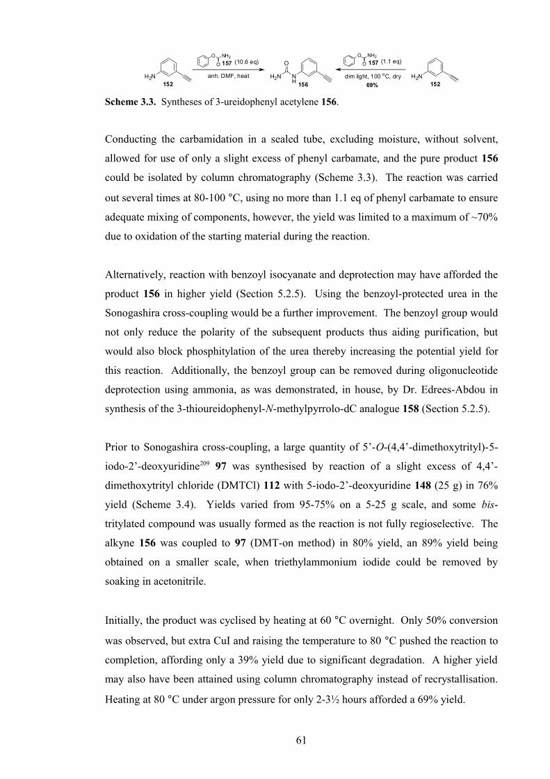
H2N152
NH
H2N
O NH2
O (1.1 eq)
dim light, 100 oC, dry156
O
H2N152
O NH2
O (10.6 eq)
anh. DMF, heat
69%
157 157
Scheme 3.3. Syntheses of 3-ureidophenyl acetylene 156.
Conducting the carbamidation in a sealed tube, excluding moisture, without solvent,
allowed for use of only a slight excess of phenyl carbamate, and the pure product 156
could be isolated by column chromatography (Scheme 3.3). The reaction was carried
out several times at 80-100 °C, using no more than 1.1 eq of phenyl carbamate to ensure
adequate mixing of components, however, the yield was limited to a maximum of ~70%
due to oxidation of the starting material during the reaction.
Alternatively, reaction with benzoyl isocyanate and deprotection may have afforded the
product 156 in higher yield (Section 5.2.5). Using the benzoyl-protected urea in the
Sonogashira cross-coupling would be a further improvement. The benzoyl group would
not only reduce the polarity of the subsequent products thus aiding purification, but
would also block phosphitylation of the urea thereby increasing the potential yield for
this reaction. Additionally, the benzoyl group can be removed during oligonucleotide
deprotection using ammonia, as was demonstrated, in house, by Dr. Edrees-Abdou in
synthesis of the 3-thioureidophenyl-N-methylpyrrolo-dC analogue 158 (Section 5.2.5).
Prior to Sonogashira cross-coupling, a large quantity of 5’-O-(4,4’-dimethoxytrityl)-5-
iodo-2’-deoxyuridine209 97 was synthesised by reaction of a slight excess of 4,4’-
dimethoxytrityl chloride (DMTCl) 112 with 5-iodo-2’-deoxyuridine 148 (25 g) in 76%
yield (Scheme 3.4). Yields varied from 95-75% on a 5-25 g scale, and some bis-
tritylated compound was usually formed as the reaction is not fully regioselective. The
alkyne 156 was coupled to 97 (DMT-on method) in 80% yield, an 89% yield being
obtained on a smaller scale, when triethylammonium iodide could be removed by
soaking in acetonitrile.
Initially, the product was cyclised by heating at 60 °C overnight. Only 50% conversion
was observed, but extra CuI and raising the temperature to 80 °C pushed the reaction to
completion, affording only a 39% yield due to significant degradation. A higher yield
may also have been attained using column chromatography instead of recrystallisation.
Heating at 80 °C under argon pressure for only 2-3½ hours afforded a 69% yield.
61

NH
O
ONO
OH
O
MeO
MeO
I
(3.0 eq)
Pd(Ph3)4 (0.1 eq), CuI (0.4 eq)
dist. Et3N (5.0 eq), anh. DMF
argon, rt, darkNH
O
ONO
OH
DMTO
CuI (1.1 eq)
dist. MeOH/Et3N (7:3, v/v)
80oC, dark, argon
O
OH
DMTO N
N
O
O
HN
80-90%
69%
97
159
160
NH
H2N
O NH
O
H2N
H2NO
156
(2 x 1.2 eq)
dist. DIPEA (2.5 eq)
69%
70N
PO
Cl
dist. THF, argon, rt
CN
O
O
DMTO N
N
O
O
HN
PO
N
CN
146
H2NO
NH
O
ONO
OH
HO
I
148
DMTCl 112 (1.1-1.3 eq)
dist. pyridine, argon, rt
75-89%
Scheme 3.4. Synthetic route to 6-(3-ureidophenyl)-furano-dT phosphoramidite monomer 146.
Phosphitylation was pushed to ~90% completion using 2 × 1.2 eq of the chloro-
phosphitylating agent 70. Some of the phosphoramidite co-eluted with the starting
material thus reducing the yield slightly further, and some material was lost during
precipitation. The phosphoramidite was precipitated after chromatography from CH2Cl2
into hexane, to remove remaining hydrolysed reagent (difficult to visualise by TLC),
and triethylamine, which would adversely affect subsequent DNA synthesis. A minor
nucleoside-based impurity was also detected after reaction, which was removed during
purification, presumed later to be due to further phosphitylation of the primary urea in
presence of excess reagent. Product 146 was finally afforded in 69% yield, containing
negligible impurities.
3.2.3 Synthesis of 6-(3-Acetamidophenyl)-furano-dT Monomer 147207
The third target, a 3-acetamidophenyl-modified furano-dT phosphoramidite 147 was
proposed as an alternative to the urea. Although the acetamide can form only one
hydrogen-bond with G (c.f. APP), the amide NH is more acidic than the aniline NH, and
may potentially form a stronger H-bond thereby increasing binding affinity.
The arylalkyne for this synthesis, 3-acetamidophenyl acetylene 161, was synthesised in
94% yield from 3-ethynylaniline 152 following a similar method to that published by
Kabalka et al.210 for synthesis of o-iodoacetanilide. The product was purified by
trituration with water as described by Arnold et al.,211 (who prepared 161 using acetic
anhydride and acetic acid), followed by aqueous extraction. Using only 0.15 eq Et3N on
62

a 1.8 mmol scale, reduced the yield by 11%, and when repeated on an 18 mmol scale,
un-neutralised HCl caused degradation to afford a 43% yield, reduced to 38% after
recrystallisation (chloroform/hexane) (Scheme 3.5).
H2N NH
OAcCl (1.0-1.1 eq)
dist. Et3N (0.15 eq)
dry/degassed Et2O, 0 oC-rt
81/38%152 161
H2N152
AcCl (1.0 eq)
dist. Et3N (0.95 eq)
dry/degassed Et2O, 0 oC-rt97%
Scheme 3.5. Synthesis of 3-acetamidophenyl acetylene 161.
Sonogashira cross-coupling proceeded to completion using alkyne 161, but the product
162 proved difficult to separate from a close-running unknown impurity, affording a
final yield of 75%. Cyclisation and phosphitylation under the same conditions as
previously described afforded the cyclised nucleoside 163 then phosphoramidite
monomer 147 in 89% and 69% yields respectively. The phosphitylation was not pushed
to completion with extra reagent beyond ~80%, as the hydrolysed reagent can prove
difficult to fully remove after purification (Scheme 3.6).
NH
O
ONO
OH
O
MeO
MeO
I(3.0 eq)
Pd(Ph3)4 (0.1 eq), CuI (0.4 eq)
dist. Et3N (5.0 eq), anh. DMF
argon, rt, dark
NH
O
ONO
OH
DMTO
CuI (1.1 eq)
dist. MeOH/Et3N (7:3, v/v)
80oC, dark, argon
O
OH
DMTO N
N
O
O
HN75%
89%97
162
163
NH
O
NH
O
O
161
(1.2 eq)
dist. DIPEA (2.5 eq), dist. THF, argon, rt
69%
70N P O
Cl
CN
O
O
DMTO N
N
O
O
HN
PO
NCN
147
O
Scheme 3.6. Synthetic route to 6-(3-acetamidophenyl)-furano-dT phosphoramidite monomer
147.
3.3 Post-Synthetic Conversion of Furano-dT to N-Methylpyrrolo-dC
The three furano-dT phosphoramidite monomers 145-147 were incorporated into
triplex-forming oligonucleotides (TFOs) for triplex melting studies, by the automated
DNA synthesis method on solid-support (CPG), as detailed in Section 9.2.
63

Following DNA synthesis, the oligonucleotides were deprotected and cleaved from the
solid-support using 30 wt % aqueous methylamine at room temperature for 6-24 hours
(Scheme 3.7).
NH
N
N
O
NH
N
O
O
O
O
O
O
RO N
N
HN
O
PO
NC
O
O
NH
N
N
O
NH2N
O
OH
O
O
O
RO N
N
NH2
O
PO
HO
RNH2, rt / 55 oC, 4-24 hrs
(R = H, Me)
R = oligo
O
NH
O
CPGn
Scheme 3.7. General deprotection/cleavage of oligonucleotides highlighting key groups.
Methylamine, in addition to deprotection and resin-cleavage, ring-opens the furano-dT
nucleobase to form a 5-aroylmethyl-N-methyl-dC derivative. The methylamine
insertion reaction occurred quantitatively in 6 hours, although some TFOs were allowed
to deprotect overnight or longer to ensure complete reaction (Scheme 3.8).
O
O
R'O N
N
O
O
R
O
OR'
R'O N
N
O
N
R
O
OR'
R'O N
N
O
HNR O
30% aq. MeNH2
Oligo
DOWEX 50Wx8 (H+-form)
16-36 hrs
R = Me, ArylR' = Oligo
keto-N-methyl-dCfurano-dT N-methylpyrrolo-dC
6-24 hrs
Scheme 3.8. Post-synthetic conversion of furano-dT to N-methylpyrrolo-dC in oligonucleotide.
After removal and washing of the resin, the remaining aqueous solution was
concentrated in vacuo at 50-60 °C and the residue was re-dissolved in distilled water (1
mL). The oligonucleotide was then treated with DOWEX 50WX8 sulfonic acid resin
(200/400 mesh, H+-form), at room temperature for 18-36 hours, to recyclise the keto-N-
methyl-dC nucleotide to form the desired N-methylpyrrolo-dC derivative (Scheme 3.8).
Initially, oligonucleotides were HPLC-purified before acid-catalysed (DOWEX 50)
cyclisation and analysed by MALDI-TOF to check extent of reaction (acidic matrix for
MALDI can cause limited additional cyclisation). Once the methylamine insertion
reaction was determined to be quantitative, oligonucleotides were treated with DOWEX
resin straightaway after deprotection/insertion as described above.
64

After treatment with DOWEX, the resin was removed by filtration, washed with water
and the oligonucleotide solution was concentrated in vacuo and desalted (Sephadex
NAP™ column). The post-synthetically modified oligonucleotides were analysed by
analytical HPLC, capillary gel electrophoresis (CE) (Section 7.3.1), MALDI-TOF and
electrospray MS (negative mode). Unfortunately, the acid-catalysed re-cyclisation was
not quantitative, only proceeding to 50-80% completion.
The desired cyclised TFO could be isolated by HPLC for oligonucleotides to be used in
UV melting studies (Section 4.1). However, HPLC could not resolve TFOs used for
fluorescence melting studies. These TFOs are labelled with a large, lipophilic quencher
molecule (DABCYL), which dominates in the HPLC chromatogram thus reducing
resolution. The two components were resolved by CE, but cannot be purified by this
method hence were used in fluorescence melting studies as a mixture. The
uncyclised/ring-opened modification, as a C analogue, could potentially bind to GC not
CG as desired.
The standard method for deprotection/cleavage of oligonucleotides is treatment with
conc. aqueous ammonia at room temperature or 55 °C for several hours. If these TFOs
were treated in this way, ammonia would ring-open the furano-dT modification. The
ammonia-insertion product spontaneously recyclises to form the NH-pyrrolo-dC
derivative, which as a C analogue, could bind preferentially with GC. The N-methyl
group, however, prevents the methylamine ring-opened nucleotide from cyclising
spontaneously, necessitating acid catalysis. This is presumably due in part to steric bulk
of the methyl group.
The post-synthetic conversion of these modifications, although not ideal due to
incomplete cyclisation, afforded TFOs that were successfully used in UV and
fluorescence melting studies. The results from these are discussed in Chapter 4.
Further discussion of oligonucleotide deprotection conditions are given in the study,
detailed in Chapter 7.
65

3.4 Synthesis of 5-(3-aminoprop-1-ynyl)-dU (pdU) monomer 16439,117
Prior to melting studies, approximately 5 g of 5-(3-aminoprop-1-ynyl)-dU (pdU)
phosphoramidite monomer 164 was synthesised. 5’-O-(4,4’-Dimethoxytrityl)-5-iodo-
2’-deoxyuridine 97 and N-trifluoroacetyl propargylamine 68 were coupled under
Sonogashira Pd-catalysed cross-coupling conditions in two 10 g scale reactions, to
afford, after combined purification, the alkynyl-nucleoside product 165 in 80% yield
(16.6 g). Following phosphitylation of 5 g of 165 using the chloro-phosphitylating
reagent 70, under standard conditions, the phosphoramidite monomer 164 was afforded
in 84% yield (43 bottles for oligonucleotide synthesis).
NH
O
ONO
OH
O
MeO
MeO
I(1.4 eq)
Pd(Ph3)4 (0.1 eq), CuI (0.2 eq)
dist. Et3N (7.0 eq), anh. DMF
argon, rt, dark
NH
O
ONO
OH
DMTO
80%97
165
(1.1 eq)
dist. DIPEA (2.5 eq)
84%
70N
PO
Cl
dist. THF, argon, rt
CNO
NH
F3C68
NH
O
F3C
OP
O
N
CN
NH
O
ONO
DMTO
164
NH
O
F3C
Scheme 3.9. Synthesis of 5-(3-aminoprop-1-ynyl)-dU (pdU) phosphoramidite monomer 164.
This phosphoramidite monomer 164 was used for synthesis of TFOs for melting studies
(Chapter 4).
3.5 Conclusion
Three furano-dT phosphoramidite monomers 145-147 were synthesised for assessment
as monomers for recognition of the CG base pair within a DNA triple helix motif. The
common synthetic route (Sonogashira cross-coupling, Cu(I)-catalysed cyclisation and
phosphitylation) was successful for each monomer, affording moderate to very good
yields for almost every step.
Two main issues were encountered during synthesis. The high polarity of the
substrates, once cyclised, introduced problems in purification, even when tritylated.
Removal of close-running impurities and starting material also presented minor
difficulties. In addition, the cyclised nucleosides demonstrated significantly reduced
66

reactivity at the 3’-OH and aniline NH2. This presented problems in trifluoroacetylation
and necessitated forcing the phosphitylation to completion with extra reagent, which is
not ideal. However, all monomers were synthesised in high purity and incorporated into
TFOs with high coupling efficiency (as determined by trityl cation conductivity
monitoring – see Section 9.2.1) for triplex melting studies (Chapter 4).
67
Chapter 4
Biophysical Studies of Furano-dT
Modified TFOs for CG Recognition
68

4. Biophysical Studies of Furano-dT Modified TFOs for CG Recognition
The three modified furano-dT phosphoramidite monomers (APPf 145, AcPPf 146, UPPf
147) and commercially available 6-methyl-furano-dT MPf monomer 144 were
incorporated into unlabelled/labelled TFOs for UV and fluorescence melting studies.
These studies were designed to test binding affinity for CG and selectivity against the
other base pairs (GC, AT, TA) at several pHs. These properties are assessed by
determining triplex melting temperatures from graphs of, change in UV absorption or
fluorescence emission of fluorophore label respectively, on triplex melting, for different
sequences, in several buffer conditions. After incorporation, the modification was post-
synthetically converted in the oligonucleotide from a furano-dT to an N-methylpyrrolo-
dC nucleotide. The procedure for this transformation is described in Section 3.3.
4.1 UV Melting Studies
Methyl- (144, MPf), trifluoroacetamidophenyl- (145, APPf) and ureidophenyl- (147, UPPf)
furano-dT phosphoramidite monomers were incorporated into TFOs (OL3-5) to assess
binding affinity for a single CG inversion in a homopurine tract, in the parallel motif. A
schematic representation of the UV melting triplex, used originally by Leumann et al.111
is shown in Figure 4.1.
5’-TTT TTM TXT MTM TMT 5’-GCT AAA AAG ACA GAG AGA TCG CGA TTT TTC TGT CTC TCT AGC-5’
Figure 4.1. UV triplex melting experiment. TFO shown in bold. M = 5-MedC, X = MP (OL3),
APP (OL4), UPP (OL5), purine duplex strand (C, OL1), pyrimidine duplex strand (G, OL2).
UV melting experiments were performed, (as described in detail in Section 9.2), on a
Varian Cary 400 Scan UV-Vis spectrophotometer monitoring at 260 nm. A 5:1 ratio of
TFO:duplex (5:1 µM) was used. Experiments were conducted at pH 6.07 and pH 6.40
(10 mM sodium phosphate, 200 mM NaCl, 1 mM Na2EDTA). A standard melt
programme was used (Programmes 1/2: 10-50-80 °C at 0.5 °C/min) with 3 or 4
heat/anneal cycles in total (Section 9.2). Condensation was not a major problem at low
temperature, affecting only the first heat/anneal cycle, hence purging the sample
69
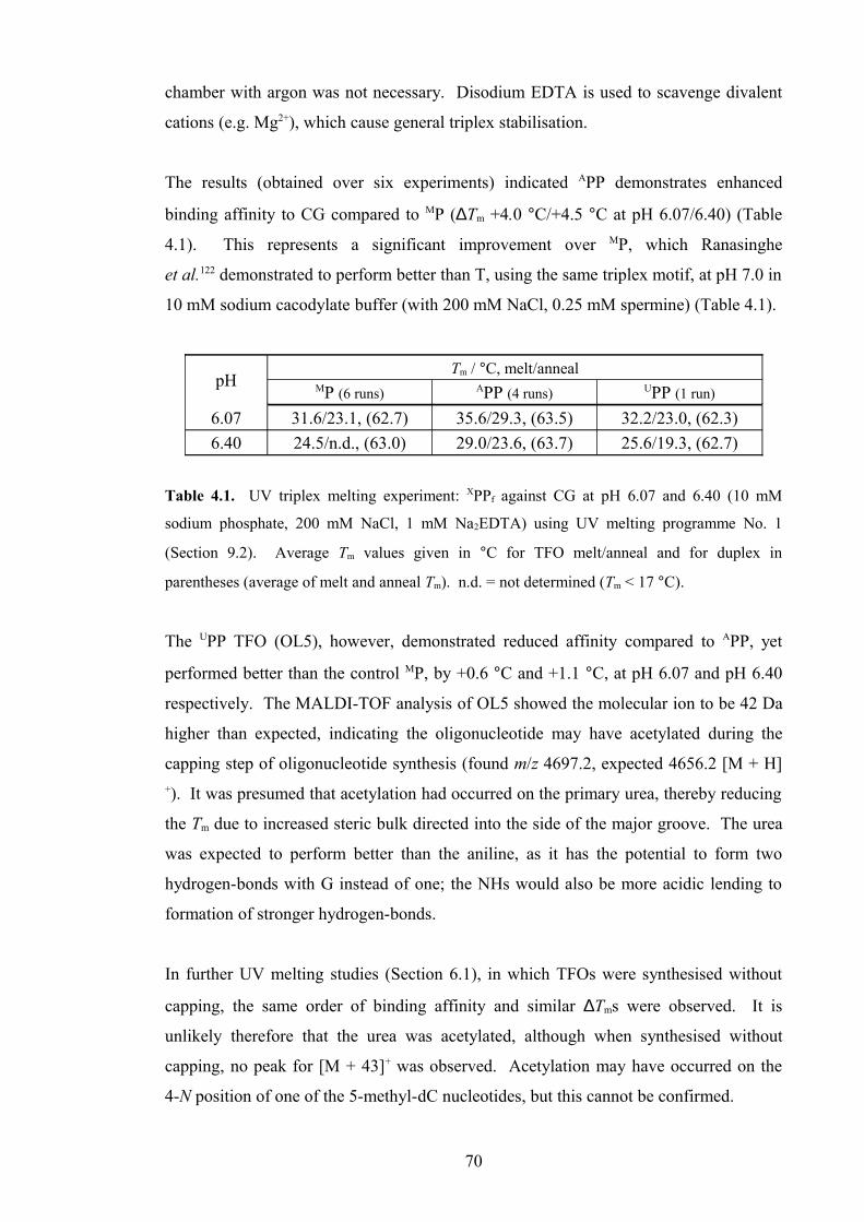
chamber with argon was not necessary. Disodium EDTA is used to scavenge divalent
cations (e.g. Mg2+), which cause general triplex stabilisation.
The results (obtained over six experiments) indicated APP demonstrates enhanced
binding affinity to CG compared to MP (∆Tm +4.0 °C/+4.5 °C at pH 6.07/6.40) (Table
4.1). This represents a significant improvement over MP, which Ranasinghe
et al.122 demonstrated to perform better than T, using the same triplex motif, at pH 7.0 in
10 mM sodium cacodylate buffer (with 200 mM NaCl, 0.25 mM spermine) (Table 4.1).
pHTm / °C, melt/anneal
MP (6 runs) APP (4 runs) UPP (1 run)
6.07 31.6/23.1, (62.7) 35.6/29.3, (63.5) 32.2/23.0, (62.3)
6.40 24.5/n.d., (63.0) 29.0/23.6, (63.7) 25.6/19.3, (62.7)
Table 4.1. UV triplex melting experiment: XPPf against CG at pH 6.07 and 6.40 (10 mM
sodium phosphate, 200 mM NaCl, 1 mM Na2EDTA) using UV melting programme No. 1
(Section 9.2). Average Tm values given in °C for TFO melt/anneal and for duplex in
parentheses (average of melt and anneal Tm). n.d. = not determined (Tm < 17 °C).
The UPP TFO (OL5), however, demonstrated reduced affinity compared to APP, yet
performed better than the control MP, by +0.6 °C and +1.1 °C, at pH 6.07 and pH 6.40
respectively. The MALDI-TOF analysis of OL5 showed the molecular ion to be 42 Da
higher than expected, indicating the oligonucleotide may have acetylated during the
capping step of oligonucleotide synthesis (found m/z 4697.2, expected 4656.2 [M + H]+). It was presumed that acetylation had occurred on the primary urea, thereby reducing
the Tm due to increased steric bulk directed into the side of the major groove. The urea
was expected to perform better than the aniline, as it has the potential to form two
hydrogen-bonds with G instead of one; the NHs would also be more acidic lending to
formation of stronger hydrogen-bonds.
In further UV melting studies (Section 6.1), in which TFOs were synthesised without
capping, the same order of binding affinity and similar ∆Tms were observed. It is
unlikely therefore that the urea was acetylated, although when synthesised without
capping, no peak for [M + 43]+ was observed. Acetylation may have occurred on the
4-N position of one of the 5-methyl-dC nucleotides, but this cannot be confirmed.
70

Significant hysteresis was observed for the melting temperature using this melt
programme, with ∆Tm values between 5.4 and 9.2 °C. This could be decreased by
reducing the rate of heating/cooling. The duplex hysteresis was within experimental
error, at less than 0.5 °C.
A graph displaying melting curves and their derivatives for APP (OL4) at pH 6.07, is
shown in Figure 4.2 below. The hysteresis between melting and annealing curves is
clearly visible.
10 20 30 40 50 60 70 800.86
0.88
0.90
0.92
0.94
0.96
0.98
0.000
0.001
0.002
0.003
0.004
0.005
0.006
0.007
Ab
so
rpti
on
Temperature / oC
Melt Curve (Abs)
Anneal Curve (Abs)
Duplex Tm
dA
/dT
Melt Derivative (dA/dT)
Anneal Derivative (dA/dT)
Triplex Tm
Figure 4.2. UV Melting curves/derivatives for APP (OL4) against CG at pH 6.07 (10 mM
sodium phosphate, 200 mM NaCl, 1 mM Na2EDTA). Data is smoothed and un-normalised.
These initial studies indicated that the 3-aminophenyl moiety significantly enhanced
binding affinity over a methyl group. The urea moiety, unexpectedly demonstrated
poorer binding than the amine. This result necessitated further UV melting and other
studies for confirmation and to test other analogues.
4.2. Fluorescence Melting Studies
Fluorescence melting studies were conducted to assess binding affinity and selectivity,
and the sequence-dependency of these recognition properties. Fluorescence melting
71

measures change in fluorescence due separation of fluorophore and fluorescence
quencher moieties on duplex and TFO respectively, on triplex melting.
4.2.1 Binding Affinity/Selectivity study
Methyl- (144, MPf), trifluoroacetamidophenyl- (145, APPf), acetamidophenyl (147, AcPPf)
and ureidophenyl- (146, UPPf) furano-dT phosphoramidite monomers were incorporated
into labelled TFOs for initial fluorescence melting studies. The target was a single CG
inversion in a homopurine tract of a hairpin duplex, in the parallel motif. The initial
triplex motif is represented below in Figure 4.3.
5’-Q-PMM TPM TXP TPT PTM PT 5’-F-GT GTT AGG AAG AYA AAA AAG AAC TGG T-H CA CAA TCC TTC TZT TTT TTC TTG ACC A-H
Figure 4.3. Fluorescence triplex melting experiment. TFO shown in bold. (M = 5-MedC,
P = 5-(3-aminoprop-1-ynyl)-dU (pdU), X = MP (OL10), APP (OL11), AcPP (OL12), UPP
(OL13), Q = DABCYL (quencher), F = FAM (fluorophore), H = HEG (hexaethylene glycol),
hairpin duplex YZ = CG (OL6), GC (OL7), AT (OL8), TA (OL9).
Fluorescence melting studies were conducted (as described in detail in Section 9.2), on
a Roche LightCycler®. Initial experiments were performed at pH 6.0 (20 mM NaOAc,
200 mM NaCl), and a 10:1 ratio of TFO:duplex (5:0.5 µM) was used. Runs in these
experiments were performed up to three times in duplicate, using LC Programme 1 (95-
30-95 °C at 0.2 °C/min, see Section 9.2) (Table 4.2). These sequences were used for
fluorescence melting only.
There is a stark contrast between some of these results and the previous UV melting
study. Whereas APP previously demonstrated a significantly higher Tm than MP against
CG (∆Tm +4.0-4.5 °C), these results place the Tm of APP, 1.2 °C lower than MP. This
observation could not be easily explained, although the buffer and sequence had been
changed. Despite these observations, the urea (UPP) showed the strongest binding
affinity towards CG as originally expected and the order of stability (UPP>AcPP≈APP),
excluding MP, correlates with the predicted hydrogen-bonding motif.
72
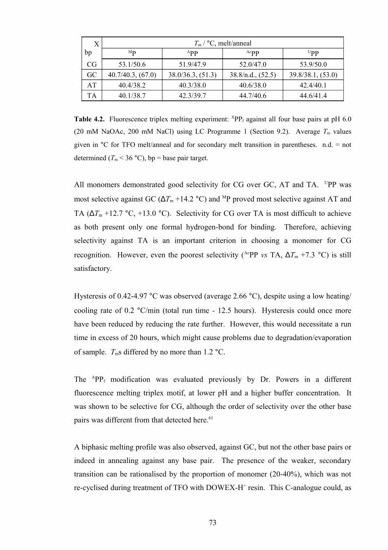
Xbp
Tm / °C, melt/annealMP APP AcPP UPP
CG 53.1/50.6 51.9/47.9 52.0/47.0 53.9/50.0
GC 40.7/40.3, (67.0) 38.0/36.3, (51.3) 38.8/n.d., (52.5) 39.8/38.1, (53.0)
AT 40.4/38.2 40.3/38.0 40.6/38.0 42.4/40.1
TA 40.1/38.7 42.3/39.7 44.7/40.6 44.6/41.4
Table 4.2. Fluorescence triplex melting experiment: XPPf against all four base pairs at pH 6.0
(20 mM NaOAc, 200 mM NaCl) using LC Programme 1 (Section 9.2). Average Tm values
given in °C for TFO melt/anneal and for secondary melt transition in parentheses. n.d. = not
determined (Tm < 36 °C), bp = base pair target.
All monomers demonstrated good selectivity for CG over GC, AT and TA. UPP was
most selective against GC (∆Tm +14.2 °C) and MP proved most selective against AT and
TA (∆Tm +12.7 °C, +13.0 °C). Selectivity for CG over TA is most difficult to achieve
as both present only one formal hydrogen-bond for binding. Therefore, achieving
selectivity against TA is an important criterion in choosing a monomer for CG
recognition. However, even the poorest selectivity (AcPP vs TA, ∆Tm +7.3 °C) is still
satisfactory.
Hysteresis of 0.42-4.97 °C was observed (average 2.66 °C), despite using a low heating/
cooling rate of 0.2 °C/min (total run time - 12.5 hours). Hysteresis could once more
have been reduced by reducing the rate further. However, this would necessitate a run
time in excess of 20 hours, which might cause problems due to degradation/evaporation
of sample. Tms differed by no more than 1.2 °C.
The APPf modification was evaluated previously by Dr. Powers in a different
fluorescence melting triplex motif, at lower pH and a higher buffer concentration. It
was shown to be selective for CG, although the order of selectivity over the other base
pairs was different from that detected here.61
A biphasic melting profile was also observed, against GC, but not the other base pairs or
indeed in annealing against any base pair. The presence of the weaker, secondary
transition can be rationalised by the proportion of monomer (20-40%), which was not
re-cyclised during treatment of TFO with DOWEX-H+ resin. This C-analogue could, as
73

previously mentioned (Section 3.3) form a triplet with GC, affording a higher Tm than
expected, for a triplex that should contain a disfavourable X.GC triplet (Figure 4.4).
dR
dR NN
N
O
HH
NN
N O
N
N
HH
H
H
X.GC
N
NO
dR N
R
dR
dRN
N
N
O
HH
NN
N O
N
N
HH
H
H
Xuncyclised.GC
N
NO
dR
N
HHH
RO
Figure 4.4. Putative X.GC and Xuncyclised.GC triplets.
The observed secondary Tm value against GC should be similar to that against CG
(~52 °C) as was observed for APP, AcPP and UPP. However, the value is significantly
higher for MP (67.0 °C), which is difficult to rationalise. Replacing the aryl group for a
methyl group should reduce the potential for beneficial interactions within the triple
helix, unless the aryl group in fact confers disfavourable steric/hydrophobic
interactions. Furthermore, comparing with the many modifications that have been made
to the 5-position of C-derivatives for achieving GC recognition (Section 1.2.1.2), it is
highly unlikely that the 5-(acetylmethyl)- group of uncyclised/ring-opened MPf is
stabilising enough to account for this Tm. Other secondary structures and interactions
may also be contributing factors.
4.2.2 Sequence study
The two possible explanations for reversal in the order of stability of MP and APP
between the initial UV and fluorescence melting studies were a change in buffer/pH,
and changing the sequence. It was hypothesised, that altering the neighbouring
nucleosides to the modification under examination within the TFO may affect binding
affinities and selectivity.
New TFOs were therefore synthesised, containing the central trimers, TXT (OL14),
PXT (OL15) and PXP (OL16) (X = APPf, P = pdU) to compare with the TXP-containing
TFO (OL11) already investigated. It was thought that presence of pdU next to the XPP
74

modification may change binding affinity/selectivity or mask the effects of the
modification, and may account for the differences between the two methods.
5’-Q-PMM TPM VXW TPT PTM PT 5’-F-GT GTT AGG AAG ACA AAA AAG AAC TGG T-H CA CAA TCC TTC TGT TTT TTC TTG ACC A-H
Figure 4.5. Fluorescence triplex melting experiment. TFO shown in bold. (M = 5-MedC,
P = 5-(3-aminoprop-1-ynyl)-dU (pdU), X = APP & VXW = TXT (OL14), PXT (OL15), TXP
(OL11), PXP (OL16); VXW = TXP & X = MP (OL10), AcPP (OL12), UPP (OL13), Q =
DABCYL (quencher); F = FAM (fluorophore), H = HEG (hexaethylene glycol), hairpin duplex
(CG, OL6).
The initial fluorescence melting experiments (Figure 4.5) were repeated using LC
Programmes 1 and 2 (95-30-95 °C at 0.2 °C/min) at pH 6.2 (10 mM sodium phosphate,
200 mM NaCl). Experiments were run once or twice in triplicate and a 10:1 ratio of
TFO:duplex (5:0.5 µM) was used as before. The aims of these experiments were not
only to obtain confirmation of previous results, but to investigate how the change of
sequence described above affects binding, and for comparison between analogous
furano-dT (XPPf) and N-methylpyrrolo-dC (XPPp) modified TFOs (see Section 6.1 for
results from XPPp-modified TFOs).
XSeq.
Tm / °C, melt/annealMP APP AcPP UPP
TXT – 43.2/39.1 * *
PXT – 49.3/43.1 * *
TXP 49.7/43.7 49.9/43.0 48.4/43.0 51.0/45.0
PXP – 53.6/49.0 * *
Table 4.3. Fluorescence triplex melting experiment: XPPf against CG in four TFOs (OL14,
OL15, OL11, OL16) at pH 6.2 (10 mM sodium phosphate, 200 mM NaCl) using LC
Programme 1 and 2 (Section 9.2). Average Tm values given in °C for TFO melt/anneal.
* Data obtained for XPPp-modified TFOs is not shown (Section 6.2).
Hysteresis was much greater than previously observed (4.08-6.91 °C, average 5.61 °C)
and annealing data was of poorer quality than in previous experiments. However, the
melting data was clear and Tm values differed by no more than 1.0 °C.
75

These experiments confirm that the urea (UPP) has the highest binding affinity in the
TXP-containing TFO. MP and APP appear as effective as each other and slightly better
than the acetamide (AcPP), although experimental error may account for variation as the
difference in Tm values is small.
When T is replaced by pdU, the Tm increases because pdU forms additional charge-
stabilising interactions with the sugar-phosphate backbone (Section 1.2.1.1). In the APP-modified series of TFOs, as for any modification, for each substitution the Tm
would be expected to increase by the same amount, and Tm should be equal for the TXP-
and PXT-containing TFOs (OL11, OL15) as only the order is changed. However, the
Tm for TXP (OL11) is 0.6 °C higher than PXT (OL15). Also the increase in Tm from
TXT to TXP is greater than for PXT to PXP.
These observations indicate that placing pdU on the 5’-side of XPP is slightly
destabilising compared to the 3’-side. It is reasonable therefore, to suggest there may be
interaction between the 3-aminoprop-1-ynyl group of pdU and the anilino group of APP,
slightly affecting recognition of CG. This difference was most significant for UPP
(XPPp-modified TFO, see Section 6.2) with a ∆Tm value of 3.0 °C. It is also reasonable
to suggest that due to this destabilisation, replacing the second T with pdU has less
stabilising effect than expected.
Finally, data for AcPP and UPP confirmed that the Tms for furano-dT and N-
methylpyrrolo-dC modified TFOs were approximately the same, only differing by 0.7
and 0.1 °C respectively. These results led into a large study, using N-methylpyrrolo-dC
monomers, the results of which are discussed in Chapter 6.
4.3 Fluorescence Melting Mismatch Experiment
Experiments were conducted to test if triplex-mediated recognition of a base pair
mismatch can occur using the XPPf modifications tested above. This may also provide
information on binding motif of the X.CG triplet. All four modifications were assessed
against a hairpin duplex containing a CC mismatch. Fluorescence melting was
conducted using LC Programme 4 (95-30-95 °C at 0.25 and 0.2 °C/min) at pH 6.0 (20
76

mM NaOAc, 200 mM NaCl). Experiments were run once in duplicate and a 10:1 ratio
of TFO:duplex (5:0.5 µM) was used as before (Table 4.4).
5’-Q-PMM TPM TXP TPT PTM PT 5’-F-GT GTT AGG AAG ACA AAA AAG AAC TGG T-H CA CAA TCC TTC TCT TTT TTC TTG ACC A-H
Figure 4.6. Fluorescence triplex melting mismatch experiment. TFO shown in bold. (M =
5-MedC, P = 5-(3-aminoprop-1-ynyl)-dU (pdU), X = MP (OL10), APP (OL11), AcPP (OL12), UPP
(OL13), Q = DABCYL (quencher), F = FAM (fluorophore), H = HEG (hexaethylene glycol),
mismatched hairpin duplex (CC, OL17).
These results show a drop in Tm of 4.5 °C for MP when CG is replaced by a CC
mismatch. This is expected as even though MP is proposed to hydrogen-bond only to
one C of the CC base pair as is the case for CG, distortions due to the CC mismatch
would cause destabilisation of the triplex as well.
Tm / °C, melt/annealMP.CG MP.CC APP.CC AcPP.CC UPP.CC
53.1/50.8 48.6/47.1 48.9/45.9 49.3/46.2 50.0/48.4
Table 4.4. Fluorescence triplex melting mismatch experiment: XPPf against CC mismatch
hairpin duplex (OL17) at pH 6.0 (20 mM NaOAc, 200 mM NaCl) using LC Programme 4
(Section 9.2). Average Tm values given in °C for TFO melt/anneal. MP.CG data from this
experiment is shown for comparison.
The CC mismatch also reduces the duplex melting temperature by ~8 °C compared to
that of the CG hairpin duplex. In addition, the derivative curve is markedly different to
the duplexes containing matched base pairs, as it curves towards an apparent melting
transition (~74 °C) just below duplex melting. It is possible that other structures may be
present, or that this is an unexplained feature of the melting profile of a mismatch-
containing hairpin duplex, the latter of which appears more likely.
77

30 40 50 60 70 80 90
2
4
6
8
10
12 MP.CC
MP.CG
Flu
ore
sce
nce
(F
1)
Temperature/oC30 40 50 60 70 80 90
-0.35
-0.30
-0.25
-0.20
-0.15
-0.10
-0.05
0.00
0.05 MP.CC derivative
MP.CG derivative
-d(F
1)/d
t
Temperature/oC
melting transition?
Triplex Tm
Figure 4.7. Fluorescence melting curves/derivatives for MP (OL10) against CG (red, OL6) and
CC (black, OL17) at pH 6.0 (20 mM NaOAc, 200 mM NaCl). Data is smoothed and un-
normalised.
Melting temperatures for all modifications were very similar; the urea performing
marginally better than the rest. This is consistent with the proposed binding model, in
which differentiation between modifications is achieved through additional hydrogen-
bonds to G of the CG base pair. Altering the hydrogen-bond pattern, in conjunction
with triplex distortion due to the mismatch, reduces these differences.
4.4 Conclusion
All melting studies demonstrated the effectiveness of the XPP modifications at
recognising CG with similar or better binding affinity/selectivity than MP, which itself is
a large improvement on T.122 Although it was unknown why order of stability differed
between UV and fluorescence melting studies, this was investigated in further studies
(Chapter 6). At this stage it was presumed to be caused by change in sequence
(particularly of neighbouring nucleotides), buffer and pH, but this was/could not be
confirmed.
The significant effect of neighbouring groups on the modification under investigation,
was also identified. Results indicated that the modified nucleoside 5-(3-aminoprop-1-
ynyl)-dU 14 (pdU, P), interacts differently with XPP depending on which side it is
located, giving rise to a difference in Tm. This neighbouring group interaction
introduces important implications for design of TFOs, particularly for use as
78

oligotherapeutics. Understanding the interactions between modified nucleosides is
vital, so that negative interactions can be minimised.
Melting experiments were also conducted against a CC mismatch in the same hairpin
duplex motif. It was demonstrated that differences in Tms between TFOs is reduced as
key hydrogen-bonding residues are removed when G is replaced with C. The reduction
in overall duplex and triplex Tm is expected, due to the CC mismatch.
It would also be useful to test the CT and CA mismatches to identify if any
differentiation could be made between modifications. Both CT and CA contain
potential hydrogen-bonding acceptors on T and A, therefore some differentiation or
stabilisation may be observed.
Assessing recognition of common base pair mismatches/mutations could also provide
valuable information when designing TFOs for therapeutic use. Depending on location
of mismatches and on which strand the mutation is located, it may be possible to bind to
both desired and undesired sequences. As binding primarily occurs to one side of the
base pair, only mutations to the other base should be tolerated.
79

Chapter 5
Amine-Modified N-Methylpyrrolo-dC
Nucleosides for CG Recognition
80
5. Amine-Modified N-Methylpyrrolo-dC Nucleosides for CG Recognition
5.1 Introduction
In previous studies (Sections 3-4), N-methylpyrrolo-dC modifications were incorporated
into TFOs as the furano-dT analogues then post-synthetically converted. Although the
methylamine-insertion reaction was quantitative for each oligonucleotide, the acid-
catalysed cyclisation reaction using acidic DOWEX 50WX8 ion-exchange resin, to
reform the bicyclic core, did not proceed to completion. The major problem with this
method, in addition to reduced yield of TFO, is difficulty in purification when labeled
with DABCYL. For this reason, TFOs for fluorescence melting studies were used as
the inseparable mixture containing approx. 20-40% uncyclised modification. This did
not affect recognition of the CG base pair, although the concentration of correct TFO
was lowered as a result. However, in fluorescence melting studies against GC, a second
minor melting transition was observed at a similar or substantially higher temperature
than against CG. This was proposed to be due to the small proportion of the uncyclised
modification, which, as a C-analogue, exhibits GC recognition properties (also Sections
4.2 and 6.2). The very high temperature of one of these transitions could not be
rationalised, however, unless the ring-opened modification is exceptionally strong at
binding, which is very unlikely.
To overcome this problem, it was proposed, in house, to synthesise monomers as the N-
methylpyrrolo-dC derivatives. Assuming this bicyclic structure was stable to
deprotection conditions (i.e. could not be ring-opened by NH3 or MeNH2, in aqueous
solution), no post-synthetic modification would be required. Ranasinghe synthesised, in
house, the protected 6-aminoethyl and 6-aminopropyl- monomers 166,167 as the
N-methylpyrrolo-dC derivatives (AEPp and APPp), via methylamine insertion and acid-
catalysed cyclisation, on the untritylated furano-dT nucleosides (c.f. post-synthetic
modification of TFO – Section 3.3) (Scheme 5.1).
81
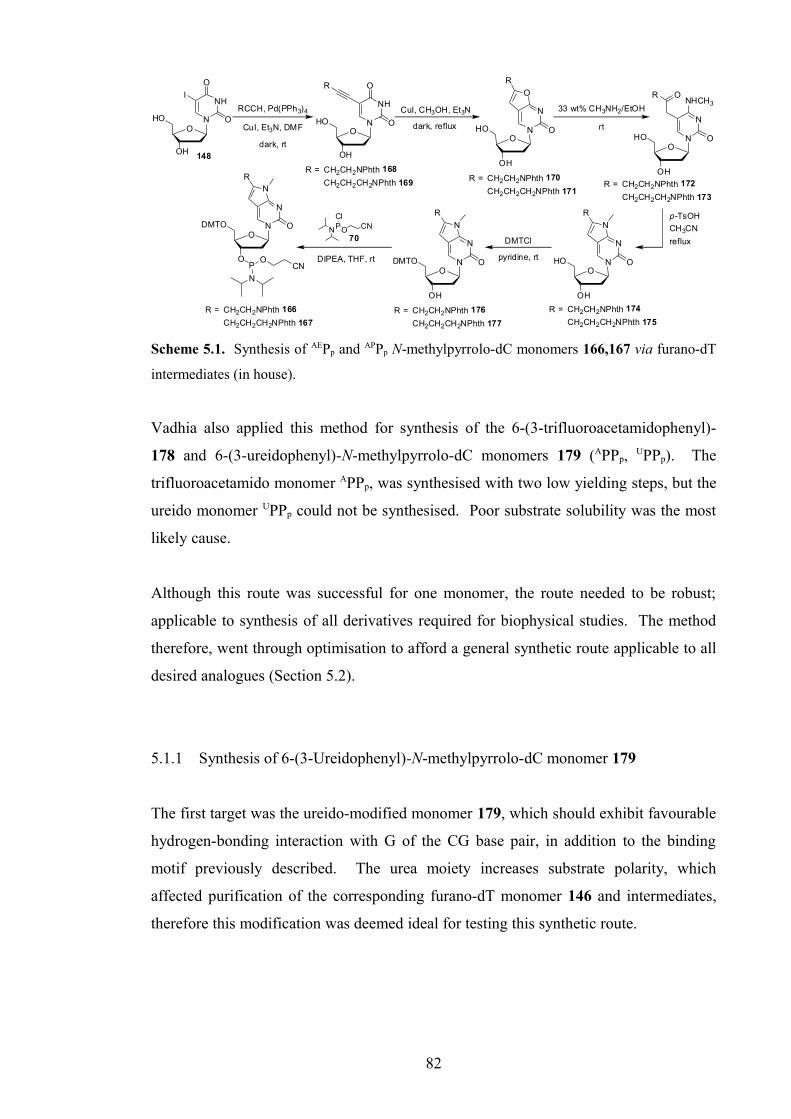
O
OH
HO N
NH
O
O
I
O
OH
HO N
NH
O
OR
O
OH
HO N
N
O
O
R
O
O
DMTO N
N
O
N
PO
N
CN
O
OH
HO N
N
O
ONHCH3
R
O
OH
DMTO N
N
O
N
R
O
OH
HO N
N
O
N
R
R
148
R = CH2CH2NPhth 168
CH2CH2CH2NPhth 169 R = CH2CH2NPhth 170
CH2CH2CH2NPhth 171R = CH2CH2NPhth 172
CH2CH2CH2NPhth 173
R = CH2CH2NPhth 174
CH2CH2CH2NPhth 175R = CH2CH2NPhth 176
CH2CH2CH2NPhth 177
RCCH, Pd(PPh3)4
CuI, Et3N, DMF
CuI, CH3OH, Et3N
dark, reflux
33 wt% CH3NH2/EtOH
rt
refluxDMTCl
pyridine, rtDIPEA, THF, rt
CH3CN
p-TsOH
dark, rt
R = CH2CH2NPhth 166
CH2CH2CH2NPhth 167
70NPO
ClCN
Scheme 5.1. Synthesis of AEPp and APPp N-methylpyrrolo-dC monomers 166,167 via furano-dT
intermediates (in house).
Vadhia also applied this method for synthesis of the 6-(3-trifluoroacetamidophenyl)-
178 and 6-(3-ureidophenyl)-N-methylpyrrolo-dC monomers 179 (APPp, UPPp). The
trifluoroacetamido monomer APPp, was synthesised with two low yielding steps, but the
ureido monomer UPPp could not be synthesised. Poor substrate solubility was the most
likely cause.
Although this route was successful for one monomer, the route needed to be robust;
applicable to synthesis of all derivatives required for biophysical studies. The method
therefore, went through optimisation to afford a general synthetic route applicable to all
desired analogues (Section 5.2).
5.1.1 Synthesis of 6-(3-Ureidophenyl)-N-methylpyrrolo-dC monomer 179
The first target was the ureido-modified monomer 179, which should exhibit favourable
hydrogen-bonding interaction with G of the CG base pair, in addition to the binding
motif previously described. The urea moiety increases substrate polarity, which
affected purification of the corresponding furano-dT monomer 146 and intermediates,
therefore this modification was deemed ideal for testing this synthetic route.
82

5.1.1.1 Synthesis via untritylated furano-dT derivative
The first method closely followed the synthetic routes used by Ranasinghe122 and
Vadhia.206 This route proceeds via the ‘free’ (unprotected) furano-dT nucleoside 180,
which was synthesised via the Sonogashira and cyclisation reactions discussed in
Section 3.2.2.
Following Cu(I)-catalysed cyclisation, Ranasinghe and Vadhia ring-opened the ‘free’
nucleoside 180 with 33% ethanolic methylamine, before acid-catalysed re-cyclisation.
Ranasinghe also, successfully ring-closed a 6-butyl-derivative, by refluxing in
acetonitrile with 1 mol% toluene-sulfonic acid, to obtain an 88% yield over these two
steps. Vadhia used 80% aqueous acetic acid, at 80 °C to afford the re-cyclised aniline-
derivative 143 in 21% yield. Subsequent tritylation proceeded with difficulty in 9%
yield. In both methods the acid was difficult to remove, and for the latter case, acetic
acid may have also affected column purification and certainly tritylation, although poor
to moderate solubility of the product 143 may also have played a role. Vadhia could not
re-cyclise the ureido-derivative using acetic acid, most likely due to poor solubility.
In an attempt optimise this route, the above synthesis was first repeated (Scheme 5.2).
5-Iodo-2’-deoxyuridine 148 was coupled three times with 3-ureidophenyl acetylene 156
via the previously described Sonogashira reaction, finally yielding a ~1:1 mixture of
cyclised 180 and uncyclised nucleoside 181, containing some alkyne dimer.
O
OH
HO N
NH
O
O
I
O
OH
HO N
NH
O
O
O
OH
HO N
N
O
O
HNH2N
O
O
OH
HO N
N
O
N
HNH2N
O
O
O
DMTO N
N
O
N
HNH2N
O
PO
N
CN
(~1:1 mixture with 181)
NHH2N
O
(3.0 eq)
Pd(Ph3)4 (0.1 eq), CuI (0.4 eq)
dist. Et3N (5.0 eq), anh. DMF
argon, rt, dark
CuI (1.1 eq)
dist. CH3OH/Et3N (7:3, v/v)
80oC, dark, argon
54%
NH
H2N
O
155
(1.1 eq)
dist. DIPEA (2.5 eq)
70NPO
Cl
dist. THF, argon, rt
CN
147
i) 33 wt% MeNH2/EtOH, rt
ii) p-TsOH (1 mol%)
anh. CH3CN, reflux
i) DMTCl (1.1 eq)
pyridine, rt
ii)
180
181
182179
X
Scheme 5.2. First route to UPPp monomer 179.
83

The product mixture 180,181 was refluxed with CuI in dry methanol and
triethylamine.121 However, the mixture was poorly soluble, even at 80 °C under argon
pressure, and NMR analysis of the crude material confirmed no change had occurred
from the 1:1 mixture.
Addition of DMF solubilised the substrate, but failed to illicit cyclisation, even at 90 °C.
Tritylation of the nucleoside mixture was also problematic due to poor solubility in
pyridine. Excess DMTCl and some heating were necessary to afford some reaction, but
a mixture of mono- and bis-DMT nucleosides was afforded.
It was noted that leaving the Sonogashira cross-coupling reaction for longer may have
resulted in complete cyclisation. However, due to solubility issues, an alternative
synthetic route was sought.
5.1.1.2 Synthesis via tritylated furano-dT derivative
In order to overcome problems with solubility, the DMT-protected furano-dT
nucleoside 160 was used, synthesised as described in Section 3.2.2.
O
OH
DMTO N
N
O
O
HNH2N
O
O
OH
HO N
N
O
N
HNH2N
O
O
O
DMTO N
N
O
N
HNH2N
O
PO
N
CN
(1.1 eq)
dist. DIPEA (2.5 eq)
70NP O
Cl
dist. THF, argon, rt
CN
33 wt% CH3NH2/EtOH, rt
DMTCl (1.1 eq)
pyridine, argon, rt
160
182
179
DOWEX resin (H+)
DMF/H2O (2:1 v/v), 60 oC
O
OH
DMTO N
N
O
ONHCH3
NHH2N
O
61%
183
O
OH
DMTO N
N
O
N
HNH2N
O
184
Scheme 5.3. Second revised route to UPPp monomer 179.
Alternative sources of acid to p-TsOH and acetic acid were sought, to prevent
detritylation if possible, but mainly for ease of removal after reaction. Cyclised
nucleoside 160 was dissolved in 33 wt% methylamine/ethanol at rt, to afford the ring-
84

opened product 183 in near quantitative conversion. The crude material was dissolved
in anhydrous DMF, divided into two, and agitated with DOWEX-50WX8 resin
(pyridinium form) or silica gel/Et3N, at 30 °C for 1 day, then 50 °C for 1 day and 70 °C
for 12 hours. No change was observed using silica gel, and the DOWEX caused some
detritylation, despite release of pyridine from the resin. The ring-opened compound 183
was also isolated (61% yield) in a larger-scale reaction.
In order to avoid detritylation by exposure to acid, heat alone was assessed as a method
of cyclisation. After quantitative ring-opening with methylamine, strong heating of the
resulting solution caused significant degradation. Refluxing the crude material in
acetonitrile and triethylamine at 90 °C under argon pressure overnight afforded an
unidentified product, less polar than the starting material, plus some degradation. This
was non-fluorescent on TLC and was presumed to be the 4N-methyl-imino-dC
derivative. Heating the mixture in ethanolic methylamine at 70 °C overnight also
afforded ~50% conversion to the same unidentified, and undesired product, in one step.
Successful cyclisation was achieved using DOWEX 50WX8 resin, in the protonated
form. Following ring-opening, the residue was heated at 60 °C overnight in DMF/H2O
(2:1 v/v) with DOWEX 50 (H+-form) affording the cyclised, detritylated nucleoside
182. Clearly, protonated DOWEX resin is a suitable, easily removable source of acid,
however, it was decided this route was not practical for synthesis of this monomer. The
high polarity/poor solubility would cause problems for subsequent purification and
tritylation, as observed by Vadhia. Removal then replacement of the DMT protecting
group is also not atom-efficient.
5.1.1.3 Synthesis via derivatisation of 5-iodo-4N-methyl-dC 185
The third revision to the original route involved initial conversion of tritylated 5-iodo-
2’-deoxyuridine substrate 97 to the 4N-methyl-2’-deoxycytidine derivative 185.
Although 5-iodo-2’-deoxycytidine is commercially available, the 4N-methylated
analogue is not, but several methods of uridine to cytidine conversion are presented in
the literature.
85

The 4-carbonyl group is first activated and converted into a good leaving group by a
variety of reagents, before reaction with a suitable nucleophile (e.g. NH3, MeNH2). This
conversion was conducted using N-methylimidazole (NMI) in presence of POCl3,
conditions published by Herdewijn et al..212 The reaction proceeds via the 4-N-
methylimidazoloyl-dC intermediate 186, which is not isolated from the reaction
(Scheme 5.4).
O
O
DMTO N
N
O
N
HNH2N
O
PO
N
CN
179
O
OH
DMTO N
NH
O
O
I
97
O
OAc
DMTO N
NH
O
O
I
187O
OAc
DMTO N
N
O
N+
I
186
N
O
OH
DMTO N
N
O
HN
I
185
O
OH
DMTO N
N
O
HNNH
H2N
O
188
O
OH
DMTO N
N
O
N
HNH2N
O
184
Cl-
Ac2O (1.7 eq)
dist. pyridine
91%
POCl3 (4.0 eq), NMI (12.7 eq)
dist. pyridine, argon, 0 oC-rt
i) 40 wt% CH3NH2/H2O, rt
ii) 33 wt% CH3NH2/EtOH, rt
46-90%
70NP O
ClCN
(1.1 eq)
dist. DIPEA (2.5 eq)
51%
79% (over 2 steps)
CuI (1.1 eq)
dist. Et3N, anh. DMF
NH
H2N
O
(3.0 eq)
Pd(PPh3)4 (0.1 eq)
CuI (0.4 eq), dist. Et3N (5.0 eq)
anh. DMF, argon, dark, rt
156
anh. DMF, dist. THF
argon, 0 oC
argon, rt
argon, dark, 125 oC
+ alkynedimer impurity
Scheme 5.4. Third revised route to UPPp monomer 179.
After acetylation of the 3’-OH group in 91% yield (Scheme 5.4), the protected
nucleoside 187 was treated with phosphorous oxychloride and NMI in pyridine to form
the yellow intermediate 186 over 30-60 minutes. Treatment with 40 wt% aqueous
methylamine for several hours afforded the partially deacetylated 4N-methyl-dC
nucleoside. Further treatment with 33 wt% methylamine/ethanol, before or after
aqueous workup completed the deacetylation.
On a 1.4 g scale, the product 185 was obtained in a 90% yield, after column
chromatography using CH2Cl2/CH3OH as eluent. Reaction on a 6.8 g scale afforded the
compound in 80% yield after difficult removal of a significant quantity of NMI. This
was removed by 4 columns (different eluents) and recrystallisation from toluene. On an
8.3 g scale, NMI was removed with difficulty by 2 columns and successive extraction
with diethyl ether, followed by recrystallisation from ethyl acetate (46% yield).
86

When later resynthesised in house, by Dr. Mastoura Edrees-Abdou, stirring the reaction
at rt overnight after addition of 40 wt% aqueous methylamine was sufficient to
completely deacetylated, therefore no further treatment with 33 wt% ethanolic
methylamine was required. Also, careful column chromatography using methanol/
CH2Cl2 as eluent afforded the product in 84% yield.
Commonly, conversion via the 4-(1,2,4-triazol-1-yl)- intermediate is used, using a large
excess of 1,2,4-triazole in presence of excess POCl3.213-215 The intermediate was often
isolated, although Sanghvi et al.213 reported an increased yield if this intermediate was
not isolated, and have conducted this reaction up to kilo scale, in yields of 65-85%.
Other reagent systems used for this conversion include 3-nitro-1,2,4-triazole/diphenyl
phosphorochloridate,216 l-(mesitylene-2-sulfonyl)-3-nitro-1,2,4-1H-triazole (MSNT)/
cat. Ph2PO4 (forms the 3-nitro-1,2,4-triazolyl- intermediate),216 2,4,6-mesitylenesulfonyl
chloride/DMAP,95 2,4,6-triisopropylbenzenesulfonyl chloride/DMAP,217,218 triiso-
propylsilyl chloride/DMAP,219 and 2- and 4-nitrophenol/POCl3.216,220,221
Sonogashira cross-coupling with 3-ureidophenyl acetylene 156 gave the desired alkynyl
product 188, with a large quantity of the alkyne dimer impurity, and a minor unknown
impurity. The alkyne dimer was partially removed by recrystallisation from methanol.
Following cross-coupling, the nucleoside/alkyne dimer mixture was treated with copper
iodide in dry, refluxing CH3OH/Et3N at 80 °C under McGuigan’s conditions,121 but only
partial cyclisation was observed over 2 hours.
One of few literature examples of cyclisation of 5-alkynyl-dC derivatives was published
by Inoue et al..222,223 A protected 4N-acetyl-5-ethynyl-dC nucleoside was cyclised by
heating at 125 °C in DMF for 10 minutes, in presence of CuI (0.55 eq).
The ureido-modified Sonogashira product 188, containing the alkyne dimer impurity,
was successfully cyclised under similar conditions, to give the desired product in 55%
then 79% yield, over two steps, finally using a slight excess of CuI (1.1 eq) and longer
reaction time (40 minutes). The alkyne dimer impurity from the previous reaction was
then easily removed by column chromatography.
87

The cyclised nucleoside 184 was subsequently phosphitylated to afford the desired UPPp
monomer 179 for incorporation into TFOs. During the first small-scale attempt, using
the chloro-phosphitylating reagent 70, a larger quantity was added in error affording
41% yield of the bis-phosphoramidite (reaction on primary urea) plus 19% yield of
desired phosphoramidite 179. Once the reactivity of the urea NH2 was highlighted, the
less reactive bis-aminophosphitylating reagent 189 (Figure 5.1) was used, in order to
minimise secondary reaction. This necessitated 2.6 eq of reagent, 1.0 eq of activator
diisopropylamine hydrotetrazolide (DIHT) 190 and very long reaction time. During this
time, significant degradation occurred, with formation of a viscous yellow syrup, from
which the product was extracted in 29% yield. No bis-phosphitylation was observed.
NP
N
OCN
N
NN
HH189 190
Figure 5.1. Bis-amino phosphitylating reagent 189 and activator, DIHT 190.
DIHT 190 is used in preference to tetrazole due to greater stability and solubility, and
lower acidity and hygroscopicity.224
Phosphitylation using the chloro-reagent 70 (1.1 eq) was, however, chosen finally, and
the product was obtained in 51% yield after column chromatography and precipitation.
The reaction was stopped after 2 hrs due to appearance of some bis-phosphitylated
material. In all phosphitylation reactions, anhydrous DMF was added to solubilise the
polar starting material. In total, 6 bottles of monomer (100-120 mg each) were obtained
for DNA synthesis.
5.1.2 Synthesis of 6-(3-Acetamidophenyl)-N-methylpyrrolo-dC monomer 191
The acetamide-modified monomer 191 was synthesised by the same route as described
above. After Sonogashira cross-coupling of 3-acetamidophenyl acetylene 161 and 5-
iodo-4N-methyl-dC nucleoside 185, the resultant product 192/alkyne-dimer mixture was
treated under Inoue’s conditions222,223 and the cyclised acetamido nucleoside 193 was
isolated in excellent 84% yield over the two steps (Scheme 5.5).
88

O
O
DMTO N
N
O
N
HN
O
PO
N
CN
191
O
OH
DMTO N
N
O
HN
I
185
O
OH
DMTO N
N
O
HNNH
O
192
O
OH
DMTO N
N
O
N
HN
O
193
70NP O
ClCN
(1.1 eq)
dist. DIPEA (2.5 eq)
86%
84% (over two steps)
CuI (1.1 eq), dist. Et3N (10.0 eq)
anh. DMF, argon, dark, 125 oC
NH
O
(3.0 eq)
Pd(PPh3)4 (0.1 eq), CuI (0.4 eq)
dist. Et3N (5.0 eq), anh. DMF
argon, dark, rt
161
anh. DMF, dist. THF, argon, rt
+ alkyne dimer impurity
Scheme 5.5. Synthesis of AcPPp monomer 191.
Phosphitylation was carried out using the chloro-reagent 70 in anhydrous THF/DMF to
afford the phosphoramidite 191 in excellent 86% yield. This substrate had no
competing reaction sites and the reaction proceeded to completion. Despite the high
polarity once cyclised, purification proved easier than for the ureido nucleoside 179. In
total, 13 bottles of monomer (70-120 mg each) were obtained for DNA synthesis.
5.1.3 Synthesis of 6-(3-Trifluoroacetamidophenyl)-N-methylpyrrolo-dC monomer 178
Synthesis of the trifluoroacetamido-modified monomer 178 was achieved via a slightly
altered route. Following Sonogashira coupling of 3-ethynylaniline (3-aminophenyl
acetylene) 152 with 5-iodo-4N-methyl-dC nucleoside 185 (93-95% yield), cyclisation
was carried out under Inoue’s conditions.222,223
O
O
DMTO N
N
O
N
HNF3C
O
PO
N
CN
178
O
OH
DMTO N
N
O
HN
I
185
O
OH
DMTO N
N
O
HNH2N
194
O
OH
DMTO N
N
O
N
HNF3C
O
197
70NPO
ClCN
(1.1 eq)
dist. DIPEA (2.5 eq)
70%
H2N (3.0 eq)
Pd(PPh3)4 (0.1 eq), CuI (0.4 eq)
dist. Et3N (5.0 eq), anh. DMF
argon, dark, rt
152
95%
THF, argon, rt
i) TMSCl (3.5/5.0 eq), dist. pyridine, rt
ii) (CF3CO)2O (2.0/1.5 eq), 0 oC
iii) H2O, rt
O
OH
DMTO N
N
O
N
H2N
195
CuI (1.1 eq)
dist. Et3N (10.0 eq), anh. DMF
argon, dark, 125 oC
80%
32/53%
Scheme 5.6. Synthesis of APPp monomer 178.
89

The first cyclisation attempt afforded only 32% yield of product 195, plus 12% yield of
a bis-tritylated product 196 and some untritylated (free) nucleoside 143. During
workup, addition of acetone, methanol or dichloromethane (to precipitate impurities)
had caused precipitation of what was thought at that stage to be a mixture of product
and copper salts. The mixture was therefore re-dissolved in DMF and filtered, which
unfortunately proved unsuccessful in removing significant impurities. After removing
the resulting large volume of DMF under high vacuum at elevated temperature (40-50
°C), the dark brown residue was dissolved/suspended in dichloromethane and filtered,
successfully removing impurities as originally intended. It was discovered, however,
that during DMF removal, some detritylation (through prolonged heating) and
retritylation of the product was observed. Retritylation was shown by NMR analysis to
have occurred on the less hindered, though less reactive aniline NH2, not the 3’-OH
group. This is possible as the DMT+ cation is capable of reacting further in absence of
water (removed under heat/high vacuum). On repeating this reaction, DCM was used to
precipitate impurities, and a yield of 80% was achieved with no DMT migration.
The aniline-NH2 of 195 was trifluoroacetylated using trifluoroacetic anhydride with
transient protection of the 3’-OH with a trimethylsilyl group. This method was used in
house, by Dr. Hong Li, to protect the 4-NH2 group of the C-nucleoside and GC
recognition monomer, 3-methyl-4-aminopyridine (MAP), during an attempt to optimise
the original synthesis.65 In previously described synthesis (Section 3.2.1), the aniline-
NH2 of the furano-dT analogue 154 was reacted with ethyl trifluoroacetate in only 35%
yield with significant degradation due to essential prolonged heating (Section 3.2.1).
This alternative method obviated this poor yield but introduced other problems.
During the procedure, the 3’-OH group is protected using trimethylsilyl chloride
(TMSCl) in pyridine. Upon completion, trifluoroacetic anhydride is added, followed by
water. Addition of water, in presence of pyridine/trifluoroacetic acid/HCl cleaves the
TMS group affording the product. On the first attempt, despite adding further
equivalents of TMSCl (upto 3.5 eq), some trifluoroacetylation at the 3’-position had
occurred; this bis-trifluoroacetylated compound 198 proving very difficult to separate
by column chromatography. Although the conversion to products was very good, only
a 32% yield of the desired product 197 could be obtained pure. Some triethylamine was
added after reaction, before removal of solvent, to prevent detritylation.
90

In the second attempt, in efforts to prevent further reaction, a larger excess of TMSCl
(5.0 eq) was added at the start. Although the 3’-protection occurred more quickly than
before, after addition of trifluoroacetic anhydride, white fumes were observed and the
reaction mixture darkened slightly due to partial detritylation from HCl exposure.
Triethylamine was added immediately to prevent further degradation, however, this
subsequently prevented complete TMS cleavage by addition of water, as the solution
was now too basic. Aqueous workup with 15% aq NH3 removed excess HCl, affording
a 1:1 mixture of TMS-protected product and unprotected nucleoside 197. Conveniently,
the TMS group was cleaved during column chromatography on silica, and the product
was afforded in 53% yield. Little bis-trifluoroacetylated compound 198 was observed,
but the yield was compromised due to detritylation.
Dr. Edrees-Abdou later repeated this reaction, in house, using 20 eq of ethyl
trifluoroacetate, 22 eq of Et3N and 1 eq of DMAP in CH2Cl2 at 50 °C overnight. The
product 197 was afforded in a maximum yield of 65%, despite some degradation.
These conditions again highlight the low reactivity of this aniline due in part, to
extended conjugation with the N-methylpyrrolopyrimidin-2-one bicycle.
For phosphitylation, the bis-amino phosphitylating reagent 189 was tested first.
However, poor conversion to products combined with significant degradation on the
column led to only a 24% yield of desired monomer 178. This material was
incorporated into an oligoT DNA strand for deprotection tests (Section 7), however, the
monomer coupled very poorly (~20%) due to the small quantity obtained (39 mg).
Phosphitylation was most successful using the chloro-phosphitylating reagent 70 in
70% yield. Despite an extended reaction time (4¾ hours), excess reagent (2.6 eq) and
prolonged purification time, little degradation was observed and all hydrolysed excess
reagent was removed. Two bottles (90 mg each) were obtained for incorporation into
TFOs. The monomer contained a significant quantity of Et3N.HCl, which did not affect
coupling efficiency during oligonucleotide synthesis. Presence of the free amine would
have inhibited coupling (hinders acid-catalysed detritylation step).
91

5.1.4 Synthesis of 6-(3-Guanidinylphenyl)-N-methylpyrrolo-dC monomer 199
The guanidinium moiety was expected to exhibit the same binding pattern as the urea,
yet contribute additional stability by charge-stabilisation; the guanidine is protonated at
physiological pH. The guanidinyl-modified monomer (GPPp) proved difficult to
synthesise, however, possessing both acid- and base-sensitive groups, necessitating
careful manipulation, especially during phosphitylation.
Several methods have been employed for introduction of guanidinyl groups into DNA,
both during synthesis41,225 or post-synthetically on the oligonucleotide.43,44 Although
much easier to introduce post-synthetically, this method is not selective as all exposed
primary amines capable of reacting, can react. The main issue with the former approach
lies in compatibility with DNA synthesis. Protecting groups must be stable to the acidic
(TCA or DCA) and oxidising (iodine) conditions encountered during oligonucleotide
synthesis (excludes using BOC and Cbz for example). Using the free-guanidinium
moiety significantly reduces the monomer’s solubility and therefore the coupling
efficiency. The protecting group 2-cyanoethoxycarbonyl- (CEOC) is used as it is stable
to oligonucleotide synthesis. However, the groups are base-labile, and removal of just
one of these groups from the guanidinyl moiety (most labile on C=N) compromises
oligonucleotide synthesis (investigated in house).
5.1.4.1 Improved Synthesis of Guanidinylating Reagent 200
The first step was synthesis of the base-sensitive guanidinylating reagent, N,N’-bis-[(2-
cyanoethoxy)carbonyl]-S-methyl-isothiourea 200.
NCOH
O O
O
NNC
O
O
O NH
O
NC
S
N O
O
CN
DSC 202 (1.3 eq), dist. pyridine (1.4 eq)
anh. CH3CN, 4Å sieves, argon, rt
94% (94%)
i) 201 (2.0 eq), NaHCO3 (3.0 eq)
H2N NH
SMe
.1/2 H2SO4
205
degassed CH2Cl2/H2O (2:1 v/v), argon, rt
ii) 201 (1.0 eq), dist. CH2Cl2, argon, rt
201203
200
89% (35%)
Scheme 5.7. Synthesis of guanidinylating reagent 200. (lit.42,225,226 yield in parentheses).
92

The first step in the route to this reagent 200 was synthesis of the NHS-carbonate, (2-
cyanoethyl)-N-succinimidyl carbonate (CEOC-succinimide) 201, using di-succinimidyl
carbonate (DSC) 202 under anhydrous conditions. The literature protocol42,226 requires
that DSC is added, followed by pyridine, to a solution of 2-cyanoethanol (3-
hydroxypropionitrile, 3-HPN) 203 upon which the suspension solubilises and a final
yield of 94%, or 87% after further purification (column chromatography) is obtained.
In order to reduce reaction time (7 hours), the concentration was increased almost three-
fold, however, DSC did not dissolve after 1 hour as expected although the reaction
proceeded to completion after 4 hours. After isolation (93%), NMR analysis in d6-
DMSO (much greater solubility than in CDCl3) indicated a large degree of
decomposition had occurred. It was hypothesised that the initially high concentration of
3-HPN 203 in the reaction had caused further reaction with the product, due to poor
solubility of DSC 202. The corresponding by-product 204 could decarboxylate in
presence of base, producing acrylonitrile and re-forming 3-HPN 203 (Scheme 5.8).
This appeared substantiated by significant gas evolution during aqueous workup with aq
NaHCO3 (sat. soln:water, 1:1 v/v), and signals for NHS and acrylonitrile in the NMR
spectrum. Following later recrystallisation and column chromatography, the compound
was isolated in 75% yield, despite fears over loss due to degradation.
NCOH
O O
O
NN
O
O
O
O
OHN
O
O
NC CO2+
O O
O
NNC
O
O
O O
O
NC CN
202 201
204aqueous base
203
NHS
Scheme 5.8. Hypothesised mechanism for by-product 204 formation.
The reaction was repeated on a smaller scale at a similar concentration to the literature,
and the order of addition was reversed. DSC was stirred in anhydrous CH3CN for 10
minutes, pyridine was added, followed by 3-HPN 203 dropwise 10 minutes later. The
suspension dissolved after 20 minutes and the reaction was complete after 1 hour.
Following column chromatography, even after 3 weeks in storage, the pure compound
201 was afforded in 86% yield. MS analysis of both reaction products indicated
formation of the correct product with a small quantity of NHS present. NMR analysis
in CDCl3 confirmed this also. CEOC-succinimide 201 was discovered to be unstable in
93

DMSO due in part to presence of water. Also, changing the order of addition of
reagents increased the yield by 11% on the previous attempt. On scaling up the reaction
to 200 mmol, at twice the concentration in the literature, the reaction was complete
within 1.5 hours and a 94% yield was obtained.
The literature protocol41,225,226 for formation of the guanidinylating reagent is a two-
phase reaction (CH2Cl2:conc. aq NaHCO3, 1:1 v/v), coupling the organic-soluble NHS-
carbonate 201 with water-soluble S-methylisothiourea hemisulfate 205. The literature
yield of 35% is poor and not cost-effective on a large-scale due to the price of DSC.
Due to base-sensitivity of the product and in order to increase the yield, the reaction was
tested in CH2Cl2:H2O and THF:H2O (4:1 v/v), with only 6 equivalents of NaHCO3, 2.5
eq of CEOC-succinimide 201, and using argon-degassed solvents. After workup, TLC
indicated a ~3:2 mixture of mono- 206 to bis-modified 200 isothiourea (Figure 5.1).
The sp2 nitrogen is more difficult to functionalise due to higher protecting group lability
under basic conditions. To push the reaction to completion, each reaction was dried,
dissolved in CH2Cl2 or THF, split into two and treated with 1.5 eq of CEOC-
succinimide with or without 1 eq of pyridine. Only reaction in CH2Cl2, especially
without pyridine, achieved significant further reaction over 26 hours even under non-
anhydrous conditions.
O NH
O
NC
S
N O
O
CN
200
O NH
O
NC
S
NH
206
Figure 5.2. Mono- 206 and bis-modified 200 S-methylisothioureas.
Using this knowledge, the reaction was scaled up (87 mmol), at a higher concentration
than in the literature under the best conditions from the tests. The two-step procedure
worked very efficiently, benefitted by using argon-degassed/distilled solvents, 2 eq then
1 eq of CEOC-succinimide and only 3 eq of NaHCO3 in the first step. A yield of 89%
was achieved, a significant improvement on the literature method, and could be
optimised further, using less CEOC-succinimide in the second step. A yield greater
than 35% appears extremely difficult to achieve without modifying the literature
protocol.
94

5.1.4.2 Final Steps towards GPPp Monomer 199
The anilino-N-methylpyrrolo-dC intermediate 195 (synthesised following the synthetic
route for the trifluoroacetamidophenyl monomer 178, see Section 5.1.3) was
guanidinylated using guanidinylating reagent 200 under slightly different conditions to
the literature, based on experiments in house.
O
O
DMTO N
N
O
N
NH
PO
N
CN
199
O
OH
DMTO N
N
O
N
H2N
195
200O N
H
ONC
S
N O
OCN
O
OH
DMTO N
N
O
N
NH
207
N
HN
O
O
O ONC
NCN
HN
O
O
O ONC
NC
dist. pyridine (5.2 eq), dist. CH2Cl235-50 oC, argon
42%
(2.5 eq)70
NP O
ClCN
(1.1 eq)
dist. DIPEA (2.0 eq), THFargon, rt
Scheme 5.9. Synthesis of GPPp monomer 199.
The reaction was conducted in CH2Cl2 with pyridine (instead of DMF with
triethylamine) at 35 °C instead of room temperature due to the previously demonstrated
low reactivity of the aniline NH2. Only 1.1 eq of guanidinylating reagent 200 was
added initially, using only 0.5 eq of pyridine due to base-sensitivity. Guanidinylation of
a primary alkylamine-modified nucleoside, in house, required only 0.5 eq of pyridine.
Over more than 1½ days, further reagent and pyridine were added (upto 2.5 eq and 5.2
eq respectively) and the temperature was increased to 50 °C. Although prolonged
reaction time and temperature caused some degradation/cleavage of CEOC-groups,
these conditions were necessary to ensure adequate reaction occurred. The product was
purified by column chromatography, on pyridine-neutralised silica gel, using eluent
containing no base, affording a 42% yield. Neutralising the silica gel is important to
prevent cleavage of the acid-sensitive DMT group, yet excess base is washed from the
column and base is omitted from the eluent to prevent base-mediated cleavage of the
CEOC protecting groups from the product 207.
Phosphitylation was not straight-forward as during storage, some CEOC-cleavage was
observed, likely to be caused due to presence of byproduct CH3SH. The reaction was
tested on this material, using 1.1 eq and 1.5 eq of the chloro-phosphitylating reagent 70
and 1.5 eq and 2.0 eq of DIPEA. Literature methods41,225,226 use the bis-amino reagent
95

189 as DIPEA can cause CEOC-cleavage. However, the low aniline reactivity
necessitated these conditions, and less DIPEA was used. The products of both reactions
(better conversion for the latter) were combined and purified twice by column
chromatography and precipitation. However, due to phosphitylation of the single
unprotected guanidinyl nitrogen, the product (208) of which eluted similarly to the
desired product 199, the monomer could not be obtained pure. 31P-NMR indicated
peaks for both phosphoramidites at ~150 ppm, and signals at ~ 71 ppm corresponding to
the N-phosphoramidyl group (Figure 5.2).
O
O
DMTO N
N
O
N
NH
PO
N
CN
208
N
HN
P
N
O ONC
ONC
white text
Figure 5.3. Expansions of 31P-NMR of GPPp phosphoramidite mixture 199,208, and structure of
bis-phosphoramidite 208. Bis-phosphoramidyl peaks labelled with an asterisk.
Although the 1H-NMR spectrum was too complicated to assign, both the desired
phosphoramidite 199 and bis-phosphoramidite 208 were also detected by MS.
When the monomer was re-synthesised in house, by Dr. Edrees-Abdou, guanidinylation
was achieved using 2.5 eq of GR 200, 5 eq of pyridine in refluxing CH2Cl2 under argon
pressure (50 °C) for 2 days, in 44% yield. Phosphitylation using 1.8 eq of chloro-
phosphitylating reagent 70 with 2 eq of DIPEA at rt afforded the desired GPPp monomer
after 2 hours reaction time, in 58% yield. The monomer precursor 207 was precipitated
from DCM into hexane to remove residual pyridine and methanethiol and no
degradation was encountered during storage.
96

5.1.5 Synthesis of 6-(3-Thioureidophenyl)-N-methylpyrrolo-dC monomer 158
The final modification investigated was a thioureido moiety. The greater acidity of
thiourea N-H bonds compared to the analogous urea, should result in strengthening of
the proposed hydrogen-bonds with G of the CG base pair.
The initial synthetic route to the thiourea-modified monomer (TUPPp) 158 closely
followed the procedure for the UPPp monomer 179 (Scheme 5.10). After synthesis of
the modified alkyne 208, Sonogashira cross-coupling, cyclisation and phosphitylation
were expected to afford the desired monomer 158.
O
O
DMTO N
N
O
N
HNH2N
S
PO
N
CN
158
O
OH
DMTO N
N
O
HN
I
185
O
OH
DMTO N
N
O
HNNH
H2N
S
213
O
OH
DMTO N
N
O
N
HNH2N
S
214
70NPO
ClCN
(1.1 eq)
dist. DIPEA (2.5 eq)
CuI (1.1 eq), dist. Et3N (10.0 eq)
anh. DMF, argon, dark, 125 oC
NH
H2N
S
(2.0-2.5 eq)
Pd(PPh3)4 (0.5-0.7 eq), CuI (1.1 eq)
dist. Et3N (5.0 eq), anh. DMFargon, dark, rt
209
quant.
THF, argon, rt
X
Scheme 5.10. Initial synthetic route to TUPPp monomer 158.
Synthesis of the alkyne for Sonogashira cross-coupling, 3-thioureidophenyl acetylene
209, was first attempted using one of two methods published by Saxena et al.227 for
synthesis of N-arylthioureas. Refluxing 3-ethynylaniline 152 in 15% aq HCl with
ammonium thiocyanate, however, afforded only 6% yield of product 209 with some
recovery of starting material. Poor solubility despite strong heat and vigorous stirring
may have hindered the reaction (Scheme 5.11).
NH
NH
SO
211
H2N152
dry acetone, argon, rt
(1.0 eq)
NH
H2N
S
209
NH4SCN (1.1 eq)
15% aq HCl, reflux, dark
5% aq NaOH, argon, 90 oC
6%
79%
75%NH
O
212
Benzamide Side-Product
N
OC
S
210
Scheme 5.11. Synthetic routes to 3-thioureidophenyl acetylene 209.
97

After noting that isothiocyanates could be used for formation of aryl-thioureas,227,228
3-EA 152 was treated with trimethylsilyl isothiocyanate (TMS-NCS) in dry Et2O, with
and without base (DIPEA). No desired thiourea could be observed; only thiocyanic
acid salts of DIPEA or 3-EA were formed, which were broken down during workup.
Although potentially a very convenient route to primary ureas due to facile deprotection
of the TMS-thiourea product, TMS-NCS appears to hydrolyse before the poorly
reactive aniline can react.
The other method published by Saxena227 was used successfully to afford the desired
alkyne 209 in 59% yield over two steps. 3-Ethynylaniline 152 was reacted with benzoyl
isothiocyanate 210 cleanly in 75% yield. Cleavage of the benzoyl group of 211 was
achieved by heating at 90 °C in 5% aq NaOH for 15 minutes in 79% yield. Neither step
required chromatography and little benzamide byproduct 212 was produced (removed
by filtration) (Scheme 5.11).
Sonogashira cross-coupling reactions throughout this work have used the catalyst,
Pd(PPh3)4. This posed a problem for this step, as due to the high affinity of sulphur for
palladium, it was likely that the thiourea moiety may bind to palladium, either poisoning
or consuming the catalyst.
Small-scale test reactions demonstrated that much higher quantities of Pd catalyst and
CuI were required to elicit any reaction other than minimal alkyne dimerisation. Two
other products were also detected by TLC, which appeared to correspond to Pd-thiourea
complexes of the alkynylated product 213 and alkyne 209.
This reaction was subsequently conducted on a 1.3 g and 0.3 g scale using 0.5-0.7 eq of
Pd(PPh3)4, 1.1 eq of CuI and 2.0-2.5 eq of alkyne, during which, both starting material
and alkyne were consumed. After column chromatography, the product from the larger-
scale reaction was divided. Aqueous workup was unsuccessful removing inorganic
impurities, however, passing through a benzyl-thiol StratoSpheres™ SPE cartridge (PL-
BnSH MP Resin) successfully cleaved the residual thiourea complexed-Pd and removed
excess Pd from the mixture.
After combining all batches, a portion of the Sonogashira product 213 was treated under
Inoue’s conditions for cyclisation.222,223. Unexpectedly, after 40 minutes, nothing had
98

occurred except precipitation of a black solid. The mixture was filtered, concentrated in
vacuo, dried and treated again under the same conditions, after confirming the starting
material had remained unchanged. The starting material was mostly consumed, as
determined by TLC analysis using several solvent systems, over 1½ hours hence the
reaction was worked up and purified twice by column chromatography. TLC analysis
of the resulting seven fractions indicated a complex mixture of products, some
fluorescent and all UV active. The desired cyclised product 214 could not be detected
by MS; only the analogous ureido compound 184 was identified. It was subsequently
discovered that in recent work by Shibahara et al.,229 a variety of thiocarbonyl
compounds were oxidatively desulfurised in presence of copper (I) chloride (20 mol%),
oxygen and heat (80 °C) in excellent yield under the right conditions. Under the
conditions of Inoue, it is possible that traces of oxygen, combined with strong heating
(125 °C) and CuI (1.1 eq), may have induced oxidative desulfurisation and other
degradation products. This explains the presence of the urea “oxidation product” 183 in
the mixture.
The remaining uncyclised starting material was treated, without CuI, under the
otherwise same conditions affording only complete detritylation of the starting material.
This was recovered and treated under Inoue’s conditions, at 50 °C this time, but no
change was observed after 24 hours. It was decided to abandon this synthetic route and
a new route was proposed.
O
OH
DMTO N
N
O
N
H2N
195
O
OTBDMS
DMTO N
N
O
N
H2N
215
O
OTBDMS
DMTO N
N
O
N
NH
216
NH
SO
O
OH
DMTO N
N
O
N
HN
214
H2NS
OP
O
N
CN
158
ODMTO N
N
O
N
HNH2N
S
TBDMSCl, imidazole
dist. pyridine, argon, rt
PhCONCS 210
dry acetone, argon, rt
ii) NH3 / CH3OH, argon, heat
i) TBAF, THF, argon, rt70NP O
ClCN
(1.1 eq)
dist. DIPEA (2.5 eq)
THF, argon, rt
Scheme 5.12. Revised route to TUPPp monomer 158.
99
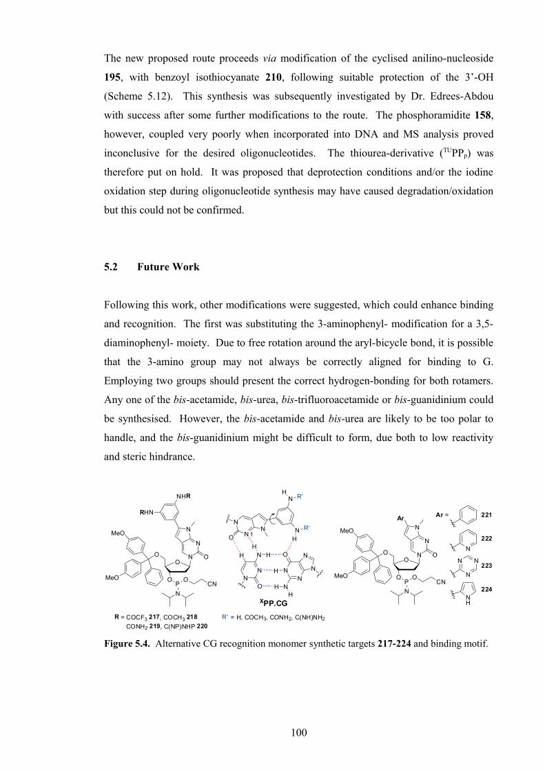
The new proposed route proceeds via modification of the cyclised anilino-nucleoside
195, with benzoyl isothiocyanate 210, following suitable protection of the 3’-OH
(Scheme 5.12). This synthesis was subsequently investigated by Dr. Edrees-Abdou
with success after some further modifications to the route. The phosphoramidite 158,
however, coupled very poorly when incorporated into DNA and MS analysis proved
inconclusive for the desired oligonucleotides. The thiourea-derivative (TUPPp) was
therefore put on hold. It was proposed that deprotection conditions and/or the iodine
oxidation step during oligonucleotide synthesis may have caused degradation/oxidation
but this could not be confirmed.
5.2 Future Work
Following this work, other modifications were suggested, which could enhance binding
and recognition. The first was substituting the 3-aminophenyl- modification for a 3,5-
diaminophenyl- moiety. Due to free rotation around the aryl-bicycle bond, it is possible
that the 3-amino group may not always be correctly aligned for binding to G.
Employing two groups should present the correct hydrogen-bonding for both rotamers.
Any one of the bis-acetamide, bis-urea, bis-trifluoroacetamide or bis-guanidinium could
be synthesised. However, the bis-acetamide and bis-urea are likely to be too polar to
handle, and the bis-guanidinium might be difficult to form, due both to low reactivity
and steric hindrance.
NN
N
O
HH
NN
NO
N
N
HH
H
H
XPP.CG
N
NON N
H
N
N
OO
O
O
N
RHN
P
N
OCN
MeO
MeO
R = COCF3 217, COCH3 218
CONH2 219, C(NP)NHP 220
R' = H, COCH3, CONH2, C(NH)NH2
1
NH
R'
R'
NHR
N
N
OO
O
O
N
Ar
P
N
OCN
MeO
MeO
N
N
N
N
NH
Ar = 221
222
223
224
Figure 5.4. Alternative CG recognition monomer synthetic targets 217-224 and binding motif.
100

It may also be beneficial to change the aromatic ring to a heterocycle such as pyridine,
triazine, pyrrole or imidazole. Altering hydrophilicity and electronic properties may
have beneficial effects on binding affinity.
The first target, however, is 6-phenyl-N-methylpyrrolo-dC phosphoramidite 220. It is
important to confirm what role the aromatic ring plays in triplex binding and
recognition, and whether the modifications do benefit binding or could even be omitted.
In addition, it would be necessary therefore to investigate how changing the ring,
whether aromatic (phenyl, napthyl), heteroaromatic (pyridine, triazine, pyrrole, thiazole)
or non-aromatic (cyclohexyl, cyclopentyl), affects binding, before further derivatisation.
The phenyl monomer 221 has been synthesised in house, in excellent overall yield by
Dr. Edrees-Abdou, and biophysical studies are in progress.
5.3 Conclusion
Three N-methylpyrrolo-dC monomers (APPp 178, AcPPp 191, UPPp 179), were
synthesised for incorporation into TFOs for biophysical studies. All were synthesised
from the key intermediate, 5’-(4,4’-dimethoxytrityl)-5-iodo-4N-methyl-2’-deoxy-
cytidine 185, made from the 2’-deoxyuridine analogue 97 in two steps. Sonogashira
cross-coupling124 and a modified copper (I)-catalysed cyclisation222,223 were used to form
the derivatised bicyclic base in typically good to excellent yield.
Problems were again encountered with the low reactivity of the aniline NH2 (c.f. Section
3), necessitating prolonged reaction times, a higher reagent excess, and raised
temperature (e.g. trifluoroacetylation, guanidinylation). Care was also taken for the
guanidinium monomer (GPPp) and precursor, to avoid cleavage of the base-labile
protecting groups, whilst preventing cleavage/activation of the acid-sensitive DMT and
phosphoramidite moieties. A revised method for synthesis of the guanidinylating
reagent 200 from CEOC-succinimide 201 was also evaluated, affording a final semi-
optimised yield of 89%, compared to the literature yield of 35%.41,225,226 This allows for
a much more accessible synthesis, owing to cost of DSC.
The fourth monomer, GPPp 199, was synthesised later in house, after synthetic
investigation (see above), and a fifth monomer, TUPPp 158, although eventually
101

synthesised later in house, could not be incorporated and studied as desired. Although
the thiourea moiety should have imparted extra stability over the urea in the triplex, it is
unlikely this modification will be continued. Instability during oligonucleotide
synthesis and/or deprotection introduced too many problems. However, there are
several new directions for future work, and the syntheses and biophysical studies
(Section 6) already undertaken are promising.
The precursors to the four monomers that were incorporated into TFOs, were also
deprotected and fluorescence measurements were taken. This is discussed in detail in
Section 6.3.
102

Chapter 6
Biophysical Studies of
N-Methylpyrrolo-dC Modified
TFOs for CG Recognition
103

6. Biophysical Studies of N-Methylpyrrolo-dC Modified TFOs for CG
Recognition
Four modified N-methylpyrrolo-dC phosphoramidite monomers (APPp 178, AcPPp 191, UPPp 179, GPPp 199) were incorporated into unlabelled/labelled TFOs for UV and
fluorescence melting studies. The initial study (from Section 4.2.2) was designed to
compare both post-synthetically modified furano-dT, and N-methylpyrrolo-dC
nucleosides at CG recognition in the four sequences introduced in Chapter 4. Following
this work, the main biophysical studies were carried out. These were again designed to
test binding affinity for CG and selectivity against the other base pairs (GC, AT, TA) at
several pHs. In addition to determination of the best modification for CG recognition,
the other aim was to compare UV and fluorescence melting data (collected using the
same buffer conditions), which appeared not to correlate in previous studies.
6.1 Initial Fluorescence Melting Study
This section discusses the N-methylpyrrolo-dC results from the ‘Sequence Study’
introduced in Section 4.2.2.
5’-Q-PMM TPM VXW TPT PTM PT 5’-F-GT GTT AGG AAG ACA AAA AAG AAC TGG T-H CA CAA TCC TTC TGT TTT TTC TTG ACC A-H
Figure 6.1. Fluorescence triplex melting experiment. TFO shown in bold. M = 5-MedC,
P = 5-(3-aminoprop-1-ynyl)-dU (pdU), X = AcPP & VXW = TXT (OL18), PXT (OL19), TXP
(OL20), PXP (OL21), or X = UPP & VXW = TXT (OL22), PXT (OL23), TXP (OL24), PXP
(OL25), Q = DABCYL (quencher), F = FAM (fluorophore), H = HEG (hexaethylene glycol),
hairpin duplex (CG, OL6).
As previously described, these fluorescence melting experiments (Figure 6.1) were
conducted using LC Programmes 1 and 2 (95-30-95 °C at 0.2 °C/min) at pH 6.2
(10 mM sodium phosphate, 200 mM NaCl). Experiments were run once in triplicate
and a 10:1 ratio of TFO:duplex (5:0.5 µM) was used.
104

XSeq.
Tm / °C, melt/annealAcPP UPP
TXT 42.1/38.0 44.0/42.0
PXT 49.0/44.0 48.1/45.1
TXP49.1/44.0
(48.4/43.0)51.1/47.1
(51.0/45.0)
PXP 54.0/49.0 54.5/50.0
Table 6.1. Fluorescence triplex melting experiment: AcPPp and UPPp against CG, each in four
TFOs (AcPP – OL18-OL21 & UPP – OL22-OL25) at pH 6.2 (10 mM sodium phosphate, 200
mM NaCl) using LC Programme 1 and 2 (Section 9.2). Average Tm values for TFO melt/anneal
given in °C. Data obtained for XPPf-modified TFOs is shown for comparison, in parentheses
(Section 4.2.2).
These results confirm the previous findings, that placing pdU (P) on the 5’-side of the
modification X (XPP) has a slightly destabilising effect compared to the 3’-side, notably
for the urea (UPP). The results for the TXP TFOs for both furano-dT (in parentheses)
and N-methylpyrrolo-dC modifications were similar (within experimental error), hence
previous furano-dT experimental data can be corroborated.
The trends identified when examining the combined XPPf and XPPp results, correlate
with those in the main fluorescence melting studies, despite differences in buffer
composition and pH.
In addition, hysteresis was on average 1.5 °C lower for XPPp than for XPPf modifications.
This may rise from the higher TFO purity when no post-synthetic modification is
required (Sections 3.3, 5.1).
6.2 Primary UV Melting Study
A comprehensive UV melting study was conducted in cooperation with Dr. Edrees-
Abdou, to examine the binding affinity/selectivity of the four monomers, in comparison
also with the natural base T. Data was collected at pH 6.2, 6.6 and 7.0, and at pH 7.0
with 2 mM spermine added. Spermine is a general/non-specific DNA stabilising agent
and component in all eukaryotic cells, which is used in this context, to increase the
stability of a duplex/triplex if the Tm is too low to be easily measured. Prior to this
105

work, large quantities of the pdU phosphoramidite monomer 164 were synthesised by
Dr. Edrees-Abdou and Dr. Imenne Bouamaied for synthesis of the TFOs used in this
both studies.
5’-TTT TTM VXW MTM TMT 5’-GCT AAA AAG AYA GAG AGA TCG CGA TTT TTC TZT CTC TCT AGC-5’
Figure 6.2. UV triplex melting experiment. TFO shown in bold. M = 5-MedC, P = 5-(3-
aminoprop-1-ynyl)-dU (pdU), VXW = TTT (OL32), TTP (OL33), PTT (OL34), PTP (OL35);
X = APP & VXW = TXT (OL36), TXP (OL37), PXT (OL38), PXP (OL39); X = AcPP & VXW
= TXT (OL40), TXP (OL41), PXT (OL42), PXP (OL43); X = UPP & VXW = TXT (OL44),
TXP (OL45), PXT (OL46), PXP (OL47); X = GPP & VXW = TXT (OL48), TXP (OL49), PXT
(OL50), PXP (OL51); purine strand Y = C (OL1), G (OL26), A (OL27), T (OL28), pyrimidine
strand Z = G (OL2), C (OL23), T (OL30), A (OL31).
UV melting experiments were performed (as described previously, and in detail in
Section 9.2), using a 5:1 ratio of TFO:duplex (5:1 µM). Experiments were conducted at
pH 6.2, 6.6, 7.0 and pH 7.0 + 2 mM spermine (10 mM sodium phosphate, 200 mM
NaCl, 1 mM Na2EDTA). All oligonucleotides for this study were synthesised without
capping to avoid undesired acetylation of TFOs.
Standard melt programme No. 3 was used (15-50-80 °C at 0.25-0.5 °C/min) with 5 heat/
anneal cycles in total (Section 9.2). Condensation was not a problem at low
temperature (15 °C).
There are several trends, which can be drawn from the data given below in Table 6.2.
Firstly, at pH 6.2, against CG in sequence TXT, (Table 6.2, Lines 1, 6, 19, 28, 37) the
aniline/amine (APP, OL36) outperforms all other monomers, and the poorest monomer
(AcPP, OL40) gives a Tm that is 1.7 °C greater than T. This result appears to contradict
the original order of stability as derived from the proposed hydrogen-bonding triplet
motif. The proposed motif places the urea (UPP) and guanidinium (GPP) as the strongest
binders, as they can form two hydrogen-bonds and the latter also contributes additional
charge-stabilisation. However, these results suggest steric or electronic factors may
play a role, where such groups cause destabilisation relative to the sterically non-
hindering amino group.
106

X Sequence Base Pair Buffer Tm / °C Hysteresis BufferDuplex 6.2 6.6 7.0 7.0 SP
T
TXT CG 6.2 25.6/22.9 2.7 1 CG 61.3 62.8 61.6 63.6TXT
CG 6.6
19.4/n.d. n.d. 2 GC 60.5 - - 63.4TXP n.d. n.d. 3 AT 58.4 - - 61.8PXT 23.2/20.1 3.1 4 TA 58.6 - - 61.7PXP 22.4/18.8 3.6 5
APPp
TXT
CG
6.2
30.0/26.3 3.7 6
GC 21.1/18.1 3.0 7 Hysteresis 1.1 – 5.9 °CAT 21.4/18.4 3.0 8
TA n.d. n.d. 9 Average Hysteresis 3.7 °CTXT
CG 6.6
22.7/18.5 4.2 10
TXP 24.0/19.8 4.2 11 Average duplex ∆Tm
due to spermine+ 2.9 °C
PXT 25.7/21.2 4.5 12
PXP 26.8/21.9 4.9 13
TXT CG 7.0 n.d. n.d. 14
TXT
CG
7.0 SP
30.4/29.4 1.0 15
GC 20.2/n.d. n.d. 16
AT 17.5/n.d. n.d. 17
TA n.d. n.d. 18
AcPPp
TXT
CG
6.2
27.3/23.1 4.2 19
GC 20.4/n.d. n.d. 20
AT 20.4/n.d. n.d. 21
TA 20.3/n.d. n.d. 22
TXT
CG 6.6
21.0/16.8 4.2 23
TXP 21.1/n.d. n.d. 24
PXT 22.6/19.4 3.2 25
PXP 22.7/18.3 4.4 26
TXT CG 7.0 n.d. n.d. 27
UPPp
TXT
CG
6.2
29.2/24.4 4.8 28
GC 20.1/n.d. n.d. 29
AT 21.5/18.5 3.0 30
TA 21.6/19.5 2.1 31
TXT
CG 6.6
22.7/18.5 4.2 32
TXP 23.4/18.3 5.1 33
PXT 24.1/18.3 5.8 34
PXP 24.9/19.0 5.9 35
TXT CG 7.0 n.d. n.d. 36
GPPp
TXT
CG
6.2
27.9/25.0 2.9 37
GC 19.5/n.d. n.d. 38
AT 20.7/n.d. n.d. 39
TA 17.8/n.d. n.d. 40
TXT
CG 6.6
21.4/18.0 3.4 41
TXP 21.1/18.0 3.1 42
PXT 21.6/18.6 3.0 43
PXP 22.5/20.3 2.2 44
TXT CG 7.0 n.d. n.d. 45
Table 6.2. UV triplex melting experiment: X against YZ at pH 6.2, 6.6, 7.0 and 7.0 + 2 mM
spermine (10 mM sodium phosphate, 200 mM NaCl, 1 mM Na2EDTA) using UV melting
programme No. 3 (Section 9.2). Average Tm values for TFO melt/anneal and duplex (average of
melt and anneal Tm), and hysteresis given in °C. n.d. = not determined (Tm < 17 °C).
107

At pH 6.6 (Lines 2, 10, 23, 32, 41), the urea and aniline (APP) show similar binding
affinity, greater than the guanidinium, which should be the strongest binder.
At pH 6.6, against CG for all the pdU-containing TFOs, the aniline is again the
strongest binder, followed by the urea. The order of stability for the other modifications
varies, such that for PXT (OL50) and PXP (OL51), the guanidinium is of equal or lower
stability than T. This may is attributed to disfavourable steric interaction and also
charge-repulsion with the pdU on the 5’-side as previously discussed.
All monomers demonstrated a very high selectivity for CG at pH 6.2, over the other
base pairs, by 6.9-8.6 °C. For all monomers except UPP, the second highest affinity was
for AT not TA, the latter of which has often been observed for CG recognition
monomers (Section 1.2.2). A putative X.AT triplet can be drawn to support this
observation, with hydrogen bonds between N3 and anilino NH of XPP and the NH2 of A
and C4=O of T. Only UPP and GPP show significant differences (> 1 °C) between the Tms
against the other base pairs. In addition, the aniline demonstrates very high selectivity
for CG, by 10.2 °C at pH 7.0 with 2 mM spermine, where the second highest affinity
was for GC (Lines 15-18).
Examination of the data for the pdU-containing TFO series, obtained at pH 6.6,
identifies significant differences from the initial fluorescence melting data discussed in
Sections 4.2 and 6.1. The main difference is order of stability, where higher Tms are
observed for PXT- rather than TXP-oligonucleotides. It was deduced from previous
fluorescence melting data, that disfavourable interaction between the pdU and XPP
amino-modified side-chains occurred when pdU was located on the 5’-side. However,
the reverse is observed here, and the difference between TXP and PXT is pronounced
for the aniline (1.7 °C) and acetamide (1.4 °C). Otherwise increase in Tm with each T-
pdU substitution varies from 0.1 to 1.3 °C. Interestingly, the TXT TFO (X = GPP,
OL48) is 0.2 °C more stable than the corresponding TXP oligonucleotide (OL49), but
this may simply be due to experimental error. The results for T at pH 6.6, appear
spurious, where the order of stability (TXP < TXT < PXP < PXT) cannot be easily
explained.
In summary, this study highlights the high selectivity of all monomers for CG over the
other base pairs. The order of binding affinity, however, contradicts the proposed triplet
108

binding motif, although the urea remains a strong binder. These observations suggest
the aniline or urea would be highly suitable candidates for CG recognition, the latter
perhaps particularly at higher pH. Selectivity at pH 7.0 with 2 mM spermine should be
carried out for the urea to confirm whether the urea does indeed outperform the aniline
at higher pH. Without spermine, no Tm could be measured for any monomer at pH 7.0.
The acetamide and guanidinium should also be assessed against CG at pH 7.0 + 2 mM
spermine to determine whether the effect of the positive-charge of the guanidinium is
beneficial to binding at higher pH, where little protonation of other amine
functionalities should occur. The acetamide performed poorest throughout and the
guanidinium proved less effective than expected.
6.3. Primary Fluorescence Melting Study
The main fluorescence melting study was conducted in cooperation with Dr. Edrees-
Abdou, to confirm the previous results and make more accurate comparisons between
monomer recognition capabilities. Data was collected using the same buffer systems as
for the UV melting study so that direct comparisons between the studies could be made.
5’-Q-PMM TPM VXW TPT PTM PT 5’-F-GT GTT AGG AAG AYA AAA AAG AAC TGG T-H CA CAA TCC TTC TZT TTT TTC TTG ACC A-H
Figure 6.3. Fluorescence triplex melting experiment. TFO shown in bold. M = 5-MedC,
P = 5-(3-aminoprop-1-ynyl)-dU (pdU), VXW = TTT (OL60), PTT (OL61), TTP (OL62), PTP
(OL63); X = APP & VXW = TXT (OL52), PXT (OL53), TXP (OL54), PXP (OL55); X = AcPP
& VXW = TXT (OL18), PXT (OL19), TXP (OL20), PXP (OL21); X = UPP & VXW = TXT
(OL22), PXT (OL23), TXP (OL24), PXP (OL25); X = GPP & VXW = TXT (OL56), PXT
(OL57), TXP (OL58), PXP (OL59);. Q = DABCYL (quencher), F = FAM (fluorophore), H =
HEG (hexaethylene glycol), hairpin duplex YZ = CG (OL6), GC (OL7), AT (OL8), TA (OL9).
109

X Sequence Base Pair Buffer Tm / °C Hysteresis
T
TXT
CG 6.2
44.3/41.0 3.3 1
TXP 47.5/45.9 1.6 2
PXT 48.4/47.8 0.7 3
PXP 51.1/48.7 2.4 4
APPp
TXT
CG
6.2
44.1/40.9 3.2 5
GC 36.7/32.9 3.8 6
AT 33.9/n.d. n.d. 7
TA 36.7/33.8 2.9 8
PXP
CG
6.2
56.8/52.9 3.9 9
GC 45.8/42.9 2.9 10
AT 43.3/40.9 2.4 11
TA 47.2/43.9 3.3 12
TXT
CG
6.6
37.5/32.9 4.6 13
GC n.d. n.d. 14
AT n.d. n.d. 15
TA n.d. n.d. 16
TXPCG 6.6
43.2/35.9 7.3 17
PXT 42.2/35.4 6.8 18
PXP 48.5/41.4 7.1 19
TXTCG 7.0
n.d. n.d. 20
PXP 40.6/n.d. n.d. 21
TXT CG 7.0 SP 36.0/33.5 2.5 22
AcPPp
TXT
CG
6.2
43.9/40.8 3.1 23
GC 38.8/35.9 2.9 24
AT 35.0/32.7 2.3 25
TA 41.9/38.0 3.9 26
PXP
CG
6.2
55.2/50.2 5.0 27
GC 46.9/43.8 3.1 28
AT 45.0/39.9 5.1 29
TA 50.4/47.0 3.4 30
TXPCG 6.2
50.3/46.1 4.2 31
PXT 51.0/46.0 5.0 32
TXT
CG
6.6
36.2/32.8 3.4 33
GC n.d. n.d. 34
AT n.d. n.d. 35
TA 33.9/n.d. n.d. 36
TXPCG 6.6
43.8/35.7 8.1 37
PXT 43.8/35.7 8.1 38
PXP 46.5/37.8 8.7 39
TXTCG 7.0
n.d. n.d. 40
PXP 39.9/n.d. n.d. 41
TXT CG 7.0 SP 36.0/34.2 1.8 42
Table 6.3. Fluorescence triplex melting experiment: X against YZ at pH 6.2, 6.6, 7.0 and 7.0
+ 2 mM spermine (10 mM sodium phosphate, 200 mM NaCl, 1 mM Na2EDTA) using LC
Programme 3 (Section 9.2). Average Tm values for TFO melt/anneal and values for hysteresis
given in °C. n.d. = not determined (Tm < 33 °C). Hysteresis: 0.7–9.3 °C, average hysteresis: 4.0
°C. Continued on following page.
110

X Sequence Base Pair Buffer Tm / °C Hysteresis
UPPp
TXT
CG
6.2
44.3/40.9 3.4 43
GC 39.9/34.4 5.5 44
AT 35.1/32.8 2.3 45
TA 40.7/36.9 3.8 46
PXP
CG
6.2
58.1/53.5 4.6 47
GC 46.0/45.0 1.0 48
AT 43.8/40.8 3.0 49
TA 49.7/45.1 4.6 50
TXPCG 6.2
52.0/48.9 3.1 51
PXT 50.8/45.8 5.0 52
TXT
CG
6.6
38.0/n.d. n.d. 53
GC n.d. n.d. 54
AT n.d. n.d. 55
TA 33.5/n.d. n.d. 56
TXPCG 6.6
44.2/38.8 5.4 57
PXT 43.0/37.7 5.3 58
PXP 48.2/43.9 4.3 59
TXTCG 7.0
n.d. n.d. 60
PXP 39.9/32.8 7.1 61
TXT CG 7.0 SP 37.4/34.8 2.6 62
GPPp
TXT
CG
6.2
47.2/45.8 1.4 63
GC 36.9/35.9 1.0 64
AT 37.0/35.9 1.1 65
TA 39.7/38.5 1.2 67
PXP
CG
6.2
56.0/51.4 4.6 68
GC 45.0/40.9 4.1 69
AT 47.0/44.5 2.5 70
TA 51.0/47.2 3.8 71
TXT
CG
6.6
40.3/35.0 5.3 72
GC n.d. n.d. 73
AT 30.9/n.d. n.d. 74
TA 33.0/n.d. n.d. 75
TXPCG 6.6
46.2/36.9 9.3 76
PXT 43.7/36.8 6.9 78
PXP 47.2/41.9 5.3 79
TXTCG 7.0
32.7/n.d. n.d. 80
PXP 38.5/n.d. n.d. 81
TXT CG 7.0 SP 39.0/37.9 1.1 82
Table 6.3 cont. Fluorescence triplex melting experiment. Continued from previous page.
Fluorescence melting studies were performed (as described previously, and in detail in
Section 9.2), on a Roche LightCycler®. Experiments were performed at pH 6.2, 6.6 and
7.0, and at pH 7.0 + 2 mM spermine (10 mM sodium phosphate, 200 mM NaCl, 1 mM
Na2EDTA), and a 10:1 ratio of TFO:duplex (5:0.5 µM) was used. Runs in these
experiments were performed up to three times in triplicate, using LC Programme 3 (95-
27-95 °C at 0.2 °C/min, see Section 9.2).
111

At pH 6.2, against CG in sequence TXT, (Table 6.3, Lines 1, 5, 23, 43, 63), GPP
outperforms the other monomers by 2.9 °C, however, the Tm of the poorest monomer
(AcPP) is 0.4 °C below T. The aniline, urea and T have similar Tms. At pH 6.6 also
(Lines 13, 33, 53, 72), the guanidinium monomer has the strongest binding affinity by
2.3 °C, followed by the urea, aniline then acetamide. In the PXP sequence at pH 6.2
(Lines 9, 27, 47, 68), the urea has the highest Tm by 1.3 °C, followed surprisingly by the
aniline then guanidinium and acetamide, which is 4.1 °C above T. A similar pattern
was observed for TXT and PXP at pH 7.0 and 7.0 with spermine.
The guanidinium monomer performed best, as expected, in the TXT sequence but not
for PXP. It is possible that the presence of a protonated amine species either side of the
positively-charged guanidinium may have destabilised it significantly, below that of the
aniline as observed. Otherwise, apart from the acetamide performing slightly worse
than the aniline, the order of stability can be rationalised.
A different situation was encountered for the PXT and TXP sequences. At pH 6.6, for
TXP (Lines 17, 37, 57, 76), the guanidinium and urea performed best, whereas for PXT
(Lines 18, 38, 58, 77), these appear destabilised such that the acetamide has a similar Tm
to GPP. This indicates that both the guanidinium and urea have a negative interaction
with pdU when they are located on the 5’-side (PXT) as originally noted (Section 4.2.2).
It is also perhaps surprising, that changing from PXT to PXP has a significant effect on
the order of binding affinity, whereas changing from TXT to TXP makes little
difference. It was originally thought that the proposed negative interaction was due to
steric hindrance between the aminopropynyl group of pdU and modified phenyl group
of the monomer. However, charge-repulsion may also play an important role.
All monomers demonstrate good to high selectivity for CG over the other base pairs, at
all pHs tested in both TXT and PXP sequences. In all cases, the second most stable
binding is to TA as originally expected. The least stable binding occurs to GC then AT,
or to AT then GC for GPP. For TXT at pH 6.2, the guanidinium and aniline demonstrate
the highest selectivity, by 7.5 °C, and the acetamide the least, by 2.0 °C. For TXT at pH
6.6, the guanidinium is most selective (∆Tm 7.4 °C) and the acetamide the least (∆Tm 2.4
°C), and for PXP at pH 6.2, the aniline is most selective (∆Tm 9.6 °C), followed by the
urea (∆Tm 8.4 °C), the guanidinium (∆Tm 5.0 °C) then the acetamide (∆Tm 4.7 °C).
112

The high selectivity of GPP and UPP compared to AcPP can be rationalised by the extra
potential hydrogen-bond, which can effect differentiation between the binding motifs of
CG and the other base pairs more effectively. It does not explain the good selectivity
for the aniline possessing only one potential hydrogen-bond donor, although in the PXP
sequence, general destabilisation of GPP and UPP, due to neighbouring pdU nucleotides,
may reduce their selectivity relative to the aniline.
Data for the pdU-containing TFOs demonstrates greater stability for TXP over PXT for
aniline, urea and guanidinium, and the reverse for the acetamide, in all cases (not tested
at pH 6.2 for APP and GPP). The greatest difference between TXP and PXT occurs for
GPP (∆Tm 2.5 °C), is moderate for UPP (∆Tm 1.2 °C) and APP (∆Tm 1.1 °C) and small for
AcPP (av. ∆Tm 0.4 °C).
Charge-repulsion may explain this difference for the guanidinium monomer, and steric
hindrance for the urea, but there must be alternative reasons for the observed differences
for the aniline and acetamide.
The fluorescence melting study results suggest the aniline, urea or guanidinium would
prove suitable candidates. The guanidinium has the greatest binding affinity in most of
the conditions tested and the aniline proved the most selective, followed by the
guanidinium and urea. Due to sensitivity of intermediates in the synthesis of the
guanidinium monomer, either the urea or aniline are probably more suitable.
6.4. Melting Study Comparison
The fluorescence melting study data (Section 6.3) correlates with previous fluorescence
melting data (Sections 4.2.2, 6.1), but not with the UV melting study data (Section 6.2).
Importantly, the order of binding affinity has stark differences between the two studies.
It is possible that the sequence composition may be partly responsible for the observed
differences. Examining the neighbouring triplets to the central three-triplet triplex core
of the UV and fluorescence melting motifs, only the triplet on the 5’-side is the same
(M+.GC) (Figure 6.4).
113

..TTM VXW MTM.. ..TPM VXW TPT.....AAG AYA GAG... ...AAG AYA AAA......TTC TZT CTC... ...TTC TZT TTT...
UV melting Fluorescence Melting
Figure 6.4. Central sections of UV and fluorescence melting triplex motifs highlighting
matched triplets (boxed) and differences (red).
It is difficult to explain all the differences between the two studies, however, by
analysing the differences in the sequence around the central core, without extensive and
time-consuming melting and NMR experiments. It is likely that the general triplex
shape, general TFO composition and whether the duplex part is a hairpin or comprises
separate strands, contributes to the differences in trends between systems.
In order to rule out the possibility that the method of analysis is responsible, although
unlikely, UV melting experiments should be performed using the fluorescence melting
triplex motif. The fluorophore and quencher should not affect the trends, only causing a
slight general increase in Tm. The oligonucleotides could also be resynthesised without
DABCYL and FAM, although this would create significant work.
If the method of analysis is proven not to be a factor, the differences must be sequence-
dependent. This would have considerable implications when designing TFOs for gene-
targeting and oligotherapeutics. Depending on the sequence and also interaction with
other modified/un-natural nucleotides and recognition monomers, the same monomer
may have significant differences in selectivity and binding affinity. Optimising both of
these properties is vitally important for achieving mixed-sequence recognition under
physiological conditions.
6.5. Fluorescence Properties of CG Recognition Monomers
The synthesis and fluorescent properties of pyrrolo-C nucleobases, nucleosides and
PNA monomers have been studied in detail by Robert Hudson et al.120,230-235 and others119
as they can be used as fluorescent reporter groups in SNP analysis236 and for
fluorimetric detection of guanosine.120,233 Fluorescence decreases significantly only on
duplex hybridisation with G, such that the change is visible to the naked eye on
irradiation of the solution with long-wave UV light (365 nm).
114

These pyrrolo-dC compounds have not, however, been evaluated as probes or
fluorophores in triplexes, and only the triplex-forming properties of N-methylpyrrolo-
dC derivatives have been studied thus far.67,122,207 For this purpose, the fluorescence
properties of the four deprotected nucleosides, APP 143, AcPP 225, UPP 182 and GPP 226
were evaluated to afford an indication of their usefulness in this application. Their
deprotection procedures are described in Section 9.2.6 (Scheme 6.1)
O
OH
DMTO N
N
O
N
RHN
R = H 195
O
OH
HO N
N
O
N
RHN
3% TCA/DCM
COCH3 193
CONH2 184
O
OH
DMTO N
N
O
N
NH
O
OH
HO N
N
O
N
NH
226
N
HN
O
ONC
O ONC
NH
H2N
i) 3% TCA/DCM, Et2O, rt
ii) 20% Et2NH/CH3CN, rt, 24h
iii) 40% NH3/H2O : 7N NH3
/CH3OH (1:2 v/v), rt, 60h
207
R = H 143
COCH3 225
CONH2 182
Et2O, rt
Scheme 6.1. Deprotection of N-methylpyrrolo-dC nucleosides 184,193,195,207.
The excitation and emission wavelengths were measured in HPLC grade methanol (APP
143, AcPP 225) or anhydrous DMF (UPP 182, GPP 226) using a Varian Cary 400 Scan
UV-Visible Spectrophotometer and a Varian Cary Eclipse Fluorescence
Spectrophotometer respectively (Section 9.2). The extinction coefficient was calculated
for each excitation maximum using the Beer-Lambert Law (A = εcL; A = absorbance,
ε = molar extinction coefficient, c = molar concentration, L = pathlength/cm – see
Section 9.2) and all data is detailed in Table 6.4 below.
X λabs1 ε1 λabs2 ε2 λabs3 ε3 λem λem–λabs1APPp 356.3 3730 237.8 16680 203.2 12100 448.5 92.2AcPPp 355.5 5530 243.4 33060 202.8 19130 458.0 102.5UPPp 358.5 15140 287.4 13740 276.4 13410 429.5 71.0GPPp 366.6 6690 270.2 22380 – – 458.0 91.4
Table 6.4. UV/Fluorescence data for N-methylpyrrolo-dC nucleosides 143,225,182,226:
λabs1,2,3 = excitation/absorption maxima (nm), λem = emission wavelength (nm) from excitation at
λabs1, ε = molar extinction coefficient (L mol-1 cm-1), λem–λabs1 = Stokes Shift (nm).
Absorption and emission spectra are displayed in Figure 6.5. Absorption data has been
truncated for samples run in DMF due to noise caused by loss of solvent transparency
115

below ~270 nm. Emission data has been scaled for easy visual comparisons between
curves.
200 250 300 350 400 450 500 550 600
0.0
0.5
1.0
1.5
2.0
-10
0
10
20
30
40
50
60
70
80
90
100 APP
AcPP
UPP
GPP
Ab
so
rban
ce
λ / nm
Em
ission
Inten
sity
/ a.u.
Figure 6.5. UV absorption (solid line) and fluorescence emission (dashed line) spectra for
N-methylpyrrolo-dC nucleosides. Properties measured at different concentrations. UPPp and GPPp absorption data were truncated below ~260 nm. Emission data scaled by scale factor = 1
(APPp), 0.1 (AcPPp), 10 (UPPp), 0.6 (GPPp). Fluorescence spectra obtained by excitation at λabs1
maxima (350-370 nm).
The urea, which demonstrated good binding affinity and selectivity, has the highest
extinction coefficient (15140 L mol-1 cm-1) by greater than a factor of 2. The Stokes
shift, although lowest at 71.0 nm, is still very good. The acetamide, despite performing
poorest in melting studies, has the highest Stokes Shift (102.5 nm), comparable with
that of the 6-arylpyrrolo-C nucleobases 227-229 and PNA monomers 230,231 of
Hudson et al..230,231 The guanidine demonstrated a good Stokes Shift (91.4 nm) and has
the second highest extinction coefficient of 6690 L mol-1 cm-1, thus may show most
potential as a triplex probe. Additionally, all emission spectra maxima have minimal
spectral overlap with their corresponding excitation maxima.
116

NH
N
O
NH
R
N
N
O
NH
R
OEt
O
R = H 227
NO2 228
R = NO2 230
OCH3 229
OCH3 231
Figure 6.6. 6-Arylpyrrolo-C derivatives 227-231 of Hudson et al..230,231
All nucleosides emit close to the excitation wavelength of DABCYL (471 nm), and
therefore could be used instead of fluorescein (FAM) in fluorescence melting studies, or
in combination with other acceptors for FRET analysis. There may also be a significant
change in fluorescence emission on triplex hybridisation, analogous to that observed by
Hudson in the duplex, on pairing with G.120,233 3’-Pyrene-modified dT has also found
use as a probe, showing marked increase in fluorescence on triplex formation,
depending on position within the triplex.237 Fluorescence was strongly dependent on
correct hybridisation, which offered the potential for detection of duplex mutations.
The quantum yields should be determined next, for absolute confirmation of the best N-
methylpyrrolo-dC probe candidate.
6.6 Conclusions & Future Work
The final melting studies confirm the efficacy of these monomers to specifically
recognise a CG inversion in a homopurine tract. Comparing the results from both
studies, the monomers of choice should be the urea and the aniline. The aniline
outperformed the other monomers in the UV melting study for both binding affinity and
selectivity, and the urea also demonstrated its efficacy. The guanidinium monomer
demonstrated the highest binding affinity in the fluorescence melting study, followed by
the urea. However, the aniline proved slightly more selective and the urea less so on
average, and these are easier to synthesise than the guanidinium. The acetamide
performed relatively poorly throughout both studies, comparibly with T, except where
placing pdU on the 5’-side caused destabilisation of the other monomers.
It is difficult to rationalise the differences between the two studies, which should be
well correlated. Differences such as the change in order of stability of the pdU-
117

containing triplexes, and relative binding affinities and selectivity have not yet been
explained, but several factors may contribute and there are some studies, which can be
carried out to examine this. The three best monomers have been tested against real gene
targets, and are currently being re-synthesised for further tests, in conjunction with other
recognition monomers. Such mixed-sequence triplex-based recognition has already
been evaluated,67 and gene targeting studies are in progress.
The 6-phenyl-N-methylpyrrolo-dC phosphoramidite monomer 221 was synthesised by
Dr. Edrees-Abdou, as detailed in Section 5.2, in excellent yield and melting studies are
underway. Further modifications for assessment are described also in Section 5.2.
Finally, the fluorescence properties of the four CG recognition monomer nucleosides
show potential for their use as probes within triple helices and in fluorescence studies,
including melting. The Stokes Shifts and extinction coefficients are comparable with
the NH-pyrrolo-C analogues of Hudson et al.,230,231 which have already found use in
fluorimetric detection of G.120,233 Quantum yields must be determined, however, so the
most efficient substrate can be chosen.
118

Chapter 7
Oligonucleotide Deprotection Study
119
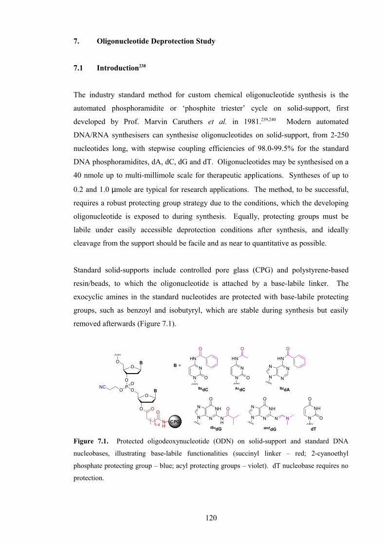
7. Oligonucleotide Deprotection Study
7.1 Introduction238
The industry standard method for custom chemical oligonucleotide synthesis is the
automated phosphoramidite or ‘phosphite triester’ cycle on solid-support, first
developed by Prof. Marvin Caruthers et al. in 1981.239,240 Modern automated
DNA/RNA synthesisers can synthesise oligonucleotides on solid-support, from 2-250
nucleotides long, with stepwise coupling efficiencies of 98.0-99.5% for the standard
DNA phosphoramidites, dA, dC, dG and dT. Oligonucleotides may be synthesised on a
40 nmole up to multi-millimole scale for therapeutic applications. Syntheses of up to
0.2 and 1.0 µmole are typical for research applications. The method, to be successful,
requires a robust protecting group strategy due to the conditions, which the developing
oligonucleotide is exposed to during synthesis. Equally, protecting groups must be
labile under easily accessible deprotection conditions after synthesis, and ideally
cleavage from the support should be facile and as near to quantitative as possible.
Standard solid-supports include controlled pore glass (CPG) and polystyrene-based
resin/beads, to which the oligonucleotide is attached by a base-labile linker. The
exocyclic amines in the standard nucleotides are protected with base-labile protecting
groups, such as benzoyl and isobutyryl, which are stable during synthesis but easily
removed afterwards (Figure 7.1).
BO
O
O
O
O
O B
PO
NCO
CPG
N
N
HN
O
O
NH
N
N
O
NH
N
O
N
N
HN
O
O
NH
N
N
O
NN N
N
N
N
HN
N
O
O
NH
O
1-4CPG
B =
BzdC AcdC BzdA
iBudG dmfdG
N
NH
O
O
dT
Figure 7.1. Protected oligodeoxynucleotide (ODN) on solid-support and standard DNA
nucleobases, illustrating base-labile functionalities (succinyl linker – red; 2-cyanoethyl
phosphate protecting group – blue; acyl protecting groups – violet). dT nucleobase requires no
protection.
120

Non-standard nucleosides, either natural or synthetically modified, are incorporated by
the same method, appropriately protected with base-labile or other labile groups.
Alternative compatible protecting groups for primary/exocyclic amines include
phthalimide, trifluoroacetyl and phenoxyacetyl. Hydroxyl groups may be protected as
the TBDMS- or allyl-ether for example. The protecting group lability can be altered
depending on the oligonucleotide sensitivity and desired deprotection conditions.
7.2 Deprotection Conditions for Oligonucleotides238
The standard reagents for deprotection of non-modified oligodeoxynucleotides (ODNs),
are concentrated (~33 wt%) aqueous ammonia and concentrated (40 wt%) aqueous
methylamine. The oligonucleotide may be cleaved from the solid-support under mild
conditions (conc. aq ammonia, rt, 1 hour) before heating or further treatment, or the
support may be removed from the synthesis column and all deprotections carried out in
one step. Treatment at rt overnight or at 55 °C for 4-6 hours is usually sufficient to
ensure full deprotection of shorter ODNs. Using a 1:1 mixture of conc. aq ammonia
and conc. aq methylamine (AMA) may also be used to significantly reduce deprotection
time for nucleosides such as AcdC, BzdA and iBudG. AMA can also be used for quick
cleavage of DNA from the solid-support (5-15 minutes). Methylamine and AMA
should not be used for deprotection of BzdC, however, as there is precedent for attack of
the C4-position by alkylamines, to form the 4N-alkyl-dC derivative. Oligonucleotides
may also be deprotected using pressurised ammonia or methylamine gas, following
cleavage from the support.
Standard deprotection conditions are also usually sufficient for deprotection of other
base-labile groups. Phthalimide may be cleaved using methylamine or hydrazine, and
allyl groups can be removed by automated treatment with palladium (0), such as a
solution of Pd(PPh3)4 in chloroform, containing 5% acetic acid and 2.5% N-
methylmorpholine,241,242 before removal from the synthesiser for cleavage/deprotection.
Oligonucleotides may also be treated with diethylamine before cleavage from the
resin,243,244 or by addition of nitromethane during deprotection.245 These reagents cleave
2-cyanoethyl groups from the phosphate esters and scavenge acrylonitrile, a by-product
of oligonucleotide synthesis. Nitromethane acts predominantly as a scavenger of
acrylonitrile. Without such treatments, a small proportion of dT nucleotides are
121

cyanoethylated at the N3-position. Such adducts however, do not form readily when
deprotecting using methylamine, which also acts as a scavenger. Ammonia is not
nucleophilic enough to act as an effective scavenger.
Washing of the solid-support with 20% diethylamine/acetonitrile is incorporated into
the automated synthesis procedure on some new DNA/RNA synthesisers. 2-Cyanoethyl
groups are cleaved and acrylonitrile scavenged before cleavage from the solid-
support.246 Diethylamine will only cause cleavage from solid-support upon prolonged
exposure (>5 hours).
7.3 Oligonucleotide Deprotection Study
7.3.1 Background
Oligonucleotides synthesised for the initial UV and fluorescence melting studies
(Chapters 4, 6) were analysed by analytical HPLC and either electrospray or MALDI-
TOF MS. Analysis of the non-labelled furano-dT modified TFOs for UV melting,
showed that after deprotection with 30 wt% aqueous methylamine (rt, 6-24 hours), and
treatment with DOWEX-H+ resin, cyclisation of the methylamine ring-opened
intermediate was not quantitative (Figure 7.2). However, HPLC purification of these
oligonucleotides was successful as resolution was obtained.
Figure 7.2. HPLC traces of UV melting TFOs (OL4,5) following first HPLC purification and
treatment with DOWEX-H+ resin.
OL4
OL5
122

HPLC analysis of the DABCYL-labelled TFOs for fluorescence melting, however,
could not resolve the components. The large lipophilic DABCYL moiety causes the
labelled DNA to adhere strongly to the stationary phase, thus causing reduction in
resolution (”streaking”). MS analysis of several of the first DABCYL-labelled N-
methylpyrrolo-dC modified TFOs (OL18-21, AcPPp; OL23, UPPp), indicated they were
clean following HPLC purification, and they were subsequently used in melting studies.
HPLC analysis of some of the crude mixtures before purification (OL18-21), identified
a normal distribution of minor impurities around the major peak. Analytical ion-
exchange (IONEX) chromatography of HPLC-purified TFO OL19, resolved a small
possible failure sequence peak, and due to the long retention time was deemed
unsuitable as a method of purification (Figure 7.3).
Figure 7.3. HPLC traces of AcPPp-modified TFOs (OL18-21) before purification and IONEX
trace of OL19 after HPLC purification.
Following this work, a Capillary Electrophoresis system was acquired, from which very
high resolution chromatograms could be obtained. CE analysis of several of the
DABCYL-modified TFOs, resolved each into three peaks for both furano-dT (OL14, APPf) and N-methylpyrrolo-dC modified TFOs (OL20, AcPPp; OL23, UPPp) (Figure 7.4).
This result could be rationalised for the furano-dT modified TFO, as a mixture of
cyclised (highest retention time), uncyclised/ring-opened TFOs and a sequence where
one phosphoramidite coupling did not occur (failure sequence, lowest retention time).
The observation is harder to explain for the N-methylpyrrolo-dC modified TFOs
OL19
123

(deprotected using conc. aq NH3), where only ring-opening by ammonia or failure
sequences could be held accountable. Failure sequences are usually capped by
treatment with acetic anhydride, before the oxidation step in each cycle of
oligonucleotide synthesis. If synthesised without capping, failure sequences, which are
usually minor, may become more prominent, especially when using synthetically-
modified nucleosides. However, this may account only for the smaller peak of lowest
retention time, and ring-opening by ammonia appears unlikely. Secondary structures
may also provide an explanation but are even more unlikely.
Figure 7.4. CE traces of DABCYL-modified TFOs (OL14, OL20, OL23).
Concurrently there were problems with TFOs containing the GC-recognition monomer,
bis-amino-U (BAU). Following incorporation into the TFO, the oligonucleotide was
deprotected using 40 wt% aqueous methylamine. Methylamine was also required for
cleavage of the phthalimide protecting group on the 2’-moiety (Scheme 7.1). Despite
previous successful deprotections and melting studies,35-37 problems were later
encountered. Although HPLC analysis of the TFOs indicated they were pure, melting
studies afforded poor results, i.e. little or no triplex formation. On receipt of the CE
machine, analysis of some of these TFOs revealed a mixture of peaks, which could only
be accounted for by excessive degradation of the TFO, not by failure sequences alone.
This suggested that the deprotection conditions must have changed since the previous
TFOs containing this monomer were synthesised, deprotected and purified.
OL14 OL20 OL23
124

NO
O
O
O
PO
NCO NH
O
O
ON
O
O
NH
F3C
O
NO
O
O
O
PHO
O NH
O
O
ONH2
H2N
40 wt % aq MeNH2
rt, 15 hrs
Scheme 7.1. BAU nucleotide deprotection in TFO.
After these results, it was decided to investigate oligonucleotide deprotection conditions
to determine the optimum conditions for monomers used within the research group.
7.3.2 Deprotection Study
A series of oligonucleotides were synthesised on a 1.0 µmole scale (with capping) based
on oligoT (T12), containing the monomers under examination (Figure 7.5).
TTT TTT TTX TTT TTT TYT YTY TTT TZZ ZTZ ZTZ TZT
Figure 7.5. Sequences for oligonucleotide deprotection study; X = T (OL64), pdU (OL65), BAU
(OL68), AcPPp (OL69), S (OL70), GpdU (OL74), MOEpU (OL71), AET (OL72); Y = pdU (OL66);
Z = pdU (OL67).
After each oligonucleotide synthesis, the solid-support was removed from the column.
Small portions of the resin (5-8 mg) were deprotected for varying lengths of time, at
different temperatures using conc. aq ammonia, 20 wt% aq MeNH2 and 40 wt% aq
MeNH2. A list of all deprotections for each oligonucleotide is given in Table 7.1.
Following deprotection, the resin was removed and solvent evaporated, the
oligonucleotide residue was dissolved in distilled water (1.0 mL) and desalted using a
disposable Sephadex NAP™ 10 column. All samples were then analysed, without
HPLC purification, by CE. ES and MALDI-TOF MS analyses were taken as
appropriate. Analytical HPLC and IONEX were also used for several samples.
125

ODN X SequenceDeprotection Conditions MS
Concn Base Temp Time/h ES MALDI
OL64 – TTT TTT TTT TTTconc NH3 55 °C 520% MeNH2 rt 1540% MeNH2 rt 15
OL65 pdU TTT TTT TTX TTT
conc NH3 rt 5 conc NH3 rt 15 conc NH3 55 °C 5conc NH3 55 °C 15 20% MeNH2 rt 1540% MeNH2 rt 15
OL66 pdU TTT TXT XTX TTT
conc NH3 rt 1 **conc NH3 rt 1
conc NH3 rt 5conc NH3 rt 15conc NH3 55 °C 4 conc NH3 55 °C 15 20% MeNH2 rt 520% MeNH2 rt 15 40% MeNH2 rt 15
OL67 pdU TXX XTX XTX TXTconc NH3 55 °C 1
**conc NH3 55 °C 1
OL68 BAU TTT TTT TTX TTT
conc NH3 55 °C 15 conc AMA rt 1520% MeNH2 rt 5 20% MeNH2 rt 820% MeNH2 rt 15 20% MeNH2 rt 30
OL69 AcPPp TTT TTT TTX TTT
conc NH3 rt 5 conc NH3 55 °C 4 20% MeNH2 rt 540% MeNH2 rt 6 40% MeNH2 rt 15
OL70 S base TTT TTT TTX TTT
conc NH3 rt 5 conc NH3 rt 15 conc NH3 55 °C 5 conc NH3 55 °C 15 20% MeNH2 rt 540% MeNH2 rt 15
OL71 MOEpU TTT TTT TTX TTTconc NH3 rt 5conc NH3 55 °C 520% MeNH2 rt 5
OL72 AET TTT TXT XTX TTT conc NH3 55 °C 4
OL73 AET TTT TTT TTX TTT conc NH3 55 °C 4
OL74 GpdU TTT TTT TTX TTTconc NH3 rt 5
tBuNH2:MeOH:H2O (1:1:2) rt 21
Table 7.1. Oligonucleotides, deprotection conditions and MS analysis. Optimum conditions
evaluated are in bold. **Column washed with 20% Et2NH/MeCN for 10 minutes before
removal of resin for deprotection.
126

7.3.2.1 Control T12 Oligonucleotide
Control oligonucleotide T12 (OL64) was deprotected successfully by three methods. No
degradation was observed by CE, hence even the strongest deprotection conditions
(40 wt% aq MeNH2) caused no degradation of the DNA backbone (Figure 7.6).
Figure 7.6. CE traces for deprotections of T12 (OL64).
It was deduced therefore, that any impurities observed for other oligonucleotides must
be due to failure sequences (n-1, n-2 etc.), degradation of the modified nucleotide, or
fragmentation of the oligonucleotide due to the modification.
7.3.2.2 Test Oligonucleotides – 5-(3-Aminopropynyl)-dU (pdU)
The first modified monomer to be tested, pdU, was incorporated into three sequences
with one, three and seven insertions.
conc. aq NH3, 55 °C, 5 hrs 20% aq MeNH2, rt, 15 hrs 40% aq MeNH2, rt, 15 hrs
127

Figure 7.7. CE traces for deprotections of T8XT3 (X = pdU, OL65).
The best conditions for T8XT3 (X = pdU, OL65), giving the least degradation, are conc.
aq ammonia at rt for 5 hours (Figure 7.7). A longer time does not significantly affect
the impurity profile. Heating at 55 °C for 5 hours is also suitable, but heating overnight
(15 hrs) caused further appearance of impurities. A 20 wt% aqueous solution of
methylamine also deprotected causing little degradation overnight.
O
O
O N
HN
O
OP
OH
O
O
TTTTTTTT
POHO
O TTT
NH2
O
OH
OP
OH
O
O
TTTTTTTTNHCH3
40% MeNH2, rt, 15 hrs
Mol. Wt.: 2580.8Mol. Wt.: 3627.4
5'5'
3'3'
Scheme 7.2. Proposed cleavage/degradation of T8XT3 by methylamine at high concentration.
However, it is very clear that using conc. aq methylamine (40 wt%) causes formation of
a significant impurity (2581 Da) plus other minor peaks. This major impurity can be
explained by cleavage of the oligonucleotide at the 3’-side of pdU, and displacement of
the alkynyl-U nucleobase by methylamine (Scheme 7.2, Figure 7.8).
conc. NH3, 55 °C, 5 hrs 20% MeNH2, rt, 15 hrs 40% MeNH2, rt, 15 hrsconc. NH3, rt, 15 hrs conc. NH3, 55 °C, 15 hrs
128

Figure 7.8. MALDI-TOF MS spectra for two deprotections of T8XT3 (X = pdU, 3627.4 Da,
OL65). * Internal T10 and T15 references.
Similar observations were made for three additions of pdU (T4XTXTXT3, OL66). The
mildest successful deprotection conditions were treatment with conc. aq ammonia at rt
for 1 hour only, after washing the resin with 20% Et2NH/CH3CN for 10 minutes.
Without the diethylamine wash, impurities were present. For this sample, this was due
either to poor coupling of pdU, perhaps caused by presence of pyridine in
phosphoramidite, or too little time for complete deprotection, not the lack of a
diethylamine wash. Under these conditions, 1 hour is the shortest possible time for
cleavage from the resin, but may not be long enough for consistent deprotection of all
protecting groups (Figures 7.9, 7.10).
Figure 7.9. CE traces for deprotections of T4XTXTXT3 (X = pdU, OL66).
conc. NH3, rt, 5 hrs
40% MeNH2, rt, 15 hrs
20% MeNH2, rt, 15 hrs40% MeNH2,
rt, 15 hrs20% MeNH2, rt, 5 hrsconc. NH3, 55 °C, 4 hrsconc. NH3, rt, 1 hr
129

Heating at 55 °C for only 4 hours with conc. aq ammonia formed several small
impurities, and treatment overnight at rt with 40 wt% methylamine caused extensive
oligonucleotide degradation. Treatment with 20 wt% methylamine at rt could be used
for 5 hours, but impurities formed over 15 hours.
Figure 7.10. ES MS spectra for two deprotections of T4XTXTXT3 (X = pdU, 3705.5 Da,
OL66). Data deconvoluted to resolution of 1 Da.
Oligonucleotide TX3TX2TXTXT (X = pdU, OL67) was successfully deprotected under
mild conditions (conc. aq ammonia, 55 °C, 1 hr) with and without a prior diethylamine
wash. The small impurities were slightly greater without washing and failure sequences
became slightly more prominent. A slightly longer deprotection time (~2 hours) may
have removed some of these trace impurities. HPLC and IONEX traces were similar
for both (Figure 7.11).
A small impurity was present in these oligonucleotides (3651 Da in OL66), due to
incorporation of a single dU nucleoside instead of pdU. The dU impurity was formed
during the Sonogashira cross-coupling reaction in synthesis of the pdU phosphoramidite
monomer 164 and was present at approximately 2-3%. When used in synthesis of the
deprotection study oligonucleotides, it only became noticeable on multiple additions.
When oligonucleotide OL66 was re-synthesised using a different batch of monomer,
with and without capping for comparison, only trace baseline impurities were
noticeable. Synthesis was as effective with capping as without capping indicating
absence of failure sequences.
conc. NH3, rt, 1 hr
conc. NH3, 55 °C, 15 hrs
130

Figure 7.11. HPLC/CE traces for deprotections of TX3TX2TXTXT (X = pdU, OL67).
7.3.2.3 Test Oligonucleotides – Bis-amino-U (BAU)
The next monomer under test was bis-amino-U (BAU). The best conditions for
deprotection were discovered to be 20 wt% aq methylamine, at rt for 5 hours.
Figure 7.12. CE traces for deprotections of T8XT3 (X = BAU, OL68).
Little change was noted after 8 hours, but after 15 and 30 hours notable impurities had
formed. Heating at 55 °C in conc. aq ammonia for 15 hours, and treating with AMA
(conc. aq NH3/40 wt% aq MeNH2, 1:1 v/v) at rt for 15 hours produced a similar
significant pattern of impurities (Figure 7.12).
20% Et2NH/MeCNconc. NH3, 55 °C, 1 hr conc. aq NH3, 55 °C, 1 hr
AMA, rt, 15 hrs conc. NH3, 55 °C, 15 hrs20% MeNH2, rt, 30 hrs20% MeNH2, rt, 15 hrs20% MeNH2, rt, 5 hrs
131

Figure 7.13. MALDI-TOF/ES MS spectra for two deprotections of T8XT3 (X = BAU, 3686.5
Da, OL68). ES data deconvoluted to resolution of 1 Da. * Internal T10 and T15 references.
Only a ‘minus T’ failure sequence (3382.3 Da) was identified by MS. The other MS
peaks could not be assigned (Figure 7.13). Methylamine is required for phthalimide
cleavage, but a high concentration causes significant degradation.
7.3.2.4 Test Oligonucleotides – 6-(3-Acetamidophenyl)-N-methylpyrrolo-dC (AcPPp)
The acetamide monomer for CG recognition was incorporated into a ‘T12
oligonucleotide’ (T8XT3, X = AcPPp, OL69) and proved stable to conc. aq ammonia at rt
for 5 hours, and 55 °C for 4 hours, and also to 20 wt% aq methylamine at rt for 5 hours
(Figure 7.14). Heating at 55 °C in conc. aq ammonia for 15 hours caused more
significant degradation than AMA at rt for the same time.
20% MeNH2, rt, 5 hrs
conc. NH3, 55 °C, 15 hrs
132

Figure 7.14. CE traces for deprotections of T8XT3 (X = AcPPp, OL69).
Concentrated aq methylamine, however, primarily caused major conversion to the
cleavage product (2581 Da) shown in Scheme 7.2 (Figure 7.15).
Figure 7.15. ES MS spectra for two deprotections of T8XT3 (X = AcPPp, 3744.6 Da, OL69).
Data deconvoluted to resolution of 1 Da.
This was originally presumed to be the deacetylated oligonucleotide, but was confirmed
otherwise by ES and MALDI-TOF MS. No ring-opening of the N-
methylpyrrolopyrimidine nucleobase by methylamine was observed.
AMA, rt, 15 hrs conc. NH3,55 °C, 15 hrs
20% MeNH2, rt, 30 hrs20% MeNH2,rt, 15 hrs
20% MeNH2,rt, 5 hrs
conc. NH3, rt, 5 hrs
40% MeNH2, rt, 15 hrs
133
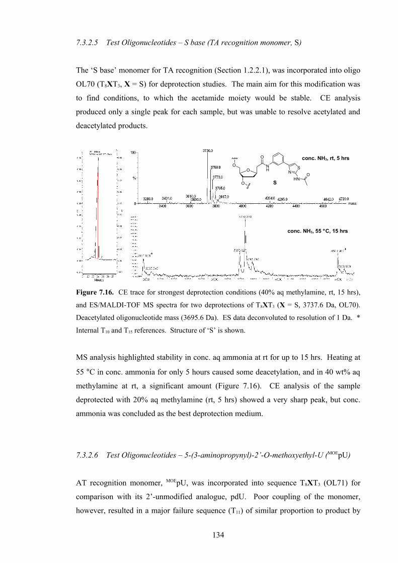
7.3.2.5 Test Oligonucleotides – S base (TA recognition monomer, S)
The ‘S base’ monomer for TA recognition (Section 1.2.2.1), was incorporated into oligo
OL70 (T8XT3, X = S) for deprotection studies. The main aim for this modification was
to find conditions, to which the acetamide moiety would be stable. CE analysis
produced only a single peak for each sample, but was unable to resolve acetylated and
deacetylated products.
Figure 7.16. CE trace for strongest deprotection conditions (40% aq methylamine, rt, 15 hrs),
and ES/MALDI-TOF MS spectra for two deprotections of T8XT3 (X = S, 3737.6 Da, OL70).
Deacetylated oligonucleotide mass (3695.6 Da). ES data deconvoluted to resolution of 1 Da. *
Internal T10 and T15 references. Structure of ‘S’ is shown.
MS analysis highlighted stability in conc. aq ammonia at rt for up to 15 hrs. Heating at
55 °C in conc. ammonia for only 5 hours caused some deacetylation, and in 40 wt% aq
methylamine at rt, a significant amount (Figure 7.16). CE analysis of the sample
deprotected with 20% aq methylamine (rt, 5 hrs) showed a very sharp peak, but conc.
ammonia was concluded as the best deprotection medium.
7.3.2.6 Test Oligonucleotides – 5-(3-aminopropynyl)-2’-O-methoxyethyl-U (MOEpU)
AT recognition monomer, MOEpU, was incorporated into sequence T8XT3 (OL71) for
comparison with its 2’-unmodified analogue, pdU. Poor coupling of the monomer,
however, resulted in a major failure sequence (T11) of similar proportion to product by
conc. NH3, rt, 5 hrs
conc. NH3, 55 °C, 15 hrs
O
O
O
O
NH N
S
HNO
S
134

CE. Of the three conditions tested (Table 7.1), heating at 55 °C in conc. aq ammonia
for 5 hrs caused an increase in the apparent failure sequence peak and a small increase
in the general impurity profile over the other two.
7.3.2.7 Test Oligonucleotides – Other AT recognition monomers
AT recognition monomer, 2’-aminoethoxy-T (AET) was incorporated into T8XT3 (OL72)
and T4XTXTXT3 (OL73) and both were deprotected at 55 °C in conc. aq ammonia for 4
hours. Both samples produced very clean HPLC traces hence this method was deemed
suitable, and this should be applicable also at rt.
5-(3-Guanidinylprop-1-ynyl)-dU (GpdU) was incorporated once (T8XT3, OL74) and
treated under two deprotection conditions. Deprotection with conc. aq ammonia (rt, 5
hrs) caused formation of two major peaks plus impurities by CE (Figure 7.17).
Figure 7.17. CE traces for deprotections of T8XT3 (X = GpdU, OL74).
One major peak (~22.8 minutes) appears to correspond to the oligonucleotide with a
fully-protected guanidinyl moiety. The other (~21.9 minutes) may correspond to a
stable triazinedione derivative, caused by cyclisation due to attack by ammonia. This
was observed by Prakash et al.225 when deprotecting oligonucleotides, containing 2-
cyanoethyloxycarbonyl- (CEOC) protected guanidinyl moieties, with ammonia (Figure
7.18). Prakash et al. cleaved the CEOC groups in 50% aq piperidine at rt for 24 hours,
prior to heating at 55 °C in conc. ammonia. Treating with 50% aq piperidine also
tBuNH2:MeOH:H2O, rt, 21 hrsconc. NH3, rt, 5 hrs
135

cleaved the 2-cyanoethyl phosphate protecting groups and cleaved the oligonucleotide
from the resin.
HN NH
N
O
ONC
OONC
O
O
O N
NH
O
ONH3NH
NH
N
O
ONC
O
H2N
bis-CEOC-GpdU
NH
NH
N
O
HN
O
O
O N
NH
O
O
O
triazinedione derivative
Figure 7.18. Possible mechanism of formation of triazinedione derivative.225
It was later discovered that another secondary amine, diethylamine, could be used
instead of piperidine. Washing the column with 20% Et2NH/CH3CN for at least 30
minutes prior to deprotection, was sufficient to remove the most labile CEOC group
thus preventing triazine formation. It is likely that methylamine would attack the
CEOC groups in the same manner but the steric bulk of the methyl group might prevent
cyclisation after attack.
The second deprotection of OL74 was carried out in tBuNH2:CH3OH:H2O (1:1:2 v/v/v)
at rt for 21 hours. Tert-butylamine was used to cleave all protecting groups at the same
time. It was presumed that the sterically-hindering tert-butyl group would prevent
attack of the CEOC groups and cyclisation. These conditions, however, proved too
mild, producing a mixture of bis-CEOC, mono-CEOC and fully deprotected
oligonucleotides in an approximate ratio of 9:5:1 (Figure 7.17).
7.4 Conclusion
The results of this study highlight the need for suitable/compatible deprotection
conditions for each modified nucleotide within the oligonucleotide. It cannot be
assumed that the conditions used for one modification, will be safe to use for another, as
proven by the compatibility of most modifications to deprotection with conc. ammonia
at rt, except the CEOC-protected guanidinyl-modified nucleotide.
136

Concentrated (40 wt%) aq methylamine was proven too strong for deprotection of all
oligonucleotides containing modified non-natural nucleotides. It is likely that previous
deprotections of BAU-containing oligonucleotides using conc. aq methylamine were
successful, due to evaporation of methylamine over time from the bottle in use, thus
reducing the effective concentration.
In general, conc. aq ammonia can be used to deprotect most modified oligonucleotides,
either at rt or sometimes with heating. Methylamine is needed for cleaving phthalimide
groups, and may be required for cleavage of more stable protecting groups.
Washing the column with 20% Et2NH/CH3CN solution for 10-20 minutes before
removal from the synthesiser, was incorporated into the standard oligonucleotide
synthesis protocol, on all synthesisers where it was not already included and could be
easily achieved. It cleanly cleaves 2-cyanoethyl groups and scavenges acrylonitrile, and
proves highly effective at removing CEOC-protecting groups on guanidinyl-modified
nucleotides.
These results were used for choosing deprotection conditions for oligonucleotides used
within the research group, including for the TFOs used in the primary melting studies
(Sections 6.2, 6.3). For all TFOs used in these melting studies, following synthesis, the
solid-support column was washed with 20% Et2NH/CH3CN for 20 minutes, or 3 hours
for GPPp-containing TFOs (OL48-51, OL56-59). Each oligonucleotide was then
deprotected in conc. aq ammonia at rt for 12 hours, with or without 2 hours at 50 °C, or
at rt for 24 hours followed by heating at 50 °C for 1 hour to ensure complete
deprotection. CE analysis indicated good to high purity for most TFOs after one HPLC
purification. Where impurities constituted a significant proportion, the TFO was
carefully repurified, collecting only the centre of the peak. Impurities did not, however,
hinder performance in melting studies.
137

Chapter 8
Conclusion
138

8. Conclusion
8.1. Modified 6-Oxocytidine Nucleosides for GC Recognition
Synthesis of a bis-amino-modified 6-oxocytidine monomer 64, was re-investigated as a
potential GC recognition monomer, which exhibits pH-independent binding within a
triplex. Previous published studies70,71 reported pH-independent triplex formation using 6-oxoC nucleotides up to pH 8, and enhanced binding to GC at higher pH compared to C.
Modifications to this nucleoside,11,70,76 however, produced varied or negative results,
hence further modifications were proposed to introduce protonated amine
functionalities, which should enhance triplex stability.
Several synthetic investigations were carried out, in order to assess reaction feasibility.
Palladium-catalysed alkynylation was ruled out as a method of introduction of amine
functionality at the 5-position due to inactivity of the halogenated substrate or
degradation. Modification by amine- or thiol-halide exchange was investigated with
limited success, but this requires extensive optimisation if a robust synthetic route is to
be found. Feasibility of 2’-modification was also investigated, although this requires
further work. Optimised synthetic routes to some key intermediates have been
developed.
8.2 Amino-Modified Bicyclic Nucleosides for CG Recognition
Recognition of pyrimidine.purine base pairs is a greater challenge than
purine.pyrimidine base pairs as fewer hydrogen-bonds are presented for binding in the
major groove. Previous published research describes the development of recognition
monomers from T, based around the bicyclic C analogue, N-methylpyrrolo-dC.122 This
nucleobase retains the C2 carbonyl group, vital for CG recognition, whilst blocking GC
recognition by replacing an H for a methyl group at the 4N position.
The latest design had employed an aminoalkyl group for targeting of hydrogen-bonding
residues on G across the base pair. This research examined various meta-aminophenyl
groups; rigidified linkers better placed for binding.
139

Furano-dT 145-147 and N-methylpyrrolo-dC phosphoramidite monomers 178,179,191
(199 – synthesis repeated later in house) were synthesised for assessment. All were
synthesised by a common synthetic route (Sonogashira cross-coupling, Cu(I)-catalysed
cyclisation and phosphitylation) with some modifications, starting from DMT-protected
5-iodo-2’-deoxyuridine 97, and analogue 5-iodo-N-methyl-2’-deoxycytidine 185
respectively. Yields ranged from moderate to excellent and most steps were optimised
despite high substrate polarity.
Furano-dT monomers underwent post-synthetic modification to the N-methylpyrrolo-dC
nucleotide after incorporation into DNA. N-Methylpyrrolo-dC monomers could be
incorporated with no further modification necessary. Although post-synthetic
modification was not complete, and HPLC purification was unable to resolve the
mixture for DABCYL-labelled oligonucleotides, both performed equally in melting
studies.
Thiourea-modified monomer 158 (synthesis finished later in house) could not be
incorporated into TFOs due to issues with stability to oligonucleotide synthesis or
deprotection, hence could not be assessed in biophysical studies.
Extensive melting studies demonstrated the effectiveness of these monomers at
recognising CG with high selectivity and good binding affinity compared to previous
monomers (T, 4HT, MP). The order of stability was altered, however, between UV and
fluorescence melting studies. This was proposed to be due to changes in target
sequence and local environment around the CG inversion target, but requires further
investigation.
The effect of neighbouring groups on the modification was also highlighted. The CG
recognition monomers appear to interact differently with 5-(3-aminoprop-1-ynyl)-dU
(pdU, P), depending on which side it is located. This neighbouring group interaction
introduces important implications when using TFOs as oligotherapeutics, as all
interactions must be taken in to account for successful development.
140

8.3 Oligonucleotide Deprotection
A study was conducted to determine the optimum deprotection conditions for
monomers used within the research group, after experiencing several problems.
Concentrated aq ammonia was determined the best medium for deprotection at rt or
sometimes with heating. Most monomers also tolerated 20% aq methylamine for
approximately 5 hours. Deprotection times of 15 hours or greater caused some
degradation for several monomers, and the standard deprotection media, conc. aq
methylamine and AMA, as used for non-modified DNA, caused significant to extensive
degradation and strand cleavage for everything except the oligonucleotide T12.
A column wash with 20% Et2NH/CH3CN solution was introduced on to DNA
synthesisers where not already incorporated, after demonstrating efficient cleavage of 2-
cyanoethyl phosphate protecting groups and prevention of cyanoethylation by
scavenging of acrylonitrile. This wash also proved very effective at partial deprotection
of the guanidine moiety of two monomers, thus avoiding subsequent side-reactions
during deprotection.
141

Chapter 9
Experimental
142

9. Experimental
9 Synthesis
10 General
HPLC grade acetonitrile, methanol and water were purchased from Fisher Scientific.
Pyridine, dichloromethane, triethylamine and diisopropylethylamine were distilled over
calcium hydride; methanol, ethanol and isopropanol over magnesium and iodine;
phosphorous oxychloride over sodium wire, and tetrahydrofuran over sodium wire and
benzophenone before use. Anhydrous N,N-dimethylformamide and 1,2-dichloroethane
were purchased from Aldrich. Diethyl ether was purchased from Fisher Scientific and
dried by passing through a column of dry alumina then degassed at low temperature, or
argon-degassed over 4Å molecular sieves. Molecular sieves were oven-dried at greater
than 120 °C over 2 days or microwave-activated. Acetone was purchased from Fisher
Scientific and dried over Drierite (anhydrous CaSO4). 6-Aminouracil was purchased
from Aldrich, recrystallised from boiling water and dried over P2O5 before use. Iodine/
pyridine/THF/water solution (0.02 M), phosphitylating reagents and DIHT were
purchased from Link Technologies. N-Iodosuccinimide was purchased from Aldrich
and recrystallised from 1,4-dioxane/carbon tetrachloride before use. Deuterated NMR
solvents were purchased from Apollo Scientific Ltd. DOWEX 50WX8 ion-exchange
resin was purchased from Aldrich and washed with 0.2 M HCl before treatment. All
other reagents were purchased from Aldrich, Lancaster, Avocado, Alfa Aesar, BDH,
Fisher Scientific, Acros and Fluka, and used without purification. All reactions
requiring absence of oxygen were carried out under an atmosphere of argon. Glassware
was oven-dried overnight before use, for reactions requiring exclusion of moisture.
Reactions were monitored by TLC, using Merck Kieselgel 60 F254 or Machenary-Nagel
Alugram Sil G/UV254 silica gel plates (0.22 mm thickness, aluminium backed).
Compounds were visualised by irradiation at 254/365 nm, or staining with
p-anisaldehyde (A’), potassium permanganate (B’), ninhydrin (C’, D’),
10% H2SO4/ethanol (E’) or ceric sulfate (F’), followed by heating.
143

A’ – p-anisaldehyde / glacial AcOH / conc. H2SO4 / ethanol (9.2:3.8:12.5:338 v/v)
B’ – KMnO4 / K2CO3 / 5% aq NaOH / water (3:20 g/5:300 mL)
C’ – ninhydrin (5 g) / acetone (100 mL)
D’ – 0.3% w/v ninhydrin / butan-1-ol / 3% v/v glacial AcOH
E’ – 10% v/v conc. sulfuric acid / ethanol
F’ – 8% w/v Ce(SO4)2.2H2SO4 hydrate / 15% v/v sulphuric acid
Column chromatography was carried out under air or argon pressure using Fisher
Scientific DAVISIL 60Å (35-70 micron) silica gel. Silica gel was pre-equilibrated with
triethylamine or pyridine for purification of acid-senstive compounds.
Proton and carbon NMR spectra were recorded at 300/400 MHz and 75/100 MHz,
respectively, using either a Bruker AC300 or Bruker DPX400 spectrometer. Fluorine
and phosphorous NMR spectra were recorded at 282 MHz and 121 MHz respectively,
using a Bruker AC300 spectrometer. NMR spectra were recorded in deuterated
chloroform or dimethylsulfoxide. Chemical shifts are given in ppm and spectra are
calibrated to the residual solvent peak.247 J values are correct to within 0.5 Hz.
Assignment was aided by DEPT-135,1H–1H COSY, HMQC and HMBC experiments
and Lorentz-Gauss resolution enhancement data reprocessing (Win-NMR Lite).
Low-resolution mass spectra were recorded using electrospray ionisation (ES) on a
Fisons VG platform instrument, or on a Waters ZMD quadrupole mass spectrometer in
HPLC grade acetonitrile, methanol or water, or using electron ionisation (EI) on a
ThermoQuest TraceMS single quadrupole GC-MS instrument in dichloromethane.
High-resolution mass spectra were recorded in HPLC grade acetonitrile, methanol or
water using electrospray ionisation on a Bruker APEX III FT-ICR mass spectrometer.
IR spectra were recorded on a Satellite FT-IR instrument using a ‘Golden Gate’ or
‘Smart Orbit’ adapter, and visualised using Win First-lite or OMNIC software
respectively. All absorptions are measured in cm-1 and are described as broad (br),
weak (w), medium (m), strong (s) or very strong (vs).
Melting points were recorded on a Gallenkamp Electrothermal melting point apparatus
and uncalibrated.
144

Elemental (CHN) Thermal Combustion Analysis was carried out by MEDAC Ltd.,
Egham, Surrey, UK.
145

9.1.2 Experimental Procedure
1,3,5-Tri-O-acetyl-2-O-(2-phthalimidoethyl)-D-ribofuranose, 6537
O
OO
OSi
Si
O
89
1α
2
5
N1'
2'
O
OC28H45NO8Si2Mol. Wt.: 579.83
OMe
3' 4' 5'
6'
conc. H2SO4 (cat.)
AcOH / Ac2O (1:1, v/v), argon, rt
O
OAcO
AcO
65
1
2
5
N1'
2'
O
O
5'
6'
OAc
C21H23NO10Mol. Wt.: 449.41
To a stirred solution of 89 (3.31 g, 5.71 mmol) in glacial acetic acid/acetic anhydride
(1:1 v/v, 30 mL), under an argon atmosphere, conc. sulfuric acid (0.35 mL, 6.57 mmol,
1.1 eq) was added slowly. The reaction mixture was stirred at rt for 1 hour. Saturated
aq sodium bicarbonate (250 mL) was added carefully to the dark green reaction
mixture, which was extracted with CH2Cl2 (3 × 100 mL). The combined organic
fraction was washed with sat. aq sodium bicarbonate and sat. aq KCl, dried (Na2SO4)
and concentrated in vacuo to yield an orange-brown oil/solid. Following purification by
column chromatography (1:1, hexane/ethyl acetate), and drying under high vacuum, the
product 65 was isolated, as an anomeric mixture (α:β ca 2:1), as a very pale yellow oil
(2.11 g, 4.70 mmol, 82% yield).
Rf 0.17 (1:1, hexane/ethyl acetate, A’); 1H NMR (400 MHz, CDCl3) δ signals due to α
anomer 7.86-7.80 (2H, m, H5’), 7.74-7.68 (2H, m, H6’), 6.07 (1H, s, H1), 5.00 (1H, dd, J
= 4.8, 6.8 Hz, H3), 4.35-4.30 (1H, m, H4), 4.29 (1H, t, J = 4.8 Hz, H5), 4.14-4.08 (1H,
m, H5), 4.12-4.07 (1H, m, H2), 3.94-3.83 (2H, m, H2’), 3.83-3.72 (2H, m, H1’), 2.05,
2.04 (3H, s, COCH3), 2.00 (3H, s, COCH3), signals due to β anomer 7.86-7.80 (2H, m,
H5’), 7.74-7.68 (2H, m, H6’), 6.29 (1H, d, J = 4.5 Hz, H1), 5.13 (1H, dd, J = 2.5, 6.5 Hz,
H3), 4.38 (1H, br dd, J = 3.3, 7.3 Hz, H4), 4.24 (1H, dd, J = 3.5, 12.6 Hz, H5), 4.06 (1H,
dd, J = 5.5, 12.1 Hz, H5), 3.96 (1H, dd, J = 4.5, 6.5 Hz, H2), 3.94-3.83 (2H, m, H2’),
3.83-3.72 (2H, m, H1’), 2.07, 2.03, 1.96 (3H, s, COCH3); 13C NMR (100 MHz, CDCl3)
δ signals due to α anomer 170.6, 170.4, 169.5 (COCH3), 168.2 (C3’), 134.2 (C6’), 132.2
(C4’), 123.4 (C5’), 98.9 (C1), 80.8 (C2), 79.5 (C4), 71.9 (C3), 67.8 (C1’), 63.8 (C5), 37.6
(C2’), 21.2, 20.8, 20.5 (COCH3), signals due to β anomer 171.2, 170.5, 170.0 (COCH3),
168.2 (C3’), 134.1 (C6’), 132.3 (C4’), 123.3 (C5’), 94.8 (C1), 81.4 (C4), 77.9 (C2), 69.7
(C3), 68.6 (C1’), 63.4 (C5), 37.6 (C2’), 21.1, 20.9, 20.7 (COCH3); LRMS (ES+): m/z (%):
546 (38), 472 ([M + Na]+, 100).
146

Analytical results consistent with reported data.37
N-Trifluoroacetyl propargylamine, 68117,170
O
NH
F3CH2N
O
OF3C(1.3 eq)
dry MeOH, argon, 0 oC - rtC3H5N
Mol. Wt.: 55.08 C5H4NOF3Mol. Wt.: 151.09
9868
Propargylamine (3.71 g, 67.3 mmol) 98 was added dropwise to an ice-cooled, stirred
solution of ethyl trifluoroacetate (10.4 mL, 87.6 mmol, 1.3 eq) in distilled methanol (50
mL) under an argon atmosphere. After addition, the solution was allowed to warm to rt
and stirred for 15 hrs. The reaction mixture was concentrated in vacuo and redissolved
in CH2Cl2 (200 mL). The solution was washed with 10% w/v aq citric acid (2 × 100
mL) and sat aq KCl (100 mL), dried (Na2SO4) and concentrated in vacuo to give an
orange/brown oil. Following column chromatography (CH2Cl2), and drying in vacuo,
the product 68 was isolated as a very pale yellow oil (5.81 g, 38.4 mmol, 57%).
Rf 0.27, 0.33 (CH2Cl2, B’, D’), 0.53, 0.66 (1:9, methanol/CH2Cl2, B’, D’); IR (film): ν
3300 (br m, CC–H), 3092 (br w, N–H), 1703 (vs, C=O), 1551 (m, N–H), 1428, 1363,
1335 (w), 1207 (s), 1152 (br vs, C–F), 1046 (w), 997 (m), 926, 826 (w), 724 (m, C–F),
667 (m), 638 (br s), 602, 556 (m) cm-1; 19F NMR (282 MHz, CDCl3) δ -76.2 (CF3); 1H
NMR (300 MHz, CDCl3) δ 6.73 (1H, br s, NH), 4.15 (2H, dd, J = 2.6, 5.3 Hz, CH2),
2.33 (1H, t, J = 2.6 Hz, C≡CH); 13C NMR (75 MHz, CDCl3) δ 157.2 (q, J = 37.6 Hz,
COCF3), 115.7 (q, J = 287.5 Hz, COCF3), 77.2 (C≡CH), 73.3 (C≡CH), 29.8 (CH2);
LRMS (ES+): m/z : 206 ([M + CH3OH + Na]+, 100), 174 ([M + Na]+, 45); (ES–): m/z :
150 ([M – H]–, 100), 80 (57).
Analytical results consistent with reported data.117,170
147

Benzyltriethylammonium dichloroiodate 71154
i) NaI, conc. HCl, aqu. NaClO
ii) 10% aqu. NaI
iii) BnEt3N+Cl- 72, H2O
NICl2
NCl
72 71
C13H22NClMol. Wt.: 227.77
C13H22NCl2IMol. Wt.: 390.13
12
34
5
6
7
Sodium iodide (6.63 g, 44.2 mmol, 1.0 eq) was dissolved in conc. aq HCl (15.5 mL) and
cooled in an ice bath. To this, an 8.8% w/v aqueous solution of NaClO (37.4 mL, 44.1
mmol) was added slowly, with swirling, so that the temperature was kept below 20 °C.
Effervescence and a brown residue were observed. Towards the end of addition, the
bleach solution was added dropwise until all brown colouration had disappeared. A
10% aq solution of NaI was added dropwise (20 mL) until a trace amount of iodine
reappeared. The resulting yellow/orange solution was added portionwise to a solution
of benzyltriethylammonium chloride 72 (10.0 g, 44.1 mmol) in water (14 mL) to give a
brilliant yellow precipitate. The mixture was filtered, and the precipitate was washed
with water and dried under high vacuum over P2O5, to give the product salt 71 as a fine,
bright yellow powder (16.5 g, 42.2 mmol, 96% yield).
Mp 89-98 °C, lit. 84-86 °C;154 IR (solid): 3053 (w, C–H), 2984 (m, C–H), 1479 (s, C–
H, Aryl C=C), 1390 (m, C–H), 1360 (m), 1155 (m), 1004 (m), 903 (w), 796 (m), 757 (s,
Aryl C–H), 708 (vs Aryl C–H), 605 (w) cm-1; 1H NMR (300 MHz, d6-DMSO): δ 7.52
(5H, app. t, J = 1.8 Hz, CHAr), 4.48 (2H, s, H3), 3.17 (6H, q, J = 7.3 Hz, H2), 1.31 (9H,
t, J = 7.3 Hz, H1); 13C NMR (75 MHz, d6-DMSO): δ 132.5 (C5), 130.2 (C6), 129.0 (C7),
127.8 (C4), 59.5 (C3), 52.0 (C2), 7.5 (C1); LRMS (ES+): m/z (%): 192 (M+, 100); (ES–):
m/z (%): 380 (I3–, 16), 197/199/201 (ICl2
–, 4, 11:3:1), 126 (I–, 100).
Analytical results are consistent with the reported data.154
148

N1-[1-(3’,5’-O-(1,1,3,3-tetraisopropyldisiloxane-1,3-diyl)-β-D-ribofuranosyl)]-(5-
chloro-6-oxocytosin-4-yl)-N,N-dimethylformamidine, 74
BTEA-ICl2 (1.5 eq), NaHCO3 (3.0 eq)
distilled CH2Cl2, distilled MeOH, rt 1'2'3'
4'
5'
2
45
6
O
OHO
OSi
Si
O
N
NH
N
OO
NMe2
O
OHO
OSi
Si
O
N
NH
N
OO
Cl
NMe2
C24H44N4O7Si2Mol. Wt.: 556.80
C24H43N4O7ClSi2Mol. Wt.: 591.24
73 74
To a stirred solution of nucleoside 73 (0.66 g, 1.18 mmol) in distilled CH2Cl2 (20 mL)
and distilled methanol (10 mL), was added anhydrous sodium bicarbonate (0.30 g, 3.56
mmol, 3.0 eq), followed by a solution of benzyltriethylammonium dichloroiodate 71
(0.69 g, 1.77 mmol, 1.5 eq) in distilled CH2Cl2 (15 mL), and the reaction mixture was
stirred vigorously at rt for 50 minutes. The crude reaction mixture was filtered and
concentrated in vacuo to give an orange/brown gum. The residue was redissolved in
CH2Cl2 (50 mL) and washed with water (3 × 50 mL). This was further washed with sat.
aq sodium thiosulfate (50 mL), water (2 × 50 mL) and brine (2 × 50 mL), dried
(Na2SO4) and concentrated in vacuo to yield a pink foam. The compound was twice
purified by column chromatography (0-2% methanol/CH2Cl2) to yield after drying in
vacuo, the chlorinated product 74 as a white crystalline solid (0.37 g, 0.54 mmol, 46%).
Rf 0.50 (14:1, CH2Cl2/methanol, A’); Mp 94-99 °C (methanol/CH2Cl2); IR (solid): ν
3485 (w), 2942, 2891, 2864 (m, C–H), 1714 (m, C=O), 1637 (s, C=O, C=C), 1578 (vs,
C=N, C=C), 1462 (m), 1418 (s), 1371 (s, O–H), 1296, 1246 (m), 1122 (s), 1074 (s, C–
O), 1029 (vs, Si–O), 917 (m), 882, 861, 761 (s), 690 (vs, C–Cl), 587 (s) cm-1; 1H NMR
(300 MHz, CDCl3) δ 8.16 (1H, s, H7), 6.24 (1H, d, J = 1.5 Hz, H1’), 4.98 (1H, t, J = 7.0
Hz, H3’), 4.60 (1H, dd, J = 1.5, 6.2 Hz, H2’), 4.01 (2H, d, J = 5.5 Hz, H5’), 3.84 (1H, td,
J = 5.3, 7.3 Hz, H4’), 3.33 (1H, v br s, 2’-OH), 3.19, 3.12 (3H, s, NCH3), 1.15-1.02
(28H, m, CH(CH3)2); 13C NMR (75 MHz, CDCl3) δ 160.0 (C6), 156.8 (N=C7–N), 151.4
(C2, C4), 94.2 (C5–Cl), 89.4 (C1’), 82.8 (C4’), 73.7 (C2’), 73.1 (C3’), 64.1 (C5’), 41.6, 35.2
(N–CH3), 17.6, 17.5, 17.5, 17.4, 17.3, 17.3, 17.3, 17.2 (Si–CH(CH3)2), 13.4, 13.3, 12.9,
12.8 (Si–CH(CH3)2); LRMS (ES+): m/z (%): 1203 ([2M + Na]+, 44), 613 ([M + Na]+,
100); HRMS (ES+): calcd for C24H43O7N4ClSi2 (M), [M + Na]+ = 613.2251, found
613.2267.
149

2’,3’,5’-Tri-O-acetyl-6-oxocytidine, 7571,248
i) HMDS (10.0 eq), (NH4)2SO4 (0.2 eq)
ii) sugar 65, TMSOTf (1.0 eq), anh. DCE
ref lux / rt, argon
NH
NH
NH2
OO
NH
NH2
ONO
OAcAcO
AcO O5
5
1'
5'
4'
3' 2'
4
26
6275
C15H19N3O9Mol. Wt.: 385.33
C4H5N4O2Mol. Wt.: 141.1081
A suspension of dry 6-aminouracil 62 (8.00 g, 62.9 mmol, 1.1 eq) and ammonium
sulfate (1.67 mg, 12.6 mmol, 0.2 eq) in HMDS (131.5 mL, 631 mmol, 10.5 eq) was
heated to reflux, under an argon atmosphere, whilst stirring vigorously, for 24 hours,
during which the starting material dissolved. The reaction was allowed to cool to rt and
the solvent was removed under high vacuum. The cream-coloured solid was
redissolved in anhydrous DCE (150 mL) under an argon atmosphere, to which was
added acetylated sugar 65 (19.0 g, 59.8 mmol). Trimethylsilyl triflate (11.4 mL, 63.0
mmol, 1.1 eq) was carefully added dropwise and the reaction was stirred at rt, under an
argon atmosphere, for 43 hours. After concentrating in vacuo the residue was
redissolved in CH2Cl2 (250 mL), and washed with water (150 mL) and dilute aq sodium
bicarbonate (150 mL), during which a white solid precipitated out of solution. The
mixture was filtered, and following drying under high vacuum at 50 °C for 2 days, the
product β-anomer 75 was afforded as a fine white solid (16.9 g, 43.8 mmol, 73% yield).
The aqueous layer was re-extracted (CH2Cl2, 100 mL) and the combined, filtered
organic fractions were washed with sat. aq KCl (200 mL), dried (Na2SO4) and
concentrated in vacuo to yield a peach-coloured foam/solid (3.52 g) containing the
sugar 65 and α- 76 and β-anomers 75 of the product nucleoside.
Rf 0.28 (1:9 methanol/CH2Cl2, A’); Mp 197-201 °C (CH2Cl2), lit.71 228-229 °C
(ethanol/diethyl ether); IR (solid): ν 3447, 3324 (w, N–H), 3211, 2992 (w), 2911, 2789
(w, C–H), 1736 (m, C=O), 1718 (s, C=O), 1652 (s, N–H), 1586 (br m), 1537, 1427 (m),
1372 (m, C–H), 1252, 1215 (s, C–O), 1071, 1044 (s), 973, 796, 750 (m), 641 (s), 623,
582 (m), 547 (vs) cm-1; 1H NMR (400 MHz, d6-DMSO) δ 10.64 (1H, br s, NH3), 6.47
(2H, br s, NH2), 6.15 (1H, br s, H1’), 5.64 (1H, dd, J = 2.8, 6.8 Hz, H2’), 5.47 (1H, app. t,
J = 7.0 Hz, H3’), 4.55 (1H, s, H5), 4.31 (1H, dd, J = 3.0, 11.5 Hz, H5’), 4.08 (1H, ddd, J
= 3.1, 6.4, 8.0 Hz, H4’), 4.02 (1H, dd, J = 6.5, 11.0 Hz, H5’), 2.06, 2.03, 2.00 (3H, s,
COCH3); 13C NMR (100 MHz, d6-DMSO) δ 170.1, 169.5, 169.3 (COCH3), 161.9 (C6),
150

154.5 (C4–NH2), 150.4 (C2), 84.4 (C1’), 77.7 (C4’), 73.6 (C5), 72.6 (C2’), 69.9 (C3’), 63.1
(C5’), 20.5, 20.4, 20.2 (COCH3); LRMS (ES+): m/z (%): 793 ([2M + Na]+, 12), 430 (32),
408 ([M + Na]+, 100); (ES–): m/z (%): 769 ([2M – H]–, 32), 384 ([M – H]–, 100).
Analytical results consistent with reported data.71,248
5-Bromo-2’,3’,5’-tri-O-acetyl-6-oxocytidine, 79
NBS (1.0 eq), AIBN (2 mol %)
dist. THF, dark, argon, rt
C15H19N3O9Mol. Wt.: 385.33
C15H18N3O9BrMol. Wt.: 464.22
NH
NH2
ONO
OAcAcO
AcO O
5
1'
5'
4'
3' 2'
75
NH
NH2
ONO
OAcAcO
AcO O
5
79
Br
To a stirred suspension of 2’,3’,5’-tri-O-acetyl-6-oxocytidine 75 (3.05 g, 7.91 mmol)
and NBS (1.41 g, 7.92 mmol, 1.0 eq) in distilled THF (30.0 mL) was added AIBN (27.0
mg, 0.16 mmol, 2.1 mol %), and the reaction was stirred in absence of light, under an
argon atmosphere at rt for 30 minutes. The white slurry was concentrated in vacuo to
yield a white solid. Following purification by column chromatography (25-80%
acetone/toluene), and drying in vacuo, the product 79 was afforded as a powdery white
solid (3.47 g, 7.48 mmol, 95% yield) containing 2.0 mol % succinimide.
Rf 0.35 (1:1, acetone/toluene, A’), 0.41 (1:9 methanol/CH2Cl2, A’); IR (solid): ν 3412
(w, N–H), 3301 (br w, N–H), 3240, 3194, 3137 (w), 3089 (w, N–H), 3020, 2909 (w,
CH2), 1739 (s, C=O), 1718 (vs, C=O), 1628, 1557 (s, N–H), 1426 (m), 1371 (s, CH3),
1225 (vs, C–O), 1156, 1128, 1091 (s), 1040 (vs), 1013 (s), 941, 930 (m), 897, 765, 727
(s), 678 (m), 617 (s), 538 (s, C–Br) cm-1; 1H NMR (300 MHz, d6-DMSO) δ 11.00 (1H,
br s, NH3), 6.70 (2H, s, NH2), 6.16 (1H, br s, H1’), 5.62 (1H, dd, J = 2.5, 6.5 Hz, H2’),
5.49 (1H, app. t, J = 7.3 Hz, H3’), 4.33 (1H, dd, J = 3.3, 11.8 Hz, H5’), 4.11 (1H, ddd, J
= 3.0, 6.5, 8.0 Hz, H4’), 4.02 (1H, dd, J = 6.3, 11.8 Hz, H5’), 2.07, 2.03, 2.00 (3H, s,
COCH3); 13C NMR (75 MHz, d6-DMSO) δ 170.0, 170.0, 169.3 (COCH3), 158.1 (C6),
151.4 (C4-NH2), 149.2 (C2), 85.9 (C1’), 77.9 (C4’), 72.6 (C2’), 69.7 (C3’), 69.1 (C5), 62.9
(C5’), 20.5, 20.3, 20.2 (COCH3); LRMS (ES+): m/z : 949/951/953 ([2M + Na]+, 1:2:1,
52), 527/529 ([M + CH3CN + Na]+, 1:1, 40), 495/497 (1:1, 100), 486/488 ([M + Na]+,
1:1, 99); HRMS (ES+): calcd for C15H18N3O9Br (M), [M + Na]+ = 486.0124/488.0104,
151

found 486.0114/488.0097 (1:1), [2M + Na]+ = 949.0351/951.0330/953.0310, found
951.0254 (1:2:1).
5-Iodo-2’,3’,5’-tri-O-acetyl-6-oxocytidine, 83
NIS (1.0 eq), AIBN (2 mol%)
dist. THF, dark, argon, rt
C15H19N3O9Mol. Wt.: 385.33
C15H18N3O9IMol. Wt.: 511.22
NH
NH2
ONO
OAcAcO
AcO O
5
1'
5'
4'
3' 2'
75
NH
NH2
ONO
OAcAcO
AcO O
5
83
I
To a stirred suspension of 2’,3’,5’-tri-O-acetyl-6-oxocytidine 75 (5.00 g, 13.0 mmol)
and AIBN (46.3 mg, 0.28 mmol, 2.2 mol %) in distilled THF (50.0 mL) was added NIS
(2.95 g, 13.1 mmol, 1.0 eq), and the reaction was stirred in absence of light, under an
argon atmosphere, at rt for 15 minutes. The orange/brown slurry was concentrated in
vacuo to yield an orange/brown solid. A portion of the solid (~0.5 g) was dissolved in
ethyl acetate (250 mL) and washed with dilute aq Na2S2O3 (100 mL, adjusted to pH 7
using NaHCO3) until decolourised then separated immediately. The solution was
further washed with sat. aq KCl (2 × 100 mL), dried (Na2SO4) and concentrated in
vacuo to give an off-white solid (0.34 g). The remainder was suspended in ethyl acetate
(550 mL) and allowed to stand in the fridge for 2 hours. The mixture was vacuum
filtered, and the residue was washed with ice-cold ethyl acetate (3 × 150 mL) and dried
under high vacuum over P2O5, to yield the product 83 as a white, powdery solid (5.47 g)
containing 0.4 mol % succinimide (recalculated yield - 10.7 mmol, 82%).
Rf 0.35 (1:1, acetone/toluene, A’), 0.54 (tetrahydrofuran, A’); IR (solid): ν 3400, 3295
(w, N–H), 3207 (br w, N–H), 1740, 1719 (s, C=O), 1624 (s, C=C), 1551 (s, N–H), 1422
(m), 1371 (s, CH3), 1228 (vs), 1096, 1040 (s, C–O), 942, 931, 898 (m), 763 (s), 736 (m),
722 (m, CH2), 677, 647, 630 (m), 602, 571, 537 (s), 512 (m, C–I) cm-1; 1H NMR (300
MHz, d6-DMSO) δ 10.91 (1H, br s, NH3), 6.54 (2H, s, NH2), 6.17 (1H, s, H1’), 5.61
(1H, dd, J = 1.8, 6.4 Hz, H2’), 5.49 (1H, app. t, J = 7.2 Hz, H3’), 4.33 (1H, dd, J = 2.7,
11.5 Hz, H5’), 4.10 (1H, ddd, J = 2.9, 6.4, 8.0 Hz, H4’), 4.02 (1H, dd, J = 6.2, 11.5 Hz,
H5’), 2.07, 2.03, 2.00 (3H, s, COCH3); 13C NMR (100 MHz, d6-DMSO) δ 170.0, 169.5,
169.3 (COCH3), 159.3 (C6), 153.3 (C4–NH2), 149.5 (C2), 86.1 (C1’), 77.8 (C4’), 72.7
152

(C2’), 69.6 (C3’), 62.8 (C5’), 41.7 (C5-I), 20.5 (2’-OCOCH3), 20.3 (3’-OCOCH3), 20.2
(5’-OCOCH3); LRMS (ES+): m/z : 1061 ([2M + K]+, 3), 1045 ([2M + Na]+, 8), 1023
([2M + H]+, 2), 550 ([M + K]+, 24), 534 ([M + Na]+, 100), 512 ([M + H]+, 77); (ES–): m/
z (%): 1021 ([2M – H]–, 2), 608 ([M + H3PO4 – H]–, 4), 510 ([M – H]–, 100); HRMS
(ES+): calcd for C15H18N3O9I (M), [M + Na]+ = 533.9980, found 533.9993, [M + NH4]+ =
529.0426, found 529.0445, [M + H]+ = 512.0160, found 512.0184.
1-O-Methyl-3,5-O-(1,1,3,3-tetraisopropyldisiloxane-1,3-diyl)-D-ribofuranose, 8637
ii) (TIPSCl)2O (1.2 eq), dist. pyridine
0 oC to rt, argon
i) AcCl (0.06eq), dist. MeOHO
OHO
OSi
Si
OOMeOHO
OH OH
OH
84 86
C5H10O5Mol. Wt.: 150.13
C18H38O6Si2Mol. Wt.: 406.66
1
23
4
5
Acetyl chloride (0.91 mL, 12.8 mmol, 0.1 eq) in distilled methanol (50 mL) was added
portionwise to a stirred solution of D-ribose 84 (30.0 g, 200 mmol) in distilled methanol
(200 mL). The reaction mixture was stirred at rt, under an argon atmosphere, for 22
hours. Sodium bicarbonate (10.1 g, 120 mmol, 0.6 eq) was added and the mixture was
stirred for 10 minutes. The mixture was filtered and concentrated in vacuo to yield the
crude product, 1’-O-methylribofuranoside 85 as an anomeric mixture (α:β ca 1:1) as a
viscous, orange/red oil (38.3 g), which was used without further purification.
Rf 0.47, 0.32 (4:1, ethyl acetate/methanol, A’); LRMS (ES+): m/z (%): 187 ([M + Na]+,
100).
The crude product 85 (5.63 g) was co-evaporated with distilled pyridine and dried under
high vacuum before re-dissolving in distilled pyridine (40 mL). The solution was
cooled to 0 °C, under an argon atmosphere, and 1,3-dichloro-1,1,3,3-tetraisopropyl-
disiloxane (13.2 mL, 41.3 mmol, 1.4 eq) was added dropwise. The reaction mixture
was allowed to warm to rt and stirred for 15 hours, then concentrated under high
vacuum, co-evaporating with toluene. The resulting cream slurry was partitioned
between ethyl acetate (400 mL) and water (200 mL). The organic layer was washed
with water (2 × 200 mL), 2 M aq HCl (200 mL) then water (200 mL), dried (Na2SO4)
and concentrated in vacuo to yield a yellow oil. Following partial purification by
153

column chromatography (4:1, hexane/ethyl acetate), and drying under high vacuum the
product 86 was afforded, as an anomeric mixture (α:β ca 6:1), as a very pale yellow
viscous oil (15.1 g, 37.1 mmol, quant.).
Rf 0.47, 0.30 (4:1, hexane/ethyl acetate, A’), 0.74 (4:1, ethyl acetate/methanol, A’); 1H
NMR (300 MHz, CDCl3) δ signals due to α anomer 4.85 (1H, s, H1), 4.70 (1H, br t, J =
5.3 Hz, H3), 4.13-4.02 (1H, m, H2), 3.99 (1H, dd, J = 4.9, 8.9 Hz, H4), 3.85 (1H, dd, J =
2.5, 12.3 Hz, H5), 3.69 (1H, dd, J = 2.2, 12.3 Hz, H5), 3.40 (3H, s, Me), 2.75 (1H, br s,
2-OH), 1.11-0.80 (28H, m, CH(CH3)2), signals due to β anomer 4.87 (1H, d, J = 4.5 Hz,
H1), 4.50 (1H, br s, H3), 4.29-4.16 (1H, m, H4), 4.13-4.01 (2H, m, H2, H5), 3.78 (1H, br
d, J = 10.6 Hz, H5), 3.32 (3H, s, Me), 2.75 (1H, br s, 2-OH), 1.10-0.80 (28H, m,
CH(CH3)2); LRMS (ES+): m/z (%): 447 (100), 429 ([M + Na]+, 50).
Analytical results consistent with reported data.37
1-O-Methyl-2-O-methylethanoyl-3,5-O-(1,1,3,3-tetraisopropyldisiloxane-1,3-diyl)-
D-ribofuranose, 8737
(5.1 eq)
NaH (4.4 eq), anh. DMF
-5 oC to rt, argon
O
OHO
OSi
Si
OOMe
86
C18H38O6Si2Mol. Wt.: 406.66
1
23
4
5O
OO
OSi
Si
OOMe
87
1
2
5
O
OMe1'
2'
C21H42O8Si2Mol. Wt.: 478.72
OMe
OBr
Compound 86 (15.1 g, 37.1 mmol) was dissolved in anhydrous DMF, and cooled to –5 °C (ice/methanol) under an argon atmosphere. Methyl bromoacetate (18.0 mL, 190.2
mmol, 5.13 eq) was added followed by sodium hydride (60% w/w dispersion in mineral
oil) (6.48 g, 162 mmol, 4.4 eq) portionwise. The reaction was stirred under an argon
atmosphere at –5 °C for 15 minutes, then allowed to warm and stirred vigorously at rt
for 17 hours. Saturated aq KCl (100 mL) was added carefully to the stirred reaction,
and the mixture was extracted with diethyl ether (3 × 150 mL). The combined organic
fraction was dried (Na2SO4) and concentrated under high vacuum, co-evaporating with
toluene to yield a two-layered liquid (yellow/colourless). The crude compound was
twice-purified by column chromatography (5-20% ethyl acetate/hexane) to yield, after
drying under high vacuum, the product 87, as an anomeric mixture (α:β ca. 5:1), as a
white solid (10.6 g, 22.1 mmol, 60% yield).
154

Rf 0.33, 0.21 (4:1, hexane/ethyl acetate, A’); 1H NMR (300 MHz, CDCl3) δ signals due
to α anomer 4.86 (1H, s, H1), 4.48 (1H, dd, J = 4.2, 7.9 Hz, H3), 4.45 (1H, d, J = 16.7
Hz, H1’), 4.32 (1H, d, J = 16.7 Hz, H1’), 4.04 (1H, td, J = 2.9, 7.9 Hz, H4), 3.98 (1H, dd,
J = 2.9, 12.1 Hz, H5), 3.86 (1H, dd, J = 5.9, 12.1 Hz, H5), 3.83 (1H, d, J = 4.0 Hz, H2),
3.74 (3H, s, CO2CH3), 3.32 (3H, s, 1-OCH3), 1.10-0.93 (28H, m, CH(CH3)2), δ signals
due to β anomer 4.71 (1H, s, H1), 4.45 (1H, d, J = 16.7 Hz, H1’), 4.32 (1H, d, J = 16.7
Hz, H1’), 4.05-4.02 (1H, m, H4), 4.02-3.77 (4H, m, H2, H3, H5), 3.78 (3H, s, CO2CH3),
3.32 (3H, d, J = 1.0 Hz, 1-OCH3), 1.10-0.93 (28H, m, CH(CH3)2); 13C NMR (75 MHz,
CDCl3) δ signals due to α anomer 170.5 (C2’), 106.0 (C1), 83.3 (C2), 81.0 (C4), 74.2
(C3), 68.4 (C1’), 63.9 (C5), 54.9 (1-OCH3), 51.9 (CO2CH3), signals due to β anomer
171.0 (C2’) other signals not visible, indistinguishable signals due to both anomers 17.6,
17.5, 17.5, 17.5, 17.4, 17.4, 17.2, 17.1 (Si–CH(CH3)2), 13.5, 13.4, 12.9, 12.8 (Si–
CH(CH3)2); LRMS (ES+): m/z (%): 501 ([M + Na]+, 100).
Analytical results consistent with reported data.37
1-O-Methyl-2-O-(2-hydroxyethyl)-3,5-O-(1,1,3,3-tetraisopropyldisiloxane-1,3-diyl)-
D-ribofuranose, 8837
O
OO
OSi
Si
OOMe
87
1
2
5
O
OMe1'
2'
C21H42O8Si2Mol. Wt.: 478.72
LiBH4 (2.0 eq)
dist. THF, argon, rt
O
OO
OSi
Si
OOMe
88
1
2
5
OH1'
2'
C20H42O7Si2Mol. Wt.: 450.71
To a solution of compound 87 (9.13 g, 19.1 mmol) in distilled THF (80 mL), under an
argon atmosphere, LiBH4 (0.84 g, 38.5 mmol, 2.0 eq) was added portionwise and the
reaction was stirred at rt. Further LiBH4 (0.21 g, 9.6 mmol, 0.5 eq) was added after 55
minutes and the reaction was stirred for a further 20 minutes. The reaction mixture was
poured into a conical flask, cooled in an ice bath and stirred vigorously. A solution of
methanol (7 mL) in THF (50 mL) was added with extreme caution and the reaction
mixture was left to stir for 20 minutes. Methanol (30 mL) was added with care, and the
mixture was diluted with diethyl ether (150 mL). The solution was washed with water,
dried (Na2SO4) and concentrated in vacuo to yield an orange oil. Following purification
by column chromatography (14-20% ethyl acetate/hexane), and drying under high
155

vacuum, the product 88 was isolated as an anomeric mixture (α:β ca 1:3), as a very pale
yellow to colourless oil (3.10 g, 6.87 mmol, 36% yield).
Rf 0.09 (4:1, hexane/ethyl acetate, A’); 1H NMR (300 MHz, CDCl3) δ signals due to α
anomer 4.77 (1H, s, H1), 4.50 (1H, dd, J = 4.5, 7.8 Hz, H3), 4.05-3.96 (1H, m, H4), 4.00
(1H, dd, J = 5.4, 11.3 Hz, H5), 3.91-3.79 (3H, m, H5, H1’), 3.73-3.67 (2H, m, H2’), 3.54
(1H, d, J = 7.7 Hz, H2), 3.32 (3H, s, OCH3), 2.06 (1H, br s, OH), 1.10-1.00 (28H, m,
CH(CH3)2), signals due to β anomer 4.63 (1H, d, J = 7.5 Hz, H1), 4.12 (1H, app. t, J =
3.6 Hz, H4), 3.91-3.79 (1H, m, H3), 3.91-3.73 (2H, m, H2’), 3.78-3.73 (2H, m, H1’), 3.75
(1H, t, J = 4.8 Hz, H2), 3.73 (1H, dd, J = 4.8, 11.2 Hz, H5), 3.66 (1H, dd, J = 2.6, 11.2
Hz, H5), 3.47 (3H, s, OCH3), 2.06 (1H, br s, OH), 1.10-1.00 (28H, m, CH(CH3)2);
13C NMR (75 MHz, CDCl3) δ signals due to α anomer 106.1 (C1), 83.8 (C2), 79.4 (C4),
73.2 (C5), 71.8 (C3), 66.2 (C1’), 62.7 (C2’), 56.5 (OCH3), signals due to β anomer 100.5
(C1), 81.2 (C2), 73.7 (C4), 71.3 (C3), 65.0 (C1’), 63.8 (C5), 61.8 (C2’), 54.8 (OCH3),
indistinguishable signals due to both anomers 17.6, 17.6, 17.5, 17.4, 17.4, 17.4, 17.1,
17.0 (Si–CH(CH3)2), 13.6, 13.4, 12.8, 12.7 (Si–CH(CH3)2); LRMS (ES+): m/z (%): 473
([M + Na]+, 100).
Analytical results consistent with reported data.37
1-O-Methyl-2-O-(2-phthalimidoethyl)-3,5-O-(1,1,3,3-tetraisopropyldisiloxane-1,3-
diyl)-α-D-ribofuranose, 8937
O
OO
OSi
Si
OOMe
88
1
2
5
OH1'
2'
C20H42O7Si2Mol. Wt.: 450.71
PPh3 (1.1 eq), DEAD (1.1 eq)
phthalimide (1.1 eq)
dist. THF, argon, rt
O
OO
OSi
Si
O
89
1α
2
5
N1'
2'
O
OC28H45NO8Si2Mol. Wt.: 579.83
OMe
3' 4' 5'
6'
To a solution of compound 88 (2.90 g, 6.43 mmol) in distilled THF (30 mL), under an
argon atmosphere, triphenylphosphine (1.86 g, 7.07 mmol, 1.1 eq) and phthalimide
(1.04 g, 7.07 mmol, 1.1 eq) were added, followed by DEAD (1.1 mL, 6.99 mmol, 1.1
eq) dropwise, whilst cooling in a water bath. The reaction mixture was then stirred
under an argon atmosphere, at rt for 2 hours. The solvent was removed in vacuo and the
crude material was eluted through a short silica plug (4:1, hexane/ethyl acetate).
Following further purification by column chromatography (4:1, hexane/ethyl acetate),
156

and drying under high vacuum, the product α-anomer 89 was isolated as a cream-
coloured solid (3.49 g, 6.0 mmol, 94% yield).
Rf 0.26 (89, α), 0.10 (β) (4:1, hexane/ethyl acetate, A’); 1H NMR (400 MHz, CDCl3) δ
signals for α anomer 89 only 7.84 (2H, dd, J = 3.0, 5.5 Hz, H5’), 7.70 (2H, dd, J = 3.0,
5.5 Hz, H6’), 4.67 (1H, s, H1), 4.42 (1H, dd, J = 4.3, 7.8 Hz, H3), 4.09 (1H, d, J = 4.7,
9.3 Hz, H1’), 3.98-3.90 (1H, m, H1’), 3.95-3.90 (1H, m, H4), 3.94 (1H, dd, J = 3.0, 12.0
Hz, H5), 3.87-3.80 (2H, m, H2’), 3.86 (1H, dd, J = 5.8, 12.3 Hz, H5), 3.73 (1H, d, J = 4.3
Hz, H2), 3.28 (3H, s, OCH3), 1.09-0.93 (28H, m, CH(CH3)2); 13C NMR (100 MHz,
CDCl3) δ signals for α anomer 89 only 168.3 (C3’), 134.0 (C6’), 132.3 (C4’), 123.4 (C5’),
106.2 (C1), 83.0 (C2), 81.0 (C4), 73.8 (C3), 68.2 (C1’), 63.7 (C5), 54.8 (OCH3), 38.0
(C2’), 17.6, 17.5, 17.5, 17.5, 17.5, 17.4, 17.2, 17.1 (Si–CH(CH3)2), 13.5, 13.4, 12.8, 12.8
(Si-CH(CH3)2); LRMS (ES+): m/z (%): 602 ([M + Na]+, 100).
Analytical results consistent with reported data.37
1,2,3,5-Tetra-O-acetyl-β-D-ribofuranose, 91249,250
i) Ac2O / AcOH (4:3 v/v), conc. H2SO4 (cat.)
ii) NaOAc (1.5 eq), rt
OAcO
OAcAcO
AcOO
OHOH
HOOMe
C6H12O5Mol. Wt.: 164.16
C13H18O9Mol. Wt.: 318.28
85 91
1-O-Methylribofuranoside 85 (30.0 g, 183 mmol) was dissolved in acetic anhydride
(105 mL) and glacial acetic acid (80 mL), to which was added conc. sulfuric acid (3.00
mL, 56.3 mmol, 0.3 eq) dropwise with ice-cooling. The deep red-brown solution was
stirred at rt for 1 hour after which further conc. sulfuric acid (5.00 mL, 93.8 mmol, 0.5
eq) was added dropwise with ice-cooling. After stirring the solution for a further 2
hours at rt, sodium acetate (22.6 g, 275 mmol, 1.5 eq) was added, and the solution was
stirred vigorously for 10 minutes. The solvent was removed under high vacuum, co-
evaporating with ethanol/isopropanol, to yield a brown, opaque syrup. The syrup was
dissolved in chloroform (1.5 L) and washed with water (2 × 400 mL) and sat. aq KCl
(2 × 400 mL), dried (Na2SO4) and concentrated under high vacuum to afford a clear,
brown syrup. To the syrup was added ice-cold ethanol (100 mL) and the mixture was
cooled, from which crystallised the product. Following washing with ice-cold ethanol
157

(2 × 50 mL) and ice-cold diethyl ether (3 × 100 mL), and drying under high vacuum
over KOH, the product β-anomer 91 was afforded, as a white, crystalline solid (22.6 g,
71.2 mmol, 39% yield). The remaining fractions and washings were dried under high
vacuum to give a brown syrup, as an anomeric mixture (α:β ca 65:35) (37.4 g) from
which further product could not be crystallised.
Rf 0.47 (91, β), 0.40 (α) (2:1, ethyl acetate/hexane, A’), 0.30 (91, β), 0.26 (α) (1:1,
ethyl acetate/hexane, A’); Mp 79-81 °C (ethanol), lit.249,250 81-83 °C (ethanol); IR
(solid): ν 3025, 2992, 2961, 2932 (w, C–H), 1751 (s, C=O), 1740 (vs, C=O), 1445 (m),
1374 (m, C–H), 1215 (br vs, C–O), 1095, 1072, 1030 (m, C–O), 956, 913, 893, 883 (s),
815, 729, 679, 643, 621, 603, 589, 552, 536 (m) cm-1; 1H NMR (300 MHz, CDCl3) δ
signals for β anomer 91 only 6.15 (1H, s, H1), 5.34 (1H, dd, J = 4.8, 8.1 Hz, H3), 5.33
(1H, app. s, H2), 4.40-4.33 (1H, m, H4), 4.32 (1H, dd, J = 3.3, 12.1 Hz, H5), 4.14 (1H,
dd, J = 5.3, 11.9 Hz, H5), 2.11 (3H, s, COCH3), 2.08 (3H, s, COCH3), 2.08 (3H, s,
COCH3), 2.06 (3H, s, COCH3); 13C NMR (75 MHz, CDCl3) δ 170.5, 169.8, 169.5,
169.1 (COCH3), 98.3 (C1), 79.4 (C4), 74.3 (C2), 70.7 (C3), 63.8 (C5), 21.1, 20.8, 20.6,
20.6 (COCH3); LRMS (ES+): m/z (%): 659 ([2M + Na]+, 22), 382 ([M + CH3CN + Na]+,
87), 341 ([M + Na]+, 100).
Analytical results consistent with reported data.250
5’-O-(4,4’-Dimethoxytrityl)-5-iodo-2’-deoxyuridine, 97209
NH
O
ONO
OH
O
IMeO
MeO2'
5'
5NH
O
ONO
OH
HO
I
2'
5'
3'1'4'
5
7
8
9
1011
12
13
14 15
11
6
43
2 DMTCl 112 (1.1 eq)
dist. pyridine, argon, rt
C9H11N2O5IMol. Wt.: 354.10
C30H29N2O7IMol. Wt.: 656.47
97148
5-Iodo-2’-deoxyuridine 148 (25.0 g, 70.6 mmol) was dried by co-evaporation with
distilled pyridine (2 × 50 mL) under high vacuum, then dissolved in distilled pyridine
(120 mL). 4,4’-Dimethoxytrityl chloride 112 (25.1 g, 74.1 mol, 1.1 eq) was added in
five portions over 4 hours, under an argon atmosphere, whilst stirring vigorously, at rt.
The reaction mixture was stirred for a further 3.5 hours cooling in ice, then quenched
with methanol/water (1:1 v/v, 20 mL) stirring for 10 minutes at rt. The reaction mixture
158

was concentrated in vacuo and by co-evaporation with methanol/toluene. The
foam/syrup was dissolved in CH2Cl2 (400 mL), washed with water (2 × 200 mL) during
which unreacted material 148 precipitated. The aqueous was filtered and the precipitate
was washed with water (50 mL), CH2Cl2 (150 mL), ethanol (30 mL) and diethyl ether (2
× 60 mL) and dried over P2O5 to give 148 as a white powder (2.41 g. 6.82 mmol, 10%).
The combined aqueous fraction was re-extracted (CH2Cl2, 150 mL) and combined
CH2Cl2 layers were washed with sat aq KCl (3 × 150 mL), dried (Na2SO4), and
concentrated in vacuo and dried under high vacuum to give a yellow foam. Following
purification by column chromatography (50-95% ethyl acetate/hexane + 0.5% Et3N →
0.5-1.0% methanol/ethyl acetate + 0.5% Et3N → 10-15% methanol/ethyl acetate + 0.5-
1.0% Et3N), and drying under high vacuum, the desired mono-tritylated product 97, the
bis-tritylated by-product, and a mixture thereof, were afforded as a white foam (37.1 g,
56.5 mmol, 80%), a yellow foam/solid (3.41 g, 3.56 mmol, 5%) and a pale yellow foam
(2.19 g) respectively.
Rf 0.19 (97), 0.58 (bis) (5:95 methanol/CH2Cl2 + 0.3% Et3N, A’); 0.39 (97), 0.66 (bis)
(1:9 methanol/CH2Cl2 + 0.3% Et3N, A’); 0.29 (1:9 acetone/CH2Cl2 + 0.7% pyridine,
A’); 1H NMR (400 MHz, d6-DMSO) δ 11.67 (1H, NH3), 8.01 (1H, s, H6), 7.40 (2H, d,
J = 7.6 Hz, H13), 7.32 (2H, t, J = 7.3 Hz, H14), 7.29 (4H, d, J = 9.0 Hz, H9), 7.23 (1H, t,
J = 7.2 Hz, H15), 6.90 (4H, d, J = 8.8 Hz, H10), 6.10 (1H, t, J = 6.8 Hz, H1’), 5.29 (1H, br
s, 3’-OH), 4.23 (1H, br s, H3’), 3.90 (1H, dd, J = 3.6, 7.5 Hz, H4’), 3.74 (6H, s, OCH3),
3.20 (1H, dd, J = 4.8, 10.8 Hz, H5’), 3.16 (1H, dd, J = 3.3, 10.6 Hz, H5’), 2.25 (1H, td, J
= 6.8, 13.6 Hz, H2’), 2.17 (1H, ddd, J = 3.4, 6.2, 13.3 Hz H2’); 13C NMR (100 MHz, d6-
DMSO) δ 160.5 (C4), 158.1 (C11-OCH3), 150.1 (C2), 144.7 (C12), 144.2 (C6), 135.5,
135.4 (C8), 129.7 (C9), 127.9 (C14), 127.6 (C13), 126.7 (C15), 113.3 (C10), 85.8 (C7Ar3),
85.8 (C4’), 84.9 (C1’), 70.5 (C3’), 69.8 (C5-I), 63.7 (C5’), 55.0 (OCH3), 39.8 (C2’); LRMS
(ES+) m/z: 679 ([M + Na]+, 100).
Analytical results consistent with reported data.61,170,251
159

5-Bromo-6-oxocytidine, 99
NH
NH2
ONO
OAcAcO
AcO O
5
1'
5'
4'
3' 2'
79
Br
0.4 M K2CO3 / MeOH / H2O (4:1, v/v)
heat until dissolved, then rt
NH
NH2
ONO
OHOH
HO O
5
99
Br
C15H18N3O9BrMol. Wt.: 464.22
C9H12N3O6BrMol. Wt.: 338.11
Nucleoside 79 (1.57g, 3.37 mmol) was dissolved with heating in a 0.4 M solution of
K2CO3 in methanol/water (4:1, v/v) (34.0 mL, 13.6 mmol K2CO3, 4.0 eq) over 2 minutes
then stirred at rt. After 10 minutes, DOWEX 50Wx8-400 (pyridinium form) was added
with stirring until the solution was neutralised (pH 12.0→7.0). The mixture was
filtered, the DOWEX washed with methanol (2 × 20 mL), water (2 × 20 mL), and
water/methanol (1:1 v/v, 40 mL) and the filtrate was concentrated in vacuo to yield a
gum. The gum was suspended in ethanol (60 mL) and allowed to stand for 2 days in the
fridge. The powder obtained after filtration, washing and drying was hygroscopic and
was therefore treated again with DOWEX in methanol/water (1:1 v/v, 25 mL) solution
to acidify (pH 8.0→6.0). The DOWEX was washed and filtrate was concentrated in
vacuo as previously described. The solid was suspended in isopropanol (70 mL) and
allowed to stand overnight in the fridge. Following vacuum filtration, the solid was
washed with ice-cold isopropanol (10 mL) and ice-cold diethyl ether (2 × 25 mL) and
dried under high vacuum over P2O5, to afford the desired product 99, as an off-white
chalky solid (1.04 g, 3.06 mmol, 91%).
Rf 0.49 (3:1:1, n-BuOH:AcOH:water, A’); IR (solid): ν 3306 (m, N–H), 3207 (m, O–
H), 2979 (w, O–H), 2915, 2790 (w, C–H), 1716 (m, C=O), 1604 (vs, C=O), 1538 (s,
C=C), 1468, 1444 (m, CH2), 1372 (m, O–H), 1256, 1172 (w), 1107 (m, C–O), 1037 (s,
C–O), 957, 902, 760, 741, 680 (m), 624 (s), 554 (s, C–Br) cm-1; 1H NMR (400 MHz, d6-
DMSO) δ 10.80 (1H, br s, NH3), 6.58 (2H, s, NH2), 6.02 (1H, d, J = 3.8 Hz, H1’), 4.96
(1H, br d, J = 4.8 Hz, 2’-OH), 4.77 (1H, br d, J = 5.5 Hz, 3’-OH), 4.53 (1H, br t, J = 5.5
Hz, 5’-OH), 4.46 (1H, br dd, J = 4.8, 9.6 Hz, H2’), 4.07 (1H, br dd, J = 5.5, 11.1 Hz,
H3’), 3.66 (1H, td, J = 3.3, 6.0 Hz, H4’), 3.58 (1H, br td, J = 3.6, 11.5 Hz, H5’), 3.40 (1H,
td, J = 6.0, 12.0 Hz, H5’); 13C NMR (100 MHz, d6-DMSO) δ 158.8 (C6), 151.1 (C4–
NH2), 149.5 (C2), 88.1 (C1’), 84.2 (C4’), 71.0 (C2’), 70.1 (C3’), 69.6 (C5), 62.4 (C5’);
LRMS (ES+): m/z: 360/362 ([M + Na]+, 1:1, 100); (ES–): m/z: 336/338 ([M – H]–, 1:1,
160
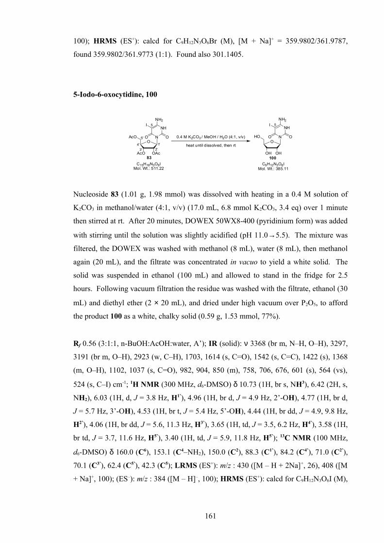
100); HRMS (ES+): calcd for C9H12N3O6Br (M), [M + Na]+ = 359.9802/361.9787,
found 359.9802/361.9773 (1:1). Found also 301.1405.
5-Iodo-6-oxocytidine, 100
C15H18N3O9IMol. Wt.: 511.22
NH
NH2
ONO
OAcAcO
AcO O
5
1'
5'
4'
3' 2'
83
I
0.4 M K2CO3 / MeOH / H2O (4:1, v/v)
heat until dissolved, then rt
NH
NH2
ONO
OHOH
HO O
5
100
I
C9H12N3O6IMol. Wt.: 385.11
Nucleoside 83 (1.01 g, 1.98 mmol) was dissolved with heating in a 0.4 M solution of
K2CO3 in methanol/water (4:1, v/v) (17.0 mL, 6.8 mmol K2CO3, 3.4 eq) over 1 minute
then stirred at rt. After 20 minutes, DOWEX 50WX8-400 (pyridinium form) was added
with stirring until the solution was slightly acidified (pH 11.0→5.5). The mixture was
filtered, the DOWEX was washed with methanol (8 mL), water (8 mL), then methanol
again (20 mL), and the filtrate was concentrated in vacuo to yield a white solid. The
solid was suspended in ethanol (100 mL) and allowed to stand in the fridge for 2.5
hours. Following vacuum filtration the residue was washed with the filtrate, ethanol (30
mL) and diethyl ether (2 × 20 mL), and dried under high vacuum over P2O5, to afford
the product 100 as a white, chalky solid (0.59 g, 1.53 mmol, 77%).
Rf 0.56 (3:1:1, n-BuOH:AcOH:water, A’); IR (solid): ν 3368 (br m, N–H, O–H), 3297,
3191 (br m, O–H), 2923 (w, C–H), 1703, 1614 (s, C=O), 1542 (s, C=C), 1422 (s), 1368
(m, O–H), 1102, 1037 (s, C=O), 982, 904, 850 (m), 758, 706, 676, 601 (s), 564 (vs),
524 (s, C–I) cm-1; 1H NMR (300 MHz, d6-DMSO) δ 10.73 (1H, br s, NH3), 6.42 (2H, s,
NH2), 6.03 (1H, d, J = 3.8 Hz, H1’), 4.96 (1H, br d, J = 4.9 Hz, 2’-OH), 4.77 (1H, br d,
J = 5.7 Hz, 3’-OH), 4.53 (1H, br t, J = 5.4 Hz, 5’-OH), 4.44 (1H, br dd, J = 4.9, 9.8 Hz,
H2’), 4.06 (1H, br dd, J = 5.6, 11.3 Hz, H3’), 3.65 (1H, td, J = 3.5, 6.2 Hz, H4’), 3.58 (1H,
br td, J = 3.7, 11.6 Hz, H5’), 3.40 (1H, td, J = 5.9, 11.8 Hz, H5’); 13C NMR (100 MHz,
d6-DMSO) δ 160.0 (C6), 153.1 (C4–NH2), 150.0 (C2), 88.3 (C1’), 84.2 (C4’), 71.0 (C2’),
70.1 (C3’), 62.4 (C5’), 42.3 (C5); LRMS (ES+): m/z : 430 ([M – H + 2Na]+, 26), 408 ([M
+ Na]+, 100); (ES–): m/z : 384 ([M – H]–, 100); HRMS (ES+): calcd for C9H12N3O6I (M),
161

[2M + H]+ = 770.9620, found 770.9570, [M + Na]+ = 407.9663, found 407.9654, [M +
H]+ = 385.9844, found 385.9834.
6-Oxocytidine, 101
NH
NH2
ONO
OAcAcO
AcO O
5
1'
5'
4'
3' 2'
75
0.4 M K2CO3 / MeOH / H2O (4:1, v/v)
heat until dissolved, then rt
NH
NH2
ONO
OHOH
HO O
5
101
C15H19N3O9Mol. Wt.: 385.33
C9H13N3O6Mol. Wt.: 259.22
Nucleoside 75 (1.55 g, 4.03 mmol) was dissolved with heating in a 0.4M solution of
K2CO3 in methanol/water (4:1 v/v) (40.0 mL, 16.0 mmol K2CO3, 4.0 eq) over 3 minutes,
then stirred at rt for 5 minutes. DOWEX 50WX8-400 (pyridinium form) was added
with stirring until the solution was slightly acidified (pH 12.0→5.5). The mixture was
filtered, the DOWEX washed with the filtrate, methanol/water (1:1 v/v, 60 mL) then
water (20 mL), and the combined filtrate was concentrated in vacuo, co-evaporating
with methanol/toluene (×5) to yield a sticky white foam. The residue was suspended in
isopropanol (50 mL) and allowed to stand in the fridge overnight. The mixture was
vacuum filtered, washed with ice-cold isopropanol (25 mL) and diethyl ether (2 × 50
mL), and dried under high vacuum over P2O5, to afford the product 101 as a powdery
white solid (0.91 g) containing 37.7 mol % potassium acetate (recalculated yield - 2.52
mmol, 71%).
Rf 0.13 (8:2:1, CH2Cl2/methanol/acetic acid, A’), 0.11 (1:4, methanol/CH2Cl2, A’); IR
(solid): ν 3334 (br w, O–H, N–H), 3191 (br w, O–H), 1704 (m, C=O), 1603 (s, C=O),
1404 (br m, O–H), 1290 (m), 1200 (w), 1098 (m), 1020 (br s, C–O), 896, 781, 768 (m),
640 (br s), 533 (vs) cm-1; 1H NMR (400 MHz, d6-DMSO) δ 7.18 (2H, br s, NH2), 6.06
(1H, d, J = 4.0 Hz, H1’), 4.47 (1H, dd, J = 4.3, 5.8 Hz, H2’), 4.44 (1H, s, H5), 4.08 (1H, t,
J = 6.0 Hz, H3’), 3.65 (1H, td, J = 2.8, 8.5 Hz, H4’), 3.56 (1H, dd, J = 2.9, 11.7 Hz, H5’),
3.40 (1H, dd, J = 5.4, 11.7 Hz, H5’); 13C NMR (100 MHz, d6-DMSO) δ 163.3 (C6),
156.8 (C4–NH2), 152.7 (C2), 86.8 (C1’), 84.1 (C4’), 73.7 (C5), 71.1 (C2’), 70.3 (C3’), 62.5
(C5’); LRMS (ES+) m/z: 282 ([M + Na]+, 100); (ES–) m/z: 356 ([M + H3PO4 – H]–, 53),
258 ([M – H]–, 100); HRMS (ES+): calcd for C9H13N3O6 (M), [3M + Na]+ = 800.2311,
162

found 800.2285, [2M + Na]+ = 541.1506, found 541.1497, [M + Na]+ = 282.0697, found
282.0700.
Analytical results consistent with reported data.71,248
5-Chloro-6-oxocytidine, 108
C15H18N3O9ClMol. Wt.: 419.77
C9H12N3O6ClMol. Wt.: 293.66
NH
NH2
ONO
OAcAcO
AcO O
5
1'
5'
4'
3' 2'
109
Cl
0.4 M K2CO3 / MeOH / H2O (4:1, v/v)
heat until dissolved, then rt
NH
NH2
ONO
OHOH
HO O
5
108
Cl
Nucleoside 109 (1.42 g, 3.38 mmol) was dissolved with heating in a 0.4 M solution of
K2CO3 in methanol/water (4:1 v/v) (33.9 mL, 13.5 mmol K2CO3, 4.0 eq) over 3 minutes
then stirred at rt. After 5 minutes, DOWEX 50WX8-400 (pyridinium form) was added
with stirring, until the solution was slightly acidified (pH 12.0→5.5). The mixture was
filtered, the DOWEX washed with methanol/water (1:1 v/v, 2 × 50 mL) then water (50
mL), and the combined filtrate was concentrated in vacuo to dryness. The residue was
suspended in ethyl acetate (60 mL) and allowed to stand in the fridge overnight. The
mixture was vacuum filtered, washed with ethyl acetate (30 mL), ethyl
acetate/isopropanol (2:1 v/v, 30 mL) and diethyl ether (2 × 30 mL), and dried under
high vacuum over P2O5, to afford a powdery white solid (1.24 g).
The solid was retreated with 0.4 M solution of K2CO3 in methanol/water (4:1 v/v) (12.7
mL, 5.08 mmol K2CO3, 1.5 eq) for 15 minutes, then DOWEX 50WX8-400 (pyridinium
form), and concentrated in vacuo to dryness. Following suspension in isopropanol (35
mL) overnight, filtration, washing and drying under high vacuum, over P2O5, the
product 108 was afforded as a white powdery solid (1.07 g) containing. 35.6 mol %
potassium acetate and 1.8 mol % succinimide (recalculated yield - 3.05 mmol, 90%).
Rf 0.52 (3:1:1, n-propanol/conc. aq NH3/water, A’); IR (solid) ν 3306 (br w, O–H, N–
H), 3213 (br w, O–H), 2980 (br w, C–H), 1714 (m, C=O), 1613 (br vs, C=O), 1434 (m),
1322, 1267 (w), 1113 (s), 1051, 1023 (s, C–O), 962, 897, 861 (m), 756 (s), 658 (s, C–
Cl), 557 (s) cm-1; 1H NMR (400 MHz, d6-DMSO) δ 6.69 (2H, v br s, NH2), 6.12 (1H, d,
163

J = 4.3 Hz, H1’), 4.73 (3H, extremely br s, OH), 4.53 (1H, dd, J = 4.5, 6.0 Hz, H2’), 4.13
(1H, t, J = 5.8 Hz, H3’), 3.67 (1H, dt, J = 2.8, 4.9 Hz, H4’), 3.56 (1H, dd, J = 2.6, 11.7
Hz, H5’), 3.41 (1H, dd, J = 4.2, 11.6 Hz, H5’); 13C NMR (100 MHz, d6-DMSO) δ 159.8
(C6), 159.2 (C4–NH2), 154.0 (C2), 87.9 (C1’), 84.3 (C4’), 80.5 (C5), 71.1 (C3’), 70.4 (C2’),
62.5 (C5’); LRMS (ES+) m/z: 316/318 ([M + Na]+, 3:1, 100); (ES–) m/z: 585/587/589
([2M – H]–, 3:2:1, 9). 292/294 ([M – H]–, 3:1, 100); HRMS (ES+): calcd for
C15H18N3O9Cl (M), [2M + Na]+ =609.0721/611.0692, found 609.0702 (2:1), [M + Na]+
= 316.0307/318.0277, found 316.0298 (3:1).
5-Chloro-2’,3’,5’-tri-O-acetyl-6-oxocytidine, 109
2) NCS (1.1 eq), AIBN (2 mol %), dist. THF, dark, argon, rt
C15H19N3O9Mol. Wt.: 385.33
NH
NH2
ONO
OAcAcO
AcO O
5
1'
5'
4'
3' 2'
75
C15H18N3O9ClMol. Wt.: 419.77
NH
NH2
ONO
OAcAcO
AcO O
5
109
Cl
1) BTEA-ICl2 71 (1.5 eq), NaHCO3 (3.0 eq), CH2Cl2, MeOH, rt
Method 1: To a stirred solution of 2’,3’,5’-tri-O-acetyl-6-oxocytidine 75 (0.39 g, 1.00
mmol) in CH2Cl2 (7 mL) and methanol (5 mL), was added sodium bicarbonate (0.25 g,
3.02 mmol, 3.0 eq), followed by a solution of iodinating agent benzyltriethylammonium
dichloroiodate 71 (0.59 g, 1.51 mmol, 1.5 eq) in CH2Cl2 (8 mL), and the reaction was
stirred vigorously at rt for 30 minutes. The reaction mixture was filtered and
concentrated in vacuo to yield a brown gum. The residue was redissolved in CH2Cl2 (40
mL), washed with sat. aq sodium thiosulfate (30 mL) and brine (2 × 40 mL), dried
(Na2SO4) and concentrated in vacuo to yield a yellow foam (466 mg). The compound
was twice purified by column chromatography (0-5% methanol/CH2Cl2) to afford, after
drying under high vacuum, the undesired chlorinated product 109 as a white foam (0.11
g, 0.26 mmol, 26% yield).
Method 2: To a stirred suspension of 2’,3’,5’-tri-O-acetyl-6-oxocytidine 75 (1.52 g,
3.95 mmol) and AIBN (13.3 mg, 0.08 mmol, 2.1 mol %) in distilled THF (15.0 mL),
was added NCS (0.56 g, 4.15 mmol, 1.1 eq), and the reaction was stirred in absence of
light, under an argon atmosphere, at rt for 18 hours. The thick white slurry was
dissolved in acetone/methanol (1:1 v/v, 100 mL), and concentrated in vacuo to dryness.
164

Following purification by column chromatography (40-60% acetone/toluene), the
desired product 109 was afforded as a white powdery solid (1.64 g) containing 29.6 mol
% succinimide (recalculated yield - 3.57 mmol, 90%).
Rf 0.37 (5:95, methanol/CH2Cl2, A’), 0.36 (1:1, acetone/toluene, A’); IR (solid): ν 3419,
3307 (w, N–H), 3198, 3140, 3087, 3024 (w), 2909 (w, C–H), 1741, 1718 (s, C=O),
1629 (s, N–H), 1559 (br s, N–H), 1430 (m), 1372 (s, C–H), 1228 (br vs, C–O), 1155
(m), 1044 (s, C–O), 939, 897, 766, 747 (m), 629 (C–Cl), 601 (m) cm-1; 1H NMR (400
MHz, d6-DMSO) δ 10.96 (2H, br s, NH2), 6.15 (1H, br s, H1’), 5.64 (1H, dd, J = 2.5, 6.5
Hz, H2’), 5.49 (1H, t, J = 7.3 Hz, H3’), 4.33 (1H, dd, J = 3.3, 11.8 Hz, H5’), 4.11 (1H,
ddd, J = 3.3, 6.4, 8.0 Hz, H4’), 4.03 (1H, dd, J = 6.3, 11.8 Hz, H5’), 2.07 (3H, s, CH3),
2.03 (3H, s, CH3), 2.00 (3H, s, CH3); 13C NMR (100 MHz, d6-DMSO) δ 170.0, 169.5,
169.3 (COCH3), 158.0 (C6), 150.5 (C4–NH2), 148.7 (C2), 85.6 (C1’), 80.3
(C5–Cl), 77.9 (C4’), 72.5 (C2’), 69.7 (C3’), 62.9 (C5’), 20.4, 20.3, 20.2 (COCH3); LRMS
(ES+) m/z: 861/863/865 ([2M + Na]+, 6:5:2, 11), 442/444 ([M + Na]+, 3:1, 100), 420/422
([M + H]+, 3:1, 26); (ES–) m/z: 418/420 ([M – H]–, 3:1, 100); HRMS (ES+): calcd for
C15H18N3O9Cl (M), [2M + Na]+ = 861.1355/863.1326/865.1296, found 861.1374 (6:4:1),
[M + Na]+ = 442.0624/444.0594, found 442.0627 (3:1).
N-(2-aminoethyl)-bis-N,N-[2-(trifluoroacetamido)ethyl]amine, 110
(2.0 eq), dist. Et3N (1.1 eq)
dist. MeOH, -5 oC, argon
F3C NH
NNH
CF3
NH2
O O
H2NN
NH2
NH2
C6H18N4Mol. Wt.: 146.23
C10H16F6N4O2Mol. Wt.: 338.25
103 110
F3C O
O12
3
4 4
35 5
66
To a stirred solution of tris(2-aminoethyl)amine 103 (0.50 mL, 3.34 mmol) in distilled
methanol (4.0 mL) and distilled Et3N (0.51 mL, 3.66 mmol, 1.1 eq), under an argon
atmosphere, at –5 °C, was added ethyl trifluoroacetate (0.80 mL, 6.72 mmol, 2.0 eq)
dropwise over 30 minutes. The mixture was stirred for a further 2.5 hours, then
concentrated in vacuo and dried under high vacuum co-evaporating with
methanol/toluene. Following purification by column chromatography (20-40% acetone/
ethyl acetate + 0.5% Et3N), bis-trifluoroacetamide 110 was afforded, following drying
under high vacuum, as a viscous, pale yellow oil (422 mg, 1.25 mmol, 37%).
165

Rf 0.21 (2:3, acetone/ethyl acetate + 0.3% Et3N, D’), 0.15 (1:9, methanol/CH2Cl2 +
0.3% Et3N, D’), 1H NMR (300 MHz, d6-DMSO) δ 9.12 (2H, extremely br s,
NHCOCF3), 3.25 (4H, t, J = 6.4 Hz, H3), 3.18 (2H, app. t, J = 7.0 Hz, H2), 2.65 (4H, q,
J = 6.6 Hz, H4), 2.59 (2H, t, J = 6.6 Hz, H1), 1.88 (2H, t, J = 1.3 Hz, NH2); LRMS
(ES+) m/z: 379 (100), 339 ([M + H]+, 13); HRMS (ES+): calcd for C10H16N4O2F6 (M),
[M + H]+ = 339.1250, found 339.1258. Found also 379.1568.
Bis-N,N-[2-(trifluoroacetamido)ethyl]ammonium trifluoroacetate, 111182,192
(3.5 eq), dist. H2O (1.2 eq)
MeCN, reflux, argon
F3C NH
H2+
NNH
CF3
O O
H2N
HN
NH2
C6H18N4Mol. Wt.: 146.23
127 111
F3C O
O
1
2 2
3 34
CF3CO2-
C10H12N3O4F9Mol. Wt.: 409.21
To a stirred solution of diethylene triamine 127 (3.20 mL, 29.6 mmol) in HPLC grade
acetonitrile (50.0 mL), was added distilled water (0.64 mL, 35.5 mmol, 1.2 eq),
followed by slow addition of ethyl trifluoroacetate (12.3 mL, 104 mmol, 3.5 eq). The
solution was heated at reflux, under argon pressure at 105 °C for 16 hours. The reaction
mixture was concentrated in vacuo and dried under high vacuum co-evaporating with
methanol (2 × 50 mL). The syrup was soaked in CH2Cl2 (100 mL) overnight, then
suspended and the resulting ‘gel’ was filtered. The solid was washed with CH2Cl2 (2 ×
100 mL) and dried under high vacuum over P2O5, to afford the product salt 111 as an
off-white, papery solid (11.3 g, 27.6 mmol, 93%).
Rf 0.44 (1:1, acetone/ethyl acetate + 0.3% Et3N, C’), 0.21 (3:7, acetone/CH2Cl2 + 0.3%
Et3N, C’), 0.31 (1:1, acetone/CH2Cl2 + 0.3% Et3N, C’); IR (solid): ν 3282 (br w, N–H,
O-H), 3118 (w, N–H), 2838 (w, N+–H), 1717, 1673 (s, C=O), 1558 (m, N–H, C=O),
1445 (w, C–H), 1175 (s, C–F), 1142 (vs, C–F), 1015, 837, 797 (m), 724 (s, CH2), 694
(br m, C–F) cm-1; 19F NMR (282 MHz, d6-DMSO) δ -73.5 (CF3CO2–), -74.2
(NHCOCF3); 1H NMR (400 MHz, d6-DMSO) δ 9.60 (2H, br s, NHCOCF3), 8.80 (2H, v
br s, R2NH2+), 3.50 (4H, br dd, J = 5.0, 9.5Hz, H2), 3.13 (4H, t, J = 6.4 Hz, H1); 13C
NMR (100 MHz, d6-DMSO) δ 159.2 (q, J = 31.6 Hz, CF3CO2–), 157.3 (q, J = 36.5 Hz,
NHCOCF3), 117.5 (q, J = 298.9 Hz, CF3CO2–), 116.2 (q, J = 287.7 Hz, NHCOCF3),
45.8 (C1), 36.3 (C2); LRMS (ES+) m/z: 296 ([M + H]+, 100); (ES–) m/z: 522
([M.CF3CO2H + CF3CO2H – H]–, 7), 408 ([M.CF3CO2H – H]–, 100), 392 ([M + H3PO4 –
166

H]+, 56), 294 ([M – H]–, 59); HRMS (ES+): calcd C8H11N3O2F6 (M), [M + H]+ =
296.0828, found 296.0823.
Analytical results consistent with reported data.192
N-[2-N’-(4-Methoxytritylamino)ethyl]-bis-N,N-(2-ethyl trifluoroacetamide), 116
F3C NH
NNH
HN
O
O
CF3O
i) TAEA 103 (7.0 eq), dist. pyridine, dist. CH2Cl2, argon, 0 oC
C30H32N4O3F6Mol. Wt.: 610.59
ii) ethyl trifluoroacetate (10.0 eq)
dist. CH2Cl2/dist. Et3N (1:1 v/v), argon, rt1
2
3
5
4
6
7
8
9
10
11
8
11
12
1314
15
115
116
Cl
H3CO
C20H17ClOMol. Wt.: 308.80
To a stirred solution of tris(2-aminoethyl)amine (TAEA) 103 (1.79 mL, 11.9 mmol, 7.0
eq) in distilled CH2Cl2 (4.0 mL), under an argon atmosphere at 0 °C, was added very
slowly dropwise in portions, a solution of MMTCl 115 (0.53 g, 1.70 mmol) in distilled
pyridine (3.5 mL), over 5.5 hours. The reaction was quenched with methanol (2 mL),
stirring for 5 minutes, then concentrated in vacuo, co-evaporating with CH2Cl2 (×5) then
under high vacuum, co-evaporating with methanol/toluene (×3). The wet bottle-green
powder was washed with hexane (2 × 50 mL) and CH2Cl2 (4 × 25 mL). The resulting
blue solid was washed a third time with CH2Cl2 (4 × 25 mL) and all combined organic
fractions were concentrated in vacuo and under high vacuum to give a brown/green oil.
The oil was dissolved in CH2Cl2 (100 mL), washed with water (100 mL), the aqueous
was re-extracted (CH2Cl2, 3 × 50 mL), and the organic fraction was dried (Na2SO4), and
concentrated in vacuo and under high vacuum, to afford the crude mono-MMT
protected polyamine, as a pale bottle green gum. (0.61 g).
Rf 0.35 (5:1:1, ethyl acetate/methanol/aq NH3, C’);
To a stirred solution of the green resin (0.40 g) in distilled CH2Cl2 (5.0 mL) and distilled
Et3N (5.00 mL), under an argon atmosphere, at rt, was added, ethyl trifluoroacetate
(1.15 mL, 9.66 mmol, 10.0 eq) dropwise, and the reaction was stirred for 3 hours. The
reaction was diluted with CH2Cl2 (100 mL), washed with water (2 × 50 mL), the
aqueous was re-extracted (CH2Cl2, 50 mL). The combined organic fraction was washed
with sat. aq KCl (50 mL), dried (Na2SO4) and concentrated in vacuo to give a
167

yellow/brown syrup. The compound was twice-purified by column chromatography
(1% methanol/CH2Cl2 + 0.5% Et3N; toluene + 0.5% Et3N → 1.5% methanol/CH2Cl2 +
0.5% Et3N) to afford the product 116 as a pale yellow foam (0.14 g, 0.23 mmol, 20%
over two steps).
Rf 0.53 (3% methanol/CH2Cl2 + 0.3% Et3N, C’, E’); IR (solid) ν 3303 (br w, water),
3057 (br w, N–H), 2952, 2837 (br w, C–H), 1703 (s, C=O), 1607, 1556 (w, Aryl C–H),
1508 (m, Aryl C–H), 1448 (m), 1297 (w), 1248 (m), 1206 (s, C–F), 1148 (br vs, C–F),
1032 (m), 903 (w), 830, 768, 750 (m, Aryl C–H), 703 (s, Aryl C–H), 583 (m) cm-1; 19F
NMR (282 MHz, d6-DMSO) δ –74.1 (COCF3); 1H NMR (400 MHz, d6-DMSO) δ 9.20
(1H, br s, NHCOCF3), 7.38 (4H, d, J = 8.0 Hz, H9), 7.29-7.23 (2H, m, H13), 7.27 (4H, d,
J = 8.5 Hz, H10), 7.16 (2H, t, J = 7.3 Hz, H11), 6.83 (2H, d, J = 9.0 Hz, H14), 3.71 (3H, s,
OCH3), 3.31 (1H, s, NHMMT), 3.19 (4H, br dd, J = 6.3, 10.8 Hz, H2), 2.58 (2H, t, J =
6.5 Hz, H6), 2.48 (4H, t, J = 6.5 Hz, H1), 2.07 (2H, dd, J = 6.0, 12.6 Hz, H5); 13C NMR
(100 MHz, d6-DMSO) δ 157.3 (C15), 156.2 (q, J = 36.0 Hz, COCF3), 146.5 (C8), 138.1
(C12), 129.5 (C13), 128.2 (C9), 127.6 (C10), 125.9 (C11), 115.9 (q, J = 288.2 Hz, COCF3),
113.0 (C14), 69.7 (C7Ar3), 54.9 (OCH3), 54.0 (C6), 52.1 (C1), 41.1 (C5), 37.3 (C2);
LRMS (ES–) m/z: 723 ([M + F3CCO2H – H]–, 38), 672 (21), 609 ([M – H]–, 13), 145
(100); HRMS (ES+): calcd for C30H32N4O3F6 (M), [M + Na]+ = 633.2271, found
633.2274.
Bis-N,N-[2-(trifluoroacetamido)ethyl]amine, 128194,252
H2N
HN
NH2
C6H18N4Mol. Wt.: 146.23
127
(2.3 eq), dist. Et3N (2.5 eq)
CH2Cl2 (distilled), -5 oC, argonF3C N
H
HN
NH
CF3
O O
F3C O
O
1
2 2
3 34
C8H11N3O2F6Mol. Wt.: 295.18
128
To a stirred solution of diethylene triamine 127 (2.00 mL, 18.5 mmol) in distilled
CH2Cl2 (40.0 mL), and distilled Et3N (6.45 mL, 46.3 mmol, 2.5 eq), under an argon
atmosphere, at -5 °C, ethyl trifluoroacetate (5.07 mL, 42.6 mmol, 2.3 eq), was added
dropwise over 1 hour. After stirring for a further 1¼ hours, the reaction mixture was
concentrated in vacuo and redissolved in argon-degassed CH2Cl2 (100 mL). The
solution was washed with degassed sat. aq KCl (2 × 50 mL), and the combined aqueous
fraction was re-extracted (degassed CH2Cl2, 2 × 30 mL). The combined CH2Cl2
168

fractions were dried (Na2SO4), concentrated in vacuo and dried under high vacuum, to
afford the product bis-trifluoroacetamide 128, as an off-white, slightly air-sensitive
solid (5.13 g, 17.4 mmol, 94%).
Rf 0.27 (3:7, acetone/CH2Cl2 + 0.3% Et3N, C’), 0.35 (1:1, acetone/CH2Cl2 + 0.3% Et3N,
C’); IR (solid): ν 3287 (w, N–H), 2945, 2850 (w, C–H), 1704 (s, C=O), 1563 (m, N–H,
C=O), 1435 (m, C–H), 1349 (w), 1183, 1140 (br vs, C–F), 950 (w), 866, 843 (m), 724
(s, CH2), 681 (br m, C–F) cm-1; 19F NMR (282 MHz, d6-DMSO) δ -74.1 (COCF3); 1H
NMR (400 MHz, d6-DMSO) δ 9.25 (2H, v br s, NHCOCF3), 3.24 (4H, t, J = 6.4 Hz,
H2), 2.64 (4H, t, J = 6.5 Hz, H1); 13C NMR (100 MHz, d6-DMSO) δ 156.3 (q, J = 36.0
Hz, NHCOCF3), 115.9 (q, J = 287.7 Hz, NHCOCF3), 47.1 (C1), 39.3 (C2); LRMS (ES+)
m/z: 296 ([M + H]+, 100); (ES–) m/z: 408 ([M + CF3CO2H – H]–, 20), 392 ([M + H3PO4
– H]–, 100), 294 ([M – H]–, 77); HRMS (ES+): calcd for C8H11N3O2F6 (M), [M + Na]+ =
318.0648, found 318.0672; [M + H]+ = 296.0828, found 296.0830.
Analytical results consistent with reported data.252
2,2’-Anhydro-6-oxocytidine and 6,2’-anhydro-6-oxocytidine, 141,142
NH
NH2
ONO
OHOH
HO O
5
1'
5'
4'
101
C9H13N3O6Mol. Wt.: 259.22
NH
NH
ONO
OH
HO O
2'
5'
1'
Ph2CO3 (1.1 eq), NaHCO3 (5 mol%)
5N
NH2
ONO
OH
HO O
2'
5'
3'1'
5
+
anh. DMF, argon, 100 oC
2 6
C9H11N3O5Mol. Wt.: 241.20
141 142
To a stirred solution of 6-oxocytidine 101 (0.20 g, 0.60 mmol) in anhydrous DMF (2.50
mL), under an argon atmosphere, was added diphenyl carbonate (0.14 g, 0.66 mmol, 1.1
eq) and sodium bicarbonate (3.0 mg, 0.04 mmol, 6.0 mol %) and the mixture was stirred
at 100 °C for 3 hours. The reaction mixture was allowed to cool to rt, then diethyl ether
(35 mL) was slowly added with rapid stirring, and the suspension was stirred for 30
minutes. The ether was decanted, and the precipitate was washed with ether (2 × 15
mL) and dried under high vacuum to afford the crude product as a hygroscopic,
powdery solid. The solid was redissolved in water/methanol (2:1, v/v, 30 mL) and
concentrated in vacuo and under high vacuum. The gum was redissolved in DMF (2
mL) and diethyl ether (25 mL) was added slowly with vigorous stirring. After stirring
169

for 30 minutes, the ether was decanted, the solid was washed with diethyl ether (2 × 75
mL) and dried in vacuo and under high vacuum to give the product as an isomeric
mixture 141,142 (ca. 1:1), as an off-white chalky solid (0.14 g, 0.59 mmol, 98%).
Rf 0.13, 0.23 (5:1:1, ethyl acetate/methanol/conc. aq NH3, A’); LRMS (ES+) m/z: 280
([M + Na]+, 24), 264 ([M + Na]+, 100).
3-N-[2’-Deoxy-5’-O-(4,4’-dimethoxytrityl)-β-D-ribofuranosyl]-6-[3-
(trifluoroacetamido)phenyl]-(2,3H)-furano[2,3-d]pyrimidine-2-one-3’-O-(2-O-
cyanoethyl-N,N-diisopropyl) phosphoramidite, 145206
(1.2 eq), dist. DIPEA (2.5 eq)
dist. THF, ARGON, rt
N
N
ONO
O
O
MeO
MeO
O
HN
P
N
OCN
PO
ClCN
C49H51N5O9F3PMol. Wt.: 941.93
C40H34N3O8F3Mol. Wt.: 741.71
N
ONO
OH
O
MeO
MeO
O
HN
4
5
6
2'
5'8
9
12
13
16
12
1''
3''
155
OF3C
70
145
4
5
6
2'
5'8
9
12
13
16
12
1''
3''
17
18
19 20
F3CO
To a solution of nucleoside 155 (0.49 g, 0.66 mmol) in distilled THF (4.0 mL) and
distilled DIPEA (0.29 mL, 1.67 mmol, 2.5 eq), strictly under an argon atmosphere and
excluding moisture, was added chloro-phosphitylating reagent 70 (0.18 mL, 0.81 mmol,
1.2 eq) dropwise, and the reaction was stirred at rt for 80 minutes. The reaction was
diluted with argon-degassed ethyl acetate (40 mL), then washed, under an argon
atmosphere, with degassed sat aq KCl (3 × 10 mL). The organic fraction was dried
(Na2SO4), the inorganics were washed with degassed ethyl acetate (3 × 10 mL) and the
combined fraction was transferred under argon and concentrated in vacuo to give a pale
yellow residue. Following purification by column chromatography (degassed 30%
ethyl acetate/CH2Cl2 + 0.5% Et3N), on silica gel pre-equilibrated with Et3N, under argon
pressure, and after drying under high vacuum, the product phosphoramidite 145 was
afforded, as a diastereomeric mixture (ca. 3:2), as an air-sensitive, off-white foam (0.40
g, 0.42 mmol, 64%).
170
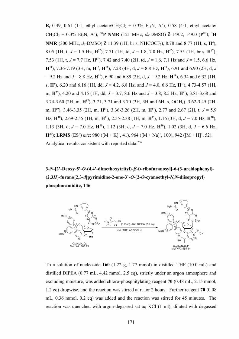
Rf 0.49, 0.61 (1:1, ethyl acetate/CH2Cl2 + 0.3% Et3N, A’), 0.58 (4:1, ethyl acetate/
CH2Cl2 + 0.3% Et3N, A’); 31P NMR (121 MHz, d6-DMSO) δ 149.2, 149.0 (PIII); 1H
NMR (300 MHz, d6-DMSO) δ 11.39 (1H, br s, NHCOCF3), 8.78 and 8.77 (1H, s, H4),
8.05 (1H, t, J = 1.5 Hz, H2”), 7.71 (1H, td, J = 1.8, 7.0 Hz, H4”), 7.55 (1H, br s, H6”),
7.53 (1H, t, J = 7.7 Hz, H5”), 7.42 and 7.40 (2H, td, J = 1.6, 7.1 Hz and J = 1.5, 6.6 Hz,
H14), 7.36-7.19 (3H, m, H15, H16), 7.28 (4H, d, J = 8.8 Hz, H10), 6.91 and 6.90 (2H, d, J
= 9.2 Hz and J = 8.8 Hz, H11), 6.90 and 6.89 (2H, d, J = 9.2 Hz, H11), 6.34 and 6.32 (1H,
s, H5), 6.20 and 6.16 (1H, dd, J = 4.2, 6.8 Hz, and J = 4.0, 6.6 Hz, H1’), 4.73-4.57 (1H,
m, H3’), 4.20 and 4.15 (1H, dd, J = 3.7, 8.6 Hz and J = 3.8, 8.5 Hz, H4’), 3.81-3.68 and
3.74-3.60 (2H, m, H17), 3.71, 3.71 and 3.70 (3H, 3H and 6H, s, OCH3), 3.62-3.45 (2H,
m, H19), 3.46-3.35 (2H, m, H5’), 3.36-3.26 (2H, m, H5’), 2.77 and 2.67 (2H, t, J = 5.9
Hz, H18), 2.69-2.55 (1H, m, H2’), 2.55-2.38 (1H, m, H2’), 1.16 (3H, d, J = 7.0 Hz, H20),
1.13 (3H, d, J = 7.0 Hz, H20), 1.12 (3H, d, J = 7.0 Hz, H20), 1.02 (3H, d, J = 6.6 Hz,
H20); LRMS (ES+) m/z: 980 ([M + K]+, 41), 964 ([M + Na]+, 100), 942 ([M + H]+, 52).
Analytical results consistent with reported data.206
3-N-[2’-Deoxy-5’-O-(4,4’-dimethoxytrityl)-β-D-ribofuranosyl]-6-(3-ureidophenyl)-
(2,3H)-furano[2,3-d]pyrimidine-2-one-3’-O-(2-O-cyanoethyl-N,N-diisopropyl)
phosphoramidite, 146
N
ONO
OH
O
MeO
MeO
O
HN
C39H36N4O8Mol. Wt.: 688.73
H2NO
24
4a
5
6
7a
2'
5'89
12
13
16
12
1''
3''
160
(1.2 eq), dist. DIPEA (2.5 eq)
dist. THF, ARGON, rt
N
N
ONO
O
O
MeO
MeO
O
HN
P
N
OCN
P O
ClCN 24
5
6
2'
5'89
12
13
16
12
1''
3''
146
17
18
1920
C48H53N6O9PMol. Wt.: 888.94
H2NO
70
To a solution of nucleoside 160 (1.22 g, 1.77 mmol) in distilled THF (10.0 mL) and
distilled DIPEA (0.77 mL, 4.42 mmol, 2.5 eq), strictly under an argon atmosphere and
excluding moisture, was added chloro-phosphitylating reagent 70 (0.48 mL, 2.15 mmol,
1.2 eq) dropwise, and the reaction was stirred at rt for 2 hours. Further reagent 70 (0.08
mL, 0.36 mmol, 0.2 eq) was added and the reaction was stirred for 45 minutes. The
reaction was quenched with argon-degassed sat aq KCl (1 ml), diluted with degassed
171

ethyl acetate (60 mL), then washed, under an argon atmosphere, with degassed sat aq
KCl (2 × 20 mL). The organic fraction was dried (Na2SO4), the inorganics were washed
with degassed ethyl acetate (2 × 10 mL) and the combined organic fraction was
transferred under argon and concentrated in vacuo to give a yellow gum. Following
purification by column chromatography (degassed 65% acetone/CH2Cl2 + 1.0% Et3N),
on silica gel pre-equilibrated with Et3N, under argon pressure, then precipitation at rt
from distilled CH2Cl2 (3 mL) into degassed hexane (150 mL), under an argon
atmosphere, the desired product 146 was afforded as a diastereomeric mixture (ca. 2:3),
as an air-sensitive, powdery white solid (1.08 g, 1.22 mmol, 69%).
Rf 0.33 (65:35, acetone/CH2Cl2 + 0.3% Et3N, A’), 0.44 (9:1, acetone/CH2Cl2 + 0.3%
Et3N, A’); 31P NMR (121 MHz, d6-DMSO) δ 149.3, 149.1 (PIII), 15.1
(HO(H)PV(O)OCH2CH2CN); 1H NMR (400 MHz, d6-DMSO) δ 8.75 and 8.74 (1H, s,
H4), 8.74 (1H, br s, NHCONH2), 7.90 (1H, br s, H2”), 7.42 and 7.40 (2H, d, J = 8.5 Hz
and J = 9.0 Hz, H14), 7.38-7.33 (1H, m, H4”), 7.34 (2H, d, J = 7.0 Hz, H15), 7.34-7.27
(2H, m, H16, H5”), 7.30 and 7.27 (4H, d, J = 9.0 Hz, H10), 7.24 (1H, ddd, J = 1.8, 3.3, 6.5
Hz, H6”), 6.91, 6.91 and 6.90 (2H, 2H and 4H, d, J = 9.0 Hz, H11), 6.21 and 6.17 (1H,
dd, J = 4.5, 6.5 Hz, H1’), 6.20 and 6.18 (1H, s, H5), 5.93 (2H, br s, CONH2), 4.69 and
4.65 (1H, td, J = 6.4, 13.1 Hz and J = 6.1, 12.5 Hz, H3’), 4.19 and 4.15 (1H, td, J = 3.5,
5.0 Hz, H4’), 3.81-3.70 and 3.71-3.62 (2H, m, H17), 3.72, 3.72 and 3.71 (3H, 3H and 6H,
s, OCH3), 3.63-3.47 (2H, m, H19), 3.46 and 3.43 (1H, dd, J = 3.4, 10.9 Hz and 3.4, 11.4
Hz, H5’), 3.43-3.38 and 3.39 (1H, m and dd, J = 2.8, 12.1 Hz, H5’), 2.77 and 2.67 (2H, t,
J = 6.0 Hz, H18), 2.64 and 2.60 (1H, td, J = 6.4, 12.8 Hz and J = 6.3, 12.4 Hz, H2’), 2.47-
2.39 (1H, m, H2’), 1.15 (3H, d, J = 6.8 Hz, H20), 1.13 (3H, d, J = 6.8 Hz, H20), 1.12 (3H,
d, J = 6.8 Hz, H20), 1.02 (3H, d, J = 6.8 Hz, H20); LRMS (ES+) m/z: 927 ([M + K]+, 9),
911 ([M + Na]+, 28), 889 ([M + H]+, 17), 618 (100).
172

3-N-[2’-Deoxy-5’-O-(4,4’-dimethoxytrityl)-β-D-ribofuranosyl]-6-(3-
acetamidophenyl)-(2,3H)-furano[2,3-d]pyrimidine-2-one-3’-O-(2-O-cyanoethyl-
N,N-diisopropyl) phosphoramidite, 147
N
ONO
OH
O
MeO
MeO
O
HN
O
24
4a5
6
7a
2'
5'89
12
13
16
12
1''
3''
163
(1.2 eq), dist. DIPEA (2.5 eq)
dist. THF, ARGON, rt
N
N
ONO
O
O
MeO
MeO
O
HN
P
N
OCN
P O
ClCN
24
5
6
2'
5'89
12
13
16
12
1''
3''
147
17
18
1920
O
70
C40H37N3O8Mol. Wt.: 687.74 C49H54N5O9P
Mol. Wt.: 887.95
To a solution of nucleoside 163 (1.00 g, 1.46 mmol) in distilled THF (8.0 mL) and
distilled DIPEA (0.64 mL, 3.67 mmol, 2.5 eq), strictly under an argon atmosphere and
excluding moisture, was added chloro-phosphitylating reagent 70 (0.39 mL, 1.75 mmol,
1.2 eq) dropwise, and the reaction was stirred at rt for 2½ hours. The reaction was
quenched with argon-degassed sat aq KCl (2 ml) stirring for 5 minutes, diluted with
degassed ethyl acetate (100 mL), then washed, under an argon atmosphere, with
degassed sat aq KCl (2 × 20 mL). The organic fraction was dried (Na2SO4), the
inorganics were washed with degassed ethyl acetate (2 × 15 mL) and the combined
fraction was transferred under argon and concentrated in vacuo to give a pale yellow
foam. Following purification by column chromatography (degassed 10-60% acetone/
CH2Cl2 + 0.5% Et3N), on silica gel pre-equilibrated with Et3N, under argon pressure,
then precipitation twice from distilled CH2Cl2 (2 mL) into degassed hexane (200/250
mL) at –78 °C/rt, under an argon atmosphere, the desired phosphoramidite 147 was
afforded as a diastereomeric mixture (ca. 2:3), as an air-sensitive, pale cream foam
(0.89 g) containing 18.9 mol % hydrolysed phosphitylating reagent (recalculated yield -
0.97 mmol, 66%).
Rf 0.42, 0.50 (2:3, acetone/CH2Cl2 + 0.3% Et3N, A’), 0.21, 0.30 (1:9, acetone/CH2Cl2 +
0.3% Et3N, A’); 31P NMR (121 MHz, d6-DMSO) δ 149.3, 149.0 (PIII), 15.1
(HO(H)PV(O)OCH2CH2CN); 1H NMR (400 MHz, d6-DMSO) δ 10.10 (1H, s,
NHCOCH3), 8.76 and 8.75 (1H, s, H4), 8.02 (1H, s, H2”), 7.59 (1H, br d, J = 7.5 Hz,
H4”), 7.44-7.40 (1H, m, H5”), 7.42-7.37 (2H, m, H14), 7.37-7.30 (2H, m, H15, H6”), 7.30
and 7.27 (4H, d, J = 8.5 Hz and J = 9.0 Hz, H10), 7.30-7.25 (1H, m, H16), 6.91, 6.91 and
173
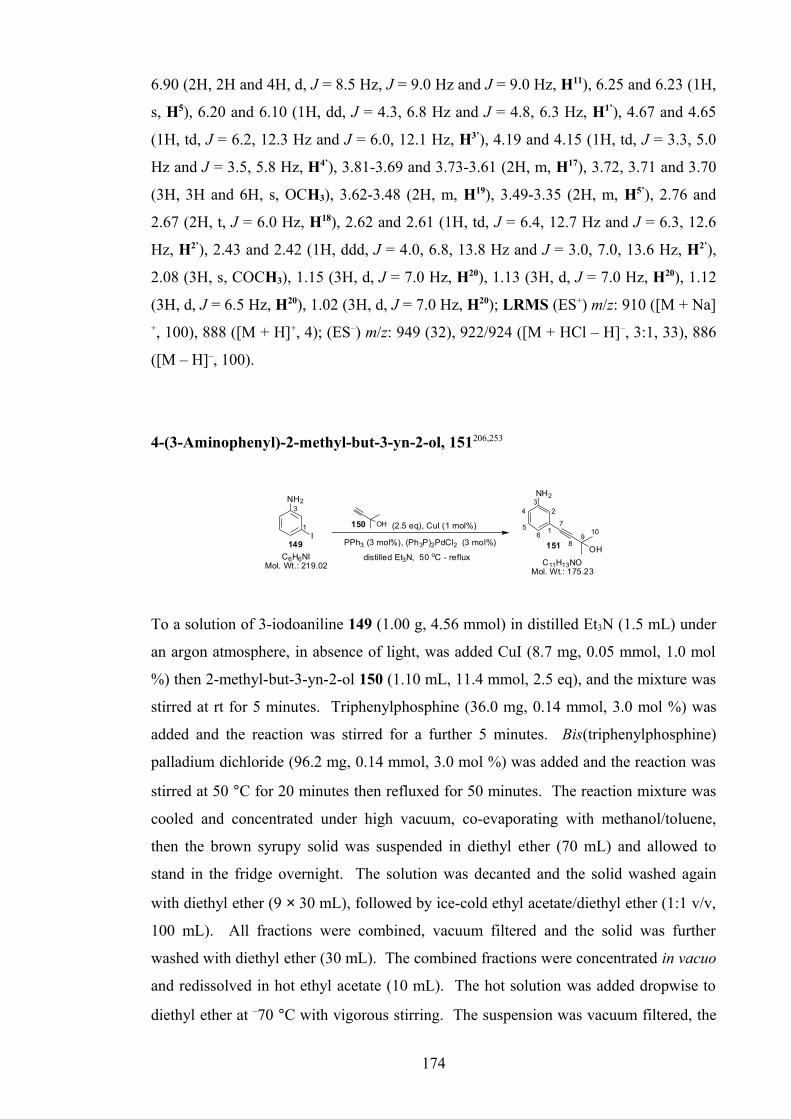
6.90 (2H, 2H and 4H, d, J = 8.5 Hz, J = 9.0 Hz and J = 9.0 Hz, H11), 6.25 and 6.23 (1H,
s, H5), 6.20 and 6.10 (1H, dd, J = 4.3, 6.8 Hz and J = 4.8, 6.3 Hz, H1’), 4.67 and 4.65
(1H, td, J = 6.2, 12.3 Hz and J = 6.0, 12.1 Hz, H3’), 4.19 and 4.15 (1H, td, J = 3.3, 5.0
Hz and J = 3.5, 5.8 Hz, H4’), 3.81-3.69 and 3.73-3.61 (2H, m, H17), 3.72, 3.71 and 3.70
(3H, 3H and 6H, s, OCH3), 3.62-3.48 (2H, m, H19), 3.49-3.35 (2H, m, H5’), 2.76 and
2.67 (2H, t, J = 6.0 Hz, H18), 2.62 and 2.61 (1H, td, J = 6.4, 12.7 Hz and J = 6.3, 12.6
Hz, H2’), 2.43 and 2.42 (1H, ddd, J = 4.0, 6.8, 13.8 Hz and J = 3.0, 7.0, 13.6 Hz, H2’),
2.08 (3H, s, COCH3), 1.15 (3H, d, J = 7.0 Hz, H20), 1.13 (3H, d, J = 7.0 Hz, H20), 1.12
(3H, d, J = 6.5 Hz, H20), 1.02 (3H, d, J = 7.0 Hz, H20); LRMS (ES+) m/z: 910 ([M + Na]+, 100), 888 ([M + H]+, 4); (ES–) m/z: 949 (32), 922/924 ([M + HCl – H]–, 3:1, 33), 886
([M – H]–, 100).
4-(3-Aminophenyl)-2-methyl-but-3-yn-2-ol, 151206,253
NH2
OH
7
89
101
23
4
56
NH2
I
OH (2.5 eq), CuI (1 mol%)
distilled Et3N, 50 oC - reflux
PPh3 (3 mol%), (Ph3P)2PdCl2 (3 mol%)
C6H6NIMol. Wt.: 219.02 C11H13NO
Mol. Wt.: 175.23
151149
1501
3
To a solution of 3-iodoaniline 149 (1.00 g, 4.56 mmol) in distilled Et3N (1.5 mL) under
an argon atmosphere, in absence of light, was added CuI (8.7 mg, 0.05 mmol, 1.0 mol
%) then 2-methyl-but-3-yn-2-ol 150 (1.10 mL, 11.4 mmol, 2.5 eq), and the mixture was
stirred at rt for 5 minutes. Triphenylphosphine (36.0 mg, 0.14 mmol, 3.0 mol %) was
added and the reaction was stirred for a further 5 minutes. Bis(triphenylphosphine)
palladium dichloride (96.2 mg, 0.14 mmol, 3.0 mol %) was added and the reaction was
stirred at 50 °C for 20 minutes then refluxed for 50 minutes. The reaction mixture was
cooled and concentrated under high vacuum, co-evaporating with methanol/toluene,
then the brown syrupy solid was suspended in diethyl ether (70 mL) and allowed to
stand in the fridge overnight. The solution was decanted and the solid washed again
with diethyl ether (9 × 30 mL), followed by ice-cold ethyl acetate/diethyl ether (1:1 v/v,
100 mL). All fractions were combined, vacuum filtered and the solid was further
washed with diethyl ether (30 mL). The combined fractions were concentrated in vacuo
and redissolved in hot ethyl acetate (10 mL). The hot solution was added dropwise to
diethyl ether at –70 °C with vigorous stirring. The suspension was vacuum filtered, the
174

solid washed with very cold diethyl ether (150 mL) and the filtrate was concentrated in
vacuo. The resultant syrup was dissolved in CH2Cl2 (70 mL) and washed with water (3
× 100 mL). The aqueous fraction was re-extracted (CH2Cl2, 200 mL), and combined
CH2Cl2 fractions were washed with sat aq KCl (100 mL), dried (Na2SO4) and
concentrated in vacuo to give an orange/brown gum. Following purification by column
chromatography (0–30% CH2Cl2/diethyl ether), and drying under high vacuum, the
product alkyne 151 was isolated as a pale yellow solid (0.43 g, 2.43 mmol, 53%).
Rf 0.29 (1:1, ethyl acetate/hexane, A’), 0.31 (1:1, CH2Cl2/diethyl ether, A’), 0.04
(CH2Cl2, A’), 0.36 (diethyl ether, A’); 1H NMR (300 MHz, d6-DMSO) δ 6.97 (1H, t, J
= 7.9 Hz, H5), 6.58 (1H, dd, J = 1.3, 2.0 Hz, H2), 6.53 (1H, ddd, J = 1.1, 2.6, 8.1 Hz,
H4), 6.50 (1H, td, J = 1.3, 7.4 Hz, H6), 5.37 (1H, s, OH), 5.14 (2H, br s, NH2), 1.44 (6H,
s, CH3); 13C NMR (75 MHz, d6-DMSO) δ 148.7 (C3–NH2), 129.0 (C5), 122.8 (C1),
118.6 (C6), 116.3 (C2), 114.0 (C4), 94.6 (ArC≡C8R), 81.1 (ArC7≡CR), 63.5
(C(CH3)2OH), 31.7 (CH3); LRMS (ES+): m/z: 277 ([M + Et3N + H]+, 10), 217 ([M +
CH3CN + H]+, 35), 176 ([M + H]+, 37), 102 ([Et3N + H]+, 100).
Analytical results consistent with reported data.253
3-Aminophenyl acetylene (or 3-ethynylaniline), 152206
NH2
OH
7
89
101
23
4
56
C11H13NOMol. Wt.: 175.23
151
KOH (2.0 eq), dry iPrOH
4Å molecular sieves, reflux
NH2
7
8
1
3
C8H7NMol. Wt.: 117.15
152
Potassium hydroxide (0.21 g, 3.80 mmol, 2.3 eq) was dissolved in dry, refluxing
isopropanol (5 mL) with 4Å molecular sieves. Immediately on dissolution, protected
alkyne 151 (0.29 g, 1.63 mmol) was added and the mixture was refluxed for 2½ hours.
Further KOH (0.10 g, 1.82 mmol, 1.1 eq) was added and the reaction was refluxed for 1
hour. The mixture was cooled and concentrated to dryness under high vacuum at rt.
Dichloromethane (40 mL) was added and the mixture was vacuum filtered. The residue
was washed with CH2Cl2 (2 × 20 mL), and the filtrate was concentrated to dryness in
vacuo and under high vacuum to give a tan-coloured solid. Following purification by
column chromatography (0–10% diethyl ether/CH2Cl2), and drying under high vacuum,
175
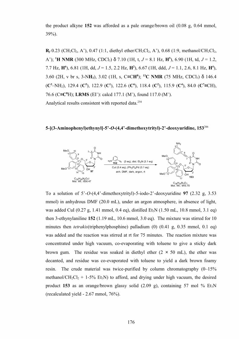
the product alkyne 152 was afforded as a pale orange/brown oil (0.08 g, 0.64 mmol,
39%).
Rf 0.23 (CH2Cl2, A’), 0.47 (1:1, diethyl ether/CH2Cl2, A’), 0.68 (1:9, methanol/CH2Cl2,
A’); 1H NMR (300 MHz, CDCl3) δ 7.10 (1H, t, J = 8.1 Hz, H5), 6.90 (1H, td, J = 1.2,
7.7 Hz, H6), 6.81 (1H, dd, J = 1.5, 2.2 Hz, H2), 6.67 (1H, ddd, J = 1.1, 2.6, 8.1 Hz, H4),
3.60 (2H, v br s, 3-NH2), 3.02 (1H, s, C≡CH8); 13C NMR (75 MHz, CDCl3) δ 146.4
(C3–NH2), 129.4 (C5), 122.9 (C1), 122.6 (C6), 118.4 (C2), 115.9 (C4), 84.0 (C7≡CH),
76.6 (C≡C8H); LRMS (EI+): calcd 177.1 (M+), found 117.0 (M+).
Analytical results consistent with reported data.254
5-[(3-Aminophenyl)ethynyl]-5’-O-(4,4’-dimethoxytrityl)-2’-deoxyuridine, 153206
NH
O
ONO
OH
O
MeO
MeO
NH2
2'
5'
5
NH
O
ONO
OH
O
MeO
MeO
I
2'
5'
5
7
81''
3''
9
10
13
14
17
13
7
8
9
1011
12
13
14 15
11
6
6H2N (3 eq), dist. Et3N (3.1 eq)
CuI (0.4 eq), (Ph3P)4Pd (0.1 eq)
anh. DMF, dark, argon, rt
C30H29N2O7IMol. Wt.: 656.47
C38H35N3O7Mol. Wt.: 645.70
153
97
152
To a solution of 5’-O-(4,4’-dimethoxytrityl)-5-iodo-2’-deoxyuridine 97 (2.32 g, 3.53
mmol) in anhydrous DMF (20.0 mL), under an argon atmosphere, in absence of light,
was added CuI (0.27 g, 1.41 mmol, 0.4 eq), distilled Et3N (1.50 mL, 10.8 mmol, 3.1 eq)
then 3-ethynylaniline 152 (1.19 mL, 10.6 mmol, 3.0 eq). The mixture was stirred for 10
minutes then tetrakis(triphenylphosphine) palladium (0) (0.41 g, 0.35 mmol, 0.1 eq)
was added and the reaction was stirred at rt for 75 minutes. The reaction mixture was
concentrated under high vacuum, co-evaporating with toluene to give a sticky dark
brown gum. The residue was soaked in diethyl ether (2 × 50 mL), the ether was
decanted, and residue was co-evaporated with toluene to yield a dark brown foamy
resin. The crude material was twice-purified by column chromatography (0–15%
methanol/CH2Cl2 + 1-5% Et3N) to afford, and drying under high vacuum, the desired
product 153 as an orange/brown glassy solid (2.09 g), containing 57 mol % Et3N
(recalculated yield - 2.67 mmol, 76%).
176

Rf 0.35 (1:9, methanol/CH2Cl2 + 0.3% Et3N, A’); 0.35 (4:1, ethyl acetate/toluene + 0.3%
Et3N, A’); Mp 116-120 °C (CH2Cl2); IR (solid): ν 3342 (w, O–H, N–H), 3232 (w, O–
H), 3056, 2973, 2931, 2834 (w, C–H), 2171 (w, C≡C), 1689 (br m, C=O), 1632 (s, N–
H), 1596 (s, C=C), 1507 (s, Aryl C=C), 1442 (C–H), 1385 (m, O–H), 1276 (s), 1246
(vs), 1174, 1087, 1030 (s, C–O), 943, 866 (m), 826 (s, C–H), 788 (s), 754, 688 (s, Aryl
C–H), 583, 515 (s) cm-1; 1H NMR (400 MHz, d6-DMSO) δ 11.3 (1H, br s, NH3), 7.97
(1H, s, H6), 7.41 (2H, d, J = 7.5 Hz, H15), 7.32–7.26 (2H, m, H16), 7.30 (2H, d, J = 9.0
Hz, H11), 7.29 (2H, d, J = 9.0 Hz, H11), 7.18 (1H, br t, J = 7.3 Hz, H17), 6.95 (1H, t, J =
7.7 Hz, H5”), 6.86 (2H, d, J = 9.0 Hz, H12), 6.84 (2H, d, J = 9.0 Hz, H12), 6.55 (1H, ddd,
J = 0.6, 2.0, 8.0 Hz, H4”), 6.52 (1H, app. t, J = 1.6 Hz, H2”), 6.33 (1H, br d, J = 7.8 Hz,
H6”), 6.14 (1H, t, J = 6.8 Hz, H1’), 5.31 (1H, br s, 3’-OH), 5.13 (2H, s, NH2), 4.28 (1H,
td, J = 3.3, 6.0 Hz, H3’), 3.95 (1H, td, J = 3.3, 4.8 Hz, H4’), 3.68 (3H, s, OCH3), 3.67
(3H, s, OCH3), 3.24 (1H, dd, J = 5.3, 10.5 Hz, H5’), 3.15 (1H, dd, J = 2.8, 10.5 Hz, H5’),
2.30 (1H, td, J = 6.7, 13.6 Hz, H2’), 2.22 (1H, ddd, J = 3.3, 6.3, 13.6 Hz, H2’); 13C NMR
(100 MHz, d6-DMSO) δ 161.4 (C4), 158.1, 158.0 (C13), 149.3 (C2), 148.5 (C3”), 144.6
(C14), 142.4 (C6), 135.6, 135.3 (C10), 129.7, 129.6 (C11), 128.8 (C5”), 127.8 (C16), 127.6
(C15), 126.6 (C17), 122.4 (C1”), 118.7 (C6”), 116.0 (C2”), 114.4 (C4”), 113.2 (C12-OMe),
98.8 (C5), 93.0 (C8), 85.9 (C4’), 85.8 (C9Ar3), 85.1 (C1’), 80.3 (C7), 70.5 (C3’), 63.7 (C5’),
54.9 (OCH3), 39.9 (C2’); LRMS (ES+): m/z: 1314 ([2M + Na]+, 22), 747 ([M + Et3N +
H]+, 100), 719 (41), 668 ([M + Na]+, 44), 646 ([M + H]+, 41); HRMS (ES+): calcd for
C38H35N3O7 (M), [M + Na]+ = 668.2367, found 668.2375. Found also DMT+: calcd
303.1380, found 303.1387.
Analytical results consistent with reported data.206
3-N-[2’-Deoxy-5’-O-(4,4’-dimethoxytrityl)-β-D-ribofuranosyl]-6-(3-aminophenyl)-
(2,3H)-furano[2,3-d]pyrimidine-2-one, 154206
N
ONO
OH
O
MeO
MeO
O
H2N
24
4a5
6
7a
2'
5'8
9
10
1112
13
14
15 16
12
CuI (1.0 eq)
dist. MeOH/Et3N (7:3 v/v)
50 oC, argon, dark
1''
3''
C38H35N3O7Mol. Wt.: 645.70
NH
O
ONO
OH
O
MeO
MeO
NH2
2'
5'
57
81''
3''
9
10
13
14
17
13
6
153 154
177
To a solution of nucleoside 153 (1.94 g, 2.48 mmol) in distilled methanol/distilled Et3N
(7:3 v/v, 15.0 mL), under an argon atmosphere, in absence of light, was added CuI (0.57
g, 2.99 mmol, 1.0 eq). The reaction mixture was stirred at 50 °C for 17 hours then
cooled to rt and concentrated in vacuo, co-evaporating with CH2Cl2 (2 × 50 mL). The
residue was dissolved/suspended in CH2Cl2 (150 mL) and filtered. The filtrate was
washed with 5% w/v aq Na2EDTA (pH 9, 4 × 50 mL), sat aq KCl (2 × 50 mL), dried
(Na2SO4), concentrated in vacuo and dried under high vacuum to afford the product 154
as an orange/brown glassy foam (1.53 g, 2.37 mmol, 96%) containing 3.5 mol %
uncyclised material 153.
Rf 0.35 (153), 0.34 (154) (1:9, methanol/CH2Cl2 + 0.3% Et3N, A’); 1H NMR (300 MHz,
d6-DMSO) δ 8.70 (1H, s, H4), 7.40 (2H, d, J = 7.5 Hz, H14), 7.34 (2H, t, J = 7.5 Hz,
H15), 7.29 (4H, d, J = 9.0 Hz, H10), 7.31-7.24 (1H, m, H16), 7.13 (1H, t, J = 7.8 Hz, H5”),
6.94-6.88 (1H, m, H2”), 6.92 (2H, d, J = 8.8 Hz, H11), 6.91 (2H, d, J = 9.0 Hz, H11), 6.84
(1H, br d, J = 7.5 Hz, H4”), 6.62 (1H, br dd, J = 1.5, 8.0 Hz, H6”), 6.13 (1H, dd, J = 4.6,
6.4 Hz, H1’), 5.42 (1H, d, J = 4.8 Hz, 3’-OH), 5.32 (2H, br s, NH2), 4.43 (1H, td, J =
5.9, 11.1 Hz, H3’), 4.04-3.99 (1H, m, H4’), 3.72 (3H, s, OCH3), 3.71 (3H, s, OCH3), 3.41
(1H, dd, J = 4.0, 10.8 Hz, H5’), 3.32 (1H, dd, J = 2.8, 10.8 Hz, H5’), 2.46 (1H, td, J =
6.6, 13.1 Hz, H2’), 2.25 (1H, ddd, J = 4.8, 6.5, 13.4 Hz, H2’); 13C NMR (100 MHz, d6-
DMSO) δ 170.9 (C7a), 158.2 (C12–OCH3), 154.5 (C2), 149.2 (C3”–NH2), 144.4 (C13),
137.3 (C4), 135.4 (C9), 135.3 (C6), 135.1 (C9), 129.8, 129.7 (C10), 129.5 (C5”), 128.6
(C1”), 128.0 (C15), 127.8 (C14), 126.8 (C16), 115.1 (C6”), 113.3 (C11), 112.0 (C4”), 109.2
(C2”), 106.7 (C4a), 97.8 (C5), 87.3 (C1’), 86.1 (C8Ar3), 85.9 (C4’), 68.8 (C3’), 62.4 (C5’),
55.0 (OCH3), 41.1 (C2’); LRMS (ES+): m/z: 917 (20), 663 ([M + NH4]+, 100), 646 ([M +
H]+, 13); HRMS (ES+): calcd for C38H35N3O7 (M), [M + Na]+ = 668.2367, found
668.2359. Found also DMT+: calcd 303.1380, found 303.1382.
Analytical results consistent with reported data.206
178

3-N-[2’-Deoxy-5’-O-(4,4’-dimethoxytrityl)-β-D-ribofuranosyl]-6-[3-
(trifluoroacetamido)phenyl]-(2,3H)-furano[2,3-d]pyrimidine-2-one, 155206
(4 x 2.0 eq), DMAP (4 x 1.0 eq)
dist. THF, argon, 80-85oC
C38H35N3O7Mol. Wt.: 645.70
dist. Et3N, 4Å molecular sieves
O
OF3C
C40H34N3O8F3Mol. Wt.: 741.71
N
ONO
OH
O
MeO
MeO
O
H2N
24
4a5
6
7a
2'
5'8
9
10
1112
13
14
15 16
12
1''
3''
154
N
ONO
OH
O
MeO
MeO
O
HN
4
5
6
2'
5'89
12
13
16
12
1''
3''
155
OF3C
To a stirred solution of nucleoside 154 (1.37 g, 2.12 mmol) and N,N-dimethylamino
pyridine (DMAP) (0.26 g, 2.13 mmol, 1.0 eq) in distilled THF (10.0 mL), under an
argon atmosphere, with 4Å molecular sieves, was added distilled Et3N (0.15 mL) then
ethyl trifluoroacetate (0.50 mL, 4.24 mmol, 2.0 eq). The solution was stirred at rt for 2
minutes, then heated to reflux under argon pressure (80 °C). Further DMAP (1.0 eq)
and ethyl trifluoroacetate (2.0 eq) were added after 6 hours and 21½ hours. The
temperature was then raised to 85 °C. Further DMAP (0.5 eq) and ethyl trifluoroacetate
(1.0 eq) were added at 30 hours. At 53 hours, the reaction mixture was cooled to rt,
dissolved in CH2Cl2/methanol (1:1 v/v, 60 mL) and concentrated in vacuo, co-
evaporating with CH2Cl2 (4 × 40 mL) to give an orange/brown solid. Following
purification by column chromatography (70-90% ethyl acetate/CH2Cl2 + 0.5% Et3N),
and drying under high vacuum, the desired product 155 was afforded as an off-white
foam (0.55 g, 0.74 mmol, 35%).
Rf 0.23 (9:1, ethyl acetate/CH2Cl2 + 0.3% Et3N, A’); 0.42 (1:9, methanol/CH2Cl2 + 0.3%
Et3N, A’); 1H NMR (400 MHz, d6-DMSO) δ 11.40 (1H, br s, NHCOCF3), 8.75 (1H, s,
H4), 8.06 (1H, t, J = 1.8 Hz, H2”), 7.72 (1H, td, J = 2.0, 7.3 Hz, H4”), 7.57 (1H, dd, J =
1.7, 7.7 Hz, H6”), 7.53 (1H, t, J = 7.7 Hz, H5”), 7.41 (2H, dd, J = 1.5, 8.1 Hz, H14), 7.34
(2H, t, J = 8.1 Hz, H15), 7.31-7.25 (1H, m, H16), 7.29 (2H, d, J = 8.8 Hz, H10), 7.29 (2H,
d, J = 9.2 Hz, H10), 6.92 (2H, d, J = 9.1 Hz, H11), 6.91 (2H, d, J = 9.2 Hz, H11), 6.33
(1H, s, H5), 6.16 (1H, dd, J = 4.4, 6.6 Hz, H1’), 5.43 (1H, d, J = 4.8 Hz, 3’-OH), 4.43
(1H, td, J = 5.2, 10.5 Hz, H3’), 4.04 (1H, dd, J = 4.5, 8.1, H4’), 3.71 (3H, s, OCH3), 3.70
(3H, s, OCH3), 3.41 (1H, dd, J = 4.0, 10.8 Hz, H5’), 3.32 (1H, dd, J = 3.3, 11.0 Hz, H5’),
2.54-2.44 (1H, m, H2’), 2.27 (1H, ddd, J = 4.6, 6.4, 13.5 Hz, H2’); 13C NMR (100 MHz,
d6-DMSO) δ 170.9 (C7a), 158.2 (C12–OCH3), 154.7 (q, 2JCF = 36.9 Hz, COCF3), 153.6
(C2), 144.4 (C13), 138.2 (C4), 137.4 (C6), 137.3 (C3”–NHR), 135.4, 135.1 (C9), 129.9
179

(C5”), 129.8, 129.7 (C10), 128.8 (C1”), 128.0 (C15), 127.8 (C14), 126.8 (C16), 121.9 (C4”),
121.5 (C6”), 116.7 (C2”), 113.3 (C11), 113.2 (q, 1JCF = 227.4 Hz, COCF3), 106.4 (C4a),
99.6 (C5), 87.6 (C1’), 86.1 (C8Ar3), 86.0 (C4’), 68.8 (C3’), 62.4 (C5’), 54.9 (OCH3), 41.1
(C2’); LRMS (ES+) m/z: 764 ([M + Na]+, 100); (ES–) m/z: 740 ([M – H]–, 100).
Analytical results consistent with reported data.206
3-Ureidophenyl acetylene, 156206
H2N152
C8H7NMol. Wt.: 117.15
13
NH
H2N
O NH2
O(1.1 eq)
dim light, 100 oC, dryC9H8N2O
Mol. Wt.: 160.17
156
O13
7 8157
3-Ethynylaniline 152 (3.00 mL, 26.6 mmol) and dry phenyl carbamate 157 (7.31 g, 53.3
mmol, 2.0 eq) were heated in a sealed tube, under an argon atmosphere, excluding
moisture, in dim light at 90 °C for 12 hours. The mixture was cooled to rt, transferred
using warm methanol/acetone, and dried under high vacuum. Following purification by
column chromatography (5-50% acetone/CH2Cl2), and drying under high vacuum, the
desired urea 156 was afforded, as an off-white, papery solid (2.93 g, 18.3 mmol, 69%).
Rf 0.31 (1:9, methanol/CH2Cl2 + 0.3% Et3N, A’, F’), 0.04 (1:9, acetone/CH2Cl2 + 0.3%
Et3N, A’, F’); IR (solid): ν 3467, 3329 (m, N–H), 3302 (w N–H), 3238(m, N–H), 2100
(w, C≡C), 1649 (vs, C=O), 1586, 1575 (s, Aryl C–H), 1540 (br vs, Aryl C–H), 1473 (s),
1413 (m), 1325 (s), 1284, 1256, 1162 (m), 1120 (br w), 1029 (w), 890 (m, Aryl C–H),
801 (m), 773 (m, Aryl C–H), 683 (s), 643 (br s), 587 (s) cm-1; 1H NMR (300 MHz, d6-
DMSO) δ 8.61 (1H, s, NH), 7.64 (1H, t, J = 1.7 Hz, H2), 7.32 (1H, ddd, J = 1.1, 2.0, 8.2
Hz, H4), 7.22 (1H, t, J = 7.9 Hz, H5), 7.00 (1H, td J = 1.3, 7.3 Hz, H6), 5.89 (2H, s,
NH2), 4.08 (1H, s, C≡CH8); 13C NMR (100 MHz, d6-DMSO) δ 155.9 (CONH2), 140.8
(C3), 129.0 (C5), 124.3 (C6), 121.8 (C1), 120.5 (C2), 118.4 (C4), 83.8 (C7≡CH), 80.0
(C≡C8H); LRMS (ES+) m/z: 343 ([2M + Na]+, 21), 183 ([M + Na]+, 70), 102 ([Et3N +
H]+, 100); (ES–) m/z: 159 ([M – H]–, 100); HRMS (ES+): calcd for C9H8N2O (M), [M +
Na]+ = 183.0529, found 183.0530, [2M + Na]+ = 343.1171, found 343.1170.
Analytical results consistent with reported data.206
180

5-[(3-Ureidophenyl)ethynyl]-5’-O-(4,4’-dimethoxytrityl)-2’-deoxyuridine, 159
NH
H2N
O
C39H36N4O8Mol. Wt.: 688.73
NH
O
ONO
OH
O
MeO
MeO
NH
2'
5'
5
NH
O
ONO
OH
O
MeO
MeO
I
2'
5'
5
7
81''
3''
910
13
14
17
13
78
11
12
15
11
6
6
(3 eq), dist. Et3N (5.0 eq)
CuI (0.4 eq), Pd(Ph3P)4 (0.1 eq)
anh. DMF, dark, argon, rt
C30H29N2O7IMol. Wt.: 656.47 159
97
156
O
H2N
To a solution of 5’-O-(4,4’-dimethoxytrityl)-5-iodo-2’-deoxyuridine 97 (3.01 g, 4.58
mmol) in anhydrous DMF (15.0 mL), under an argon atmosphere, in absence of light,
were added distilled Et3N (3.30 mL, 23.7 mmol, 5.0 eq), CuI (0.35 g, 1.83 mmol, 0.4
eq), then alkyne 156 (2.20 g, 13.7 mmol, 3.0 eq). The mixture was stirred for 15
minutes then tetrakis(triphenylphosphine) palladium (0) (0.53 g, 0.46 mmol, 0.1 eq)
was added and the reaction was stirred at rt for 3 hours. The reaction mixture was
concentrated under high vacuum, co-evaporating with CH3CN to give a dark brown
foam/gum. Following purification by column chromatography (5-8% methanol/CH2Cl2
+ 0.5% Et3N), and drying under high vacuum, the desired product 159 was afforded as a
pale orange foam (2.71 g) containing 34 mol % Et3N (recalculated yield - 3.66 mmol,
80%).
Rf 0.23 (1:9, methanol/CH2Cl2 + 0.3% Et3N, A’), 0.27 (7:3 acetone/CH2Cl2 + 0.3%
Et3N, A’); IR (solid): ν 3348 (w, O–H), 3208, 3067, 2931 (w, N–H), 2832 (w, C–H),
1669 (br vs, C=O, C=C), 1605, 1580 (s, N–H), 1546, 1506, 1456, 1435 (s), 1342, 1302
(m), 1274 (s), 1247 (vs, C–O), 1175 (s), 1088, 1030 (br s, C–O), 826 (s, Aryl C–H),
789, 753, 698, 585 (s) cm-1; 1H NMR (400 MHz, d6-DMSO) δ 11.71 (1H, br s, NH3),
8.57 (1H, s, NHCONH2), 8.02 (1H, s, H6), 7.59 (1H, t, J = 1.8 Hz, H2”), 7.41 (2H, d, J =
7.5 Hz, H15), 7.31-7.26 (1H, m, H16), 7.30 (2H, d, J = 9.0 Hz, H11), 7.29 (2H, d, J = 8.8
Hz, H11), 7.24 (1H, ddd, J = 1.0, 2.1, 8.2 Hz, H4”), 7.17 (1H, br t, J = 7.4 Hz, H17), 7.14
(1H, t, J = 7.9 Hz, H5”), 6.85 (2H, d, J = 8.8 Hz, H12), 6.84 (2H, d, J = 9.0 Hz, H12), 6.65
(1H, td, J = 1.3, 6.5 Hz, H6”), 6.14 (1H, t, J = 6.7 Hz, H1’), 5.90 (2H, s, CONH2), 5.32
(1H, d, J = 4.3 Hz, 3’-OH), 4.29 (1H, td, J = 2.5, 6.5 Hz, H3’), 3.95 (1H, td, J = 3.3, 5.0
Hz, H4’), 3.67 (6H, s, OCH3), 3.25 (1H, dd, J = 5.3, 10.5 Hz, H5’), 3.14 (1H, dd, J = 2.8,
10.5 Hz, H5’), 2.31 (1H, td, J = 6.8, 13.6 Hz, H2’), 2.22 (1H, ddd, J = 3.5, 6.3, 13.6 Hz,
181

H2’); 13C NMR (100 MHz, d6-DMSO) δ 161.4 (C4), 158.1, 158.0 (C13–OCH3), 155.8
(CONH2), 149.3 (C2), 144.7 (C14), 142.8 (C6), 140.6 (C3”–NHR), 135.6, 135.3 (C10),
129.7, 129.6 (C11), 128.7 (C5”), 127.8 (C16), 127.6 (C15), 126.7 (C17), 124.0 (C6”), 122.4
(C1”), 120.0 (C2”), 118.0 (C4”), 113.2 (C12), 98.6 (C5), 92.4 (C8), 86.0 (C4’), 85.8 (C9Ar3),
85.1 (C1’), 81.2 (C7), 70.5 (C3’), 63.7 (C5’), 54.9 (OCH3), 39.9 (C2’); LRMS (ES+) m/z:
711 ([M + Na]+, 100), 303 ([DMT+], 9); HRMS (ES+): calcd for C39H36N4O8 (M), [M +
Na]+ = 711.2425, found 711.2409, [M + NH4]+ = 706.2877, found 706.2856. Found also
301.1413.
3-N-[2’-Deoxy-5’-O-(4,4’-dimethoxytrityl)-β-D-ribofuranosyl]-6-(3-ureidophenyl)-
(2,3H)-furano[2,3-d]pyrimidine-2-one, 160
NH
O
ONO
OH
O
MeO
MeO
NH
2'
5'
57
81''
3''
9
10
13
14
17
13
6
159
O
H2N
N
ONO
OH
O
MeO
MeO
O
HN
CuI (1.1 eq)
dist. MeOH/Et3N (7:3 v/v)
80 oC, argon, dark
C39H36N4O8Mol. Wt.: 688.73
H2NO
24
4a
5
6
7a
2'
5'8
9
10
11
12
13
14
15 16
12
1''
3''
160
To a solution of nucleoside 159 (2.13 g, 3.09 mmol) in distilled/argon-degassed
methanol/Et3N (7:3 v/v, 15.0 mL), with 4Å molecular sieves, in absence of light and
under an argon atmosphere, was added CuI (0.65 g, 3.40 mmol, 1.1 eq), and the mixture
was stirred at reflux under argon pressure (80 °C) for 3.5 hours. After cooling to rt, the
reaction cake was dissolved/suspended in CH2Cl2 (150 mL), filtered, and the solid was
washed with CH2Cl2 (2 × 50 mL). The combined filtrate was washed with 5% w/v aq
Na2EDTA (pH 9, 4 × 100 mL) and sat aq KCl (100 mL), dried (Na2SO4), and
concentrated in vacuo to give a yellow solid. The product was crystallised by addition
of methanol (10 mL) and the mixture was allowed to stand in the fridge for 2 days. The
solid was filtered, washed with the filtrate (×2) then cold diethyl ether (-8 °C, 3 × 10
mL), and dried under high vacuum over KOH, to afford the desired product 160 as a
pale cream-coloured fine crystalline solid (1.34 g). The filtrate was concentrated in
vacuo from which two further crops were obtained (0.11 g, 0.03 g) (combined yield
1.47 g, 2.14 mmol, 69%).
182

Rf 0.18 (9:1, acetone/CH2Cl2 + 0.3% Et3N), 0.23 (acetone + 0.3% Et3N); 0.33 (1:9,
methanol/CH2Cl2 + 0.3% Et3N); Mp 160-169 °C (dec.); IR (solid): ν 3472, 3305 (m, N–
H, O–H), 2932, 2836 (w, C–H), 1663 (s, C=O), 1606 (s, N–H), 1584 (s, C=N), 1547
(m), 1508, 1492 (s, Aryl C=C), 1445, 1408 (m), 1382 (m, C–H), 1342 (m), 1301, 1247
(s), 1174 (vs), 1072 (br s, C–O), 1031 (vs), 972, 912, 878 (m), 828 (s, C–H), 773 (s),
701 (s, Aryl C–H), 583, 532 (s) cm-1; 1H NMR (400 MHz, d6-DMSO) δ 7.73 (1H, br s,
NHCONH2), 8.72 (1H, s, H4), 7.90 (1H, s, H2”), 7.40 (2H, d, J = 7.5 Hz, H14), 7.37-7.31
(4H, m, H15, H4”, H5”), 7.31-7.24 (1H, m, H16), 7.28 (4H, d, J = 8.3 Hz, H10), 7.28-7.22
(1H, m, H6”), 6.92 (2H, d, J = 8.8 Hz, H11), 6.91 (2H, d, J = 8.8 Hz, H11), 6.19 (1H, s,
H5), 6.16 (1H, dd, J = 4.5, 6.3 Hz, H1’), 5.92 (2H, s, CONH2), 5.42 (1H, d, J = 4.9 Hz,
3’-OH), 4.44 (1H, td, J = 5.6, 10.8 Hz, H3’), 4.02 (1H, dd, J = 3.5, 7.5 Hz, H4’), 3.72
(3H, s, OCH3), 3.71 (3H, s, OCH3), 3.41 (1H, dd, J = 3.7, 10.8 Hz, H5’), 3.34-3.27 (1H,
m, H5’), 2.47 (1H, td, J = 6.5, 13.1 Hz, H2’), 2.26 (1H, ddd, J = 4.6, 6.5, 13.6 Hz, H2’);
13C NMR (100 MHz, d6-DMSO) δ 170.9 (C7a), 158.2 (C12–OCH3), 155.9 (CONH2),
153.7 (C2), 144.5 (C6), 141.3 (C13), 137.8 (C4), 137.7 (C3”–NHR), 135.3, 135.0 (C9),
129.8, 129.8 (C10), 129.4 (C5”), 128.4 (C1”), 128.0 (C15), 127.7 (C14), 126.9 (C16), 118.7
(C6”), 117.2 (C4”), 113.3 (C11), 113.2 (C2”), 106.6 (C4a), 98.6 (C5), 87.4 (C1’), 86.1
(C8Ar3), 85.9 (C4’), 68.8 (C3’), 62.4 (C5’), 55.0 (OCH3), 41.1 (C2’); LRMS (ES+) m/z:
743 ([M + CH3OH + Na]+, 100), 711 ([M + Na]+, 74); (ES–) m/z: 687 ([M – H]–, 100);
HRMS (ES+): calcd for C39H36N4O6 (M), [M + Na]+ = 711.2425, found 711.2389, [M +
H]+ = 689.2606, found 689.2572. Found also DMT+ (calcd 303.1380, found 303.1385).
3-Acetamidophenyl acetylene, 161211
H2N NH
O
AcCl (1.0-1.1 eq), dist. Et3N (0.15-1.0 eq)
dry/degassed Et2O, 0 oC - rt
13
152 161
C8H7NMol. Wt.: 117.15
C10H9NOMol. Wt.: 159.18
13
78
Method 1: To a stirred solution of 3-ethynylaniline 152 (0.20 mL, 1.78 mmol) in
dry, degassed diethyl ether (5.0 mL) at 0 °C, under an argon atmosphere, was added
distilled Et3N (0.04 mL, 0.29 mmol, 0.16 eq), followed by acetyl chloride (0.14 mL,
1.97 mmol, 1.1 eq) in dry, degassed diethyl ether (4.0 mL) dropwise. The reaction
mixture was stirred for 2 hours, allowed to warm to rt, then stirred for a further 4.5
hours. The reaction mixture was filtered, the solid was washed with diethyl ether (3 ×
183

10 mL), and combined ether fractions were concentrated in vacuo and dried under high
vacuum to give a pale yellow oil. Following trituration with water (3.0 mL), the
product 161 was afforded, following drying under high vacuum over KOH, as a pale
cream-coloured waxy solid (228 mg, 1.43 mmol, 81%).
Method 2: To a stirred solution of 3-ethynylaniline 152 (3.50 mL, 31.1 mmol) and
distilled Et3N (4.37 mL, 31.4 mmol, 1.0 eq) in dry, degassed diethyl ether (15.0 mL), at
0 °C, under an argon atmosphere, was added acetyl chloride (2.23 mL, 31.4 mmol, 1.0
eq) in dry, degassed diethyl ether (25.0 mL), dropwise over 45 minutes. The reaction
was stirred for a further 15 minutes then allowed to warm to rt over 30 minutes. The
mixture was filtered, the white precipitate was washed with diethyl ether (3 × 35 mL)
and combined ether fractions were concentrated in vacuo to give a cream-coloured
solid. The solid was triturated with water (60 mL) to which was added diethyl ether
(100 mL) and the layers were separated. The ether layer was washed with water (50
mL), combined aqueous layers were re-extracted (diethyl ether, 20 mL), and combined
ether fractions were concentrated in vacuo and under high vacuum to afford the desired
product 161 a pale brown oil, which crystallised on standing to an off-white
chalky/waxy solid (4.79 g, 30.1 mmol, 97%).
Rf 0.40 (5:95, methanol/CH2Cl2 + 0.3% Et3N, A’); Mp 92-94 °C (chloroform/hexane),
lit.211 94-96 °C (CCl4); IR (solid): ν 3300, 3282 (m, CC–H), 1666 (m, C=O), 1605, 1583
(m, Aryl C–H), 1556 (m, C=O), 1481 (m, Aryl C–H), 1425, 1401, 1370, 1308, 1289,
1255 (m), 1015 (w), 880, 786 (m, Aryl C–H), 721, 678, 648 (m), 601 (s), 541 (m) cm-1;
1H NMR (300 MHz, d6-DMSO) δ 10.00 (1H, s, NHCOCH3), 7.77 (1H, app. t, J = 1.7
Hz, H2), 7.54 (1H, ddd, J = 1.1, 2.0, 8.1 Hz, H4), 7.30 (1H, t, J = 7.9 Hz, H5), 7.13 (1H,
td, J = 1.3, 7.7 Hz, H6), 4.13 (1H, s, C≡CH8), 2.05 (3H, s, COCH3); 13C NMR (100
MHz, d6-DMSO) δ 169.1 (COCH3), 140.1 (C3), 129.7 (C5), 126.8 (C6), 122.5 (C1),
122.4 (C2), 120.1 (C4), 84.0 (C7≡CH), 81.0 (C≡C8H), 24.6 (COCH3); LRMS (ES+) m/z:
160 ([M + H]+, 100), 182 ([M + Na]+, 11), 201 ([M + CH3CN + H]+, 7), 223 ([M +
CH3CN + Na]+, 17), 242 ([M + AcOH + Na]+, 12); (ES–) m/z: 158 ([M – H]–, 100);
HRMS (ES+): calcd for C10H9NO (M), [2M + Na]+ = 341.1260, found 341.1239, [M +
Na]+ = 182.0576, found 182.0565, [M + H]+ = 160.0757, found 160.0755.
184

5-[(3-Acetamidophenyl)ethynyl]-5’-O-(4,4’-dimethoxytrityl)-2’-deoxyuridine, 162
NH
O
NH
O
ONO
OH
O
MeO
MeO
NH
2'
5'
5
NH
O
ONO
OH
O
MeO
MeO
I
2'
5'
5
7
81''
3''
910
13
14
17
13
78
11
12
15
11
6
6
(3 eq), dist. Et3N (5.0 eq)
CuI (0.4 eq), Pd(Ph3P)4 (0.1 eq)
anh. DMF, dark, argon, rt
C30H29N2O7IMol. Wt.: 656.47
162
97
161
O
C40H37N3O8Mol. Wt.: 687.74
To a solution of 5’-O-(4,4’-dimethoxytrityl)-5-iodo-2’-deoxyuridine 97 (3.02 g, 4.61
mmol) in anhydrous DMF (15.0 mL), under an argon atmosphere, in absence of light,
were added distilled Et3N (3.20 mL, 23.0 mmol, 5.0 eq), CuI (0.35 g, 1.84 mmol, 0.4
eq), then alkyne 156 (2.20 g, 13.8 mmol, 3.0 eq). The mixture was stirred for 15
minutes then tetrakis(triphenylphosphine) palladium (0) (0.53 g, 0.46 mmol, 0.1 eq)
was added and the reaction was stirred at rt for 2 hours. After concentration under high
vacuum and co-evaporation with methanol/toluene, the dark brown foam was purified
four times by column chromatography (3-10% methanol/CH2Cl2 + 0.5% Et3N; 20-50%
acetone/CH2Cl2 + 0.5% Et3N; 40-100% acetone/CH2Cl2 + 0.5% Et3N; 92:6:2-82:16:2,
ethyl acetate/methanol/conc. aq NH3). The desired product 162 was afforded, after
drying under high vacuum, as a pale orange foam (2.37 g, 3.44 mmol, 75%).
Rf 0.39 (1:9, methanol/CH2Cl2 + 0.3% Et3N, A’), 0.33 (82:16:2, ethyl acetate/methanol/
conc. aq NH3, A’), 0.64 (1:4, methanol/ethyl acetate + 0.3% Et3N, A’); 1H NMR (400
MHz, d6-DMSO) δ 11.63 (1H, extremely br s, NH3), 9.98 (1H, s, NHCOCH3), 8.04
(1H, s, H6), 7.74 (1H, t, J = 1.5 Hz, H2”), 7.45 (1H, br dd, J = 1.2, 8.0 Hz, H4”), 7.41
(2H, d, J = 7.5 Hz, H15), 7.30 (2H, d, J = 9.0 Hz, H11), 7.30-7.25 (1H, m, H5”), 7.29 (2H,
d, J = 9.0 Hz, H11), 7.22 (1H, t, J = 8.0 Hz, H16), 7.16 (1H, t, J = 7.3 Hz, H17), 6.85 (2H,
d, J = 8.5 Hz, H12), 6.84 (2H, d, J = 9.0 Hz, H12), 6.74 (1H, br d, J = 7.5 Hz, H6”), 6.14
(1H, t, J = 6.8 Hz, H1’), 5.32 (1H, br s, 3’-OH), 4.29 (1H, br s, H3’), 3.95 (1H, td, J =
3.2, 5.0 Hz, H4’), 3.66 (6H, s, OCH3), 3.25 (1H, dd, J = 5.3, 10.8 Hz, H5’), 3.15 (1H, dd,
J = 2.5, 10.5 Hz, H5’), 2.32 (1H, td, J = 6.8, 13.6 Hz, H2’), 2.22 (1H, ddd, J = 3.5, 6.5,
13.6 Hz, H2’); 13C NMR (100 MHz, d6-DMSO) δ 168.4 (COCH3), 161.4 (C4), 158.0,
158.0 (C13–OCH3), 149.3 (C2), 144.7 (C14), 142.9 (C6), 139.3 (C3”–NHR), 135.6, 135.3
(C10), 129.7, 129.6 (C11), 128.8 (C5”), 127.8 (C16), 127.6 (C15), 126.6 (C17), 125.8 (C6”),
122.5 (C2”), 121.3 (C4”), 119.1 (C1”), 113.2 (C12), 98.4 (C8), 92.0 (C7), 86.0 (C4’), 85.8
185

(C9Ar3), 85.2 (C1’), 81.6 (C5), 70.4 (C3’), 63.7 (C5’), 54.9 (OCH3), 40.0 (C2’), 24.0
(COCH3); LRMS (ES+) m/z: 710 ([M + Na]+, 100), 705 ([M + NH4]+, 8); (ES–) m/z: 784
([M + H3PO4 – H]–, 100), 732 ([M + HCO2H – H]–, 45), 686 ([M – H]–, 12); HRMS
(ES+): calcd for C40H37N3O8 (M), [M + Na]+ = 710.2473, found 710.2480.
3-N-[2’-Deoxy-5’-O-(4,4’-dimethoxytrityl)-β-D-ribofuranosyl]-6-(3-acetamido-
phenyl)-(2,3H)-furano[2,3-d]pyrimidine-2-one, 163
NH
O
ONO
OH
O
MeO
MeO
NH
2'
5'
57
81''
3''
910
13
14
17
13
6
162
O
N
ONO
OH
O
MeO
MeO
O
HN
CuI (1.1 eq)
dist. MeOH/Et3N (7:3 v/v)
80 oC, argon, dark
O
24
4a5
6
7a
2'
5'8
9
10
11
12
13
14
15 16
12
1''
3''
163C40H37N3O8
Mol. Wt.: 687.74
To a solution of nucleoside 159 (1.94 g, 2.82 mmol) in distilled/argon-degassed
methanol/Et3N (7:3 v/v, 20.0 mL), with 4Å molecular sieves, in absence of light and
under an argon atmosphere, was added CuI (0.59 g, 3.10 mmol, 1.1 eq), and the mixture
was stirred at reflux under argon pressure (80 °C) for 2 hours. The reaction was diluted
with methanol (20 mL), filtered, and the filtrate was concentrated in vacuo. The green
foam was dissolved in CH2Cl2 (100 mL) and washed with 5% w/v aq Na2EDTA (pH 10,
5 × 100 mL). The combined aqueous fraction was re-extracted (CH2Cl2, 2 × 70 mL)
and the combined CH2Cl2 layer was washed with sat aq KCl (2 × 100 mL), dried
(Na2SO4), and concentrated in vacuo to give a green foam. Following purification by
column chromatography (20-60% acetone/ethyl acetate + 0.5-1.0% Et3N), and drying
under high vacuum, the desired cyclised product 163 was afforded as a pale yellow
foam (1.73 g, 2.52 mmol, 89%).
Rf 0.31 (1:2, acetone/ethyl acetate + 0.3% Et3N, A’), 0.62 (1:4, methanol/ethyl acetate +
0.3% Et3N, A’); 1H NMR (400 MHz, d6-DMSO) δ 10.10 (1H, s, NHCOCH3), 8.73 (1H,
s, H4), 8.03 (1H, t, J = 1.5 Hz, H2”), 7.59 (1H, td, J = 1.6, 7.8 Hz, H4”), 7.44-7.40 (1H,
m, H5”), 7.41 (2H, d, J = 7.5 Hz, H14), 7.37 (1H, td, J = 1.5, 8.0 Hz, H6”), 7.34 (2H, t, J
= 8.0 Hz, H15), 7.29 (2H, d, J = 9.0 Hz, H10), 7.28 (2H, d, J = 9.0 Hz, H10), 7.27 (1H, td,
J = 1.3, 7.0 Hz, H16), 6.92 (2H, d, J = 9.0 Hz, H11), 6.91 (2H, d, J = 9.0 Hz, H11), 6.23
186
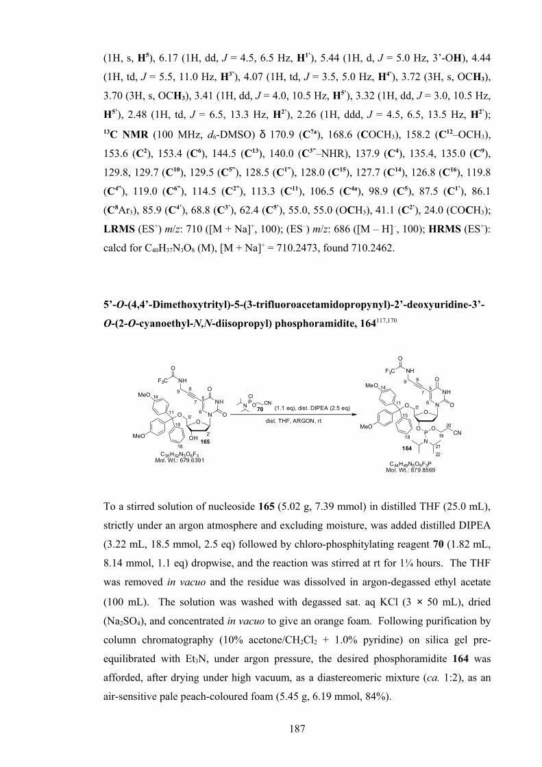
(1H, s, H5), 6.17 (1H, dd, J = 4.5, 6.5 Hz, H1’), 5.44 (1H, d, J = 5.0 Hz, 3’-OH), 4.44
(1H, td, J = 5.5, 11.0 Hz, H3’), 4.07 (1H, td, J = 3.5, 5.0 Hz, H4’), 3.72 (3H, s, OCH3),
3.70 (3H, s, OCH3), 3.41 (1H, dd, J = 4.0, 10.5 Hz, H5’), 3.32 (1H, dd, J = 3.0, 10.5 Hz,
H5’), 2.48 (1H, td, J = 6.5, 13.3 Hz, H2’), 2.26 (1H, ddd, J = 4.5, 6.5, 13.5 Hz, H2’);
13C NMR (100 MHz, d6-DMSO) δ 170.9 (C7a), 168.6 (COCH3), 158.2 (C12–OCH3),
153.6 (C2), 153.4 (C6), 144.5 (C13), 140.0 (C3”–NHR), 137.9 (C4), 135.4, 135.0 (C9),
129.8, 129.7 (C10), 129.5 (C5”), 128.5 (C1”), 128.0 (C15), 127.7 (C14), 126.8 (C16), 119.8
(C4”), 119.0 (C6”), 114.5 (C2”), 113.3 (C11), 106.5 (C4a), 98.9 (C5), 87.5 (C1’), 86.1
(C8Ar3), 85.9 (C4’), 68.8 (C3’), 62.4 (C5’), 55.0, 55.0 (OCH3), 41.1 (C2’), 24.0 (COCH3);
LRMS (ES+) m/z: 710 ([M + Na]+, 100); (ES–) m/z: 686 ([M – H]–, 100); HRMS (ES+):
calcd for C40H37N3O8 (M), [M + Na]+ = 710.2473, found 710.2462.
5’-O-(4,4’-Dimethoxytrityl)-5-(3-trifluoroacetamidopropynyl)-2’-deoxyuridine-3’-
O-(2-O-cyanoethyl-N,N-diisopropyl) phosphoramidite, 164117,170
O
MeO
MeO
11
14
15
18
O
O
OH
N
NH
O5'
2'
5
165
NHF3C
O
C35H32N3O8F3Mol. Wt.: 679.6391
O
O
O
O N
NH
O5'
5
164
NHF3C
O
P
N
OCN
C44H49N5O9F3PMol. Wt.: 879.8569
6
7
89MeO
MeO
11
14
15
18
6
7
89
19
20
21
22
(1.1 eq), dist. DIPEA (2.5 eq)70N
PCl
O CN
dist. THF, ARGON, rt
To a stirred solution of nucleoside 165 (5.02 g, 7.39 mmol) in distilled THF (25.0 mL),
strictly under an argon atmosphere and excluding moisture, was added distilled DIPEA
(3.22 mL, 18.5 mmol, 2.5 eq) followed by chloro-phosphitylating reagent 70 (1.82 mL,
8.14 mmol, 1.1 eq) dropwise, and the reaction was stirred at rt for 1¼ hours. The THF
was removed in vacuo and the residue was dissolved in argon-degassed ethyl acetate
(100 mL). The solution was washed with degassed sat. aq KCl (3 × 50 mL), dried
(Na2SO4), and concentrated in vacuo to give an orange foam. Following purification by
column chromatography (10% acetone/CH2Cl2 + 1.0% pyridine) on silica gel pre-
equilibrated with Et3N, under argon pressure, the desired phosphoramidite 164 was
afforded, after drying under high vacuum, as a diastereomeric mixture (ca. 1:2), as an
air-sensitive pale peach-coloured foam (5.45 g, 6.19 mmol, 84%).
187

Rf 0.41 (1:9, acetone/CH2Cl2 + 0.5% pyridine, A’); 31P NMR (121 MHz, d6-DMSO) δ
148.9, 148.5 (PIII); 1H NMR (400 MHz, d6-DMSO) δ 11.66 (NH3), 9.98 (1H, s,
CONH), 7.97 and 7.96 (1H, s, H6), 7.42-7.38 (2H, m, H16), 7.33-7.28 (2H, m, H17), 7.29
and 7.27 (4H, d, J = 7.8 Hz and J = 8.0 Hz, H12), 7.25-7.19 (1H, m, H18), 6.89 and 6.87
(4H, d, J = 8.8 Hz and J = 9.0 Hz, H13), 6.10 and 6.08 (1H, t, J = 6.7 Hz and J = 6.8 Hz,
H1’), 4.50 and 4.47 (1H, ddd, J = 3.6, 6.7, 14.2 Hz and J = 3.9, 6.6, 14.2 Hz, H3’), 4.10
and 4.06 (2H, s, H9), 4.08-4.00 (1H, m, H4’), 3.77-3.67 (1H, m, H19), 3.74 (3H, s,
OCH3), 3.73 (3H, s, OCH3), 3.67-3.59 (1H, m, H19), 3.60-3.44 (1H, m, H21), 3.33-3.24
(1H, m, H5’), 3.19 and 3.14 (1H, dd, J = 3.3, 10.5 Hz and J = 3.0, 10.3 Hz, H5’), 2.75
and 2.64 (2H, t, J = 5.9 Hz, H20), 2.44 and 2.43 (1H, dd, J = 6.8, 13.6 Hz and J = 6.9,
13.9 Hz, H2’), 2.35 and 2.30 (1H, ddd, J = 3.9, 6.4, 13.7 Hz and J = 3.6, 6.4, 13.9 Hz,
H2’), 1.13 (3H, d, J = 6.8 Hz, H22), 1.11 (3H, d, J = 7.0 Hz, H22), 1.10 (3H, d, J = 6.8 Hz,
H22), 0.99 (3H, d, J = 6.8 Hz, H22); LRMS (ES+) m/z: 902 ([M + Na]+, 100), 897 ([M +
NH4]+, 38), 880 ([M + H]+, 32), 303 (DMT+, 24); (ES–) m/z: 914/916 ([M + HCl – H]–,
2:1, 21), 878 ([M – H]–, 97), 825 ([M – CH2CHCN – H]–, 100).
Analytical results consistent with reported data.117,170
5’-O-(4,4’-Dimethoxytrityl)-5-(3-trifluoroacetamidopropynyl)-2’-deoxyuridine,
165117,170,255
O
MeO
MeO
78
11
12
15
O
(1.4 eq)
O
OH
O N
NH
O5'
2'
5
165
O
O
OH
N
NH
O
I
5'
2'
5
97
Pd(PPh3)4 (0.1 eq), CuI (0.2 eq)
dist. Et3N (7.0 eq), anh DMF, argon, rt
C30H29N2O7IMol. Wt.: 656.46
NHF3C
O
C35H32N3O8F3Mol. Wt.: 679.64
6
MeO
MeO
10
14
18
6
7
89
11
14
F3C NH
O
68
To a stirred solution of 5’-O-(4,4’-dimethoxytrityl)-5-iodo-2’-deoxyuridine 97 (10.1 g,
15.4 mmol) in anhydrous DMF (70.0 mL), under an argon atmosphere, was first added
distilled Et3N (15.0 mL, 108 mmol, 7.0 eq) followed by, in absence of light, CuI
(0.59 g, 3.09 mmol, 0.2 eq) and 3-(trifluoroacetamido)prop-1-yne 68 (2.41 mL, 21.5
mmol, 1.4 eq). After stirring for 20 minutes, tetrakis(triphenylphosphine) palladium (0)
(1.78 g, 1.54 mmol, 0.1 eq) was added and the reaction was stirred at rt for 1¾ hours,
when solid Na2EDTA (1.20 g) was added. After stirring for 5 minutes, the reaction
188

mixture was concentrated under high vacuum to give a dark brown syrup. The syrup
was dissolved in ethyl acetate (300 mL) and washed with 5% w/v aq Na2EDTA (pH 9, 3
× 150 mL) and sat. aq KCl (2 × 150 mL), dried (Na2SO4), and concentrated in vacuo
and under high vacuum to give a golden brown foam. This reaction was repeated on the
same scale as described above.
Following joint purification by column chromatography (10-30% acetone/CH2Cl2 +
1.0% pyridine), the desired product 165 was afforded, after drying under high vacuum,
as a very pale orange foam (16.6 g, 24.4 mmol, 80%). A mixture of starting material 97
and product 165 (1.84 g) was also isolated.
Rf 0.19 (1:9, acetone/CH2Cl2 + 0.5% pyridine, A’, B’); 19F NMR (282 MHz, d6-DMSO)
δ -73.9 (NHCOCF3); 1H NMR (400 MHz, d6-DMSO) δ 11.66 (NH3), 9.98 (1H, s,
NHCOCF3), 7.93 (1H, s, H6), 7.40 (2H, d, J = 7.5 Hz, H16), 7.31 (2H, t, J = 7.5 Hz,
H17), 7.29 (2H, d, J = 9.0 Hz, H12), 7.28 (2H, d, J = 9.0 Hz, H12), 7.22 (1H, tt, J = 1.5,
7.3 Hz, H18), 6.89 (2H, d, J = 9.0 Hz, H13), 6.89 (2H, d, J = 9.0 Hz, H13), 6.10 (1H, t, J =
6.7 Hz, H1’), 5.32 (1H, d, J = 4.5 Hz, 3’-OH), 4.27 (1H, td, J = 4.0, 10.3 Hz, H3’), 4.06
(2H, br d, J = 3.8 Hz, H9), 3.92 (1H, td, J = 3.3, 5.3 Hz, H4’), 3.74 (6H, s, OCH3), 3.26
(1H, dd, J = 5.4, 10.4 Hz, H5’), 3.09 (1H, dd, J = 2.9, 10.4 Hz, H5’), 2.29 (1H, td, J =
6.8, 13.6 Hz, H2’), 2.19 (1H, ddd, J = 3.8, 6.3, 13.3 Hz, H2’); 13C NMR (100 MHz, d6-
DMSO) δ 161.5 (C4), 158.1, 158.1 (C14–OMe), 156.0 (q, 2JCF = 36.9 Hz, COCF3), 149.3
(C2), 144.8 (C15), 143.7 (C6), 135.6, 135.2 (C11), 129.7, 129.6 (C12), 127.8 (C17), 127.5
(C16), 126.6 (C18), 115.7 (q, 1JCF = 288.7 Hz, COCF3), 113.2, 113.2 (C13), 97.8 (C5), 87.4
(C10Ar3), 85.8 (C4’,C7), 85.1 (C1’), 75.1 (C8), 70.4 (C3’), 63.8 (C5’), 55.0 (OCH3), 39.7
(C2’), 29.4 (C9); LRMS (ES+) m/z: 803 ([M + Et3N + Na]+, 14), 718 ([M + K]+, 14), 702
([M + Na]+, 100), 697 ([M + NH4]+, 5), 303 (DMT+, 5); (ES–) m/z: 678 ([M – H]–, 100).
Analytical results consistent with reported data.170,255
189

3-N-[2’-Deoxy-5’-O-(4,4’-dimethoxytrityl)-β-D-ribofuranosyl]-6-(3-
trifluoroacetamidophenyl)-(2,3H)-N-methylpyrrolo[2,3-d]pyrimidine-
2(7H)-one-3’-O-(2-O-cyanoethyl-N,N-diisopropyl) phosphoramidite, 178
O
O
O N
N
O
N
4
5
6
MeO
MeO
12
16
5'
2'
8
3''
O
OH
O N
N
O
N
4
5
6
MeO
MeO
12
16
5'
2'
8
3''
C41H37N4O7F3Mol. Wt.: 754.75
P
N
OCN17
18
1920
C50H54N6O8F3PMol. Wt.: 954.97
HNHN
F3CO
F3CO
197
(1.1 eq), dist. DIPEA (2.5 eq)70N
PCl
O CN
dist. DCM, ARGON, rt12
12
178
To a stirred solution of nucleoside 197 (5.02 g, 7.39 mmol) in distilled CH2Cl2 (25.0
mL), strictly under an argon atmosphere and excluding moisture, was added distilled
DIPEA (3.22 mL, 18.5 mmol, 2.5 eq) followed by chloro-phosphitylating reagent 70
(1.82 mL, 8.14 mmol, 1.1 eq) dropwise, and the reaction was stirred at rt for 4¾ hours.
The reaction mixture was diluted with argon-degassed CH2Cl2 (15 mL) and washed,
under an argon atmosphere, with degassed sat. aq KCl (15 mL). The aqueous was re-
extracted (degassed CH2Cl2, 3 × 5 mL) and combined CH2Cl2 fractions were dried
(Na2SO4), and concentrated in vacuo and dried under high vacuum to give a yellow
foam. Following purification by column chromatography (degassed 1% acetone/
CH2Cl2 + 0.5% Et3N → 5% acetone/CH2Cl2 + 0.5% Et3N), on silica gel pre-equilibrated
with Et3N, under argon pressure, then precipitation at rt from degassed CH2Cl2 (3 mL)
into degassed hexane (180 mL), the desired phosphoramidite 178 was afforded, after
drying under high vacuum, as a diastereomeric mixture (ca. 2:1), as a pale yellow foam
(0.18 g) containing 38.0 mol % Et3N.HCl (recalculated yield - 0.18 mmol, 70%).
Rf 0.50, 0.57 (1:1, acetone/CH2Cl2 + 0.5% Et3N, A’), 0.29, 0.34 (1:4, acetone/CH2Cl2 +
0.5% Et3N, A’), 0.18, 0.25 (CH2Cl2 + 0.5% Et3N, A’); 31P NMR (121 MHz, d6-DMSO)
δ 149.1, 148.9 (PIII); 19F NMR (282 MHz, d6-DMSO) δ -73.5 (NHCOCF3); 1H NMR
(400 MHz, d6-DMSO) δ 11.47 (NHCOCF3), 8.71 and 8.68 (1H, s, H4), 7.85 (1H, br s,
H2”), 7.75 (1H, br dd, J = 1.1, 8.2 Hz, H4”), 7.54 (1H, t, J = 8.0 Hz, H5”), 7.42 and 7.40
(2H, d, J = 8.3 Hz and J = 8.8 Hz, H14), 7.16 (1H, br dd, J = 0.9, 7.9 Hz, H6”), 7.34-7.28
(2H, m, H15), 7.30 and 7.27 (4H, d, J = 8.5 Hz and J = 8.8 Hz, H10), 7.28-7.23 (1H, m,
H16), 6.89, 6.88 and 6.87 (2H, 2H and 4H, d, J = 8.8 Hz, J = 9.0 Hz and J = 8.5 Hz,
190

H11), 6.30 and 6.26 (1H, dd, J = 5.1, 6.4 Hz and J = 5.0, 6.5 Hz, H1’), 5.69 and 5.65 (1H,
s, H5), 4.68 and 4.64 (1H, td, J = 5.3, 10.7 Hz and J = 5.6, 11.2 Hz, H3’), 4.17 and 4.13
(1H, dd, J = 3.5, 7.8 Hz and J = 3.8, 8.0 Hz, H4’), 3.81-3.68 (1H, m, H17), 3.70, 3.69 and
3.69 (3H, 3H and 6H, s, OCH3), 3.69-3.59 (1H, m, H17), 3.61-3.47 (2H, m, H19), 3.49
(3H, s, NCH3), 3.46-3.28 (2H, m, H5’), 2.77 and 2.67 (1H, t, J = 5.9 Hz, H18), 2.60 and
2.58 (1H, td, J = 6.7, 13.3 Hz and J = 6.5, 13.2 Hz, H2’), 2.35 (1H, td, J = 6.0, 12.2 Hz,
H2’), 1.15 (3H, d, J = 6.8 Hz, H20), 1.13 (3H, d, J = 6.8 Hz, H20), 1.12 (3H, d, J = 6.8 Hz,
H20), 1.01 (3H, d, J = 6.8 Hz, H20); LRMS (ES+) m/z: 977 ([M + Na]+, 100), 955 ([M +
H]+, 13); (ES–) m/z: 953 ([M – H]–, 100); HRMS (ES+): calcd for C50H54N6O8F3P (M),
[M + Na]+ = 977.3585, found 977.3591; [M + H]+ = 955.3766, found 955.3783. Found
also DMT+ (calcd 303.1380, found 303.1070).
3-N-[2’-Deoxy-5’-O-(4,4’-dimethoxytrityl)-β-D-ribofuranosyl]-6-(3-ureidophenyl)-
(2,3H)-N-methylpyrrolo[2,3-d]pyrimidine-2(7H)-one-3’-O-(2-O-cyanoethyl-N,N-
diisopropyl) phosphoramidite, 179
O
OH
O N
N
O
N
HNH2N
O
188
4
5
6
MeO
MeO
12
16
4a
7a
5'
2'
8
3''
(1.1 eq), DIHT 190 (0.5 eq)
C40H39N5O7Mol. Wt.: 701.77
O
O
O N
N
O
N
HNH2N
O
179
P
N
OCN
C49H56N7O8PMol. Wt.: 901.98
MeO
MeO
8
12
16
2'
5'4
5
3''
17
18
1920
189
12
12
1''
NPN
O CN1)
(1.1 eq), dist. DIPEA (2.5 eq)70N
PCl
O CN2)
dist. THF/anh. DMF (3:1 v/v), ARGON, rt
dist. THF/anh. DMF (5:1 v/v), ARGON, rt
Method 1: To a stirred solution of cyclised nucleoside 188 (0.22 g, 0.32 mmol) in
distilled THF (1.5 mL) and anhydrous DMF (0.5 mL), under an argon atmosphere and
excluding moisture, was added DIHT 190 (0.03 g, 0.16 mmol, 0.5 eq) followed by
phosphitylating reagent 189 (0.11 mL, 0.36 mmol, 1.1 eq), and the reaction was stirred
at rt. After 18½ hours, further reagent 189 was added (0.05 mL, 0.16 mmol, 0.5 eq). At
22 hours, further DIHT 190 (27.8 mg, 0.16 mmol, 0.5 eq) was added, then at 24 hours,
further reagent 189 was added (0.10 mL, 0.32 mmol, 1.0 eq). After 41 hours, THF was
removed in vacuo, and the syrup was dissolved in argon-degassed ethyl acetate (5 mL).
The sticky yellow precipitate was washed with degassed ethyl acetate (3 × 5 mL), and
the combined solution was washed with degassed sat. aq NaHCO3 (3 × 8 mL) and sat.
191

aq KCl (2 × 8 mL), dried (Na2SO4), concentrated in vacuo and dried under high vacuum
to give a viscous yellow syrup. Following purification by column chromatography
(degassed 2% methanol/acetone + 0.5% Et3N) on silica gel pre-equilibrated with Et3N,
under argon pressure, and drying under high vacuum, the desired phosphoramidite 179
was afforded as a diastereomeric mixture (ca. 1:1), as an air-sensitive, pale yellow foam
(0.08 g, 0.09 mmol, 29%).
Method 2: To a stirred solution of cyclised nucleoside 188 (0.92 g, 1.31 mmol) in
distilled THF (5.0 mL) and anhydrous DMF (1.0 mL), under an argon atmosphere and
excluding moisture, was added distilled DIPEA (0.58 mL, 3.30 mmol, 2.5 eq) followed
by chloro-phosphitylating reagent 70 (0.33 mL, 1.46 mmol, 1.1 eq), and the reaction
was stirred at rt for 2 hours. The reaction mixture was concentrated in vacuo and under
high vacuum to give a viscous orange syrup, which was dissolved in argon-degassed
CH2Cl2 (80 mL). The solution was washed with degassed sat. aq KCl (60 mL) and the
aqueous was re-extracted (degassed CH2Cl2, 3 × 10 mL). The combined CH2Cl2
fractions were dried (Na2SO4) and concentrated in vacuo and under high vacuum to give
an orange foam. Following purification by column chromatography (degassed acetone
+ 0.5% Et3N → 2% methanol/acetone + 0.5% Et3N) on silica gel pre-equilibrated with
Et3N, under argon pressure, and precipitation at rt from degassed CH2Cl2 (6 mL) into
degassed hexane (350 mL), the desired phosphoramidite 179 was afforded, after drying
under high vacuum, as a diastereomeric mixture (ca. 1:1), as an air-sensitive pale
yellow foam (0.60 g, 0.67 mmol, 51%).
Rf 0.30 (9:1, acetone/CH2Cl2 + 0.3% Et3N, A’), 0.24, 0.31 (5:95, methanol/acetone +
0.3% Et3N, A’), 0.17 (acetone + 0.3% Et3N, A’); 31P NMR (121 MHz, d6-DMSO) δ
149.2, 149.0 (PIII); 1H NMR (400 MHz, d6-DMSO) δ 8.69 (1H, s, NHCONH2), 8.66
and 8.63 (1H, s, H4), 7.63 (1H, br s, H2”), 7.44-7.38 (1H, m, H4”), 7.42 and 7.40 (2H, d,
J = 8.0 Hz and J = 8.5 Hz, H14), 7.34 (1H, t, J = 7.9 Hz, H5”), 7.34-7.24 (3H, m,
H15,H16), 7.30 and 7.27 (4H, d, J = 8.0 Hz and J = 8.8 Hz, H10), 7.02 (1H, br d, J = 7.5
Hz, H6”), 6.89 and 6.87 (2H, d, J = 8.3 Hz and J = 8.6 Hz, H11), 6.89 and 6.87 (2H, d, J
= 8.5 Hz, H11), 6.31 and 6.27 (1H, app. t, J = 5.8 Hz, H1’), 5.92 (2H, br s, NH2), 5.63
and 5.59 (1H, s, H5), 4.68 and 4.64 (1H, td, J = 5.4, 16.1 Hz and J = 5.9, 17.9 Hz, H3’),
4.17 and 4.13 (1H, dd, J = 3.4, 7.7 Hz and J = 3.8, 8.0 Hz, H4’), 3.81-3.68 (1H, m, H17),
3.71, 3.70 and 3.70 (3H, 3H and 6H, s, OCH3), 3.69-3.61 (1H, m, H17), 3.63-3.49 (2H,
m, H19), 3.46 (3H, s, NCH3), 3.49-3.44 and 3.41 (1H, m and dd, J = 3.5, 11.5 Hz, H5’),
192

3.46-3.41 and 3.37 (1H, m and dd, J = 2.9, 10.8 Hz, H5’), 2.77 and 2.67 (1H, t, J = 5.9
Hz, H18), 2.59 (1H, td, J = 6.7, 13.6 Hz, H2’), 2.34 (1H, td, J = 6.0, 11.9 Hz, H2’), 1.15
(3H, d, J = 7.3 Hz, H20), 1.13 (3H, d, J = 7.3 Hz, H20), 1.12 (3H, d, J = 6.8 Hz, H20),
1.02 (3H, d, J = 6.8 Hz, H20); LRMS (ES+) m/z: 940 ([M + K]+, 8), 924 ([M + Na]+, 45),
902 ([M + H]+, 78), 303 (DMT+, 100); HRMS (ES+): calcd for C49H56N7O8P (M), [M +
Na]+ = 924.3820, found 924.3825; [M + H]+ = 902.4001, found 902.4000. Found also
DMT+ (calcd 303.1380, found 303.1381).
5’-O-(4,4’-Dimethoxytrityl)-5-[(3-ureidobenzoyl)methyl]-4N-methyl-2’-
deoxycytidine, 183
N
ONO
OH
O
MeO
MeO
O
HNH2N
O
24
4a
5
6
7a
2'
5'8
9
10
11
12
13
14
15 16
12
1''
3''
160
33 wt% MeNH2/EtOH, rt, argon
O
OH
O N
N
O
MeO
MeO
HNO
8
5'
2'
5
6
7
NH
O
H2N3''
9
C39H36N4O8Mol. Wt.: 688.73 C40H41N5O8
Mol. Wt.: 719.78
183
13
13
17
A suspension of nucleoside 160 (0.20 g, 0.29 mmol) in 33 wt% MeNH2/ethanol (2.50
mL) was stirred under an argon atmosphere, at rt for 1 hour. The suspension dissolved
over 25 minutes. The solvent was removed in vacuo to give a green crystalline solid.
Following purification by column chromatography (5-20% methanol/ethyl acetate +
0.5% Et3N), and drying under high vacuum, the desired ring-opened nucleoside 183 was
afforded as a yellow glassy solid (0.13 g, 0.18 mmol, 61%).
Rf 0.11 (9:1, acetone/ethyl acetate + 0.3% Et3N, A’), 0.25 (1:4, methanol/ethyl acetate +
0.3% Et3N, A’); LRMS (ES+) m/z: 758 ([M + K]+, 10), 742 ([M + Na]+, 30), 720 ([M +
H]+, 5), 303 (DMT+, 100); (ES–) m/z: 816 ([M + H3PO4 – H]–, 100), 718 ([M – H]–, 41),
675 ([M – O=C=NH – H]–, 52); HRMS (ES+): calcd for C40H41N5O8 (M), [M + H]+ =
720.3028, found 720.3024.
193

3-N-[2’-Deoxy-5’-O-(4,4’-dimethoxytrityl)-β-D-ribofuranosyl]-6-(3-ureidophenyl)-
(2,3H)-N-methylpyrrolo[2,3-d]pyrimidine-2(7H)-one, 184
O
OH
O N
N
O
HN
NHH2N
O
188
O
OH
O N
N
O
N
HNH2N
O
184
(3.0 eq)
Pd(PPh3)4 (0.1 eq)
dist. Et3N (5.0 eq)
CuI (1.1 eq)
dist. Et3N (10.0 eq)
anh. DMF, 4Å sieves
C40H39N5O7Mol. Wt.: 701.77
6
4
2'
5'
4
5MeO
MeO
78
11
12
15
6
MeO
MeO
12
16
1''
3''
7
84a
7a
5'
2'
8
3''HN
O
OH
O N
N
O
MeO
MeO
I
5'
2'
5
7
185
C31H32N3O6IMol. Wt.: 669.51
11
15
11
NH
H2N
O
156
CuI (0.4 eq), anh. DMF
argon, dark, rt
argon, dark, 125 oC
To a solution of 5’-O-(4,4’-dimethoxytrityl)-5-iodo-4N-methyl-2’-deoxycytidine 185
(1.25 g, 1.87 mmol) in anhydrous DMF (6.0 mL), under an argon atmosphere and in
absence of light, were added distilled Et3N (1.30 mL, 9.33 mmol, 5.0 eq), CuI (0.14 g,
0.75 mmol, 0.4 eq), then alkyne 156 (0.90 g, 5.61 mmol, 3.0 eq). The reaction mixture
was stirred for 15 minutes then tetrakis(triphenylphosphine) palladium (0) (0.53 g, 0.46
mmol, 0.1 eq) was added and the reaction was stirred at rt for 2¼ hours. The reaction
mixture was diluted with methanol/acetone (1:1 v/v, 10 mL), filtered and the residue
was washed with methanol/acetone (1:1 v/v, 3 × 10 mL). The combined filtrate was
concentrated in vacuo and under high vacuum, co-evaporating with methanol/toluene to
give a dark brown foam. The crude material was partially-purified by column
chromatography (80-100% acetone/CH2Cl2 + 0.5% Et3N → 5-20% methanol/acetone +
0.5% Et3N), to afford, after drying under high vacuum, the product 188 plus alkyne-
dimer impurity, as a pale brown foam (1.49 g).
Rf 0.18 (188), 0.12 (alkyne dimer) (1:9, methanol/CH2Cl2 + 0.3% Et3N, A’), 0.11 (188),
0.21 (alkyne dimer) (4:1, acetone/CH2Cl2 + 0.3% Et3N, A’), 0.44 (188 + alkyne dimer),
(1:9, methanol/acetone + 0.3% Et3N, A’).
To a stirred solution of partially-purified product 188 (1.48 g) in anhydrous DMF (10.0
mL) and distilled Et3N (2.60 mL, 18.7 mmol, 10.0 eq), under an argon atmosphere, in
absence of light, were added 4Å molecular sieves then CuI (0.39 g, 2.05 mmol, 1.1 eq),
and the reaction was stirred at 125 °C for 40 minutes. The mixture was allowed to cool
and diluted with methanol (20 mL). The mixture was filtered, the residue was washed
with methanol (3 × 10 mL) and the combined filtrate was concentrated in vacuo and
under high vacuum to give a green foam/resin. The compound was twice-purified by
column chromatography (0-10% methanol/acetone + 0.5% Et3N), then by precipitation
194

from methanol/acetone (1:1 v/v, 8 mL) into diethyl ether (230 ml), to afford, after
drying under high vacuum, the desired N-methylpyrrolo-dC product 184, as a very pale
yellow powdery solid (1.03 g, 1.47 mmol, 79% over two steps).
Rf 0.24 (1:9, methanol/acetone + 0.3% Et3N, A’), 0.49 (1:4, methanol/CH2Cl2 + 0.3%
Et3N, A’); IR (solid): ν 3324 (br m, O–H, N–H), 2932, 2836 (w, C–H), 1651 (s, C=O),
1606 (s, C=C), 1556 (vs, C=N), 1507 (s, Aryl C=C), 1479 (s), 1441 (m), 1402 (s), 1338,
1303 (m), 1248 (s, O–H), 1176 (s), 1095, 1032 (s, C–O), 904 (m), 828 (s, C–H), 773,
726 (s), 701 (s, Aryl C–H), 659, 584, 534 (s) cm-1; 1H NMR (400 MHz, d6-DMSO) δ
8.71 (1H, s, NHCONH2), 8.63 (1H, s, H4), 7.63 (1H, t, J = 1.8 Hz, H2”), 7.41 (1H, ddd,
J = 1.0, 2.0, 7.0 Hz, H4”), 7.41 (2H, d, J = 7.0 Hz, H14), 7.34 (1H, t, J = 8.0 Hz, H5”),
7.32 (2H, tt, J = 1.5, 7.0 Hz, H15), 7.29 (2H, d, J = 9.0 Hz, H10), 7.28 (2H, d, J = 8.5 Hz,
H10), 7.28 (1H, t, J = 8.0 Hz, H16), 7.02 (1H, td, J = 1.3, 7.5 Hz, H6”), 6.89 (2H, d, J =
9.0 Hz, H11), 6.89 (2H, d, J = 9.0 Hz, H11), 6.26 (1H, dd, J = 5.0, 6.5 Hz, H1’), 5.92 (2H,
s, CONH2), 5.59 (1H, s, H5), 5.40 (1H, d, J = 4.8 Hz, 3’-OH), 4.43 (1H, qn, J = 5.5 Hz,
H3’), 4.00 (1H, dd, J = 4.0, 8.0 Hz, H4’), 3.70 (3H, s, OCH3), 3.69 (3H, s, OCH3), 3.46
(3H, s, NCH3), 3.40 (1H, dd, J = 4.0, 10.5 Hz, H5’), 3.29 (1H, dd, J = 2.5, 10.5 Hz, H5’),
2.44 (1H, td, J = 6.3, 13.1 Hz, H2’), 2.18 (1H, ddd, J = 5.0, 6.5, 13.6 Hz, H2’); 13C NMR
(100 MHz, d6-DMSO) δ 158.5 (C7a), 158.1 (C12–OCH3), 155.9 (CONH2), 153.5 (C2),
144.5 (C13), 142.6 (C6), 140.9 (C3”–NHR), 136.2 (C4), 135.4, 135.1 (C9), 130.8 (C1”),
129.8, 129.7 (C10), 129.1 (C5”), 127.9 (C15), 127.7 (C14), 126.8 (C16), 120.9 (C6”), 118.1
(C4”), 117.4 (C2”), 113.3 (C11), 107.6 (C4a), 98.6 (C5), 86.7 (C1’), 86.1 (C8Ar3), 85.7
(C4’), 69.0 (C3’), 62.6 (C5’), 55.0 (OCH3), 41.5 (C2’), 29.3 (NCH3); LRMS (ES+) m/z:
1425 ([2M + Na]+, 8), 724 ([M + Na]+, 100), 702 ([M + H]+, 50); HRMS (ES+): calcd
for C40H39N5O7 (M), [M + Na]+ = 724.2742, found 724.2733. Found also DMT+ (calcd
303.1380, found 303.1382); Anal. Calcd C40H39N5O7 (701.77), C 68.46%, H 5.60%, N
9.97%, found C 65.70%, H 5.82%, N 9.35%.
195

5’-O-(4,4’-Dimethoxytrityl)-5-iodo-4N-methyl-2’-deoxycytidine, 185
HN
i) POCl3 (3.8 eq), NMI (12.6 eq), dist. pyridine, 0 oC-rtO
OH
O N
N
O
MeO
MeO
I
5'
2'
5
7
185
ii) 40 wt% MeNH2/H2O, 0 oC-rt
iii) 33 wt% MeNH2/EtOH, rt
C31H32N3O6IMol. Wt.: 669.51
O
O
OAc
O N
NH
O
MeO
MeO
I
5'
2'
5
7
187
C32H31N2O8IMol. Wt.: 698.50
11
15
11
11
15
11
Method 1: To a stirred solution of acetylated nucleoside 187 (1.44 g, 2.07 mmol) and
N-methylimidazole (2.10 mL, 26.3 mmol, 12.8 eq) in distilled pyridine (50.0 mL),
under an argon atmosphere, at 0 °C, distilled phosphorous oxychloride (0.73 mL, 7.83
mmol, 3.8 eq) was added dropwise over 10 minutes. The yellow suspension of
intermediate 186 was stirred at rt for 1 hour. A 40 wt% MeNH2/water solution (10.0
mL, 116 mmol MeNH2, 55.9 eq MeNH2) was added whilst cooling in ice, and the
cloudy solution was stirred at rt for 90 minutes. The pyridine was removed under high
vacuum, and the mixture was diluted with water (200 mL). The aqueous solution was
extracted with CH2Cl2 (4 × 100 mL), then the combined organic layer was washed with
sat. aq KCl (200 mL) and dried (Na2SO4). Triethylamine (6.0 mL) was added to prevent
detritylation and the solution was concentrated in vacuo, and dried under high vacuum.
The viscous brown oil was dissolved in 33 wt% MeNH2/ethanol (10.0 mL, 80.3 mmol
MeNH2, 38.8 eq MeNH2) and stirred at rt for 40 minutes, then concentrated in vacuo
and dried under high vacuum. Following purification by column chromatography (0-
2% methanol/CH2Cl2 + 0.5% Et3N), and drying under high vacuum, the 4N-methyl-dC
nucleoside 185 was afforded, as a pale yellow foam (1.24 g, 1.86 mmol, 90%).
Method 2: Acetylated nucleoside 187 (6.85 g, 9.80 mmol) was treated with NMI and
POCl3 in pyridine, then 40 wt% MeNH2/water and worked up, as described in Method
1. After treatment with 33 wt% MeNH2/ethanol as described above, the resulting syrup
was twice-purified by column chromatography (0-3% methanol/CH2Cl2 + 0.5% Et3N, 0-
15% acetone/CH2Cl2 + 0.5% Et3N → 60% acetone/CH2Cl2 + 0.5% Et3N) to yield a
syrup containing significant NMI. Two further purifications (30-100% acetone/toluene
+ 0.5% Et3N; 10-25% acetone/toluene + 1.0% pyridine) failed to remove NMI, however
the product was successfully recrystallised from toluene (100 mL), and dried under high
vacuum over KOH to afford the product 185 as a fluffy white crystalline solid (5.26 g,
7.86 mmol, 80%).
196

Method 3: Acetylated nucleoside 187 (8.27 g, 11.8 mmol) was treated with NMI and
POCl3 in pyridine, then 40 wt% MeNH2/water and the reaction concentrated as
described in Method 1. The resulting syrup was treated with 33 wt% MeNH2/ethanol as
described above, without an aqueous workup, then twice-purified by column
chromatography (0-100% acetone/CH2Cl2 + 0.5% Et3N → 5-15% methanol/acetone +
1.0% Et3N). The orange/brown syrup was further purified by a combination of
precipitation from toluene into diethyl ether, removal of NMI by soaking in diethyl
ether, and recrystallisation from ethyl acetate to afford the product 185 as a white to off-
white powdery/crystalline solid (3.62 g, 5.40 mmol, 46%).
Rf 0.40 (1:9, methanol/CH2Cl2 + 0.3% Et3N, A’), 0.36 (4:1, acetone/CH2Cl2 + 0.3%
Et3N, A’), 0.57 (1:9, methanol/acetone + 0.3% Et3N, A’); Mp 141-144 °C (ethyl
acetate/diethyl ether); IR (solid): ν 3291 (br w, N–H), 3059, 2930, 2835 (w, C–H), 1634
(s, C=O), 1606 (vs, C=O, C=C), 1547 (s, C=O), 1505 (vs, C=C), 1397 (m, O–H), 1284
(s, C–O), 1246 (vs, C–N), 1173 (s), 1092, 1030 (m, C–O), 825, 779 (s, Aryl C=C), 700,
583 (s) cm-1; 1H NMR (400 MHz, d6-DMSO) δ 7.93 (1H, s, H6), 7.40 (2H, d, J = 7.5
Hz, H13), 7.32 (2H, t, J = 7.3 Hz, H14), 7.29 (4H, d, J = 9.0 Hz, H9), 7.22 (1H, tt, J = 2.0,
7.3 Hz, H15), 7.06 (1H, q, J = 4.5 Hz, NHCH3), 6.89 (4H, d, J = 9.0 Hz, H10), 6.12 (1H,
dd, J = 6.2, 7.4 Hz, H1’), 5.25 (1H, d, J = 4.3 Hz, 3’-OH), 4.20 (1H, dt, J = 3.3, 6.4 Hz,
H3’), 3.91 (1H, dd, J = 3.3, 6.5 Hz, H4’), 3.74 (6H, s, OCH3), 3.20 (1H, dd, J = 3.3, 10.8
Hz, H5’), 3.17 (1H, dd, J = 4.5, 10.8 Hz, H5’), 2.80 (3H, d, J = 4.5 Hz, NHCH3), 2.21
(1H, ddd, J = 3.0, 6.0, 13.3 Hz, H2’), 2.09 (1H, td, J = 6.8, 13.6 Hz, H2’); 13C NMR (100
MHz, d6-DMSO) δ 163.0 (C4), 159.8 (C11–OMe), 155.3 (C2), 146.8 (C6), 146.4 (C12),
137.2, 137.1 (C8), 131.4 (C9), 129.6 (C14), 129.4 (C13), 128.4 (C15), 115.0 (C10), 87.5
(C7Ar3), 87.4 (C4’), 87.0 (C1’), 72.4 (C3’), 65.4 (C5’), 59.9 (C5–I), 56.8 (OCH3), 42.4
(C2’), 30.4 (NHCH3); LRMS (ES+) m/z: 692 ([M + Na]+, 12), 670 ([M + H]+, 3), 303
(DMT+, 100); (ES–) m/z: 668 ([M – H]–, 100); HRMS (ES+): calcd for C31H32N3O6I (M),
[M + K]+ = 708.0967, found 708.0962, [M + Na]+ = 692.1228, found 692.1219, [M + H]+ = 670.1409, found 670.1414. Found also DMT+ (calcd 303.1380, found 303.1386);
Anal. Calcd C31H32N3O6I (669.51), C 55.61%, H 4.82%, N 6.27%, found C 54.80%, H
4.75%, N 6.05%.
3’-O-Acetyl-5’-O-(4,4’-dimethoxytrityl)-5-iodo-2’-deoxyuridine, 187209
197

acetic anhydride (1.5 eq)
O
O
OH
O N
NH
O
MeO
MeO
I
5'
2'
5
7
97
8
11
12
15
C30H29N2O7IMol. Wt.: 656.46
dist. pyridine, 0 oC, argon
6
11
O
O
OAc
O N
NH
O
MeO
MeO
I
5'
2'
5
7
187
C32H31N2O8IMol. Wt.: 698.50
11
15
11
To a stirred solution of nucleoside 97 (10.0 g, 15.2 mmol) in distilled pyridine (40.0
mL), under an argon atmosphere, at 0 °C was added acetic anhydride (7.20 mL, 76.3
mmol, 5.0 eq) dropwise over 20 minutes. The reaction was stirred at 0 °C for 15
minutes then allowed to warm to rt. After 2¾ hours, the reaction mixture was
concentrated in vacuo and under high vacuum, co-evaporating with acetone (2 × 50 mL)
and diethyl ether (2 × 50 mL), to give white foam/syrup. The syrup was dissolved in
ethyl acetate (300 mL) and washed with water (3 × 75 mL). The aqueous was re-
extracted (ethyl acetate, 40 mL) and combined organic fractions were washed with sat.
aq KCl (120 mL), dried (Na2SO4) and concentrated in vacuo and under high vacuum to
give a white foam. Following purification by column chromatography
(0-2% methanol/CH2Cl2 + 0.5% Et3N), and drying under high vacuum, the desired
product 187 was afforded as a white foam (10.3 g, 14.7 mmol, 97%).
Rf 0.31 (1:9, acetone/CH2Cl2 + 0.3% Et3N, A’), 0.48 (5:95, methanol/CH2Cl2 + 0.3%
Et3N, A’), 0.33 (7:3, ethyl acetate/hexane, A’); IR (solid): ν 3056 (w, C–H, N–H),
2933, 2835 (w, C–H), 1683 (br s, C=O), 1606, 1506 (m, Aryl C=C), 1442 (m, C–H),
1379 (w, CH3), 1244 (vs, C–O), 1174 (s), 11.05 (m), 1073 (s), 1027 (s, C–O), 826, 754,
700 (s, Aryl C–H), 583 (s), 545 (m) cm-1; 1H NMR (400 MHz, d6-DMSO) δ 11.75 (1H,
s, NH3), 8.05 (1H, s, H6), 7.39 (2H, d, J = 8.0 Hz, H13), 7.32 (2H, t, J = 7.3 Hz, H14),
7.28 (2H, d, J = 9.0 Hz, H9), 7.28 (2H, d, J = 9.0 Hz, H9), 7.23 (1H, tt, J = 1.1, 7.2 Hz,
H15), 6.89 (4H, d, J = 8.8 Hz, H10), 6.10 (1H, dd, J = 6.1, 8.2 Hz, H1’), 5.23 (1H, td, J =
2.4, 6.4 Hz, H3’), 4.07 (1H, td, J = 2.9, 4.5 Hz, H4’), 3.74 (6H, s, OCH3), 3.32 (1H, dd, J
= 4.9, 10.4 Hz, H5’), 3.22 (1H, dd, J = 3.1, 10.4 Hz, H5’), 2.44 (1H, td, J = 7.0, 11.5 Hz,
H2’), 2.33 (1H, ddd, J = 2.0, 6.0, 14.3 Hz, H2’), 2.03 (3H, s, COCH3); 13C NMR (100
MHz, d6-DMSO) δ 169.9 (COCH3), 160.5 (C4), 158.1 (C11–OCH3), 150.1 (C2), 144.6
(C12), 144.1 (C6), 135.3, 135.2 (C8), 129.7 (C9), 127.9 (C14), 127.6 (C13), 126.7 (C15),
113.3 (C10), 86.0 (C7Ar3), 84.7 (C1’), 83.1 (C4’), 74.1 (C3’), 70.2 (C5–I), 63.4 (C5’), 55.0
(OCH3), 36.8 (C2’), 20.7 (COCH3); LRMS (ES+) m/z: 721 ([M + Na]+, 12), 303 (DMT+,
100); (ES–) m/z: 697 ([M – H]–, 100); HRMS (ES+): calcd for C32H31N2O8I (M), [M +
198

Et3N + H]+ = 800.2402, found 800.2369; [M + Na]+ = 721.1017, found 721.1007. Found
also DMT+ (calcd 303.1380, found 303.1383).
3-N-[2’-Deoxy-5’-O-(4,4’-dimethoxytrityl)-β-D-ribofuranosyl]-6-(3-
acetamidophenyl)-(2,3H)-N-methylpyrrolo[2,3-d]pyrimidine-2(7H)-one-3’-
O-(2-O-cyanoethyl-N,N-diisopropyl) phosphoramidite, 191
C41H40N4O7Mol. Wt.: 700.78 C50H57N6O8P
Mol. Wt.: 901.00
O
OH
O N
N
O
N
HN
O
193
4
5
6
MeO
MeO
12
16
4a
7a
5'
2'
8
3''
O
O
O N
N
O
N
HN
O
191
P
N
OCN
MeO
MeO
8
12
16
2'
5'4
5
3''
17
18
1920
12
12
1''
(1.1 eq), dist. DIPEA (2.5 eq)70N
PCl
O CN
dist. THF/anh. DMF (7:9 v/v), ARGON, rt
To a stirred solution of nucleoside 193 (1.37 g, 1.95 mmol) in distilled THF (3.5 mL)
and anhydrous DMF (4.5 mL), strictly under an argon atmosphere and
excluding moisture, was added distilled DIPEA (0.86 mL, 4.91 mmol, 2.5 eq)
followed by chloro-phosphitylating reagent 70 (0.48 mL, 2.15 mmol, 1.1 eq)
dropwise, and the reaction was stirred at rt for 1¼ hours. The THF was
removed in vacuo and the residue was dissolved in argon-degassed ethyl
acetate (60 mL). The solution was washed with degassed sat. aq KCl (3 × 40
mL), dried (Na2SO4), and concentrated in vacuo and dried under high vacuum
to give a pale orange foam. Following purification by column chromatography
(7:3, acetone/CH2Cl2 + 0.5% Et3N), on silica gel pre-equilibrated with Et3N,
under argon pressure, the desired phosphoramidite 191 was afforded, after
drying under high vacuum, as a diastereomeric mixture (ca. 1:1), as an air-
sensitive, pale milky yellow foam (1.51 g, 1.68 mmol, 86%).
Rf 0.33, 0.46 (4:1, acetone/CH2Cl2 + 0.3% Et3N, A’); 31P NMR (121 MHz, d6-DMSO) δ
149.2, 149.0 (PIII); 1H NMR (400 MHz, d6-DMSO) δ 10.08 (1H, s, NHCOCH3), 8.68
and 8.65 (1H, s, H4), 7.79 (1H, br s, H2”), 7.62 (1H, br d, J = 8.0 Hz, H4”), 7.42 (1H, t, J
= 7.8 Hz, H5”), 7.42 and 7.40 (2H, d, J = 8.0 Hz and J = 8.5 Hz, H14), 7.34-7.29 (2H, m,
H15), 7.30 and 7.28 (4H, d, J = 9.0 Hz, H10), 7.30-7.23 (1H, m, H16), 7.16 (1H, br d, J =
199

7.5 Hz, H6”), 6.89, 6.88 and 6.87 (2H, 2H and 4H, d, J = 9.0 Hz, J = 8.5 Hz and J = 9.0
Hz, H11), 6.31 and 6.27 (1H, app. t, J = 5.8 Hz, H1’), 5.66 and 5.63 (1H, s, H5), 4.68 and
4.65 (1H, ddd, J = 5.5, 16.3 Hz and J = 6.0, 16.7 Hz, H3’), 4.18 and 4.14 (1H, dd, J =
3.3, 7.8 Hz and J = 3.8, 8.3 Hz, H4’), 3.82-3.69 (1H, m, H17), 3.70, 3.70 and 3.69 (3H,
3H and 6H, s, OCH3), 3.70-3.60 (1H, m, H17), 3.62-3.48 (2H, m, H19), 3.47 (3H, s,
NCH3), 3.47 and 3.43-3.39 (1H, dd and m, J = 4.5, 11.0 Hz, H5’), 3.44 and 3.38 (1H, dd,
J = 3.5, 10.5 Hz and J = 3.0, 10.5 Hz, H5’), 2.77 and 2.67 (1H, t, J = 6.0 Hz, H18), 2.61
and 2.59 (1H, td, J = 6.9, 13.4 Hz and J = 6.5, 13.1 Hz, H2’), 2.35 (1H, td, J = 6.3, 13.1
Hz, H2’), 2.08 (3H, s, COCH3), 1.15 (3H, d, J = 7.0 Hz, H20), 1.13 (3H, d, J = 7.0 Hz,
H20), 1.12 (3H, d, J = 7.0 Hz, H20), 1.02 (3H, d, J = 7.0 Hz, H20); LRMS (ES+) m/z: 939
([M + K]+, 5), 923 ([M + Na]+, 100), 901 ([M + H]+, 9); HRMS (ES+): calcd for
C50H57N6O8P (M), [M + Et3N + H]+ = 1002.5253, found 1002.5261; [M + Na]+ =
923.3868, found 923.3886; [M + H]+ = 901.4048, found 901.4033.
3-N-[2’-Deoxy-5’-O-(4,4’-dimethoxytrityl)-β-D-ribofuranosyl]-6-(3-
acetamidophenyl)-(2,3H)-N-methylpyrrolo[2,3-d]pyrimidine-2(7H)-one,
193
Pd(PPh3)4 (0.1 eq)
dist. Et3N (5.0 eq)
dist. Et3N (10.0 eq)
anh. DMF, 4Å sievesCuI (0.4 eq), anh. DMF
argon, dark, rt
argon, dark, 125 oCO
OH
O N
N
O
HN
NH
O
192
O
OH
O N
N
O
N
HN
O
193
(3.0 eq) CuI (1.1 eq)
6
4
2'
5'
4
5MeO
MeO
78
11
12
15
6
MeO
MeO
12
16
1''
3''
7
84a
7a
5'
2'
8
3''HN
O
OH
O N
N
O
MeO
MeO
I
5'
2'
5
7
185
C31H32N3O6IMol. Wt.: 669.51
11
15
11
NH
O
161
C41H40N4O7Mol. Wt.: 700.78
To a solution of 5’-O-(4,4’-dimethoxytrityl)-5-iodo-4N-methyl-2’-deoxycytidine 185
(1.69 g, 2.53 mmol) in anhydrous DMF (6.0 mL), under an argon atmosphere and in
absence of light, were added distilled Et3N (1.77 mL, 12.7 mmol, 5.0 eq), CuI (0.19 g,
1.01 mmol, 0.4 eq), then alkyne 161 (1.21 g, 7.60 mmol, 3.0 eq). The reaction mixture
was stirred for 20 minutes then tetrakis(triphenylphosphine) palladium (0) (0.29 g, 0.25
mmol, 0.1 eq) was added and the reaction was stirred at rt for 1 hour. The reaction
mixture was concentrated under high vacuum, co-evaporating with acetone to give a
black/brown foam. The residue was dissolved in CH2Cl2 (100 mL) and washed with 5%
w/v aq Na2EDTA (pH 9, 3 × 70 mL). The aqueous was re-extracted (CH2Cl2, 2 × 50
mL) and combined organic fractions were washed with sat. aq KCl (100 mL), dried
200

(Na2SO4), and concentrated in vacuo and under high vacuum to give a golden brown
foam. The crude material was partially-purified by column chromatography (10-30%
acetone/CH2Cl2 + 1.0% pyridine → 50-70% acetone/CH2Cl2 + 1.0% pyridine), to afford,
after drying under high vacuum, the product 192 plus alkyne-dimer impurity, as a
yellow glassy foam (1.99 g).
Rf 0.16 (3:2, acetone/CH2Cl2 + 0.3% Et3N, A’), 0.04 (1:4, acetone/CH2Cl2 + 0.5%
pyridine, A’), 0.28 (4:1, acetone/CH2Cl2 + 0.3% Et3N, A’), 0.32 (4:1, acetone/CH2Cl2 +
1.0% pyridine, A’); 1H NMR (400 MHz, d6-DMSO) δ 9.97 (1H, s, NHCOCH3), 8.00
(1H, s, H6), 7.79 (1H, s, H2”), 7.46 (1H, ddd, J = 1.0, 2.0, 8.5 Hz, H4”), 7.43 (1H, q, J =
5.0 Hz, NHCH3), 7.40 (1H, dd, J = 1.3, 8.3 Hz, H15), 7.29 (2H, d, J = 9.0 Hz, H11), 7.29-
7.25 (1H, m, H5”), 7.28 (2H, d, J = 8.5 Hz, H11), 7.24 (2H, t, J = 7.8 Hz, H16), 7.15 (1H,
tt, J = 1.3, 7.3 Hz, H17), 6.90 (1H, td, J = 1.3, 7.8 Hz, H6”), 6.84 (2H, d, J = 9.0 Hz, H12),
6.83 (2H, d, J = 9.0 Hz, H12), 6.16 (1H, t, J = 6.8 Hz, H1’), 5.28 (1H, d, J = 4.5 Hz, 3’-
OH), 4.26 (1H, dt, J = 3.5, 6.7 Hz, H3’), 3.97 (1H, td, J = 3.0, 5.0 Hz, H4’), 3.66 (6H, s,
OCH3), 3.23 (1H, dd, J = 5.0, 10.5 Hz, H5’), 3.15 (1H, dd, J = 2.5, 10.5 Hz, H5’), 2.86
(3H, d, J = 4.5 Hz, NHCH3), 2.27 (1H, ddd, J = 3.5, 6.3, 13.6 Hz, H2’), 2.16 (1H, td, J =
6.7, 13.6 Hz, H2’), 2.06 (3H, s, COCH3); 13C NMR (100 MHz, d6-DMSO) δ 168.4
(COCH3), 161.7 (C4–NHCH3), 158.0, 158.0 (C13–OCH3), 153.2 (C2), 144.7 (C14), 143.0
(C6), 139.1 (C3”), 135.6, 135.3 (C10), 129.7, 129.6 (C11), 128.5 (C5”), 127.8 (C16), 127.6
(C15), 126.6 (C17), 126.4 (C6”), 122.5 (C1”), 121.9 (C2”), 119.4 (C4”), 113.2, 113.1 (C12),
94.3 (C8), 90.3 (C7), 85.9 (C4’), 85.8 (C9Ar3), 85.6 (C1’), 80.4 (C5), 70.7 (C3’), 63.8 (C5’),
54.9 (OCH3), 40.8 (C2’), 27.8 (NHCH3), 23.9 (COCH3); LRMS (ES+) m/z: 723 ([M +
Na]+, 100), 303 (DMT+, 29); (ES–) m/z: 699 ([M – H]–, 100); HRMS (ES+): calcd for
C41H40N4O7 (M), [M + H]+ = 701.2970, found 701.2974. Found also DMT+ (calcd
303.1380, found 303.1381).
To a stirred solution of partially-purified alkynylated product 192 (1.92 g) in anhydrous
DMF (8.5 mL) and distilled Et3N (3.40 mL, 24.4 mmol, 10.0 eq), under an argon
atmosphere and in absence of light, were added 4Å molecular sieves then CuI (0.51 g,
2.68 mmol, 1.1 eq), and the reaction was stirred at 125 °C for 35 minutes. The mixture
was allowed to cool then filtered and the filtrate was concentrated under high vacuum to
give a sticky green/brown foam. The residue was dissolved in CH2Cl2 (150 mL) and
filtered, and the filtrate was washed with 5% w/v aq Na2EDTA (pH 9, 3 × 100 mL), sat.
aq KCl (100 mL), dried (Na2SO4), and concentrated in vacuo and under high vacuum to
201

give a green/orange foam. Following purification by column chromatography (0-10%
methanol/acetone + 0.5% Et3N), and drying under high vacuum, the desired cyclised
product 193 was afforded, as a yellow powdery solid (1.43 g, 2.04 mmol, 84% over two
steps).
Rf 0.09 (4:1, acetone/CH2Cl2 + 0.3% Et3N, A’), 0.14 (4:1, acetone/CH2Cl2 + 1.0%
pyridine, A’); 1H NMR (400 MHz, d6-DMSO) δ 10.08 (NHCOCH3), 8.65 (1H, s, H4),
7.79 (1H, br s, H2”), 7.61 (1H, br d, J = 8.0 Hz, H4”), 7.42 (1H, t, J = 8.0 Hz, H5”), 7.40
(2H, d, J = 7.0 Hz, H14), 7.32 (2H, t, J = 7.0 Hz, H15), 7.29 (2H, d, J = 8.5 Hz, H10), 7.28
(2H, d, J = 9.0 Hz, H10), 7.25 (1H, t, J = 7.0 Hz, H16), 7.16 (1H, br d, J = 8.0 Hz, H6”),
6.90 (2H, d, J = 9.0 Hz, H11), 6.89 (2H, d, J = 9.0 Hz, H11), 6.26 (1H, dd, J = 5.3, 6.3
Hz, H1’), 5.62 (1H, s, H5), 5.39 (1H, d, J = 5.0 Hz, 3’-OH), 4.43 (1H, td, J = 5.5, 11.0
Hz, H3’), 4.01 (1H, dd, J = 4.0, 8.0 Hz, H4’), 3.70 (3H, s, OCH3), 3.69 (3H, s, OCH3),
3.47 (3H, s, NCH3), 3.40 (1H, dd, J = 4.0, 11.0 Hz, H5’), 3.30 (1H, dd, J = 3.0, 11.0 Hz,
H5’), 2.45 (1H, td, J = 6.5, 13.6 Hz, H2’), 2.19 (1H, ddd, J = 5.0, 6.5, 13.6 Hz, H2’), 2.08
(3H, s, COCH3); 13C NMR (100 MHz, d6-DMSO) δ 168.5 (COCH3), 158.5 (C7a), 158.1
(C12), 153.4 (C2), 144.5 (C13), 142.2 (C6), 139.6 (C3”), 136.4 (C4), 135.4, 135.1 (C9),
130.9 (C1”), 129.8, 129.7 (C10), 129.2 (C5”), 127.9 (C15), 127.7 (C14), 126.8 (C16), 122.8
(C6”), 119.2 (C4”), 118.5 (C2”), 113.3 (C11), 107.5 (C4a), 98.9 (C5), 86.7 (C1’), 86.1
(C8Ar3), 85.7 (C4’), 69.0 (C3’), 62.6 (C5’), 55.0 (OCH3), 41.5 (C2’), 29.3 (NCH3), 24.0
(COCH3); LRMS (ES+) m/z: 803 ([M + Et3N + H]+, 14), 723 ([M + Na]+, 100); (ES–) m/
z: 699 ([M – H]–, 100); HRMS (ES+): calcd for C41H40N4O7 (M),[M + H]+ = 701.2970,
found 701.2965. Found also DMT+ (calcd 303.1380, found 303.1379).
5’-O-(4,4’-Dimethoxytrityl)-5-[(3-aminophenyl)ethynyl]-4N-methyl-2’-
deoxycytidine, 194
H2N (3 eq), dist. Et3N (5.0 eq)
CuI (0.4 eq), (Ph3P)4Pd (0.1 eq)
anh. DMF, dark, argon, rt
152O
OH
O N
N
O
HN
I
185
O
OH
O N
N
HN
194C31H32N3O6I
Mol. Wt.: 669.51
MeO
MeO
78
11
12
15
5
6
4
2'
5'
6
2'
5'
MeO
MeO
910
13
14
17
1''
3''
7
8
O
C39H38N4O6Mol. Wt.: 658.74
NH2
11
5
202
To a solution of 5’-O-(4,4’-dimethoxytrityl)-5-iodo-4N-methyl-2’-deoxycytidine 185
(0.97 g, 1.45 mmol) in anhydrous DMF (4.0 mL), under an argon atmosphere and in
absence of light, were added distilled Et3N (1.0 mL, 7.25 mmol, 5.0 eq) then CuI (0.11
g, 0.58 mmol, 0.4 eq) and alkyne 152 (0.49 mL, 4.35 mmol, 3.0 eq). The reaction
mixture was stirred for 20 minutes then tetrakis(triphenylphosphine) palladium (0) (0.17
g, 0.15 mmol, 0.1 eq) was added and the reaction was stirred at rt for 1½ hours. The
reaction mixture was concentrated under high vacuum, co-evaporating with
methanol/toluene/acetone to give an orange/brown foam. The residue was dissolved in
methanol/acetone (1:1 v/v, 40 mL) and filtered. The solid was washed with
methanol/acetone (1:1 v/v, 2 × 20 mL), and the combined filtrate was concentrated in
vacuo and dried under high vacuum to give an orange/brown foam. Following
purification by column chromatography (0-3% methanol/CH2Cl2 + 0.5% Et3N), and
drying under high vacuum, the product 194 was afforded, as a pale orange foam (0.91 g,
1.39 mmol, 95%).
Rf 0.31 (1:9, methanol/CH2Cl2 + 0.3% Et3N); 0.31 (4:1, acetone/CH2Cl2 + 0.3% Et3N,
A’), 0.03 (1:4, acetone/CH2Cl2 + 0.3% Et3N, A’); IR (solid): ν 3337 (br m, O–H, N–H),
2934, 2835 (w, C–H), 1640 (s, C=O, C=C), 1596 (s, Aryl C–C), 1556 (s), 1505 (vs,
C=N), 1444 (m, C–H), 1403 (m), 1352 (m, C–H), 1285 (m, O–H), 1246, 1174 (s), 1086
(br s, C–O), 1029 (br s), 942 (m), 866 (m, C–H), 826 (s, Aryl C–H), 780 (s), 726 (m),
686 (Aryl-C–H), 583, 537, 518 (s) cm-1; 1H NMR (400 MHz, d6-DMSO) δ 7.94 (1H, s,
H6), 7.40 (1H, dd, J = 1.1, 8.4 Hz, H15), 7.35 (1H, q, J = 4.8 Hz, NHCH3), 7.30-7.25
(2H, m, H16), 7.29 (2H, d, J = 8.0 Hz, H11), 7.29 (2H, d, J = 9.0 Hz, H11), 7.17 (1H, tt, J
= 1.0, 7.3 Hz, H17), 6.96 (1H, t, J = 7.8 Hz, H5”), 6.86 (2H, d, J = 8.5 Hz, H12), 6.83 (2H,
d, J = 8.8 Hz, H12), 6.61 (1H, t, J = 1.6 Hz, H2”), 6.57 (1H, ddd, J = 1.1, 2.4, 8.0 Hz,
H4”), 6.46 (1H, ddd, J = 1.1, 2.5, 7.5 Hz, H6”), 6.15 (1H, app. t, J = 6.7 Hz, H1’), 5.30
(1H, d, J = 4.3 Hz, 3’-OH), 5.11 (1H, br s, NH2), 4.25 (1H, td, J = 3.1, 9.0 Hz, H3’),
3.97 (1H, td, J = 2.4, 4.8 Hz, H4’), 3.68 (3H, s, OCH3), 3.67 (3H, s, OCH3), 3.23 (1H,
dd, J = 5.0, 10.5 Hz, H5’), 3.14 (1H, dd, J = 2.5, 10.3 Hz, H5’), 2.86 (3H, d, J = 4.8 Hz,
NHCH3), 2.27 (1H, ddd, J = 3.0, 5.9, 13.4 Hz, H2’), 2.13 (1H, td, J = 6.8, 13.6 Hz, H2’);
13C NMR (100 MHz, d6-DMSO) δ 161.7 (C4–NHCH3), 158.0, 158.0 (C13–OCH3), 153.2
(C2), 148.3 (C14), 144.6 (C3”), 142.5 (C6), 135.6, 135.2 (C10), 129.7, 129.6 (C11), 128.7
(C5”), 127.8 (C16), 127.6 (C15), 126.6 (C17), 122.4 (C1”), 119.2 (C6”), 116.4 (C2”), 114.5
(C4”), 113.2, 113.2 (C12), 95.3 (C8), 90.6 (C7), 85.9 (C4’), 85.8 (C9Ar3), 85.6 (C1’), 79.0
(C5), 70.8 (C3’), 63.7 (C5’), 54.9 (OCH3), 40.8 (C2’), 27.9 (NHCH3); LRMS (ES+) m/z:
203

681 ([M + Na]+, 100); HRMS (ES+): calcd for C41H40N4O7 (M), [M + Na]+ = 681.2684,
found 681.2671; [M + H]+ = 659.2864, found 659.2847. Found also DMT+ (calcd
303.1380, found 303.1381).
3-N-[2’-Deoxy-5’-O-(4,4’-dimethoxytrityl)-β-D-ribofuranosyl]-6-(3-aminophenyl)-
(2,3H)-N-methylpyrrolo[2,3-d]pyrimidine-2(7H)-one, 195
O
OH
O N
N
HN
NH2
194
6
4
2'
5'
MeO
MeO
910
13
14
17
1''
3''
7
8
O
C39H38N4O6Mol. Wt.: 658.75
O
OH
O N
N
O
N
H2N
195
CuI (1.1 eq)
dist. Et3N (10.0 eq)
anh. DMF, 4Å sieves4
5
6
MeO
MeO
12
16
4a
7a
5'
2'
8
3''
O
OH
O N
N
O
N
HN
196
4
5
6MeO
MeO
16
4a
7a
5'
3''
8
12
17
25
C60H56N4O8Mol. Wt.: 961.11
OMe
OMe21
+
21
( )
argon, dark, 125 oC
Method 1: To a stirred solution of alkynylated product 194 (2.21 g, 3.36 mmol) in
anhydrous DMF (9.0 mL) and distilled Et3N (4.70 mL, 33.7 mmol, 10.0 eq), under an
argon atmosphere, in absence of light, were added 4Å molecular sieves then CuI (0.70
g, 3.70 mmol, 1.1 eq), and the reaction was stirred at 125 °C for 50 minutes. The
mixture was allowed to cool, filtered and the filtrate was concentrated under high
vacuum, co-evaporating with methanol/toluene/acetone to give a dark brown/gold foam.
The foam was dissolved in DMF (80 mL), filtered, the solid was washed with DMF (3 ×
60 mL) and the combined filtrate was concentrated under high vacuum at 35 °C to give
a sticky, dark brown foam/gum. The mixture was finally suspended/dissolved in
CH2Cl2 (100 mL), soaked overnight and filtered. The solid residue was washed
(CH2Cl2, 3 × 50 mL), and the combined filtrate was concentrated in vacuo to give a
sticky orange/brown foam. Following purification by column chromatography (50-
100% acetone/CH2Cl2 + 0.5% Et3N → 5% methanol/acetone + 0.5% Et3N), and drying
under high vacuum, the desired product 195 and bis-tritylated nucleoside 196 were
isolated, as pale orange foams (195, 0.71 g, 1.07 mmol, 32%; 196, 0.38 g, 0.39 mmol,
12%).
204

Method 2: Cyclised nucleoside 195 was prepared using nucleoside 194 (0.88 g, 1.34
mmol), by the same procedure as in Method 1, with a reaction time of 30 minutes. The
mixture was allowed to cool, filtered, and the filtrate was concentrated under high
vacuum to give a viscous brown syrup. The residue was suspended/dissolved in CH2Cl2
(50 mL) and filtered. The solid residue was washed (CH2Cl2, 2 × 25 mL) and the
combined filtrate was concentrated in vacuo and under high vacuum to give a brown
foam. Following purification by column chromatography (50-100% acetone/CH2Cl2 +
0.5% Et3N), and drying under high vacuum, the desired cyclised product 195 was
afforded, as a pale brown foam (0.71 g, 1.07 mmol, 80%).
Product 195:
Rf 0.08 (5:95, methanol/CH2Cl2 + 0.3% Et3N, A’), 0.14 (4:1, acetone/CH2Cl2 + 0.3%
Et3N, A’); IR (solid): ν 3336 (w, O–H, N–H), 3064, 2930, 2835 (w, C–H), 1699 (w),
1651 (s, C=O), 1605 (s, C=C), 1553 (s, C=N), 1507 (s, Aryl C–C), 1476 (s, N–H),
1445, 1398 (s), 1338 (m), 1301 (s), 1246 (vs), 1174 (s), 1095 (s, C–O), 1030 (vs), 969,
914 (m), 827 (s, C–H), 772, 726 (s), 701 (s, Aryl C–H), 659, 584, 530 (s) cm-1;
1H NMR (400 MHz, d6-DMSO) δ 8.63 (1H, s, H4), 7.41 (2H, d, J = 7.3 Hz, H14), 7.32
(2H, t, J = 7.0 Hz, H15), 7.29 (4H, d, J = 8.8 Hz, H10), 7.25 (1H, tt, J = 1.8, 7.3 Hz, H16),
7.13 (1H, t, J = 7.8 Hz, H5”), 6.90 (2H, d, J = 8.8 Hz, H11), 6.89 (2H, d, J = 9.0 Hz, H11),
6.67 (1H, t, J = 1.8 Hz, H2”), 6.64 (1H, ddd, J = 0.8, 2.2, 8.0 Hz, H4”), 6.60 (1H, td, J =
1.3, 8.0 Hz, H6”), 6.26 (1H, dd, J = 5.0, 6.3 Hz, H1’), 5.51 (1H, s, H5), 5.42 (1H, d, J =
4.8 Hz, 3’-OH), 5.26 (2H, br s, NH2), 4.44 (1H, td, J = 5.3, 10.5 Hz, H3’), 4.00 (1H, dd,
J = 3.9, 7.9 Hz, H4’), 3.71 (3H, s, OCH3), 3.70 (3H, s, OCH3), 3.44 (3H, s, NCH3), 3.39
(1H, dd, J = 3.9, 10.7 Hz, H5’), 3.30 (1H, d, J = 2.6, 10.9 Hz, H5’), 2.44 (1H, td, J = 6.5,
13.1 Hz, H2’), 2.19 (1H, ddd, J = 5.1, 6.4, 13.4 Hz, H2’); 13C NMR (100 MHz, d6-
DMSO) δ 158.5 (C7a), 158.2 (C12–OCH3), 153.5 (C2), 149.0 (C3”–NHR), 144.5 (C13),
143.4 (C6), 135.9 (C4), 135.5, 135.2 (C9), 131.1 (C1”), 129.8, 129.7 (C10), 129.3 (C5”),
128.0 (C15), 127.8 (C14), 126.9 (C16), 115.6 (C6”), 114.3 (C4”), 113.3 (C11,C2”), 107.7
(C4a), 98.0 (C5), 86.6 (C1’), 86.1 (C8Ar3), 85.6 (C4’), 69.0 (C3’), 62.6 (C5’), 55.0 (OCH3),
41.5 (C2’), 29.4 (NCH3); LRMS (ES+) m/z: 681 ([M + Na]+, 100); HRMS (ES+): calcd
for C39H38N4O6 (M), [M + Na]+ = 681.2684, found 681.2677. Found also DMT+ (calcd
303.1380, found 303.1381); Anal. Calcd C39H38N4O6 (658.75), C 71.11%, H 5.81%, N
8.50%, found C 69.90%, H 6.11%, N 8.41%.
Analytical results are consistent with the reported data.206
205

3-N-[2’-Deoxy-5’-O-(4,4’-dimethoxytrityl)-β-D-ribofuranosyl]-6-[3-(4,4’-
dimethoxytrityl)aminophenyl]-(2,3H)-N-methylpyrrolo[2,3-d]pyrimidine-2(7H)-
one, 196:
Rf 0.33 (4:1, acetone/CH2Cl2 + 0.3% Et3N, A’); 1H NMR (400 MHz, d6-DMSO) δ 8.57
(1H, s, H4), 7.40 (2H, d, J = 7.3 Hz, H14), 7.33-7.26 (4H, m, H15,H23), 7.28 (4H, d, J =
8.8 Hz, H10), 7.26-7.19 (3H, m, H16,H24), 7.22 (4H, d, J = 8.8 Hz, H19), 7.16 (1H, tt, J =
1.6, 7.0 Hz, H25), 6.97 (1H, t, J = 7.9 Hz, H5”), 6.89 (2H, d, J = 9.0 Hz, H11), 6.88 (2H,
d, J = 9.5 Hz, H11), 6.85 (4H, d, J = 9.0 Hz, H20), 6.64 (1H, br dd, J = 1.4, 8.2 Hz, H6”),
6.54 (1H, d, J = 7.8 Hz, H4”), 6.51 (1H, t, J = 1.8 Hz, H2”), 6.22 (1H, dd, J = 5.3, 6.3 Hz,
H1’), 5.39 (1H, d, J = 4.5 Hz, 3’-OH), 5.38 (1H, s, NHDMT), 4.40 (1H, qn, J = 5.5 Hz,
H3’), 3.98 (1H, dd, J = 3.8, 8.0 Hz, H4’), 3.69 (6H, s, C21–OCH3), 3.69 (3H, s, C12–
OCH3), 3.68 (3H, s, C12–OCH3), 3.36 (1H, dd, J = 3.8, 10.4 Hz, H5’), 3.28 (1H, d, J =
2.8, 10.5 Hz, H5’), 3.12 (3H, s, NCH3), 2.41 (1H, td, J = 6.4, 12.8 Hz, H2’), 2.15 (1H,
ddd, J = 5.3, 6.3, 13.1 Hz, H2’); 13C NMR (100 MHz, d6-DMSO) δ 158.3 (C7a), 158.2,
158.1 (C12–OCH3), 157.6 (C21–OCH3), 153.4 (C2), 147.2 (C3”–NHR), 145.7 (C22), 144.4
(C13), 143.2 (C6), 137.2 (C18), 135.9 (C4), 135.6, 135.2 (C9), 129.9 (C19,C1”), 129.8,
129.6 (C10), 128.7 (C23), 128.2 (C5”), 127.9 (C15), 127.8 (C14,C24), 126.8 (C16), 126.4
(C25), 116.8 (C6”), 116.4 (C4”), 115.3 (C2”), 113.3, 113.1 (C11,C20), 107.5 (C4a), 98.0
(C5), 86.6 (C1’), 86.0 (C8Ar3), 85.6 (C4’), 69.5 (C17Ar3), 69.0 (C3’), 62.6 (C5’), 55.0
(OCH3), 41.5 (C2’), 29.1 (NCH3); LRMS (ES+) m/z: 1063 ([M + Et3N + H]+, 16), 983
([M + Na]+, 59), 961 ([M + H]+, 27), 303 (DMT+, 100).
3-N-[2’-Deoxy-5’-O-(4,4’-dimethoxytrityl)-β-D-ribofuranosyl]-6-(3-
trifluoroacetamidophenyl)-(2,3H)-N-methylpyrrolo[2,3-d]pyrimidine-2(7H)-one,
197
C39H38N4O6Mol. Wt.: 658.74
O
OH
O N
N
O
N
195
i) TMSCl (3.5/5.0 eq)
ii) (CF3CO)2O (2.0/1.5 eq), 0 oC
iii) H2O, rt
4
5
6
MeO
MeO
12
16
4a
7a
5'
2'
8
3''
O
OH
O N
N
O
N
197
4
5
6
MeO
MeO
12
16
5'
2'
8
3''
C41H37N4O7F3Mol. Wt.: 754.75
O
O
O N
N
O
N
198
4
5
6
MeO
MeO
12
16
5'
2'
8
3''
O
CF3
C43H36N4O8F6Mol. Wt.: 850.76
+
H2N HN HN
F3CO
F3CO
12 12 12
( )dist. pyridine, argon, rt
206

Method 1: To a stirred solution of nucleoside 195 (0.52 g, 0.79 mmol) in distilled
pyridine (4.0 mL), under an argon atmosphere and excluding moisture, at rt, was added
TMSCl (0.15 mL, 1.5 eq) slowly. After stirring for 2 hours, further TMSCl (0.20 mL,
2.0 eq) was added, and the reaction was stirred for a further 30 minutes. The reaction
mixture was cooled to 0 °C, then trifluoroacetic anhydride (0.22 mL, 2.0 eq) was added
dropwise. The reaction was stirred at 0 °C for 10 minutes, then allowed to warm to rt
for 25 minutes. Water (1.0 mL) was added and the pale yellow solution was stirred for
25 minutes. The reaction mixture was reduced to approximately one-third volume, then
was diluted with CH2Cl2 (150 mL), adding Et3N (1.5 mL) to prevent detritylation. The
solution was washed with water (60 mL), the aqueous was re-extracted (CH2Cl2, 2 × 50
mL), and combined CH2Cl2 fractions were washed with sat. aq KCl (140 mL), dried
(Na2SO4) and concentrated in vacuo and dried under high vacuum to give a yellow gum.
The compound was twice-purified by column chromatography (50-80% acetone/
CH2Cl2 + 0.5% Et3N; 10-60% acetone/CH2Cl2 + 0.5% Et3N → 5% methanol/acetone +
0.5% Et3N), from which the product 197 was isolated, after drying under high vacuum,
as a pale orange foam (0.19 g, 0.25 mmol, 32%). A mixture containing product 197 and
bis-trifluoroacetylated compound 198 (ca. 11:2) was also isolated (0.31 g).
Method 2: Product 197 was prepared using nucleoside 195 (0.51 g, 0.78 mmol) as
described in Method 1, using 5 eq of TMSCl, with a total reaction time of 3½ hours.
After addition of trifluoroacetic anhydride, white fumes were observed, hence Et3N (2.0
mL) was added, before water, to prevent detritylation. Water (2.0 mL) was then added
and the orange/brown solution was stirred for 2 hours at rt. The reaction mixture was
diluted with CH2Cl2 (300 mL) then washed with water (300 mL). The aqueous was re-
extracted (CH2Cl2, 2 × 50 mL), and the combined CH2Cl2 fraction was washed with
15% aq NH3 (100 mL) and sat. aq KCl (140 mL), dried (Na2SO4) and concentrated in
vacuo to give a brown gum. Following purification by column chromatography (5-80%
acetone/CH2Cl2 + 0.5% Et3N), and drying under high vacuum, the desired product 197
was afforded, as a pale orange foam (0.31 g, 0.41 mmol, 53%).
Rf 0.31 (197), 0.41 (198) (7:3, acetone/CH2Cl2 + 0.5% Et3N, A’), 0.08 (197) (1:4,
acetone/CH2Cl2 + 0.5% Et3N, A’); 19F NMR (282 MHz, d6-DMSO) δ -73.6
(NHCOCF3); 1H NMR (400 MHz, d6-DMSO) δ 11.36 (NHCOCF3), 8.68 (1H, s, H4),
7.83 (1H, br s, H2”), 7.75 (1H, td, J = 0.9, 8.3 Hz, H4”), 7.55 (1H, t, J = 8.0 Hz, H5”),
7.40 (2H, d, J = 8.0 Hz, H14), 7.37 (1H, d, J = 8.2 Hz, H6”), 7.32 (2H, t, J = 7.3 Hz, H15),
207

7.29 (4H, d, J = 8.3 Hz, H10), 7.28 (4H, d, J = 9.0 Hz, H10), 7.24 (1H, t, J = 7.5 Hz, H16),
6.89 (2H, d, J = 8.5 Hz, H11), 6.88 (2H, d, J = 9.0 Hz, H11), 6.25 (1H, app. t, J = 5.7 Hz,
H1’), 5.65 (1H, s, H5), 5.42 (1H, d, J = 4.3 Hz, 3’-OH), 4.42 (1H, qn, J = 5.0 Hz, H3’),
4.00 (1H, dd, J = 3.9, 7.4 Hz, H4’), 3.69 (3H, s, OCH3), 3.69 (3H, s, OCH3), 3.49 (3H, s,
NCH3), 3.39 (1H, d, J = 7.3 Hz, H5’), 3.35 (1H, d, J = 7.3 Hz, H5’), 2.45 (1H, td, J = 6.5,
13.1 Hz, H2’), 2.19 (1H, ddd, J = 5.5, 6.1, 13.2 Hz, H2’); 13C NMR (100 MHz, d6-
DMSO) δ 158.6 (C7a), 158.2 (C12–OCH3), 154.7 (q, 2JCF = 36.9 Hz, COCF3), 153.4 (C2),
144.5 (C13), 141.6 (C6), 136.7 (C4,C3”), 135.5, 135.2 (C9), 131.3 (C1”), 129.8, 129.7
(C10), 129.6 (C5”), 128.0 (C15), 127.8 (C14), 126.8 (C16), 125.4 (C6”), 121.3 (C4”), 120.6
(C2”), 115.7 (q, 1JCF = 286.7 Hz, COCF3), 113.3 (C11), 107.5 (C4a), 99.3 (C5), 86.7 (C1’),
86.1 (C8Ar3), 85.7 (C4’), 68.9 (C3’), 62.6 (C5’), 55.0 (OCH3), 41.5 (C2’), 29.4 (NCH3);
LRMS (ES+) m/z: 804 (16), 793 ([M + K]+, 15), 777 ([M + Na]+, 99), 303 (DMT+, 100);
HRMS (ES+): calcd for C41H37N4O7F3 (M), [M + Na]+ = 777.2507, found 777.2509.
Found also DMT+ (calcd 303.1380, found 303.1384).
Analytical results are consistent with the reported data.206
N,N’-Bis-[(2-cyanoethoxy)carbonyl]-S-methyl-isothiourea, 20041,225,226
NC O O
ON
O
O
i) (2.0 eq), NaHCO3 (3.0 eq)201 NC
O NH
O
N
SMe
O
O
CN
C10H12N4O4SMol. Wt.: 284.29
200H2N NH
SMe
.1/2 H2SO4
C2H7N2O2S1.5Mol. Wt.: 139.19
205degassed DCM/H2O (2:1 v/v), argon, rt
NC O O
ON
O
O
ii) (1.0 eq), dist. DCM, argon, rt
201
12
3
4
5
2'
3'
4'
5'
A mixture of S-methylisothiourea hemisulfate 205 (6.03 g, 43.3 mmol) and NaHCO3
(10.9 g, 130 mmol, 3.0 eq) in argon-degassed, distilled water (50.0 mL) was stirred,
under an argon atmosphere, at rt, for 30 minutes. To this was added degassed CH2Cl2
(150.0 mL) followed by (2-cyanoethyl)-N-succinimidyl carbonate (CEOC-succinimide)
201 (18.9 g, 86.6 mmol, 2.0 eq) and the mixture was stirred vigorously, under an argon
atmosphere, at rt for 8 hours. The mixture was diluted with CH2Cl2 (100 mL) and water
(100 mL), agitated and separated. The aqueous layer was re-extracted (CH2Cl2, 3 × 100
mL) and combined CH2Cl2 fractions were dried (Na2SO4) and concentrated in vacuo and
under high vacuum to give a viscous, cloudy yellow syrup.
208

The syrup was redissolved in distilled CH2Cl2 (65.0 mL), to which CEOC-succinimide
201 (9.47 g, 43.3 mmol, 1.0 eq) was added and the mixture was stirred under an argon
atmosphere, at rt for 14½ hours. The yellow solution was diluted with CH2Cl2 (135 mL)
then washed with sat. aq KCl (80 mL). The aqueous fraction was re-extracted (CH2Cl2,
2 × 30 mL) and the combined organic fractions were dried (Na2SO4) and concentrated
in vacuo to give a cloudy, yellow syrup. Following purification by column
chromatography (5:95, ethyl acetate/CH2Cl2), and drying under high vacuum, the
desired product 200 was afforded as a hard, powdery white solid (11.0 g, 38.7 mmol,
89%). A mixture of reagent 201 and product 200 (ca. 7:3) (0.90 g) was also isolated.
Rf 0.46 (1:3, ethyl acetate/CH2Cl2, B’, F’), 0.28 (1:9, ethyl acetate/CH2Cl2, B’); 1H
NMR (400 MHz, CDCl3) δ 11.77 (1H, s, NH), 4.40 (2H, t, J = 6.3 Hz, H3), 4.36 (2H, t,
J = 6.0 Hz, H3’), 2.78 (4H, t, J = 6.5 Hz, CH2CN), 2.44 (3H, s, SCH3); 13C NMR (100
MHz, CDCl3) δ 173.7 (NHC1=N), 160.4 (NC2=O), 150.8 (NHC2’=O), 116.8 (C5N),
116.3 (C5’N), 61.0 (C3), 60.6 (C3’), 18.1 (CH2CN), 14.8 (SCH3); LRMS (ES+) m/z: 307
([M + Na]+, 100), 285 ([M + H]+, 12), 277 (11), 236 (17).
Analytical results are consistent with the reported data.225
(2-Cyanoethyl)-N-succinimidyl carbonate, 20142,226
NCOH NC
O O
O
N
O
O
DSC 202 (1.3 eq), dist. pyridine (1.4 eq)
anh. MeCN, 4Å molecular sieves, argon, rtC3H5NOMol. Wt.: 71.08 C8H8N2O5
Mol. Wt.: 212.16
203201
A suspension of di-succinimidyl carbonate (DSC) 202 (69.3 g, 271 mmol, 1.3 eq) in
anhydrous acetonitrile (300 mL), with 4Å molecular sieves, was stirred vigorously at rt,
under an argon atmosphere for 35 minutes. Distilled pyridine (23.6 mL, 291 mmol, 1.4
eq) was added and the mixture was stirred for 15 minutes. 3-Hydroxypropionitrile (3-
HPN) 203 (14.2 mL, 208 mmol) was added dropwise over 35 minutes then the mixture
was stirred, at rt, for 1½ hours. The pale yellow mixture was filtered, the residue was
washed with CH3CN (2 × 30 mL) and the combined filtrate was concentrated in vacuo
and under high vacuum to give a viscous yellow syrup. The syrup was dissolved in
argon-degassed CH2Cl2 (500 mL) and washed with degassed sat. aq NaHCO3 (2 × 250
mL). The combined aqueous fraction was re-extracted (degassed CH2Cl2, 3 × 50 mL)
209

and combined CH2Cl2 fractions were washed with degassed sat. aq KCl (2 × 200 mL),
dried (Na2SO4) and concentrated in vacuo and under high vacuum to give a white waxy
solid. The solid was co-evaporated with anhydrous CH3CN (2 × 50 mL) and dried
under high vacuum, then triturated with diethyl ether (2 × 150 mL) and dried under high
vacuum, to afford the desired product 201 as a slightly waxy, white solid (42.5 g)
containing 5.1 mol % N-hydroxysuccinimide (recalculated yield - 195 mmol, 94%).
Rf 0.33 (1:3, ethyl acetate/CH2Cl2, B’, F’), 0.17 (1:9, ethyl acetate/CH2Cl2, B’, F’); IR
(solid): ν 3001, 2968, 2941 (w, C–H), 2262 (w, C≡N), 1810, 1781 (s, C=O), 1727 (br
vs, carbonate C=O), 1474, 1430 (w), 1364 (m), 1263, 1231 (s, C–O), 1196 (vs, C–O),
1076 (s, C–O), 992, 968 (s), 861, 813 (m), 763, 718 (s), 642 (s, N–O), 575, 460 (m)
cm-1; 1H NMR (400 MHz, CDCl3) δ 4.52 (2H, t, J = 6.5 Hz, CH2O), 2.85 (4H, s,
succinimide-CH2), 2.84 (2H, t, J = 6.5 Hz, CH2CN); 13C NMR (100 MHz, CDCl3) δ
168.4 (succinimide-NCO), 151.3 (OCO2), 115.6 (CN), 64.8 (CH2O), 25.6 (succinimide-
CH2), 18.0 (CH2CN); LRMS (ES+) m/z: 276 ([M + CH3CN + Na]+, 30), 235 ([M + Na]+, 100).
Analytical results are consistent with the reported data.42
3-N-[2’-Deoxy-5’-O-(4,4’-dimethoxytrityl)-β-D-ribofuranosyl]-6-(3-{N,N’-bis-[(2-
cyanoethoxy)carbonyl]guanidinyl}phenyl)-(2,3H)-N-methylpyrrolo[2,3-
d]pyrimidine-2(7H)-one, 207
C39H38N4O6Mol. Wt.: 658.74
O
OH
O N
N
O
N
195
(2.5 eq)
dist. pyridine (5.2 eq), dist. DCM, argon, 35-50 oC
4
5
6
MeO
MeO
12
16
4a
7a
5'
2'
8
3''
O
OH
O N
N
O
N
207
4
5
6
MeO
MeO
12
16
5'
2'
8
3''
H2N
NH
12
12
N
HN
O O
NC
O
ONC
17
18
19
20
21
2223
24
25
C48H46N8O10Mol. Wt.: 894.93
NC O NH
O
N
S
O
OCN
200
To a stirred solution of nucleoside 195 (0.39 g, 0.60 mmol) and guanidinylating reagent
200 (0.19 g, 0.66 mmol, 1.1 eq) in distilled CH2Cl2 (4 mL), under an argon atmosphere,
was added distilled pyridine (24.1 µL, 0.30 mmol, 0.5 eq) and the solution was stirred at
35 °C. Further distilled pyridine (0.05 mL, 0.62 mmol, 1.0 eq) was added at 16½ hours
210

and 19 hours, when distilled CH2Cl2 (1.0 mL) was added and the temperature was raised
to 50 °C. Molecular sieves (4Å) and distilled pyridine (0.05 mL, 0.62 mmol, 1.0 eq)
were added at 20½ hours, distilled CH2Cl2 (2.0 mL) at 22½ hours, and further distilled
pyridine (0.08 mL, 0.93 mmol, 1.6 eq) and guanidinylating reagent 200 (0.24 g, 0.84
mmol, 1.4 eq) at 23½ hours. After a further 17 hours, the reaction mixture was diluted
with CH2Cl2 (100 mL), filtered and washed with water (50 mL). The aqueous layer was
re-extracted (CH2Cl2, 2 × 30 mL) and combined organic fractions were washed with sat.
aq KCl (75 mL), dried (Na2SO4), and concentrated in vacuo and under high vacuum to
give a golden brown foam. Following purification by column chromatography (50-
100% acetone/CH2Cl2), on silica gel pre-equilibrated with pyridine, the desired
guanidinylated product 207 was afforded, as a yellow foam (0.23 g, 0.25 mmol, 42%).
A mixture of product 207 and starting material 195 (ca. 1:2) (0.03 g) was also isolated.
Rf 0.37 (4:1, acetone/CH2Cl2, A’, E’); IR (solid): ν 3271 (br w, O–H, N–H), 2933, 2837
(w, C–H), 2255 (w, C≡N), 1732 (m, C=O), 1650 (s, C=O, C=N), 1622 (s, C=C), 1607,
1555 (s, C=O), 1507 (s, Aryl C–C), 1424, 1398, 1282, 1229, 1175 (s), 1114, 1083 (s, C–
O), 1057, 1029 (s), 907 (m), 827 (s, Aryl C–H), 791 (s), 770 (s, Aryl C–H), 727 (s,
CH2), 700 (s, Aryl C–H), 658, 583, 531 (s) cm-1; 1H NMR (400 MHz, CDCl3) δ 11.57
(1H, s, CONHCN), 9.91 (1H, s, NHAr), 8.52 (1H, s, H4), 7.48 (1H, t, J = 1.8 Hz, H2”),
7.23 (1H, ddd, J = 1.0, 2.0, 8.0 Hz, H4”), 7.18-7.15 (1H, m, H5”), 7.15 (2H, d, J = 7.0
Hz, H14), 7.04 (4H, d, J = 9.0 Hz, H10), 7.04-6.99 (1H, m, H16), 6.98 (2H, t, J = 9.0 Hz,
H15), 6.95 (1H, br d, J = 8.5 Hz, H6”), 6.54 (2H, d, J = 9.0 Hz, H11), 6.53 (2H, d, J = 9.0
Hz, H11), 6.19 (1H, t, J = 5.5 Hz, H1’), 5.29 (1H, s, H5), 4.39 (1H, q, J = 5.5 Hz, H3’),
4.19 (2H, br t, J = 5.8 Hz, H24), 4.00 (2H, br t, J = 5.8 Hz, H20), 3.90 (1H, td, J = 3.0, 5.0
Hz, H4’), 3.46 (3H, s, OCH3), 3.45 (3H, s, OCH3), 3.34 (3H, s, NCH3), 3.26 (1H, dd, J =
3.0, 11.0 Hz, H5’), 3.21 (1H, dd, J = 3.3, 10.8 Hz, H5’), 2.59-2.52 (1H, m, H2’), 2.53 (2H,
br t, J = 6.0 Hz, H23), 2.41 (2H, br t, J = 5.8 Hz, H19), 2.11 (1H, ddd, J = 5.5, 6.5, 13.6
Hz, H2’); 13C NMR (100 MHz, CDCl3) δ 163.5 (C17=N), 159.3 (C7a), 159.1, 158.0 (C12),
155.3 (NC18=O), 154.0 (C2), 153.6 (NHC22=O), 144.8 (C13), 142.7 (C6), 136.8 (C4),
136.5 (C3”–NHR), 136.1, 135.9 (C9), 132.3 (C1”), 130.6, 130.5 (C10), 129.9 (C5”), 128.6
(C15), 128.5 (C14), 127.5 (C16), 126.1 (C6”), 123.3 (C2”), 123.2 (C4”), 117.2 (C21≡N),
116.4 (C25≡N), 113.8 (C11), 109.2 (C4a), 100.4 (C5), 88.2 (C1’), 87.4 (C8Ar3), 86.6 (C4’),
70.9 (C3’), 63.0 (C5’), 61.5 (C19), 60.3 (C23), 55.6 (OCH3), 43.0 (C2’), 30.4 (NHCH3),
18.4 (C20,C24); LRMS (ES+) m/z: 918 ([M + Na]+, 100), 895 ([M + H]+, 10); HRMS
211

(ES+): calcd for C48H46N8O10 (M), [M + Na]+ = 917.3229, found 917.3234. Found also
DMT+: calcd 303.1380, found 303.1386.
3-Thioureidophenyl acetylene, 209
NH
H2N
S
209
C9H8N2SMol. Wt.: 176.24
H2N152
C8H7NMol. Wt.: 117.15
137
8 137
8NH4SCN (1.1 eq)
15% aq HCl, ref lux, dark
Method 1: Ammonium thiocyanate (1.12 g, 14.7 mmol, 1.1 eq) was dissolved, with
care, in 15% aq HCl (1.5 mL). This was added to a stirred emulsion of 3-ethynylaniline
152 (1.50 mL, 13.3 mmol) in 15% aq HCl (1.3 mL), at rt. The mixture was refluxed, in
absence of light, for 9¼ hours. The brown reaction mixture was allowed to cool, then
diluted with ethyl acetate (180 mL) and washed with water (2 × 40 mL) and sat. aq KCl
(40 mL). The organic layer (suspension) was removed and concentrated in vacuo to
give a golden-brown foam/gum. The gum was dissolved/suspended in acetone (100
mL), filtered, the residue was washed with acetone (2 × 20 mL) and the combined
filtrate was concentrated in vacuo. The resulting gum was suspended in diethyl ether
(50 mL) and allowed to soak overnight. The mixture was filtered, washed with diethyl
ether (4 × 25 mL) and combined ether fractions were concentrated in vacuo and under
high vacuum to give a yellow syrup. Following purification by column chromatography
(0-8% methanol/CH2Cl2 + 0.5% Et3N), and drying under high vacuum, the desired
product 209 was afforded as an orange solid (0.14 g, 0.79 mmol, 6%). Starting material
152 was also recovered (0.41 g, 3.53 mmol, 27%).
NH
NH
SO
211
C16H12N2OSMol. Wt.: 280.34
137
89
12
NH
H2N
S
209
C9H8N2SMol. Wt.: 176.24
137
85% aq NaOH, argon, 90 oC
Method 2: Benzoylthiourea 211 (2.54 g, 9.06 mmol) was added, in one portion, to
stirred 5% aq NaOH (45.0 mL) under argon, at 90 °C. After stirring for 15 minutes, the
suspension was filtered hot to remove the benzamide byproduct 212, then cooled and
acidified to pH 5.0 using 15% aq HCl. The mixture was then basified to pH 8.0 using
212

conc. aq NH3 and filtered. The solid was washed with water (3 × 10 mL) then dried
under high vacuum over P2O5 for 2 days, to afford the desired deprotected thiourea 209
as a pale, cream-coloured powdery solid (1.26 g, 7.15 mmol, 79%).
Rf 0.17 (5:95, acetone/CH2Cl2, A’); Mp 145-148 °C (CH2Cl2); IR (solid): ν 3418, 3394
(m, N–H), 3208 (m), 3160 (br s, water), 3015, 2981 (m, Aryl C–H), 2680 (w), 1618 (s,
N–H), 1596 (m), 1580 (m, Aryl C–C), 1526, 1514, 1480, 1468 (s), 1423, 1411 (m),
1297, 1248 (s, N–CS), 1155 (m), 1065 (s, C=S), 999, 888, 831 (m), 786, 711, 688, 667,
632, 622, 579 (s), 518 (br vs) cm-1; 1H NMR (400 MHz, d6-DMSO) δ 9.75 (1H, s, NH),
7.62 (1H, t, J = 1.8 Hz, H2), 7.54 (2H, extremely br s, NH2), 7.42 (1H, ddd, J = 1.0, 2.0,
8.0 Hz, H4), 7.32 (1H, t, J = 8.0 Hz, H5), 7.07 (1H, td, J = 1.3, 7.7 Hz, H6), 4.17 (1H, s,
C≡CH8); 13C NMR (100 MHz, d6-DMSO) δ 181.2 (C=S), 139.5 (C3), 129.0 (C5), 127.4
(C6), 125.6 (C2), 123.5 (C4), 121.8 (C1), 83.2 (C7≡CH), 80.8 (C≡C8H); LRMS (ES+) m/
z: 353 ([2M + H]+, 100), 199 ([M + Na]+, 78), 177 ([M + H]+, 73); HRMS (ES+): calcd
for C9H8N2S (M), [3M + Na]+ = 551.1117, found 551.1121; [2M + Na]+ = 375.0709,
found 375.0710; [M + Na]+ = 199.0300, found 199.0301.
N-Benzoyl-N’-(3-ethynylphenyl) thiourea, 211
NH
NH
SO
211
C16H12N2OSMol. Wt.: 280.34
H2N152
C8H7NMol. Wt.: 117.15
N
OCS
210 (1.0 eq)
dry acetone, argon, rt
12
3
45
6
78
910
11
12
To a stirred solution of 3-ethynylaniline 152 (1.50 mL, 13.3 mmol) in dry acetone (30.0
mL), under an argon atmosphere, at rt, was added a solution of benzoyl isothiocyanate
210 (1.82 mL, 13.5 mmol, 1.0 eq) in dry acetone (20.0 mL) dropwise over 12 minutes.
The solution was stirred at rt for 1 hour, then concentrated in vacuo to give a pale
yellow papery solid. Cold dry acetone (15 mL) was added and the mixture was cooled
at –10 °C for 2 hours. The mixture was filtered cold, then washed with cold, dry acetone
(2 × 5 mL) and diethyl ether (2 × 15 mL), then dried under high vacuum over P2O5 to
afford the desired product 211 as a pale yellow papery solid. Four further crops were
obtained from the filtrate. (Combined yield - 2.81 g, 10.0 mmol, 75%).
213

Rf 0.69 (5:95, acetone/CH2Cl2, A’); Mp 138-140 °C (acetone); IR (solid): ν 3346 (br w,
N–H), 3259 (s, alkyne C–H), 2991 (br m, Aryl C–H), 1663 (m, C=O), 1600 (s, Aryl C–
C), 1557, 1517 (s, C=O), 1486, 1449 (s), 1338 (s, N–CS), 1299 (s), 1255 (N–CS), 1188,
1165 (m), 1140 (s, C=S), 1098, 1078 (s), 1024, 1000, 975, 929 (m), 878 (s, Aryl C–H),
783 (vs, Aryl C–H), 740 (m), 702 (vs, Aryl C–H), 677, 655 (vs), 591, 533, 515 (s) cm-1;
1H NMR (300 MHz, CDCl3) δ 12.61 (1H, s, CONHCS), 9.09 (1H, s, CSNHAr), 7.90
(2H, dd, J = 1.3, 6.0 Hz, H10), 7.86 (1H, t, J = 1.5 Hz, H2), 7.76 (1H, td, J = 2.2, 7.1 Hz,
H4), 7.67 (1H, tt, J = 1.5, 7.3 Hz, H12), 7.55 (2H, tt, J = 1.7, 7.5 Hz, H11), 7.41 (1H, td, J
= 1.8, 7.7 Hz, H6), 7.37 (1H, t, J = 7.7 Hz, H5), 3.12 (1H, s, C≡CH8); 13C NMR (100
MHz, CDCl3) δ 178.6 (C=S), 167.1 (C=O), 137.8 (C3), 134.0 (C12), 131.7 (C9), 130.6
(C5), 129.4 (C11), 129.0 (C6), 127.6 (C10), 127.6 (C2), 124.7 (C4), 123.1 (C1), 82.9
(C7≡CH), 78.2 (C≡C8H); LRMS (ES+) m/z: 344 ([M + CH3CN + Na]+, 50), 330 (49),
303 ([M + Na]+, 100), 281 ([M + H]+, 33); HRMS (ES+): calcd for C16H12N2OS (M),
[3M + Cu]+ = 903.1302, found 903.1038, [2M + Cu]+ = 623.0631, found 623.0492;
[2M + H]+ = 561.1413, found 561.1358; [M + H]+ = 281.0743, found 281.0742.
5’-O-(4,4’-Dimethoxytrityl)-5-[(3-thioureidophenyl)ethynyl]-4N-methyl-2’-
deoxycytidine, 213
(2.5 eq), dist. Et3N (5.0 eq)
CuI (1.1 eq), (Ph3P)4Pd (0.7 eq)
anh. DMF, dark, argon, rt
209O
OH
O N
N
O
HN
I
185 O
OH
O N
N
HN
213C31H32N3O6IMol. Wt.: 669.51
MeO
MeO
78
11
12
15
5
6
4
2'
5'
6
2'
5'
MeO
MeO
910
13
14
17
1''
3''
7
8
O
NH
11
5
H2N
S
NH
H2N
S
C40H39N5O6SMol. Wt.: 717.83
To a solution of 5’-O-(4,4’-dimethoxytrityl)-5-iodo-4N-methyl-2’-deoxycytidine 185
(0.31 g, 0.46 mmol) in anhydrous DMF (2.0 mL), under an argon atmosphere and in
absence of light, were added distilled Et3N (0.33 mL, 2.33 mmol, 5.0 eq), CuI (0.10 g,
0.51 mmol, 1.1 eq), then alkyne 209 (0.20 g, 1.16 mmol, 2.5 eq). The mixture was
stirred for 1 hour then tetrakis(triphenylphosphine) palladium (0) (0.38 g, 0.33 mmol,
0.7 eq) was added and the reaction was stirred at rt for 1¼ hours. The reaction mixture
was concentrated under high vacuum, co-evaporating with methanol/toluene, to give an
214

orange foam/glass. The residue was dissolved/suspended in methanol/acetone (1:1 v/v,
40 mL), and filtered. The solid was washed with methanol/acetone (1:1 v/v, 2 × 30 mL)
and the combined filtrate was concentrated in vacuo and under high vacuum to give a
golden orange foam/resin. Following purification by column chromatography (80-
100% acetone/CH2Cl2 + 0.5% Et3N), and drying under high vacuum, the desired product
213 was afforded as a yellow foam/resin (0.42 g) containing 21.1 mol % Et3N.HI, 22.3
mol % triethylamine, and 4.0 mol% starting material 185, with some Pd-based
impurities (recalculated yield - 0.47 mmol, quant.).
Rf 0.23 (5:95, methanol/ethyl acetate + 0.5% Et3N, A’), 0.17 (4:1, acetone/CH2Cl2 +
0.5% Et3N); 1H NMR (400 MHz, d6-DMSO) δ 9.68 (1H, s, NH), 8.01 (1H, s, H6), 7.49
(1H, br s, H2”), 7.44-7.39 (1H, m, NHCH3), 7.42 (2H, d, J = 7.5 Hz, H15), 7.37 (1H, br
d, J = 8.3 Hz, H4”), 7.32-7.27 (1H, m, H5”), 7.30 (2H, d, J = 9.0 Hz, H11), 7.30 (2H, d, J
= 9.0 Hz, H11), 7.26 (2H, t, J = 8.0 Hz, H16), 7.17 (1H, t, J = 7.3 Hz, H17), 6.96 (1H, br
d, J = 7.5 Hz, H6”), 6.85 (2H, d, J = 9.0 Hz, H12), 6.84 (2H, d, J = 9.0 Hz, H12), 6.16
(1H, t, J = 6.5 Hz, H1’), 5.30 (1H, d, J = 4.5 Hz, 3’-OH), 4.28 (1H, td, J = 3.5, 9.5 Hz,
H3’), 3.98 (1H, td, J = 3.3, 4.5 Hz, H4’), 3.67 (3H, s, OCH3), 3.66 (3H, s, OCH3), 3.22
(1H, dd, J = 5.0, 10.5 Hz, H5’), 3.16 (1H, dd, J = 2.8, 10.3 Hz, H5’), 2.87 (3H, d, J = 5.0
Hz, NHCH3), 2.28 (1H, ddd, J = 3.3, 6.0, 13.3 Hz, H2’), 2.15 (1H, td, J = 6.7, 13.6 Hz,
H2’), 2.08 (2H, s, NH2); 13C NMR (100 MHz, d6-DMSO) δ 181.2 (CSNH2), 161.6 (C4),
158.0, 158.0 (C13), 153.2 (C2), 144.7 (C3”), 143.0 (C6), 139.0 (C14), 135.6, 135.3 (C10),
129.7, 129.6 (C11), 128.5 (C5”), 127.9 (C15), 127.6 (C16,C6”), 126.7 (C17), 126.1 (C2”),
123.9 (C4”), 122.5 (C1”), 113.2, 113.2 (C12), 94.1 (C8), 90.3 (C7), 85.9 (C4’), 85.8
(C9Ar3), 85.7 (C1’), 80.8 (C5), 70.6 (C3’), 63.7 (C5’), 55.0, 54.9 (OCH3), 40.9 (C2’), 27.9
(NHCH3); LRMS (ES+) m/z: 740 ([M + Na]+, 100), 177 ([M + H]+, 73); HRMS (ES+):
calcd for C40H39N5O6S (M), [M + K]+ = 756.2253, found 756.2260; [M + Na]+ =
740.2513, found 740.2550; [M + H]+ = 718.2694, found 718.2696.
215
9.2 Biophysical Studies
9.2.1 Synthesis of Oligonucleotides
Oligonucleotides were synthesised by ATDBio Ltd. and Dr. Bouamaied on an Applied
Biosystems 394 automated DNA/RNA synthesiser or an ÄKTA™ oligopilot plus DNA/
RNA synthesiser using a standard solid-phase 0.2 or 1.0 µmole phosphoramidite
synthesis cycle. The cycle comprises acid-catalysed (TCA) detritylation, coupling,
capping (Ac2O) and oxidation (iodine/pyridine/THF). Stepwise coupling efficiencies
and overall yields were determined by an automated trityl cation conductivity
monitoring function and in all cases were >96%.
All synthesised phosphoramidite monomers were dissolved in anhydrous DNA grade
acetonitrile or freshly distilled CH2Cl2 and filtered through a Millipore Millex®-FH
syringe filter (0.45µm, 13/25 mm). After removing the solvent in vacuo, the monomer
was dissolved in freshly distilled CH2Cl2 and aliquots corresponding to 100-120 mg
each were transferred in to dry, sealed, argon-flushed ABI reagent bottles. After drying
to constant weight, under high vacuum over KOH for 2-3 days, the monomers were
stored under positive argon pressure at –20 °C until required.
All phosphoramidite monomers were dissolved in anhydrous DNA grade acetonitrile
(or freshly distilled CH2Cl2 if poorly soluble) to a concentration of 0.1 M immediately
before use.
9.2.2. Deprotection of Oligonucleotides
Oligonucleotides were deprotected/cleaved from solid-support by various conditions.
Capping was carried out using acetic anhydride. Washing with 20% diethylamine/
acetonitrile was introduced in the latter half of the project.
OL1, 2, 6-9, 17: conc. aq ammonia, 55 °C, 5 hours (Capping only).
OL3-5: 30% aq methylamine, rt, 6-18 hours (Capping only), followed by treatment
with DOWEX-H+ resin for 15-17 hours.
216

OL10-13: 30% aq methylamine, rt, 18 hours (Capping only), followed by treatment
with DOWEX-H+ resin for 15-17 hours.
OL14-16: 30% aq methylamine, rt, 6 hours (Capping only), followed by treatment with
DOWEX-H+ resin for 15-17 hours.
OL18-25, 32, 33, 35-37, 40, 41, 43-59: conc. aq ammonia, rt, 24 hours, plus 1 hour at
50 °C (DEA Wash only).
OL26-31: conc. aq ammonia, 55 °C, 5 hours (DEA Wash plus Capping).
OL34, 38, 39, 42: conc. aq ammonia, rt, 12 hours, plus 2 hours at 50 °C (DEA Wash
only).
OL60-63: conc. aq ammonia, rt, 12 hours (DEA Wash only).
The deprotection conditions used for oligonucleotides (OL64-70) in the Deprotection
Study are discussed in Chapter 7.
9.2.3 Purification of Oligonucleotides
Oligonucleotides were purified by reversed-phase HPLC on a Gilson HPLC system
using an Brownlee Aquapore column (C8, 8 × 250 mm, 300Å pore size). The system
was controlled by Gilson 7.12 software, and oligonucleotide elution was monitored by
UV absorption at 297 nm for non-modified and 310 nm for modified oligonucleotides.
The following purification protocol was used: Gradient of acetonitrile in ammonium
acetate buffer from 0-50% buffer B over 30 minutes (flow rate: 4 mL/min; buffer A: 0.1
M ammonium acetate, pH 7.0; buffer B: 0.1 M ammonium acetate with 50%
acetonitrile, pH 7.0).
9.2.4 Analysis of Oligonucleotides
Mass spectra of oligonucleotides were recorded in cooperation with Dr. Edrees-Abdou,
using electrospray ionisation (ES) on a Fisons VG platform instrument in HPLC grade
water, with tripropylamine to aid ionisation; or by Dr. Hong Li, using matrix-assisted
laser desorption/ionisation time-of-flight (MALDI-TOF) analysis on a
ThermoBioAnalysis Dynamo MALDI-TOF mass spectrometer in positive ion mode
217

using internal Tn standards.256 ES MS data was deconvoluted/reprocessed using the
Maximum Entropy facility in MassLynx software version 2.22.
CE analysis was conducted by Louise Hagon (ATDBio Ltd.), using a Beckman Coulter
P/ACE™ MDQ Capillary Electrophoresis System, using the 32 Karat Software MDQ
UV application, at a concentration of ~4 OD/mL. An ssDNA 100-R gel with Tris-
Borate-7 M Urea system was used.
Analytical HPLC and ion-exchange analysis were conducted by ATDBio Ltd.
9.2.5 UV Melting Studies
UV Melting experiments were performed, partly in cooperation with Dr. Edrees-Abdou,
on a Varian Cary 400 Scan UV-Visible Spectrophotometer in Hellma® SUPRASIL
synthetic quartz cuvettes (10 mm pathlength, 1.5 mL sample volume), monitoring at
260 nm, using Cary WinUV Thermal application software. Buffers were prepared
according to the literature procedures,257 allowed to warm to rt and the pH checked
before use.
9.2.5.1 Set-Up Procedure
Aqueous solutions of the TFO and duplex strands were mixed in a 5:1 ratio in an
eppendorf tube and lyophilised. The residue was redissolved in 1.5 mL of the correct
buffer, pH 6.1, 6.2, 6.4, 6.6, 7.0 or 7.0 + 2 mM spermine (10 mM sodium phosphate,
200 mM NaCl, 1 mM Na2EDTA) to afford a 5 µM:1 µM concentration of TFO:duplex.
The samples were degassed for 1 minute using a sonic-bath, then filtered into the
cuvettes though Kinesis regenerated cellulose syringe filters (0.45 µm, 15 mm). The
samples were subjected to the desired melt programme alongside a matched cell
reference blank.
218

9.2.5.2 UV Melt Programmes
Programme 1
Stage Temp (°C) Rate (°C/min) Hold time (min) Data Interval (°C)
Melt 1 10→80 10.0 5 1.0
Anneal 1 80→10 1.0 2 1.0
Melt 2 10→80 0.5 2 0.25
Anneal 2 80→10 0.5 2 0.25
Melt 3 10→50 0.5 2 0.25
Anneal 3 50→20 0.5 2 0.25
Programme 2
Stage Temp (°C) Rate (°C/min) Hold time (min) Data Interval (°C)
Melt 1 10→80 10.0 2 1.0
Anneal 1 80→10 1.0 2 1.0
Melt 2/3 10→80 0.5 2 0.25
Anneal 2/3 80→10 0.5 2 0.25
Melt 4 10→50 0.5 2 0.25
Anneal 4 50→20 0.5 0 0.25
Programme 3
Stage Temp (°C) Rate (°C/min) Hold time (min) Data Interval (°C)
Melt 1 20→80 10.0 2 1.0
Anneal 1 80→15 0.5 20 0.1
Melt 2/3 15→80 0.5 2 0.1
Anneal 2/3 80→15 0.5 20 0.1
Melt 4/5 15→50 0.25 2 0.1
Anneal 4 50→15 0.25 20 0.1
Anneal 5 50→20 10.0 0 1.0
Following a fast melt/anneal for equilibration of denaturation of the triplex, heat/cool
cycles up to 80 °C were recorded, followed by cycles up to 50 °C, at a slower rate and
smaller data interval, for more accurate determination of the triplex Tm and reduction of
hysteresis.
9.2.5.3 Data Analysis
Tm values were derived from the derivatives of melting curves for each experiment
using Cary WinUV Thermal application software or OriginPro 7.5.
219

9.2.6 Fluorescence Melting Studies
Fluorescence melting experiments were conducted, partly in cooperation with Dr.
Edrees-Abdou, on a Roche LightCycler® 1.5 instrument in LightCycler glass capilliaries
(20 µL volume) using Roche LightCycler Software Version 3.5. The LightCycler has
one excitation source (488 nm) and change in fluorescence was monitored at 520 nm.
9.2.6.1 Set-Up Procedure
Master solutions (5 µM) of each oligonucleotide involved in each LightCycler run were
made. For each experiment within that run, 4.5 µL and 45 µL of the appropriate duplex
and TFO respectively were lyophilised in 100 µL eppendorf tubes then redissolved in
90 µL of the correct buffer, pH 6.0 (20 mM NaOAc, 200 mM NaCl), pH 6.2 (10 mM
sodium phosphate, 200 mM NaCl) or, pH 6.2, 6.6, 7.0 or 7.0 + 2 mM spermine (10 mM
sodium phosphate, 200 mM NaCl, 1 mM Na2EDTA), to afford a 10:1 ratio of
TFO:hairpin duplex (5:0.5 µM). Samples containing only the duplex were used as
references within each run. The samples were mixed by vortex then centrifugation, and
were degassed in a sonic bath for 1 minute. Aliquots of 20 µL were pipetted into the
top of two or three LightCycler tubes per sample, which left one or two spare aliquots.
The tubes were capped and centrifugated gently to avoid breakage, then loaded into the
carousel and subjected to the desired LC melt programme.
9.2.6.2 LC Melt Programmes
Programme 1
Stage Temp (°C) Step Rate (°C/s) Hold time (s) Rate (°C/min)
Denaturation 30→95 20.0 300 4.0
Hold 95 – 600 –
Annealing 95→30 1.0 300 0.2
Incubation 30 – 3600 –
Melting 30→95 1.0 300 0.2
Hold 95 – 600 –
Cool 95→30 20.0 1, 5, 300 ≥4.0
220
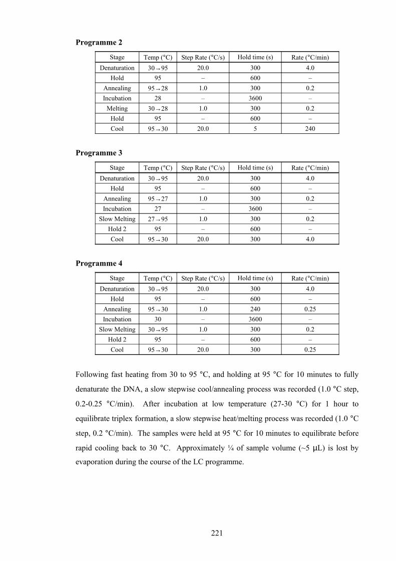
Programme 2
Stage Temp (°C) Step Rate (°C/s) Hold time (s) Rate (°C/min)
Denaturation 30→95 20.0 300 4.0
Hold 95 – 600 –
Annealing 95→28 1.0 300 0.2
Incubation 28 – 3600 –
Melting 30→28 1.0 300 0.2
Hold 95 – 600 –
Cool 95→30 20.0 5 240
Programme 3
Stage Temp (°C) Step Rate (°C/s) Hold time (s) Rate (°C/min)
Denaturation 30→95 20.0 300 4.0
Hold 95 – 600 –
Annealing 95→27 1.0 300 0.2
Incubation 27 – 3600 –
Slow Melting 27→95 1.0 300 0.2
Hold 2 95 – 600 –
Cool 95→30 20.0 300 4.0
Programme 4
Stage Temp (°C) Step Rate (°C/s) Hold time (s) Rate (°C/min)
Denaturation 30→95 20.0 300 4.0
Hold 95 – 600 –
Annealing 95→30 1.0 240 0.25
Incubation 30 – 3600 –
Slow Melting 30→95 1.0 300 0.2
Hold 2 95 – 600 –
Cool 95→30 20.0 300 0.25
Following fast heating from 30 to 95 °C, and holding at 95 °C for 10 minutes to fully
denaturate the DNA, a slow stepwise cool/annealing process was recorded (1.0 °C step,
0.2-0.25 °C/min). After incubation at low temperature (27-30 °C) for 1 hour to
equilibrate triplex formation, a slow stepwise heat/melting process was recorded (1.0 °C
step, 0.2 °C/min). The samples were held at 95 °C for 10 minutes to equilibrate before
rapid cooling back to 30 °C. Approximately ¼ of sample volume (~5 µL) is lost by
evaporation during the course of the LC programme.
221

9.2.6.3 Data Analysis
Tm values were derived from the negative derivatives of melting curves for each
experiment using the “Manual Tm” feature in Roche LightCycler Software Version 3.5.
9.2.7 Determining UV/Fluorescence Properties of Fluorescent Nucleosides
9.2.7.1 Deprotection of Nucleosides
DMT-protected nucleosides (143,182,225,226) were treated up to three times with 3%
TCA/DCM followed by removal of DCM in vacuo and trituration with diethyl ether
(×3). The aniline 143, acetamide 225 and urea 182 deprotected nucleosides were
afforded in this way as pale yellow to orange powders. The guanidine nucleoside (non-
protonated) was further treated with 20% Et2NH/CH3CN for 24 hours, concentrated in
vacuo, then treated with a mixture of 40% NH3/H2O and 7N NH3/CH3OH (1:2 v/v) for
60 hours, before concentration in vacuo and recrystallisation (CH3OH/Et2O). The fully
deprotected nucleoside 226 was afforded as a yellow-orange powder. Treatment with
20% diethylamine solution cleaved only one 2-cyanoethoxycarbonyl (CEOC) protecting
group from the guanidine moiety. Subsequent treatment with ammonia was necessary
to cleave the second. Treatment with ammonia only would have resulted in cyclisation
to form a stable triazine analogue of the guanidine moiety as reported in work by
Prakash et al.225 (Section 7.3.2.7).
9.2.7.2 UV Absorption Measurements
Solutions of nucleosides were made in either HPLC grade methanol (APPp 143, AcPPp
225) or anhydrous DMF (UPPp 182, GPPp 226) up to 0.174 µM (APPp), 0.109 µM (AcPPp),
0.151 µM (UPPp) and 0.154 µM (GPPp) concentrations. The solution being tested was
filtered through a Millipore Millex®-FH syringe filter (0.45µm, 13 mm) into a Hellma®
SUPRASIL synthetic quartz cuvette (10 mm pathlength, 1.5 mL sample volume). UV
absorption/excitation spectra were recorded at this and 2-3 lower concentrations by
successive dilution by factor of 2, on a Varian Cary 400 Scan UV-Visible
Spectrophotometer using Cary WinUV Scan application software. Spectra for
222

compounds run in DMF were dominated by noise below 260 nm, due to loss of solvent
transparency to UV light below ~270 nm.
Extinction coefficients (ε / L mol-1 cm-1) were calculated using the Beer-Lambert Law
(Equation 9.1)
A = ε.c.l
Equation 9.1. Beer-Lambert Law; A = absorbance (dimensionless), ε = molar extinction
coefficient (L mol-1 cm-1), c = concentration (M), l = pathlength (cm).
For each absorption/excitation maximum, the extinction coefficient was calculated by a
three-step process:
1. At each concentration measured, the value was calculated using Equation 9.1,
then the average of those values was taken (ignoring outliers).
2. The value was given as the gradient of a plot of Absorbance vs Molar
Concentration, ignoring significant outliers.
3. The average of the two values was taken to give an extinction coefficient for
each absorption/excitation maximum.
9.2.7.3 Fluorescence Measurements
Solutions of nucleosides were made in either HPLC grade methanol (APPp 143, AcPPp
225) or anhydrous DMF (UPPp 182, GPPp 226) up to concentrations of 1-3 mg/mL. The
solution being tested was filtered through a Millipore Millex®-FH syringe filter
(0.45µm, 13 mm) into a Hellma® SUPRASIL synthetic quartz cuvette for fluorimetry
(10 mm pathlength, 1.5 mL sample volume). An emission spectrum was run for each
sample, by irradiation at the principal absorption maximum (λ ~360 nm) for that
sample, on a Varian Cary Eclipse Fluorescence Spectrophotometer using Cary Eclipse
Scan application software. Each spectrum was an average of 10 consecutive scans from
380-600 nm.
223
References
1. Blackburn, G.M., Gait, M.J., Loakes, D., Williams, D.M., Nucleic Acids in
Chemistry and Biology, 3rd ed., RSC Publishing, 2006.
2. Neidle, S., DNA Structure and Recognition, 1st ed., IRL Press, Oxford
University Press Inc., New York, 1994.
3. Watson, J.D., The Double Helix: A New Critical Edition, Weidenfeld and
Nicolson Ltd., London, UK, 1981.
4. Watson, J.D., Crick, F.H.C., Nature (London), 1953, 171, 737-738.
5. Franklin, R.E., Gosling, R.G., Nature (London), 1953, 171, 740-741.
6. Franklin, R.E., Gosling, R.G., Nature (London), 1953, 172, 156-157.
7. Wilkins, M.H.F., Stokes, A.R., Wilson, H.R., Nature (London), 1953, 171, 738-
740.
8. Watson, J.D., Crick, F.H.C., Nature (London), 1953, 171, 964-967.
9. Wang, A.H.-J., Quigley, G.J., Kolpak, F.J., Crawford, J.L., van Boom, J.H., van
der Mare, G., Rich, A., Nature (London), 1979, 282(5740), 680-686.
10. Fox, K.R., Darby, R.A.J., Triple helix-specific ligands, in Small Molecule DNA
and RNA Binders, ed. M. Demeunynck, C. Bailly, W.D. Wilson, WILEY-VCH
Verlag GmbH & Co., Weinheim, Germany, 2003, Vol. 2, Ch. 14, pp 360-383.
11. Rusling, D.A., Brown, T., Fox, K.R., DNA Recognition by Triple Helix
Formation, in Sequence-specific DNA Binding Agents, ed. M.J. Waring, RSC
Publishing, Cambridge, UK, 2006, Ch. 1, pp 1-27.
12. Thuong, N.T., Hélène, C., Angew. Chem. Int. Ed. Engl., 1993, 32(5), 666-690.
13. Vasquez, K.M., Glazer, P.M., Q. Rev. Biophys., 2002, 35(1), 89-107.
14. Abu-Daya, A., Brown, P.M., Fox, K.R., Nucleic Acids Res., 1995, 23(17), 3385-
3392.
15. Felsenfeld, G., Davies, D.R., Rich, A., J. Am. Chem. Soc., 1957, 79, 2023-2024.
16. Asensio, J.L., Brown, T., Lane, A.N., Structure, 1999, 7(1), 1-11.
17. Winters, T.A., Curr. Opin. Mol. Ther., 2000, 2(6), 670-681.
18. Praseuth, D., Guieysse, A.L., Helene, C., BBA-Gene Struct. Expr., 1999,
1489(1), 181-206.
19. Karympalis, V., Kalopita, K., Zarros, A., Carageorgiou, H., Biochemistry
(Moscow), 2004, 69(8), 855-860.
20. Carbone, G.M., Napoli, S., Valentini, A., Cavalli, F., Watson, D.K., Catapano,
C.V., Nucleic Acids Res., 2004, 32(14), 4358-4367.
224
21. Majumdar, A., Puri, N., Cuenoud, B., Natt, F., Martin, P., Khorlin, A., Dyatkina,
N., George, A.J., Miller, P.S., Seidman, M.M., J. Biol. Chem., 2003, 278(13),
11072-11077.
22. Buchini, S., Leumann, C.J., Curr. Opin. Chem. Biol., 2003, 7(6), 717-726.
23. Moser, H.E., Dervan, P.B., Science, 1987, 238(4827), 645-650.
24. Antony, S., Arimondo, P.B., Sun, J.S., Pommier, Y., Nucleic Acids Res., 2004,
32(17), 5163–5173.
25. Woynarowski, J.M., Adv. DNA Seq. Spec. Agents, 2002, 4, 1-27.
26. Seidman, M.M., Glazer, P.M., J. Clin. Invest., 2003, 112(4), 487-494.
27. Guillonneau, F., Guieysse, A.L., Nocentini, S., Giovannangeli, C., Praseuth, D.,
Nucleic Acids Res., 2004, 32(3), 1143-1153.
28. Kalish, J.M., Seidman, M.M., Weeks, D.L., Glaze, P.M., Nucleic Acids Res.,
2005, 33(11), 3492–3502.
29. Ma, Z., Taylor, J.-S., Proc. Natl. Acad. Sci. U. S. A., 2000, 97(21), 11159-11163.
30. Bello-Roufai, M., Roulon, T., Escude, C., Chem. Biol., 2004, 11(4), 509-516.
31. Whiteford, N., Haslam, N., Weber, G., Prügel-Bennett, A., Essex, J.W., Roach,
P.L., Bradley, M., Neylon, C., Nucleic Acids Res., 2005, 33(19), e171.
32. Hoogsteen, K., Acta Crystallogr., 1959, 12, 822-823.
33. Fox, K.R., Curr. Med. Chem., 2000, 7(1), 17-37.
34. Gowers, D.M., Fox, K.R., Nucleic Acids Res., 1999, 27(7), 1569-1577.
35. Sollogoub, M., Dominguez, B., Fox, K.R., Brown, T., Chem. Commun., 2000,
23, 2315-2316.
36. Sollogoub, M., Darby, R.A.J., Cuenoud, B., Brown, T., Fox, K.R., Biochemistry,
2002, 41(23), 7224-7231.
37. Osborne, S.D., Powers, V.E.C., Rusling, D.A., Lack, O., Fox, K.R., Brown, T.,
Nucleic Acids Res., 2004, 32(15), 4439-4447.
38. Cuenoud, B., Casset, F., Husken, D., Natt, F., Wolf, R.M., Altmann, K.-H.,
Martin, P., Moser, H.E., Angew. Chem. Int. Ed., 1998, 37(9), 1288-1291.
39. Bijapur, J., Keppler, M.D., Bergqvist, S., Brown, T., Fox, K.R., Nucleic Acids
Res., 1999, 27(8), 1802-1809.
40. Froehler, B.C., Wadwani, S., Terhorst, T.J., Gerrard, S.R., Tetrahedron Lett.,
1992, 33(37), 5307-5310.
41. Prakash, T.P., Puschl, A., Lesnik, E., Mohan, V., Tereshko, V., Egli, M.,
Manoharan, M., Org. Lett., 2004, 6(12), 1971-1974.
225

42. Manoharan, M., Prakash, T.P., Barber-Peoc'h, I., Bhat, B., Vasquez, G., Ross,
B.S., Cook, P.D., J. Org. Chem., 1999, 64(17), 6468-6472.
43. Deglane, G., Abes, S., Michel, T., Prévot, P., Vives, E., Debart, F., Barvik, I.,
Lebleu, B., Vasseur, J.-J., ChemBioChem, 2006, 7(4), 684 – 692.
44. Roig, V., Asseline, U., J. Am. Chem. Soc., 2003, 125(15), 4416-4417.
45. Lee, J.S., Woodsworth, M.L., Latimer, L.J.P., Morgan, A.R., Nucleic Acids Res.,
1984, 12(16), 6603-6614.
46. Xodo, L.E., Manzini, G., Quadrifoglio, F., Van der Marel, G.A., Van Boom,
J.H., Nucleic Acids Res., 1991, 19(20), 5625-31.
47. Povsic, T.J., Dervan, P.B., J. Am. Chem. Soc., 1989, 111(8), 3059-3061.
48. Singleton, S.F., Dervan, P.B., Biochemistry, 1992, 31(45), 10995-11003.
49. Barawkar, D.A., Kumar, V.A., Ganesh, K.N., Biochem. Biophys. Res. Commun.,
1994, 205(3), 1665-1670.
50. Barawkar, D.A., Rajeev, K.G., Kumar, V.A., Ganesh, K.N., Nucleic Acids Res.,
1996, 24(7), 1229–1237.
51. Rajeev, K.G., Jadhav, V.R., Ganesh, K.N., Nucleic Acids Res., 1997, 25(21),
4187–4193.
52. Doronina, S.O., Behr, J.-P., Tetrahedron Lett., 1998, 39(7), 547-550.
53. Robles, J., Grandas, A., Pedroso, E., Tetrahedron, 2001, 57(1), 179-194.
54. Robles, J., Grandas, A., Pedroso, E., Nucleos. Nucleot. Nucl., 2003, 22(5-8),
1085-1087.
55. Ono, A., Ts’o, P.O.P., Kan, L.-S., J. Am. Chem. Soc., 1991, 113(10), 4032-4033.
56. Ono, A., Ts’o, P.O.P., Kan, L.-S., J. Org. Chem., 1992, 57(11), 3225-3230.
57. Christensen, C., Eldrup, A.B., Haaima, G., Nielsen, P.E., Bioorg. Med. Chem.
Lett., 2002, 12(21), 3121-3124.
58. Nielsen, P.E., Egholm, M., Bèrg, R.H., Buchardt, O., Science, 1991, 254(5037),
1497-1500.
59. Egholm, M., Buchardt, O., Nielsen, P.E., Bèrg, R.H., J. Am. Chem. Soc., 1992,
114(5), 1895-1897.
60. Sollogoub, M., Fox, K.R., Powers, V.E.C., Brown, T., Tetrahedron Lett., 2002,
43(17), 3121-3123.
61. Powers, V.E.C., Synthesis and studies of nucleoside analogues in triplex
forming oligonucleotides, PhD Thesis, University of Southampton,
Southampton, UK, 2004.
226

62. Bates, P.J., Laughton, C.A., Jenkins, T.C., Capaldi, D.C., Roselt, P.D., Reese,
C.B., Neidle, S., Nucleic Acids Res., 1996, 24(21), 4176-4184.
63. Cassidy, S.A., Slickers, P., Trent, J.O., Capaldi, D.C., Roselt, P.D., Reese, C.B.,
Neidle, S., Fox, K.R., Nucleic Acids Res., 1997, 25(24), 4891-4898.
64. Hildbrand, S., Leumann, C.J., Angew. Chem. Int. Ed. Engl., 1996, 35(17), 1968-
1970.
65. Hildbrand, S., Blaser, A., Parel, S.P., Leumann, C.J., J. Am. Chem. Soc., 1997,
119(24), 5499-5511.
66. Reese, C.B., Wu, Q., Org. Biomol. Chem., 2003, 1(18), 3160-3172.
67. Rusling, D.A., Powers, V.E.C., Ranasinghe, R.T., Wang, Y., Osborne, S.D.,
Brown, T., Fox, K.R., Nucleic Acids Res., 2005, 33(9), 3025-3032.
68. von Krosigk, U., Benner, S.A., J. Am. Chem. Soc., 1995, 117(19), 5361-5362.
69. Hutter, D., Benner, S.A., J. Org. Chem., 2003, 68(25), 9839-9842.
70. Berressem, R., Engels, J.W., Nucleic Acids Res., 1995, 23(17), 3465-3472.
71. Parsch, U., Engels, J.W., Chem. Eur. J., 2000, 6(13), 2409-2424.
72. Xiang, G., Soussou, W., McLaughlin, L.W., J. Am. Chem. Soc., 1994, 116(24),
11155-11156.
73. Xiang, G., Bogacki, R., McLaughlin, L.W., Nucleic Acids Res., 1996, 24(10),
1963-1970.
74. Xiang, G., McLaughlin, L.W., Tetrahedron, 1998, 54(3-4), 375-392.
75. Brodin, P., Pinskaya, M., Parsch, U., Bischerour, J., Leh, H., Romanova, E.,
Engels, J.W., Gottikh, M., Mouscadet, J.F., Nucleos. Nucleot. Nucl., 2001, 20(4-
7), 481-486.
76. Wang, Y., Rusling, D.A., Powers, V.E.C., Lack, O., Osborne, S.D., Fox, K.R.,
Brown, T., Biochemistry, 2005, 44(15), 5884-5892.
77. Hunziker, J., Priestley, E.S., Brunar, H., Dervan, P.B., J. Am. Chem. Soc., 1995,
117(9), 2661-2662.
78. Brunar, H., Dervan, P.B., Nucleic Acids Res., 1996, 24(11), 1987-1991.
79. Koshlap, K.M., Schultze, P., Brunar, H., Dervan, P.B., Feigon, J., Biochemistry,
1997, 36(9), 2659-2668.
80. St. Clair, A., Xiang, G., McLaughlin, L.W., Nucleos. Nucleot. Nucl., 1998,
17(5), 925-938.
81. Koh, J.S., Dervan, P.B., J. Am. Chem. Soc., 1992, 114(4), 1470-1478.
82. Radhakrishnan, I., Patel, D.J., Priestley, E.S., Nash, H.M., Dervan, P.B.,
Biochemistry, 1993, 32(41), 11228-11234.
227

83. Priestley, E.S., Dervan, P.B., J. Am. Chem. Soc., 1995, 117, 4761-4765.
84. D’Costa, M., Kumar, V.A., Ganesh, K.N., J. Org. Chem., 2003, 68(25), 9839-
9842.
85. Marfurt, J., Parel, S.P., Leumann, C.J., Nucleic Acids Res., 1997, 25(10), 1875-
1882.
86. Miller, P.S., Bhan, P., Cushman, C.D., Trapane, T.L., Biochemistry, 1992,
31(29), 6788-93.
87. Krawczyk, S.H., Milligan, J.F., Wadwani, S., Moulds, C., Froehler, B.C.,
Matteucci, M.D., Proc. Natl. Acad. Sci. U. S. A., 1992, 89(9), 3761-3764.
88. Jetter, M.C., Hobbs, F.C., Biochemistry, 1993, 32(13), 3249-3254.
89. Lin, S.-B., Wu, C.-M., Lin, W.-C., Kan, L.-S., J. Chin. Chem. Soc. (Taipei),
1999, 46(5), 693-697.
90. Young, S.L., Krawczyk, S.H., Matteucci, M.D., Toole, J.J., Proc. Natl. Acad.
Sci. U. S. A., 1991, 88, 10023-10026.
91. Durland, R.H., Rao, T.S., Jayaraman, K., Revankar, G.R., Bioconjugate Chem.,
1995, 6(3), 278-282.
92. Staubli, A.B., Dervan, P.B., Nucleic Acids Res., 1994, 22(13), 2637-2642.
93. Flanagan, W.M., Wagner, R.W., Grant, D., Lin, K.-Y., Matteucci, M.D., Nature
Biotech., 1999, 17, 48-52.
94. Matteucci, M.D., von Krosigk, U., Tetrahedron Lett., 1996, 37(29), 5057-5060.
95. Lin, K.-Y., Jones, R.J., Matteucci, M., J. Am. Chem. Soc., 1995, 117(13), 3873-
3874.
96. Sandin, P., Wilhelmsson, L.M., Lincoln, P., Powers, V.E.C., Brown, T.,
Albinsson, B., Nucleic Acids Res., 2005, 33(16), 5019–5025.
97. Wilhelmsson, L.M., Holmén, A., Lincoln, P., Nielsen, P.E., Nordén, B., J. Am.
Chem. Soc., 2001, 123(10), 2434-2435.
98. Guianvarc’h, D., Benhida, R., Fourrey, J.-L., Maurisse, R., Sun, J.-S., Chem.
Commun., 2001, (18), 1814–1815.
99. Guianvarc’h, D., Fourrey, J.-L., Maurisse, R., Sun, J.S., Benhida, R., Bioorg.
Med. Chem., 2003, 11(13), 2751–2759.
100. Griffin, L.C., Kiessling, L.L., Beal, P.A., Gillespie, P., Dervan, P.B., J. Am.
Chem. Soc., 1992, 114(21), 7976-7982.
101. Koshlap, K.M., Gillespie, P., Dervan, P.B., Feigon, J., J. Am. Chem. Soc., 1993,
115(17), 7908-7909.
228

102. Wang, E., Koshlap, K.M., Gillespie, P., Dervan, P.B., Feigon, J., J. Mol. Biol.,
1996, 257(5), 1052–1069.
103. Blommers, M.J.J., Natt, F., Jahnke, W., Cuenoud, B., Biochemistry, 1998,
37(51), 17714-17725.
104. Puri, N., Majumdar, A., Cuenoud, B., Miller, P.S., Seidman, M.M.,
Biochemistry, 2004, 43(5), 1343-1351.
105. Buchini, S., Leumann, C.J., Eur. J. Org. Chem., 2006, (14), 3152–3168.
106. Yoon, K., Hobbs, C.A., Koch, J., Sardaro, M., Kutny, R., Weis, A.L., Proc.
Natl. Acad. Sci. U. S. A., 1992, 89(9), 3840-3844.
107. Obika, S., Hari, Y., Sekiguchi, M., Imanishi, T., Angew. Chem. Int. Ed., 2001,
40(11), 2079-2081.
108. Obika, S., Hari, Y., Sekiguchi, M., Imanishi, T., Chem. Eur. J., 2002, 8(20),
4796-4802.
109. Hari, Y., Obika, S., Sekiguchi, M., Imanishi, T., Tetrahedron, 2003, 59(27),
5123-5128.
110. Obika, S., Hari, Y., Sugimoto, T., Sekiguchi, M., Imanishi, T., Tetrahedron
Lett., 2000, 41(46), 8923-8927.
111. Prévot-Halter, I., Leumann, C.J., Bioorg. Med. Chem. Lett., 1999, 9(18), 2657-
2660.
112. Prévot, I., Leumann, C.J., Helv. Chim. Acta, 2002, 85(2), 502-515.
113. Buchini, S., Leumann, C.J., Tetrahedron Lett., 2003, 44(27), 5065-5068.
114. Buchini, S., Leumann, C.J., Angew. Chem. Int. Ed., 2004, 43(30), 3925 –3928.
115. Rao, M.S., Esho, N., Sergeant, C., Dembinski, R., J. Org. Chem., 2003, 68(17),
6788-6790.
116. Robins, M.J., Barr, P.J., J. Org. Chem., 1983, 48(11), 1854-1862.
117. Cruickshank, K.A., Stockwell, D.L., Tetrahedron Lett., 1988, 29(41), 5221-
5224.
118. Woo, J., Meyer Jr., R.B., Gamper, H.B., Nucleic Acids Res., 1996, 24(13),
2470–2475.
119. Berry, D.A., Jung, K.-Y., Wise, D.S., Sercel, A.D., Pearson, W.H., Mackie, H.,
Randolph, J.B., Somers, R.L., Tetrahedron Lett., 2004, 45(11), 2457–2461.
120. Hudson, R.H.E., Ghorbani-Choghamarani, A., Synlett, 2007, (6), 870-873.
121. McGuigan, C., Barucki, H., Blewett, S., Carangio, A., Erichsen, J.T., Andrei, G.,
Snœck, R., Clercq, E.D., Balzarini, J., J. Med. Chem., 2000, 43(26), 4993-4997.
229

122. Ranasinghe, R.T., Rusling, D.A., Powers, V.E.C., Fox, K.R., Brown, T., Chem.
Commun., 2005, (20), 2555-2557.
123. Sonogashira, K., Tohda, Y., Hagihara, N., Tetrahedron Lett., 1975, 50, 4467-
4470.
124. Hobbs Jr., F.W., J. Org. Chem., 1989, 54(14), 3420-3422.
125. Chin, T.-M., Chung, K.-Y., Chen, J.-J., Lin, W.-C., Kan, L.S., J. Chin. Chem.
Soc. (Taipei), 1999, 46(5), 751-758.
126. Chin, T.-M., Tseng, M.-H., Chung, K.-Y., Hung, F.-S., Lin, S.-B., Kan, L.-S., J.
Biomol. Struct. Dyn., 2001, 19(3), 543-553.
127. Gianolio, D.A., McLaughlin, L.W., J. Am. Chem. Soc., 1999, 121(26), 6334-
6335.
128. Guzzo-Pernell, N., Tregear, G.W., Haralambidis, J., Lawlor, J.M., Nucleosides
Nucleotides, 1998, 17(7), 1191-1207.
129. Huang, C.-Y., Bi, G., Miller, P.S., Nucleic Acids Res., 1996, 24(13), 2606-2613.
130. Huang, C.-Y., Cushman, C.D., Miller, P.S., J. Org. Chem., 1993, 58(19), 5048-
5049.
131. Huang, C.-Y., Miller, P.S., J. Am. Chem. Soc., 1993, 115(22), 10456-10457.
132. Guzzo-Pernell, N., Lawlor, J.M., Tregear, G.W., Haralambidis, J., Aust. J.
Chem., 1998, 51(11), 965-972.
133. Zimmerman, S.C., Schmitt, P., J. Am. Chem. Soc., 1995, 117(43), 10769-10770.
134. Lengeler, D., Weisz, K., Tetrahedron Lett., 2001, 42(8), 1479–1481.
135. Mertz, E., Mattei, S., Zimmerman, S.C., Org. Lett., 2000, 2(19), 2931-2934.
136. Xiao, Z., Weisz, K., J. Phys. Org. Chem., 2007, 20(10), 771-777.
137. Mertz, E., Mattei, S., Zimmerman, S.C., Bioorg. Med. Chem., 2004, 12(6),
1517-1526.
138. Purwanto, M.G.M., Lengeler, D., Weisz, K., Tetrahedron Lett., 2002, 43(1), 61-
64.
139. Purwanto, M.G.M., Weisz, K., Curr. Org. Chem., 2003, 7(5), 427-446.
140. Purwanto, M.G.M., Weisz, K., Tetrahedron Lett., 2006, 47(23), 3849–3852.
141. Durland, R.H., Rao, S., Bodepudi, V., Seth, D.M., Jayaraman, K., Revankar,
G.R., Nucleic Acids Res., 1995, 23(4), 647-653.
142. Sasaki, S., Taniguchi, Y., Takahashi, R., Senko, Y., Kodama, K., Nagatsugi, F.,
Maeda, M., J. Am. Chem. Soc., 2003, 126(2), 516-528.
143. de la Paz, M.L., González, C., Vicent, C., Chem. Commun., 2000, (5), 411–412.
144. McBride, L.J., Caruthers, M.H., Tetrahedron Lett., 1983, 24(3), 245-248.
230

145. Carreño, M.C., Ruano, J.L.G., Sanz, G., Toledo, M.A., Urbano, A., Tetrahedron
Lett., 1996, 37(23), 4081-4084.
146. Das, B., Kundu, N.G., Synth. Commun., 1988, 18(8), 855-868.
147. Ge, P., Kirk, K.L., J. Org. Chem., 1997, 62(10), 3340-3343.
148. Hudson, R.H.E., Li, G., Tse, J., Tetrahedron Lett., 2002, 43(8), 1381-1386.
149. Robins, M.J., Barr, P.J., Giziewicz, J., Can. J. Chem., 1982, 60, 554-557.
150. Boden, N., Bushby, R.J., Cammidge, A.N., Headdock, G., Tetrahedron Lett.,
1995, 36(47), 8685-8686.
151. Harrowven, D.C., Woodcock, T., Howes, P.D., Tetrahedron Lett., 2002, 43(51),
9327-9329.
152. Kajigaeshi, S., Kakinami, T., Yamasaki, H., Fujisaki, S., Manabu, K., Okamoto,
T., Chem. Lett., 1987, (11), 2109-2112.
153. Quintin, J., Lewin, G., Tetrahedron Lett., 2004, 45, 3635–3638.
154. Kosynkin, D.V., Tour, J.M., Org. Lett., 2001, 3(7), 991-992.
155. Kajigaeshi, S., Kakinami, T., Moriwaki, M., Watanabe, M., Fujisaki, S.,
Okamoto, T., Chem. Lett., 1988, (5), 795-798.
156. Mitra, S.S., Sreekumar, K., Indian J. Chem., Sect. B, 1997, 36B(2), 133-136.
157. Mitra, S.S., Sreekumar, K., React. Funct. Polym., 1997, 32(3), 281-291.
158. Mitra, S.S., Sreekumar, K., Polym. Int., 1997, 42(2), 173-178.
159. Mitra, S.S., Sreekumar, K., Polymer, 1997, 38(6), 1363-1366.
160. Haszeldine, R.N., Sharpe, A.G., J. Chem. Soc., 1952, 993-1001.
161. Hajipour, A.R., Arbabian, M., Ruoho, A.E., J. Org. Chem., 2002, 67(24), 8622-
8624.
162. Pan, B.-C., Chen, Z.-H., Chu, E., Chu, M.-Y.W., Chu, S.-H., Nucleosides
Nucleotides, 1998, 17(12), 2367-2382.
163. Hirota, K., Sako, M., Sajiki, H., Heterocycles, 1997, 46, 547-554.
164. Djerassi, C., Lenk, C.T., J. Am. Chem. Soc., 1953, 75(14), 3493-3495.
165. Markiewicz, W.T., Padyukova, N.S., Samek, Z., Smrt, J., Collect. Czech. Chem.
Commun., 1980, 45(6), 1860-1865.
166. Mitsunobu, O., Synthesis, 1981, 1-28.
167. Vorbrüggen, H., Krolikiewicz, K., Bennua, B., Chem. Ber., 1981, 114(4), 1235-
1245.
168. Robins, M.J., Barr, P.J., Tetrahedron Lett., 1981, 22(5), 421-424.
169. Wang, L., Yan, J., Li, P.H., Wang, M., Su, C.N., J. Chem. Res., Synop., 2005,
(2), 112-115.
231
170. Bijapur, J., Factors Affecting the Stability of Nucleic Acids, PhD Thesis,
University of Southampton, Southampton, UK, 1999.
171. Alami, M., Crousse, B., Ferri, F., J. Organomet. Chem., 2001, 624, 114-123.
172. Bhattacharya, S., Sengupta, S., Tetrahedron Lett., 2004, 45, 8733-8736.
173. Negishi, E.-i., Qian, M., Zeng, F., Anastasia, L., Babinski, D., Org. Lett., 2003,
5(10), 1597-1600.
174. Vincent, P., Beaucourt, J.-P., Pichat, L., Tetrahedron Lett., 1981, 22, 94947.
175. Stille, J.K., Simpson, J.H., J. Am. Chem. Soc., 1987, 109, 2138-2152.
176. Phillips, A.P., J. Am. Chem. Soc., 1951, 73(3), 1061-1062.
177. Gütschow, M., Hecker, T., Thiele, A., Hauschildt, S., Eger, K., Bioorg. Med.
Chem., 2001, 9(4), 1059-1065.
178. Lin, T.-S., Guo, J.-Y., Schinazi, R.F., Chu, C.K., Xiang, J.-N., Prusoff, W.H., J.
Med. Chem., 1988, 31(2), 336-340.
179. Storek, M.J., Suciu, A., Verdine, G.L., Org. Lett., 2002, 4(22), 3867-3869.
180. Azam, A.T.M.Z., Moriguchi, T., Shinozuka, K., Chem. Commun., 2006, (3),
335-337.
181. Ben-Gaied, N., Zhao, Z., Gerrard, S.R., Fox, K.R., Brown, T., ChemBioChem,
2009, in press.
182. Asseline, U., Chassignol, M., Draus, J., Durand, M., Maurizot, J.-C., Bioorg.
Med. Chem., 2003, 11(16), 3499-3511.
183. Yang, W., Giandomenico, C.M., Sartori, M., Moore, D.A., Tetrahedron Lett.,
2003, 44(12), 2481-2483.
184. Brederick, H., Gottfried, K., Wieland, H., Chem. Ber., 1959, 92(3), 583-594.
185. Schroeder, E.F., G. D. Searle & Co. 5-Halo-6-Amino-Uracils and Derivatives
Thereof. US 2731465, 17th January 1956.
186. Rybár, A., Hesek, D., Szemes, F., Alföldi, J., Collect. Czech. Chem. Commun.,
1990, 55(9), 2257-2269.
187. Zhang, L., Zhang, Y.J., Tetrahedron Lett., 2006, 47(5), 775-778.
188. Kupchan, S.M., Bondesson, G., Davies, A.P., J. Med. Chem., 1972, 15(1), 65-
68.
189. Taylor, E.C., Loux, H.M., Falco, E.A., Hitchings, G.H., J. Am. Chem. Soc.,
1955, 77(8), 2243 - 2248.
190. Fieser, L.F., Brown, R.H., J. Am. Chem. Soc., 1949, 71(11), 3609-3614.
191. Hansen, B., Acta Chem. Scand., 1957, 11, 537-540.
232

192. O'Sullivan, M.C., Dalrymple, D.M., Tetrahedron Lett., 1995, 36(20), 3451-
3452.
193. Cheng, C.-C., Lu, Y.-L., Chem. Commun., 1998, 253-254.
194. Koščová, S., Budĕšínský, M., Hodačová, J., Collect. Czech. Chem. Commun.,
2003, 68(4), 744-750.
195. Lee, Y., Sarjeant, A.A.N., Karlin, K.D., Chem. Commun., 2006, 621-623.
196. Zoller, U., Three-Membered Rings Containing Sulfur, in Small Ring
Heterocycles, Chemistry of Heterocyclic Compounds: A Series of Monographs,
ed. A. Hassner, John Wiley & Sons 1983, Vol. 42, Part 1, Ch. III, pp 333-629.
197. Choi, J., Yoon, N.M., Synth. Commun., 1995, 25(17), 2655-2663.
198. Zheng, T.-C., Burkart, M., Richardson, D.E., Tetrahedron Lett., 1999, 40(4),
603-606.
199. Cummins, L.L., Owens, S.R., Risen, L.M., Lesnik, E.A., Freier, S.M., McGee,
D., Guinosso, C.J., Cook, P.D., Nucleic Acids Res., 1995, 23(11), 2019-2024.
200. Legorburu, U., Reese, C.B., Song, Q., Tetrahedron, 1999, 55(17), 5635-5640.
201. Reese, C.B., Song, Q. 2'-Substituted RNA Preparation. WO 00/56747, 28th
September 2000.
202. Manfredini, S., Baraldi, P.G., Durini, E., Porcu, L., Angusti, A., Vertuani, S.,
Solaroli, N., De Clercq, E., Karlsson, A., Balzarini, J., Bioorg. Med. Chem. Lett.,
2001, 11(10), 1329-1332.
203. Ross, B.S., Springer, R.H., Tortorici, Z., Dimock, S., Nucleos. Nucleot. Nucl.,
1997, 16(7-9), 1641-1644.
204. Reichman, U., Chu, c.K., Hollenberg, K.A., Watanabe, K.A., Fox, J.J.,
Synthesis, 1976, (8), 533-534.
205. McGee, D.P.C., Zhai, Y., Nucleos. Nucleot. Nucl., 1996, 15(11-12), 1797-1803.
206. Vadhia, S.J., The Effects of Modified Nucleosides on DNA Duplexes and
Triplexes, PhD Thesis, University of Southampton, Southampton, UK, 2007.
207. Gerrard, S.R., Srinivasan, N., Fox, K.R., Brown, T., Nucleos. Nucleot. Nucl.,
2007, 26(10-12), 1363-1367.
208. Melissaris, A.P., Litt, M.H., J. Org. Chem., 1994, 59(19), 5818-5821.
209. Classon, B., Samuelsson, B., Acta Chem. Scand. Ser. B, 1985, 39(6), 501-504.
210. Kabalka, G.W., Wang, L., M., P.R., Tetrahedron, 2001, 57(38), 8017-8028.
211. Arnold, F.E., Reinhardt, B.A., Hedberg, F.L. Aromatic enyne compounds and
their synthesis. US 4162265, 24th July 1979.
233

212. Herdewijn, P., Balzarini, J., Baba, M., Pauwels, R., Aerschot, A.V., Janssen, G.,
Clercq, E.D., J. Med. Chem., 1988, 31(10), 2040-2048.
213. Divakar, K.J., Sawant, C.M., Mulla, Y.A., Zemse, D.V., Sitabkhan, S.M., Ross,
B.S., Sanghvi, Y.S., Nucleos. Nucleot. Nucl., 2003, 22(5-8), 1321-1325.
214. Lyttle, M.H., Walton, T.A., Dick, D.J., Carter, T.G., Beckman, J.H., Cook,
R.M., Bioconjugate Chem., 2002, 13(5), 1146-1154.
215. San-Félix, A., Velázquez, S., Pérez-Pérez, M.J., Balzarini, J., Clercq, E.D.,
Camarasa, M.J., J. Med. Chem., 1994, 37(4), 453-460.
216. Miah, A., Reese, C.B., Song, Q., Nucleosides Nucleotides, 1997, 16(1&2), 53-
65.
217. Jin, Y.H., Liu, P., Wang, J., Baker, R., Huggins, J., Chu, C.K., J. Org. Chem.,
2003, 68(23), 9012-9018.
218. Song, G.Y., Paul, V., Choo, H., Morrey, J., Sidwell, R.W., Schinazi, R.F., Chu,
C.K., J. Med. Chem., 2001, 44(23), 3985-3993.
219. Shi, J., Du, J., Ma, T., Pankiewicz, K.W., Patterson, S.E., Tharnish, P.M.,
McBrayer, T.R., Stuyver, L.J., Otto, M.J., Chu, C.K., Schinazic, R.F., Watanabe,
K.A., Bioorg. Med. Chem., 2005, 13, 1641-1652.
220. Holmes, S.C., Gait, M.J., Nucleos. Nucleot. Nucl., 2003, 22(5-8), 1259-1262.
221. Nyilas, Á., Chattopadhyaya, J., Acta Chem. Scand. Ser. B, 1986, 40(10), 826-
830.
222. Inoue, H., Imura, A., Ohtsuka, E., Nippon Kagaku Kaishi, 1987, (7), 1214-1220.
223. Inoue, H., Otsuka, E., Imura, A. Preparation of fluorescent nucleosides or
nucleotides and their use for DNA hybridization probes. JP99-255499, 11th July
1987.
224. Nurminen, E.J., Mattinen, J.K., Lönnberg, H., J. Chem. Soc., Perkin Trans. 2,
2001, (11), 2159–2165.
225. Prakash, T.P., Püschl, A., Manoharan, M., Nucleos. Nucleot. Nucl., 2007, 26(2),
149-159.
226. Segev, D. System for delivering therapeutic agents into living cells and cell
nuclei. US 20060160763, 20th July 2006.
227. Saxena, A.K., Pandey, S.K., Seth, P., Singh, M.P., Dikshit, M., Carpy, A.,
Bioorg. Med. Chem., 2001, 9, 2025-2034.
228. Bernat, J., Kristian, P., Guspanova, J., Imrich, J., Busova, T., Collect. Czech.
Chem. Commun., 1997, 62, 1491-1496.
234

229. Shibahara, F., Suenami, A., Yoshida, A., Murai, T., Chem. Commun., 2007,
(23), 2354-2356.
230. Hudson, R.H.E., Dambenieks, A.K., Heterocycles, 2006, 68(7), 1325-1328.
231. Hudson, R.H.E., Dambenieks, A.K., Viirre, R.D., Synlett, 2004, (13), 2400-
2402.
232. Hudson, R.H.E., Dambenieks, A.K., Viirre, R.D., Nucleos. Nucleot. Nucl., 2005,
24(5-7), 581-584.
233. Hudson, R.H.E., Ghorbani-Choghamarani, A., Nucleos. Nucleot. Nucl., 2007,
26(6&7), 533-537.
234. Hudson, R.H.E., Moszynski, J.M., Synlett, 2006, (18), 2997-3000.
235. Wojciechowski, F., Hudson, R.H.E., J. Am. Chem. Soc., 2008, 130(38), 12574-
12575.
236. Yoshimoto, K., Xu, C.-Y., Nishizawa, S., Haga, T., Satake, H., Teramae, N.,
Chem. Commun., 2003, (24), 2960-2961.
237. Van Daele, I., Bomholt, N., Filichev, V.V., Van Calenbergh, S., Pedersen, E.B.,
ChemBioChem, 2008, 9, 791-801.
238. Caruthers, M.H., Beaucage, S.L., Unit 3.3, in Current Protocols in Nucleic Acid
Chemistry, ed. S.L. Beaucage, D.E. Bergstrom, G.D. Glick, R.A. Jones, John
Wiley & Sons Inc. 2002, Vol. 1.
239. Beaucage, S.L., Caruthers, M.H., Tetrahedron Lett., 1981, 22(20), 1859-1862.
240. Matteucci, M., Caruthers, M.H., J. Am. Chem. Soc., 1981, 103(11), 3185-3191.
241. Albericio, F., Kates, S.A. Automated allyl deprotection in solid-phase synthesis.
US 5777077, 7th July 1998.
242. Albericio, F., Kates, S.A. Automated allyl deprotection in solid-phase synthesis.
US 5861351, 19th January 1999.
243. Hsiung, H.M. Process for de-cyanoethylating blocked nucleotides. US 4419509,
30th March 1983.
244. Hsiung, H.M. Process for de-cyanoethylating blocked nucleotides. US 4426517,
24th August 1984.
245. Umemoto, T., Wada, T., Tetrahedron Lett., 2005, 46(24), 4251-4253
246. Bhan, A., Holmberg, L. Method for deprotecting oligonucleotides. US 6887990,
3rd May 2005.
247. Gottlieb, H.E., Kotlyar, V., Nudelman, A., J. Org. Chem., 1997, 62(21), 7512-
7515.
248. Winkley, M.W., Robins, R.K., J. Chem. Soc. C, 1969, (5), 791 - 796.
235

249. Guthrie, R.D., Smith, S.C., Biochem. Prep., 1971, 13, 1-3.
250. Kam, B.L., Barascut, J.-L., Imbach, J.-L., Carbohydr. Res., 1979, 69(1), 135-
142.
251. Pike, A.R., Ryder, L.C., Horrocks, B.R., Clegg, W., Elsegood, M.R.J., Connolly,
B.A., Houlton, A., Chem. Eur. J., 2002, 8(13), 2891-2899.
252. Hay, M.P., Pruijn, F.B., Gamage, S.A., Liyanage, H.D.S., Kovacs, M.S.,
Patterson, A.V., Wilson, W.R., Brown, J.M., Denny, W.A., J. Med. Chem.,
2004, 47(2), 475-488.
253. Bleicher, L.S., Cosford, N.D.P., Herbaut, A., McCallum, J.S., McDonald, I.A.,
J. Org. Chem., 1998, 63(4), 1109-1118.
254. Bosch, E., Jeffries, L., Tetrahedron Lett., 2001, 42(46), 8141–8142.
255. Battersby, T.R., Ang, D.N., Burgstaller, P., Jurczyk, S.C., Bowser, M.T.,
Buchanan, D.D., Kennedy, R.T., Benner, S.A., J. Am. Chem. Soc., 1999,
121(42), 9781-9789.
256. Langley, G.J., Herniman, J.M., Davies, N.L., Brown, T., Rapid Commun. Mass
Spectrom., 1999, 13(17), 1717-1723.
257. Sambrook, J., Fritsch, E.F., Maniatis, T., Molecular Cloning: a laboratory
manual, Vol. 3, 2nd ed., Cold Spring Harbor Laboratory Press, New York,
U.S.A., 1989.
236

Appendices
237

A: List of Synthesised Compounds
Cmpd No. Structure Name
65
O
OAcO
AcO1
2
5
N1'
2'
O
O
5'
6'
OAc 1,3,5-tri-O-acetyl-2-O-(2-phthalimidoethyl)-D-
ribofuranose
68O
NH
F3C N-trifluoroacetyl propargylamine
71 NICl2
12
34
5
6
7
benzyltriethylammonium dichloroiodate
741'
2'3'
4'
5'
2
45
6
O
OHO
OSi
Si
O
N
NH
N
OO
Cl
NMe2
N1-[1-(3’,5’-O-(1,1,3,3-tetraisopropyldisiloxane-1,3-
diyl)-β-D-ribofuranosyl)]-(5-chloro-6-oxocytosin-4-
yl)-N,N-dimethylformamidine
75NH
NH2
ONO
OAcAcO
AcO O
5
1'
5'
4'
3' 2'
2’,3’,5’-tri-O-acetyl-6-oxocytidine
791'
5'
4'
3' 2'
NH
NH2
ONO
OAcAcO
AcO O
5Br
5-bromo-2’,3’,5’-tri-O-acetyl-6-oxocytidine
831'
5'
4'
3' 2'
NH
NH2
ONO
OAcAcO
AcO O
5I
5-iodo-2’,3’,5’-tri-O-acetyl-6-oxocytidine
85O
OHOH
HOOMe1
23
4
5
1-O-methyl-D-ribofuranose
86O
OHO
OSi
Si
OOMe1
23
4
51-O-methyl-3,5-O-(1,1,3,3-tetraisopropyldisiloxane-
1,3-diyl)-D-ribofuranose
87O
OO
OSi
Si
OOMe1
2
5
O
OMe1'
2'
1-O-methyl-2-O-methylethanoyl-3,5-O-(1,1,3,3-
tetraisopropyldisiloxane-1,3-diyl)-D-ribofuranose
88O
OO
OSi
Si
OOMe1
2
5
OH1'
2'
1-O-methyl-2-O-(2-hydroxyethyl)-3,5-O-(1,1,3,3-
tetraisopropyldisiloxane-1,3-diyl)-D-ribofuranose
238

89O
OO
OSi
Si
O1α
2
5
N1'
2'
O
O
OMe
3' 4' 5'
6'
1-O-methyl-2-O-(2-phthalimidoethyl)-3,5-O-(1,1,3,3-
tetraisopropyldisiloxane-1,3-diyl)-α-D-ribofuranose
91OAc
O
OAcAcO
AcO
1
5
1,2,3,5-tetra-O-acetyl-β-D-ribofuranose
97
NH
O
ONO
OH
O
IMeO
MeO2'
5'
5
7
8
9
1011
12
13
14 15
11
5’-O-(4,4’-dimethoxytrityl)-5-iodo-2’-deoxyuridine
99NH
NH2
ONO
OHOH
HO O
5Br
5-bromo-6-oxocytidine
100NH
NH2
ONO
OHOH
HO O
5I
5-iodo-6-oxocytidine
101NH
NH2
ONO
OHOH
HO O
5
6-oxocytidine
108NH
NH2
ONO
OHOH
HO O
5Cl
5-chloro-6-oxocytidine
109NH
NH2
ONO
OAcAcO
AcO O
5Cl
5-chloro-2’,3’,5’-tri-O-acetyl-6-oxocytidine
110F3C N
H
NNH
CF3
NH2
O O12
3
4 4
35 5
66
N-(2-aminoethyl)-bis-N,N-[2-
(trifluoroacetamido)ethyl]amine
111 F3C NH
H2+
NNH
CF3
O O
1
2
34
CF3CO2-
4 bis-N,N-[2-(trifluoroacetamido)ethyl]ammonium
trifluoroacetate
116
F3C NH
NNH
HN
O
O
CF3O
1
23
5
4
6
7
8
9
10
11
8
11
12
1314
15
N-[2-N’-(4-methoxytritylamino)ethyl]-bis-N,N-(2-ethyl
trifluoroacetamide)
128 F3C NH
HN
NH
CF3
O O
1
2 2
3 34 1 4
bis-N,N-[2-(trifluoroacetamido)ethyl]amine
239

141N
NH2
ONO
OH
HO O
2'
5'
1'
52
2,2’-anhydro-6-oxocytidine
142NH
NH
ONO
OH
HO O
2'
5'
1'
5
6
6,2’-anhydro-6-oxocytidine
143
O
OH
HO N
N
O
N
H2N
4
5
6
4a
7a
5'
2'
3''
3-N-[2’-deoxy-β-D-ribofuranosyl]-6-(3-aminophenyl)-
(2,3H)-N-methylpyrrolo[2,3-d]pyrimidine-2(7H)-one
145
N
ONO
O
O
MeO
MeO
O
HN
P
N
OCN
4
5
6
2'
5'8
9
12
13
16
12
1''
3''
17
18
19 20
F3CO 3-N-[2’-deoxy-5’-O-(4,4’-dimethoxytrityl)-β-D-
ribofuranosyl]-6-[3-(trifluoroacetamido)phenyl]}-
(2,3H)-furano[2,3-d]pyrimidine-2-one-3’-O-(2-O-
cyanoethyl-N,N-diisopropyl) phosphoramidite
146
N
ONO
O
O
MeO
MeO
O
HN
P
N
OCN
24
5
6
2'
5'89
12
13
16
12
1''
3''
17
18
1920
H2NO 3-N-[2’-deoxy-5’-O-(4,4’-dimethoxytrityl)-β-D-
ribofuranosyl]-6-(3-ureidophenyl)-(2,3H)-furano[2,3-
d]pyrimidine-2-one-3’-O-(2-O-cyanoethyl-N,N-
diisopropyl) phosphoramidite
147
N
ONO
O
O
MeO
MeO
O
HN
P
N
OCN
24
5
6
2'
5'89
12
13
16
12
1''
3''
17
18
1920
O 3-N-[2’-deoxy-5’-O-(4,4’-dimethoxytrityl)-β-D-
ribofuranosyl]-6-(3-acetamidophenyl)-(2,3H)-
furano[2,3-d]pyrimidine-2-one-3’-O-(2-O-cyanoethyl-
N,N-diisopropyl) phosphoramidite
151
NH2
OH
7
89
101
23
4
56
4-(3-aminophenyl)-2-methyl-but-3-yn-2-ol
152
NH2
7
8
1
3
3-aminophenyl acetylene, 3-ethynylaniline
240

153NH
O
ONO
OH
O
MeO
MeO
NH2
2'
5'
57
81''
3''
9
10
13
14
17
13
6
5-[(3-aminophenyl)ethynyl]-5’-O-(4,4’-
dimethoxytrityl)-2’-deoxyuridine
154N
ONO
OH
O
MeO
MeO
O
H2N
24
4a5
6
7a
2'
5'8
9
10
1112
13
14
15 16
12
1''
3''
3-N-[2’-deoxy-5’-O-(4,4’-dimethoxytrityl)-β-D-
ribofuranosyl]-6-(3-aminophenyl)-(2,3H)-furano[2,3-
d]pyrimidine-2-one
155N
ONO
OH
O
MeO
MeO
O
HN
4
5
6
2'
5'89
12
13
16
12
1''
3''
OF3C
3-N-[2’-deoxy-5’-O-(4,4’-dimethoxytrityl)-β-D-
ribofuranosyl]-6-[3-(trifluoroacetamido)phenyl]}-
(2,3H)-furano[2,3-d]pyrimidine-2-one
156 NH
H2N
O13
7 83-ureidophenyl acetylene
159 NH
O
ONO
OH
O
MeO
MeO
NH
2'
5'
57
81''
3''
910
13
14
17
13
6
O
H2N
5-[(3-ureidophenyl)ethynyl]-5’-O-(4,4’-
dimethoxytrityl)-2’-deoxyuridine
160N
ONO
OH
O
MeO
MeO
O
HNH2N
O
24
4a5
6
7a
2'
5'8
9
10
11
12
13
14
15 16
12
1''
3''
3-N-[2’-deoxy-5’-O-(4,4’-dimethoxytrityl)-β-D-
ribofuranosyl]-6-(3-ureidophenyl)-(2,3H)-furano[2,3-
d]pyrimidine-2-one
161NH
O13
78
3-acetamidophenyl acetylene
162 NH
O
ONO
OH
O
MeO
MeO
NH
2'
5'
57
81''
3''
910
13
14
17
13
6
O
5-[(3-acetamidophenyl)ethynyl]-5’-O-(4,4’-
dimethoxytrityl)-2’-deoxyuridine
241

163N
ONO
OH
O
MeO
MeO
O
HN
O
24
4a5
6
7a
2'
5'8
9
10
11
12
13
14
15 16
12
1''
3''
3-N-[2’-deoxy-5’-O-(4,4’-dimethoxytrityl)-β-D-
ribofuranosyl]-6-(3-acetamidophenyl)-(2,3H)-
furano[2,3-d]pyrimidine-2-one
164
O
O
O
O N
NH
O5'
5
NHF3C
O
P
N
OCN
MeO
MeO
11
14
15
18
6
7
89
19
20
21
22
5’-O-(4,4’-dimethoxytrityl)-5-(3-
trifluoroacetamidopropynyl)-2’-deoxyuridine-3’-O-(2-
O-cyanoethyl-N,N-diisopropyl) phosphoramidite
165
O
O
OH
O N
NH
O5'
2'
5
NHF3C
O
MeO
MeO
10
14
18
6
7
89
14
5’-O-(4,4’-dimethoxytrityl)-5-(3-
trifluoroacetamidopropynyl)-2’-deoxyuridine
178O
O
O N
N
O
N
4
5
6
MeO
MeO
12
16
5'
2'
8
3''
P
N
OCN17
18
1920
HNF3C
O
12
3-N-[2’-deoxy-5’-O-(4,4’-dimethoxytrityl)-β-D-
ribofuranosyl]-6-(3-trifluoroacetamidophenyl)-(2,3H)-
N-methylpyrrolo[2,3-d]pyrimidine-2(7H)-one-3’-O-(2-
O-cyanoethyl-N,N-diisopropyl) phosphoramidite
179O
O
O N
N
O
N
HNH2N
O
P
N
OCN
MeO
MeO
8
12
16
2'
5'4
5
3''
17
18
1920
12
3-N-[2’-deoxy-5’-O-(4,4’-dimethoxytrityl)-β-D-
ribofuranosyl]-6-(3-ureidophenyl)-(2,3H)-N-
methylpyrrolo[2,3-d]pyrimidine-2(7H)-one-3’-O-(2-O-
cyanoethyl-N,N-diisopropyl) phosphoramidite
182
O
OH
HO N
N
O
N
HNH2N
O
2'
5'4
5
3''
3-N-[2’-deoxy-β-D-ribofuranosyl]-6-(3-ureidophenyl)-
(2,3H)-N-methylpyrrolo[2,3-d]pyrimidine-2(7H)-one
242

183
O
OH
O N
N
O
MeO
MeO
HNO
8
5'
2'
5
6
7
NH
O
H2N3''
9
13
13
17
5’-O-(4,4’-dimethoxytrityl)-5-[(3-
ureidobenzoyl)methyl]-4N-methyl-2’-deoxycytidine
184O
OH
O N
N
O
N
HNH2N
O
4
5
6
MeO
MeO
12
16
4a
7a
5'
2'
8
3''
3-N-[2’-deoxy-5’-O-(4,4’-dimethoxytrityl)-β-D-
ribofuranosyl]-6-(3-ureidophenyl)-(2,3H)-N-
methylpyrrolo[2,3-d]pyrimidine-2(7H)-one
185
HN
O
OH
O N
N
O
MeO
MeO
I
5'
2'
5
7
11
15
11
5’-O-(4,4’-dimethoxytrityl)-5-iodo-4N-methyl-2’-
deoxycytidine
187
O
O
OAc
O N
NH
O
MeO
MeO
I
5'
2'
5
7
11
15
11
3’-O-acetyl-5’-O-(4,4’-dimethoxytrityl)-5-iodo-2’-
deoxyuridine
188
O
OH
O N
N
O
HN
NHH2N
O
6
4
2'
5'
MeO
MeO
78
11
12
15
1''
3''
7
85’-O-(4,4’-dimethoxytrityl)-5-[(3-
ureidophenyl)ethynyl]-4N-methyl-2’-deoxycytidine
191O
O
O N
N
O
N
HN
O
P
N
OCN
MeO
MeO
8
12
16
2'
5'4
5
3''
17
18
1920
12
3-N-[2’-deoxy-5’-O-(4,4’-dimethoxytrityl)-β-D-
ribofuranosyl]-6-(3-acetamidophenyl)-(2,3H)-N-
methylpyrrolo[2,3-d]pyrimidine-2(7H)-one-3’-O-(2-O-
cyanoethyl-N,N-diisopropyl) phosphoramidite
192
O
OH
O N
N
O
HN
NH
O
6
4
2'
5'
MeO
MeO
78
11
12
15
1''
3''
7
8 5’-O-(4,4’-dimethoxytrityl)-5-[(3-
acetamidophenyl)ethynyl]-4N-methyl-2’-
deoxycytidine
243

193O
OH
O N
N
O
N
HN
O
4
5
6
MeO
MeO
12
16
4a
7a
5'
2'
8
3''
3-N-[2’-deoxy-5’-O-(4,4’-dimethoxytrityl)-β-D-
ribofuranosyl]-6-(3-acetamidophenyl)-(2,3H)-N-
methylpyrrolo[2,3-d]pyrimidine-2(7H)-one
194O
OH
O N
N
HN
6
2'
5'
MeO
MeO
910
13
14
17
1''
3''
7
8
O
NH2
5 5’-O-(4,4’-dimethoxytrityl)-5-[(3-
aminophenyl)ethynyl]-4N-methyl-2’-deoxycytidine
195O
OH
O N
N
O
N
H2N
4
5
6
MeO
MeO
12
16
4a
7a
5'
2'
8
3''
3-N-[2’-deoxy-5’-O-(4,4’-dimethoxytrityl)-β-D-
ribofuranosyl]-6-(3-aminophenyl)-(2,3H)-N-
methylpyrrolo[2,3-d]pyrimidine-2(7H)-one
196
O
OH
O N
N
O
N
HN
4
5
6MeO
MeO
16
4a
7a
5'
3''
8
12
17
25
OMe
OMe21
21
3-N-[2’-deoxy-5’-O-(4,4’-dimethoxytrityl)-β-D-
ribofuranosyl]-6-[3-(4,4’-
dimethoxytrityl)aminophenyl]-(2,3H)-N-
methylpyrrolo[2,3-d]pyrimidine-2(7H)-one
197O
OH
O N
N
O
N
4
5
6
MeO
MeO
12
16
5'
2'
8
3''HN
F3CO
12
3-N-[2’-deoxy-5’-O-(4,4’-dimethoxytrityl)-β-D-
ribofuranosyl]-6-(3-trifluoroacetamidophenyl)-(2,3H)-
N-methylpyrrolo[2,3-d]pyrimidine-2(7H)-one
200 NCO N
H
O
N
SMe
O
O
CN12
3
4
5
2'
3'
4'
5' N,N’-bis-[(2-cyanoethoxy)carbonyl]-S-methyl-
isothiourea
201 NCO O
O
N
O
O
(2-cyanoethyl)-N-succinimidyl carbonate
207
O
OH
O N
N
O
N
4
5
6
MeO
MeO
12
16
5'
2'
8
3''NH
12
N
HN
O O
NC
O
ONC
17
18
19
20
21
2223
24
25 3-N-[2’-deoxy-5’-O-(4,4’-dimethoxytrityl)-β-D-
ribofuranosyl]-6-(3-{N,N’-bis-[(2-
cyanoethoxy)carbonyl]guanidinyl}phenyl)-(2,3H)-N-
methylpyrrolo[2,3-d]pyrimidine-2(7H)-one
244
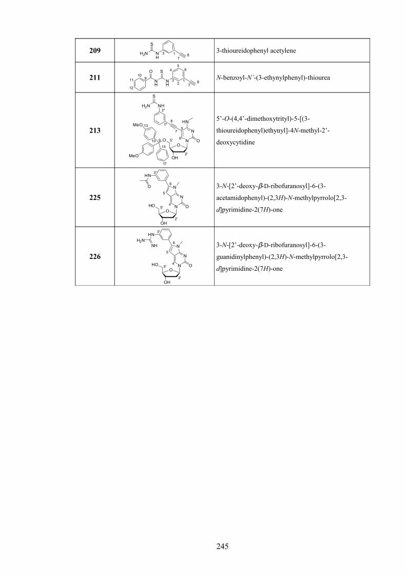
209 NH
H2N
S
137
83-thioureidophenyl acetylene
211NH
NH
SO
12
3
45
6
78
910
11
12
N-benzoyl-N’-(3-ethynylphenyl)-thiourea
213
O
OH
O N
N
HN
6
2'
5'
MeO
MeO
910
13
14
17
1''
3''
7
8
O
NH
5
H2N
S
5’-O-(4,4’-dimethoxytrityl)-5-[(3-
thioureidophenyl)ethynyl]-4N-methyl-2’-
deoxycytidine
225
O
OH
HO N
N
O
N
4
5
6
5'
2'
3''HN
O 3-N-[2’-deoxy-β-D-ribofuranosyl]-6-(3-
acetamidophenyl)-(2,3H)-N-methylpyrrolo[2,3-
d]pyrimidine-2(7H)-one
226
O
OH
HO N
N
O
N
4
5
6
5'
2'
3''HN
H2NNH 3-N-[2’-deoxy-β-D-ribofuranosyl]-6-(3-
guanidinylphenyl)-(2,3H)-N-methylpyrrolo[2,3-
d]pyrimidine-2(7H)-one
245

B: Oligonucleotide List
ODN No.
SequenceExpected
MassExpected [M+H]+
Observed Mass
Notes
1 GCT AAA AAG ACA GAG AGA TCG 6521.34 - *
2 CGA TCT CTC TGT CTT TTT AGC 6329.17 - *
3 TTT TTM TMPfT MTM TMT 4535.04 4536.05 4535.16
4 TTT TTM TAPPfT MTM TMT 4612.13 4613.13 4611.98
5 TTT TTM TUPPfT MTM TMT 4655.15 4656.164654.83 / 4697.25
partial acetylation
6F GTG TTA GGA AGA CAA AAA AGA ACT GGT HHA CCA GTT CTT TTT TGT CTT CCT AAC AC
17843.96 - * CG hd
7F GTG TTA GGA AGA GAA AAA AGA ACT GGT HHA CCA GTT CTT TTT TCT CTT CCT AAC AC
17843.96 - * GC hd
8F GTG TTA GGA AGA AAA AAA AGA ACT GGT HHA CCA GTT CTT TTT TTT CTT CCT AAC AC
17842.97 - * AT hd
9F GTG TTA GGA AGA TAA AAA AGA ACT GGT HHA CCA GTT CTT TTT TAT CTT CCT AAC AC
17842.97 - * TA hd
10 Q PMM TPM TMPfP TPT PTM PT 5807.10 5808.105807.41 / 5827.63
partially uncyclised
11 Q PMM TPM TAPPfP TPT PTM PT 5884.18 5885.195884.66 / 5902.71
partially uncyclised
12 Q PMM TPM TAcPPfP TPT PTM PT 5926.22 5927.235926.67 / 5943.79
partially uncyclised
13 Q PMM TPM TUPPfP TPT PTM PT 5927.21 5928.215927.31 / 5945.78
partially uncyclised
14 Q PMM TPM TAPPfT TPT PTM PT 5845.14 - 5846.7
15 Q PMM TPM PAPPfT TPT PTM PT 5884.18 - 5882.5
16 Q PMM TPM PAPPfP TPT PTM PT 5923.17 - 5963.6 [M+2Na-2H]
17F GTG TTA GGA AGA CAA AAA AGA ACT GGT HHA CCA GTT CTT TTT TCT CTT CCT AAC AC
17803.93 - * CC hd
18 Q PMM TPM TAcPPpT TPT PTM PT 5887.04 - 5889.1
19 Q PMM TPM PAcPPpT TPT PTM PT 5926.11 - 5929.6
20 Q PMM TPM TAcPPpP TPT PTM PT 5926.11 - 5929.9
21 Q PMM TPM PAcPPpP TPT PTM PT 5965.14 - 5966.1
22 Q PMM TPM TUPPpT TPT PTM PT 5888.07 - 5890.5
23 Q PMM TPM PUPPpT TPT PTM PT 5927.10 - 5929.3 (+ 5875.4)
24 Q PMM TPM TUPPpP TPT PTM PT 5927.10 - 5928.8
25 Q PMM TPM PUPPpP TPT PTM PT 5966.13 - 5968.3
26 GCT AAA AAG AGA GAG AGA TCG 6561.37 - *
27 GCT AAA AAG AAA GAG AGA TCG 6545.37 - *
28 GCT AAA AAG ATA GAG AGA TCG 6536.36 - *
29 CGA TCT CTC TCT CTT TTT AGC 6289.14 - *
30 CGA TCT CTC TTT CTT TTT AGC 6304.15 - *
31 CGA TCT CTC TAT CTT TTT AGC 6313.17 - *
32 TTT TTM TTT MTM TMT 4496.99 - 4499.2
246

33 TTT TTM TTP MTM TMT 4536.03 - 4541.0 (+ 4236.8)
34 TTT TTM PTT MTM TMT 4536.03 - 4537.4
35 TTT TTM PTP MTM TMT 4575.07 - 4578.0
36 TTT TTM TAPPpT MTM TMT 4611.14 - 4611.9
37 TTT TTM TAPPpP MTM TMT 4650.18 - 4652.6
38 TTT TTM PAPPpT MTM TMT 4650.18 - 4651.8
39 TTT TTM PAPPpP MTM TMT 4689.21 - 4690.0
40 TTT TTM TAcPPpT MTM TMT 4653.18 - 4653.8
41 TTT TTM TAcPPpP MTM TMT 4692.21 - 4696.4
42 TTT TTM PAcPPpT MTM TMT 4692.21 - 4693.2
43 TTT TTM PAcPPpP MTM TMT 4731.25 - 4733.1
44 TTT TTM TUPPpT MTM TMT 4654.17 - 4655.3
45 TTT TTM TUPPpP MTM TMT 4693.20 - 4696.6
46 TTT TTM PUPPpT MTM TMT 4693.20 - 4697.0
47 TTT TTM PUPPpP MTM TMT 4732.24 - 4734.1
48 TTT TTM TGPPpT MTM TMT 4653.18 - 4653.9
49 TTT TTM TGPPpP MTM TMT 4692.22 - 4694.4
50 TTT TTM PGPPpT MTM TMT 4692.22 - 4694.1
51 TTT TTM PGPPpP MTM TMT 4731.25 - 4732.2
52 Q PMM TPM TAPPpT TPT PTM PT 5845.17 - 5849.2
53 Q PMM TPM PAPPpT TPT PTM PT 5884.21 - 5884.8
54 Q PMM TPM TAPPpP TPT PTM PT 5884.21 - 5886.0
55 Q PMM TPM PAPPpP TPT PTM PT 5923.24 - 5924.2
56 Q PMM TPM TGPPpT TPT PTM PT 5887.21 - 5887.8
57 Q PMM TPM PGPPpT TPT PTM PT 5926.25 - 5928.6
58 Q PMM TPM TGPPpP TPT PTM PT 5926.25 - 5926.1
59 Q PMM TPM PGPPpP TPT PTM PT 5965.28 - 5967.5
60 Q PMM TPM TTT TPT PTM PT 5731.00 - 5730.4
61 Q PMM TPM PTT TPT PTM PT 5770.03 - 5771.0
62 Q PMM TPM TTP TPT PTM PT 5770.03 - 5770.5
63 Q PMM TPM PTP TPT PTM PT 5809.07 - 5809.7
64 TTT TTT TTT TTT 3588.35 -
Oligonucleotides for
Deprotection Study
(Chapter 7)
65 TTT TTT TTP TTT 3627.39 -
66 TTT TPT PTP TTT 3705.46 -
67 TPP PTP PTP TPT 3861.61 -
68 TTT TTT TTBAU TTT 3686.46 -
69 TTT TTT TTAcPPp TTT 3744.54 -
70 TTT TTT TTS TTT 3737.57 -
71 TTT TTT TTMOEpU TTT 3701.47 -
72 TTT TTT TTAET TTT 3647.42 -
73 TTT TAETT AETTAET TTT 3765.56 -
74 TTT TTT TTGP TTT 3669.43 -
Table. ES MS (1 dp), MALDI-TOF MS (2 dp), Q = DABCYL (quencher), F = FAM
(fluorophore), H = HEG. Key nucleotides marked in bold. * No MS analysis
necessary.
247

C: Conferences Attended, Posters, Presentations and Publication
July 2007 Year 3 Postgraduate Talks, School of Chemistry, University of
Southampton, UK. – Presentation
June 2007 3rd Nucleic Acids Summer School, University of Southern
Denmark, Odense, Denmark. – Presentation
March 2007 SCI Postgraduate Symposium, UCL, London, UK. – Presentation
November 2006 Year 2 Postgraduate Poster Day, School of Chemistry, University
of Southampton, UK. – Poster
September 2006 XVII International Roundtable Conference on Nucleosides,
Nucleotides and Nucleic Acids, Bern, Switzerland. – Poster
– Gerrard, S.R., Srinivasan N., Fox, K.R., Brown, T. ‘CG Base Pair Recognition within
DNA Triple Helices using N-Methyl-3H-pyrrolo[2,3-d]pyrimidin-2(7H)-one
Nucleoside Analogues.’ Nucleos. Nucleot. Nucl. 2007, 26(10-12), 1363-1367.
DOI: 10.1080/15257770701533958
– Ben Gaied, N., Zhao, Z., Gerrard, S.R., Fox, K.R., Brown, T. ‘Potent Triple Helix
Stabilisation by 3’ and 5’, 3’-Modified Triplex Forming Oligonucleotides.’
ChemBioChem 2009, in press.
DOI: 10.1002/cbic.200900232
248