university of missouri detection and surveillance sheila grant department of biological engineering...
TRANSCRIPT
University of Missouri
DETECTION AND SURVEILLANCE
Sheila Grant
Department of Biological Engineering
UMC
University of Missouri
Goal
• Goal is to ensure that early and accurate detection is available for important pathogens and zoonotic pathogens in various environments and deployment mechanisms
University of Missouri
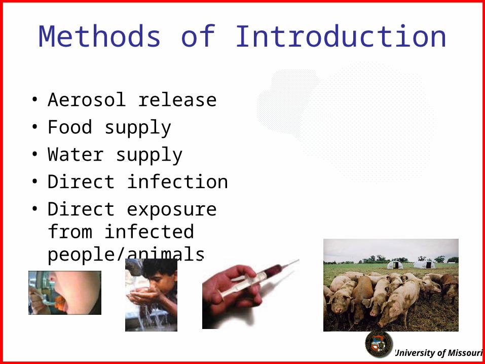
Methods of Introduction
• Aerosol release• Food supply• Water supply• Direct infection• Direct exposure from
infected people/animals
University of Missouri
How to Protect?
Biological sensors
Field RT-PCR
Syndromic surveillance
University of Missouri
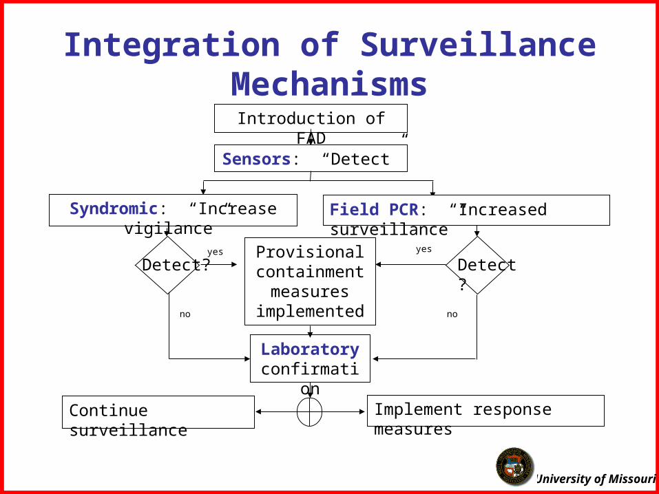
Introduction of FAD
Sensors: “Detect”
Syndromic: “Increase vigilance” Field PCR: “Increased surveillance”
Detect? Detect?Provisional containment
measures implemented
yes yes
Laboratory confirmation
no no
Continue surveillance Implement response measures
Integration of Surveillance Mechanisms
University of Missouri
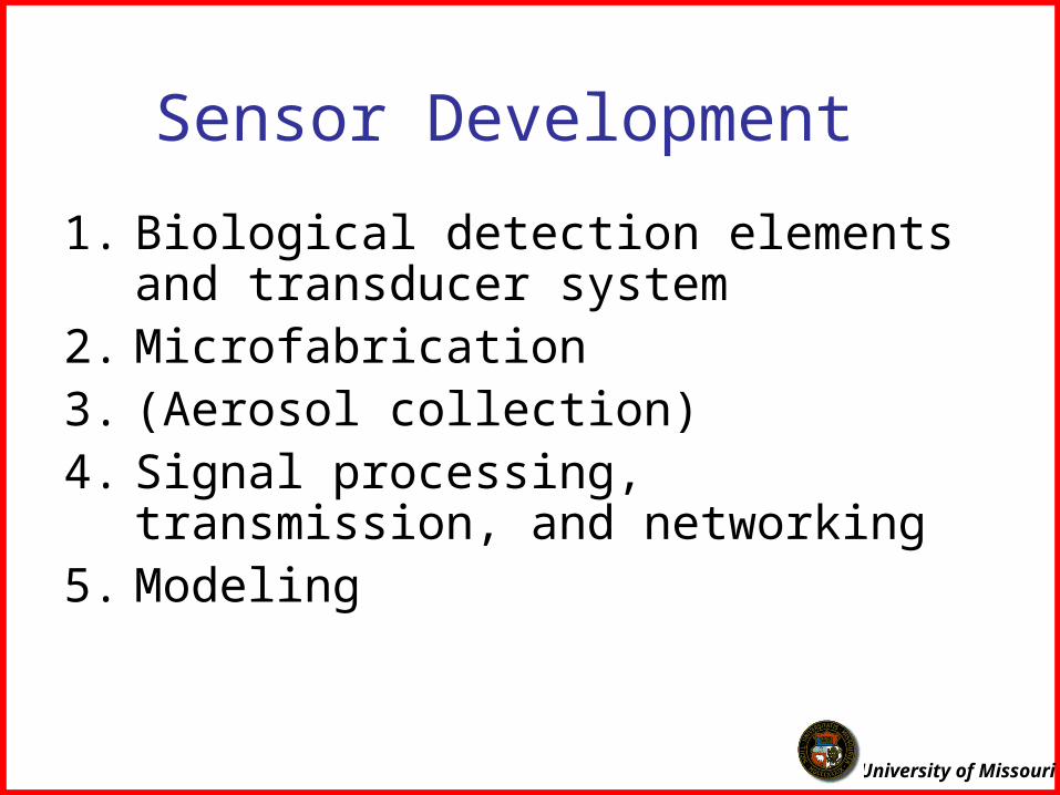
Sensor Development
1. Biological detection elements and transducer system
2. Microfabrication3. (Aerosol collection)4. Signal processing, transmission, and
networking5. Modeling
University of Missouri
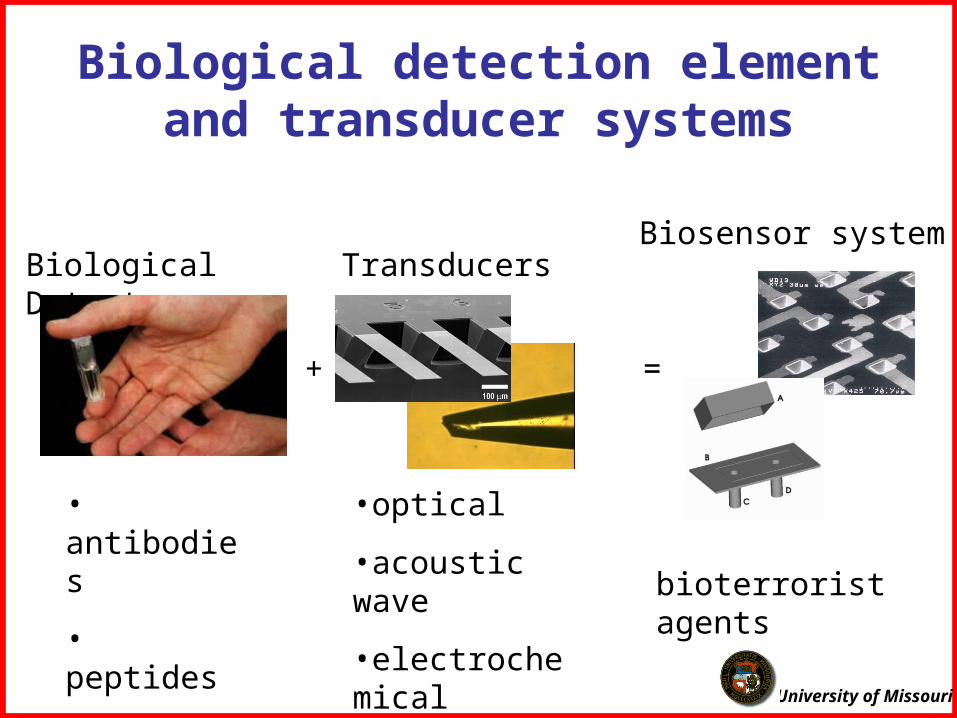
Biological detection element and transducer systems
Biological Detectors
• antibodies
• peptides
• receptors
=
bioterrorist agents
Biosensor systemTransducers
•optical
•acoustic wave
•electrochemical
+

University of Missouri
FRET Immunosensor
Protein A
Do
1
2
• measures the conformational changes that occurs when antibodies bind to select agents
• technique can eliminate false positives since only viable agents can elicit a conformational change.
University of Missouri
Sensing PeptidesMechanical:
Shear Horizontal-SAW (SH-SAW) biosensors will detect enzymes in an aqueous solution. This device will detect a change in wave propagation speed as the targeted enzyme in solution cleaves a specific peptide-construct, vastly increasing specificity.
Optical:Additionally, labeled peptide-constructscan be immobilized to gold nanoparticles, which effectively quenchesfluorescence. Upon interactions with target enzymes, the peptide is cleaved and fluorescence is enhanced.
University of Missouri
Upon SNARE cleavage, Au particles are releasedUpconversion is possible to detect
Microsphere doped with Erbium at the surface
980 nm laser
SNARE
Au nanoparticle
Au nanoparticles spoil Q-factorUpconversion is inhibited
Ring resonator toxin sensor using fluorescence method
University of Missouri
Microfabrication and NanotechnologyPeristaltic Micro-pumpsNanoporous waveguide materials
University of Missouri
Inline Detection using liquid core wave-guide (LCW)
Analyte solution being pumped in
Detector
Excitation source
Meandering Type Micro-mixer
Anodic Bonding between the two substrates
Waste Chamber
Micro channel with a Liquid core wave-guide
Output Reservoir
Input Reservoir
Light guide
Light guide
Excitation Window
WaterPDMS
Nanoporous Silica
Cross Detection using solid core wave-guide
Signal detection on a chip
University of Missouri
Integrated Fluorescence Assay on a Chip
LAS ER
D etec tor-c overedw all
M eas urem entF low c ell
Referenc eD iode
Ac c eptorD iode
D onorD iode
D
F 1 F 2
Short light pulses are generated by the laser and directed onto the sensor fluorophore inside the flowcell.
University of Missouri
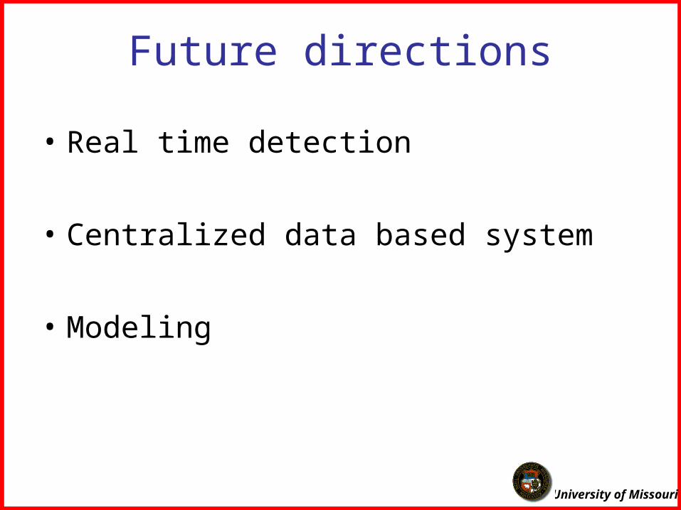
Future directions
• Real time detection
• Centralized data based system
• Modeling
University of Missouri
Acknowledgements• Xudong (Sherman) Fan• Frank Feng• Shubhra Gangopadhyay• Kevin Gillis• Mark Haidekker• Susan Lever• Darcy Lichlyter
Graduate Students• Shantanu Bhattacharya• Rosalynn Manor• Mary Pierce (now employed by MRI)• Lisa Boettcher