university of california, davis health systems · university of california, davis health systems...
TRANSCRIPT

THE PAUL R. LIPSCOMB ALUMNI SOCIETY PRESENTS:
————————————————
UNIVERSITY OF CALIFORNIA, DAVIS
HEALTH SYSTEMS
DEPARTMENT OF ORTHOPAEDIC SURGERY
GRADUATE RESEARCH SYMPOSIUM ————————————————
FRIDAY, JUNE 17, 2016

Welcome to the
2016 Paul R. Lipscomb Alumni Society
Graduate Research Symposium
This outstanding gathering is an opportunity for our
department to highlight scientific as well as clinical
research, and to reconnect with clinical faculty and
alumni who have served our department over the
years. Our special guests this year are: Stuart B.
Goodman, M.D., Ph.D., the Robert L. and Mary
Ellenburg Professor of Surgery, and Professor with
Tenure in the Department of Orthopaedic Surgery at
Stanford University; and Cosimo De Bari, M.D., Ph.D.,
Professor of Translational Medicine and Honorary
Consultant Rheumatologist at the University of Aber-
deen in Scotland, where he heads the Regenerative
Medicine Group in the Musculoskeletal Research
Program.
Most importantly, this is an occasion to commemorate
the graduation of 5 residents into the ranks of ortho-
paedic surgery. While always a bittersweet occasion,
this day validates the wonderful camaraderie and con-
tinuity of our field.
Thank you for being part of this memorable event.
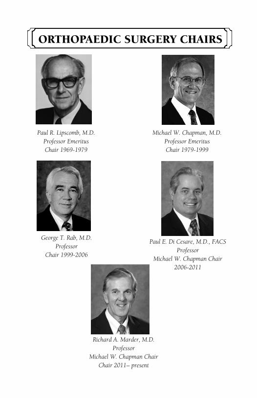
ORTHOPAEDIC SURGERY CHAIRS
Paul R. Lipscomb, M.D.
Professor Emeritus
Chair 1969-1979
Michael W. Chapman, M.D.
Professor Emeritus
Chair 1979-1999
George T. Rab, M.D.
Professor
Chair 1999-2006
Paul E. Di Cesare, M.D., FACS
Professor
Michael W. Chapman Chair
2006-2011
Richard A. Marder, M.D.
Professor
Michael W. Chapman Chair
Chair 2011– present
VISITING PROFESSORS
1982 — Robert B. Winter, M.D.
1983 — Anthony Catterall, M.D.
1984 — Euguene E. Bleck, M.D.
1985 — Paul P. Griffin, M.D.
1986 — M. Mark Hoffer, M.D.
1987 — Robert B. Salter, M.D.
1988 — Colin F. Moseley, M.D.
1989 — James R. Gage, M.D.
1990 — James F. Kellman, M.D.
1991 — David S. Bradford, M.D.
1992 — Adrian E. Flatt, M.D.
1993 — Augusto Sarmiento, M.D.
1994 — M. Mark Hoffer, M.D.
1995 — James R. Andrews, M.D.
1996 — James R. Urbaniak, M.D.
1997 — Stuart L. Winstein, M.D.
1998 — Robert A. Mann, M.D.
1999 — Joseph M. Lane, M.D.
2000 — Andrew J. Weiland, M.D.
2001 — Joel M. Matta, M.D.
2002 — Terry R. Trammell, M.D.
2003 — Kaye E. Wilkins, M.D.
2004 — Richard Gelberman, M.D.
2005 — Robert H. Hensinger, M.D.
2006 — James Heckman, M.D.
2007 — Thomas A. Einhorn, M.D.
2008 — Joseph A. Buckwalter, M.D.
2009 — Peter J. Stern, M.D.
2010 — Joseph Borrelli, Jr., M.D.
2011 — Keith Bridwell, M.D.

VISITING PROFESSORS
2012 — Gary G. Poehling, M.D.
2013 — Robert Anderson, M.D.
2014 — Jeffrey Eckardt, M.D.
2015 — J. Tracy Watson, M.D.
2015 — Matthew L. Warman, M.D.
2016 — Stuart B. Goodman, M.D.
2016 — Cosimo De Bari, Ph.D.

Stuart B. Goodman received his B.Sc., M.D. and M.Sc. (Institute of Medical Science) from the University of Toronto, and his Ph.D. in Orthopedic Medical Science from Lund University in Sweden. He is a Fellow of the Royal College of Surgeons (Canada), the American Academy of Orthopaedic Surgeons and the American College of Surgeons. Dr. Goodman's clinical practice concentrates on adult reconstructive surgery. His clinical research interests center on the outcome of surgery for arthritis including primary and of revision total joint replacement, juvenile arthritis, and osteone-crosis of the hip and knee. His basic science interests center on biocompatibility of orthopaedic implants, inflammation, and musculoskeletal tissue regeneration and repair. Dr. Goodman is a member of numerous academic organizations including the AAOS Biological Implants Committee (Chairman), and is a former member of the AAOS Biomedical Engineering Commit-tee. He is a member of the Hip Society, Knee Society and AAHKS, a consultant to the Orthopaedic and Rehabilitation Devices Advisory Panel of the FDA, and former vice-chairman of the Musculoskeletal Tissue Engineering study section at NIH.
Stuart B. Goodman, M.D. M.Sc.
Ph.D. F.R.C.S.C. F.A.C.S. F.B.S.E.
Professor of Surgery—Robert L. and
Mary Ellenburg
Professor & Chief of Orthopaedic
Surgery—Stanford University
Fellow of the Institute of Chemistry,
Engineering and Medicine for Human
Health (ChEM-H)—Stanford University

Dr. Goodman is on the editorial board of the Journal of
Orthopaedic Research (Associate Editor), Clinical Or-
thopaedics (Deputy Editor-Hip Society Liason), Bio-
materials (Associate Editor), Journal of Arthroplasty,
Journal of Biomedical Materials Research, and other
journals, and is a manuscript reviewer for over 20 jour-
nals in the fields of orthopaedic surgery, arthritis, bioen-
gineering and biomaterials. Dr. Goodman has pub-
lished over 400 peer-reviewed manuscripts in medical
and bioengineering journals. Dr. Goodman and co-
workers have received awards for their research from
the Society for Biomaterials, Orthopaedic Research Soci-
ety, the American Orthopaedic Association, Western Or-
thopaedic Association, and the Association of Bone and
Joint Surgeons. Dr. Goodman was awarded the Clem-
son Award for Basic Research from the Society For Bio-
materials in May 2000. He was the President of the So-
ciety For Biomaterials (2001-2) and served on the Board
of Directors of the Orthopaedic Research Society. Dr.
Goodman served as Co-Chair for the 1995, 2000 and
2007 NIH/AAOS-sponsored workshops on Implant
Wear. Dr. Goodman was recognized as a Fellow, Bio-
materials Science and Engineering (FBSE) by the Inter-
national Union of Societies, Biomaterials Science and
Engineering in May 2004, a Fellow of the Japanese Soci-
ety of the Promotion of Science in 2011, and a Fellow of
the American Institute of Medical and Biological Engi-
neers in 2012.

Professor Cosimo De Bari is a clinically active rheumatologist with expertise in regenerative medicine for musculoskeletal applications.
Dr. De Bari graduated in Medicine (summa cum laude) from the University of Bari (Italy), where he underwent specialist training in Rheumatolo-gy. He then obtained his Ph.D. from the Univer-sity of Leuven (Belgium). In 2003 Cosimo moved to King's College London, where in 2005 he was awarded an MRC Clinician Scientist Fellowship.
Since September 2007 Dr. De Bari is Professor of Translational Medicine at the University of Aber-deen, where he leads the Arthritis & Regenera-tive Medicine Laboratory.
Cosimo De Bari, M.D. Ph.D. F.R.C.P. Professor of Translational Medicine & Hon. Consultant Rheumatologist Institute of Medical Sciences University of Aberdeen United Kingdom
FACULTY
University of California, Davis Health System
Robert H. Allen, M.D.
Associate Professor, Hand, Upper Extremity, and Microvascular Surgery
Kyriacos A. Athanasiou, Ph.D., Ph.M.
Professor, Orthopaedic Research and Biomedical Engineering
Christopher O. Bayne, M.D.
Assistant Professor, Hand, Upper Extremity, and Microvascular Surgery
Blaine A. Christiansen, Ph.D.
Assistant Professor, Orthopaedic Research Laboratory
Jonathan G. Eastman, M.D.
Assistant Professor, Trauma Service
Ellen P. Fitzpatrick, M.D.
Assistant Professor, Trauma Service
David P. Fyhrie, Ph.D.
Professor, Orthopaedic Research Laboratory
Mauro M. Giordani, M.D.
Professor and Chief of Adult Reconstructive Service
Eric Giza, M.D.
Associate Professor, Chief of Foot and Ankle Service
Dominik R. Haudenschild, Ph.D.
Associate Professor, Orthopaedic Research Laboratory
Brian M. Haus, M.D.
Assistant Professor, Pediatric Orthopaedic Service
Yashar Javidan, M.D.
Assistant Professor, Adult and Pediatric Spine Service
Eric O. Klineberg, M.D.
Associate Professor, Adult and Pediatric Spine Service
Christopher D. Kreulen, M.D.
Assistant Professor, Foot and Ankles Service
J. Kent Leach, Ph.D.
Associate Professor, Orthopaedic Research Laboratory, and Biomedical
Engineering
Cassandra A. Lee, M.D.
Associate Professor, Sports Medicine Service
Mark A. Lee, M.D.
Professor, Trauma Service

Richard A. Marder, M.D.
Professor , Chief of Sports Medicine Service, and Michael W. Chapman Chair
John P. Meehan, M.D.
Professor, Adult Reconstructive Service
Gavin C.T. Pereira, M.B.B.S., F.R.C.S.
Assistant Professor, Adult Reconstructive Service
A. Hari Reddi, Ph.D.
Distinguished Professor, Lawrence J. Ellison Chair of Molecular Biology,
Director of Orthopaedic Research Laboratories
Rolando F. Roberto, M.D.
Professor, Chief of Adult and Pediatric Spine Service, and Executive Vice Chair
Robert M. Szabo, M.D.
Professor, Chief of Hand, Upper Extremity, and Microvascular Surgery
James M. Van Den Bogaerde, M.D.
Associate Professor, Sports Medicine Service
Barton L. Wise, M.D.
Associate Professor, Orthopaedic Research, and Internal Medicine
Philip R. Wolinsky, M.D.
Professor, Chief of Trauma Service
FACULTY
Shriners Hospital for Children, Northern California
Jennette L. Boakes, M.D.
Fellowship Director, Clinical Professor Pediatric Orthopaedic Service
Jon R. Davids, M.D.
Clinical Professor, Assistant Chief of Pediatric Orthopaedic Surgery
Michelle A. James, M.D.
Professor, Chief of Pediatric Orthopaedic Service
Vedant A. Kulkarni, M.D.
Assistant Clinical Professor, Pediatric Orthopaedic Service
Joel A. Lerman, M.D.
Associate Clinical Professor, Pediatric Orthopaedic Service
Debra J. Templeton, M.D.
Assistant Professor, Pediatric Orthopaedic Service

Friday, June 17, 2016 7:00 AM Continental Breakfast 7:30 AM Welcome - Department Chair Richard A. Marder, M.D. 7:40 AM Introduction of Guest Speaker (Research) A. Hari Reddi, Ph.D. 7:45 AM BASIC SCIENCE VISITING PROFESSOR: Cosimo De Bari, M.D., Ph.D.—University of Aberdeen in Scotland- Mesenchymal Stem Cells Find Their Niche in Cell Therapy for Joint Disorders 8:45 AM RESEARCH FACULTY: J. Kent Leach, Ph.D. - Adhesion vs. Cohesion: Tipping the Scales for Cell-based Therapies 9:00 AM 2015-2016 DICKENSON RESEARCH RESIDENT: Nasser Heyrani, M.D. –Mesenchymal Stem Cell Spheroids in Critical-Sized Bone Defect Repair 9:15 AM Morning Break 9:25 AM Introduction of Guest Speaker (Clinical) John P. Meehan, M.D. 9:30 AM VISITING PROFESSOR: Stuart B. Goodman, M.D., Ph.D. – Stanford University — Can We Preserve Joints Afflicted by Osteonecrosis?
10:30 AM RESIDENT: Laurence E. Cook, M.D. - Case Series of Tantalum Grafting for Structurally Unstable Bone Defects in Post Traumatic Reconstruction
10:45 AM RESIDENT: William B. T. Kent, M.D. - Fracture Morphology of AO/OTA 31-A Trochanteric Fractures: A 3D CT Study with an Emphasis on Coronal Fragments

11:00 AM RESIDENT: Jason J. Kim, M.D. - Does Changing the Mechanical Axis Lead to MUA after TKA?
11:15 AM RESIDENT: Motasem I. Refaat, M.D. - Binding to COMP Reduces the BMP2 Dose for Spinal Fusion in a Rat Model. 11:30 AM RESIDENT: Dora A. R. Storelli, M.D. - The Effect of Radioscapholunate Fusion with and without Distal Scaphoid and Triquetrum Excision on Capitolunate Contact Pressures 11:45 AM DEPARTMENT PHOTOS 12:15 PM VISITING PROFESSOR CASE PRESENTATIONS FROM RESIDENTS (60 minute working lunch) – Moderators: Mauro Giordani, M.D.; John P. Meehan, M.D. 1:20 PM CLINICAL CHALLENGES PANEL: Panelists: Domingo A. Hallare, M.D.; Gavin C. Pereira, M.B.B.S, F.C.R.S.; Paul M. Sasaura, M.D.; Philip R. Wolinsky, M.D.: Femoral Neck Fracture: To Fix or Replace? 2:00 PM RECENT GRADUATE KEYNOTE LECTURE: Safdar N. Khan, M.D. - What I Would Have Done Differently in My First Years after Graduation 2:30 PM Kevin K. Howe, M.D. - The Rotation Doesn't End... 2:45 PM H. David Moehring, M.D. - My Worst Case since Graduation and How I Solved It 3:00 PM Discussion 3:15 PM Adjournment

Laurence E. Cook, M.D. Chief Resident
Education:
Case Western Reserve University
School of Medicine
M.D. (2011)
University of California, Berkeley
B.A. Molecular and Cellular Biology (2007)
Next Step:
Fellowship — UC Davis, Orthopaedic Trauma
Career Objective:
To become a diversely skilled Orthopaedic traumatolo-
gist and physician educator. To strive for efficient and
excellent care for my patients and their families. To
strive for technical excellence in every procedure that I
undertake.
I would like to be known as an expert educator in addi-tion to an expert surgeon.
Spouse:
Lauren Cook
Children: Evelyn Clare Cook
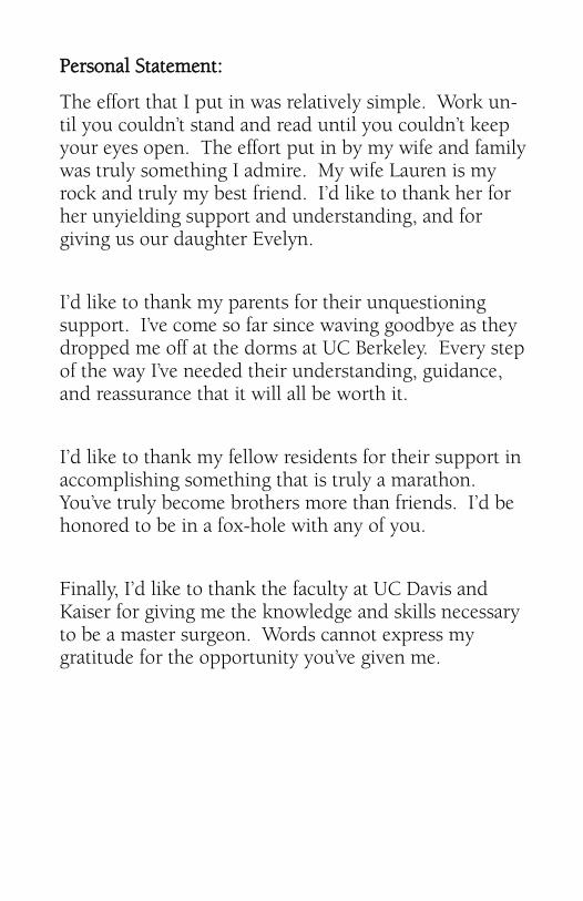
Personal Statement:
The effort that I put in was relatively simple. Work un-til you couldn’t stand and read until you couldn’t keep your eyes open. The effort put in by my wife and family was truly something I admire. My wife Lauren is my rock and truly my best friend. I’d like to thank her for her unyielding support and understanding, and for giving us our daughter Evelyn.
I’d like to thank my parents for their unquestioning support. I’ve come so far since waving goodbye as they dropped me off at the dorms at UC Berkeley. Every step of the way I’ve needed their understanding, guidance, and reassurance that it will all be worth it.
I’d like to thank my fellow residents for their support in accomplishing something that is truly a marathon. You’ve truly become brothers more than friends. I’d be honored to be in a fox-hole with any of you.
Finally, I’d like to thank the faculty at UC Davis and Kaiser for giving me the knowledge and skills necessary to be a master surgeon. Words cannot express my gratitude for the opportunity you’ve given me.

Research
Title:
Case Series of Tantalum Grafting for Structurally Unstable Bone Defects in Post Traumatic Reconstruction
Authors:
Mark Lee M.D., Laurence Cook, M.D.
Purpose: The purpose of this study was to determine whether tantalum augmentation for unstable bone defects would incorporate with bone and maintain post reconstructive morphology. We hypothesized that tantalum will demonstrate incorporation with bone and maintain construct stability in the midterm follow-up.
Methods: After IRB approval we conducted a retrospective chart review of our institutional trauma database to identify patients that received a porous tantalum implant for traumatic bone defects over a two-year period. Each patient’s chart was reviewed for need for an additional procedure due to infection, malunion, pain or nonun-ion. Radiographs were also reviewed for the graft incor-poration and maintenance of the original reconstructive alignment.

Results: 9 patients were identified for this study, 4 male and 5 females, with an average age of 54 years old (range 22-69). The average length of follow-up was 18 months. Seven injuries were to the tibia and two to the femur. Tantalum was utilized in seven patients for reoperation due infected nonunion and in two for reoperation for traumatic bone loss after fixation. The average volume Tantalum was 56 cm2 (Range 14-274 cm2). The average length of the defect was 3.2 cm (range 1.6-6.5). All patients were able to ambulate without pain at the most recent follow-up visit except for one who required a hinged total knee arthroplasty for pain and instability. Post-op morphology was maintained in all patients including the patient who later had a revision. All patients showed graft integration at their most recent postoperative radiograph. One patient required intervention for a postoperative infection that subsequently resolved with superficial debridement and antibiotic therapy.
Conclusion: Tantalum demonstrated excellent incorporation and long term maintenance of post reconstructive morphology. Additionally, eight of the nine patients were able to bear weight without pain at final follow-up. We believe that tantalum may represent a viable alternative for unstable bone defects especially in a setting where large volume allografts are not an option due to infectious or structural concerns.

William B.T. Kent, M.D. Chief Resident
Education:
Michigan State University College of Human Medicine
M.D. (2011)
University of California, Santa Barbara
B.A. Business Economics (2006)
Next Step:
Fellowship — UC San Diego, Orthopaedic Trauma
Career Objective:
To provide the highest level of care to all my patients. To continue to grow professionally through lifelong learning, research and education. To continue to strive to be better.

Personal Statement:
It has been an awesome five years. There are a lot of people I need to thank. First and foremost, I need to thank my family and friends. Mom, Dad, Andrew, and Michael thank you for your love and support throughout the years. Corey- thank you for your understanding, patience, love and support each and everyday. I am excited for the future and our next adventure. For the rest of my close friends and family- thank you for your encouragement and support throughout the years.
I’m truly grateful for my experience here at UC Davis. I feel very honored and privileged to have trained under so many outstanding surgeons. Thank you for your mentorship, teaching and patience. I have learned countless skills and lessons I will use everyday in my career. I am forever indebted to you. For all my co-residents and junior residents, thanks for the hard work and good times. Keep up the tradition and I look forward to seeing you guys in the near future.
Research Topic:
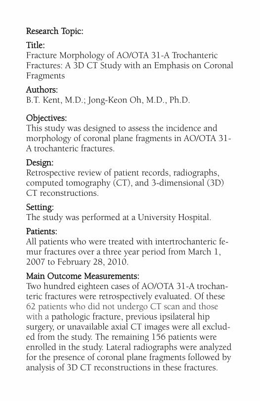
Title: Fracture Morphology of AO/OTA 31-A Trochanteric Fractures: A 3D CT Study with an Emphasis on Coronal Fragments
Authors: B.T. Kent, M.D.; Jong-Keon Oh, M.D., Ph.D.
Objectives: This study was designed to assess the incidence and morphology of coronal plane fragments in AO/OTA 31-A trochanteric fractures.
Design: Retrospective review of patient records, radiographs, computed tomography (CT), and 3-dimensional (3D) CT reconstructions.
Setting: The study was performed at a University Hospital.
Patients: All patients who were treated with intertrochanteric fe-mur fractures over a three year period from March 1, 2007 to February 28, 2010.
Main Outcome Measurements: Two hundred eighteen cases of AO/OTA 31-A trochan-teric fractures were retrospectively evaluated. Of these 62 patients who did not undergo CT scan and those with a pathologic fracture, previous ipsilateral hip surgery, or unavailable axial CT images were all exclud-ed from the study. The remaining 156 patients were enrolled in the study. Lateral radiographs were analyzed for the presence of coronal plane fragments followed by analysis of 3D CT reconstructions in these fractures.

The incidence of the coronal fragments identified on the lateral radiograph and 3D CT reconstructions were both calculated. Coronal fragment morphology was described based upon the origin and exit points of fracture lines and the number of fragments.
Results: On plain radiographs, a coronal plane fracture was identified in 59 cases, an incidence of 37.8% (59/156). In comparison, 3D CT reconstructions identified coronal plane fractures in 138 cases for an incidence of 88.4% (138/156). 3D CT reconstructions identified coronal fracture fragments in 81.9% (50/61) of AO/OTA 31-A1 cases, 94.5% (69/73) of 31-A2 cases, and 86.3% (19/22) of 31-A3 cases. The incidence of coronal fractures identified on plain radiographs of all 3 AO/OTA 31-A1, 31-A2, 31-A3 groups was all lower when compared to the incidence of coronal fractures identified on 3D CT (p<0.0001). Of the 138 cases that had coronal plane fracture, 82 cases (59.4%) had a single coronal fragment (GT fragment 35 cases, GLT fragment 19 cases, GLPC fragment 28 cases). The remaining 56 cases (40.5%) had two coronal fragments.
Conclusions: There is a high incidence of coronal fragments in intertrochanteric femur fractures when analyzed with 3D CT reconstructions. These coronal fragments are difficult to identify on plain radiographs. Knowledge of the incidence and morphology of coronal fragments helps to avoid potential intraoperative pitfalls.

Jason J. Kim, M.D. Chief Resident
Education:
University of California, San Diego
School of Medicine
M.D. (2009)
University of California, San Diego
B.S. Bioengineering (2004)
Next Step:
Fellowship – University of Utah, Adult Reconstruction
Career Objective:
I plan to continue this journey in Orthopaedic Surgery
by pursuing efficiency, safety, and innovation in man-
agement of hip and knee degenerative joint disease and
replacement.

Personal Statement:
My journey in Orthopaedics has been longer than most. But I am proud to say that the experience was worth it. I feel fortu-nate that I was given the opportunity to pursue my dream here at UC Davis and I will never forget the relationship I have made throughout this journey. I need to thank Dr. Rob-erto, Dr. Eastman, Dr. Wolinsky, and Dr. Lee for supporting me when I was a 5809 fellow and giving me the chance to prove my commitment. Thank you Margaret for keeping the residents in good standing and Dr. Pereira, Dr. Giordani, and Dr. Meehan for showing me the path away from Trauma. (I still love trauma).
Research Topic:
Title: Does changing the mechanical axis lead to MUA after TKA?
Total knee arthroplasty (TKA) outcomes have not matched total hip arthroplasty outcomes despite the improvements in techniques, implant design and use of navigation. The out-comes of TKA have not improved beyond 80%. Philosophies of implant position have changed and have been refined over the years and currently, the accepted wisdom is that the limb undergoing a TKA should be brought as close to the Mechan-ical axis as possible.
Various factors have been implicated in influencing the out-come of the TKA. It has been proposed that changing a pa-tient’s natural alignment to a mechanical aligned knee may not favor appropriate ligament tensions and may lead to un-happy patients and sometimes manipulation under anaesthe-sia to improve the range of motion of the knee.
We hypothesized that the change in mechanical axis from pre-op to post op following TKA is a risk factor for MUA. We conducted a retrospective analysis of patients who underwent TKA from 2005 to 2014 and compared those that underwent MUA to control group.

Motasem I. Refaat, M.D. Chief Resident
Education:
University of Illinois College of Medicine
M.D. (2011)
University of California, Davis
B.S. Exercise Physiology (2006)
Next Step:
Fellowship – Orthopedic Trauma, Harborview Medical
Center. Seattle, Washington
Career Objective:
My career objectives are to provide quality fracture care to all my patients with a smile on my face. To affect the lives of my patients and my community for the better, give back to those in need, and to make my father and mother proud.
Personal Statement:
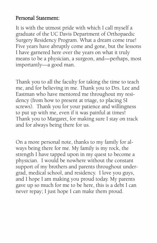
It is with the utmost pride with which I call myself a graduate of the UC Davis Department of Orthopaedic Surgery Residency Program. What a dream come true! Five years have abruptly come and gone, but the lessons I have garnered here over the years on what it truly means to be a physician, a surgeon, and—perhaps, most importantly—a good man.
Thank you to all the faculty for taking the time to teach me, and for believing in me. Thank you to Drs. Lee and Eastman who have mentored me throughout my resi-dency (from how to present at triage, to placing SI screws). Thank you for your patience and willingness to put up with me, even if it was painful at times! Thank you to Margaret, for making sure I stay on track and for always being there for us.
On a more personal note, thanks to my family for al-ways being there for me. My family is my rock, the strength I have tapped upon in my quest to become a physician. I would be nowhere without the constant support of my brothers and parents throughout under-grad, medical school, and residency. I love you guys, and I hope I am making you proud today. My parents gave up so much for me to be here, this is a debt I can never repay; I just hope I can make them proud.

Research Topic:
Title:
Binding to COMP Reduces the BMP2 Dose for Spinal Fu-sion in a Rat Model.
Authors:
Refaat M, Klineberg EO, Fong MC, Garcia TC, Leach JK, Haudenschild DR. Study Design:
Test the effect of Cartilage Oligomeric Matrix Protein (COMP) on enhancing rhBMP-2 induced spinal fusion in a prospective 8-week interventional trial of spinal fusion in rats. Objective:
To determine whether the amount of BMP-2 required to achieve spinal fusion in a pre-clinical model can be re-duced by the addition of COMP. Summary of Background Data:
Bone morphogenetic proteins (BMPs) are applied clini-cally at supraphysiological doses to promote spinal fu-sion by inducing osseous growth, but dose-related limi-tations include ectopic bone formation and local inflam-matory reactions. COMP is a matricellular BMP-binding protein expressed during endochondral ossification and fracture healing. In-vitro studies demonstrate enhanced activity of BMP bound to COMP. We hypothesized that BMP bound to COMP could achieve equivalent spinal fusion rates at lower doses and with fewer complications.

Methods:
Posterolateral intertransverse process spinal fusion at L4-L5 was performed in 36 Lewis rats. COMP (10 μg) was tested with or without "low-dose" rhBMP-2 (2 μg), and the results were compared with the "low dose" (2 μg rhBMP-2) and "high-dose" (10 μg rhBMP-2) groups. All groups utilized insoluble collagen bone matrix carri-er (ICBM). Fusion was evaluated by radiology, histology, and manual palpation. BMP release kinetics were evalu-ated in-vitro.
Results:
Fusion grading of microCT images demonstrated that the fusion rate with the COMP+LoBMP was statistically equivalent to HiBMP, and significantly better than LoBMP without COMP. These results were confirmed with radiographs and manual palpation. BMP release kinetics suggest that COMP increased local concentra-tions of BMP due to decreased growth factor retention on the scaffold.
Conclusions:
COMP enhances BMP-induced bone formation, ena-bling lower doses of BMP to achieve the same level of spinal fusion. COMP may function by affecting the availability and biological presentation of BMP-2. A de-crease of BMP-2 required for fusion may reduce dose-related adverse effects, surgical costs, and improve clini-cal outcomes.

Dora A.R. Storelli, M.D. Chief Resident
Education:
University of California, Davis
School of Medicine
M.D. (2011)
California State University, Fresno
B.S. Biology (2007)
Next Step:
Fellowship — University of California, San Francisco,
Hand and Upper Extremity Orthopaedic Surgery
Career Objective:
To become a highly capable and skillful hand/upper
extremity surgeon for patients of all ages. I look for-
ward to a practice that will involve a broad spectrum of
upper extremity care, ranging from arthroplasty to
trauma.
Spouse:
Julian Storelli
Children: Luka Rendulic Storelli

Personal Statement:
When I think about where I am today; I am incredibly humbled and grateful. Graduation is not only a time for me to mark an end to my training, but also a time to thank the individuals and the institution that has al-lowed me to grow and become the surgeon that I am today.
A large part of my life in the past five years has been my wonderful husband, Julian, who probably didn’t realize that when he met me, he was starting a residency as well. He has been my greatest support, through both good days and bad. He has lived and dealt with me through the struggles involved with training, patiently telling me that there is an end, and that I will finish. I am beyond grateful to him. I love you and our wonder-ful baby boy.
My parents are the second group of people I would like to acknowledge. Mila and Adam Rendulic taught me that hard work will get you places, and that if you’re stubborn enough, eventually you will get what you want. Volim vas puno.
Lastly, the institution. UC Davis has been my home for the past nine years. I am grateful to this department for teaching me how to be a surgeon and for sharing your patients for my experience.
Medicine is a humbling teacher, I look forward to never stop learning.

Research Topic:
Title: The Effect of Radioscapholunate Fusion with and without Distal Scaphoid and Triquetrum Excision on Capitolunate Contact Pressures
Authors: Christopher Bayne, M.D., Dora Storelli, M.D., Nasser Heyrani, M.D., Sean McNary, Ph.D.
Introduction: Radiocarpal arthritis can be managed with limited radiocarpal fusion. In patients with iso-lated radiocarpal arthritis, with sparing of the mid-carpal joint, radioscapholunate (RSL) fusion is an op-tion for treatment. This procedure preserves adjacent joints, however does restrict range of motion and can lead to eventual adjacent joint degeneration. In order to address this issue of motion; distal scaphoid exci-sion and triquetrum excision have been proposed as possible solutions. To date, no study has compared RSL alone to RSL with distal scaphoid or triquetrum excision, nor the effect of RSL with distal scaphoid and triquetrum excision to contact pressures in adja-cent joints.
Materials and Methods: Ten wrist specimens, pre-screened for underlying deformity or arthritis, were dissected, with a ligament sparing capsulotomy per-formed. Pressure film was inserted into the capi-tolunate joint. The specimen was loaded with 35 N of force. The measurements were repeated for specimens with no fusion, RSL fusion, RSL fusion with distal scaphoid excision, and lastly, RSL fusion with distal scaphoid and triquetrum excision.
Contact pressure film was scanned into the computer; data obtained from film was converted into contact area and pressure measurements.

Results thus far: See figures 1, 2, 3
With the initial available data, RSL fusion with distal scaphoid excision with and without triquetrum excision shows an increase in contact area and force.
Discussion: With this very early review of data; RSL fusion with distal scaphoid and triquetrum excision appears to have trends of increased contact pressures. This modification to gain motion with selective carpal fusion may lead to increased adjacent joint degenera-tion; however this warrants further investigation.
Figure 1: Figure 2:
Figure 3: