university microfilms international...4.7.1. cytochalasin b 127 4.7.1.1. effects on overlapping...
TRANSCRIPT
INFORMATION TO USERS
This material was produced from a microfilm copy of the original document. Whilethe most advanced technological means to photograph and reproduce this documenthave been used, the quality is heavily dependent upon the quality of the originalsubmitted.
The following explanation of techniques is provided to help you understandmarkings or patterns which may appear on this reproduction.
1. The sign or "target" for pages apparently lacking from the documentphotographed is "Missing Page(s)". If it was possible to obtain the missingpagels) or section, they are spliced into the film along with adjacent paqes,This may have necessitated cutting thru an image and duplicating adjacentpages to insure you complete continuity.
2. When an image on the film is obliterated with a large round black mark, itis an indication that the photographer suspected that the copy may havemoved during exposure and thus cause a blurred image. You will find agood image of the page in the adjacent frame.
3. When a map, drawing or chart, etc., was part of the material beingphotographed the photographer followed a definite method in"sectioning" the material. It is customary to begin photoing at the upperleft hand corner of a large sheet and to continue photoing 'from left toright in equal sections with a small overlap. If necessary, sectioning iscontinued again - beginning below the first row and continuing on untilcomplete.
4. The majority of users indicate that the textual content is of greatest value,however, a somewhat higher quality reproduction could be made from"photographs" if essential to the understanding of the dissertation. Silverprints of "photographs" may be ordered at additional charge by writingthe Order Department, giving the catalog number, title, author andspecific pages you wish reproduced.
5. PLEASE NOTE: Some pages may have indistinct print. Filmed asreceived.
University Microfilms International300 North Zeeb RoadAnn Arbor, Michigan 48106 USA
51. John's Road, Tyler's GreenHigh Wycombe, Bucks, England HP10 8HR

78-1057
SINGLEY, Carl Thomas, 1943-AN ANALYSIS OF GASTRULATION INLOLIGO PEALE I •
University of Hawaii,Ph.D., 1977Zoology
University Microfilms International, Ann Arbor. Michigan 48106
AN ANALYSIS OF GASTRULATION
IN LOLIGO PEALEI
A DISSERTATION SUBMITTED TO THE GRADUATE DIVISION OF THEUNIVERSITY OF HAWAII IN PARTIAL FULFILLMENT
OF THE REQUIREMENTS FOR THE DEGREE OF
DOCTOR OF PHILOSOPHY
IN ZOOLOGY
AUGUST 1977
By
Carl T. Singley
Dissertation Committee:
John M. Arnold, ChairmanRuth G. Kleinfeld
Michael G. HadfieldSidney J. Townsley
Samuel R. Haley

Acknowledgments
I thank the people of the Marine Biological Laboratory,
Woods Hole, Massachusetts for use of the laboratory·s facil
ities, their help, and their friendship. I also thank
Lois D. Williams-Arnold for her critical and editorial
comments, and Dr. Virginia Peters of the Woods Hole
Oceanographic Institute for her help with scanning electron
microscopy.
Work on this project was begun during the post-session
of the embryology course at The Marine Biological Laboratory.
This work has been supported, in part, by grants from the
National Science Foundation and National Institute of Health
to Dr. J. M. Arnold, and by the Tarr Foundation.
iii
Abstract
Time-lapse cinemicrography, light microscopy, and
electron microscopy were used to study the cellular events
and processes of gastrulation in Loligo pealei. Early
cleavage (cleavages 1 through 7) are reviewed and later
divisions (8 through 11), which occur coincidentally with
the onset of gastrulation, are described. Cleavage of the
blastodisc results in a single-layered blastoderm. After
eighth cleavage a specific population of blastomeres
spreads vegetally over presumptive middle-layer cells
(= mesendodermal layer of older literature) by the active
extension of lamellipodia and filopodia. The process of
overlapping is essentially epibolic. After the presumptive
middle-layer cells are overlapped they cleave (ninth clea
age) perpendicular to the axis of overlap. The processes
of overlapping and cell division together produce a ring
of cells between the blastoderm and the yolk cytoplasmic
layer.
After segregation of the middle layer the yolk
syncytium is formed by the vegetal movements of blastocone
nuclei and retraction of the furrows of the blastocones.
Furrow retraction is a result of the dissociation of the
contractile bands of microfilaments from the bases of
furrows and a combined folding and vesiculation of the
plasma membranes of the furrows. After their initial
iv
vegetal movements, the yolk syncytial nuclei divide twice
by mitosis with the formation of a circumferential, transi
tory furrow. The yolk syncytial nuclei then come to lie
beneath the two-layered blastoderm by means of nuclear
movements and epibolic sprearling of the blastoderm.
The ultrastructural features of cellular extensions
of overlapping cells in Loligo are similar to those of
other cells during locomotion. Lamellipodia and filopodia
extend from leading edges of overlapping cells and make
20 nm appositional contacts with underlying cells. Over
lapping cell processes frequently contain arrays of
oriented microfilaments, but few microtubules. Progressive
extension and retraction of these cell processes results
in the overlapping cells being positioned above the
presumptive middle-layer cells.
The significance of cell movement relative to that of
cell division in cephalopod gastrulation was assessed
using the inhibitors colchicine and cytochalasin B. Cell
movement of overlapping and yolk syncytium form~tion are
inhibited by application of 0.2 ~g/ml cytochalasin B,
but not by lO-4M colchicine. In relation to gastrulation,
cytochalasin B inhibits the formation of a two-layered
blastoderm whereas colchicine does not.
Descriptive and experimental results are discussed in
relation to concepts of cephalopod gastrulation and current
v

concepts of mechanisms of cell movement. A generalized
description of cephalopod gastrulation is presented and
discussed with regard to recent literature on cephalopod
development.
vi

TABLE OF CONTENTS
Page
ACKNOWLEDGMENTS. .. . . . . .. .. . . . . . . ... .. .. . . .. . . . . .. . .. . iii
ABSTRACT. . . . . . . . . . . . . . . . . . . . . . . . . . . . . . . . . . . . . . . . . . . . . i v
LIST OF ILLUSTRATIONS - FIGURES..... x
LIST OF ILLUSTRATIONS - PLATES..... xi
1. INTRODUCTION.... 1
2. MATERIALS AND METHODS............................ 11
2.1. Acquisition and Maintenance ofExperimental Animals........ 11
2.2. Light Microscopy, Transmission ElectronMiscroscopy, and Scanning ElectronMicroscopy................................ 12
2.3. Time-Lapse Cinemicrography................ 15
2.4. Experimental Inhibition of CellMovement and Division..................... 17
2.5. Terminology of Cephalopod Development..... 19
3. RESULTS.......................................... 21
3.1. Cleavage Pattern in Loligo pealei......... 21
3.1.1. Establishment of the Blastodermby Cleavage....................... 22
3.1.2. Post Cleavage Division and theFirst Phase of Gastrulation....... 32
3.2. Cell Movements of Gastrulation inLoligo peale;............................. 40
3.3. Establishment of the Yolk Syncytium....... 57
3.3.1. Observation of Normal Development. 57
3.3.2. Observations of ExperimentallyAltered Devel.opment........... .... 70
vii

Page
3.4. Observations with the ScanningElectron Microscope....................... 74
3.4.1. Late Cleavage Stage Blastodermand Blastocones................... 74
3.4.2. Overlapping Cells duringMiddle Layer-Segregation 75
3.5. Observation with the TransmissionElectron Microscope........................ 78
3.5.1. Early Cleavage Blastoderm......... 78
3.5.2. Late Cleavage Blastoderm...... .... 84
3.5.3. Overlapping Cells................. 87
3.5.4. Secondary Epibolic Phase.. 95
3.6. Experimental Inhibition of Cell Movementand Di vi s ion. . . . . . . . . . . . . . . . . . . . . . . . . . . . . . 98
3.6.1. Effects of Cytochalasin B..... .... 98
3.6.1.1. Middle-Layer Segregation 99
3.6.1.2. Yolk Syncytium Formation 105
3.6.2. Effects of Colchicine 108
4. DISCUSSION AND CONCLUSIONS 111
4.1. Cleavage: Establishment of theBlastoderm 111
4.2. Cell Division and Movement in Middle-Layer Segregation 114
4.3. Yolk Syncytium Formation 117
4.4. Electron Microscopy: Questions ofFixation................................. 121
4.5. Scanning Electron Microscopy..... ... ..... 123
viii

Page
4.6. Transmission Electron Microscopy 124
4.6.1. Cleavage Blastoderm 124
4.6.2. Overlapping Cells 125
4.7. Experiments with Inhibitors 127
4.7.1. Cytochalasin B 127
4.7.1.1. Effects on OverlappingMovements 129
4.7.2
4.7.1.2. Effects on YolkSyncytium Formation 130
Effects on Colchicine 133
4.8. A Theory of Epiboly in Cephalopods 135
4.8.1. Evidence for the Model; Literatureon Biochemistry and Cell Movement .. 138
4.8.2. Evidence for the Model: A Studyof Loligo 144
4.8.3. Suggested Further Investigation 145
5. APPENDIX 146
5.1. A Reappraisal of Arnold's (1965a) StagingBased on Observations from this Study 146
6. LITERATURE CITED 149
ix

LIST OF ILLUSTRATIONS
Figure Description Page
1. Perfusion Apparatus for Time-Lapse Filming 16
2. Symmetry of the Egg and Embryo 23
3. Extent and Rate of Cell Movement duringOverlapping I 53
4. Extent and Rate of Cell Movement duringOverlapping II 55
5. A Model of Cephalopod Epiboly 137
x
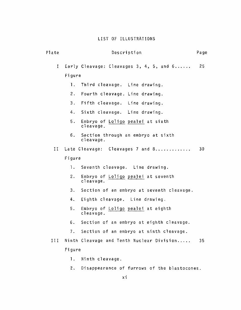
LIST OF ILLUSTRATIONS
Plate Description Page
I Early Cleavage: Cleavages 3, 4, 5, and 6...... 25
Figure
l. Third cleavage. Line drawing.
2. Fourth cleavage. Line drawing.
3 . Fifth cleavage. Li ne drawing.
4. Si xth cleavage. Line drawing.
5. Embryo of Lo1igo pea1ei at 5 i x thcleavage.
6. Section through an embryo at sixthcleavage.
II Late Cleavage: Cleavages 7 and 8..... ..... ... 30
Figure
1. Seventh cleavage. Line drawing.
2. Embryo of Lo1igo pea1ei at seventhcleavage.
3. Section of an embryo at seventh cleavage.
4. Eighth cleavage. Line drawing,
5. Embryo of Lo1igo pea1ei at eighthcleavage.
6. Section of an embryo at eighth cleavage.
7. Section of an embryo at ninth cleavage.
III Ninth Cleavage and Tenth Nuclear Division..... 35
Figure
1. Ninth cleavage.
2. Disappearance of furrows of the b1astocones.
xi

Plate
III
Description
3. Movement of blastocona1 nuclei.
Page
4. Prophase of tenth nuclear division.
IV Tenth Nuclear Division. 38
Figure
1. End of mitosis and formation of thetransitory furrow.
2. Disappearance of the transitory furrow.
3. Movements of yolk syncytium nuclei.
4. Nuclear movement and onset of epiboly.
V Cinemicrographic Series I. Cleavages 5 and 6. 42
VI Cinemicrographic Series I. Cleavages 7 and 8. 44
VII Cinemicrographic Series I. Germ LayerSegre gati on. . . . . . . . . . . . . . . . . . . . . . . . . . . . . . . . . . . 46
VIII Cinemicrographic Series I. Germ LayerSegregation................................... 48
IX Cinemicrographic Series II. Cleavages 7to 9.......................................... 50
X Cinemicrographic Series II. Ninth Cleavage... 58
XI Cinemicrographic Series II. Ninth Cleavageand Estab1 i shment of the Yo1 k Syncyti urn.. . . . . . 60
XII Cinemicrographic Series II. Establishmentof the Yolk Syncytium...... 62
XIII Undercutting Furrows of B1astocones........... 66
Figure
1. Light micrograph of margin of blastodermat eighth cleavage.
2. Electron micrograph of a b1astocone.
xii

Plate
XII I
Description
3. Base of an undercutting furrow.
4. Microfi1aments at the base of a furrow
Page
XIV Furrow Retraction in a B1astocone 68
Figure
1. Light micrograph of margin of blastodermat ninth cleavage.
2. Electron micrograph of a retracting furrow.
3. High magnification micrograph of a furrow.
XV Yolk Syncytium and Microtubu1es............... 71
Fi gu re
1. Edge of the blastoderm during earlyepiboly.
2. Microtubu1es in yolk syncytium.
3. Associations of microtubu1es.
XVI Scanning Electron Microscopy: Eighth Cleavage 76
Figure
1. Scanning electron micrograph of eighthcleavage.
2. Margin of blastoderm
XVII Scanning Electron Microscopy: OverlappingCells 79
Figure
1. Relations of overlapping cells.
2. Cell surface activity
xii i

Plate Description Page
XVIII Intercellular Junctions and Contacts.......... 81
Figure
1. Cell-cell apposition at eighthcleavage.
2. Apical junction at ninth cleavage.
3. Septate junction.
4. Intercellular interdigitation.
5. Intercellular interdigitation.
XIX Germ Layer Segregation....... 85
Figure
1. Transmission electron micrograph ofthe margin of the blastoderm.
2. Tip of a lamellipodium.
3. Leading lamella of an overlapping cell.
XX Cell Overlapping.............................. 89
Figure
1. Transmission electron micrograph ofan overlapping lamellipodium.
2. Membranous vesicles in overlapping cells.
3. Fusion of vesicles with membrane.
XXI Germ Layer Segregation....... 93
Fi gure
1. Overlapping cell.
2. Surface papillae and microfilaments.
3. Orientation of cortical microfilaments.
xiv
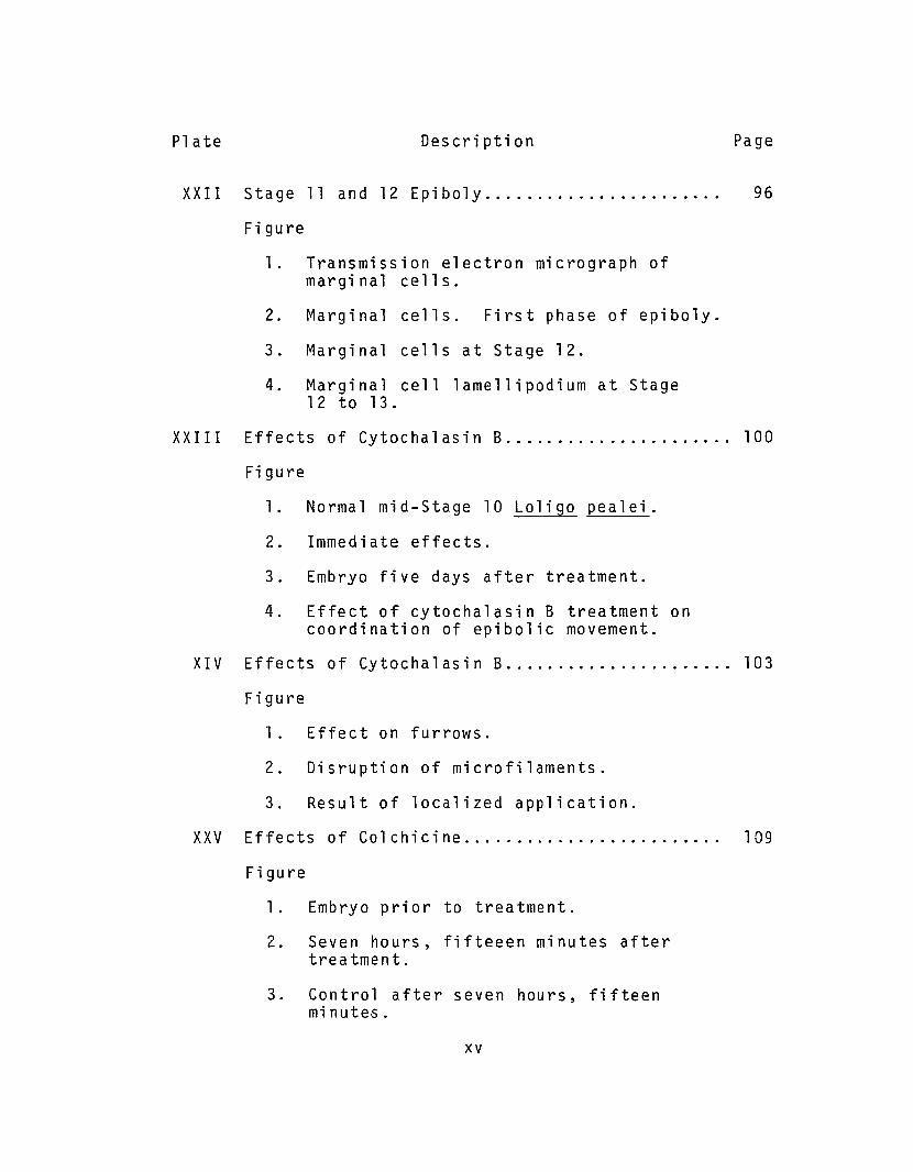
Plate Description Page
XXII Stage 11 and 12 Epiboly.... 96
Figure
1. Transmission electron micrograph ofmarginal cells.
2. Marginal cells. First phase of epiboly.
3. Marginal cells at Stage 12.
4. Marginal cell lamellipodium at Stage12 to 13.
XXIII Effects of Cytochalasin B 100
Fi gu re
1. Normal mid-Stage 10 Loligo pealei.
2. Immediate effects.
3. Embryo five days after treatment.
4. Effect of cytochalasin B treatment oncoordination of epibolic movement.
XIV Effects of Cytochalasin B 103
Figure
1. Effect on furrows.
2. Disruption of microfilaments.
3. Result of localized application.
XXV Effects of Col chi ci ne. . . . . . .. . . . . . . . .. . . . . . . .. 109
Figure
1. Embryo prior to treatment.
2. Seven hours, fifteeen minutes aftertreatment.
3. Control after seven hours, fifteenmi nut es .
xv

Pl ate Description Page
XXV 4. Control after nineteen hours.
5. Treated embryo after nineteen hours.
6. Seven days after treatment at eleventhnuclear division.
xvi
1. INTRODUCTION
Gastrulation is of fundamental importance in the
embryogenesis of all Metazoa. Since the pioneering work
of Ho1tfreter (1939, 1943, 1944), it has become increasingly
clear that gastrulation involves cellular shape change and
translocation (see Ba1insky, 1970; Johnson, 1974; Berri11
and Karp, 1976; Trinkaus, 1976). For example, cellular
shape changes have been implicated as a driving force in
amphibian gastrulation (Baker, 1965) and in vegetal plate
formation in echinoderms (Dan and Okazuki, 1956; Dan 1960;
Gustafson, 1963; Gibbins, et a1., 1969). Formation of
pseudopodia and ruffled membranes by mesodermal cells of
amphibians indicate that active migration of individual
cells is also involved (Nakatsuji, 1974; Johnson, 1976).
The appearance of chorda mesodermal cells with scanning
electron microscopy indicates that these cells trans10cate
during gastrulation by means of 1ame11ipodia and filopodia
(Monroy, et a1., 1976). Active cell movements have been
observed during the formation of the archenteron and
mesenchyme in echinoderms (Gustafson and Wolpert, 1967;
Gustafson and Toneby, 1971 l and during epiboly in te1eosts
(Trinkaus, 1973; Wourms, 1972). Avian gastrulation also
appears to involve trans1ocations by various cells (Tre1sted,
et a1., 1967; Hay, 1968; Nicolet, 1971). Although there is

now considerable information concerning the processes of
gastrulation, as Trinkaus (1976) has recently stated, there
is still a great deal to be learned.
The concept of cephalopod gastrulation has a history
paralleling the development of the general concept of
gastrulation. Many of the earliest observations of cepha
lopod development dealt almost exclusively with external
morphology (e.g. Kolliker, 1844; Lankester, 1875; Grenacher,
1874; Brooks, 1880). Writers who examined and described
gastrulation may be categorized according to their explana
tion of the supposed mechanisms of gastrulation. Regarding
the segregation of the middle (= mesendodermal) layer, early
studies fall into three groups. The majority of writers
described the formation of the middle layer in cephalopods
as a process of mitotic delamination comparable to what was
then thought to be the mechanism of presumptive mesoderm
formation in birds, teleosts and mammals (Metschnikoff,
1867; Ussow, 1875; Bruce, 1886, Watase, 1891; Teichmann,
1903). Horizontal divisions occurring in either a restricted
area (Watase, 1891) or throughout the single-layered
blastoderm (Metschnikoff, 1867; Ussow, 1874, 1875, 1881)
were thought to give rise to a second layer of cells
between the yolk and the blastoderm, thereby producing a
"Gastrula" stage embryo. The most descriptive writer,
Ussow (1875) gave the following account:
2
II ••• the cells, which are continually undergoing further division in a longitudinal direction,begin also to divide gradually in a transversedirection, the division commencing at the lowerperiphery and advancing towards the center....As the result of this transverse division a secondgerm-lamella is produced, at first only in themedian ring and in the segment pa r t ." (Ussow,1875, p. 109).
Disputing the idea of delamination, Korschelt (J892;
and in Korschelt and Heider, 1900, 1936) reported that the
middle layer was established by a modified form of invagina-
tion which he termed "{mm i qr a t t on.." He thought that cells
near the edge of the blastoderm migrated into the sub-
blastodermal area. Korschelt proposed that II. the edge
of the germ-disc corresponds to the blastopore which is
filled by the large y ol kvp l uq" (Korschelt and Heider, 1900,
p. 279). This homologization of the blastodermal perimeter
with the blastopore was tenuous, admittedly having been
based principally on the proximity of the stomodeal
invagination to the perimeter in Grenacher's (1874) Oegopsid
embryo. Korschelt's opinions concerning this homology and
Naef's (1928) later extensions of the idea were discussed
by Sacarrao (1949, 1952 a, b, c, 1953).
Leading a third group of writers, Bobretzky (1877)
observed development in Loligo vulgaris and described the
middle layer as formed by means of a folding under at the
edge of the blastoderm (Umschlagsrand). This view of
middle layer formation was further espoused by Vialleton
3

(1888) and Faussek (1896, 1900). Naef (1928), in his
extensive monograph on the Cephalopoda, embraced this
theory to derive a scheme for the evolution of the cepha
lopod embryo. Naef proposed a hypothetical form having an
amount of yolk and form of development intermediate between
Prosobranchs and Cephalopods. His interpretation, however,
ignores obvious differences in the origin and ultimate fate
of the germinal layers in these two groups (for a detailed
discussion of Naef's views, see Sacarrao, 1953; Arnold, 1971).
Other attempts were made to find an intermediate form of
development among extant molluscan species. Conklin (1907)
found that the origin and fate of the germ layers in the
large (1.7 mm diameter), yolky eggs of the whelk Busycon
corresponded precisely with those of other prosobranchs.
He found that the same was true for Crepidula which also
has a large (400 urn), yo1ky egg (Conklin, 1897). Naef's
theories have gained little support and it is generally
accepted that the cephalopod form of development is unique
among the molluscs (Raven, 1958; Arnold, 1971).
Sacarrao (1953) discussed at length problems of segre
gation and fates of germ layers in cephalopods. His inter
pretations have gained widest acceptance in recent literature
(Arnold, 1965, 1971; Arnold, et al., 1974; Fields, 1965;
Raven, 1958; Meister, 1972; De Leo, 1972; Fuchs, 1973;
Fioron;, 1974; Fioroni and Meister, 1974). Sacarrao
reaffirmed the idea that the middle layer constituted a
4
complex endomesoderm which arose by means of a mitotic
delamination of the originally single-layered blastoderm.
However, he observed in only one instance a mitotic figure
which would give rise to a cell in the middle region. This
provoked the remark that" .•. la delamination du blastodisque
est tres probablement un acte unique. 1I 1 (Sacarrao, 1953
p. 34}. Though he favored the idea of mitotic delamination,
Sacarrao was aware of the limitation and dangers of inter
pretation solely from fixed materials. Thus he wrote the
following:
II ••• on ne peut pas rejecter ~ priori l'idee d'unemigration de quelques elements vers la profondeur.La position oblique d'une ou autre cellule de lamarge du blastoderm, comme qu'enchevauchee parl'element vot s t n , fait penser ~ cette pos s t bt l i tf .... 112(se car r'ao , 1953 p . 34).
Sacarrao's allusion to the possibility of the movement of
cells during germ layer segregation has been ignored by
most subsequent investigators of cephalopod development.
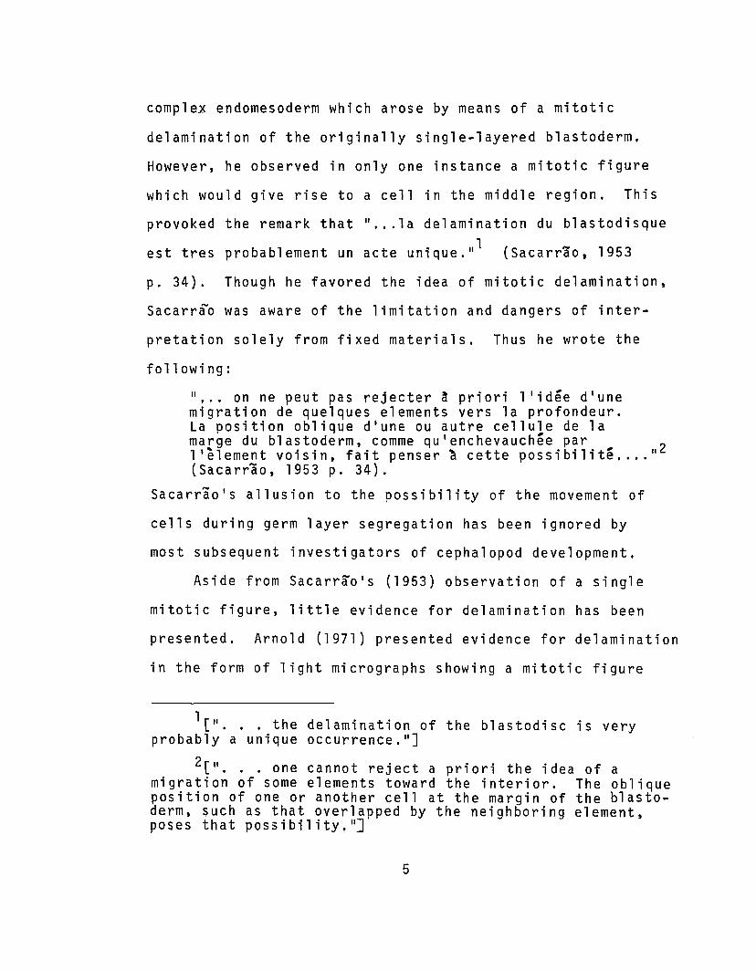
Aside from Sacarrao's (1953) observation of a single
mitotic figure, little evidence for delamination has been
presented. Arnold (1971) presented evidence for delamination
in the form of light micrographs showing a mitotic figure
1[" ..• the delamination of the blastodisc is veryprobably a unique occurrence. lI
]
2[ I'. . . 0 ne can not r e j ectaprio r i the ide a 0 f amigration of some elements toward the interior. The obliqueposition of one or another cell at the margin of the blastoderm, such as that overlapped by the neighboring element,poses that possibility!"]
5

oriented so that it might give rise to a middle layer of
cells. Both Sacarrao's and Arno1d 's evidence are, however,
open to a different interpretation.
Recently, Fioroni (1974) reiterated Korsche1t 's
opinion that middle-layer formation takes place by means
of immigration of cells from the periphery of the blastoderm.
Fioroni's opinion, like that of Korsche1t, was based upon
study of sections of paraffin-embedded embryos observed
with light microscopy. Neither investigator presented
evidence, other than rather interpretive drawings of what
he observed. It seems unreasonable to expect to be able to
interpret the dynamic phenomena of gastrulation solely by
observing sections of fixed embryos. Fioroni concluded
by refuting Naef's theory of invagination at an enlarged
blastopore and proposing a mechanism of middle-layer
formation involving unverified cell movements.
Another aspect of the problem of cephalopod gastrula-
tion is the establishment of the yolk syncytium (= yolk
epithelium, yolk membrane or periblast of older literature)
(see Arnold, 1971; Arnold and Williams-Arnolds, 1976).
Various conflicting descriptions are found in the early
literature. Lankester (1875) felt that the yolk syncytium
nuclei were derived from the yolk mass as was, at that time,
thought of the so-called lI yol k nuclei ll in arthropods and verte
brates. Ussow (1875) did not recognize the syncytial
nature of the yolk syncytial layer and termed it the
6
"intestino ... fibrous layer." He described the "cells"
forming this layer as derived by delamination from the
middle layer. Ussow felt that the middle layer ultimately
produced the muscular envelope of the intestinal tract
and ink sac, and the walls of the circulatory system,
Vialleton (1888) coined the name "yolk epithelium" to
describe the yolk syncytium although he seems to have
recognized its syncytial quality. Vialleton described the
"yolk epithelium" as forming from the marginal elements of
the blastoderm which he named blastocones. The blastocones,
he saw, underwent an "essential alteration," Their cellular
nature was lost giving rise to a "plasmodium" that moved by
"ingression" under the middle layer to the animal apex of
the egg as well as down around the yolk mass. Vialleton
considered the "yolk epithelium" to be purely endodermal in
nature.
Watase (1891) first recognized the role of the yolk
syncytium as a transitory digestive organ. He felt that
it functioned in the digestion of the yolk during embryo
genesis~ and that it produced no adult tissue. Korschelt
(1900) used Vialleton's term "yolk epithelium" to describe
a layer he saw as consisting of very flattened cells. He
did not discuss its origin, although he recognized the
yolk syncytial layer's role in embryogenesis. Teichmann
(1903) thought, wrongly, that the middle layer and yolk
7

syncyttum together constituted the definitive endoderm.
He also thought that mesoderm arose from infoldings of
the ectoderm. Teichmann did not discuss the formation of
the yolk syncytium. The role of the yolk syncytium was
later clarified by the investigations of Portmann and
Bidder (1928) and of Boletzky (1967, 1975). These authors
showed conclusively that the yolk syncytium functioned as
a digestive organ and that it was not incorporated into the
definitive endoderm which, as Kolliker (1844) had earlier
recognized, developed from a small plaque of middle-layer
cells. Sacarrao (1953) agreed with Vialleton's description
of the mode of formation and syncytial character of the
yolk syncytium. Sacarrao (1953) considered that the
endoderm in cephalopods was divided into two ontogenetically
and functionally distinct tissues: the yolk syncytium was
derived from the blastocones and had a transitory role,
digesting yolk during development; the definitive endoderm
of the adult was derived from the complex endomesodermal
layer.
More recently Fuchs (1973) and Fioroni (1974) have
confirmed, in various octopods, the fact that the yolk
syncytium nuclei arise from the blastocones, However,
neither of these writers has addressed questions of the
cellular basis of yolk syncytium formation. Both report
that, in Eledone, nuclei and cytoplasm of the yolk
8

syncytium lying under the center of the blastoderm are
derived by detachment and incorporation of cells from
that area of the blastoderm. Fuchs (1973) summarizes
his findings as follows:
111m Innern der Keimscheibe. Die Bi1dungdes Syncytium vollzieht sich in diesem Fall amDetachierungsort, direkt unter dem Blastoderm.Vie1leicht erhellt daraus, warum nach gewissenalteren Autoren (Ussow 1881; Korschelt 1892) dasDotterepithel aus dem Entomesoderm hervorgeht.Freilich wollen wir damit nicht sagen, d~
Entomesodermzel1en bei der Bi1dung des Dotterepithels mitwirken. Vielmehr differenziert sichdas Dotterepithe1 direkt aus den BlastomerenGleichzeitig 10der sogar noch vor den Entomeso-de rmze l l en ." (Fuchs, 1973 p. 37)
This manner of yolk syncytium development is found in
no other cephalopod species. Furthermore, it conflicts
with the theory of an organogenetic inductive function of
the yolk syncytium (Arnold and Williams-Arnold, 1974, 1976)
and with the conservative nature of the cleavage patterns
of most l phalopods. Even with these more recent studies
knowledge of the mechanism of yolk syncytium nucleation is
little advanced beyond Vial1eton 's (1888) description.
l[IIIn the interior of the germinal disc. Theformation of the syncytium occurs in this case at thepoint of attachment directly beneath the blastoderm.Maybe this clarifies the fact that according to someprevious authors (Ussow, 1881; Korschelt, 1892) the yolkepithelium develops from endomesoderm. Certainly we donot say with this that the endomesodermal cells have apart in the formation of the yolk epithelium. Rather theyolk epithelium develops directly from the b1astomeresafter or even before the formation of the endomesoderma1cel1s."]
9

The investigations reported here provide a description
of gastrulation in Loligo pealei. Observations made using
techniques of light microscopy, electron microscopy and
time-lapse cinemicrography are employed to describe:
1) the later phases of cleavage in order to clarify the
ontogenetic history of the germ layers, particularly the
middle-layer cells; 2) the events of middle-layer segrega
tion; and 3) the events and mechanism of yolk syncytium
formation. Observational data are used to present a
generalized description of cephalopod gastrulation and
are discussed in relation to recent literature on cephalo
pod development.
Results of experiments with the inhibitors colchicine
and cytochalasin B further describe and differentiate the
morphogenetic mechanisms involved in segregation of the
middle layer and formation of the yolk syncytium. Data
from these experiments are compared with observational
data on the normal development of Loligo pealei. Observa
tional and experimental data are discussed in relation
to cephalopod gastrulation and to problems of morpho
genetic cell movement. A testable model is presented
as a possible explanation of the mechanism of epibolic
cell movement in Loligo.
10

2. MATERIALS AND METHODS
2.1. Acquisition and Maintenance of Experimental Animals
Adult Lo1igo pealei were obtained from the Supply
Department of the Marine Biological Laboratory at Woods
Hole, Massachusetts. These animals were maintained for
short periods (3 to 6 days) in fiberglass tanks (0.50 m X
0.75 m Xl. 10 m) supplied with fresh, running sea water
(ca. 0.35 m in depth) at a temperature of 15° to 21°C.
No attempts were made to feed adults to extend their
maintenance capacity (see Summers and McMahon, 1970, 1974;
Summers, et a1., 1974).
Fertilized eggs of h. pea1ei were obtained by
artificially inducing egg laying (Arnold, 1962). Embryos
were separated from their surrounding jelly layers using
fine forceps. Deje11ied embryos were placed in Syracuse
watchg1asses with fresh sea water for observation. Those
to be maintained for longer periods were placed in Stender
dishes (62 mm X 36 mm) containing sea water (15 to 20 m1)
at room temperature (15° to 21°C). Sea water was changed
at least three times daily. A maximum of twelve embryos
was maintained in any dish.
Adult Euprymna sco1opes were captured and maintained
as previously reported (Arnold, et a1., 1972). There is
no known stimulus for egg laying behavior in this species.
To obtain eggs from E. sco1opes a mature adult of each sex
11
was placed in a common tank. Copulation occurred only at
night; usually after 2200 hours. The duration of copula
tion varied from 20 minutes to over 60 minutes. The
females usually laid eggs within one week after copulation
and then died within the following two weeks. E. scolopes
embryos were observed and maintained as described for
L. pealei.
2.2. Methods of Light Microscopy, TransmissionElectron Microscopy, and ScanningElectron Microscopy
For the study of early cleavage stages and cell
lineage, embryos were fixed according to the method of
Timmermans, Geilenkirchen, and Verdonk (1970). Whole
embryos were fixed in a mixture of formalin, ethanol and
acetic acid (FAA), treated with 95% ethanol for 24 hours,
and stained by a modified Feulgen stain (Pearse, 1968
p. 648). Stained embryos were dehydrated, cleared in
xylene and mounted, whole or with the vegetal half removed,
on standard microscope slides. Some FAA-Feulgen treated
and Bouin's fixed embryos were embedded in Epon (Luft,
1961} and sectioned at 1 ~m for observation with the
light microscope. Whole-mounted embryos were observed
with a compound microscope and camera lucida drawings made
of the various stages.
Embryos were fixed for transmission electron microscopy
using several methods. The procedure giving the best and
12
most consistent results was that of Palade (1952). This
was a single fixation procedure using 1% osmium tetroxide
in a veronal acetate buffer at pH 6,8. Embryos were fixed
for ten to twenty minutes and rinsed in several changes of
50% ethanol. Dehydration continued through a graded
ethanol series into propylene oxide. Dehydrated embryos
were embedded in Epon (Luft, 1961). Sections 60 nm to
90 nm in thickness, as judged by their interference colors,
were taken with a diamond knife using a Reichert OM-U2
Ultramicrotome. Sections were picked up on copper mesh
grids and stained with saturated uranyl acetate in 50%
methanol (Stempak and Ward, 1964) and lead citrate
(Venable and Coggeshall, 1965). Stained sections were
observed using a Philips EM 201 electron microscope.
Thi ck sec t i 0 f, S 11 to 1. 5 f.l m) 0 f 0 smi um fix ed, Ep0 n
embedded embryos were made with glass knives for observa
tion with the light microscope. Thick sections were
mounted on standard glass slides and stained with either
Richardson's methylene blue-azure II mixture (Richardson,
et al. 1960) or a sequential methylene blue, azure II, and
basic fuchsin series as described by Humphry and Pittman
(1974 ).
Other fixation procedures used for electron microscopy
were as follows:
1, Embryos were prefixed in 2.5% glutaraldehyde
buffered to pH 7.2 with 0.2 M Millonig1s phosphate, adjusted
13

to 960 milliosmoles with NaCl. Prefixation was followed
by three buffer rinses. Post fixation was accomplished
in 1% osmium tetroxide in Millonig's phosphate buffer at
pH 7.2 for 15 to 20 minutes (Cloney and Florey~ 1968).
2. Embryos were prefixed for 1 hour in 2% glutaralde
hyde in 0.1 M cacodylate buffered sea water at pH 7.2,
which was adjusted to 1.5% sucrose. Prefixation was
followed by two 15 minute rinses in 0.1 M cacodylate in
sea water plus 5% sucrose. Post fixation was accomplished
in 2% osmium tetroxide in distilled water followed by two
15 minute rinses in 0.1 M cacodylate in distilled water.
3. Embryos were prefixed for 1/2 to 1 hour in 2.6%
glutaraldehyde in 0.1 M collidine in sea water at pH 7.4
followed by a 30 minute rinse in filtered sea water.
Embryos were post fixed in 1% osmium tetroxide in collidine
buffered sea water at pH 7.4, rinsed in filtered sea water
and dehydrated (Arnold~ personal communication).
Embryos prepared for scanning electron microscopy
were fixed as for transmission electron microscopy~
dehydrated in acetone and dried in a Sorvall Critical
Point Drying System (Du Pont-Sorvall Inc., Newtown~ CT)
using Freon 113. Dried specimens were coated with carbon
and gold and observed using a JOELCO Scanning Electron
Microscope.
14
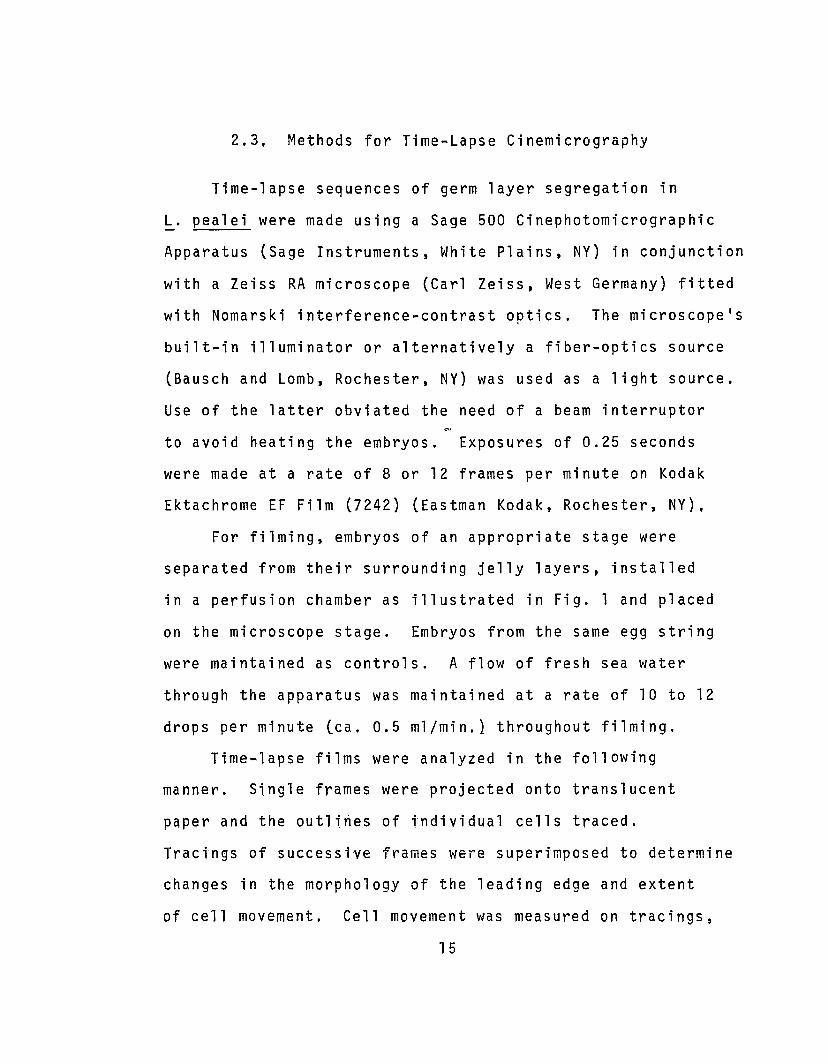
2.3. Methods for Time-Lapse Cinemicrography
Time-lapse sequences of germ layer segregation in
L. pealei were made using a Sage 500 Cinephotomicrographic
Apparatus (Sage Instruments, White Plains, NY) in conjunction
with a Zeiss RA microscope (Carl Zeiss, West Germany) fitted
with Nomarski interference-contrast optics. The microscope1s
built-in illuminator or alternatively a fiber-optics source
(Bausch and Lomb, Rochester, NY) was used as a light source.
Use of the latter obviated the need of a beam interruptor
to avoid heating the embryos. Exposures of 0.25 seconds
were made at a rate of 8 or 12 frames per minute on Kodak
Ektachrome EF Film (7242) (Eastman Kodak, Rochester, NY),
For filming, embryos of an appropriate stage were
separated from their surrounding jelly layers, installed
in a perfusion chamber as illustrated in Fig. 1 and placed
on the microscope stage. Embryos from the same egg string
were maintained as controls. A flow of fresh sea water
through the apparatus was maintained at a rate of 10 to 12
drops per minute (ca. 0.5 ml/min.} throughout filming.
Time-lapse films were analyzed in the following
manner. Single frames were projected onto translucent
paper and the outliries of tndividual cells traced.
Tractngs of successive frames were superimposed to determine
changes in the morphology of the leading edge and extent
of cell movement. Cell movement was measured on tracings,
15
-------------------------:
[ I
[@4 r= -- ~
€l))Ef""' -~ :::::=F=
L
Iow
E ,CS I LJT~<r""---_-=----~
R
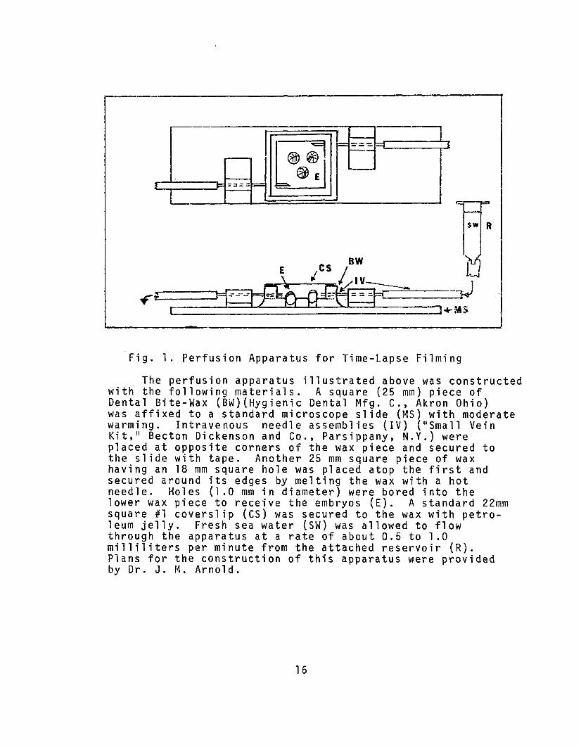
Fig. 1. Perfusion Apparatus for Time-Lapse Filming
The perfusion apparatus illustrated above was constructedwith the following materials. A square (25 mm) piece ofDental Bite-Wax (BW)(Hygienic Dental Mfg. C., Akron Ohio)was affixed to a standard microscope slide (MS) with moderatewarming. Intravenous needle assemblies (IV) ("S mall VeinKit," Becton Dickenson and Co., Parsippany, N.Y.) wereplaced at opposite corners of the wax piece and secured tothe slide with tape. Another 25 mm square piece of waxhaving an 18 mm square hole was placed atop the first andsecured around its edges by melting the wax with a hotneedle. Holes (1.0 mm in diameter) were bored into thelower wax piece to receive the embryos (E). A standard 22mmsquare #1 coverslip (CS) was secured to the wax with petroleum jelly. Fresh sea water (SW) was allowed to flowthrough the apparatus at a rate of about 0.5 to 1.0milliliters per minute from the attached reservoir (R).Plans for the construction of this apparatus were providedby Dr. J. M. Arnold.
16

projected images, and prints of rephotographed frames of
16mm films. Cell movement was determined by measuring the
distance between the position of a cell's leading edge in
any frame and its position at an arbitrary starting point
(time = O). Time = 0 was chosen as the frame in which
minimal overlap was exhibited by those cells which were
to overlap the ring of presumptive middle-layer cells
after eighth cleavage. Measurements were made at three sites
along the leading edge of each cell. The average of these
measurements was taken as the distance moved by each cell
over the interval between measured frames. Frames were
chosen such that the interval between frames was approximately
10 minutes. Rates of movement were determined by dividing
measurements of the distance moved by the time interval
over which the movement occurred. Time intervals between
measurements were determined by multiplying the film
exposure interval (i .e. at 8 frames/minute the exposure
interval was 7.5 seconds) by the number of frames plus one
between measured frames. Suitable sequences were
rephotographed on 35mm Kodak Plus X film (Plates V through
XI I ) •
2.4. Methods of Experimental Inhibitionof Cell Movement and Division
Colchicine and cytochalasin B were used in attempts
to specifically inhibit mitosis and cell motility during
11

early phases of germ layer segregation in Loligo pealei.
In each experiment, control and experimental embryos were
taken from a single egg string. Each string contains
about 180 eggs all of which have been fertilized simultane
ously and develop synchronously, Each experimental and
control group consisted of 10 or 15 embryos. At least four
experimental replications were made for any single time/
concentration combination. Experimental results were
discarded if more than ten percent of the controls showed
aberrant development. This situation occurred only with
lI end of the season ll animals.
Cytochalasin B solutions were made up according to
the method of Carter (1967 a). Cytochalasin B was dissolved
in dimethyl sulfoxide at a concentration of 1 mg/ml and
stored at _4 0 to _20 0 C until used. Serial dilutions of
the stock solution were made with fresh, filtered sea water
to concentrations of 0.05,0.1,0.2,0.5,1.0 and 2.0Ilgm/m1.
Control embryos were maintained in sea water alone and in
appropriate concentrations of dimethyl sulfoxide in sea
water corresponding. Embryos at specific stages were
treated with each of the above concentrations of cytochalasin
B for periods of 5, 10, 30, 60 minutes and continuous
immersion.
A few embryos (4) were treated by regional application
of cytochalasin B using the method of Arnold and Williams
Arnold (1976). Restricted regions of the blastoderm were
18

treated by applying ( for 10 min.) a small glass probe
(ca. 75 m to 100 m at the tip) which had been coated
with agar, dried, and soaked in 2.0 ~g/ml cytochalasin B.
Colchicine was dissolved in sea water to a concentra
tion of 10-4M. Initially, embryos of various stages were
treated with this solution for periods ranging from 7ive
minutes to one hour. Because the block of mitosis produced
by colchicine at this concentration is irreversible in
Loligo, variation of treatment time was discontinued in
favor of a single ten or fifteen minute treatment followed
by at least three five minute rinses in fresh sea water.
After treatment, the embryos were maintained for
periods of three to five days. Daily observations were
made and any changes in the morphology of the treated
embryos over the maintenance period were recorded. Control
and treated embryos were fixed for either light or electron
microscopy at the end of the experimental period.
2.5. Terminology of Cephalopod DevelopmentUsed in This Study
Except as noted below the terminology for cephalopod
development used herein will be that of Arnold (1971) and
Arnold and Williams-Arnold (1976). A review of the literature
on cephalopod development reveals some confusion regarding
certain terms descriptive of the structure of the embryo.
This has led to considerable misinterpretation by some
19

workers, The problems are primarily semantic, but nonethe
less real, The following are often the source of miscon
ception and are defined as they are used here:
Yolk Cytoplasmic Layer. This is the uncleaved portion
original egg cytoplasm before it is invaded by yolk
syncytial nuclei. This portion of the embryo is usually
lumped with the yolk syncytium. I feel, however, that
it must be renamed because of functional and morphological
differences and to lend clarity to descriptive analysis.
Yolk Syncytium. The yolk syncytium is that portion
of the yolk cytoplasmic layer which has become nucleated
and thus constitutes d true syncytium. This structure is,
on the average. more than twice as thick as the yolk
cytoplasmic layer, and is associated with active yolk
digestion. This term corresponds to the terms yolk
epithelium and periblast of the literature.
Yolk Syncytium Nuclei. The nuclei of the yolk
syncytium. These are larger than the nuclei of the blasto
meres, are greatly flattened when they lie beneath the
blastoderm, have dispersed chromatin, and are more
basophtlfc than blastoderm nuclei. This corresponds to
the term yolk epithelium nuclei of the literature.
Blastomere. As in the usual sense, this term is used
to describe the cells resulting from the complete cleavage
of the egg cytoplasm. In cephalopods the blastomeres are
distinguished from the blastocones by not being continuous
20
with the yolk cytoplasmic layer. This term has caused
confusion by being used in the literature to describe all
the products of cleavage.
Blastocones. These are the products of the partial
cleavage of the blastodisc which are defined by the
meridional and undercutting furrows and which remain con
tinuous with the yolk cytoplasmic layer. Because they
remain throughout their history continuous with the yolk
cytoplasmic layer, they cannot, by any rigid definition,
be called cells. The term IIblastocone cell ll of the liter
ature is therefore discarded in favor of the simpler
blastocone.
3. RESULTS
3.1. Cleavage Pattern in Loligo pealei
The cleavage pattern in Loligo was followed using
living and FAA-Feulgen treated embryos and time-lapse
cinemicrography. The principal questions of interest
with regard to cleavage are: 1) What is the lineage of
the cells from which the middle layer arises? 2) Are the
middle-layer cells products of a delaminating cleavage,
blastomeres which migrate inward from the periphery of
the blastoderm or are they derived in some other manner?
3) What is the lineage of the yolk syncytium nuclei?
Information relating to each of these questions is
presented below.
21

3.1.1. Establishment of the Blastoderm by Cleavage
The early cleavage pattern of Loligo pealei has
previously been described to seventh cleavage (Watase,
1891; Arnold, 1971). Little has been said, however, about
the later cleavages just prior to and through germ layer
segregation. The following description of cleavage
includes a brief redescription of the egg and first seven
cleavages in order to lend clarity and continuity to
descriptions of the later cleavages.
The egg of Loligo is ovate with one end more pointed
than the other, much like a hen1s egg. Prior to ferti
lization the egg cytoplasm exists as a thin layer sur-
rounding the mass of yolk (see Arnold, 1971). Fertilization
initiates a streaming of the cytoplasm toward the pointed
(animal) end of the egg. There, part of the cytoplasm
forms a thickened cap, the blastodisc. At this stage the
uncleaved egg possesses a symmetry which is maintained through
out development, and corresponds to the axes of the adult
animal (Fig. 2; Watase, 1891; Arnold, 1971). The animal
vegetal plane of the egg corresponds to the caudo-cephalic
plane of the adult. The egg is bisected by the anterior
posterior plane, but asymmetrical around the dorsal-
ventral axis. Viewed from the side, the anterior portion
of the egg is more convex and thus more massive than the
posterior. In addition, the place of the zygotic
22
D D
V-4----.:+--'-\-D
ey. __________
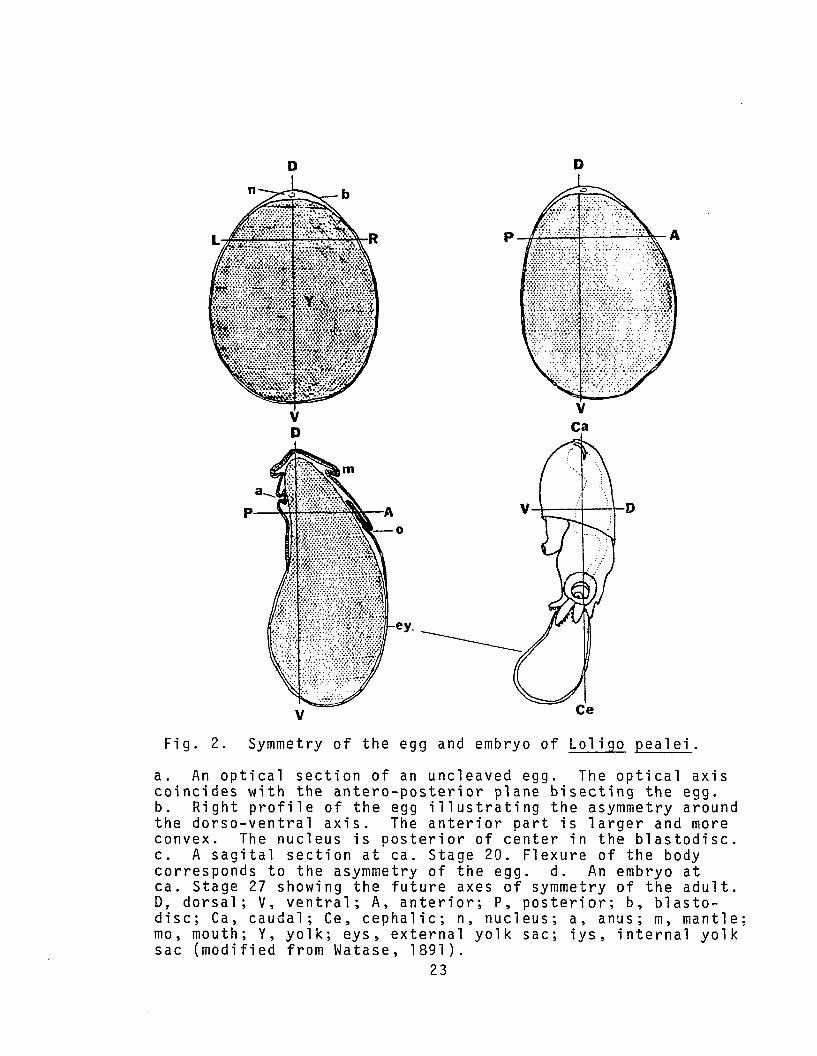
vFig. 2. Symmetry of the egg and embryo of Loligo pealei.
a. An optical section of an uncleaved egg. The optical axiscoincides with the antero-posterior plane bisecting the egg.b. Right profile of the egg illustrating the asymmetry aroundthe dorso-ventral axis. The anterior part is larger and moreconvex. The nucleus is posterior of center in the blastodisc.c. A sagital section at ca. Stage 20. Flexure of the bodycorresponds to the asymmetry of the egg. d. An embryo atca. Stage 27 showing the future axes of symmetry of the adult.D, dorsal; V, ventral; A, anterior; P, posterior; b, blastodisc; Ca, caudal; Ce, cephalic; n, nucleus; a, anus; m, mantle;mo, mouth; Y, yolk; eys, external yolk sac; iys, internal yolksac (modified from Watase, 1891).
23

nucleus is asymmetric, being disposed to the posterior of
a transverse line bisecting the blastodisc (Fig. 2). This
anterior-posterior asymmetry persists throughout embryogenesis
and may be used to orient within the blastoderm during
cleavage and gastrulation stages (Arnold, 1971).
First cleavage (Stage 4 of Arnold, 1965) separates the
future right and left halves of the embryo along its
anteroposterior plane (Arnold, 1965). The furrows of the
first three cleavages cut through the cytoplasm of the
blastodisc, but the cytoplasm remains continuous at the
periphery of the resultant "cell s." A second furrow
divides the blastodisc somewhat symmetrically into an
anterior and posterior portion. The posterior portion is
the smaller due to the above-mentioned acentric displace
ment of the zygotic nucleus.
The third cleavage (Stage 6) furrow is asymmetric in
the right and left halves. In the posterior half the third
furrow forms at right angles to the second furrow, but
occurs at an angle of 30° to the perpendicular anterior of
the second furrow (PLATE I, Fig. 1). Thus the daughter
"c e l l s " adjacent to the first furrow in the anterior half
are larger than those in the posterior half. It has been
suggested that this asymmetry is a remnant of ancestral
Spiralian relationships (Arnold and Williams-Arnold, 1976).
At third cleavage a mitotic asynchrony becomes more
evident. The posterior daughter "cell s" lag somewhat
24

N(J1
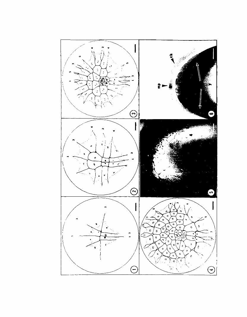
PLATE I
Early Cleavage: Cleavages 3, 4, 5, and 6
Figs. 1 to 4 are camera 1ucida drawings of FAA-Feu1gen treated embryos ofLoligo pea1ei. Numerals indicate the cleavage number of the corresponding furrow.Dotted lines indicate the positions of undercutting furrow bases. The figures areoriented with the anterior-posterior axes of the embryos vertical. Nuclei aredrawn as they appear with Feu1gen stain. Mitotic phases appear as follows:o Interphase, ~ Early Prophase, ~ Late Prophase, I Metaphase, 11 Anaphase,
and ~ Telophase. Bars = 0.1 mm (ca. 75 X).
Fig. 1. Third cleavage (Stage 6). The third cleavage furrows are unequalin the posterior half and symmetrical in the right and left halves. Note themitotic phase lag of the posterior-medial bastocones.
Fig. 2. Fourth cleavage (Stage 7). Fourth cleavage is roughly parallel tothe second furrow and forms the first four b1astomeres. The degree of mitoticphase lag has increased.
Fig. 3. Fifth cleavage (Stage 8). Cleavage planes are radial except in theblastocones adjacent to the second furrow and in the posterior-medial blastocones.
Fig. 4. Sixth cleavage (Stage 9). All blastocones and the large blastomeresabutting the second furrow in Stage 8 have cleaved trans-radially. The orientationof cleavage in central blastomeres varies.
Fig. 5. Sixth cleavage embryo of Lo1igo pea1ei. View is of the right posterior quadrant. Numerals indicate furrows as above. Bar = 0.1 mm (ca. 75 X).
Fig. 6. Section through a sixth cleavage embryo illustrating the sing1elayered aspect of the blastoderm at this stage. The section plane is parallel andjust posterior to the second furrow. The blastocones (be), chorion (ch), yolk (y)and micropyle (mp) are indicated. Bar = 0.1 mm (ca. 100 X).
...\' \.
I
-' -, ,~.-\\ ...""
'"
// ..N
\..
(.\
...
...
(\ 1 )••
\~~
behind the anterior II cel l sll as regards their mitotic phase.
The period of the mitotic cycle at this stage is 50 to 60
minutes at 21°C. After second cleavage, undercutting
furrows begin to separate the cleavage elements from the
underlying yolk (see Arnold and Williams-Arnold, 1976).
Fourth cleavage (Stage 7) produces the first four com
plete blastomeres. These are surrounded by twelve radially
arranged elements, the blastocones (Vialleton, 1888) (PLATE
I, Fig. 2). The blastocones are confluent with the yolk
cytoplasmic layer and remain so through ninth cleavage.
The mitotic phase lag of the posterior nuclei is more
evident at fourth cleavage. The nuclei of the blastomeres
and blastocones adjacent the first furrow lag behind the
remaining synchronously dividing nuclei (PLATE I, Figs. 2
and 3). Nuclei of the smallest blastomeres and of the
blastocones which lie along the posterior midline are in an
earlier phase of mitosis than the remaining nuclei. Thus
the asynchrony is symmetrical in the right and left halves
of the embryo. The mitotic phase difference of the blasto
meres and blastocones lying along the posterior midline is
very apparent through tenth cleavage. After tenth cleavage
the numbers of cells and the presence of the middle layer
prevent a clear determination of the pattern.
The pattern of cleavages five through nine are diffi
cult to describe simply. For convenience and descriptive
continuity, the terms chosen here to describe furrow
27

orientation are the following:
1). Radial furrow. With reference to the circular
geometry of the blastodisc and blastoderm, this is a furrow
which lies roughly along a radius of the blastodisc or
blastoderm.
2). Trans-radial furrow. Again with reference to
blastoderm geometry, a furrow which lies at right angles
(or approximately right angles) to a radius of the
blastoderm. These terms are in no way meant to imply a
relation to an overall pattern of cleavage, vide radial
cleavage versus spiral cleavage.
Furrows of the fifth cleavage (Stage 8) which divide
the anterolateral and posterolateral blastocones of the
fourth cleavage embryo are oriented radially (PLATE I.
Fig. 3). The remaining furrows of the fifth cleavage are
trans-radial. The fifth cleavage blastoderm thus consists
of six small cells at its center and eight large cells
anteriorly and laterally. This blastoderm is surrounded
by eighteen blastocones (PLATE I, Fig. 3).
Sixth cleavage (Stage 9) is trans-radial in all blasto
cones and large blastomeres (compare PLATE I, Figs. 3 and
4). Sixth cleavage establishes two concentric rings of
blastomeres surrounding eight small central blastomeres.
The blastomeres together constitute the blastoderm which
is surrounded by eighteen blastocones. Plate I, Fig. 5
is a photograph of the right rear quadrant of an embryo
28
having completed sixth cleavage. Furrow orientation can
easily be discerned. In cross section the blastoderm is
seen as a single layer of blastomeres boardered by
blastocones (PLATE I, Fig. 6).
Furrows of the seventh cleavage (Stage 9) are radial
in all blastocones posterior to the second furrow except
the postero-medial two in which they are trans-radial
(PLATE II, Fig. 1). Anteriorly, seventh cleavage divides
the two blastocones boardered by furrows three and five
at sixth cleavage by radial furrows. The remaining blasto
cones divide trans-radially.
Seventh cleavage is also trans-radial in most divisions
of outer ring blastomeres. Blastomeres adjacent to the
second furrow, both anteriorly and posteriorly, however,
are divided radially. The central blastomeres formed by
sixth cleavage are divided trans-radially by seventh
cleavage. The cleavage pattern is consistent to this point,
but may show some variation within the central group of
blastomeres. Completion of seventh cleavage yields a
blastoderm consisting of about 104 cells arrayed in
approximately concentric rings. The blastoderm is
surrounded by blastocones now numbering 24 (or occasionally
26 if the lateral most blastocones in the posterior portion
divide radially at sixth cleavage). Plate II, Fig. 2
illustrates an embryo of L. pealei at seventh cleavage.
29

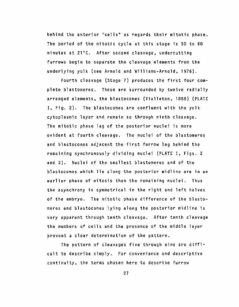
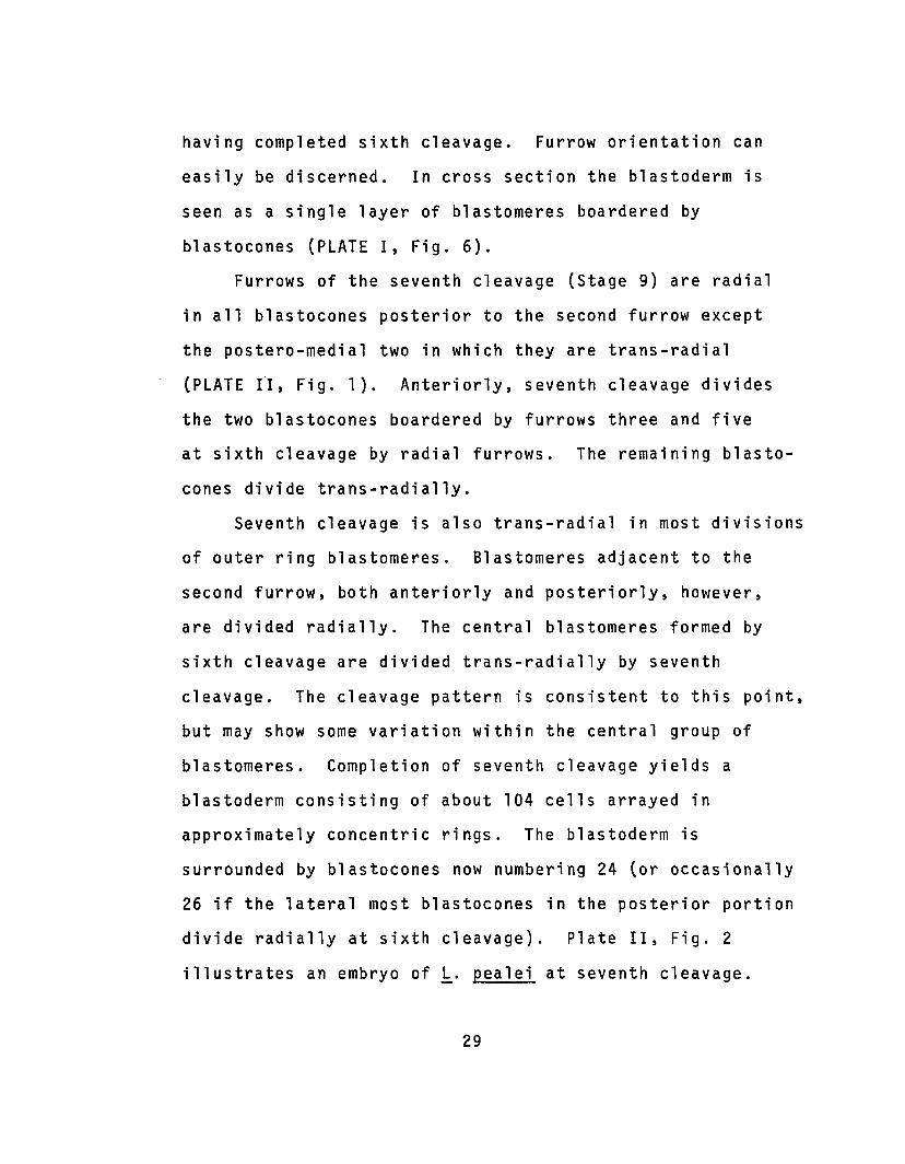
PLATE II
Late Cleavage: Cleavages 7 and 8
Fig. 1. Seventh cleavage (Stage 9); see Plate I legend. Blastomeres of thesixth cleavage outer ring are, in general, cleaved trans-radially. The large posterior blastocones and the two anterior blastocones boardered by furrows 3 and 5 atsixth cleavage are cleaved radially. The mitotic phase lag can be seen in the blastomeres along the posterior midline. Bar = 0.1 mm (ca. 75 X).
Fig. 2.. Embryo of L~ligo ~ealei at Seventh cleavage. This is the same embryoas Plate I, Flg. 5. Bar - 0.1 ca. 75 X).
Fig. 3. Section of a seventh cleavage embryo. The blastoderm remains as asingle layer of blastomeres surrounded by blastocones. Bar = 0.1 mm (ca. 100 X).
Fig. 4. Eighth cleavage (Stage 9). Cleavage planes are radial in most blastomeres and trans-radial in all blastocones. Blastomeres now number about 230 andblastocones 24 or 26. Overlapping of the presumptive middle-layer cells begins at
w this stage. The mitotic phase lag of the posterior-medial cells is still veryo evident. Bar = 0.1 mm (ca. 75 X).
Fig. 5.Fig. 5 above.75 X).
Embryo of Loligo pealei at eighth cleavage. This is the embryo ofThe arrow indicates the area of visible overlap. Bar = 0.1 mm (ca.
Fig. 6. Section of an eighth cleavage embryo. Overlapping is moderatelyadvanced causing a depression of the yolk (open arrows). Bar = 0.1 mm (ca. 100 X).
Fig. 7. Section of a ninth cleavage (Stage 10) embryo. A ring of middlelayer cells is produced below the blastoderm (open arrows). The blastocones (bc)are still present. Bar = 0.1 mm (ca. 100 X).
e
In cross section the blastoderm is still seen as a single
layer of now smaller blastomeres surrounded by peripherally
attached blastocones (PLATE II, Fig. 6).
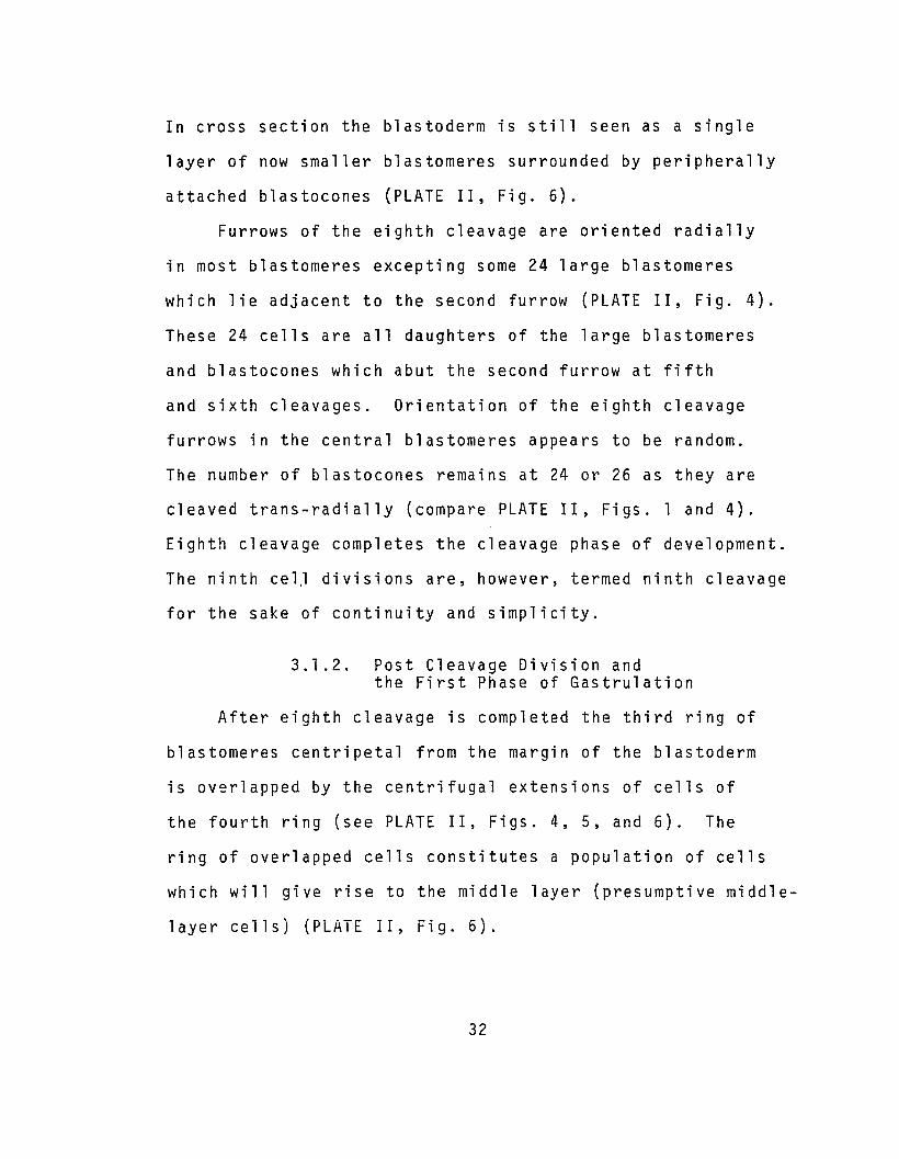
Furrows of the eighth cleavage are oriented radially
in most blastomeres excepting some 24 large blastomeres
which lie adjacent to the second furrow (PLATE II, Fig. 4).
These 24 cells are all daughters of the large blastomeres
and blastocones which abut the second furrow at fifth
and sixth cleavages. Orientation of the eighth cleavage
furrows in the central blastomeres appears to be random.
The number of blastocones remains at 24 or 26 as they are
cleaved trans-radially (compare PLATE II, Figs. 1 and 4).
Eighth cleavage completes the cleavage phase of development.
The ninth ce l.l divisions are, however, termed ninth cleavage
for the sake of continuity and simplicity.
3.1.2. Post Cleavage Division andthe First Phase of Gastrulation
After eighth cleavage is completed the third ring of
blastomeres centripetal from the margin of the blastoderm
is overlapped by the centrifugal extensions of cells of
the fourth ring (see PLATE II, Figs. 4, 5, and 6). The
ring of overlapped cells constitutes a population of cells
which will give rise to the middle layer (presumptive middle-
layer cells) (PLATE II, Fig. 6).
32

At ninth cleavage the blastomeres of the two most
peripheral rings are divided radially, whereas in the
centripedally adjacent ring of presumptive middle-layer
cells the furrows are oriented trans-radially. The
blastomeres of the fourth ring in from the periphery of
the blastoderm (overlapping cells) also divide trans
radially. This trans-radial division of the presumptive
middle-layer cells in conjunction with their overlapping
by the centripetally adjacent cells is one of the two basic
morphogenetic processes of germ layer segregation in Loligo.
Interference with either of these processes, as described
later, produces an abnormal segregation of the middle-layer.
The orientation of division in the central blastomeres
is less consistent at ninth cleavage. Division has also
become increasingly asynchronous. The blastoderms of
FAA-Feulgen treated embryos at ninth cleavage display a
symmetrical pattern of asynchrony. Symmetrically arrayed
bands of cells in the right and left halves of the blasto
dermere seen to be in a phase of mitosis which is later
with respect to the cells anterior to the bands and earlier
with respect to those posterior of the band. Thus an
anterior-posterior gradient of division rate is established
which persists through early phases of gastrulation. With
time-lapse cinemicrography the pattern of asynchrony is
visible as waves of division which originate along the
anterior midline and then sweep toward the posterior midline.
33
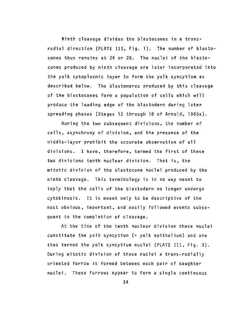
Ninth cleavage divides the b1astocones in a trans
radial direction (PLATE III, Fig. 1). The number of b1asto
cones thus remains at 24 or 26. The nuclei of the b1asto
cones produced by ninth cleavage are later incorporated into
the yolk cytoplasmic layer to form the yolk syncytium as
described below. The b1astomeres produced by this cleavage
of the b1astocones form a population of cells which will
produce the leading edge of the blastoderm during later
spreading phases (Stages 12 through 18 of Arnold, 1965a).
During the two subsequent divisions, the number of
cells, asynchrony of division, and the presence of the
middle-layer prohibit the accurate observation of all
divisions. I have, therefore, termed the first of these
two divisions tenth nuclear division. That is, the
mitotic division of the b1astocone nuclei produced by the
ninth cleavage. This terminology is in no way meant to
imply that the cells of the blastoderm no longer undergo
cytokinesis. It is meant only to be descriptive of the
most obvious, important, and easily followed events subse
quent to the completion of cleavage.
At the time of the tenth nuclear division these nuclei
constitute the yolk syncytium (= yolk epithelium) and are
thus termed the yolk syncytium nuclei (PLATE III, Fig. 3).
During mitotic division of these nuclei a trans-radially
oriented furrow is formed between each pair of daughter
nuclei. These furrows appear to form a single continuous
34

wU1
PLATE III
Ninth Cleavage and Tenth Nuclear Division
Fig. 1. Ninth cleavage (Early Stage 10). The b1astocones have undergone ninthcleavage and are delineated by the meridional and undercutting furrows. In thisembryo the furrows have already begun to retract. At the perimeter of the blastodermthe middle-layer cells are visible beneath the outer layer of cells. Elapse time =0.00. 250 X.
Fig. 2. Ninth cleavage (Early Stage 10). The meridional and undercuttingfurrows of blastocones have almost disappeared as retraction continues. The framingof this micrograph has shifted somewhat vegeta11y. Elapse time = 0:17. 250X.
Fig. 3. Ninth cleavage (Early Stage 10). The blastocone nuclei have movedvegetally into the yolk cytoplasmic layer to produce the yolk syncytium. Note thatthe blastomeres produced by ninth cleavage of the blastocones have moved toward themargin of the blastoderm. A lenticulate mass of cytoplasm surrounds each yolksyncytium nucleus. The ring of middle-layer cells produces a slight depression inthe underlying yolk at the edge of the blastoderm. Elapse time = 1.16. 250 X.
Fig. 4. Tenth nuclear division (Mid-Stage 10). The yolk syncytium nuclei arein prophase of the tenth nuclear division. The peripheral blastomeres have movedfarther toward the edge of the blastoderm. The yolk depression is more prominentat this time. Elapse time = 2:05. 250 X.


circumferential ring around the embryo (PLATE IV, Fig. 1).
The circumferential furrow is transitory, existing for
about one hour. Contraction of the filamentous band at the
base of the furrow produces a depression in the surface of
the embryo, but it does not cut through the syncytial cyto
plasm to the yolk. Later the furrow relaxes and disappears
(PLATE IV, Fig. 2). Thus although cleavage furrows are
formed during this division, a syncytium remains. The
maintenance of the syncytial character of this layer is of
critical importance for the later movements of the yolk
syncytium nuclei discussed below.
During tenth nuclear division there is an increase in
the number of middle-layer cells. This increase causes a
deepening of the depression in the yolk near the edge of
the blastoderm (PLATE III, Fig. 4). As the number of
middle-layer cells increases, their absence in the area
under the center of the blastoderm produces a "central
yolk papilla" (see Arnold, 1971). The middle layer is
eventually completed by continued division of the middle
layer cells. The blastomeres produced by ninth cleavage
of the blastocones begin to spread around the perimeter
of the blastoderm (compare for example PLATE III, Figs. 3
and 4; PLATE IV, Figs. 1 to 4).
Eleventh nuclear division ;s similarly designated by
the second mitotic division of the yolk syncytium nuclei.
Prior to eleventh nuclear division the proximal ring
37
wco
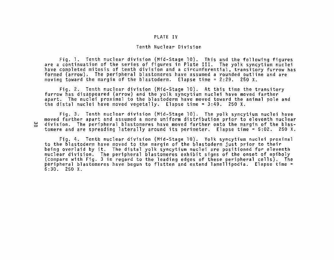
PLATE IV
Tenth Nuclear Division
Fig. 1. Tenth nuclear division (Mid-Stage 10). This and the following figuresare a continuation of the series of figures in Plate III. The yolk syncytium nucleihave completed mitosis of tenth division and a circumferential, transitory furrow hasformed (arrow). The peripheral blastomeres have assumed a rounded outline and aremoving toward the margin of the blastoderm. Elapse time = 2:29. 250 x.
Fig. 2. Tenth nuclear division (Mid-Stage 10). At this time the transitoryfurrow has disappeared (arrow) and the yolk syncytium nuclei have moved fartherapart. The nuclei proximal to the blastoderm have moved toward the animal pole andthe distal nuclei have moved vegetally. Elapse time = 3:49. 250 X.
Fig. 3. Tenth nuclear division (Mid-Stage 10). The yolk syncytium nuclei havemoved farther apart and assumed a more uniform distribution prior to eleventh nucleardivision. The peripheral blastomeres have moved farther onto the margin of the blastomere and are spreading laterally around its perimeter. Elapse time = 5:02. 250 x.
Fig. 4. Tenth nuclear division (Mid-Stage 10). Yolk syncytium nuclei proximalto the blastoderm have moved to the margin of the blastoderm just prior to theirbeing overlaid by it. The distal yolk syncytium nuclei are positioned for eleventhnuclear division. The peripheral blastomeres exhibit signs of the onset of epiboly(compare with Fig. 3 in regard to the leading edges of these peripheral cells). Theperipheral blastomeres have begun to flatten and extend lamellipodia. Elapse time =6:30. 250 X.

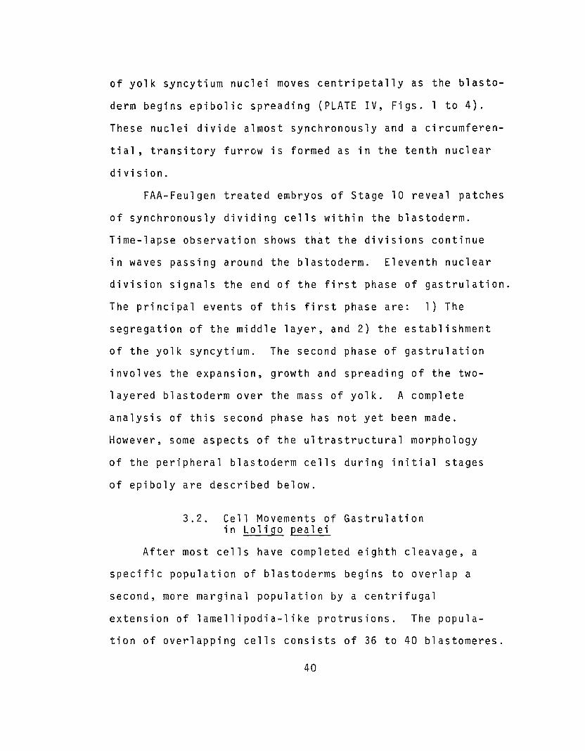
of yolk syncytium nuclei moves centripetally as the blasto
derm begins epibolic spreading (PLATE IV, Figs. 1 to 4).
These nuclei divide almost synchronously and a circumferen-
tial, transitory furrow is formed as in the tenth nuclear
division.
FAA-Feulgen treated embryos of Stage 10 reveal patches
of synchronously dividing cells within the blastoderm.
Time-lapse observation shows that the divisions continue
in waves passing around the blastoderm. Eleventh nuclear
division signals the end of the first phase of gastrulation.
The principal events of this first phase are: 1) The
segregation of the middle layer, and 2) the establishment
of the yolk syncytium. The second phase of gastrulation
involves the expansion, growth and spreading of the two
layered blastoderm over the mass of yolk. A complete
analysis of this second phase has not yet been made.
However, some aspects of the ultrastructural morphology
of the peripheral blastoderm cells during initial stages
of epiboly are described below.
3.2. Cell Movements of Gastrulationin Loligo pealei
After most cells have completed eighth cleavage, a
specific population of blastoderms begins to overlap a
second, more marginal population by a centrifugal
extension of lamellipodia-like protrusions. The popula
tion of overlapping cells consists of 36 to 40 blastomeres.
40

These are arranged in a ring located three blastomeres in
from the perimeter of the blastoderm (PLATE II, Fig. 3 and
PLATE VI, T=2:10). The presumptive middle-layer cells are
segregated into the area between the blastoderm and the
yolk by the processes of overlapping and ninth cleavage.
Time-lapse cinemicrography reveals the dynamic character
of the overlapping cell movements.
Plates V through XII are reproductions of sequences
of frames from two time-lapse films of germ layer segrega
tion in Loligo pealei. The pattern of late cleavage can
be seen clearly. Concentric rings of cells are established
by seventh cleavage (PLATES V and VI). After eighth
cleavage centrifugally directed cell protrusions progres
sively overlap adjacent cells. The onset of overlapping
can be seen in several cells (PLATE VI, T=2:10, T=2:20 and
PLATE IX, T~0:25.00 and T=0:37.30). These cells are
followed through successive stages (see Plate captions for
details). The extension and flattening of the overlapping
cells progresses during the interval between eighth and
ninth cleavage until the presumptive middle-layer cells are
one-fifth to one-quarter overlapped (see PLATE VII,
T=2:30 and T=2:40; PLATE IX, T=0:37.30 to T=l :03.00).
Just prior to ninth cleavage the overlapping cells round
up and their leading edges appear less active (PLATE VII,
T=2:50; PLATE IX, T=l :03.00). After ninth cleavage the
activity of the leading edges increases ~nd the cells spread
41

PLATE V
Cinemicrographic Series T.
Cleavages 5 and 6
Plates V through VIII present a series of frames from atime-lapse cinemicrographic film of middle layer segregationin Loligo pealei. The view of the embryo is of the leftanterior quadrant of the blastoderm. Magnification is 320 Xas indicated by the first frame. Elapse time designationsare in hours and minutes.
T=O:OO. Fifth cleavage (Stage 8) • Cleavage of theblastocones is radial.
T=O : 10. Fifth cleavage (Stage 8) . The blastoconenuclei are entering prophase of the sixth cleavage division.
T=0:20. Fifth cleavage (Stage 8) • Sixth cleavagemitosis.
T=0:30. Fifth cleavage (Stage 8) . Sixth cleavagemitosis.
T=0:40. Sixth cleavage (Stage 9). Sixth cleavage ofall blastocones is trans-radial.
T=0:50. Sixth cleavage (Stage 9). The nuclei of theblastomeres are entering prophase of the seventh cleavagedivision.
T=l :00. Sixth cleavage (Stage 9). The nuclei are inmitosis of seventh cleavage.
42
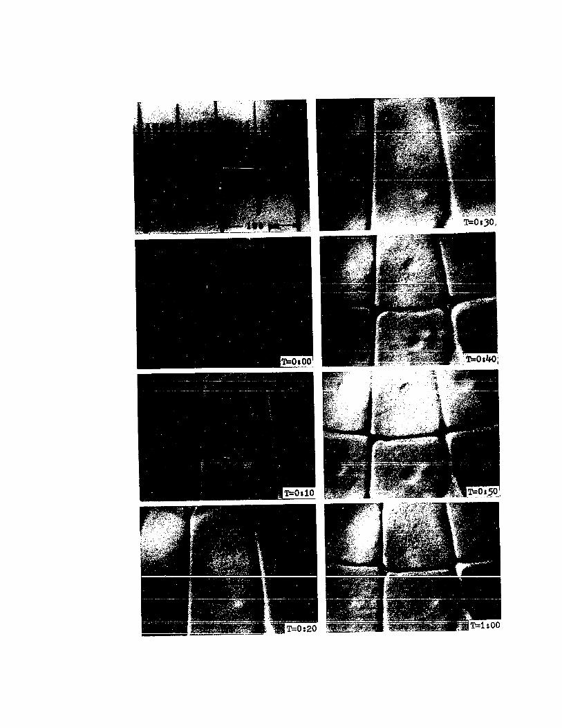

PLATE VI
Cinemicrographic Series I.
Cleavages 7 and 8
T=1:10. Late sixth cleavage (Stage 9). The nuclei arein early anaphase of mitosis prior to seventh cleavage. Thebeginning formation of seventh cleavage furrows is visibleas slight indentations along the radial furrows (arrows).
T=1:20. Seventh cleavage (Stage 9). Nuclei of the cellswithin the frame are in late telophase. Orientation of thecleavage furrows is trans-radial.
T=1:30. Seventh cleavage (Stage 9). The nuclei havereformed and moved to the centers of the cells.
T=1:40. Seventh cleavage (Stage 9). The cells are inthe G1 phase of the cell cycle. Note that the nuclei haveincreased in size.
T=l :50. Seventh cleavage (Stage 9). Nuclei are in mitosisprior to eighth cleavage. Note the asynchrony of mitosis.
T=2:00. Eighth cleavage (Stage 9). The cells in theupper-middle and left of the frame are undergoing cytokinesis.Asynchrony of division is more evident in this frame.
T=2:10. Eighth cleavage (Stage 9). The first signs ofoverlapping of the presumptive middle-layer cells are visible(arrow). The b1astocones are cleaved trans-radially by eighthcleavage.
44

'~'~'~3"p,~'".,~ .~, '. ~ -
. -

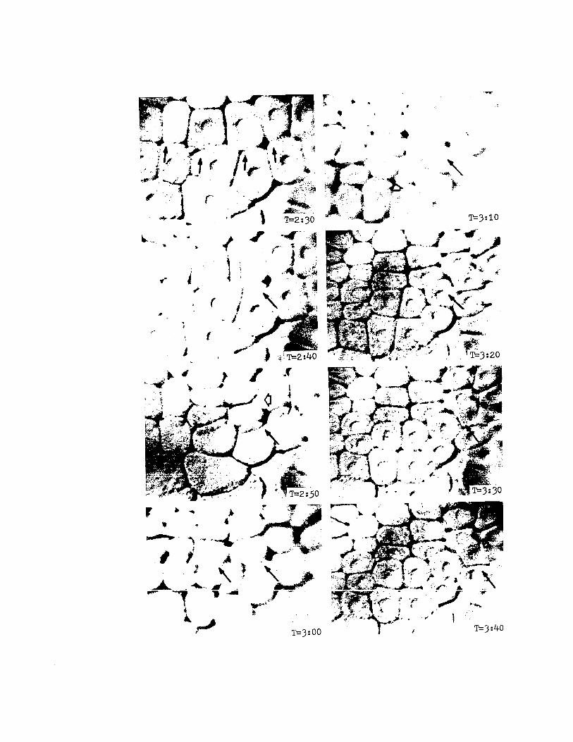
PLATE VII
Cinemicrographic Series I.
Germ Layer Segregation
T=2:30. Late eighth cleavage (Stage 9). Overlappingis visible in all cells of the overlapping ring (arrows).Close scrutiny of this frame reveals that the leading edgesare uneven and have numerous small pseudopodia.
T=2:40. Late eighth cleavage (Stage 9). Overlappinghas increased, the cells are beginning to retract their leadingedges (arrow), and are rounding up prior to ninth cleavage.These cells now overlap the presumptive middle-layer cellsby more than 10 u m.
T=2:50. Early ninth cleavage (Stage 9-10). Two overlapping cells have undergone ninth cleavage (open arrow).The two vegetal-most daughter cells of the above cleavageare still somewhat rounded at their leading edges (closedarrow) as they begin to flatten and spread.
T=3:00. Ninth cleavage (Early Stage 10). Leading edgesof the overlapping cells again show pseudopodial activity(arrows).
T=3:10. Ninth cleavage (Early Stage 10). Although thisframe is somewhat out of focus, the arrow indicates the location of the leading edge of overlap. Some variation in thecleavage pattern is evident. For example, the presumptivemiddle-layer cell in the center of the field has cleavedradially whereas the others are cleaving trans-radially(open arrow).
T=3:20. Ninth cleavage (Early Stage 10). Presumptivemiddle-layer cells are one-fourth to one-half overlapped. Theleading edge of overlap is indicated by the arrow.
T=3:30 .. Ninth cleavage (Early Stage 10). Presumptivemiddle-layer cells are now one-half to two-thirds overlapped(arrow). The leading edges of the overlapping cells showsigns of intense activity.
T=3:40. Ninth cleavage (Stage 10). Presumptive middlelayer cells are now approximately three-fourths overlapped(arrow). Note the rounded aspect of the centripetal marginsof the middle-layer cells. The appearance of the middle-layer cells contrasts sharply with that of the overlapping cells.
46
6
•
II
·.......<
r-..
l..... .. ~
.f...~
1,), I
'II. I
,.,.
.•,,.-(
J
T=3:10.\ , -..,..
....J ~..--" ..-- ~.
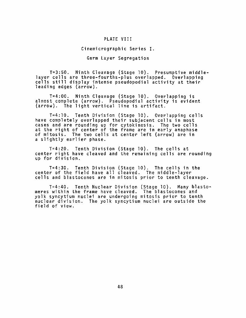
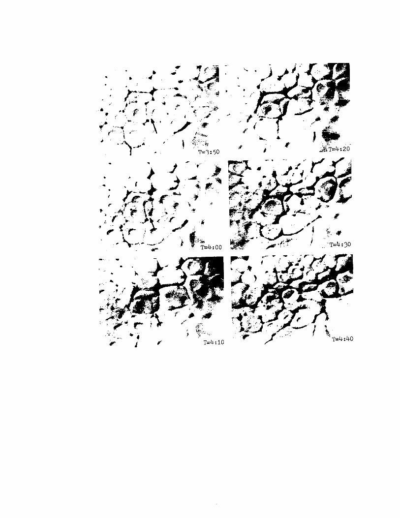
PLATE VIII
Cinemicrographic Series I.
Germ Layer Segregation
T=3:50. Ninth Cleavage (Stage 10). Presumptive middlelayer cells are three-fourths-plus overlapped. Overlappingcells still display intense pseudopodial activity at theirleading edges (arrow).
T=4:00. Ninth Cleavage (Stage 10). Overlapping isalmost complete (arrow). Pseudopodial activity is evident(arrow). The light vertical line is artifact.
T=4:10. Tenth Division (Stage 10). Overlapping cellshave completely overlapped their subjacent cells in mostcases and are rounding up for cytokinesis. The two cellsat the right of center of the frame are in early anaphaseof mitosis. The two cells at center left (arrow) are ina slightly earlier phase.
T=4:20. Tenth Division (Stage 10). The cells atcenter right have cleaved and the remaining cells are roundingup for division.
T=4:30. Tenth Division (Stage 10). The cells in thecenter of the field have all cleaved. The middle-layercells and blastocones are in mitosis prior to tenth cleavage.
T=4:40. Tenth Nuclear Division (Stage 10). Many blastomeres within the frame have cleaved. The blastocones andyolk syncytium nuclei are undergoing mitosis prior to tenthnuclear division. The yolk syncytium nuclei are outside thefield of view.
48
..
...•
#I• -.k ~ cj" 'f \~"
.. -"', . .:>,."';','~ _..~~.<.'r/'" .,'". -.. ,.' .''''r* ". ,,~ ... .l-... ,./ "'i~J1i!
r- ..t' T=4:00
T=4:10

PLATE IX
Cinemicrographic Series II.
Cleavages 7 to 9 and Germ Layer Segregation
Plates IX through XII presen~ a series of frames froma time-lapse cinemicrographic film of germ layer segregationand yolk syncytium formation in Loligo pealei. Magnificationin this series is approximately 210 times normal. Elapse timedesignation is in hours, minutes and seconds.
T=O:OO.OO. Seventh Cleavage (Stage 9). Seventh cleavageis trans-radial in all blastomeres and blastocones within theframe.
T=0:12.30. Seventh Cleavage (Stage 9). All nuclei arein prophase of eighth cleavage mitosis.
T=0:25.00. Eighth Cleavage (Stage 9). All blastomereshave completed or are completing eighth cleavage. Some evidenceof the initiation of overlapping can be seen in the form of abroad pseudopod extending over the subjacent presumptive middlelayer cell (arrow).
T=0:37.30. Eighth Cleavage (Stage 9). Most nuclei are ininterphase of mitosis. The pseudopod of the previous framehas broadened and overlapping has begun in adjacent cells.
T=0:50.30. Eighth Cleavage (Stage 9-10). Overlappinghas advanced and small pseudopodial processes can be seen atthe margins of the leading edges (arrow).
T=1:03.00. Eighth Cleavage (Stage 9-10). The nuclei ofthe overlapping cells are entering prophase of the ninthcleavage division. The cells are rounded. The presumptivemiddle-layer cells appear to be about one-fourth overlapped.
T=l :15.30. Ninth Cleavage (Early Stage 10). Ninthcleavage has occurred in most blastomeres and blastocones.Cleavage in the ring of overlapping cells is indicated bythe open arrow. Flattening ~nd extension of overlappingprocesses is resumed (arrow).
T=1:28.00. Ninth Cleavage (Early Stage 10). Overlappingcontinues as the presumptive middle-layer cells cleave (arrow).
50

'1'=1:28.00
r:r;;O:25.00
1'=0:37.30

more rapidly over the underlying presumptive middle-layer
cells (PLATE VII, T=3:00 to PLATE VIII, T=3:50; PLATE IX,
T=l :15.30 to T=2:05.30). During spreading the leading
margins of these flattened cellular protrusions send out
smaller processes which extend and retract at a greater
rate than the rate of advance of the cell margins (PLATE
VII, T=3:00 and PLATE IX, T=l :15.30). The number of
lamellipodia and filopodia each cell develops varies with
time. These pseudopodial processes disappear completely
just prior to and during cytokinesis. As cellular exten
sions make contact with underlying cells, they broaden
and coalesce. This activity produces an overall vectorial
flow of overlapping cells over the presumptive middle-layer
cells.
Overlapping accelerates subsequent to ninth cleavage
(PLATE VII, T=3:00 to PLATE VIII, T=4:00 and PLATE IX,
T=1:15.30 to PLATE X, T=2:18.00). The change in the rate
of overlapping is further illustrated in Figs. 3 and 4.
Measurements of the rate of overlapping show that the
rate of oriented movement reaches a maximum of approximately
1.4 m per minute (Fig. 4A) between ninth cleavage and tenth
nuclear division. Although the overlapping cells constitute
an epithelial sheet, Figs. 3 and 4 illustrate that each
cell moves somewhat independently of adjacent cells. Figs.
38 and 48 show that the rate of movement is not constant over
the period observed and they illustrate the retraction of
52
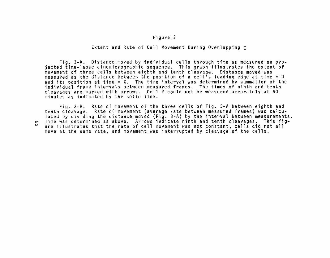
<.J1W
Figure 3
Extent and Rate of Cell Movement During Overlapping I
Fig. 3-A. Distance moved by individual cells through time as measured on projected time-lapse cinem;crographic sequence. This graph illustrates the extent ofmovement of three cells between eighth and tenth cleavage. Distance moved wasmeasured as the distance between the position of a cell's leading edge at time = 0and its position at time = X. The time interval was determined by summation of theindividual frame intervals between measured frames. The times of ninth and tenthcleavages are marked with arrows. Cell 2 could not be measured accurately at 60minutes as indicated by the solid line.
Fig. 3-B. Rate of movement of the three cells of Fig. 3-A between eighth andtenth cleavage. Rate of movement (average rate between measured frames) was calculated by dividing the distance moved (Fig. 3-A) by the interval between measurements.Time was determined as above. Arrows indicate ninth and tenth cleavages. This figure illustrates that the rate of cell movement was not constant, cells did not allmove at the same rate, and movement was interrupted by cleavage of the cells.
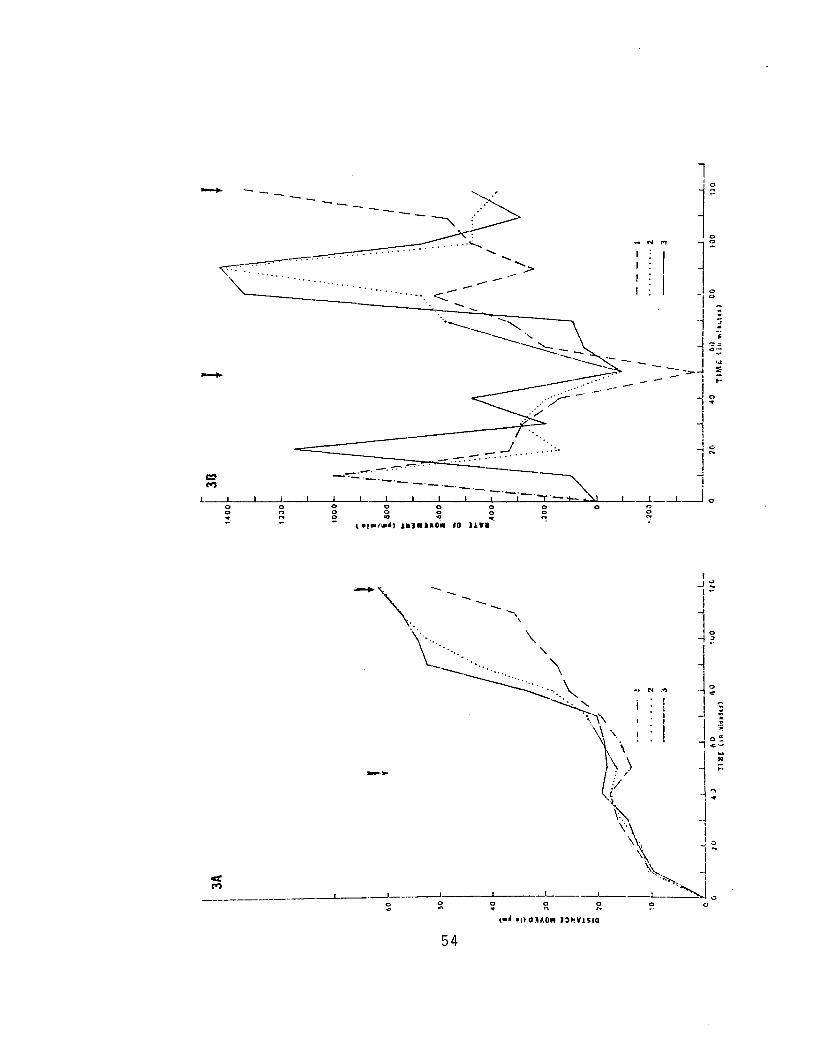
-
-
--
I_.~.~.:7'"'..~..... ""'.
54

U1U1
Figure 4
Extent and Rate of Cell Movement During Overlapping II
Fig. 4-A. Distance moved by individual cells through time as measured on projected time-lapse cinemicrographic sequences. This figure illustrates the extentof movement of two cells over the period between eighth and tenth cleavages. Distance moved was measured as the distance between the position of the cell's leadingedge at time = 0 and its position at time = X. The time interval was determined bysummation of the individual frame intervals between measured frames. The times ofninth and tenth cleavages are marked with arrows.
Fig. 4-B. Rate of movement of the two cells of Fig. 4-A between eighth andtenth cleavages. Rate of movement (average rate between measured frames) was calculatedby dividing the distance moved (Fig. 4-A) by the period between measurements. Timewas determined as above. Arrows indicate ninth and tenth cleavages. This figureillustrates as in Fig. 3-B that the rate of movement was not constant and that therate of movement was reduced during cytokinesis.

!Do"!
,oo~
,COCO..
111J~ lAO. JG un
ji
I:
.......
oo
0'""'iii
56
.....III
i•...
"';IiI ..
~ J:,~·o
0co

rounding up of the leading edge prior to cleavage. During
cytokinesis the rate of movement is reduced to near or below
zero. Although the number of cells whose rates of movement
were measured was necessarily limited by movements of the
cells in and out of focus, Figs. 3 and 4 illustrate some
behavioral characteristics of the overlapping cells. The
significance of this behavior is discussed below.
3.3. Establishment of the Yolk Syncytium
3.3.1. Observation of Normal Development
The yolk syncytium is established by the incorporation
into the yolk cytoplasmic layer of the nuclei of blastocones
produced by ninth cleavage. Blastocones are, in essence,
pockets within the continuous yolk cytoplasmic layer. They
are formed by the membranes of meridional and undercutting
furrows.
The sequence of events in the establishment of the
yolk syncytium was followed by time-lapse cinemicrography
as illustrated in PLATES X, XI, and XII. After ninth
cleavage (Plate X, T=3:08.00) lateral and undercutting
furrows of the blastocones retract toward the blastoderm
and disappear (PLATE XI, T=3:20.30). The blastocone nuclei
move vegetally (compare PLATE X, T=3:08.00 through PLATE XI,
T=3:45.30). The vegetal movement of the nuclei and the re
traction of the furrows leave the nuclei in a syncytial ring
around the perimeter of the blastoderm. At this stage
57
PLATE X
Cinemicrographic Series II.
Ninth Cleavage
T=1:40.30. Ninth Cleavage (Early Stage 10). The centripetal ring of cells resulting from the ninth cleavage ofthe presumptive middle-layer cells is completely overlaid.The cells of this ring can be seen lying beneath the overlap cells (bracketed area). The active margin of the overlapping ring of cells is indicated (arrow).
T=l :53.00. Ninth Cleavage (Stage 10). Overlappingcontinues. The pesudopodial activity of the leading edgeis visible (arrow).
T=2:05.00. Ninth Cleavage (Stage 10). The centrifugalring of cells produced by the ninth cleavage of the presumptive middle-layer cells is now completely overlapped. Themiddle-layer ring of cells is now two cells wide and can beseen beneath the overlap cells. Active overlapping continues(arrow).
T=2:18.00. Ninth Cleavage (Stage 10). The cells in thecenter of the field are in mitosis and are rounding up fortenth cleavage which is trans-radial in these cells. Notethe retracted leading edge (arrow).
T=2:30.30. Ninth Cleavage (Stage 10). The majority ofthe b1astomeres appear to have undergone ninth cleavage. Theblastocones have entered mitosis prior to ninth cleavage(broad arrow). The margin of overlap is indicated (arrow).
T=2:43.00. Ninth Cleavage (Stage 10). The b1astoconesare entering cytokinesis of ninth cleavage. Furrow orientation is trans-radial in this division of the b1astocones.Note that the b1astomeres produced by eighth cleavage of theb1astocones are moving up onto the edge of the blastoderm.The arrow again indicates the margin of overlap.
T=2:55.30. Ninth Cleavage (Mid-Stage 10). Ninthcleavage is now complete within the field of view.
T=3:08.00. Ninth Cleavage (Mid-Stage 10). The daughternuclei of blastocone ninth cleavage mitosis have moved apart(broad arrows) and the b1astocone furrows have begun to retract.The middle layer is visible beneath the surface cells (bracket).
58
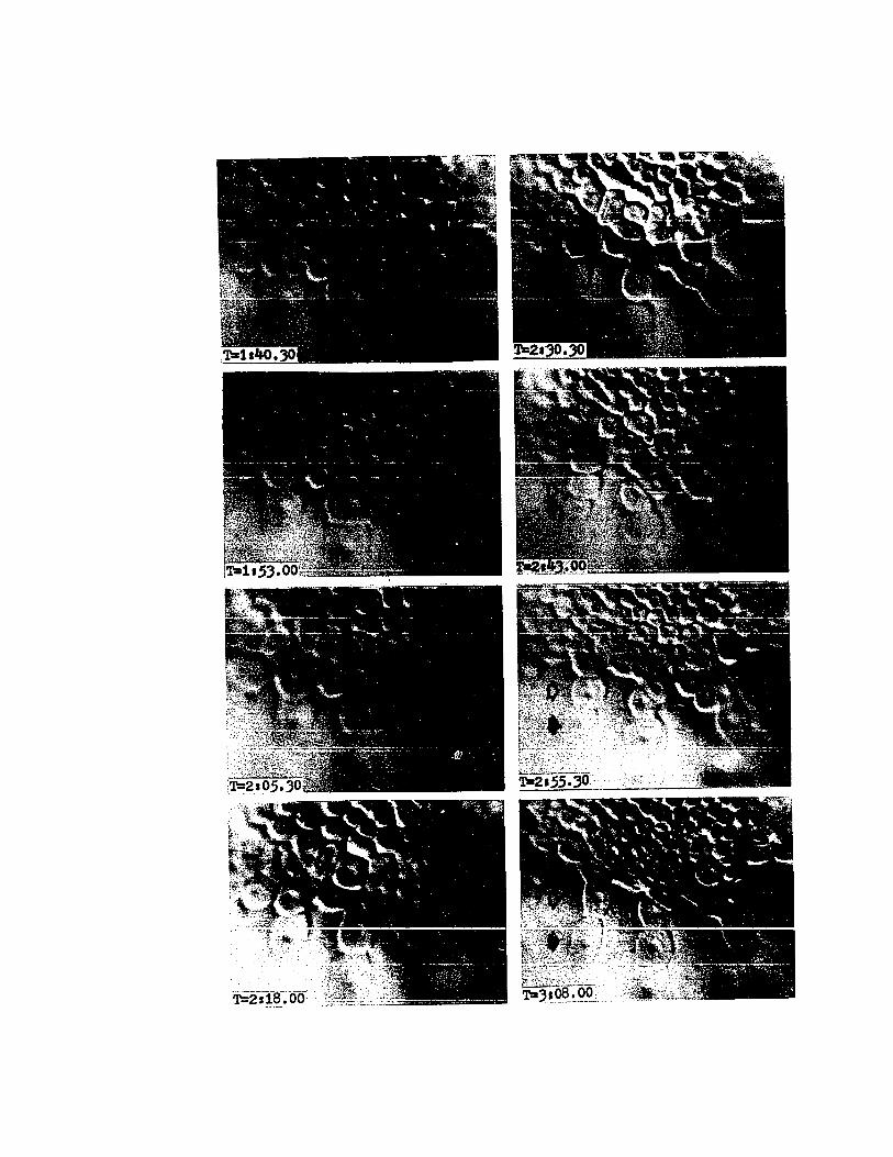

PLATE XI
Cinemicrographic Series II.
Ninth Cleavage and Establishment of the Yolk Syncytium
T=3:20.30. Ninth Cleavage (Mid-Stage 10). The blasto-cone furrows are retracting. The blastomeres of eighth cleavage of the blastocones have moved up onto the edge of the blastoderm (arrowheads). The middle layer is no longer clearlydiscernable.
T=3:33.00. Ninth Cleavage (Mid-Stage 10). The blastoconefurrows are almost completely retracted and the blastoconenuclei have moved vegetally. These nuclei now constitute asyncytium and are termed the yolk syncytium nuclei. The blastocone ninth cleavage blastomeres are rounding up and movingtoward the blastoderm. The blastocone ei~hth cleavage daugh-ter blastomeres are indicated (arrowheads). .
T=3:45.30. Ninth Cleavage (Mid-Stage 10). The blastocone ninth cleavage daughter blastomeres continue to movetoward the blastoderm (open arrow). Blastocone eighth daughter blastomeres are indicated (arrowheads).
T=3:58.00. Ninth Cleavage (Mid-Stage 10). The blastocone ninth cleavage daughter blastomeres continue to move toward the blastoderm (open arrow). The yolk syncytium nucleihave not changed position (solid arrows).
T=4:10.30. Ninth Cleavage (Mid-Stage 10). The processesof the previous frames are continued (broad arrows). The yolksyncytium nuclei are entering prophase of Tenth Nuclear Division (small arrow). The black dot is an artifact.
T=4:23.00. Tenth Nuclear Division (Late Stage 10). Theblastomeres followed above continue to move onto the blastoderm(open arrow and arrowhead). The yolk syncytium nuclei are inmitosis. The nucleus indicated by the broad, solid arrow isnow in late prophase and the nucleus indicated by the smallarrows is in late anaphase.
T=4:35.30. Tenth Nuclear Division (Late Stage 10). Theblastomeres followed above continue to move onto the blastoderm(open arrow and arrowhead). The framing here has shifted vegetally and to the apparent right. The yolk syncytium nuclei continue mitosis (arrows).
T=4:48.00. Tenth Nuclear Division (Late Stage 10). Theblastodermal cells are in various stages of cleavage. Theblastomere indicated above continues moving onto the blastoderm(arrowhead).
60


PLATE XII
Cinemicrographic Series II.
Establishment of the Yolk Syncytium
T=5:00.30. Tenth Nuclear Division (Late Stage 10).The proximal ring of yolk syncytium nuclei has moved a shortdistance toward the animal pole (arrow). The peripheralblastodermal cells are flattening in preparation for movements of epiboly.
T=5:13.00. Tenth Nuclear Division (Early Stage 11).Some blastodermal cells can be seen undergoing eleventhcleavage. The proximal ring of yolk syncytium nuclei isseen moving beneath the margin of the blastoderm by meansof autonomous movement toward the animal pole and vegetalspreading of the blastoderm (arrow).
T=5:25.30. Tenth Nuclear Division (Early Stage 11).Epiboly is beginning and small pseudopodia are seen on theflattened margins of the peripheral cells of the blastoderm.
T=5:38.00. Tenth Nuclear Division (Early Stage 11).The proximal ring of yolk syncytium nuclei now lies beneaththe expanding margin of the blastoderm (arrow).
T=5:50.30. Tenth Nuclear Division (Early Stage 11).The proximal yolk syncytium nuclei are completely overlaidby the blastoderm and continue their autonomous movementtoward the animal pole (arrow).
T=6:03.00. Tenth Nuclear Division (Mid-Stage 11).The yolk syncytium nuclei followed previously are indicatedlying beneath the blastoderm (arrow). The margin of theblastoderm consists of a single layer of flattened polygonalcells. This peripheral ring of cells remains as a singlelayer throughout epiboly and contributes to the formationof the external yolk sac. The distal yolk syncytium nucleiare entering prophase of eleventh nuclear division. (arrowhead).
T=6:15.30. Tenth Nuclear Division (Mid-Stage 11). Theyolk syncytium nuclei continue in mitosis. Epiboly continues.
T=6:28.00. Tenth Nuclear Division (Mid-Stage 11).Epibolic and nuclear movements continue. The distal yolksyncytium nucleus indicated (arrowhead) is in late anaphase.The proximal yolk syncytium nucleus indicated (arrow) isflattening and so appears to be getting larger. This stagecompletes the first phases of gastrulation. Gastrulationsubsequently involves a continuation of epibolic spreadingof the blastoderm and cytodifferentiation of the germ layers.
62
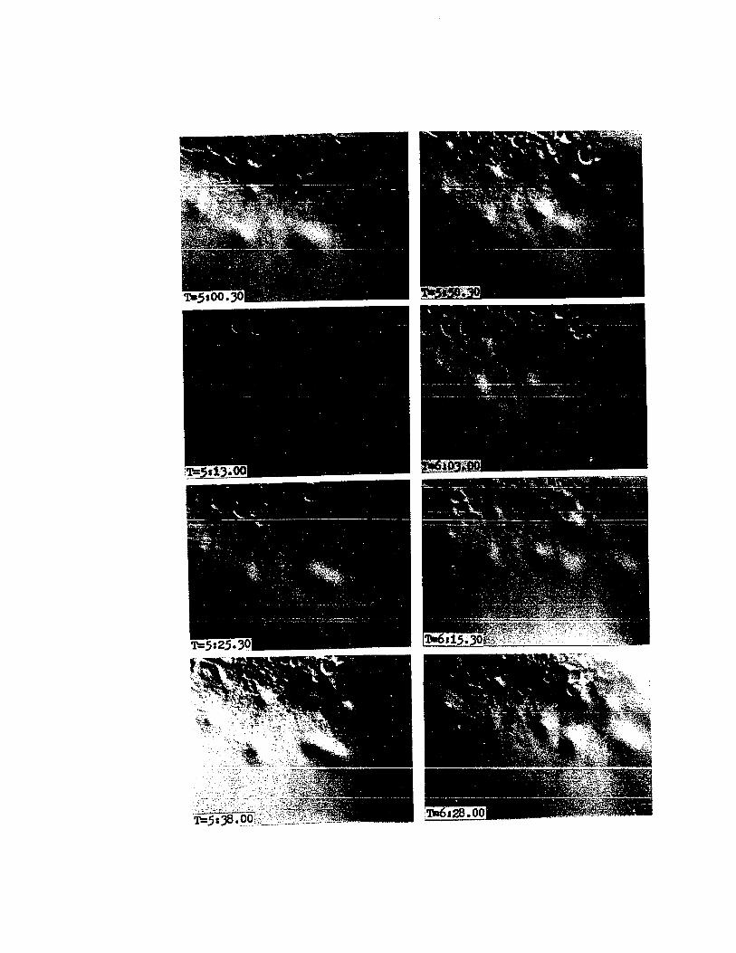

each nucleus is spheroidal and lies within a lenticulate
portion of the common cytoplasm producing a slight depression
in the underlying yolk.
During their vegetal movements the nuclei undergo a
series of relatively rapid oscillations as seen by time
lapse cinemicrography. The nuclear membrane exhibits
surface activity much like the surface activity of cultured
cells during blebbing locomotion, and the entire nucleus
moves back and forth. Similar oscillatory movements were
observed in no other nuclei. The function of these move
ments is unknown. Measurements of the vegetal movement
of fifteen blastocone nuclei were taken from photographic
sequences of yolk syncytium formation. Over their approxi
mately one hour existence, these nuclei moved vegetally on
the average a distance of 37.5 ~m.
The blastomeres produced by ninth cleavage of the
blastocones round up and are shown by time-lapse to move
up onto the perimeter of the blastoderm (see PLATE X,
T=2:55.30 to PLATE XI, T=4:23.00). These blastomeres
spread out around the perimeter of the blastoderm where
they form the leading edge during the second phase of
epibolic spreading of the blastoderm (PLATE XI, T=3:45.30
to T=4:23.00). These blastomeres establish a population
of cells which will give rise to some of the extraembryonic
tissues of the external yolk sac. Ninth cleavage has thus
64

established two unique populations of cells (i .e. the
initial ring of middle-layer cells and marginal cells of
the blastoderm) and a population of syncytial nuclei.
The marginal cells of the blastoderm and the yolk syncytium
are entirely extraembryonic.
Retraction of undercutting and meridional furrows of
the blastocones involves a folding up of the furrows begin
ning at their bases and a possible vesiculation of the
furrow membranes as revealed by electron microscopy. At
ninth cleavage these furrows appear as long parallel
membranes separated by approximately 10 nm and terminating
in an expanded bulb-shaped structure typical of furrow
bases in Lo1igo (PLATE XIII, Figs. 2 to 4: see also Arnold,
1971, 1974). Associated with the cytoplasmic face of the
furrow base membrane is a band of 5 nm to 7 nm filaments
(PLATE XIII, Fig. 4). The undercutting furrows extend
through the syncytial cytoplasm for a distance of as much
as 175 ~m. During retraction the furrows become highly
folded especially at the bases (PLATE XIV, Figs. 1 and 2).
The microfilamentous band seen associated with normal
furrows is noticeably absent in retracting furrows. This
folded area of the base apparently moves toward the blasto
derm as judged by the progressively shorter furrows seen
in embryos fixed at progressively later stages. In later
stages of retraction the furrows often appear as strings of
membranous vesicles.
65

PLATE XIII
Undercutting Furrows of Blastocones
Fig. 1. Section of an osmiun tetroxide fixed, Eponembedded ninth cleavage (early Stage 10) Loligo pealei.This figure shows the organization of the edge of the blastoderm and blastocones early in ninth cleavage. Note the location of the blastocone (BC), middle-layer ring of cells (MLR)and the overlapping cell (OLC). The small, round, light areasbeneath the cells are yolk platelets in various stages ofdigestion. 250 X.
Fig. 2. Electron micrograph of the area of the undercutting furrow (UCF) indicated by the bracket in Fig. 1. Incross section the furrow is seen as two parallel membranesseparated by a space of approximately 100 Angstroms. At thefurrow base the membranes diverge forming a bulb-like profile.Associated with the furrow base membrane is a band of microfilaments (MF and see Fig. 4). The platelet at the far rightis in an early stage of digestion.
Fig. 3. This micrograph presents the base of the undercutting furrow in more detail (FB). The furrow membranes atthis stage are parallel and exhibit little gross undulationor folding. Small undulations, however, produce apparentdiscontinuities in the membrane structure as the cross sectionalplanes of the membranes tilt through the plane of section.The syncytial cytoplasm consists primarily of mitochondria,ribosomes and smooth endoplasmic reticulum. The fibrillarmaterial (FM) between the chorion (CH) and the cytoplasmiclayer lying above the yolk (Y) is the remnant of the fertilization membrane (see Arnold and Williams-Arnold, 1976). Yolkplatelets are each surrounded by a unit membrane. Someevidence of membrane vesiculation is present near the furrowbase. 22,200 X.
Fig. 4. Higher magnification micrograph of a furrowbase illustrating the microfilamentous band (MF). The microfilamentous material associated with the furrow base isusually more abundant on the side of the furrow nearest theyolk. 50,000 X.
66
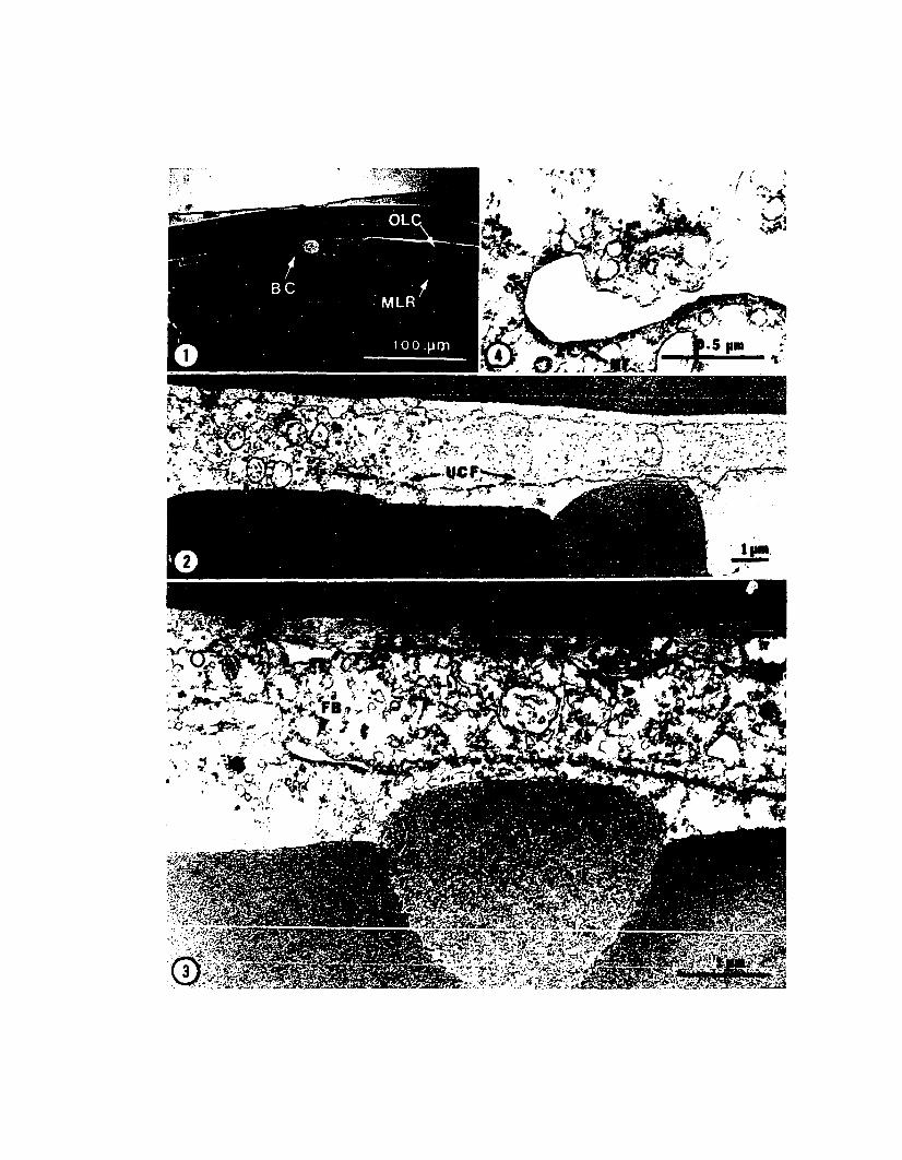
.. ,
<:«. • ,.'1f, .. ~', '
~,j • •• ..I .-
. i ,.:",;>-\\
~i
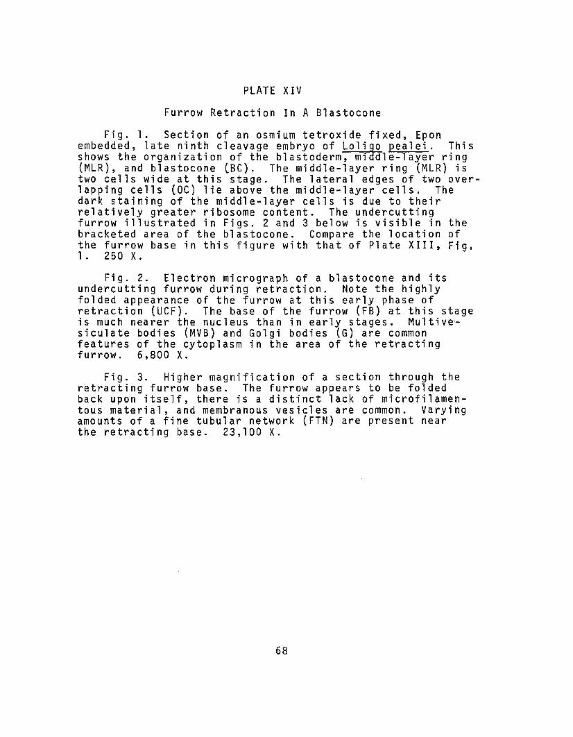
PLATE XIV
Furrow Retraction In A Blastocone
Fig. 1. Section of an osmium tetroxide fixed, Eponembedded, late ninth cleavage embryo of Lolig~ pealei. Thisshows the organization of the blastoderm, miaale-layer ring(MLR), and blastocone (Be). The middle-layer ring (MLR) istwo cells wide at this stage. The lateral edges of two overlapping cells (DC) lie above the middle-layer cells. Thedark staining of the middle-layer cells is due to theirrelatively greater ribosome content. The undercuttingfurrow illustrated in Figs. 2 and 3 below is visible in thebracketed area of the blastocone. Compare the location ofthe furrow base in this figure with that of Plate XIII, Fig.1. 250 X.
Fig. 2. Electron micrograph of a blastocone and itsundercutting furrow during retraction. Note the highlyfolded appearance of the furrow at this early phase ofretraction (UCF). The base of the furrow (FB) at this stageis much nearer the nucleus than in early stages. Multivesiculate bodies (MVB) and Golgi bodies (G) are commonfeatures of the cytoplasm in the area of the retractingfurrow. 6,800 X.
Fig. 3. Higher magnification of a section through theretracting furrow base. The furrow appears to be foldedback upon itself, there is a distinct lack of microfilamentous material, and membranous vesicles are common. Varyingamounts of a fine tubular network (FTN) are present nearthe retracting base. 23,100 X.
68

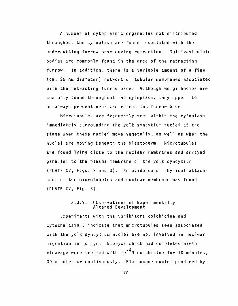
A number of cytoplasmic organelles not distributed
throughout the cytoplasm are found associated with the
undercutting furrow base during retraction. Multivesiculate
bodies are commonly found in the area of the retracting
furrow. In addition, there is a variable amount of a fine
(ca. 25 nm diameter) network of tubular membranes associated
with the retracting furrow base. Although Golgi bodies are
commonly found throughout the cytoplasm, they appear to
be always present near the retracting furrow base.
Microtubules are frequently seen within the cytoplasm
immediately surrounding the yolk syncytium nuclei at the
stage when these nuclei move vegetally, as well as when the
nuclei are moving beneath the blastoderm. Microtubules
are found lying close to the nuclear membranes and arrayed
parallel to the plasma membrane of the yolk syncytium
(PLATE XV, Figs. 2 and 3). No evidence of physical attach-
ment of the microtubu1es and nuclear membrane was found
(PLATE XV, Fig. 3).
3.3.2. Observations of ExperimentallyAltered Development
Experiments with the inhibitors colchicine and
cytocha1asin B indicate that microtubu1es seen associated
with the yolk syncytium nuclei are not involved in nuclear
migration in Lo1igo. Embryos which had completed ninth
cleavage were treated with 10- 4M colchicine for 10 minutes,
30 minutes or continuously. B1astocone nuclei produced by
70
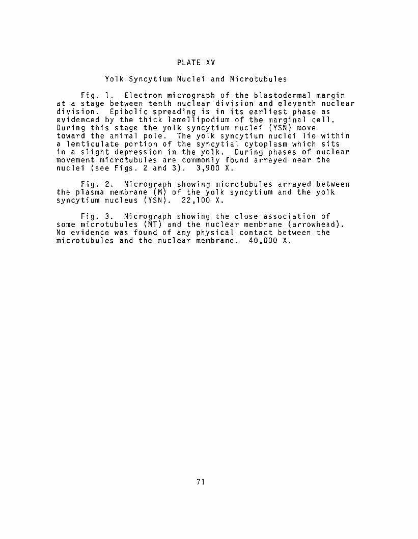
PLATE XV
Yolk Syncytium Nuclei and Microtubules
Fig. 1. Electron micrograph of the blastodermal marginat a stage between tenth nuclear division and eleventh nucleardivision. Epibolic spreading is in its earliest phase asevidenced by the thick lamellipodium of the marginal cell.During this stage the yolk syncytium nuclei (YSN) movetoward the animal pole. The yolk syncytium nuclei lie withina lenticulate portion of the syncytial cytoplasm which sitsin a slight depression in the yolk. During phases of nuclearmovement microtubules are commonly found arrayed near thenuclei (see Figs. 2 and 3). 3,900 X.
Fig. 2. Micrograph showing microtubules arrayed betweenthe plasma membrane (M) of the yolk syncytium and the yolksyncytium nucleus (YSN). 22,100 X.
Fig. 3. Micrograph showing the close association ofsome microtubules (MT) and the nuclear membrane (arrowhead).No evidence was found of any physical contact between themicrotubules and the nuclear membrane. 40,000 X.
71

~
.. :~.,-"-_.

ninth cleavage division in treated embryos migrated out of
the blastocones and continued vegetally at a rate compara
ble to the rate of advance of blastodermal spreading in
controls (PLATE XXV, Fig. 6). Colchicine irreversibly
blocked the maintenance of microtubules in embryos as
judged by electron microscopy and totally inhibited
mitosis. The results of colchicine treatment indicate
that the migration of the yolk syncytium nuclei is indepen
dent of the spreading of the blastoderm.
In contrast with the results of colchicine treatment,
nuclear migration was totally inhibited during treatment
of embryos with 0.02 ~gm/ml of cytochalasin B. Inhibition
occurred only during treatment and migration resumed after
removal of the cytochalasin. After removal of the cyto
chalasin from embryos treated for ten minutes at ninth
cleavage the nuclei renewed their movements in both vegetal
and animal directions. In cases where treatment extended
through tenth nuclear division both daughter nuclei moved
in a vegetal direction after removal of the cytochalasin.
Prolonged treatment (3D minutes or longer at 0.02 ~gm/ml)
elicited a retraction of the leading edges of the periph
eral blastoderm cells. After removal of the cytochalasin
the proximal ring of yolk syncytium nuclei was out beyond
the perimeter of the blastoderm, whereas in control embryos
these nuclei lay just beneath the edge of the blastoderm.
In such cases completion of gastrulation was totally
73

inhibited. The yolk syncytium nuclei moved vegetally
while the blastodermal cells eventually formed a clump at
the animal apex and the embryo died (PLATE XXIII, Figs.
3 and 4).
3.4. Observations with the ScanningElectron Microscope
3.4.1. Late Cleavage Stage Blastodermand Blastocones
At eighth cleavage (Late Stage 9) the embryo of
Loligo pealei consists of a single layered blastoderm
comprised of ca. 230 blastomeres arranged in approximately
concentric rings and surrounded by 24 or 26 blastocones
(PLATE II, Figs. 4 and 5; PLATE XVI, Fig. 1). Plate XVI,
Fig. 1 illustrates the overall appearance of the embryo
at eighth cleavage. The remnants of the fertilization
membrane (FM) are pulled back to reveal the blastoderm.
The second complete ring of cells in from the margin of
the blastoderm constitutes the population of presumptive
middle-layer cells (PMC). The presumptive middle-layer
cells in the embryo illustrated are partially overlapped
by the centripetally adjacent ring of cells (OC). The
free surfaces of the blastomeres possess numerous small
blebs, filopodia (termed microspikes by Weiss, 1961) and
intermediate sorts of protrusions. These small cellular
protrusions are more numerous near the cells' apices.
These protrusions extend out of the cells and into the
74
intrachorionic space or across the intercellular space
where their tips make non-junctional contact with neigh-
boring cells (PLATE XVI, Fig. 2; PLATE XVII, Figs. 1 and 2).
The filopodia range in diameter from 0.2 ~m to 0.4 ~m and
are thicker at the base tapering toward the tip. Their
length ranges from less than 1.0 ~m to over 10.0 ~m.
Some blastomeres lack large numbers of filopodia, have
a more rounded appearance, and do not closely abut adjacent
cells apically. For example, in Plate XVI, Fig. 1, two
radially arrayed bands of blastomeres exhibiting these
characteristics can be seen. Rounding up and loss of surface
activity are common phenomena associated with cell division
(e.g. Revel, 1974). Furthermore, the number and distri-
bution of cells having these characteristics is precisely
that of mitotic cells observed in FAA-Feulgen treated
embryos of the same stage. It is assumed, therefore, that
the cells which show little surface activity have entered
mitosis. The blastocones of the embryo in Plate XVI,
Fig. 1 also have little surface activity and so are probably
undergoing mitosis. FAA-Feulgen treated embryos of this
stage often display mitotic figures in all but the pos-
teromedial blastocones.
3.4.2. Overlapping Cells duringMiddle-Layer Segregation
The blastomeres, which with time-lapse cinemicrography
were found to spread over the presumptive middle-layer cells,
75
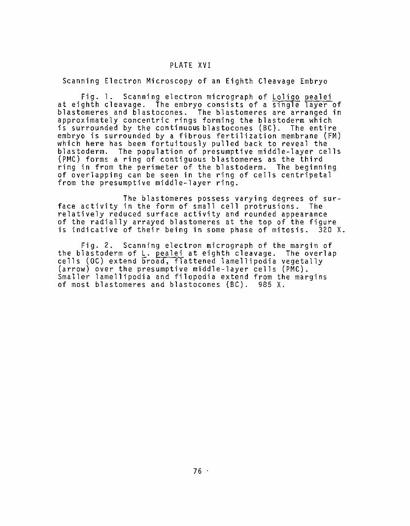
PLATE XVI
Scanning Electron Microscopy of an Eighth Cleavage Embryo
Fig. 1. Scanning electron micrograph of Loligo pealeiat eighth cleavage. The embryo consists of a single layer ofblastomeres and blastocones. The blastomeres are arranged inapproximately concentric rings forming the blastoderm whichis surrounded by the continuous blastocones (BC). The entireembryo is surrounded by a fibrous fertilization membrane (FM)which here has been fortuitously pulled back to reveal theblastoderm. The population of presumptive middle-layer cells(PMC) forms a ring of contiguous blastomeres as the thirdring in from the perimeter of the blastoderm. The beginningof overlapping can be seen in the ring of cells centripetalfrom the presumptive middle-layer ring.
The blastomeres possess varying degrees of surface activity in the form of small cell protrusions. Therelatively reduced surface activity and rounded appearanceof the radially arrayed blastomeres at the top of the figureis indicative of their being in some phase of mitosis. 320 X.
Fig. 2. Scanning electron micrograph of the margin ofthe blastoderm of ~. pealei at eighth cleavage. The overlapcells (OC) extend broad, flattened lamellipodia vegetally(arrow) over the presumptive middle-layer cells (PMC).Smaller lamellipodia and filopodia extend from the marginsof most blastomeres and blastocones (BC). 985 X.
76 .
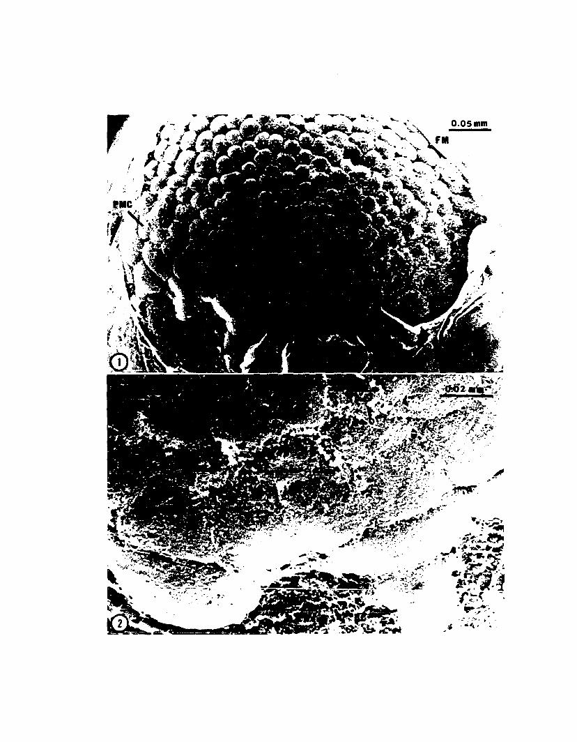
r
.. .
.~..
- ... -'~";' .,. - ..~. ~
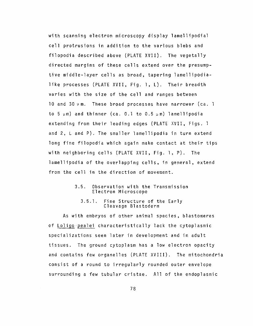
with scanning electron microscopy display lamellipodial
cell protrusions in addition to the various blebs and
filopodia described above (PLATE XVII). The vegetally
directed margins of these cells extend over the presump
tive middle-layer cells as broad, tapering lamellipodia
like processes (PLATE XVII, Fig. 1, L). Their breadth
varies with the size of the cell and ranges between
10 and 30 ~m. These broad processes have narrower (ca. 1
to 5 ~m) and thinner (ca. 0.1 to 0.5 ~m) lamellipodia
extending from their leading edges (PLATE XVII, Figs. 1
and 2, Land P). The smaller lamellipodia in turn extend
long fine filopodia which again make contact at their tips
with neighboring cells (PLATE XVII, Fig. 1, P). The
lamellipodia of the overlapping cells, in general, extend
from the cell in the direction of movement.
3.5. Observation with the TransmissionElectron Microscope
3.5.1. Fine Structure of the EarlyCleavage Blastoderm
As with embryos of other animal species, blastomeres
of Loligo'pealei characteristically lack the cytoplasmic
specializations seen later in development and in adult
tissues. The ground cytoplasm has a low electron opacity
and contains few organelles (PLATE XVIII). The mitochondria
consist of a round to irregularly rounded outer envelope
surrounding a few tubular cristae. All of the endoplasmic
78
PLATE XVII
Scanning Electron Microscopy of Overlapping Cells
Fig. 1. Scanning electron micrograph of the blastodermof Loligo pealei at eighth cleavage. This micrographillustrates in more detail the relation of the overlap cellsto the presumptive middle-layer cells. A broad, thicklamellipodium (L) is seen extending smaller lamellipodialprocesses (p) from its leading edge. These, in turn, possesssmaller filopodial protrusions. In general these processesare extended in the direction of overlapping (arrow). Smallblebs are also a common feature of the cells' surfaces atthis stage. 4,500 X.
Fig. 2. Scanning electron micrograph of an eighth cleavageembryo illustrating cell surface activity. The broad lamellipodium (L, twoheaded arrow) is spreading in the direction ofthe arrow as it extends and contacts underlying cells viasmall lamellipodial (P) and filopodial (F) processes. Mostblastomeres exhibit similar filopodial activity. An area ofpresumed junction formation (arrowhead) displays less surfaceactivity than other areas of cell-cell apposition. 45,000 X.
79
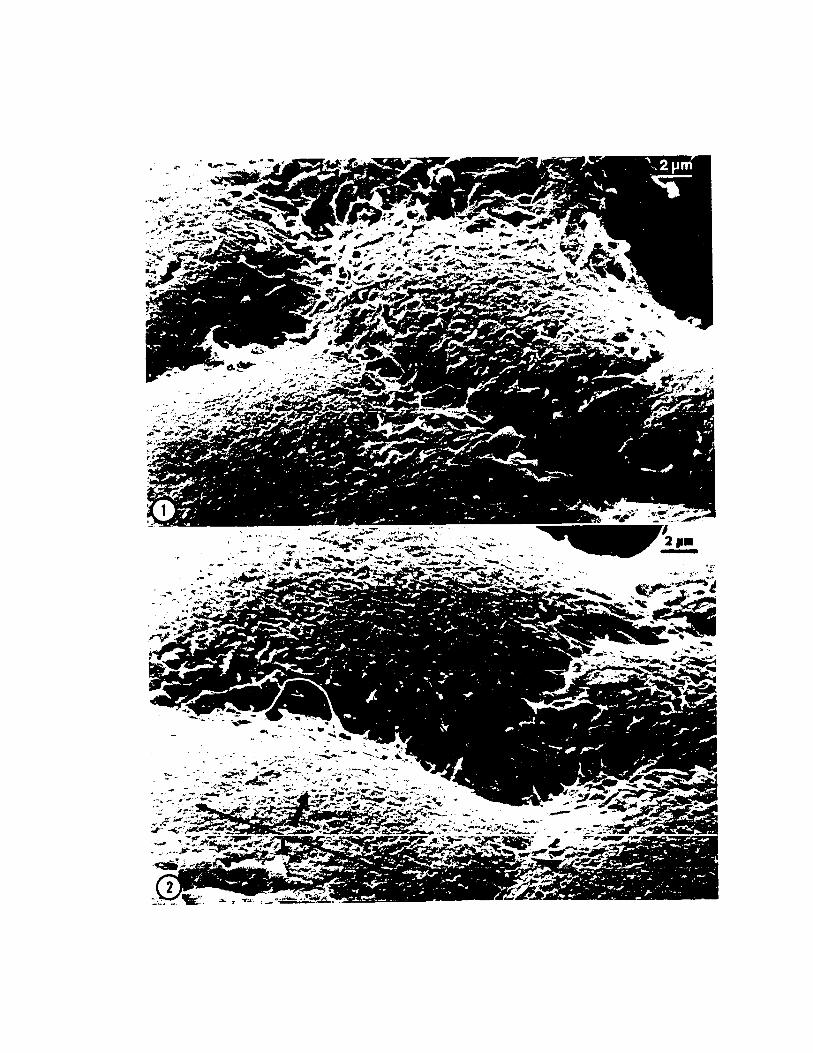
PLATE XVIII
Intercellular Junctions and Contacts
Fig. 1. Electron micrograph of an apical intercellularapposition of blastomeres in an eighth cleavage embryo. Theapposition at its apex is 20 nm to 25 nm wide. No septa areseen in apical appositions prior to eighth cleavage. Notethe microspike (filopodium) bridging the gap between cells.These filopodia make 20 nm non-junctional appositions withneighboring cells. These processes typically contain bundlesof microfilaments. 13,000 X.
Fig. 2. Micrograph of an apical intercellular junctionin a late ninth cleavage embryo. Within the 10 nm to 15 nmarea of apposition 5 nm by 15 nm septa are appearing, but witha greater than 15. nm center to center distance. The distensionof the membrane ( between arrows) is due to fixative solutionbeing hypo-osmotic (see Lord and Dibona, 1976). A Golgi body(G) is seen cut tangential to the forming face. Evidence offilopodial activity is seen at the apex of these cells. 25,600X.
Fig. 3. Septate Junction. This micrograph illustratesthe form of a typical septate junction below the apical desmosome in a Stage 11 embryo. The inter-membrane distance isconstant at 15 nm in septate areas. The spot-like closeappositions above and below the septate region are indicativeof maculate desmosomes. However, these are not clear in thismicrograph. 63,000 X.
Fig. 4. Intercellular interdigitation. Microvillioften extend from one cell into another. Interdigitationssuch as this are sometimes the remnant of cleavage furrowformation, but are often seen in other areas as well. TheGolgi (G) at the right is cut somewhat tangential to theforming face. 20,000 X.
Fig. 5. Intercellular interdigitation similar to thatin Fig. 4 above. Membranous vesicles are often seen oppositethe tips of microvillar processes. These inter~igitations
may be a means of intercellular communication ( Arnold andWilliams-Arnold, 1976). 28,000 X.
81


reticulum present in cleavage stages is of the smooth
variety and ribosomes are distributed evenly throughout
the cytoplasm individually rather than as polysomes. The
Golgi complex is quite extensive. Cross sections of
blastomeres reveal an average of six to ten dictyosomes
which appear to be distributed in no particular pattern.
Multivesiculate bodies are rarely seen during early
cleavage, but may be seen in areas of furrow vesiculation,
for example, in late phases of cytokinesis.
The blastomeres of early through late cleavage develop
no organized intercellular junctions, but commonly form
20 nm to 25 nm non-junctional appositions (PLATE XVIII,
Fig. 1). At the apex of most of these appositions
filopodia extend from one cell to another. These
filopodia contain bundles of oriented microfilaments
(PLATE XVIII, Figs. 1 and 2). Filopodia also form non
junctional appositions with adjacent cells. Various
interdigitations and projections of one cell into another,
similar to those reported in later stages (Arnold and
Williams-Arnold, 1976), are seen throughout cleavage
(PLATE XVIII). Arnold and Williams-Arnold (1976) have
suggested that these processes may playa role in inter
cellular communication.
83

3.5.2. Fine Structure of the Late Cleavage Blastoderm
A number of changes occur in the blastoderm after
eighth cleavage. One of the first, which is evident even
in thick epon sections, is a marked increase in the RNA
content of the presumptive middle-layer cells. The
difference in RNA content between blastoderm and presump
tive middle-layer cells is seen by electron microscopy as
a difference in numbers of ribosomes per cell (PLATE XIX,
Fig. l).
Subsequent to ninth cleavage the blastomeres in the
outer layer of the blastoderm begin to develop more
structured intercellular junctions. The initial junction
observed is the belt desmosome (Zonula Adherens).
These junctions appear between cells at their apices and
form a stable bond surrounding the apex of each cell
(see Staehelin, 1974). The continuity of these junctions
was confirmed by observation of serial sections of blasto
derms at this stage. At first the membranes in the area of
junction formation display an apparent thickening as an
electron dense material is deposited on their cytoplasmic
faces (PLATE XVIII, Fig. 1). At the same time there is
an accumulation of a finely granular material between the
membranes. At this time the intermembrane distance ranges
from 20 nm to 25 nm. In some cases microfilamentous
material is seen associated with the membranes in the area
of junction formation.
84
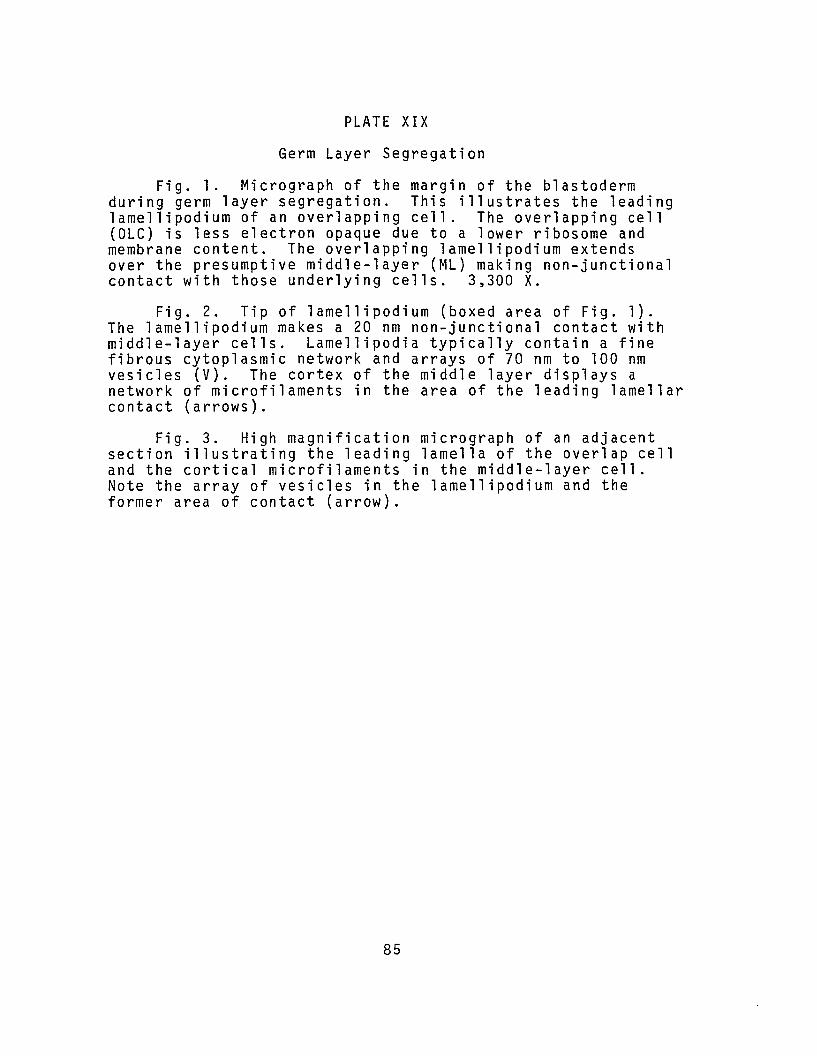
PLATE XIX
Germ Layer Segregation
Fig. 1. Micrograph of the margin of the blastodermduring germ layer segregation. This illustrates the leadinglame1lipodium of an overlapping cell. The overlapping cell(OLe) is less electron opaque due to a lower ribosome andmembrane content. The overlapping 1ame1lipodium extendsover the presumptive middle-layer (ML) making non-junctionalcontact with those underlying cells. 3,300 x.
Fig. 2. Tip of lamel1ipodium (boxed area of Fig. 1).The 1amellipodium makes a 20 nm non-junctional contact withmiddle-layer cells. Lame11ipodia typically contain a finefibrous cytoplasmic network and arrays of 70 nm to 100 nmvesicles (V). The cortex of the middle layer displays anetwork of microfilaments in the area of the leading lamellarcontact (arrows).
Fig. 3. High magnification micrograph of an adjacentsection illustrating the leading lamella of the overlap celland the cortical microfi1aments in the middle-layer cell.Note the array of vesicles in the lame1lipodium and theformer area of contact (arrow).
85
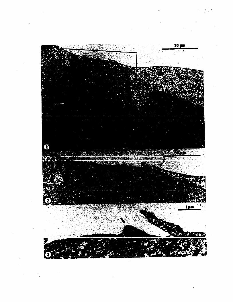
-;.,.-:, ............
10 ......

Below the area of desmosome formation the inter
membrane distance ranges from 10 nm to 20 nm. This region
develops into a fairly extensive septate junction. The
sequence of events with regard to septate junction forma
tion in Loligo is the same as that described for sea urchins
(Gilula, 1973). At first the membranes are separated by a
variable intermembrane space, are not necessarily parallel,
and are not bridged by septa (PLATE XVIII, Fig. 1). The
intermembrane distance is progressively more constant at
15 nm as septa appear in increasing numbers. Septa are,
in the electron microscope, seen as electron opaque bars
which bridge the intermembrane space. They are about 5 nm
thick, 15 nm in width, and appear between the membranes
with a 15 nm center to center spacing (PLATE XVIII, Figs. 2
and 3). The maximum number of septa per junction was not
determined definitely. However, in ninth cleavage embryos,
as many as 30 bars were counted. This contrasts with the
number which Gilu1a (1973) reports for sea urchins, but may
reflect the condition early in junction development
(Staehelin, 1974).
3.5.3. Fine Structure of the Overlapping Cells
Apart from their overall shape and possession of
elongate cell processes, overlap cells appear similar to
other b1astomeres. Their centripetal (with regard to the
geometry of the blastoderm) borders are rounded whereas the
87

opposite sides extend vegeta11y over the presumptive
middle-layer cells as large tapered processes (PLATE XIII,
Fig. 1; PLATE XIX). From the leading edges of these
processes extend smaller 1ame11ipodia which, in turn,
possess fine fi1opodial processes. The filopodia make
20 nm non-junctional appositions with neighboring cells.
The appositions of the overlap and middle-layer cells are
in all aspects non-junctional. No tight or maculae adherens
junctions were observed in cleavage or early gastrulation
stage embryos.
Lamellipodia of overlap cells were found to range in
length to about 15 ~m and from about 0.25 ~m to 0.50 ~m in
thickness. These lamellipodia contain few visible micro
filaments and no microtubules were ever observed within
such processes (PLATE XIX; PLATE XX, Fig. 1). Microfi1a
ments exist as a fine network either dispersed within the
microspikes or associated with the plasma membrane.
Overlapping cell 1amellipodia form extensive non
junctional appositions with the underlying presumptive
middle-layer cells as described above. Plate XX, Fig. 1
illustrates a lame11ipodium with a terminal microspike.
The leading edge of the lamellipodium is more hyaline than
the rest of the cell: Multivesiculate bodies (MVB) are
often found within the lamellipodia, but also frequently in
the underlying cells in the area of overlapping cell contact.
Multivesicu1ate bodies in cleavage stage embryos are
88
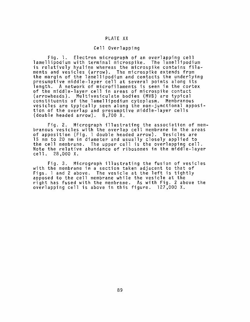
PLATE XX
Cell Overl appi ng
Fig. 1. Electron micrograph of an overlapping celllamellipodium with terminal microspike. The lamellipodiumis relatively hyaline whereas the microspike contains filaments and vesicles (arrow). The microspike extends fromthe margin of the lamellipodium and contacts the underlyingpresumptive middle-layer cell at several points along itslength. A network of microfilaments is seen in the cortexof the middle-layer cell in areas of microspike contact(arrowheads). Multivesiculate bodies (MVB) are typicalconstituents of the lamellipodium cytoplasm. Membranousvesicles are typically seen along the non-junctional apposition of the overlap and presumptive middle-layer cells(double headed arrow). 8,700 X.
Fig. 2. Micrograph illustrating the association of membranous vesicles with the overlap cell membrane in the areasof apposition (Fig. 1 double headed arrow). Vesicles are15 nm to 20 nm in diameter and usually closely applied tothe cell membrane. The upper cell is the overlapping cell.Note the relative abundance of ribosomes in the middle-layerce 11. 28,000 X.
Fig. 3. Micrograph illustrating the fusion of vesicleswith the membrane in a section taken adjacent to that ofFigs. 1 and 2 above. The vesicle at the left is tightlyapposed to the cell membrane while the vesicle at theright has fused with the membrane. As with Fig. 2 above theoverlapping cell is above in this figure. 127,000 X.
89
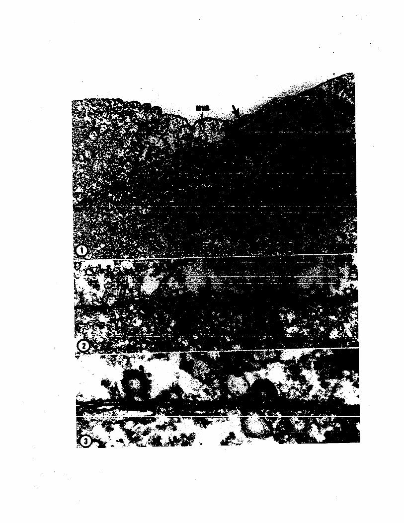

assemblages of 50 nm to 70 nm diameter membranous vesicles
surrounded by another unit membrane. Although they are
common features of late cleavage b1astoderma1 cells, their
function there is unknown at this time.
Filopodia typically contain numerous 70 nm to 100 nm
membranous vesicles arrayed along their major axes (PLATE
XIX, Fig. 3; PLATE XX, Fig. 1). The presence of these
vesicles suggests a possible mechanism for cell membrane
expansion in these actively extending processes. Bray
(1973) has suggested from similar observations a mechanism
for membrane assembly in neuronal growth cones. In growth
cones vesicles are believed to be moved along the axon by
means of a polarized actinomyosin system. The vesicles
then fuse with the plasma membrane at the tip of the
lengthening cell process. No evidence of vesicular fusion
was observed in filopodia in Lo1igo b1astomeres although
arrays of vesicles were common.
Membranous vesicles are, however, often seen lying
along and fusing with the overlap cell membrane in the area
of contact with middle-layer cells somewhat centripetal
from the leading edge (PLATE XX). These vesicles range in
diameter from 150 nm to 200 nm and are lined along their
inner surfaces by a slightly opaque material (PLATE XX,
Fig. 2). This class of vesicles is not derived from mu1ti
vesiculate bodies, but is more in the size range of Go1gi
produced vesicles. The 150 nm to 200 nm vesicles are
91
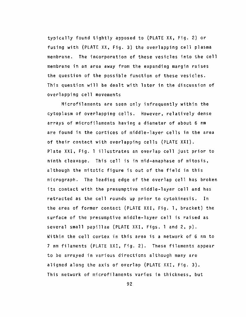
typically found tightly apposed to (PLATE XX, Fig. 2) or
fusing with {PLATE XX, Fig. 3} the overlapping cell plasma
membrane. The incorporation of these vesicles into the cell
membrane in an area away from the expanding margin raises
the question of the possible function of these vesicles.
This question will be dealt with later in the discussion of
overlapping cell movements
Microfilaments are seen only infrequently within the
cytoplasm of overlapping cells. However, relatively dense
arrays of microfilaments having a diameter of about 6 nm
are found in the cortices of middle-layer cells in the area
of their contact with overlapping cells (PLATE XXI).
Plate XXI, Fig. 1 illustrates an overlap cell just prior to
ninth cleavage. This r.ell is in mid-anaphase of mitosis,
although the mitotic figure is out of the field in this
micrograph. The leading edge of the overlap cell has broken
its contact with the presumptive middle-layer cell and has
retracted as the cell rounds up prior to cytokinesis. In
the area of former contact (PLATE XXI, Fig. 1, bracket) the
surface of the presumptive middle-layer cell is raised as
several small papillae (PLATE XXI, Figs. 1 and 2, pl.
Within the cell cortex in this area is a network of 6 nm to
7 nm filaments (PLATE XXI, Fig. 2). These filaments appear
to be arrayed in various directions although many are
aligned along the axis of overlap (PLATE XXI, Fig. 3).
This network of microfilaments varies in thickness, but
92
PLATE XXI
Germ Layer Segregation
Fig. 1. Overlapping cell. This micrograph illustratesan overlap cell (OLC) during mitosis. The mitotic figureis outside the frame to the left. The cell is in mid-anaphase. The marginal lamellipodium (L) of the overlappingcell (OLC) has retracted as the cell rounds up prior tocytokinesis. Note the area of the underlying middle-layercell (ML) formerly in contact with the extended lamellipodium(bracket). Within this area are several short papillae orsurface protrusions. The cortex in this area contains anetwork of microfilaments. 4,000 X.
Fig. 2. Higher magnification micrograph of the bracketedarea of Fig. 1 showing in greater detail the surface papillaeand the microfilamentous network (MF). The surface papillaeare former points of contact with the overlap cell lamellipodium seen at the left. 24,600 X.
Fig. 3. High magnification micrograph illustratingthe surface papillae and cortical microfilaments. Thispapilla contains several membranous vesicles and numerousfilaments. The cortical microfilaments are 6 nm to 7 nmin diameter and appear to be oriented in all directions.However, numerous filaments can be seen running parallel tothe axis of overlapping. 85,000 X.
93
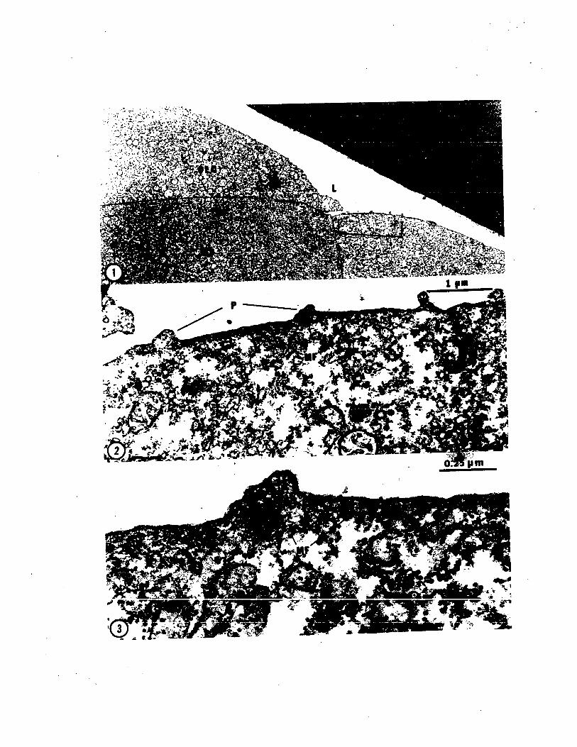

averages about 140 nm thick. The microfilaments extend
into the papillae which may also contain numbers of mem
branous vesicles (PLATE XXI, Fig. 3). Similar cortical
arrays of microfilaments are seen in Plate XIX and Plate
XX, Fig. 1.
3.5.4. The Secondary Epibolic Phase
Subsequent to segregation of the middle layer and
establishment of the yolk syncytium, the blastoderm begins
to spread over the yolk in a second phase of epiboly. The
blastomeres produced by ninth cleavages of blastocones
moved up onto the margin of the blastoderm during formation
of the yolk syncytium. Shortly after eleventh nuclear
division they begin epibolic spreading by extending broad,
highly flattened lamellipodia (PLATE XXII, Figs. 1 and 2).
These cells move across the underlying middle-layer cells
and onto the yolk syncytium where they assume a position
around the margin of the blastoderm (PLATE XXII, Fig. 3).
There they continue to extend flattened lamellipodia and to
spread vegetally. The lamellipodia produced by these cells
are much broader and thinner than those produced by over
lapping cells during middle-layer segregation (compare
PLATE XIX and PLATE XXII). During somewhat later stages
(Stages 11 to 12) these marginal cells spread out to become
very flattened and polygonal in outline (PLATE XXII, Fig.
4). These cells continue to extend and retract their
95

PLATE XXII
Stage 11 and 12 Epiboly
Fig. 1. Electron micrograph illustrating the firstphase of epibolic spreading of the blastoderm (Stage 11).The marginal cells of the Stage 11 blastoderm extend lamellipodia that are usually thinner and broader than seen duringoverlapping. These cells move over the middle-layer cellsand onto the yolk syncytium where they constitute the leading margin during epiboly. The lamellipodia often extendaway from the middle-layer cells and into the intrachorionicspace as illustrated here. Note the presence and locationof microfilaments (MF) in the cortex of the middle-layercell (ML). 2,700 x.
Fig. 2. Micrograph similar to Fig. 1 illustrating theinitial phase of epiboly. Here the lamellipodium is muchthinner than in the adjacent micrograph. Multivesiculatebodies (MVB) are typical components of the cytoplasm especially in the area of the lamellipodia. 9,200 X.
Fig. 3. Electron micrograph illustrating the marginof the blastoderm at Stage 12. The blastoderm is beginningits vegetal expansion. The marginal cells (Me) extend longlamellipodia which lie over the yolk syncytium. Theselamellipodia establish no junctions with the yolk syncytium,but maintain a simple appositional contact. At this stageall yolk syncytium nuclei (YSN) lie beneath the blastoderm.4,300 X.
Fig. 4. Micrograph illustrating another marginal celllamellipodium at Stage 12 to 13. Here the lamellipodium isquite long (16.5 m) and thin (from 1.4 m at the base to0.1 m near the tip). The epibolic spreading of the blastoderm constitutes the last phase of gastrulation. 3,700 X.
96

marginal lamellipodia throughout at least the initial
phases of spreading of the blastoderm.
3.6. Experimental Inhibition of CellMovement and Division
Embryos in various phases of Stages 9 and 10 were
subjected to the inhibitors colchicine or cytochalasin B
at several time/concentration combinations as outlined in
Methods and Materials above. These inhibitors were used
in an attempt to: 1) dissect the events of gastrulation
as a means of determining the relative importance of the
morphogenetic events of cell movement and cell division to
the segregation of the middle layer; and 2) relate the
basic mechanisms of cell movement in cephalopod gastrula
tion to theories of cell movement mechanisms in general.
3.6.1. Effects of Cytochalasin B
Time/concentration combinations greater than 0.2 ~gm/ml
for 30 minutes produced extensive short term and irreversible
long term effects. High concentrations and/or extended
treatment times completely inhibited normal development.
Such treatments almost invariably yielded embryos consisting
of a cap of cells situated at the apex of the yolk mass.
The variation of long term effects in relation to the stage
of application of short pulses of cytochalasin B has been
analyzed by Arnold and Williams~Arnold (1974) and so will
not be detailed here. Of greater concern are the effects
of cytochalasin B on the early phases of gastrulation.
98

Tne period from sixth cleavage through germ layer segre
gation to the beginning of epiboly is encompassed by Arnold's
(l965a) Stages 9 through 11. The major events of thi s peri ad
are: 1) the active overlapping of presumptive middle-
layer cells; 2) ninth cleavage division, and tenth and
eleventh nuclear divisions; 3) establishment of the yolk
syncytium by means of coordinated nuclear migration and
blastocone furrow retraction; and 4) epibolic cell move
ments. It therefore appeared necessary to determine more
specifically the stage of development of embryos at the
beginning of treatment in order to accurately assess the
effects of the inhibitors. The effects of cytochalasin
(as well as colchicine) differed markedly depending on the
specific morphogenetic stages of embryos at the time of
application. Data on cytochalasin effects presented here
are therefore restricted to the effects of 0.2 ~gm/ml
cytochalasin B applied for ten to thirty minutes at very
specific embryonic stages.
3.6.1.1. Middle-Layer Segregation
Embryos at mid- to late eighth cleavage were treated
with cytochalasin B at 0.2 ~gm/ml for 10 minutes to determine
the effect of this inhibitor on cell movements of overlapping.
Within the first 5 to 7 minutes of treatment the visible
detail of the cleavage furrows decreased almost to invisi
bility (PLATE XXIII, Fig. 2). At the same time, overlapping
99
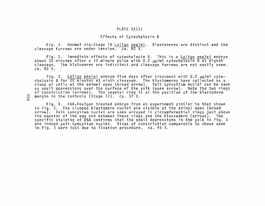
.......oo
PLATE XXIII
Effects of Cytochalasin B
Fig. 1. Normal mid-Stage 10 Loligo pealei. Blastomeres are distinct and thecleavage furrows are under tension. ca. 80 X.
Fig. 2. Immediate effects of cytochalasin B. This is a Loligo pealei embryoabout 30 minutes after a 10 minute pulse with 0.2 ~g/ml cytochalasin B at eighthcleavage. The blatomeres are indistinct and cleavage furrows are not easily seen.ca. 80 X.
Fig. 3. Loligo pealei embryo five days after treatment with 0.2 ~g/ml cytochalasin B for 20 minutes at ninth cleavage. The blastomeres have collected as aclump of cells at the animal apex (broad arrow). Yolk syncytium nuclei can be seenas small depressions over the surface of the yolk (open arrow). Note the two ringsof constriction (arrows). The vegetal ring is at the position of the blastodermmargin in the controls (Stage 17). ca. 37 X.
Fig. 4. FAA-Feulgen treated embryo from an experiment similar to that shownin Fig. 3. The clumped blastomere nuclei are visible at the animal apex (broadarrow). Yolk syncytium nuclei are seen arrayed in circumferential rings just abovethe equator of the egg and between these rings and the blastoderm (arrows). Thespecific staining of DNA confirms that the small depressions in the yolk in Fig. 3are indeed yolk syncytium nuclei. Rings of constriction comparable to those seenin Fig. 3 were lost due to fixation procedure. ca. 45 X.


lamellipodia retracted. These effects were reversed upon
removal of the cytochalasin. Furrows became more definite
within 5 to 10 minutes and overlapping cell movements and
cytokinesis were observed within 30 minutes.
Treatment of eighth cleavage embyros for 30 minutes
with 0.2 ~gm/ml cytochalasin B, in addition to the above
effects, produced numerous multinucleate blastomeres. In
some cases the multinucleate character of the blastomeres
persisted and development was arrested. In most cases,
however, the blastomeres did not remain multinucleate and
the embryos developed normally to hatching. A few cases
were also noted in which development appeared normal, but
the )rgans were reduced in size (e.g. see Arnold and
Williams-Arnold, 1974; Fig. 6, Class 2).
Examination of cytochalasin treated embryos by
electron microscopy revealed that the microfilamentous
bands at the bases of ~leavage furrows became disassociated
from the furrow membranes. The furrows retracted toward
the surface of the blastomeres and the microfilamentous
bands took the form of irregular masses of granular
material (PLATE XXIV, Figs. 1 and 2). Furrow retraction
occurred with newly formed furrows and in lateral and
undercutting furrows of blastocones, but not with completed
furrows.
The short term effects of cytochalasin treatment of
embryos at ninth cleavage were similar to those seen at
102
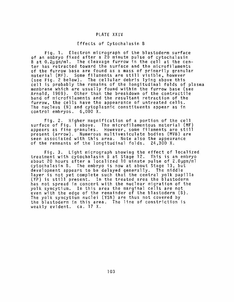
PLATE XXIV
Effects of Cytochalasin B
Fig. 1. Electron micrograph of the blastoderm surfaceof an embryo fixed after a 20 minute pulse of cytochalasinB at 0.2~gm/ml. The cleavage furrow in the cell at the center has retracted toward the surface and the microfilamentsof the furrow base are found as a mass of primarily granularmaterial (MF). Some filaments are still visible, however(see Fig. 2 below). The cellular debris lying above thiscell is probably the remains of the longitudinal folds of plasmamembrane which are usually found within the furrow base (seeArnold, 1969). Other than the breakdown of the contractileband of microfilaments and the resultant retraction of thefurrow, the cells have the appearance of untreated cells.The nucleus (N) and cytoplasmic constituents appear as incontrol embryos. 6,000 X.
Fig. 2. Higher magnification of a portion of the cellsurface of Fig. 1 above. The microfilamentous material (MF)appears as fine granules. However, some filaments are stillpresent (arrow). Numerous multivesiculate bodies (MVB) areseen associated with this area. Note also the appearanceof the remnants of the longitudinal folds. 24,300 X.
Fig. 3. Light micrograph showing the effect of localizedtreatment with cytochalasin B at Stage 12. This is an embryoabout 20 hours after a localized 10 minute pulse of 2.0~gm/ml
cytochalasin B. The embryo is now at about Stage 13, butdevelopment appears to be delayed generally. The middlelayer is not yet complete such that the central yolk papilla(YP) is still present. In the treated area the blastodermhas not spread in concert with the nuclear migration of theyolk syncytium. In this area the marginal cells are noteven with the edge of the remainder of the blastoderm (B).The yolk syncytium nuclei (YSN) are thus not covered bythe blastoderm in this area. The line of constriction isweakly evident. ca. 17 X.
103


eighth cleavage; that is, furrows and overlapping lamelli
podia retracted. Long term effects were somewhat varied.
In most cases 10 minute treatment with 0.2 ~gm/ml cyto
chalasin produced minimal long term effects. Approximately
12% of the embryos treated at this stage developed organs
of reduced size (see Arnold and Williams-Arnold, 1974; Fig.
6, Class 2.)
Treatment of ninth cleavage embryos for 30 minute at
0.2 ~gm/ml had more severe long term effects. In this case
the lateral and undercutting furrows of blastocones retracted
to a greater degree than with a ten minute treatment. These
furrow retractions released the blastocone nuclei into the
yolk cytoplasmic layer, prematurely establishing the yolk
syncytium. These effects were not reversed upon removal of
the cytochalasin. The yolk syncytium nuclei eventually
moved vegetally around the yolk mass while the blastomeres
formed a clump of cells at the animal apex of the egg.
(PLATE XXIII, Figs, 3 and 4). The time course of vegetal
movement of the yolk syncytium nuclei correlated well with
the time course of spreading of the blastoderm over the
yolk.
3.6.1.2. Yolk Syncytium Formation
Treatment of embryos with cytochalasin B at 0.2 ~gm/ml
during tenth and eleventh nuclear division had variable
effects depending upon the specific stage of the embryo at
105
the time of treatment and upon the length of treatment.
The immediate effects of treatment at tenth division were
similar to those at other stages. Furrow definition was
reduced and newly formed furrows relaxed (PLATE XXIII, Fig.
2; PLATE XXIV, Figs. 1 and 2). In addition, post mitotic
migration of the yolk syncytium nuclei was inhibited and
the lamellipodial cell processes of marginal cells were
retracted. Subsequent to the removal of the cytochalasin
after ten minutes treatment the yolk syncytium nuclei moved
up under the margin of the blastoderm, as well as vegetally,
and the blastoderm renewed epibolic spreading. In most
cases these embryos developed normally. After thirty minutes
treatment, however, the yolk syncytium nuclei moved in a
vegetal direction only and the blastoderm did not renew its
epibolic spread. The blastomeres of these embryos eventually
formed a mass of cells at the animal apex as in the case of
extended treatment of ninth cleavage embryos (see PLATE XXIII,
Fig. 3). The yolk syncytium nuclei continued to move
vegetally preceded by a line of constriction that moved
vegetally on a time course identical to that of the margin
of the blastoderm in control embryos. Plate XXIII, Fig. 3
illustrates such an embryo when the controls were in Stage
17 (Arnold, 1965a). The yolk syncytium nuclei have a distri
bution similar to that of a normal embryo at Stage 17. The
line of constriction at the vegetal pole corresponds to the
position of the blastoderm margin at Stage 17.
106
Attempts were made to discover the cellular basis of the
line of constriction using transmission electron microscopy.
The results obtained were inconclusive. However, spontane
ously occurring abnormalities morphologically similar to
the above result of cytochalasin B treatment occur infre
quently in spawns of "end of the year" females. Observation
of these abnormal embryos with the transmission electron
microscope revealed bands of microfilaments in the cortex
of the yolk cytoplasmic layer in regions of circumferential
constriction similar to those of cytochalasin treated embryos
(not illustrated).
In an initial attempt to identify the relationship
between the ring of constriction and the spreading of the
blastoderm after middle-layer segregation, an experiment
was performed in which four Stage 12 embryos were regionally
treated with cytochalasin B by the method of Arnold and
Williams-Arnold (1976). In all embryos treated by this
method a "cleft" formed in the margin at the point of
application (PLATE XXIV, Fig. 3). In the treated region,
spreading of the blastoderm was inhibited while the remaining
portion continued to spread. In contrast to the inhibition
of blastomere movement of yolk syncytium nuclei was not
inhibited and the nuclei appeared to migrate normally. The
constriction did not appear as prominent in the treated
area. Embryos treated in the above manner developed to what
approximated Stage 15 then became very abnormal and died.
107

In summary, the effect of cytocha1asin B during middle
layer segregation is that of inhibiting cell movements of
overlapping. During the formation of the yolk syncytium,
cytochalasin B inhibits nuclear movements and the cell
movements of epibolic spreading.
3.6.2. Effects of Colchicine
Embryos of Lo1igo pealei were treated with colchicine
at a concentration of 10- 4M during various phases of
Stages 9 through 10. Colchicine treatment totally inhibited
mitotic division, an effect which was not reversible.
Colchicine does not inhibit cell motility so was used here
to differentiate, by contrast with cytochalasin effects,
the relative functions of cell division and cell movement
in middle-layer segregation.
Embryos treated with colchicine at seventh to ninth
cleavage developed two-layered b1astoderms, but did not
develop further and eventually died (PLATE XXV).
Colchicine treatment inhibited all divisions so that the
blastomeres remained large in comparison with control
embryos. Active cell movements were not inhibited and
overlapping of certain marginal cells eventually placed
these cells between the overlapping cells and yolk cytoplas
mic layer and resulted in a two-layered blastoderm
(PLATE XXV, Figs. 1 to 5). Embryos treated with colchicine
up to sixth cleavage did not develop two-layered b1astoderms.
In embryos treated at tenth or eleventh division, a two-
108

PLATE XXV
Effects of Colchicine
Fig. 1. This is a late cleavage (ca. eighth) L. pealeiembryo prior to treatment with 10-4M colchicine. The blastoderm is a single layer of cells. ca. 120 X.
Fig. 2. Loligo pealei embryo seven hours and fifteenminutes after a twenty minute pulse with 10-4M colchicine.Note the large size of the blastomeres and that the blastoderm is bilayered at its margin (arrow). The ring of cellsin the middle area has created a depression in the yolk.ca. 120 X.
Fig. 3. Control for Fig. 2. Normal embryo seven hoursand fifteen minutes after the time of treatment of the embryoin Fig. 2. Note that the blastomeres are much smaller thanthose in Fig. 2 and that a ring of cells has been producedin the middle area at the edge of the blastoderm. Note theyolk syncytium nuclei forming (open arrow). ca. 120 X.
Fig. 4. Control embryo after nineteen hours. Thisembryo is now in late Stage 11. The middle layer is wellformed and has produced a large yolk papilla (arrowhead).ca. 120 X.
Fig. 5. Colchicine treated embryo after nineteenhours. Note that the middle-layer cells have becomeflattened (arrows). The embryo has undergone littlemorphological change since T=7:15 (Fig. 2). ca. 120 X.
Fig. 6. LOli~O pealei embryo about seven days aftertreatment with 10- M colchicine at eleventh nuclear division.Most of the blastomeres have piled up at the animal apex(solid arrow). The yolk syncytium nuclei can be seen assmall depressions scattered over the yolk surface (openarrow). Note the ring of contraction seen in the yolksurface (arrowhead). ca. 37 X.
109
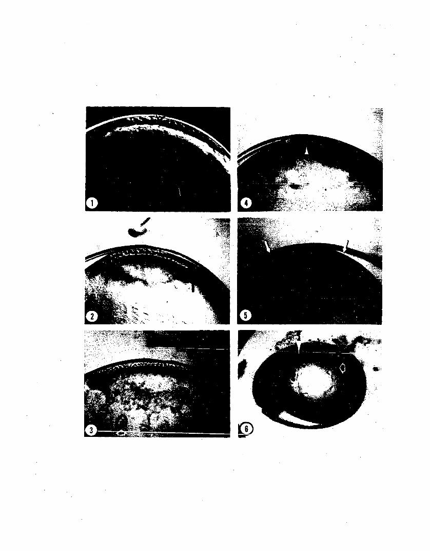

layered blastoderm developed and the yolk syncytium nuclei
formed prior to treatment moved vegetally around the yolk
mass (PLATE XXV, Fig. 6). The blastoderms of these embryos
eventually formed a clump of cells at the animal apex and
the yolk syncytium nuclei became evenly distributed through
out the syncytial cytoplasm.
4. DISCUSSION AND CONCLUSIONS
4.1. Cleavage: Establishment of the Blastoderm
The description of cleavage in Loligo pealei presented
here confirms and expands upon the previous descriptions of
Watase (1891) and Arnold (1.965, 1971). Previous descriptions
of development of this species have followed cleavage only
as far as seventh cleavage. I have expanded upon the earlier
descriptions to include eighth and ninth cleavages as well
as the two divisions subsequent to the onset of gastrulation.
The principal purpose for extending the description of
cleavage is to detail the precise timing and events of the
first phase of gastrulation.
The first through the eighth cleavages divide the
original blastodisc into progressively smaller cells pro
ducing what is termed the blastoderm. This term is some
what misleading because no true blastula is formed in
cephalopods (Arnold, 1971). Some writers have attempted,
however, to find structures in cephalopod embryos that are
homologous to structures in embryos of other organisms.
111
Fioroni (1974), for example, insists that a space
(Subgerminalhole) exists between the blastoderm and the
yolk cytoplasmic layer in cephalopods which corresponds to
the blastocoel of other molluscs. I feel, however, that
the space seen by these writers is an artifact of fixation.
The marginal blastomeres are more tightly adherent to the
underlying layers than those of the central part of the
blastoderm as is seen by dissecting away the outer layer of
the blastoderm. Only the marginal cells adhere so tightly
that attempts to remove them result in the rupture of their
membranes. This marginal adhesion probably allows the
blastoderm to act as a small osmometer. In a hypo-osmotic
fixative the small intercellular space between the blasto
derm and yolk syncytium would be inflated. On the other
hand, the term blastoderm is useful as well as descriptive
and is retained.
In addition to being the end of cleavage in the strict
est sense of the word, eighth cleavage produces those cells
which will give rise to the middle layer (presumptive middle
layer cells). The group of presumptive middle-layer cells
is comprised of a population of 48 to 52 blastomeres arrayed
in a ring near the perimeter of the blastoderm. The onset
of gastrulation in Loligo is signaled by the beginning of an
overlapping of these presumptive middle-layer cells by the
population of cells in the centripetally adjacent ring. In
addition, the cytoplasm of the presumptive middle-layer cells
112

shows signs of differentiation.
The cytoplasmic differentiation of the presumptive
middle-layer cells in Loligo appears by transmission
electron microscopy as a progressive increase in the
numbers of ribosomes, mostly as polysomes. In addition,
the presumptive middle-layer cells exhibit a differential
uptake of basic dyes beginning at eighth cleavage. While
the stains used are non-specific and the increased staining
cannot be attributed conclusively to an increase in RNA,
the affinity of RNA for basic dyes is documented ( see
Pearse, 1968). The association of an increased rate of
ribosomal RNA synthesis with the onset of gastrulation
has been confirmed in a number of embryos (for review see
Gurdon, 1974). For example, an increase in RNA content
has been found coincident with gastrulation in the gastro
pod Ilyanassa by Collier (1968, 1975). Collier's data
concern the total RNA content of whole embryos and thus
does not represent the differentiation of a specific
population of cells as implied by the increased staining
and ribosome content seen here. While the observations
presented here do not prove that a specific differentiation
of these cells has occurred, they point out a potentially
interesting avenue of inquiry.
113

4.2. Cell Division and Movement inMiddle-Layer Segregation
Ninth cleavage divides the presumptive middle-layer
cells in such a manner as to produce a ring of cells two
cells wide between the blastoderm and the yolk cytoplasmic
layer. This cleavage, along with the cell movements of
overlapping described above, is one of the two major morpho
genetic processes giving rise to the middle layer of cells.
In addition, ninth cleavage plays an essential role in
gastrulation in this species because it produces the nuclei
which give rise to the yolk syncytium as well as the blasto
meres which will become the leading edge of the blastoderm
during the second phase of epiboly.
Ninth cleavage and the overlapping of presumptive
middle-layer cells are both dynamic phenomena which have
historically been observed only in static fixed and
sectioned embryos. It is no doubt for this reason that
the theories of delamination (Ussow, 1880; Sacarrao, 1953;
Arnold, 1971; Fuchs, 1973) and immigration (Korschelt, 1892;
Fioroni, 1974) have been upheld until now. Certainly the
orientation and cleavage plane of the presumptive middle
layer cells at ninth cleavage, if seen in histological
section, would prompt either of the above theories.
The pattern of cleavage in Loligo pealei is relatively
consistent through ninth cleavage. Because of this, attempts
were made to construct a cell lineage for this species. Two
114
lineages were constructed; one used the numbering system
commonly used for spiralians (Conklin, 1907), and used by
De Leo (1972) for the squid Sepio1a rondeletii; the second
lineage attempted to record the location and divisional
relationships of b1astomeres within each quadrant of the
blastoderm. Both lineages were abandoned, however, because
the number of cell divisions subsequent to cleavage and the
presence of the middle layer prevent the clear determination
of the fates of individual or even groups of cells. In
addition, the intent of lineage construction has usually been
to provide a comparative map of the "pr-e l ocet i on" (Lillie,
1895) of adult structures within the egg and early embryo.
Cephalopods, however, have been shown to be regulative
(Ranzi, 1932; Arnold, 1965b; Arnold and Williams-Arnold,
1974, 1976). Arnold and Williams-Arnold (1974, 1976) have
shown that the organogenetic pattern exists as an inductive
pattern in the egg cortex thus precluding mosaicism. For
these reasons a detailed lineage is not presented here.
The time-lapse cinemicrographic observations of midd1e
layer segregation presented above show that the presumptive
middle-layer cells are almost completely overlapped by the
active extension of flattened processes from the adjacent
ring of cells prior to ninth cleavage. Active overlapping
of the presumptive middle-layer cells effectively places
them between the yolk cytoplasmic layer and the remaining
b1astomeres. I have, therefore, shown that the segregation
115

of the middle layer is a result of active cell movements
as Sacarrao (1953) suggested.
The appearance of active cell movements in Loligo with
time-lapse is similar to the rUffling seen with time-lapse
in cultured fibroblasts (Abercrombie, et al., 1970) and
motile activities of epithelial cells in culture (DiPasquale,
1975a). The marginal cells of epithelial sheets in culture
exhibit a variety of cell surface activities. These are
(1) extension and retraction of filopodia; (2) extension
and withdrawal of flattened lamellipodia; (3) extension of
lamellipodia accompanied by filopodial activity; (4) ruffling
of the lamellipodia; (5) blebbing; and (6) particle movement
away from the leading edge of the lamellipodium (DiPasquale,
1975a).
The overlapping cells during active overlapping are in
contact laterally, form junctions with centripetally adjacent
blastomeres and spread as a coherent sheet of cells. This
is a case of epiboly, the spreading of a sheet of prospective
ectodermal cells over the middle-layer cells of the early
embryo (see Trinkaus, 1969, 1976). Marthy (1976) has
recently reported that, in Alloteuthis media, middle-layer
segregation conforms to my earlier description (Singley,
1974) of epibolic overlapping during germ layer segregation
in L. pealei. Preliminary studies of Euprymna scolopes
indicate that middle-layer segregation occurs by a similar
process (Arnold, et al., 1972 and unpublished observations).
116
In summary~ time-lapse cinemicrography has provided
the proper perspective for the study of gastrulation in
cephalopods. The image of germ layer segregation in
Loligo pealei as a dynamic process of epiboly involving
active~ oriented cell movements contrasts sharply with the
historical concepts of delamination (Uss ow , 1881; Sacarrao,
1953; Arnold~ 1971) and immigration (Korschelt~ 1892;
Fioroni ~ 1974). In light of the evidence from time-lapse
studies presented here~ the delamination and immigration
theories are no longer tenable.
4.3. Yolk Syncytium Formation
The results presented above provide a more thorough
description of the formation of the yolk syncytium and
some insight into the mechanisms involved. The data from
time-lapse cinemicrography in particular confirm that the
yolk syncytium is established as a result of the incorpor
ation of blastocone nuclei into the yolk cytoplasmic layer
as originally described by Vialleton (1888) and recently
confirmed for several octopods (Fuchs~ 1973; Fioroni, 1974).
My studies have shown more specifically that in Loligo
pealei these nuclei are derived by the mitotic division of
blastocone nuclei preceding ninth cleavage.
Time-lapse cinemicrography further reveals that nuclei
actively migrate out of the blastocones as meridional and
undercutting furrows of the blastocones retract. The
mechanism of this nuclear movement is unknown. Similar
117
forms of nuclear migration have been seen, for example,
prior to pronuclear fusion in a sea urchin (e.g. Longo and
Anderson, 1968), during early embryogenesis in insects
(e.g. Zalokar and Erk, 1976), prior to mitosis of the
secondary nuclei in Acetabularia (Boloukh~re, 1970), during
chick lens development (Zwaan, et al., 1969; Pearce and
Zwaan, 1970), in virally induced syncytia from cultured
kidney cells (Holmes and Choppin, 1968), and in periblast
nuclei of Fundulus during epiboly (Trinkaus, 1951).
Although microtubules have been observed associated with
nuclei in some of these cases of nuclear migration (Longo
and Anderson, 1968; Boloukh~re, 1970; Holmes and Choppin,
1968), they do not appear to be required for nuclear move
ment in cases where this question has been tested experi
mentally (e.g. Kessel, 1960; Holmes and Choppin, 1968).
Instead, nuclear migration in some of these systems appears
to be microfilament (actin) mediated as judged by its
sensitivity to cytochalasin B (e.g. Zalokar and Erk, 1976).
I have shown here that the movement of blastocone nuclei
after ninth cleavage is insensitive to 10-4M colchicine,
but is inhibited by 0.2~gm/ml cytochalasin B. This suggests
that nuclear migration during yolk syncytium formation in
Loligo pealei is mediated by an actin based contractile
system.
It has been suggested that during the second phase of
epiboly, yolk syncytium nuclei are continually produced by
118
division from the margin of the blastoderm (Korschelt,
1892; Arnold, 1971). I have found no evidence for such a
derivation of yolk syncytium nuclei. In~. pealei there are
24 or 26 nuclei which migrate vegetally to form the yolk
syncytium. Subsequent to this migration they undergo two
mitotic divisions before coming to lie beneath the blastoderm.
Thus the number of yolk syncytium nuclei at Stage 12 should
be 96 or 104 as a result of these two mitotic divisions.
The numbers of yolk syncytium nuclei counted in two FAA
Feulgen treated, Epon embedded, and serially sectioned
embryos of Stages 11 to 12 were 93 and 98. A count of
89 yolk syncytium nuclei was found in a single Stage 16
embryo. Counts of numbers of yolk syncytium nuclei in
whole-mounted FAA-Feulgen treated embryos of various stages
all fall within the range of 99 to 103. My preliminary
data suggest that the two mitotic divisions of the original
24 or 26 nuclei produce the entire complement of yolk
syncytium nuclei and that no further mitotic divisions
occur. The number of yolk syncytium nuclei present through
out development has not been documented for any cephalopod
species. The figures presented here are preliminary and
are used only as an illustration of the probable manner
of yolk syncytium nucleus replication in ~. pealei. In
other species the number of nuclei produced is not necessar
ily the same as that found in Loligo pealei and the final
number would necessarily depend on the number of nuclei
119

making up the initial population.
Fuchs (1973) has suggested that the yolk syncytium
nuclei in the octopod Eledone increase in number by means
of amitotic divisions. He based this proposition on the
facts that, 1) he has never seen a mitotic figure within
the yolk syncytium, 2) the shape of some nuclei were sug
gestive of an amitotic division, and 3) the close proximity
of certain nuclei suggested a recent division. Certainly
the yolk syncytium nuclei of Loligo have a shape that is
quite variable and they are generally highly flattened
in comparison with blastodermal nuclei (Arnold, 1971).
In addition, some yolk syncytium nuclei seen in FAA-Feulgen
treated embryos have an "amoe ba-T'i ke " shape. This amoeboid
form often gives the impression that amitosis is occurring
and sections through individual yolk syncytium nuclei some
times give the appearance of amitotic divisions. All of
this, however, is only circumstantial evidence for amitosis.
Amitosis to my knowledge is found normally only in the
division of the macronucleus of some ciliate protozoans.
The lone report I have found of amitosis occurring in a
non-ciliate is that of Conklin (1903) which describes the
amitotic division of ovarian follicle cells of a cricket.
During the initial two mitotic divisions of yolk
syncytium nuclei a circumferential transitory furrow is
formed as described above. The transitory nature of this
furrow is essential to the maintenance of the syncytial
120

character of this cytoplasmic layer. The maintenance of the
syncytial character of this layer is, in turn, essential
to the subsequent movements of the nuclei within it. It
is probably also essential to the layer's development as
an embryonic digestive and circulatory organ (Boletzky,
1967; 1970; Arnold, 1971). The microfilamentous band
formed in conjunction with transitory furrowing may also
playa role in cell movement during the secondary epibolic
phases as discussed below.
The stimulus for the formation of transitory furrows
has not been investigated here. Formation of transitory
furrows is, no doubt, influenced by asters during the
mitotic divisions of the yolk syncytium nuclei. Rappaport
(1974) has shown that the asters influence the localization
and formation of cleavage furrows. Evidence of a similar
astral influence during transitory furrow formation in
Loligo is that the furrow deviates from horizontal in
areas where mitosis is oblique to the dorsal-ventral axis
of the embryo. Oblique divisions produce an undulation
in the circumferential furrow.
4.4. Electron Microscopy: Questions of Fixation
In considering the results of observations of cell
movement with electron microscopy it is necessary to
recognize that some apparent cellular substructure may be
the artifactual result of fixation. Differences in the
degree of membrane vesiculation (Spooner, et a1., 1974)
121

and the abundance of surface protrustions (Bard, et al.,
1975) in motile cells have been attributed to differences
in fixation. Fixations using osmium tetroxide, with or
without prior fixation with glutaraldehyde, for long
periods at room temperature and near neutral pH have
recently been shown to cause the destructions of actin
filaments in vitro (Maupin-Szamier and Pollard, 1977). It
is thought that osmium cleaves the actin polymer into
sma 11 pepti des.
I have found some marked differences in the amount of
cytoplasmic vesiculation present in Loligo and Octopus
cyanea embryos depending on the type of fixation procedure
used. While the use of glutaraldehyde as a fixative has
gained wide acceptance in recent years and appears to
produce the "b es t " fixation in numerous tissues, all
glutaraldehyde fixation schedules I have used on embryos
of Loligo pealei, Euprymna scolopes, and Octopus cyanea
produced large numbers of apparently abnormal vesicles and
some membrane distortion. In several cases the membranes
appeared to have delaminated. Cacodylate buffered glu
taraldehyde fixation of h. pealei embryos produced large
amounts of amorphous, electron dense material in the cyto
plasm. Colidine buffered glutaraldehyde in sea water
(Method of Arnold, in press) produced moderately good
fixation of a few cells within the blastoderm, but produced
massive vesiculation in the remaining cells. The well-
122

fixed cells were all entering prophase of mitosis. This
disparity of fixation within a single blastoderm is possi
bly the result of differences in membrane permeability
which appears to vary through the cell cycle.
Veronal acetate buffered osmium tetroxide fixation
(Palade, 1952) gave the most consistent and best overall
results. The principal variability in ultrastructure with
this procedure was encountered when the pH of the fixative
was allowed to increase to or above 7.0. At higher pH there
was more vesiculation and microfilaments were absent. This
lack of microfilaments is consistent with the results of
Maupin-Szamier and Poll.ard (1977). Fixation with the
veronal acetate buffer system at pH 6.8 for no more than
10 minutes was found to be adequate.
4.5. Scanning Electron Microscopy
Results of scanning electron microscopy confirm and
extend time-lapse observations that overlapping cell margins
actively extend broad, flattened and filiform processes
during blastodermal spreading. The morphology of motile
cell processes produced by blastodermal cells during germ
layer segregation in Loligo pealei closely resemble the
processes in numerous other cell types both in vivo and
in vitro (e.g. Nelson and Revel, 1976, 1975; Bard, et a l . ,
1975).
In general, the results of scanning electron micro
scopy show that the overlapping cells during germ layer
123
segregation in Loligo pealei are specifically differen
tiated from the other blastomeres by their possession
of flattened elongate cell processes which extend vegetally
over the adjacent ring of presumptive middle-layer cells.
4.6. Transmission Electron Microscopy
4.6.1. Cleavage Blastoderm
The general appearance of the cleavage blastomeres in
L. pealei is much like that of other embryos at early
stages (e.g. Trinkaus and Lentz, 1967). The cytoplasm
is unspecialized and contains a limited variety of
organelles; one sees no rough endoplasmic reticulum for
example. That, however, is not unexpected considering
that in the normal time course of development protein
synthesis is not "t ur ned on" to a large extent until gastru
lation (see Davidson, 1968). In addition, the blastomeres
of early cleavage stages show no junctional specializations.
A lack of junction formation during cleavage stages seems
to be common ina number of other embryos as well (e. g. Lentz
and Trinkaus, 1971; Gilula, 1973; Trelstad, et al., 1967).
During late eighth and early ninth cleavages the
blastomeres begin to develop adherens and septate type
junctions at their apices (for definitions see Staehelin,
1974). In sea urchins septate junction formation begins
during the early blastula stage (Gilula, 1973) and in
Fundulus desmosomes begin to form at the onset of epiboly
124
(Trinkaus and Lentz, 1971). It has been suggested that
junction formation may be induced by the tension placed
on the blastoderm during epiboly in Fundulus (see T~inkaus,
1976). The occurrence of a depression in the yolk beneath
the ring of middle-layer cells in Loligo while the surface
of the blastoderm remains smooth suggests that some tension
exists in the blastoderm of this embryo also. Little is
known about the development of junctions in invertebrates
but the studies of Gilula (1973) on the sea urchin indicate
that the morphology of junction development seen there is
similar to that seen in Loligo.
4.6.2. Overlapping Cells
The ultrastructural morphology of overlapping cells
in Loligo pealei as revealed by transmission electron
microscopy is, in most respects, similar to that of other
blastomeres with the exception that overlapping cells
possess cell processes typical of motile cells (e.g.
Trelstad, et al., 1967; Abercrombie, et al., 1970; Bard,
et al., 1975). The lamellipodial and filopodial processes
seen in Loligo, unlike those seen for example in chick
endothelial cells (Bard, et al., 1975) or epithelial
cells in culture (DiPasquale, 1975a and b), lack large
numbers of oriented microfilaments and these processes
in Loligo make no specialized contacts at their leading
edges with underlying cells. The absence of visible micro
filaments within active cell processes in Loligo may be
125

due either to the fact that few filaments are normally
present or that the filaments present were degraded
during fixation (Maupin~Szamier, and Pollard, 1977).
With regard to the former possibility, evidence from
mammalian tissue cells and the slime mold Physarum, for
example, indicates that a major proportion of the actin
present in the endoplasm is in the unpolymerized (G)
form (Bray and Thomas, 1976; Isenberg and Wohlfarth-Botterman,
1976). Thus only a few filaments may be necessary to support
these cell processes in Loligo.
In well-fixed embryos microfilaments appeared to have
a specific distribution within overlapping and presumptive
middle-layer cells. The concentration of microfilaments
observed in the cortex of the presumptive middle-layer cells
immediately below the area of contact with overlapping cell
processes is comparable to observations of intermediate con
tact formation between fibroblasts (Heaysman and Pagrum,
1973; Lloyd, et al., 1976) and gap junction formation between
Novikoff hepatoma cells (Johnson, et al., 1974). This distri
bution of microfilaments in Loligo has even greater similarity
to the distribution of filaments seen in areas of contact
between the yolk cytoplasmic and enveloping layers of
Fundulus during epiboly (Betchaku and Trinkaus, in prep.).
The specific localization of microfilaments in the cortices
of cells in areas of cell contact is consistent with
evidence of microfilament-mediated cell surface modulation
126
(e.g. Edelman, 1976). According to models of cell surface
modulation, surface adhesive sites could be moved over the
surface of cells by means of their trans-membrane attach
ment and interactions with actin filaments in the cell
cortex. Surface modulation is discussed in somewhat greater
detail below.
In general the results of transmission electron micro
scopy provide some insight into the cellular basis of cell
movement during the phase of overlapping.
4.7. Experiments with Inhibitors
4.7.1. Cytochalasin B
The most immediate effect of cytochalasin B treatment
was seen as a "smoothing" of the surface of the blastoderm
and a loss of visible definition of the blastomeres
(PLATE XXIII, compare Figs. 1 and 2). This effect is
probably due to the effects of cytochalasin B on the actin
based contractile system of the cells. The role of actin in
cell motility and morphology is becoming increasingly
apparent (e.g. see Allen and Taylor, 1975). Experiments
with cell extracts (e.g. Pollard, 1976; Stossel and
Hartwig, 1976) and purified actin (e.g. Kane, 1975, 1976)
indicate that the cellular activity of actin as microfila-
ments is, ;n part, a result of actin's ability to form
contractile gels. In addition, filamentous actin appears
to be most concentrated in the cortices of cells (Pollard
and Weihing, 1974). This information has provided a solid127
physical basis for the construction of models of cell
movement (Taylor, et al., 1973; Allen and Taylor, 1975).
Cytochalasin B apparently acts at or near the surface
of cells to interfere with the microfilaments located in
the cortex (Tannenbaum, et al., 1975). Cytochalasin B
injected directly into cells causes the disruption, but not
the depolymerization, of microfilaments (Luchtel, et al.,
1976). Recent evidence from in vitro studies implies that
the action of cytochalasin involves the inhibition and/or
reversal of gelation of the actin-based contractile system
(Hartwig and Stossel, 1976; Weihing, 1976a). This inhibition
appears to be directed against the interactions of actin
filaments with other proteins in the cytoplasm (Hartwig and
Stossel, 1976). Thus the loss of blastodermal definition
observed as the initial reaction to cytochalasin B treat
ment in Loligo may be the result of a gel-to-sol transfor
mation reducing the surface tension of the blastomeres.
A loss of surface tension would, in turn, reduce the small
intercellular spaces and surface curvatures of the cells
that normally interfere with the transmittance of light in
such a way as to make the cell boundaries more distinct.
Thus light passing through the blastoderm of a cytochalasin
B treated embryo would be subject to less bending and
retardation. Taking into consideration a number of other
possible explanations, this seems the most plausible.
128

Recent evidence indicates that cytochalasin B binds at
the cell surface and inhibits glucose transport (e.g. Lin
and Spudich, 1975), nucleoside transport (Plagemann and
Estensen, 1972; Plagemann, et al., 1975), and may have
other non-actin related effects (Holtzer and Sanger, 1972).
Cytochalasin B is used here to more or less specifically
inhibit cell movement over short periods of time. Although
other effects cannot be ruled out completely, I would argue
that transport effects are of no consequence in most of the
experiements performed here. First, there is little if any
glucose, other form of sugar or nucleoside in the surrounding
medium, sea water, and nutrition for the embryo is provided
by its yolk. Second, the effects of cytochalasin on cell
movement in other systems were found to be unrelated to the
inhibition of monosaccharide and nucleoside transport.
(Yamada and Wessells, 1973; Taylor and Wessels, 1973).
4.7.1 .1. Effects on Overl appi ng Movements
The effect of cytochalasin B on cell movement in Lo1igo
pea1ei occurs rapidly and reversibly. When cytochalasin is
applied during the early epibolic phase (cell overlapping)
of gastrulation, activity of the cells' margins is inhibited
and overlapping is stopped. In embryos where application
is continuous from late eighth into ninth cleavage the
marginal cells fail to migrate far enough to make contact
with the yolk syncytium and thus fail to form their normal
association with the vegetal migration of the yolk syncytium
129
nuclei. In such cases the blastoderm remains a single
layer. The effect on cell motility is reversible, however,
and the blastodermal cells later migrate to form a clump
at the animal apex (PLATE XXIII, Figs. 3 and 4). This result
indicates that formation of the early physical relationship
between vegetal migration of the yolk syncytium nuclei and
epibolic spreading of the blastoderm is essential for
epibolic spreading to occur normally.
In contrast with the results of cytochalasin treatment,
application of colchicine at eighth, or even seventh, cleav
age does not inhibit formation of a middle layer of cells
even though blastomeres can no longer divide. The results
of treatment with these two inhibitors at seventh to ninth
cleavage indicate that cell movement is the major morpho
genetic process accounting for segregation of the initial
population of middle-layer cells. On the other hand,
cell division is essential for the completion of the
middle layer.
4.7.1.2. Effects on Yolk Syncytium Formation
The effects of cytochalasin B on yolk syncytium forma
tion as outlined above indicate that the nuclear migration
observed by time-lapse cinemicrography is mediated by an
actin-based contractile system within the syncytial layer.
That migration is mediated by actin rather than microtubules
is consistent with the fact that colchicine does not inhibit
these nuclear migrations. The cytoskeletal or contractile
130
system controlling these nuclear migrations appears to be
similar to that which is suggested to control streaming of
the egg cytoplasm after fertilization and nuclear orienta
tion and distribution during cleavage (Arnold and Wi11iams
Arnold, 1970, 1974, 1976). As yet, however, there is no
morphological evidence for such a system.
Vegetal migration of the yolk syncytium nuclei appears
to be independent of epibolic spreading of the blastoderm.
Note, however, as stated above, that the inverse of this is
not true. Disruption of the initial phases of movement
disconnects these two processes in such a way that while
the nuclei continue to move vegeta11y the blastodermal
cells move to the animal apex. Yolk syncytial nuclei are
also found to move independently during gastrulation in
Fundulus. Trinkaus (1951) has shown by removing the blasto
derm of Fundulus that epibolic spreading of the yolk syncytium
layer is independent of the spreading of the blastoderm.
The results of cytochalasin application are consistent
with the idea that spreading of the blastoderm in Loligo,
as noted above, is, in contrast, dependent on the vegetal
migration of the yolk syncytium nuclei. In fact, yolk
syncytium nuclei are always found beneath the margin of the
spreading blastoderm during later phases of epiboly (Arnold,
1971). Also consistent with the idea of dependent blasto
dermal spreading is the fact that when the blastoderm is
manipulated with fine needles the upper layer of cells
131
adheres to the middle layer and later to the yolk syncytium
only at its margin. Similar marginal adhesion is evident in
sheets of epithelial cells (chick) spreading in culture
(DiPasquale, 1975a and b) and between blastoderm cells and
yolk syncytium layer during epibolic spreading in Fundulus
(Trinkaus, 1976).
Another phenomenon of interest in regard to the processes
of epiboly is the migration of the circumferential ring of
constriction seen in embryos after treatment with cytochala
sin B or colchicine during tenth nuclear division. The base
of this depression was found to possess a band of microfila
ments (not illustrated here) similar in appearance to the
band found beneath overlapping cells and at furrow bases.
Similar bands have been seen in naturally occurring abnormal
embryos. This band moved vegetally on a time course similar
to that of the blastodermal margin in controls. The band
of constriction appears to correspond to the margin of yolk
syncytium during vegetal movement. The independent movement
of this band of microfilaments and marginal adhesion of the
blastoderm suggest that blastodermal spreading may be depend
ent on and coordinated by movement of the yolk syncytium.
That is, epiboly may be controlled within the yolk syncytium.
In the few cases where it was attempted, application of
cytochalasin B to a localized area of the margin of the
blastoderm in a Stage 12 embryo produced a small area in
which the margin of the blastoderm was disassociated from
132
the margin of yolk syncytial nuclear migration (PLATE XXV,
Fig. 6). Disruption of even a small region of the blastoderm
margin, although the embryos continued to develop for two
or three days, totally disrupted gastrulation and normal
development. The results of localized application are also
consistent with the idea that epiboly is controlled by the
yolk syncytium.
The long-term effects of short pulses of cytochalasin B
on organogenesis have been dealt with by Arnold and Williams
Arnold (1974, 1976) and so will not be discussed here. In
addition, while cytochalasin B treatment during the various
phases of gastrulation produces major long-term effects,
these effects have little relation to questions concerning
the mechanism of middle-layer formation and gastrulation
movements.
4.7.2. Effects of Colchicine
Colchicine has been used here to differentiate the
relative roles of cell movement and cell division during
middle-layer segregation. Thus the effect of colchicine
as an inhibitor of cell division has been discussed above
in relation to the effects of cytochalasin B on cell movement.
It has been argued that colchicine cannot be claimed to
be a specific inhibitor of microtubular function because
colchicine also inhibits nucleoside transport (Wilson, 1975).
These two forms of inhibition would appear, however, to be
separable by experimental design. The inhibition of
133

nucleoside transport in HeLa cells was found to be competi
tive (Mizel and Wilson, 1972). Therefore it could be argued
that removal of colchicine from the culture medium should
eliminate the inhibition of nucleoside transport. In the
experiments performed with Loligo, colchicine was applied for
only a short time, and application was followed by several
rinses in fresh sea water. Treated embryos were then main
tained with daily changes of fresh sea water.
The apparent irreversibility of the inhibition of
mitosis in Loligo, during short-term experiments, is con
sistent with data on the rate of reversible dissociation
of colchicine-tubulin complex in vitro. The rate of reversi
ble dissociation of the complex as determined for porcine
brain tubulin and sea urchin tubulin in vitro corresponds
to a half-life of approximately 36 hours (Garland and Teller,
1975). The reactions of colchicine with tubulin in Loligo
are, no doubt, more complex. However, given that the rates
of reaction are similar for Loligo tubulin in vivo, the
observed irreversibility of the block of mitosis might be
explained by the slow rate of dissociation of the colchicine
tubulin complex.
Regardless of the rates of association and dissociation
of colchicine with tubulin, my observations show that colchi
cine blocked mitosis over the experimental period without
inhibiting cell movement. I feel that my use of colchicine
134
is not unjustified and that its use has served the purpose
of the experiments.
4.8. A Theory of Epiboly in Cephalopods
A number of observations made here, when considered
together, suggest a possible mechanism for the organization
and control of epiboly in Loligo. The following is provided
as an outline of a testable model of that mechanism. The
proposed model consists of the following four major components
(see also Fig. 5): 1. A circumferential band of microfila
ments is located within the cortex of the yolk cytoplasmic
layer, and also within the cortices of the presumptive middle
layer cells during the early phase of epiboly. 2. A loose
network of actin-like or possibly other filaments exists
within the cortices of the presumptive middle-layer cells
and the cortex of the yolk syncytium in addition to the
concentrated band of microfi1aments. It is not essential
that this network be present continuously. Filaments could
form precursor pools in response to the presence of the
overlying cells. 3. Adhesion of the blastoderm to the
underlying layer is mediated by specific molecules (possibly
glycoproteins) which transit the membrane such that they
are exposed at the outer surfaces of cells. On the inner
surface of the plasma membrane these molecules are attached
either directly or via a secondary protein(s) to the micro
filamentous band. The adhesive molecules may be concentrated
in specific areas via this attachment to cortical filaments.
135

4. The marginal cells of the blastoderm are motile and
produce lamellipodia and filopodia which extend and make
contacts (non-junctional) with the above adhesive areas.
This behavior occurs in a cyclical fashion.
According to this model, as the microfilaments of the
circumferential band are assembled, they move adhesive
molecules such that when the band of microfilaments is
completed a band of adhesive sites is organized on the
surface of the plasma membrane of the yolk cytoplasmic
layer (or presumptive middle-layer cell). Interaction
between the band of microfilaments and the loose network
of filaments within the yolk cytoplasmic layer causes the
band to move vegetally, simultaneously moving attached
trans-membrane adhesive molecules. This vegetal movement
of the microfilamentous band is, according to the model,
controlled at the level of microfilament-membrane interaction.
Movement may be due to a sliding of the circumferential
band of microfilaments in relation to other cortical fila
ments (see Durham, 1974), a streaming of the cytoplasm
resulting from a sol-gel-contraction sequence as proposed
in models of amoeboid movement (see Allen and Taylor, 1975),
or to a cortical contraction parallel to the cell surface
(Harris, 1976). Whatever the precise mechanism, the essential
point is that the band of adhesive sites is moved as a unit.
During vegetal movement of the band of adhesive sites
marginal cells of the blastoderm would extend filopodia and
136
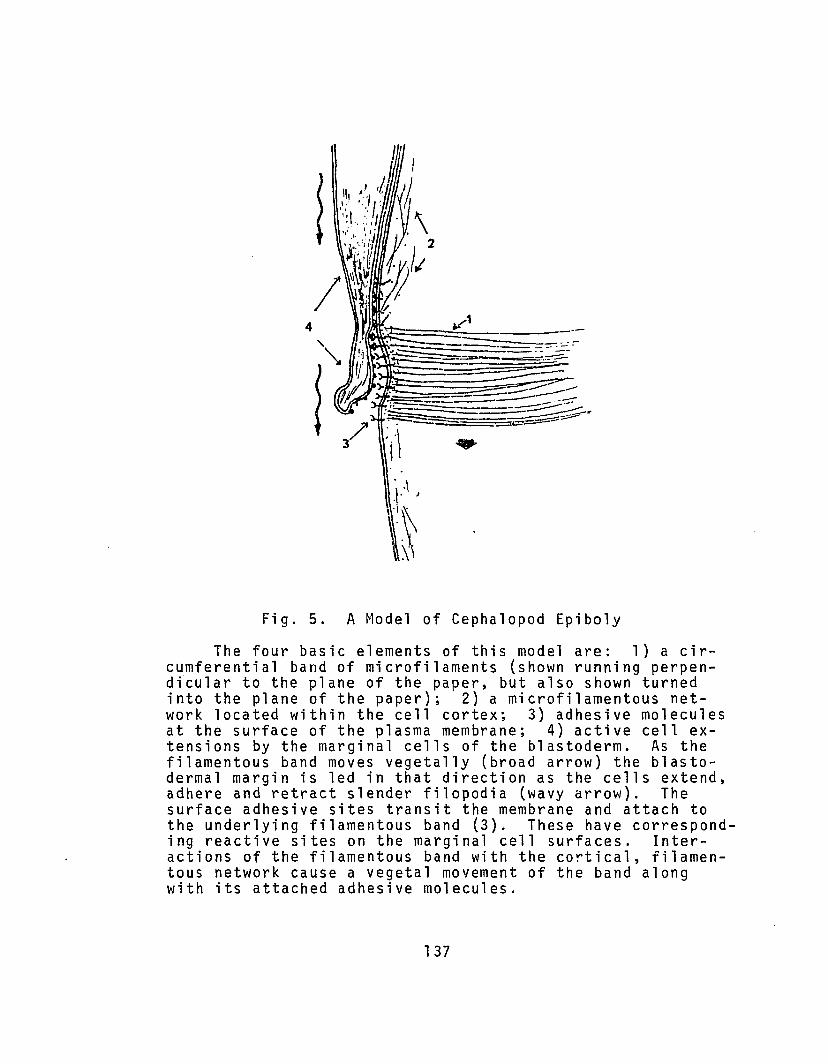
Fig. 5. A Model of Cephalopod Epiboly
The four basic elements of this model are: 1) a circumferential band of microfilaments (shown running perpendicular to the plane of the paper, but also shown turnedinto the plane of the paper); 2) a microfilamentous network located within the cell cortex; 3) adhesive moleculesat the surface of the plasma membrane; 4) active cell extensions by the marginal cells of the blastoderm. As thefilamentous band moves vegetally (broad arrow) the blastodermal margin is led in that direction as the cells extend,adhere and retract slender filopodia (wavy arrow). Thesurface adhesive sites transit the membrane and attach tothe underlying filamentous band (3). These have corresponding reactive sites on the marginal cell surfaces. Interactions of the filamentous band with the cortical, filamentous network cause a vegetal movement of the band alongwith its attached adhesive molecules.
137
1ame11ipodia which would make discrete contacts with the
adhesive sites, contract, and then disconnect. These
events would occur in a cyclical manner across the membrane
of the leading edge. During this activity the cytoplasm
of the cells would flow into their motile processes yielding
a net movement of the cells.
Thus as the band of adhesive sites moves vegeta11y,
it would lead the blastoderm in the same direction. As the
active marginal extensions retract locally the adhesive
band would move forward in that same area. Re-extension of
cell processes to the more vegetal location of adhesive
sites in conjunction with cytoplasmic flow into the proces
ses would move the cells forward to the same extent as the
yolk syncytium.
The model of cephalopod epiboly which I have presented
here is meant to suggest avenues of future research rather
than as a representation of fact. Many aspects of this
model are testable with currently available techniques.
Others must await the development of new techniques. In
general, however, this model attempts to portray epiboly
in Lo1igo in terms of current knowledge cell movement.
4.8.1. Evidence for the Model; Literatureon Biochemistry and Cell Movement
The model of cephalopod epiboly presented above is
consistent with current information concerning functions of
actin-like microfi1aments in cell motility and cell surface
138

modulation. Actin in the form of microfi1aments was first
identified by Ishikawa, Bishoff and Holtzer (1969) using the
heavy meromyosin binding technique. Actin filaments have
now been identified in a vast array of animal and some
plant species (see review of Pollard and Weihing, 1974).
In many cell types, actin filaments are found to be
especially prevalent in the cell cortex and in association
with the plasma membrane (Pollard and Ito, 1970; Isenberg
and Woh1farth-Bottermann, 1976; Mooseker, 1976; Weihing,
1976b). Actin in its monomeric (G) form may also bind to
the plasma membrane (Weihing, 1976b). Evidence further
indicates that the association of actins with membranes
is mediated by some secondary protein molecule or molecules
(e.g. Mooseker, 1976; Ti1ney, 1976). Actin associations
with certain heavy molecular weight proteins during the
contraction of gelated cell extracts (e.g. Kane, 1975,
1976) and these associations are inhibited and reversed
by cytocha1asin B (Hartwig and Stosse1, 1976).
Experiments with living cells, cell extracts and
ectoplasmic ghosts of amoeboid cells (e.g. Taylor, et a1.,
1976; Taylor, 1976; Isenberg and Woh1farth-Bottermann,
1976) indicate that the gelation and contraction of actin
in the cortical cytoplasm may be the motive force of
amoeboid movement (see Allen and Taylor, 1975). Such ideas
are further reinforced by evidence indicating that pressure
differences exist between discrete areas within the amoeboid
139

cell (see Allen, et al. 1971). The contraction of actin
containing cytoplasm in the cell cortex is the basis for
a number of models of amoeboid movement (see Allen and
Taylor~ 1975). Gell movement in developing systems must
also, however, involve specific cell surface interactions
(Trinkaus~ 1976).
The model of cephalopod epiboly presented above relies
on the fact that adhesive molecules (probably glycoproteins)
transit the plasma membrane so that their amino-terminal ~
glycosylated sequences lie exterior to the membrane and
the carboxy-terminal ends are internal and attach to a
system of microfilaments. Evidence for a trans-membrane
arrangement for glycoproteins is substantial. For example,
the best established case is that for glycophorin.
Glycophorin is the major glycoprotein of human erythrocytes
and all evidence points to a trans-membrane arrangement
(Segrest, et al. ~ 1973). All of the carbohydrate containing
amino-terminal sequences of glycophorin are found external
to the cell and the carboxy-terminal end can be attacked
enzymatically only after the inner surface of the membrane
is exposed. Other glycoproteins also appear to have a
trans-membrane arrangement. For example, certain HL-A
antigens were found to consist of a hydrophilic region at
the GOOH-terminus and a penultimate hydrophobic region
(Springer and Strominger~ 1976). This molecular structure
and the accessibility of the molecule to various reagents
140

strongly suggests a trans-membrane arrangement. Similarly,
experiments on brush-border intestinal aminopeptidase
suggest that this protein has a trans-membrane arrange
ment (Louvard, et al., 1976). During replication of
Sindbis virus, nascent coat glycoproteins are sequestered
by the lipid bilayer of host membranes (Wirth, et al.,
1977). In the virus these glycoproteins are embedded in
the virus' lipid bilayer envelope. Other evidence for the
trans-membrane arrangement of glycoproteins is reviewed by
Steck (1974) and Rothman and Lenard (1977).
Evidence of attachment of trans-membrane glycoproteins
to cytoplasmic filaments ha~ also been found in a number
of cell types. For example, Ash and Singer (1976) have
shown, with an immunofluorescent technique, that concanavalin
A receptors in normal rat kidney cells form linear arrays
20 minutes after addition of the concanavalin A. These
arrays of receptors were found superimposed on linear arrays
of intracellular myosin-containing filaments. Other evidence
for cytoplasmic-filament-membrane protein associations may
be found in the reviews of Singer (1974), Nicolson (1976),
and Edelman (1976).
Surface active sites such as antigens and concanavalin
A binding sites are able to move laterally through the plasma
membrane (e.g. Edidin and Weiss, 1972). The available
evidence strongly indicates that the mobility of surface
active sites is controlled via microfilaments and/or
141
microtubu1es linked to such sites at their cytoplasmic
ends (for review see Nicolson, 1976; Edelman, 1976). Models
attempting to explain surface site mobility propose a II s ur
face modulating assemb1 y ll consisting of: 1) a set of glyco
protein receptors that transit the membrane and confer a
specificity to the system; 2) various actin-like filaments
and associated proteins are linked to the surface glyco
protein receptors at their inner faces and produce a
coordinated movement of these surface active molecules;
3) microtubules provide anchorage of the receptors and allow
the propagation of signals to and from the cell surface.
The control of cell surface modulation may occur at the
level of interaction between act:n and other proteins
(Edelman, 1976).
There is, in addition, circumstantial evidence from
scanning electron microscope studies which suggests that
cell contact may induce a form of cell surface modulation.
For example, cell contact appears to induce the aggregation
of intramembranous particles at sites of apposition (Scott,
et a1., 1973). This particle aggregation was found to be
inhibited by very low concentrations of cytocha1asin B.
(Scott, et al., 1977).
Recent evidence suggests that cell motile behavior is
controlled, in part, by the chemical behavior of specific
cell surface glycoproteins and the chemical alterations
which they undergo during cell-cell or cell-substrate
142
associations (e.g. Roth, 1973; Moscona, 1974; Burger, et al.,
1975; Lloyd, 1975).
Trinkaus (1976) has recently formulated a theory of
epiboly in Fundulus which is consistent with the evidence
of the role of actin-like microfi1aments in cell surface
modulation and cell motility. Trinkaus and his coworkers
have shown that epiboly in Fundulus occurs in two phases.
During the first phase the margin of the enveloping layer is
very active as revealed by time-lapse and the area of con
tact between this layer and the yolk syncytial layer is
quite narrow. These observations are held to be consistent
with the idea that the enveloping layer is actively spreading
over the yolk syncytial layer during this phase. During the
second phase on the other hand, the enveloping layer margin
is quiescent and the contact zone becomes wider. Trinkaus
(1976) suggests that the second phase does not involve
active spreading of the cells, but that, on the contrary,
the enveloping layer is being pulled passively over the
autonomously spreading yolk syncytial layer. Other unpub
lished evidence is cited in support of this theory (Trinkaus,
1976). Trinkaus states, for example, that the leading edge
of the enveloping layer shows surface activity during the
first, but not the second phase. In addition, transmission
electron microscopy reveals the presence of 5 nm to 7 nm
filaments associated with the area of contact between the
enveloping layer and the yolk syncytial layer. Trinkaus
143
(1976) discusses this model of Fundulus epiboly in relation
to evidence for models of cell movement.
4.8.2. Evidence for the Model: A Study of Loligo
A band of microfilaments lying beneath the active edges
of overlapping cells has been found with transmission elec
tron microscopy. A similar band is also found in the yolk
cytoplasmic layer immediately beneath the margin of the
blastoderm during epibolic spreading. No other concentra
tions of microfilaments were observed.
Although no cortical meshwork of microfilaments was
consistently observed, a lack of their presence is not incon
sistent with the model presented here. This meshwork,
according to the model, need consist of only a few filaments
and would not necessarily be visible by electron microscopy.
No direct evidence for a band of adhesive sites has
been presented here. Although surgical manipulation shows
that marginal blastomeres adhere tightly to the yolk
syncytium and presumptive middle-layer cells, adhesion
cannot necessarily be attributed to a restricted band on
the plasma membrane. In fact, the adhesiveness may reside
instead solely in the surface of the marginal cell extensions.
The evidence presented above, from time-lapse cine
micrography and electron microscopy, shows that cell move
ment occurs during overlapping and in the marginal cells
during spreading. This evidence is adequate proof of the
144

fourth component of the model presented here. Moving cells
were observed to extend and retract filopodia and lamelli
podia in a cyclical fashion and the degree of activity
correlated with the rate of movement.
Evidence for the autonomous movement of the yolk
syncytial layer in Loligo comes from studies with inhibitors
presented above. That is, the migration of the yolk
syncytial nuclei and the contractile band of microfilaments
continues in the absence of its normal association with the
blastoderm. This movement appears to be mediated by an
actin-based system in that movement is inhibited in the
presence of cytochalasin B.
In general the model presented here is consistent with
the data on gastrulation in Loligo and with evidence for
movement in other cells.
4.8.3. Suggested Further Investigation
The model of epiboly in cephalopods presented here
suggests several areas for future investigation. Specifi
cally, evidence is needed to show that the proposed adhe
sive sites exist and are different from other surface
molecules. This might be revealed by the binding of
specific ligands to the cells' surfaces. If the proposed
sites are present, then it is of interest to reveal their
means of attachment to the microfilamentous band if such
exists. The morphology and manner of cell movement at
the blastodermal margin need further elucidation. In
145

this regard a complete time-lapse cinemicrographic investi
gation of epiboly appears warranted. Finally, the mechanism
of the control of vegetal movement needs clarification. The
inhibitors colchicine and cytocha1asin B could be used to
answer some of the questions pertaining to this problem.
5. APPENDIX
5.1. A Reappraisal of Arno1ds's (1965a) StagingBased on Observations from this Study
As a generalized description of gastrulation in
cephalopods I propose the following description in terms
of and as a revision of Arnold's (1965a) staging for Lo1igo
pea1ei. Arnold's (1965a) stages are based on major mo~pho1og
ica1 changes during embryogenesis and as such have long
served as a guide in the study of cephalopod development.
As information becomes available, however, it is clear that
the original description is inadequate. Therefore, I have
amended the descriptions of those stages to which the
information contained in these researches pertains. The
amended descriptions are as follows:
Stage 9. Sixth through eighth cleavages. Cleavage of
the central b1astomeres is largely asynchronous. Cleavage
is the peripheral rings of b1astomeres and in the b1asto-
cones is synchronous at sixth cleavage and develops as
synchronous waves of division passing from anterior to
posterior at eighth cleavage.
146
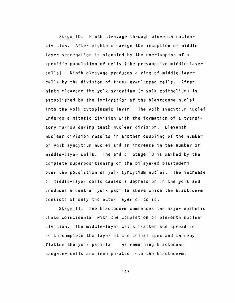
Stage 10. Ninth cleavage through eleventh nuclear
division. After eighth cleavage the inception of middle
layer segregation is signaled by the overlapping of a
specific population of cells (the presumptive middle-layer
cells). Ninth cleavage produces a ring of middle-layer
cells by the division of these overlapped cells. After
ninth cleavage the yolk syncytium (= yolk epithelium) is
established by the immigration of the blastocone nuclei
into the yolk cytoplasmic layer. The yolk syncytium nuclei
undergo a mitotic division with the formation of a transi
tory furrow during tenth nuclear division. Eleventh
nuclear division results in another doubling of the number
of yolk syncytium nuclei and an increase in the number of
middle-layer cells. The end of Stage 10 is marked by the
complete superpositioning of the bilayered blastoderm
over the population of yolk syncytium nuclei. The increase
of middle-layer cells causes a depression in the yolk and
produces a central yolk papilla above which the blastoderm
consists of only the outer layer of cells.
Stage 11. The blastoderm commences the major epibolic
phase coincidental with the completion of eleventh nuclear
division. The middle-layer cells flatten and spread so
as to complete the layer at the animal apex and thereby
flatten the yolk papilla. The remaining blastocone
daughter cells are incorporated into the blastoderm.
147
Stage 12. Completion of the middle layer of cells
at the center of the blastoderm. The peripheral cells are
greatly flattened, polygonal in outline, and have tl r uf f 1ed
membrane tl extensions at their leading edges. These cells
give rise to the external yolk sac as epiboly continues.
148

6. LITERATURE CITED
Abercrombie, M. (1970). Contact inhibition in tissue culture.In Vitro 6:128-142.
Allen, Rich. D. and D. L. Taylor. (1975) Molecular basis ofamoeboid movement. In: Molecules and Cell Movement.S. Inoue and R. E. Stephens, editors. Raven Press,New York, 239-258.
Allen, Robt. D., D.W. Francis, and R. Zeh. (1971) Directtest of the positive pressure gradient theory ofpseudopod extension and retraction in amoebae.Science 174:1237-1240.
Arnold, J. M. (1962) Mating behavior and social structurein Loligo pealii. Biol. Bull. 123:53.
Arnold, J. M. (1963) Techniques for in vitro culture ofCephalopod embryos. Assoc. Island Marine Lab. Carib.S : 19 •
Arnold, J. M. (1965a) Normal embryonic stages of the squidLoligo pealii Lesueur. Biol. Bull. 128:24-32.
Arnold, J. M. (1965b) The inductive role of the yolk epitheliumin the development of the squid Loligo pealii (Lesueur).Biol. Bull. 129:72-79.
Arnold, J. M. (1969) Cleavage furrow formation in a telolecithal egg. (Loligo pealii) I. Filaments in earlyfurrow formation. J. Cell Biol. 41 :894-914.
Arnold, J. M. (1971) Cephalopods. In: Experimental Embryology of Marine and Fresh-Water Invertebrates.Chapter 10 (G. Reverberi, ed.) North-Holland Publ.Co., Amsterdam.
Arnold, J. M. (1974) Intercellular bridges in somaticcells: Cytoplasmic continuity of blastodcrmal cellsof Loligo pealei. Differentiation 2:335-341.
Arnold, J. M., C. T. Singley, and L. D. Williams-Arnold.(1972) Embryonic development and post-hatching survival of the sepiolid squid Euprymna scolopes underlaboratory conditions. Veliger !i:36l-36S.
149

Arnold, J. M., W. C. Summers, D. L. Gilbert, R. S. Manalis,N. G. Daw, and R. J. Lasek. (1974) A laboratory guideto the use of the squid Loligo pealei. Marine Biological Laboratory, Woods Hole, Mass.
Arnold, J. M. and L. D. Williams-Arnold. (1970) The effectsof cytochalasin B on cytoplasmic movement, cleavageand subsequent development of the squid embryo. Biol.Bull. 139:413.
Arnold, J. M. and L. D. Williams-Arnold. (1974) Corticalnuclear interactions in cephalopod development:cytochalasin B effects on the informational patternin the cell surface. J. Embryol. expo Morph. 31:125.
Arnold, J. M. and L. D. Williams-Arnold. (1976) The eggcortex problem as seen through the squid eye. Am.Zoologist li:421-446.
Ash, J. F. and J. Singer. (1976) Concanavalin-A-inducedtransmembrane linkage of concanavalin A surface receptors to intracellular myosin-containing filaments.Proc. Nat. Acad. Sci. USA 73:4575-4579.
Baker, P. C. (1965) Fine structure and morphogenetic movements in the gastrula of the tree frog, ~ regi1la.J. Cell Biol. 24:95-116.
Balinsky, B. 1. (1970) An Introduction to Embryology. W. B.Saunders Co., London.
Bard, J. B. L., E. D. Hay, and S. M. Meller. (1975) Formation of the endothelium of the avian cornea: A studyof cell movement in vivo. Dev. Bio1. 42:334-361.
Be r r i 11, N. J. and G. Kar p . (1 976) 0eve lop men t. tk GrawHill Book Company, New York.
Betchaku, T. and J. P. Trinkaus. (1976) A fine-structuralanalysis of epiboly in Fundulus heteroclitus (inpreparation).
Bobretzky, N. (1877) Untersuchungen uber die Entwicklungder Cephalopoden. Nachr. Ges. Freunde Natur. Moskau24:1-73.
Boletzky, S. von. (1967) Die embryonale Ausgestaltung derFrUhen Mitteldarmanlage von Octopus vulgaris Lam. Rev.Suisse Zool. 74:555-562.
150
Boletzky, S. von. (1970) A note on the lay-out of the midgutrudiment in Loligo pealei (Le Sueur). Experientia,26: 880-881 .
Boletzky, S. von. (1975) A contribution to the study ofyolk absorption in the Cephalopoda. Z. Morph. Tiere,80:229-246.
Boloukhere, M. (1970) Ultrastructure of Acetabulariamediterranea in the course of formation of thesecondary nuclei. In: Biology of Acetabularia,145-176. (J. Brachet and S. Bonatto, eds.) AcademicPress, New York.
Bray, D. (1973) Model for membrane movements in neuralgrowth cone. Nature, Lond. 244:93-96.
Bray, D. and C. Thomas. (1976) Unpolymerized actin intissue cells, 461-474, In: R. Goldman, T. Pollard andJ. Rosenbaum (eds) Cell Motility, Book B, Cold SpringsHarbor Laboratory.
Brooks, W. K. (1880) The development of the squid Loligopeali;' Anniv. Mem. Boston Soc. Nat. Hist., 1-22.
Bruce, A. T. (1886) Segmentation of the egg and formationof the germ layers of the squid Loligo pealii. JohnsHopkins University Circulars, Vol. II, No. 54,45.
Burger, M. r~., R. S. Turn er , W. J. Kuhns , and G. Wei nba um.(1975) A possible model for cell-cell recognitionvia surface macromolecules. Phil. Trans. R. Soc.Lond., B. 271 :379-393.
Carter, S. B. (1967a) Haptotaxis and the mechanism ofcell motility. Nature, Lond. 213:256-260.
Carter, S. B. (1967b) Effects of cytochalasins on mammaliancells. Nature, Lond. 213:261-264.
Cloney, R. A. and E. Florey. (1968) Ultrastructure ofCephalopod chromatophore organs. Z. Zellforsch.89:250-280.
Collier, J. R. (1975) Nuceic acid synthesis in the normaland lobeless embryo of Ilxanassa obsoleta. Exptl. CellRes. 95:254-262.
151
Collier, J. R. and H. M. Weinstein. (1968) DNA-like RNAsynthesis during I1yanassa embryogenesis. J. CellBio1.39:27a.
Conklin, E. G. (1897) The embryology of Crepidu1a.J. Morpho1. 11:1-226.
Conklin, E. G. (1903) Amitosis in the egg follicle cellsof the cricket. Am. Nat. 37:667-675.
Conklin, E. G. (1907) The embryology of FU1gur: A studyof the influence of yolk on development. Proc. Acad.Nat. Sci., Phila. ~:320-359.
Dan, D. (1960) Cyto-embryo1ogy of echinoderms and amphibia.In: International Review of Cyt0109~, vol. 9, G. H.Bourne and J. F. Danie1li, eds., Aca ernie Press, NewYork.
Dan, K. and K. Okazaki. (1956) Cyt cvemb ryo l oqt ca l studiesof sea urchins, III. Role of the secondary mesenchymecells in the formation of the primitive gut in seaurchin larvae. Bio1. Bull. 110:29-42.
Davidson, E. H. (1968). Gene Activity in Early Development.New York: Academic Press.
De Leo, C. (1972) Data on the cell-lineage and the earlystages of development of Sepio1a ronde1etii. ActaEmbryo1. Exp. (1) :25-44.
DiPasquale, A. (1975a) Locomotory activity of epithelialcells in culture. Exp1. Cell Res. i!:191-215.
DiPasquale, A. (1975b) Locomotion of epithelial cells:Factors involved in extension of the leading edge.Exp1. Cell Res. 95:425-439.
Durham, A. C. H. (1974) A unified theory of the controlof actin and myosin in nonmusc1e movement. Cell 2:123-235.
Edelman, G. M. (1976) Surface modulation in cell recognition and cell growth. Science 192:218-226.
Edidin, M. and A. Weiss. (1972) Antigen cap formation incultured fibroblasts: A reflection of membranefluidity and of cell motility. Proc. Nat. Acad.Sci. USA 69:2456-2459.
152
Faussek, V. (1896) Zur Cepha1opoden entwick1ung. Zool.Anz. 19:496-500.
Faussek, V. (1900) Untersuchungen Uber die ~ntwick1ungder Cepha l opoden . Pubbl. Staz. Zool. Napoli 14:83237.
Fields, W. G.(1965) The structure, development, food relations,reproduction, and life history of the squid Lo1igoopa1escens Berry. State of California, Dept. ofFish and Game, Fish Bull. 131 :1-108.
Fioroni, P. (1974) Zur Entstehung des Dotterepithe1s beiverschiedenen Octopoden-Arten (Mollusca, Cephalopoda)Rev. Suisse Zool. 81:813-837.
Fioroni, P. and G. Meister. (1974) Embryo1ogie von Lo1igovulgaris Lam. Gustav Fischer Verlag, Stuttgart.
Fuchs, E. (1973) Organo- und Histogenese des Darmsystems,embryona1e B1utbi1dung und Dotterabbau bei E1edonecirrosa Lam. (Cephalopods, Octopoda) Zoo. Jb. Anat.91:31-92.
Garland, D. and D. C. Teller. (1975) A reexamination of thereaction between colchicine and tubu1in. Ann. N. Y.Acad. Sci. 253:232-238.
Gibbins, J. R., L. G. Tilney, and K. R. Porter. (1969)Microtubu1es in the formation and development ofthe primary mesenchyme in Arbacia punctu1ata. I.The distribution of microtubu1es. J. Cell Bio1.41:201-226.
Gi1u1a, N.B. (1973) Development of cell junctions. Am.Zool.13:1109-1118.
Grenacher, H. (1874) Zur Entwick1ungsgeschichte derCepha1opoden. Zeitschr. f. wiss Zool. £!:394-498.
Gurdon, J. B. (1974) The Control of Gene Expression inAnimal Development. Harvard Univ. Press,Cambridge,MA.
Gustafson,T. (1963) Cellular mechanisms in the morphogenesis of the sea urchin embryo. Exp. Cell Res.,32:570.,.589.
Gustafson, T. and M. 1. Toneby. (1971) How genes controlmorphogenesis. Am. Scientist, ~:452r462.
153

Gustafson, T. and L. Wolpert. (1967) Cellular movementand contact in sea urchin morphogenesis. Biol. Rev.42:442-498.
Harris, A. K. (1976) Is cell sorting caused by differences in the work of intercellular adhesion? Acritique of the Steinberg hypothesis. J. Theoret.Biol. 61 :267-285.
Hartwig, J. H. and T. P. Stosse1. (1976) Interactions ofactin, myosin, and an actin-binding protein ofrabbit pUlmonary macrophages. III. Effects of cytochalasin B. J. Cell Biol. 71 :295-303.
Hay, E. D. (1968) Organization and fine structure ofepithelium and mesenchyme in the developing chickembryo. In: Epithelial-Mesenchymal Interactions(R. Fleischmajor and R. E. Billingham, eds.) 31-55.Williams & Wilkins, Baltimore.
Heaysman, J. E. M. and S. M. Pegrum (1973) Early contactsbetween fibroblasts. An ultrastructural study.Exp. Cell Res. 78:71-78.
Holmes, K. V. and P. W. Choppin. (1968) On the role ofmicrotubules in movement and alignment of nuclei invirus-induced syncytia. J. Cell Biol. 39:526-543.
Holtfreter, J. (1939) Gewabeaffinitat, ein Mittel derembryonalen Formbildung. Arch. Exp. Zel1forsch.23:169-209.
Holtfreter, J. (1943) A study of the mechanics of gastrulation: Part I. J. Exp. Zool. 94:261-318.
Holtfreter, J. (1944) A study of the mechanics of gastrulation: Part II. J. Exp. Zool. ~:171-212.
Holtfreter, J. (1947) Observations on the migration, aggregation, and phagocytosis of embryonic cells. J.Morphol. 80:25-55.
Holtzer, H. and J. W. Sanger. (1972) Cytochalasin B:Microfilaments, Cell Movement and What Else? Develop.Biol., 1l.:44 t1-446.
Humphrey, C. D. and Pittman, F. E. (1974) A simple methyleneblue-azure II-basic Fuchsin stain for epoxy-embeddedtissue sections. Stain Techno1. 49:9-14.
154

Isenberg, G. and K. E. Woh1farth-Bottermann. (1976)Transformation of Cytoplasmic Actin. Cell Tiss. Res.173:495-528.
Ishikawa, H., R. Bischoff and H. Holtzer. (1969) The formation of arrowhead complexes with heavy meromyosin ina variety of cell types. J. Cell Bio1. 43:312-328.
Ivanova, O. Y., L. B. Margolis, Ju. M. Vasiliev, and 1. M.Gelfand. (1976) Effect of co1cemid on the spreading offibroblasts in culture. Exp. Cell Res. lQl:207-219.
Johnson, K. E. (1974) Gastrulation and cell interactions.In: (J. Lash and J. R. Whittaker, eds.) Concepts ofDevelopment, Sinauer Associates Inc., Stamford,Connecticut.
Johnson, K. E. (1976) Ruffling and locomotion in Rana pipiensGastrula Cells. Exp. Cell Res. lQl:71-77. ----
Johnson, R., M. Hammer, J. Sheridan, and J-P Revel. (1974)Gap junction formation between aggregated Novikoffhepatoma cells. Proc. Natn. Acad. Sci., U.S.A.71 :4536-4540.
Kane, R. E. (1975) Preparation and purification of polymerized actin from sea urchin egg extracts. J. CellBio1. 66:305-315.
Kane, R. E. (1976) Actin polymerization and interactionwith other proteins in the temperature - inducedgelation of sea urchin egg extracts. J. Cell Bio1.71 :704-714.
Kessel, R. G. (1960) The role of cell division in gastrulation of Fundulus heteroc1itus, Exp. Cell Res.20:277-282.
Ko11iker, A. (1844) Entwick1ungsgeschichte der Cepha1opoden.Zurich.
Korsche1t, E. (1892) Beitrage zur Entwick1ungsgeschichte derCepha10poden. I. Die Entstehung des Darmkana1s undNervensystems in Beziehung zur Keimb1atterfrage.Festsch. 70 Geburtstage Leuckarts (Leipzig). 345-373.
Korsche1t, E. and K. Heider. (1900) Textbook of the Embryology of Invertebrates. 235-314. (trans. from Germanby M. Bernard) The MacMi 11an Co., New York.
155

Korschelt, E. and K. Heider. (1936) Vergleichende Entwicklungsgeschichte der Tiere. 973-1009. Verlag vonGustav Fischer, Je~a.
Lankester, E. R. (1875) Observations on the developmer.t ofthe Cephalopoda. Quart. J. Microscop. Sci. ~:37-47.
Lentz, T. L. and J. P. Trinkaus. (1971) Differentiation ofthe junctional complex of surface cells in the developing Fundulus blastoderm. J. Cell Biol. 48:455-472.
Lillie, F. R. (1895) The embryology of the Uniononidae.J. Morph. lQ:1-100.
Lins, S. and J. A. Spudich. (1974) Biochemical studies onthe mode of action of cytochalasin B. J. Biol. Chem.249:5778-5783.
Lloyd, C. W. (1975) Sialic acid and the social behavior ofcells. Biol. Rev. 50:325-350.
Lloyd, C. W., D. A. Rees, C. G. Smith, and F. J. Judge.(1976) Mechanisms of cell adhesion: Early-formingjunctions between aggregating fibroblasts. J. CellSci. 22:671-684.
Longo, F. J. and E. Anderson. (1968) The fine structure ofpronuclear development and fusion in the sea urchin,Arbacia punctulata. J. Cell Biol. 39:339-368.
Lord, B. A. P. and D. R. Dibona. (1976) Role of the septatejunction in the regulation of paracellular transepithelial flow. J. Cell Biol. 71:967-972.
Louvard, D., M. Semeriva, and S. Maroux. (1976) The brushborder intestinal aminopeptidase, a transmembraneprotein as probed by macromolecular photolabelling.J. Mol. Bio1. 106:1023-1035.
Luchtel, D., J. G. Bleumink, and S. W. De Laat. (1976)The effect of injected cytochalasin B on filamentorganization in the cleaving egg of Xenopus laevis.J. Ultrastruct. Res., 54:406-419.
Luft, J. (1961) Improvements in epoxy resin embeddingmethods. J. Biophysic. and Biochem. Cytol. 1:409-414.
Marthy, H.-J. (1976) Les determinismes dans la morphogenese,contribution a 1 'embryologie experimentale des cephalopods. Ph.D Thesis, L'Universite Pierre et Marie Curie,Paris.
156
Maupin-Szamier, P. and T. D. Pollard. (1977) Factorsdetermining the destruction of actin filaments byosmium tetroxide. Biophys. J. 1I:270a.
Meister, G. (1972) Organogenese von Loligo vulgaris Lam.(Mollusca, Cephalopoda, Teuthoides, Myopsida,Loliginidae). Zool. Jb. Anat. 89:247-300.
Metschnikoff, E. (1867) Les Developpment des Sepioles.(Trans. from the Russian by E. Claparede) Arch. dessc. phy s , et nat. Vol. xxx.
Mizel, S. B. and L. Wilson. (1972) Nucleoside transportin mammalian cells. Inhibition by colchicine.Biochemestry 11:2573-2578.
Monroy, A., B. Baccetti, and S. Deni s-Doni ni. (1976)Morphological changes of the surface of the egg ofXenopus laevis in the course of development. III.S.E.M. of gastrulation. Dev. Biol. 49:250-259.
Mooseker, M. S. (1976) Actin filament-membrane attachmentin microvilli of intestinal epithelial cells. In:Cell Motility, ed. by R. Goldman, T. Pollard andJ. Rosenbaum, Book A, 631-650, Cold Springs HarborLaboratory.
Moscona, A. A. (1974) Surface specification of embryoniccells: Lectin receptors, cell recognition, and specificcell ligands. In: (A. A. Moscona, ed.) The CellSurface in Development. 67, John Wiley and Sons,New York.
Naef, A. (1928) Die Cephalopoden. Monographia 35 Fauna eFlora del Golfo di Napoli. Teil 1. Band 2.Embryologie.
Nakatsuji, N. (1974) Studies on the gastrulation ofamphibian embryos: pseudopodia in the gastrula ofBufo bufo japonicus and their significance togastrulation. J. Embryol. expo Morph., g:795-804.
Nelson, G. A. and J-P Revel. (1975) Scanning electronmicroscopic study of cell movements in the cornealendothelium of the avian embryo. Dev. Biol. 42:315333.
Nicolet, G. (1971) Avian Gastrulation. In: Advances inMorphogenesis. vol. 9,231. Academic Press, New York.
157

of theCytoplasmic
Biochem.
Nicolson, G. L. (1976) Transmembrane controlreceptors on norma1 and tumor cell s. I.influence over cell surface components.Biophys. Acta. 457:57-108.
Palade, G. E. (1952) A study of fixation for electronmicroscopy. J. Exptl. Med. ~:285-298.
Pearce, T. L. and J. Zwaan. (1970) A light and electronmicroscopic study of cell behavior and microtubulesin the embryonic chicken lens using colcemid. J.Embryol. expo Morph. £l:491-507.
Pearse, A. G. E. (1968) Histochemistry, Theoretical andAPtlied. 3rd ed., vol. I, Williams and Wilkins Co.,Ba timore.
Plagemann, P. G. W. and R. D. Estensen. (1972) CytochalasinB. VI. Competitive inhibition of nucleoside transportby cultured Novikoff rat hepatoma cells. J. CellBiol. 55:179-185.
Plagemann, P. G. W., J. H. Zylka, J. Erbe, and R. D.Estnesen. (1975) Membrane effects of cytochalasin B.Competitive inhibition of facilitated diffusion inrat hepatoma cells and other cell lines and effect onformation of functional transport sites. J. MembraneBiol. 23:77-90.
Pollard, T. D. (1976) The role of actin in the temperaturedependent gelation and contraction of extracts ofAcanthamoeba. J. Cell. Biol 68:579-60l.
Pollard, T. D. and S. Ito. (1970) Cytoplasmic filamentsof Amoeba proteus. I. The role of filaments inconsistency changes and movement. J. Cell Biol.46:267-289.
Pol 1a r d, T. D. and R. A. Wei hi ng. (1 974) Act ina nd my 0 sinand cell movement. CRC Crit. Rev. Biochem. 2:1-65.
Portmann, A. and A. M. Bidder. (1928) Yolk absorption inLoligo. Quart. J. Microscop. Sci., ll..:301-324.
Ranzi, S. (1932) Indipendenza dell'istogenesi da l l 'organogenesii. Atti, Pont. Acad. Sc. Nuovi Lincei85:27-28.
158
Rappaport, R. (1974) Cleavage. 76-98, In: (J. Lash andJ. R. Whittaker, eds.) Concepts of Development,Sinauer, Stamford, Connecticut.
Raven, C. P. (1958) Morphogenesis: The analysis ofmolluscan development. Pergamon Press, London,New York, Paris, 110-126.
Revel, J-P. (1974) Some aspects of cellular interactionsin development. In: (A. A. Moscona, ed.) The CellSurface in Development. 51, John Wiley and Sons,New York.
Richardson, K. C., L. Jarrett and F. H. Finke. (1960)Embedding in epoxy resins for ultrathin sectioningin electron microscopy. Stain Technol. 35:313.
Roth, Stephen. (1973) A molecular model for cell interactions. Quarterly Rev. Biol. 48(4):541-563.
Rothman, J. E. and J. Lenard. (1977) Membrane asymmetry.Science 195:743-753.
Saca r r'ao , G. F. (1949) Sobre as primeira1 fases da ontogenese de Tremoctopus violaceus Della Chiage. Arq.Mus. Bocage ~:1-123.
Sacarrao, G. F. (1952a) Le complexe endodermique desMollusques a oeufs riches in vitellus. Rev. Fac.Cienc. Lisboa 2:117-120.
Sacarrao, G. F. (1952b) La conception du stade Gastrulaet la gastrulation. Rev. Fac. Cienc. Lisbona 2:163170.
Sac ar ra 0, G. F. (1 952c) Remark son gas t r u1at ion i nCephalopoda. Arq. Mus. Bocage ~:43-45.
Sacar r ao , G. F. (1953) Sur la formation des feuilletsgerminatifs des Cepha10podes et 1es incertitudesde leur interpretation. Arq. Mus. Bocage £1:21-64.
Scott, R. E., L. T. Furcht, and J. H. Kersey. (1973)Changes in membrane structure associated with cellcontact. Proc. Nat. Acad. Sci. U.S.A. 73:3631-3635.
Scott, R. L, P. B. Maercklein, and L. T. Furecht. (1977)Plasma membrane intramembranous particle topographyin 3T3 and SV3T3 cells: The effect of cytochalasinB. J. Cell Sci. 23:173-192.
159

Segrest, J. P., 1. Kahane, R. L. Jackson, and V. T. Marchesi.(1973) Major glycoprotein of the human erythrocytemembrane: Evidence for an amphipathic molecularstructure. Arch. Biochem. Biophys. 155:167-183.
Singer, S. J. (1974) The molecular organization ofmembranes. Annu. Rev. Biochem. 43:805-833.
Singley, C. T. (1974) Analysis of gastrulation in Loligopealei. Symp., Biology of the Cephalopoda, 55th Ann.Western Society of Naturalists, Abstracts 22-23.
Spooner, B. S., M. A. Luduena and N. K. Wessells. (1974)Membrane fusion in the growth cone-microspike regionof embryonic nerve cells undergoing axon elongationin cell culture. Tissue and Cell 6:399-409.
Springer, T-A. and J. L. Strominger. (1976) Detergentsoluble HLA antigens contain a hydrophobic regionat the COOH-terminus and a penultimate hydrophobicregion. Proc. Nat. Acad. Sci., U.S.A. 1l:248l-2485.
Spudich, J. A. (1973) On the effects of cytochalasin Bon actin filaments. Cold Spring Harbor Symp. Quant.Biol. 37:585-593.
Spudich, J. A. and S. Lin. (1972) Cytochalasin B, itsinteractions with actin and actomyosin from muscle.Proc. Nat. Acad. Sci., U.S.A. §1.:442-446.
Steck, T. L. (1974) The organization of proteins in thehuman red blood cell membrane. A review. J. CellBio1. 62:1-19.
Staehelin, L. A. (1974) Structure and function of intercell ul ar juncti ons. I ntnl. Rev. Cyl 01. l2.: 191-283.
Stempak, J. C. and R. T. Ward. (1964) An improved stainingmethod for electron microscopy. J. Cell Biol. 22:697-701.
Stossel, T. P. and J. H. Hartwig. (1976) Interaction ofactin, myosin, and a new actin-based protein ofrabbit pulmonary macrophages. II. Role in cytoplasmicmovement and phagocytosis. J. Cell Biol. 68:602-619.
Summers, W. C. and J. J. McMahon. (1970) Survival of unfedsquid, Loligo pealei, in an aquarium. Biol. Bull138:389-396.
160

Summers, W. C. and J. J. McMahon. (1974) Studies on themaintenance of adult squid (Loligo pealei). I.Factorial survey. Biol. Bull. 146:279-290.
Summers, \L C., J. J. McMahon, and G.N.P.A. Ruppert. (1974)Studies on the maintenance of adult squid (Loligopealei). II. Empirical extensions. Biol. Bull.146:291-301.
Tannenbaum, J., S. W. Tannenbaum, L. W. l.o , G. C. Godman,and A. F. Miranda. (1975) Binding and subcellularlocalization of tritiated cytochalasin B. Exp. CellRes. 91 :47-56.
Taylor, D. L. (1976) Motile model systems of amoeboid movement. 797-822. In: R. Goldman, T. Pollard, and J.Rosenbaum (eds), Cell Motility, Book B. Cold SpringsHarbor Laboratory.
Taylor, D. L., J. Condeelis, P. L. Moore, and R. D. Allen.(1973) The contractile basis of amoeboid movement.I. The chemical control of motility in isolatedcytoplasm. J. Cell Biol. 59:378-394.
Taylor, D. L., J. A. Rhodes, and S. A. Hammond. (1976)The contractile basis of amoeboid movement.II. Structure and contractility of motile extractsand plasmalemma-ectoplasm ghosts. J. Cell Biol. 70:123-143.
Taylor, E. L. and N. K. Wessells. (1973) Cytochalasin B:Alterations in salivary gland morphogenesis not dueto glucose depletion. Develop. Biol. ~:421-425.
Teichmann, E. (1903) Die fruhe Entwicklung der Cephalopoden.Verh. Dtsch. Zool. Ges. 13:42-52.
Tilney, L. G. (1976) Nonfilamentous aggregates of actinand their association with membranes. 513-528.In: R. Goldman, T. Pollard, and J. Rosenbaum (eds).Cell Motility, Book B, Cold Springs Harbor Laboratory.
Timme rmans , L. P. M., W. L. M. Ge il en ki r chen, and N. H.Verdonk. (1970) Local accumulation of Feulgen-positivegranules in the egg cortex of Dentalium dentale L. J.Embryol. expo Morph. £l:245-252.
161

Trelstad, R. L., E. D. Hay and J-P Revel. (1967) Cellcontact during early morphogenesis in the chickembryo. Develop. Biol. ~:78-106.
Trinkaus, J. P. (1951) A study of the mechanism of epibolyin the egg of Fundulus heteroclitus. J. Exptl. Zool.118:269-320.
Trinkaus, J. P. (1969) Cells into Organs: The Forces thatShape the Embryo. Prentis Hall, Englewood Cliffs,N. J.
Trinkaus, J. P. (1973) Surface activity and locomotion ofFundulus deep cells during blastula and gastrulastages. Dev. Biol. 30:68-103.
Trinkaus, J. P. (1976) On the mechanism of metazoan cellmovements. 225-329, In: G. Poste and G. L. Nicolson(eds). The Cell Surface in Animal Development.Cell Surface Reviews vol. I.
Trinkaus, J. P. and T. L. Lentz. (1967) Surface specializations of Fundulus cells and their relation to cellmovements during gastrulation. J. Cell Biol. 32:139-153.
Ussow, M. (1874) Zoologisch embryologische Untersuchungen.Arch. Natges. 40:330-372.
Ussow, M. (1875) Zoologico-embryological investigations(Cephalopoda). The Ann. Mag. Nat. Hist., Ser. 4,~:97-113; ~:209-221; ~:317-320.
Ussow, M. (1881) Untersuchungen uber die Entwicklung derCephalopoden. Arch. Biol. ~:553-634.
Vailleton, L. (1888) Recherches sur les primeres phasesdu developpement de la seiche (Sepia officinalis).Ann. Sci. Natl. Zool. 6:1-116.
Venable, J. H. and R. Coggeshall. (1965) A simplified leadstain for use in electron microscopy. J. Cell Biol.25:407-408.
Watase, S. (1888) Observations on the development ofCephalopods. Stud. Biol. Lab. J. Hopkins Univ.4:163-183.
Watase, S. (1891) Studies on Cephalopods. I. Cleavage ofthe ovum. J. Morph. 1:274-302.
162

Weihing, R. R. (1976a) Cytochalasin B inhibits actinrelated gelation of HeLa cell extracts. J. CellBiol. 71:303-307.
Weihing, R. R. (1976b) Membrane association and polymerization of actin. 671-684. In: R. Goldman, T. Pollard,and J. Rosenbaum (eds) Cell Motility, Book B, ColdSprings Harbor Laboratory.
Wilson, L. (1975) Microtubules as drug receptors:Pharmacological properties of microtubule protein.Ann. N. Y. Acad. Sci. 253:213-231.
Wilson, L., J. R. Bamburg, S. B. Mizel, L. M. Grisham,and K. M. Creswell. (1974) Interaction of drugswith microtubule proteins. Federation Proc.33:158-166.
Wirth, D. F., F. Katz, B. Small, and H. F. Lodish, (1977)How a single Sindbis virus mRNA directs the synthesis.of one soluble protein and two integral membraneglycoproteins. Cell 10:253-263.
Wourms, J. P. (1972) The developmental biology of annualfishes. II. Naturally occurring dispersion andreaggregation of blastomeres during the developmentof annual fish eggs. J. expo Zoo1. 182:169-200.
Yamada, K. M. and N. K. Wessells. (1973) Cytochalasin B:Effects on membrane ruffling, growth cone and microspike activity, and microfilaments structure not dueto altered glucose transport. Dev. Bio1. 31 :413-420.
Zalokar, M. and 1. Erk. (1976) Division and migration ofnuclei during early embryogenesis of Drosophilamelanogaster. J. Microscopie Biol. Cell ~:97-106.
Zwaan, J., P. R. Bryan, and T. L. Pearse. (1969) Interkinetic nuclear migration during early stages of lensformation in the chicken embryo. J. Embryol. expoMorph. li:71-83.
163