università degli studi di padova dipartimento di...
TRANSCRIPT
Università degli Studi di Padova
Dipartimento di Scienze Chirurgiche, Oncologiche e Gastroenterologiche
___________________________________________________________________
SCUOLA DI DOTTORATO DI RICERCA IN
ONCOLOGIA E ONCOLOGIA CHIRURGICA
XXVII CICLO
INCREASED SURVIVAL OF CLL B CELLS IN THE PRESENCE OF MARROW MESENCHYMAL STROMAL CELLS: A NOVEL MODEL
TO DEFINE NEW TARGETS FOR THERAPY
Direttore della Scuola: Ch.mo Prof. PAOLA ZANOVELLO
Supervisore: Ch.mo Prof. GIANPIETRO SEMENZATO
Correlatore: Ch.mo Prof. LIVIO TRENTIN
Dottorando: MONICA CASTELLI

2

3
INDEX
ABBREVIATIONS……………………………………………………….… Pag. 5
ABSTRACT…………………………………………………………………. Pag. 7
RIASSUNTO………………………………………………………………… Pag. 9
INTRODUCTION…………………………………………………………...
1. Chronic Lymphocytic Leukemia……………………………………… 1.1 Incidence and epidemiology………………………………………. 1.2 Diagnosis…………………………………………………………. 1.3 Clinical features…………...……………………………………… 1.4 Clinical staging……………………………………...……………. 1.5 Biological prognostic factors……………………………...……… 1.6 Other test performed at diagnosis…………………………………. 1.7 Indications to treatment…………………………………………....
2. Neoplastic B lymphocytes…………………………………………….. 2.1 Control of apoptosis………………………………………………. 2.2 BCR-mediated signal transduction…………...…………………...
3. Cellular microenvironment in CLL…………………………………… 3.1 Nurse-like cells……………………...…………………………..... 3.2 Endothelial cell and follicular dendritic cells……………………... 3.3 T and NK cells…………………………………………………….. 3.4 Bone marrow MSCs……………………………………………….
3.4.1 Immunophenotypic characterization……………………… 3.4.2 Expansion and functional characterization………………...
3.5 Role of chemokines in CLL microenvironment…………………... 4. Treatment……………………………………………………………...
4.1 First line treatment……………...………………………………… 4.2 Second line treatment……………………………………………... 4.3 New drugs for CLL treatment……………………………………..
4.3.1 BCR signaling inhibitors………………………………….. 4.3.2 Bcl-2 inhibitors………………………………………….
Pag. 11
Pag. 11 Pag. 11 Pag. 11 Pag. 13 Pag. 13 Pag. 14 Pag. 17 Pag. 17 Pag. 18 Pag. 19 Pag. 20 Pag. 22 Pag. 23 Pag. 23 Pag. 23 Pag. 24 Pag. 25 Pag. 26 Pag. 27 Pag. 28 Pag. 28 Pag. 30 Pag. 30 Pag. 31 Pag. 33
AIM OF THE STUDY………………………………………………………. Pag. 35
MATHERIAL AND METHODS…………………...………………………
1. Patients………………………………………………………………... 2. Isolations of MSCs from CLL bone marrow………………………….. 3. Ex vivo expansion of MSCs…………………………………………… 4. Immunophenotyping of MSCs………………………………………... 5. Isolation of CLL B cells from peripheral blood samples……………... 6. Culture conditions…………………………………………………….. 7. Analysis by flow cytometry…………………………………………… 8. Polyacrilamide gel electrophoresis in SDS (SDS-PAGE)……………. 9. Western Blotting…………...………………………………………..... 10. Chemotaxis assay……………………………………………………... 11. Evaluation of CLL B cell adhesion to MSC layer…………………….. 12. Flow cytometry analysis………………...………………………….....
Pag. 37
Pag. 37 Pag. 40 Pag. 40 Pag. 41 Pag. 41 Pag. 42 Pag. 43 Pag. 43 Pag. 44 Pag. 45 Pag. 45 Pag. 46

4
13. Statistical analysis…………………………………………………….. Pag. 46
RESULTS………………...…………………………………………………..
1. MSCs isolation from bone marrow of CLL patients………………….. 2. MSCs immunophenotypic characterization…..………...…………….. 3. MSCs from CLL patients support in vitro neoplastic B cells survival.... 4. Detection of PARP 89kDa fragment reveals two subsets of CLL
clones…………………………………………………………………. 5. Cell-cell contact and soluble factors are involved in the cross-talk
between MSCs and CLL B cells………………………………………. 6. MSCs protect CLL B cells during Fludarabine and Cyclophosphamide
treatment, in vitro and in vivo…………………………………………. 7. MSCs are not able to support B leukemic cell survival after treatment
with Ibrutinib…………………………...…………………………….. 8. Ibrutinib treatment does not reduce CLL B cell migration to BM
stroma……………………………...…………………………………. 9. Ibrutinib treatment affects CLL B cell adhesion…………………..…
Pag. 47
Pag. 47 Pag. 47 Pag. 48 Pag. 49 Pag. 51 Pag. 52 Pag. 54 Pag. 55 Pag. 57
DISCUSSION…………………...…………………………………………… Pag. 59
BIBLIOGRAPHY…………………………………………………………… Pag. 65

5
ABBREVIATIONS
Abs Antibodies Ag Antigen APC Allophycocyanin APRIL A Proliferation-Inducing Ligand ATM Ataxia Teleangiectasia Mutated BAFF B cell Activating Factor Bcl-2 B cell lymphoma 2 BCR B Cell Receptor BIRC3 Baculoviral IAP Repeat Containing 3 BM Bone Marrow BR Bendamustine, Rituximab BSA Bovine Serum Albumin Btk Bruton’s tyrosine kinase CAP Cyclophosphamide, Doxorubicine, Prednisone Cbfa1 Core-binding factor alpha 1 CCL3 Chemokines C-C motif Ligand 3 CD40L CD40 Ligand CDR Complementarity Determining Region CHOP Cyclophosphamide, Doxorubicine, Vincristine, Prednisone CIRS Cumulative Illness Rating Scale CLL Chronic Lymphocytic Leukemia CM Conditionated Medium CpG Cytosine-phosphate-Guanine CR Complete Remission CT Computed Tomography CTLA-4 Cytotoxic T-Lymphocyte-Associated protein 4 CXCL12 Chemokines C-X-C motif Ligand 12 CXCR4 C-X-C motif Receptor 4 DΜΕΜ Dulbecco’s Modified Eagle Medium ERK Extracellular signal-Regulated Kinase ET-1 Endothelin-1 F/H Ficoll/Hypaque Fab Antigen binding fragment FBS Fetal Bovine Serum FC Crystallizable Fragment FCR Fludarabine, Cyclophosphamide, Rituximab FDCs Follicular Dendritic Cells FISH Interphase Fluorescent in situ Hybridization FITC Fluorescein Isothiocyanate FL Follicular Lymphoma FLU/Cy Fludarabine/Cyclophosphamide GC Germinal Centre GCLLSG German Chronic Lymphocytic Leukemia Study Group GEP Gene Expression Profile Hb Hemoglobin HLA-DR Human Leukocyte Antigen D-related HSC Hematopoietic Stem Cell HSCT Hematopoietic Stem Cell Transplantation IGVH Immunoglobulin Heavy chain Variable Region ITAM Immunoreceptor Tyrosine-based Activation Motifs LDT Lymphocyte Doubling Time

6
MBL Monoclonal B-LymphocytosisMCL Mantle Cell Lymphoma MFI Mean Fluorescence Intensity MSC-CM Mesenchymal Stromal Cell-Conditionated Medium MSCs Mesenchymal Stromal Cells MYD88 Myeloid Differentiation primary response 88 NF-κB Nuclear Factor-κB NKGD2 Natural Killer Group 2 member D NKp30 Natural Killer cell p30-related protein NLCs Nurse-Like cells NOTCH1 Notch Homolog 1, Translocation-associated ORR Overall Response Rate OS Overall Survival PARP Poli-ADP-Ribose Polymerase PBMCs Peripheral Blood Mononuclear Cells PD-1 Programmed cell Death protein 1 PD-L1 PD-1 Ligand PE Phycoerythrin PFS Progression Free Survival PKCβ Protein Kinase C β PLL Pro-Lymphocytic Leukemia Plts Platelets PMN Polymorphonuclear Neutrophil Pparγ2 Proliferator Activated Receptor γ2 PS Phosphatidylserine RBC Red Blood Cell SD Standard Deviation SF3B1 Splicing Factor 3B subunit 1 SH2 Src Homology 2 SHIP 1/2 SH2 domain containing Inositol 5-Phosphatases 1/2 SHM Somatic Hypermutations SHP1 SH2 domain containing protein tyrosine Phosphatase-1 sIg Surface Immunoglobulin SLL Small Lymphocytic Lymphoma STAT-3 Signal Transducer and Activator of Transcription 3 Syk Spleen tyrosine kinase TC Tri-Color TF Transcription Factor TGFβ Transforming Growth Factor β TTT Time To Progression VH Heavy chain Variable region WB Western Blotting WHO World Health Organization ZAP-70 Zeta-Associated Protein of 70kDa α-MEM Modified Eagle Medium αSMA+ α-Smooth Muscle Actin positive

7
ABSTRACT
Chronic Lymphocytic Leukemia (CLL) is the most common leukemia in the
Western World, accounting for about 30% of adult leukemia, and it is characterized by the
clonal expansion and accumulation of mature CD19+/CD5+/CD23+ B lymphocytes in the
peripheral blood, bone marrow and secondary lymphoid organs. Despite their apparent
longevity in patients, in vitro CLL leukemic B cells rapidly undergo spontaneous apoptosis.
The selective survival advantage is due both to intrinsic defects on apoptosis mechanism
and to signals delivered by accessory cells at the active site of the disease. Previous studies
demonstrated that mesenchymal stromal cells (MSCs), derived from bone marrow, and
CD68+ nurse-like cells, derived from peripheral blood, are involved in CLL clone longevity
and migration, suggesting a crucial role of MSCs on favouring disease progression.
Therefore, in this thesis we evaluated the effect of MSCs, the main stromal
population in the bone marrow of CLL patients, on the survival of leukemic B cells and
their role in drug resistance.
MSCs were isolated from the bone marrow of 46 CLL patients; their
immunophenotypic characterization was based on the expression of CD105, CD73 and
CD90 and the negativity of CD14, CD34, CD45 and CD31. Co-culturing MSCs and CLL
B cells, we confirmed that MSCs are able to support malignant B cell survival, providing
an in vitro culture system that closely approximates CLL microenvironment in vivo. We
observed that different leukemic clones demonstrated a large variety in the pro-survival
effect. Evaluating the cleavage pattern of PARP, we revealed two subsets of CLL clones
with different sensitivity to MSCs pro-survival signals. Our results indicate that both cell-
cell contact and soluble molecules are actors in the relationship between malignant B cells
and the MSCs, promoting CLL B cell survival and migration.
Later, we evaluated the role of the MSCs on CLL B cells during the most common
cytotoxic therapy used in clinical practice. Our data demonstrate that MSCs are able to
protect leukemic B cells from apoptosis during Fludarabine and Cyclophosphamide
treatment, both in vitro and in vivo. We tested MSCs protective role also during CLL B
cells treatment with Ibrutinib, a novel inhibitor of Btk involved in the BCR signaling
pathway, and we found that the treatment counteracts the MSC pro-survival effect. To
better understand the effect of Ibrutinib on the cross-talk between CLL B cells and MSCs,
we evaluated its role on leukemic B cell migration, also analyzing the expression levels of

8
CCR7 and CXCR4, two chemokine receptors that are central in the homing of the
neoplastic clone. We demonstrated that malignant B cell migration is not significantly
affected by the Btk inhibitor; since cell-cell contact with MSC is crucial for CLL B cell
survival, we analyzed the adhesion of leukemic B cells to MSCs after treatment with
Ibrutinib. We found a significant reduction in leukemic B cells and MSCs interactions
mediated by the CD49d integrin.
In this thesis, we demonstrate that MSCs enhance the survival of leukemic B cells
through the release of soluble factors and cell-cell direct contact and that each CLL clone
reveals a peculiar response to the anti-apoptotic signals delivered by MSCs. These
observations could be relevant to identify patients more responsive to druggable targets on
marrow microenvironment and also to find putative new strategies for CLL therapy. A
better understanding on the complexity of the cross-talk between CLL cells and their
microenvironment during CLL therapy could also help to define mechanisms of drug
resistance and treatment failure, as well to plan randomized clinical trials comparing new
compounds and their combinations with standard chemo-immunotherapy.

9
RIASSUNTO
La Leucemia Linfatica Cronica (LLC) è considerate la più comune leucemia del
mondo occidentale, rappresentando circa il 30% delle leucemie dell’adulto, ed è
caratterizzata dalla proliferazione clonale e dall’accumulo nel sangue periferico, nel
midollo osseo e negli organi linfatici secondari di linfociti B maturi CD19+/CD5+/CD23+.
Nonostante i linfociti B leucemici mostrino un’aumentata sopravvivenza nei pazienti affetti
da LLC, in vitro vanno rapidamente incontro ad apoptosi. Il vantaggio sulla sopravvivenza
è legato sia a difetti intrinseci del meccanismo di apoptosi sia a segnali forniti da cellule
accessorie, presenti nel sito attivo della malattia. Precedenti studi hanno dimostrato che le
cellule mesenchimali stromali (MSC) e le cellule accessorie (“nurse-like”) derivate
rispettivamente dal midollo osseo e dal sangue periferico, sono coinvolte nell’aumentata
longevità e mobilità del clone leucemico, suggerendo un ruolo cruciale delle MSC nel
favorire la progressione della malattia.
In questa tesi abbiamo valutato l’effetto delle MSC, la principale popolazione
stromale nel midollo osseo dei pazienti affetti da LLC, sulla sopravvivenza dei linfociti B
neoplastici e il loro ruolo sulla resistenza ai farmaci.
Le MSC sono state isolate da campioni di sangue midollare provenienti da 46
pazienti affetti da LLC; la loro caratterizzazione immunofenotipica è stata effettuata sulla
base dell’espressione di CD105, CD73 e CD90 e sulla negatività di CD14, CD34, CD45 e
CD31. Allestendo co-colture di MSC e linfociti B leucemici, abbiamo confermato la
capacità delle MSC di incrementare la sopravvivenza delle cellule B neoplastiche, fornendo
un sistema di coltura in vitro che mima profondamente il microambiente della LLC in vivo.
Abbiamo osservato una grande varietà sulla vitalità dimostrata dai diversi cloni leucemici
e, mediante la valutazione del frammento clivato della proteina PARP, abbiamo identificato
due differenti gruppi di cloni di LLC, con una diversa sensibilità ai segnali di stimolo
provenienti dalle MSC. I nostri risultati indicano che sia il diretto contatto cellula-cellula
che la presenza di molecole solubili sono coinvolte nell’interazione tra le cellule B
leucemiche e le MSC, promuovendo la sopravvivenza e la migrazione della cellula B
leucemica.
Successivamente, abbiamo valutato l’effetto delle MSC sui linfociti B neoplastici
durante un trattamento chemioterapico di uso comune nella pratica clinica. I nostri dati
hanno dimostrato che le MSC sono in grado di proteggere le cellule B leucemiche

10
dall’apoptosi durante il trattamento con Fludarabina e Ciclofosfamide, sia in vitro che in
vivo. Abbiamo esaminato il ruolo protettivo delle MSC anche durante il trattamento dei
linfociti neoplastici con Ibrutinib, un nuovo inibitore della Btk, una chinasi coinvolta nella
cascata del segnale del BCR, e abbiamo dimostrato che il trattamento delle cellule B con
Ibrutinib è in grado di contrastare l’effetto anti-apoptotico delle MSC. Per meglio definire
l’azione di Ibrutinib nell’interazione tra le cellule B di LLC e le MSC, abbiamo valutato il
suo ruolo sulla mobilità dei linfociti B leucemici, analizzando inoltre i livelli di espressione
di CCR7 e CXCR4, due recettori chemiochinici fondamentali nella migrazione del clone
neoplastico. Abbiamo dimostrato che la migrazione delle cellule B neoplastiche non è
significativamente influenzata dall’inibitore del Btk; inoltre, considerando che il diretto
contatto cellula-cellula con le MSC è di notevole importanza per la sopravvivenza dei
linfociti B leucemici, abbiamo analizzato l’adesione delle cellule B alle MSC dopo il
trattamento con Ibrutinib, evidenziando che la loro adesione era significativamente ridotta.
In questa tesi abbiamo dimostrato che le MSC incrementano la sopravvivenza delle
cellule B neoplastiche attraverso il rilascio di fattori solubili e mediante il diretto contatto
cellula-cellula, e che ogni clone leucemico rivela una peculiare risposta ai segnali anti-
apoptotici rilasciati dalle MSC. Queste osservazioni potrebbero essere determinanti al fine
di identificare i pazienti più sensibili a trattamenti mirati a colpire il microambiente
midollare ed a trovare potenziali nuove strategie terapeutiche per la LLC. Una migliore
comprensione della complessità delle interazioni tra i linfociti leucemici e il loro
microambiente nel corso del trattamento potrà inoltre aiutare a chiarire i meccanismi di
chemioresistenza e refrattarietà, così come a pianificare studi clinici randomizzati che
confrontino nuovi farmaci e la loro combinazione con i trattamenti chemio-immunoterapici
già in uso.

11
INTRODUCTION
1. Chronic Lymphocytic Leukemia
1.1 Incidence and epidemiology
Chronic Lymphocytic Leukemia (CLL) is a lymphoproliferative disorder
characterized by the clonal proliferation and accumulation of mature, typically CD5+ B
cells within the blood, bone marrow (BM), lymph node and spleen (1). Recently, it has
been reported that in CLL the capacity to generate clonal B cells might be acquired at the
hematopoietic stem cell (HSC) stage, suggesting that the primary leukemogenic event
might involve multipotent, self-renewing HSCs (2). CLL is the most prevalent leukemia in
the Western world with an incidence of about 6-9% (3). The median age diagnosis lies
between 67 and 72 years with an incidence increasing rapidly with age; more male than
female are affected by this disease and only about 10% of CLL patients are reported to be
younger than 55 years (4). The proportion of younger patients with early stage CLL and
minimal symptoms seems to increase due to more frequent blood testing. The etiology is
still unknown; moreover, there is an inherited genetic susceptibility, with an increased risk
for family members of CLL patients (5).
1.2 Diagnosis
The World Health Organization (WHO) classification of hematopoietic neoplasias
describes CLL as a leukemic lymphocytic lymphoma, distinguishable from small
lymphocytic lymphoma (SLL) only by its leukemic appearance. The diagnosis of SLL in
fact requires a number of B lymphocytes in the peripheral blood not exceeding 5,000µL.
Other lymphoma entities to be separated from CLL are marginal zone lymphoma,
lymphoplasmacytic lymphoma and mantle cell lymphoma (MCL). CLL is always a disease
of neoplastic B cells, because the entity beforehand described as T cells Chronic
Lymphocytic Leukemia (T-CLL) is now called T-cell pro-lymphocytic leukemia (T-PLL)
(6).

12
The diagnosis of CLL requires the following criteria (7):
- The presence of ≥ 5,000µL monoclonal B lymphocytes in the peripheral blood for
the duration of at least 3 months. The clonality of the circulating leukemic B cells
is confirmed by flow cytometry observing the restriction to expression of either
kappa or lambda immunoglobulin light chains; CLL B cells co-express the T-cell
antigen CD5 and B cell surface antigens CD19, CD20 and CD23 (Fig. 1). Typically,
the expression levels of surface immunoglobulin, CD20 and CD79b are lower than
normal B cells (8).
Figure 1. Cytograms of a representative case of CLL. B lymphocytes analyzed (CD19+) are positive to CD5 (panel A) and to CD23 (panel B), express one type of immunoglobuline light chain (λ, panel C), and surface IgM (sIgM), low density (panel D).
- The leukemic B lymphocytes in the blood smear are small and mature, with a
narrow border of cytoplasm, a dense nucleus lacking discernible nucleoli and a
partially aggregated chromatin (Fig. 2). Gumprecht nuclear shadows, or smudge
cells, found as cells debris, are other characteristic morphologic features found in
CLL. Larger atypical lymphocytes (pro-lymphocytes) may be seen in the blood
smear but they must not exceed 55%; the evidence of pro-lymphocytes in excess of
55% would indicate a diagnosis of pro-lymphocytic leukemia (PLL).

13
In absence of lymphoadenopathy, organomegaly, cytopaenia and clinical symptoms, the presence of fewer than 5,000/µL monoclonal B lymhocytes is defined monoclonal B-lymphocytosis (MBL), which can be detected in 5% of subjects with normal blood count. Progression to CLL occurs in 1-2% of MBL cases per year (9).
Figure 2. Peripheral blood smear of a CLL patient. Arrows indicate Gumprecht shadows typical of CLL.
1.3 Clinical features
Currently, 70-80% of CLL patients present with a low tumor burden and are frequently diagnosed from a blood count performed for an incidental reason, while other patients may present an aggressive disease (10). The patients could be asymptomatic or present B symptoms (asthenia, weight loss, fever, night sweats) and lymphadenopathy, splenomegaly and hepatomegaly. In advanced disease, we could observe hemolytic anemia (11% of cases) or autoimmune thrombocytopenia (2%). Frequently, CLL patients show hypogammaglobulinemia that could induce acquired immunodeficiency and high risk for infections. The evolution of the disease to Richter’s syndrome (a diffuse large B cell lymphoma) or to PLL could be observed in some CLL cases with a poor prognosis.
1.4 Clinical staging
In everyday clinical practice, two widely accepted staging methods co-exist, the Rai
and the Binet systems. The original Rai classification was modified to reduce from five to
three the number of prognostic groups. Both Rai and Binet staging classify patients into
three major prognostic clusters; these classifications are simple and cheap and require only
physical examination and routine laboratory tests. The Binet staging system is established
14
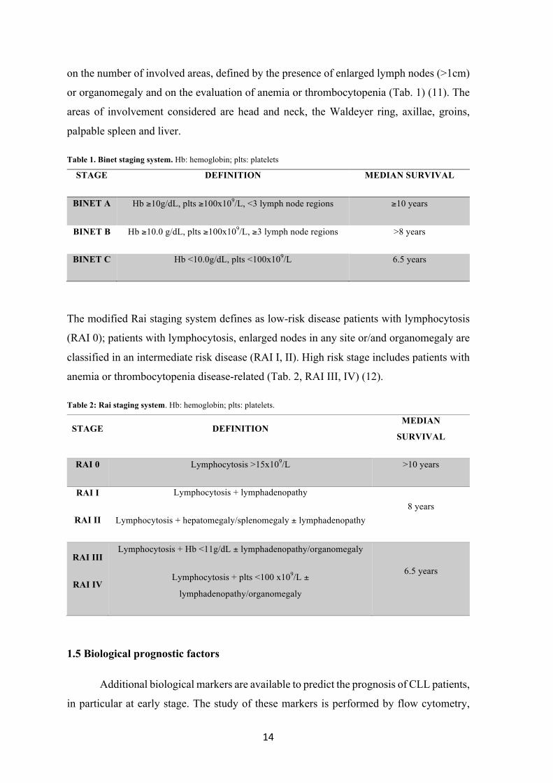
on the number of involved areas, defined by the presence of enlarged lymph nodes (>1cm)
or organomegaly and on the evaluation of anemia or thrombocytopenia (Tab. 1) (11). The
areas of involvement considered are head and neck, the Waldeyer ring, axillae, groins,
palpable spleen and liver.
Table 1. Binet staging system. Hb: hemoglobin; plts: platelets
STAGE DEFINITION MEDIAN SURVIVAL
BINET A Hb ≥10g/dL, plts ≥100x109/L, <3 lymph node regions ≥10 years
BINET B Hb ≥10.0 g/dL, plts ≥100x109/L, ≥3 lymph node regions >8 years
BINET C Hb <10.0g/dL, plts <100x109/L 6.5 years
The modified Rai staging system defines as low-risk disease patients with lymphocytosis
(RAI 0); patients with lymphocytosis, enlarged nodes in any site or/and organomegaly are
classified in an intermediate risk disease (RAI I, II). High risk stage includes patients with
anemia or thrombocytopenia disease-related (Tab. 2, RAI III, IV) (12).
Table 2: Rai staging system. Hb: hemoglobin; plts: platelets.
STAGE DEFINITION MEDIAN
SURVIVAL
RAI 0 Lymphocytosis >15x109/L >10 years
RAI I
RAI II
Lymphocytosis + lymphadenopathy
Lymphocytosis + hepatomegaly/splenomegaly ± lymphadenopathy 8 years
RAI III
RAI IV
Lymphocytosis + Hb <11g/dL ± lymphadenopathy/organomegaly
Lymphocytosis + plts <100 x109/L ±
lymphadenopathy/organomegaly
6.5 years
1.5 Biological prognostic factors
Additional biological markers are available to predict the prognosis of CLL patients,
in particular at early stage. The study of these markers is performed by flow cytometry,
15
cytogenetic and molecular biology techniques. The main markers are:
1) Somatic Hypermutations (SHM) of the Ig heavy chain variable region (VH) genes.
Conventionally, patients with <2% somatic mutations from the most similar germline gene
in both the expressed VH and VL genes were define unmutated (SHM-); mutated cases
(SHM+) were defined as those in which the CLL cells displayed ≥2% differences in either
the expressed VH or VL gene. About 50% of CLL patients present an unmutated
immunoglobulin heavy chain variable region (IGVH) status (13). CLL cells have a higher
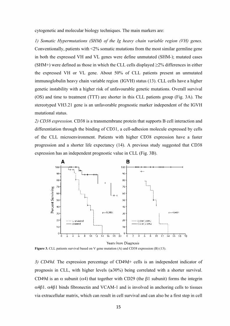
genetic instability with a higher risk of unfavourable genetic mutations. Overall survival
(OS) and time to treatment (TTT) are shorter in this CLL patients group (Fig. 3A). The
stereotyped VH3.21 gene is an unfavorable prognostic marker independent of the IGVH
mutational status.
2) CD38 expression. CD38 is a transmembrane protein that supports B cell interaction and
differentiation through the binding of CD31, a cell-adhesion molecule expressed by cells
of the CLL microenvironment. Patients with higher CD38 expression have a faster
progression and a shorter life expectancy (14). A previous study suggested that CD38
expression has an independent prognostic value in CLL (Fig. 3B).
Figure 3. CLL patients survival based on V gene mutation (A) and CD38 expression (B) (13).
3) CD49d. The expression percentage of CD49d+ cells is an independent indicator of
prognosis in CLL, with higher levels (≥30%) being correlated with a shorter survival.
CD49d is an α subunit (α4) that together with CD29 (the β1 subunit) forms the integrin
α4β1. α4β1 binds fibronectin and VCAM-1 and is involved in anchoring cells to tissues
via extracellular matrix, which can result in cell survival and can also be a first step in cell
pooled with the IgM! cases described above (bringing the totalnumber of patients studied to 63), the median survival for theunmutated group (n " 29) was 8 years and for the mutatedgroup (n " 34) was not reached for the duration of follow-up(P " .0001). Similar data were obtained for the CD38 groups:median survival for the #30% CD38! (n " 19) was 9 years,
whereas median survival for the $30% CD38! group (n " 25)was not reached (P " .0001).Gender of the B-CLL cases based on either V gene mutation
or CD38 expression. The cohort of IgM! B-CLL patients inthis study consisted of 34 males and 13 females (M:F " 2.6:1).However, the M:F ratio of the patients stratified by either Vgene mutation status or CD38 expression was very different(Table 3). In the mutated group, males and females werevirtually equally distributed, whereas in the unmutated group, amarked male predominance was found (M:F" 11:1; P " .003).A similar disparity in gender distribution was seen when thepatients were compared based on the percentages of CD38!
B-CLL cells. The numbers of males and females among the$30% CD38! group were almost equal (M:F" 1.1:1), whereasmales outnumbered females in the#30%CD38! group (M:F"7.5:1; P " .031).
DISCUSSION
The preceding data indicate that Ig V gene mutation statusand CD38 expression are distinct and reliable prognosticindicators of clinical course and outcome in B-CLL. Indeed,those patients in either the unmutated or #30% CD38! groupsexperienced a worse clinical course than those patients in the
Fig 3. Survival based on V gene mutation status and CD38 expression. (A) Kaplan-Meier plot comparing survival based on the absence(‘‘unmutated’’: . . . . . ) or presence (‘‘mutated’’: ____) of significant numbers (H2%) of V gene mutations in 47 B-CLL cases (unmutated: 24 cases;mutated: 23). Median survival of unmutated group: 9 years; median survival of mutated group not reached; P ! .0001; log-rank test). (B)Kaplan-Meier plot comparing survival based on the detection ofH30% (. . . . . ) orF30% CD38" B-CLL cells (H30%: 17 cases;F30%: 19). Mediansurvival of theH30% CD38" group: 10 years;median survival of theF30% CD38" group: not reached (P! .0001; log-rank test).
Table 2. Comparison of Modified Rai Stage at DiagnosisWith Ig VGene Mutation Status and the Percentages of CD38" B-CLLCells
Stage Unmutated Mutated
Low* 22.7% (5/22) 52.4% (11/21)Intermediate* 72.7% (16/22) 42.9% (9/21)High 4.6% (1/22) 4.7% (1/21)
P " .123
#30% CD38!
B-CLL Cells$30% CD38!
B-CLL Cells
Low† 20.0% (3/15) 50.0% (9/18)Intermediate† 73.3% (11/15) 50.0% (9/18)High 6.7% (1/15) 0.0% (0/18)
P " .138
*Comparison of V gene mutation status among patients in the lowand intermediate risk categories (P " .058; 2-tailed Fisher’s Exact test).†Comparison of CD38 expression among patients in the low and
intermediate risk categories (P " .147; 2-tailed Fisher’s Exact test).
1844 DAMLE ET AL
For personal use only.on December 11, 2015. by guest www.bloodjournal.orgFrom

16
migration. Cell migration function is very important for CLL cells survival and explains
the prognostic relevance of CD49d expression (15).
4) Intracytoplasmatic expression of protein kinase associated to TCR ζ chain of 70kDa,
ZAP-70. Zeta-associated protein of 70kDa (ZAP-70) is a cytoplasmic tyrosine kinase
working as a key signaling molecule for T lymphocytes and NK cells. ZAP-70 expression
reflects an activation state of the malignant clone and may be associated with a CLL
progression (16). ZAP-70 value could change over time in CLL; its analysis can be
performed with flow cytometry, immunohistochemistry, western blotting and Real-Time
PCR. Flow cytometry is the most useful for its diffusion and easiness of application.
5) Molecular Cytogenetics. Using Interphase Fluorescent in Situ Hybridization (FISH),
cytogenetic lesions can be identified in more than 80% of all CLL cases (17). Deletions on
the long arm of chromosome 13, involving band 13q14 [del(13q14)] represent the single
most frequently observed cytogenetic aberration, occurring in about 55% of all cases and
it is characterized by a benign CLL course. Additional frequent chromosomal aberrations
include deletions in the long arm of chromosome 11 [del(11q)]. These deletions usually
encompass band 11q23 harboring the gene Ataxia teleangiectasia mutated (ATM), which
encodes for the proximal DNA damage response kinase ATM. Patients with a del(11q)
clone show a rapid CLL progression, bulky lymphadenopathy and reduced overal survival.
Trisomy 12 is observed in 10-20% of CLL patients that demonstrated a shorter survival in
respect to those with a normal FISH. There is an association between trisomy 12 and the
presence of mutations in the Notch homolog 1, translocation-associated (NOTCH1) gene
leading to a less favorable course of the disease. Deletions of the short arm of chromosome
17 are found in 5-8% of never treated patients. These deletions usually consist in band
17p13, including the main tumor suppressor gene TP53. Del(17p) patients show marked
chemo-immunotherapy resistance (18). Mutations of TP53 are found in 4-37% of CLL
patients and are associated with a very poor prognosis (19). Whole genome sequencing
analyses identified additional recurrent mutations (20) (>5% cases at diagnosis) affecting
NOTCH1 (21), splicing factor 3B subunit 1 (SF3B1) (22), baculoviral IAP repeat
containing 3 (BIRC3) (23) and myeloid differentiation primary response (MYD88) genes.
These mutations usually coexist with some of the genetic abnormalities analyzed by the
FISH: SF3B1 mutations with del(11q), NOTCH1 with trisomy 12 and MYD88 with
del(13q); generally, these associations resulted in a poor patient outcome. There was a clear
relationship between NOTCH1 mutation and a shorter time to therapy with resistance to

17
treatment and Richter’s transformation.
6) Serum markers. According to different studies, also CD23, thymidine kinase and
β2−microglobulin may predict survival and progression-free survival. Nevertheless, their
relative value in the management of CLL patients is not validated (24), (25), (26).
1.6 Other test performed at diagnosis
In order to complete the risk assessment, the following examinations are also
recommended (27): patient performance status, medical hystory, serum chemistry
(including lactate dehydrogenase, bilirubin, serum immunoglobulins), direct antiglobulin
test, the status of relevant infections (hepatitis B and C, cytomegalovirus, human
immunodeficiency virus). Although a bone marrow biopsy is not required for diagnosis, it
is recommended for the diagnostic evaluation of unclear cytopaenias, or FISH and
molecular genetics if peripheral blood cell lymphocytosis does not allow adequate flow
cytometry analysis. Imaging studies by computed tomography (CT) scans could help to
asses the tumor load before starting treatment or to clarify unclear symptoms, but they
should not generally be performed in asymptomatic patients for clinical staging.
1.7 Indications to treatment
Patients at intermediate and high risk stages, according to the modified Rai
classification, or stage B and C, according to the Binet classification, usually benefit from
treatment but some of these patients can be only monitored until they have evidence for
progressive or symptomatic disease. The absolute lymphocyte count should not be used as
the only indication for starting therapy. Active disease should meet at least one of the
following criteria (28): 1. evidence of progressive marrow failure (development or
worsening of anemia and/or thrombocytopenia; 2. massive or progressive and symptomatic
splenomegaly; 3. massive node or progressive and symptomatic lymph nodes; 4.
progressive lymphocytosis increasing more than 50% in months or lymphocyte doubling
time (LDT) of less than 6 months (excluding factors contributing to lymphocytosis or
lymphadenopathy other than CLL); 5. autoimmune anemia and/or thrombocytopenia
poorly responding to other standard therapy; 6. constitutional symptoms as unintentional

18
weight loss of 10% or more in the last 6 months, significant fatigue, fever higher than 38°C
for more than 2 weeks and night sweats for more than 1 month without infections.
2. Neoplastic B lymphocytes
B lymphocyte participates in humoral immunity producing antibodies (Abs) in
response to antigen (Ag) stimulation. B cell can differentiate from "naive" lymphocyte to
cell secreting antibodies against specific antigens (plasma cells), or to "memory" long-lived
stimulated B lymphocyte, which is ready for rapid response to a repeated exposure of the
priming antigen. The B cell receptor (BCR) mediates B lymphocyte antigen recognition;
BCR is a multimeric complex composed by the antigen-specific surface immunoglobulin
(sIg) homodimer, linked to the plasmatic membrane through its constant region
(crystallizable fragment, Fc). The sIg antigen binding fragment (Fab) is outward and
noncovalently linked to Igα/Igβ (CD79a/CD79b) heterodimer, responsible of intracellular
signal transduction (29) (Fig. 4). The Fab region comprehends variable regions (V) of sIg
light and heavy chains that give BCR specificity for a specific antigen. In turn, V regions
are composed by three hypervariable regions, called "complementarity determining
regions" (CDR) that allow high affinity binding with the antigen. "Naive" B lymphocyte
presents an amino acid sequence identical to "germline" sequence, while the "memory" B
cell is characterized by a somatic hypermutation process that underlies the phenomenon of
affinity maturation. The "naive" B lymphocyte, after specific antigen identification, turns
on and proliferate inside lymphoid organs. Some of this progeny enters the lymphoid
follicles to create the germinal centre (GC). In the GC, Ig genes undergo somatic point
mutations leading to the formation of clones with different affinities for the antigen. Clones
are selected through contact with follicular dendritic cells expressing antigen: lymphocytes
that bind antigen with greater affinity survive, while others undergo apoptosis. CLL B cells
are small "memory" B cells blocked in G0/G1 and characterized by specific surface
markers: CD19 and CD21 are B-related, while CD5, CD23, CD25 and HLA-DR (Human
Leukocyte Antigen D-related) are not specific for B lymphocytes. In particular, malignant
B lymphocytes express markers typical of mature B cells localized in the mantle zone of
secondary lymphoid follicles. CLL pathogenesis mechanisms comprehend a defective
control of apoptosis, alterations in BCR-mediated signaling transduction and proliferative
activity and the microenvironment.

19
Figure 4. Schematic representation of the BCR. The complex is composed by a sIg, and Igα and Igβ that mediate signal transduction after antigen binding.
2.1 Control of apoptosis
The dysregulation of the programmed cell death (apoptosis) is one of the main
mechanisms in CLL pathogenesis, leading to the accumulation of CLL B cells. When CLL
B cells were cultured in vitro, a substantial proportion of them spontaneously died by
apoptosis (30). This evidence suggested that the CLL B cell defective apoptosis has to be
ascribed not only to intrinsic defects of the neoplastic cells, but also to extrinsic factors.
Malignant B cells retain the ability to respond to microenvironmental signals, but show a
specific sensitivity to anti-apoptotic signals that favour their survival and become
insensitive to pro-apoptotic signals (31). The balance between pro- and anti-apoptotic
factors is very important. The principal apoptosis regulators are proteins of the Bcl-2 family
(B-cell lymphoma-2 factors) that play a crucial role in this mechanism by inhibiting (Bcl-
2, Bcl-xL, Bcl-w, Bfl-1, and Mcl- 1) or promoting (Bax, Bak, Bcl-xS, Bid, Bik, and Hrk)
apoptosis. Heterodimerization between pro- and anti-apoptotic members, and their relative
levels, may determine the predisposition to respond to a given apoptotic stimulus. Other
intrinsic factors, involved in apoptosis control, are del(17p) and del(11q), because of the
mutation of two relevant tumor-suppressor genes, TP53 and Ataxia Teleangectasia Mutated
(ATM) (32).
25
The B-Cell Receptor (BCR) mediates antigen recognition. BCR is a
multimeric complex composed by an sIg homodimer that is linked to plasmatic
membrane24 through its constant region (crystallizable fragment, Fc); the sIg
antigen binding region (Fab) is outward and noncovalently linked to Igα/Igβ
(CD79a/CD79b) heterodimer, deputy to intracellular signal transduction42 (figure
8). The Fab region comprehends variable regions (V) of sIg light and heavy
chains that give BCR specificity for a specific antigen. In turn, V regions are
composed by three ipervariable regions, called "complementarity determining
regions" (CDR) that allow high affinity binding with the antigen.
Figura 8. Schematic representation of the BCR. The complex is composed by a sIg, and Igα and Igβ that mediate signal transduction after antigen binding.
What differentiates a "naive" B lymphocyte from a "memory" B
lymphocyte is the fact that the "naive" one presents an amino acid sequence
identical to "germline" sequence, while the "memory" one is characterized by a
different sequence. This is due to somatic hypermutation process that underlies
the phenomenon of affinity maturation.
Once recognized a specific antigen, the "naive" B lymphocyte turns on and
begins to proliferate inside lymphoid organs. Some of this progeny enters the
lymphoid follicles and forms the germinal centre (GC) characterized by an intense
proliferation. Here, Ig genes undergo point mutations that lead to the formation of
clones with different affinities for the antigen. Clones are selected through contact

20
2.2 BCR-mediated signal transduction
BCR is responsible to transmit signals that regulate B-cell fate decision and to
mediate antigen processing leading to the presentation of antigen to T cells, which allows
full activation of B cells in the effector phase (33). Antigen binding to the sIg induces
activation of upstream kinases, including spleen tyrosine kinase (Syk) and the Src kinase
Lyn, which phosphorylate immunoreceptor tyrosine-based activation motifs (ITAM) in the
cytoplasmatic tails of CD79a and CD79b. This activates the hematopoietic cell-specific
Lyn substrate (HS1) protein (34) and the related F-actin polymerization ad other upstream
kinases, including Syk, Bruton’s tyrosine kinase (Btk) and phosphoinositide 3-δ (PI3kδ)
kinases and downstream pathways, including calcium mobilization, activation of
phospholipase Cγ2, protein kinase C β (PKCβ), nuclear factor κB (NF-κB) signaling,
mitogen-activated protein kinases and nuclear transcription. Activation of phosphatases,
including Src homology 2 (SH2) domain containing protein tyrosine phosphatase-1
(SHP1), SH2 domain containing inositol 5-phosphatases 1/2 (SHIP1/2) and negative co-
receptors (CD22, CD5) contributes to negative regulation of the BCR signaling response
(Fig. 5). The precise mechanism triggering BCR activation (antigen-dependent or
independent) is still controversial, but several line of evidence support the relevant role of
BCR in CLL pathogenesis. The prognostic importance of mutational status of
immunoglobulin heavy chain variable regions (IGVH) genes indicates that CLL BCR
encounters antigens, which promote a degree of somatic hypermutations, which influence
the clinical prognosis of the disease. Gene expression profile (GEP) studies demonstrated
that BCR signaling is the key regulatory pathway activated in CLL cells in lymph nodes
(35).
Naive B cells are characterized by the presence of a functional surface
immunoglobulin of the M isotype (sIgM); in secondary lymphoid organs naive B cells
undergo further maturation, including expression of immunoglobulins of the D isotype
(sIgD). Most of CLL B cells express both sIgM and sIgD isotypes. sIgM signaling has a
dominant role and previous studies demonstrated a different responsiveness to IgM
stimulation for CLL carrying unmutated IGVH genes (U-CLL) vs mutated IGVH (M-
CLL). U-CLL are more responsive to BCR triggering whereas cells from patients with M-
CLL are generally less responsive to BCR cross-linking.

21
Figure 5. The BCR signaling pathway. BCR triggering by an antigen induces activation of early kinases (Lyn and Syk), which transduce the signal to cytoskeletal activators, including HS1 protein, and to other early effectors of the signaling response, including Btk kinase. Through the BLNK adaptor, Btk activates PLCγ2, and subsequent downstream responses, including calcium signaling (Ca2+), PKC, NFκB and ERK kinase, and nuclear transcription factor (TF). The positive co-receptor CD19 contributes to the activation of the PI3K–Akt pathway and to survival induction. The signaling response ultimately promotes activation of nuclear transcription, including CCL3 and CCL4 chemokine genes, which are then produced and secreted. The signaling response is tightly modulated by negative coreceptors (CD22, CD5) and phosphatases, including SHP1 and SHIP1/2 (41).
Prolonged extracellular signal-regulated kinase (ERK) activation after sIgM triggering
supports expression of the proto-oncogene Myc, promoting cell-cycle entry and CLL B cell
growth (36). The role of sIgD signaling is less defined but anti-IgD responsiveness was
described to impact prognosis (37). Both IgM and IgD BCRs have the same antigen
specificity and both sIgM and sIgD derived signals govern overall BCR pathway activation.
The BCR signaling patway is central to CLL activation and likely to be triggered by
antigens expressed in the tissue microenvironment. Inhibitors targeting BCR-associated
kinases, including ibrutinib and idelalisib, have changed the landscape of treatment for CLL
patients.
Fig. 2. The BCR signaling pathway. BCR triggering by an antigen induces activation of early kinases, including LYN and SYK [199], which then transduce the signal to cytoskeletal activators,including HS1 protein [112,113], and to other early effectors of the signaling response, including BTK kinase [161]. Through the BLNK adaptor, BTK activates PLCγ2, and subsequent down-stream responses, including calcium signaling (Ca2+), PKC, NFκB and ERK kinase [121,122], and nuclear transcription factors (TF). The positive co-receptor CD19 contributes to the acti-vation of the PI3K–AKTpathway and to survival induction [182]. The signaling response ultimatelypromotes activation of nuclear transcription, including CCL3 andCCL4 chemokine genes,which are then produced and secreted [35]. The signaling response is tightly modulated by negative coreceptors (e.g. CD22, CD5) and phosphatases, including SHP1 and SHIP1/2.
Fig. 3. Differences between M-CLL and U-CLL signaling pathways. M-CLL cells show constitutive phosphorylation of signaling proteins and reduced activation of the signaling responseafter BCR triggering by external antigens [121,122], including β-(1,6)-glucans [138] and rheumatoid factors (RF) [131–133,139]. U-CLL cells express BCRs specific for autoantigens, includ-ing non-muscle myosin heavy chain IIA (MYHIIA), vimentin, lupus associated ribonuclear protein Smith (Sm), single-stranded DNA (ssDNA), double-stranded DNA (dsDNA), oxidizedlow-density lipoprotein (oxLDL) as well as microbial antigens, including lipo-polysaccaride (LPS) [40,128–137]. U-CLL cells are generally highly responsive to antigenic stimulation[10,120], as well as those expressing high levels of CD38 [10,117] and ZAP70 [119].
5E. ten Hacken, J.A. Burger / Biochimica et Biophysica Acta xxx (2015) xxx–xxx
Please cite this article as: E. ten Hacken, J.A. Burger, Microenvironment interactions and B-cell receptor signaling in Chronic LymphocyticLeukemia: Implications for disease pathogenesis..., Biochim. Biophys. Acta (2015), http://dx.doi.org/10.1016/j.bbamcr.2015.07.009

22
3. Cellular microenvironment in CLL
Bone marrow (BM) precursors originate from pluripotent stem cells and are in close
contact with stromal cells. BM precursors are able to differentiate into mature virgin B
lymphocytes that migrate to peripheral lymphoid tissues searching for a foreign antigen
that will trigger B cell activation, proliferation and a second wave of differentiation. The
germinal center in secondary lymphoid organs provides the microenvironment for mature
B cells in which they can keep close contact with specialized T cells and antigen-presenting
cells. This cross-talk is regulated by chemokines, cytokines and adhesion structures and
generate B memory cells, plasma cell precursors and to the apoptosis of dangerous or
inefficient cells. As in normal B cells, the microenvironment plays an essential role also in
the natural hystory of B cell malignancies. In the BM and secondary lymphatic tissues,
CLL B cells engage complex cellular and molecular interactions with stromal cells and
matrix, that are called as “the microenvironment” (38). Several studies are gradually
defining the critical pathways for leukemic B cells and the microenvironment that could
affect cell survival and response to therapy, which now provide a rationale for targeting the
CLL microenvironment (Fig. 6). The main cellular actors are MSCs, monocyte-derived
nurse-like cells (NLCs), endothelial cells and follicular dendritic cells, T and NK cells (39).
Figure 6. Molecular interactions in the CLL microenvironment. Molecular interactions between CLL B cells and stromal cells in the BM and lymphoid tissue considered relevant for CLL B cells survival and proliferation, homing and tissue retention. BMSC: bone marrow mesenchymal stromal cell, NLC: nurse-like cell. (39).

23
3.1 Nurse-like cells
NLCs share features similar to thymic nurse cells that nurture developing
thymocytes, so they were designated as “nurse-like cells”. NCLs differentiate from
monocytes into large, round and adherent cells attracting and protecting CLL B cells from
apoptosis in a contact dependent manner. In CLL patients, NLCs can be detected in
secondary lymphoid tissues and in the spleen (40). GEPs of CLL B cells after co-culture
with NLCs, revealed an activation of BCR and NF-κB signaling pathways with similar
gene signatures in leukemic B cells isolated from neoplastic lymph nodes. NLCs are
involved in the chemotaxis and survival of CLL cells through the production of chemokines
C-X-C motif ligand 12 (CXCL12), CXCL13, expression of TNF family members, B cell
activating factor (BAFF) and a proliferation-inducing ligand (APRIL). NLCs also express
vimentin and calreticulin that are able to activate BCR on CLL B cells and CD31, which is
the ligand for CD38 (41). These findings suggest that NLCs could be a relevant model
system for studying the lymphatic tissue microenvironment in B cell malignancies.
3.2 Endothelial cells and follicular dendritic cells
Endothelial cells and follicular dendritic cells (FDCs) are additional cellular
elements with a crucial role for tissue homing and CLL B cell retention. CLL B cells bind
on the surface of microvascular endothelial cells to β1 and β2 integrins and to BAFF and
APRIL. The interaction between endothelin 1 (ET-1), exposed on B CLL cells, and the
endothelin subtype A receptor (ETAR) on endothelial cells promotes cell survival and drug
resistance (42). In vitro cultures with FDCs rescues CLL cells from apoptosis by direct cell
contact, based on ligation of CD44 on leukemic B cells. The cross-talk between CLL B
cells and FDCs dependent on CXCR5-CXCL13 and the lymphotoxin beta
receptor/lymphotoxin alpha beta signaling pathways seems to be relevant for CLL cells
retaining in lymphoid follicles and for the disease progression (43).
3.3 T and NK cells
The interaction between CD40, expressed on B cells, and CD40 ligand (CD40L) on

24
activated CD4+ T cells, is critical for the antigen presentation and the induction of normal
B cell responses. CD40 ligation is also able to activate CLL B cells promoting their
survival. In CLL the increased number of effector memory CD4+ and terminally
differentiated CD8+ lymphocytes is associated with a more advanced disease stage. CD4+
and CD8+ cells display higher expression of exhaustion markers, including programmed
cell death protein 1 (PD-1), while CLL B cells express high levels of PD-1 ligand (PD-L1);
blocking antibodies, interfering with PD-1/PD-L1 axis, are able to prevent CLL
progression and restore immune effector function (44). In CLL patients, T cells show an
increased expression of the inhibitory receptor cytotoxic T-lymphocyte-associated protein
4 (CTLA-4) and increased proliferation when CTLA-4 is blocked.
In CLL patients a defective NK-cell function is also demonstrated. The reduction
of NK cell cytotoxicity has been associated to low expression levels of the activating
receptors natural killer cell p30-related protein (NKp30) and natural killer group 2 member
D (NKGD2) (45). NK cells can produce also BAFF, interfering with NK-cell mediated
lysis after rituximab therapy.
3.4 Bone marrow MSCs
In healthy subjects MSCs represent a small fraction of the stromal cell population,
about 0.01-0.0001% of mononuclear cells, and decrease with age (1/104 in the newborn,
0.5/106 in the older age) but MSCs are the dominant stromal cell population in CLL
microenvironment. MSCs, after isolation from bone marrow and in vitro expansion in
culture systems, remain in an undifferentiated state. When exposed to specific stimuli,
MSCs are able to differentiate into the different mesodermal lineages, such as adipogenic,
osteogenic, chondrogenic and myogenic, property attesting the nature of these stem cells
(46).
MSCs provide an attachment site and growth factors for normal haematopoiesis
and, both in CLL, are thought to create in the BM a niche to support and protect CLL B
cells (47). MSCs are of mesenchymal origin and are similar to α-smooth muscle actin
(αSMA+)-positive mesenchymal stromal cells in other tissues, such as the secondary
lymphoid tissues. The observation of a diffuse increase in αSMA+ cell incorporation
throughout the stromal compartment of indolent subtype of CLL/SLL and follicular
lymphoma (FL), rather than other aggressive B-cell lymphoma subtypes, invest MSCs with
a crucial role on favouring malignant cells and disease progression (48). CLL B cells have

25
a high affinity for MSCs; in co-culture CLL cells have shown a rapid adhesion and
migration toward MSCs (49). The protective effect of MSCs is largely dependent on close
proximity between stromal cells and leukemic B cells. A murine in vivo model of CLL
demonstrated that the murine BM microenvironment consists in similar capacity to protect
CLL B cell from apoptosis and was also adequate to sustain the disease progression (50).
MSCs constitutively secrete chemokines that organize CLL B cell migration and
tissue homing and provide additional signals supporting leukemic B cells survival and
promoting drug resistance. MSCs induce up-regulation in CLL B cells of ZAP70, CD38
and the down-regulation of C-X-C motif receptor 4 (CXCR4) (51). MSCs have also been
shown to down-modulate the expression of CD20 from the surface of malignant B cells,
with possible implications for resistance to anti-CD20 antibody therapy (52). Moreover,
MSCs promote in CLL B cells glutathione synthesis and induce glycolysis through
NOTCH-mediated c-Myc activation, promoting cell survival and drug resistance (53).
Several studies support the relevance of a bidirectional cross-talk between leukemic B cells
and MSCs; CLL B cells release microvesicles enriched in activated signaling proteins and
are able to activate the Akt pathway in MSCs. MSCs activation by malignant B cells results
also in the induction of PKCβII expression and NF-κB pathway activation (54). Over the
last years, several soluble factors, cytokines and chemokines released from MSCs have
been described, involved in CLL B cell homing, survival and proliferation, which now
provide a rationale for targeting the microenvironment.
3.4.1 Immunophenotypic characterization
Without a single distinctive marker, phenotypic characterization of human MSCs is
based on their positivity for some antigens, not exclusive of the MSCs, and the absence of
some antigens, typically expressed by cells of hematopoietic origin. MSCs express the
following markers: CD44 (receptor for hyaluronic acid), CD90 and CD29 (adhesion
molecules present on stromal cells) (55), CD105 [endoglin receptor type III of
Transforming Growth Factor β (TGF-β)], CD73 (5’-ectonucleotidasi membrane, present in
cultures derived from bone marrow stromal cells), STRO-1 (antigen present in non-
hematopoietic precursors of the bone marrow) and CD54. MSCs are negatives for CD34
and CD45 (markers of hematopoietic precursors and of hematopoietic cells) and CD31
(endothelial marker).

26
3.4.2 Expansion and functional characterization
MSCs are isolated from cellular suspensions derived from ex vivo bone marrow and
resuspended in liquid medium. Mononuclear cells are seeded in plates with a 10,000
cells/cm2 density in modified Eagle Medium (α-MEM) or Dulbecco’s Modified Eagle
Medium (DMEM), added of fetal bovine serum (FBS) and antibiotics. After 24 hours some
round cells are already adherent to the plate, while the others remain in suspension and are
removed after 7 days, with the first change of the medium culture. In the following days
appear the first foci of proliferation, constituted by aggregates of highly proliferating cells
that tend to mutual confluence, condition that leads to the stop of proliferation and to the
spontaneous differentiation of the cells into pre-adipocytes. When the monolayer reaches
the semi- confluence (70-80% of surface covered by the cells), the cells are detached with
trypsin and seeded in other plates to expand the culture. After 5-7 weeks, it is possible to
obtain a homogeneous population of adherent cells with fibroblastic appearance, which
continues to proliferate up to 40 generations without spontaneously differentiating (56).
In the presence of appropriate conditioned media, MSCs are able to differentiate
into the different lines of mesodermal origin, such as the adipogenic, osteogenic,
chondrogenic and myogenic, properties attesting the nature of these stem cells. The
adipogenic differentiation is induced with medium containing dexamethasone (1µM),
insulin and 3-isobutyl-1-methylxanthine, factors that activate the pathways of lipid
synthesis. MSCs progressively accumulate lipidic drops in the cytoplasm, revealed by
specific colors as Oil Red-O or Sudan Black (Fig. 7).
Figure 7. MSCs cultures of adipogenic differentiation. a) Undifferentiated control for adipocytes; b) adipocytes; c) Oil Red-O colouration for adipocytes; d) electrophoresis of gene Proliferator Activated Receptor γ2 (Pparγ2), activated during adipogenic differentiation, and of gene house-keeping β-actin, obtained after reverse transcription of mRNA extracted from differentiated MSC and from control MSC. MSC: mesenchymal stromal cell; Differ. MSC: differentiated mesenchymal stromal cell.
MSCMSC
differenziate
Pparγ-2
Β-actina
a) b)
c)d) MSC
MSCdifferenziate
Pparγ-2
Β-actina
MSCMSC
differenziate
Pparγ-2
Β-actina
a) b)
c)d)
d)
β-actin
Differ. MSCMSC
Pparγ−2

27
The osteoblastic differentiation is induced with a culture medium containing
dexamethasone in smaller quantities than that used in the adipogenic differentiation
(0.1µM), ascorbic acid and β-glycerophosphate. The cells assume a polygonal shape and
collect in the extracellular space a mineralized matrix refracting light at optical microscope.
The mineralized matrix is revealed by intense colors, like von Kossa or alkaline
phosphatase reactions (Fig. 8).
Figure 8. MSCs cultures of osteogenic differentiation. a) Undifferentiated control for osteocytes; b) osteocytes; c) von Kossa colouration for osteocytes; d) electrophoresis of gene Core-binding factor alpha 1 (Cbfa1), activated during osteogenic differentiation, and of gene house-keeping β-actin, obtained after reverse transcription of mRNA extracted from differentiated MSC and from control MSC. MSC: mesenchymal stromal cell; Differ. MSC: differentiated mesenchymal stromal cell.
3.5 Role of chemokines in CLL microenvironment
CLL B cell chemotaxis and homing to bone marrow and lymph nodes is finely
regulated by the activation of chemokine receptors and adhesion molecules on the CLL
cells. Chemokines, as a family of about 50 peptides, were first proposed as “chemotactic
cytokines” in 1992, with a role in regulating homing of immune cells, leukocyte trafficking
and maturation (57). CLL B cell migration towards stromal cells is promoted by the
chemokine CXCL12 (previously called stromal cell derived factor 1 or SDF-1) (58),
secreted both by MSCs and NLCs (Fig. 9). The CXCR4 chemokine receptor (CD184) is
expressed on the surface of peripheral CLL B cells in response to CXCL12 gradients;
CXCR4 is regulated by receptor endocytosis after CXCL12 binding leading to low CXCR4
surface levels in lymph nodes and bone marrow were CXCL12 levels are high (59). CXCR4
is close in proximity to CD38 on the surface of leukemic B cells and CD38 synergizes with
MSCMSC
differenziate
Cbfa1
β-actina
a) b)
c) d) MSCMSC
differenziate
Cbfa1
β-actina
MSCMSC
differenziate
Cbfa1
β-actina
a) b)
c) d)
β-actin
Differ. MSC MSC
Cbfa1
28
CXCR4 signaling to promote homing and chemotaxis to CXCL12 (60). CXCR4
stimulation contributes to prolong CLL B cell survival in vitro and lead to the activation of
ERK and to the 3 activation of signal transducer and activator of transcription 3 (STAT-3)
signaling (61). On the other side, activated CLL B cells secrete high levels of the
chemokines C-C motif ligand 3 (CCL3) and CLL4 following BCR stimulation or in co-
culture with NLCs (62). CCL3 and CCL4 presumably recruit T cells and monocyte or
macrophages to tissue sites for interactions with CLL cells. High plasma levels of CCL3
and CCL4 seems to be associated with an inferior clinical outcome in CLL patients. CCL21
(also known as secondary lymphoid tissue chemokine) and CCL19, produced by the
stromal cells of extrafollicular zones of lymph nodes, are also potent B-cell chemoattractant
binding the receptor CCR7, expressed on lymphocytes. CCR7 expression is higher in
patients with lymphoadenopathy (63).
4. Treatment
The right choice of the treatment for a CLL patient is based on the evaluation of the
clinical stage of the disease, on the fitness of the patient, on the genetic risk of the leukemia
and on the treatment line (first line vs second line and response vs non response of the last
treatment) (64).
4.1 First line treatment
In patients with active symptomatic disease or advanced stage, treatment should be
started. Patients in good physical condition (“go go”) as defined by a normal creatinine
clearance and a low score at the “cumulative illness rating scale” (CIRS) (65) should
receive combination therapies such as Fludarabine and Cyclophosphamide (FLU/Cy) or
FCR (Fludarabine, Cyclophosphamide and Rituximab). Fludarabine is a purine analogue
exstensively studied in CLL; Fludarabine monotherapy produces superior overall response
rates (ORR) compared with other treatment regimens containing alkylating agents or
corticosteroids (66). Fludarabine induced more remissions than other conventional
therapies like CHOP (Cyclophosphamide, Doxorubicine, Vincristine, Prednisone), CAP
(Cyclophosphamide, Doxorubicine, Prednisone) or Chlorambucil, but did not improve
overall survival when used as single agent (67), (68). A major advance was achieved using
the combination of different treatment modalities and, particularly, the most studied
29
association chemotherapy in CLL is Fludarabine plus Cyclophosphamide. Different
randomized trials showed that FLU/Cy combination improves the complete response (CR),
OR and progression free survival (PFS) as compared to Fludarabine monotherapy. The
FLU/Cy treatment resulted in a higher frequency of neutropenias but the rate of severe
infections was not significantly increased (69), (70). In an open-label randomized trial by
the German Chronic Lymphocytic Leukemia Study Group (GCLLSG), the activity and
safety of FLU/Cy regimen (409 patients) was compared to that of FLU/Cy plus Rituximab,
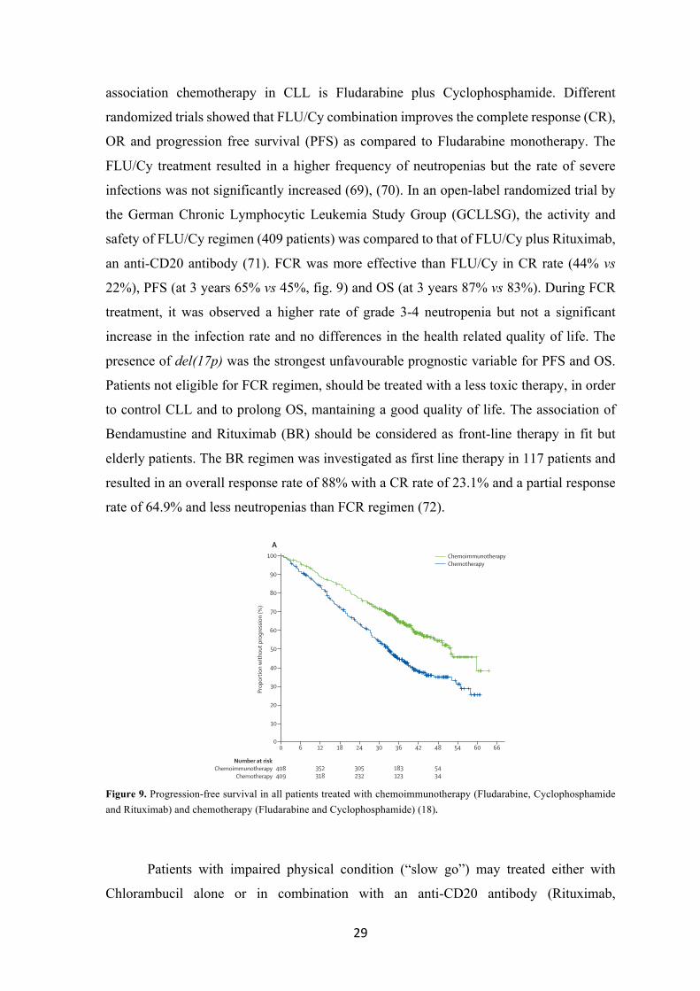
an anti-CD20 antibody (71). FCR was more effective than FLU/Cy in CR rate (44% vs
22%), PFS (at 3 years 65% vs 45%, fig. 9) and OS (at 3 years 87% vs 83%). During FCR
treatment, it was observed a higher rate of grade 3-4 neutropenia but not a significant
increase in the infection rate and no differences in the health related quality of life. The
presence of del(17p) was the strongest unfavourable prognostic variable for PFS and OS.
Patients not eligible for FCR regimen, should be treated with a less toxic therapy, in order
to control CLL and to prolong OS, mantaining a good quality of life. The association of
Bendamustine and Rituximab (BR) should be considered as front-line therapy in fit but
elderly patients. The BR regimen was investigated as first line therapy in 117 patients and
resulted in an overall response rate of 88% with a CR rate of 23.1% and a partial response
rate of 64.9% and less neutropenias than FCR regimen (72).
Figure 9. Progression-free survival in all patients treated with chemoimmunotherapy (Fludarabine, Cyclophosphamide and Rituximab) and chemotherapy (Fludarabine and Cyclophosphamide) (18).
Patients with impaired physical condition (“slow go”) may treated either with
Chlorambucil alone or in combination with an anti-CD20 antibody (Rituximab,
Articles
1168 www.thelancet.com Vol 376 October 2, 2010
(fl udarabine, p=0·6; cyclophosphamide, p=0·8). Patients in Binet stages A and B received more treatment courses (mean 5·28 [range 0–6]) than did those in Binet stage C (4·52 [0–6]; p<0·0001). For any of the three drugs, the planned dose was reduced by more than 10% in 189 (47%) of 404 patients in the chemoimmunotherapy group and 108 (27%) of 396 patients in the chemotherapy group (p<0·0001). 207 of 800 patients had dose reductions (>10%) during the fi rst to third courses
(chemotherapy 74 [19%] of 396; chemoimmunotherapy 133 [33%] of 404; p<0·0001), and dose reductions occurred in 216 of 800 patients during the fourth to sixth courses (chemotherapy 79 [20%] of 396; chemoimmunotherapy 137 [34%] of 404; p<0·0001). These dose reductions were mostly because of treatment-related haematological toxicity, particularly neutropenia and leucocytopenia (117 [62%] of 189 patients in the chemoimmunotherapy group vs 69 [64%] of 108 in the chemotherapy group; webappendix p 2).
Signifi cantly more patients were in complete remission in the chemoimmunotherapy group than in the chemotherapy group (table 2). More patients responded to treatment and more achieved a complete remission in all Binet stages (table 2). The proportion of patients who did not respond to treatment was lower in the chemoimmunotherapy group than in the chemotherapy group (39 [10%] vs 81 [20%]; p<0·0001).
PFS was longer in the chemoimmunotherapy group than in the chemotherapy group (median 51·8 months [95% CI 46·2–57·6] vs 32·8 months [29·6–36·0]; p<0·0001; fi gure 2A). At 3 years after randomisation, more patients remained progression free in the chemoimmunotherapy group than in the chemotherapy group (fi gure 2A; table 3). The risk of progression was reduced by 44% in the chemoimmunotherapy group compared with the chemotherapy group (HR 0·56 [95% CI 0·46–0·69]). An improvement in PFS was noted in all stages. Patients with disease in Binet stages B and C showed similar median PFS of 32·5 months (28·4–36·6) and 33·0 months (25·0–41·2), respectively, when treated with chemotherapy (fi gure 2B). Treatment with chemoimmunotherapy improved the median PFS to 51·8 months (47·8–56·0) in 522 patients with Binet stage B disease (HR 0·50 [95% CI 0·39–0·65]; p<0·0001) and to 40·7 months (0·73 [0·51–1·04]; p=0·081) in 252 patients with Binet stage C disease (fi gure 2B). Patients with Binet stage C disease who were given chemoimmunotherapy compared with those who were given chemotherapy showed an accumulation of several unfavourable factors (all p>0·05): age 65 years or older (44 [35%] of 126 vs 33 [26%] of 126), unmutated IGHV status (53 [54%] of 99 vs 45 [48%] of 94), elevated ZAP70 concentrations (19 [42%] of 45 vs 15 [33%] of 45), and β2 microglobulin concentrations greater than 3·5 mg/L (47 [52%] of 91 vs 37 [44%] of 84). Dose reductions of more than 10% were more common in patients with Binet stage C disease in the chemoimmunotherapy group (34 [28%] of 122 vs 61 [49%] of 124; p=0·001), mostly because of neutropenia and leucocytopenia (21 [55%] of 38 vs 42 [62%] of 68). At 3 years after randomisation, fewer patients with Binet stage B disease and Binet stage C disease in the chemotherapy group than in the chemoimmunotherapy group were progression free (table 3). The small number of patients (n=40) in Binet stage A did not allow a meaningful analysis of this subgroup, but a non-signifi cant
Figure 2: Progression-free survival in all patients (A) and in patients with Binet stage B and C chronic lymphocytic leukaemia (B)Chemoimmunotherapy=fl udarabine, cyclophosphamide, and rituximab. Chemotherapy=fl udarabine and cyclophosphamide.
Number at riskChemoimmunotherapy
Chemotherapy
0 6 12 18 24 30 36 42 48 54 60 66
352318
305232
183123
5434
408409
0
10
20
30
40
50
60
70
80
90
100
Prop
ortio
n w
ithou
t pro
gres
sion
(%)
A
0 6 12 18 24 30 36 42 48 54 60 66Time since randomisation (months)
0
10
20
30
40
50
60
70
80
90
100
Prop
ortio
n w
ithou
t pro
gres
sion
(%)
B
ChemoimmunotherapyChemotherapy
Binet B/chemoimmunotherapyBinet C/chemoimmunotherapyBinet C/chemotherapyBinet B/chemotherapy

30
Ofatumumab or Obinutuzumab), or with a dose-reduced Fludarabine containing regimen
with a CD20 antibody. In these patients the main goal of the treatment is to control
symptoms; nevertheless, the combination of Chlorambucil plus an anti-CD20 antibody
prolongs the PFS when compared with monotherapy (73). Patients with active disease and
the presence of adverse biological prognostic factors, like del(17p) or TP53 mutations, as
first line treatment, should receive FCR or an alemtuzumab-containing therapy. The
response is generally poor and short-lived; all yield response rates above 50%. In these
patients, if possible, it should be considered the treatment with novel inhibitors (Ibrutinib,
Idelalisib), the enrollment in clinical trials with new drugs or an allogeneic stem cell
transplantation (HSCT) (6) (74). Manteinance therapy in CLL cannot be generally
recommended, except for clinical trials.
4.2 Second-line treatment
As for the first-line treatment, therapy in relapsed patients should be starting only
in the presence of active and symptomatic disease. First-line treatment is repeated if the
relapsed or the progression occurs more than 24-36 months after the first therapy. If relapse
occurs within 24-36 months after first-line therapy, or the disease is refractory to any
previous treatment, the choice of therapy should be changed with other
chemoimmunotherapy combinations. Whenever possible, refractory patients should be
treated with newly approved drugs, like kinase inhibitors (Idelalisib or Ibrutinib),
Lenalidomide or enrolled in clinical trials with other new compounds (75), (76). In fit
patient with early relapse from chemoimmunotherapy and/or del(17p) or TP53 mutation
should be considered also an HSCT (77). Less fit patients could be treated with BCR
inhibitors or, if it is not present del(17p) or TP53 mutation, BR and FCR-Lite regimens
(Fig. 6).
4.3 New drugs for CLL treatment
In recent years, the CLL treatment has undergone a major innovation due to the
increasing number of very hopeful new drugs. The two main classes of novel agents are the
BCR signaling inhibitors (78) and the Bcl-2 antagonist (79) (Fig. 10); these drugs are orally

31
bioavailable and demonstrated a good efficacy and tolerability compared with conventional
chemoimmunotherapy. Furthermore, these drugs showed activity also in CLL patients with
del(17p) or TP53 mutation.
Figure 10. Survival signaling in CLL. Targeting of the BCR as a therapeutic strategy in CLL. Red symbols and letters indicate new drugs (6).
4.3.1 BCR signaling inhibitors
BCR signaling plays an important role in the development, survival, proliferation,
functional differentiation and migration of B cells. PI3Kδ, Syk and Btk are essential for
BCR signal transduction and their knockout in mouse models leads to impaired antigen-
driven maturation and expansion of B cells. In the last decade, an increasing number of B
cell malignancies (lymphomas and CLL) were ascribed on BCR signaling for proliferation
and survival (80). PI3Ks are divided into three classes and class I is composed by four
different isoforms (α, β, γ and δ). PI3Ks regulate several cell functions, including survival,
migration, chemokine receptor and integrin signaling activation. The predominant form
expressed by hematopoietic cell is PI3Kδ, harvesting a critical role in B cell homeostasis
and function. Syk activates signaling pathways downstream of the BCR, chemokine and
integrin receptor, suggesting the involvment in tissue homing and retention of activated B
cells (81). Btk is a non-receptor tyrosine kinase of the Tec family, rapidly activated by Lyn
was 60%, including complete response of 16%. Medianprogression-free survival in all patients was 13.6 months[106]. The most relevant treatment-related side effects iereviral infections. All patients had no changes in NK- or T-cellcounts.
Dasatinib. Dasatinib is a Src- and Abl- kinase inhibitorthat induces apoptosis in primary CLL cells [122]. In addi-tion Dasatinib seems to increase the apoptotic effects ofvarious agents like fludarabine, chlorambucil, sorafenib, theHSP90 inhibitor 17-DMAG, dexamethasone, or the BH3-mimetic ABT-737 [122–127]. In a Phase II study, 6 of 15patients in a Phase II study showed nodal remissions lack-ing a decrease of more than 50% in lymphocyte count,only 2 patients showed a partial remission [128]. In sum-mary, dasatinib seems effective in reduction of nodulartumor masses, but seems to lack efficacy on peripheralblood lymphocytes.
Bcl-2 inhibitors. Proteins in the B cell CLL/lymphoma 2(Bcl-2) family are key regulators of the apoptotic process[129]. The Bcl-2 family comprises proapoptotic and prosur-vival proteins. Shifting the balance toward the latter is anestablished mechanism whereby cancer cells evade apo-ptosis. Bcl-2, the founding member of this protein family, isencoded by the BCL2 gene which was initially described infollicular lymphoma as a protein in translocations involvingchromosomes 14 and 18 [130].
The Bcl-2 inhibitor ABT-263 (Navitoclax) and ABT-199. ABT-263 is a small molecule Bcl-2 family proteininhibitor that binds with high affinity (Ki! 1 nM) to multipleanti-apoptotic Bcl-2 family proteins including Bcl-XL, Bcl-2,Bcl-w, as well as Bcl-B and has a high oral bioavailability[131]. Initial studies showed very promising results for this
drug as a single agent [96]. However, its therapeutic useseemed somewhat limited by severe thrombocytopeniasbeing a prominent side effect. Therefore, the compoundwas re-engineered to create a highly potent, orally bioavail-able and Bcl-2-selective inhibitor, ABT-199 [101]. This com-pound inhibits the growth of BCL-2 dependent tumors invivo and spares human platelets. A single dose of ABT-199in three patients with refractory chronic lymphocytic leuke-mia resulted in tumor lysis within 24 hr [101]. Together,these data indicate that selective pharmacological inhibitionof BCL-2 shows promise for the treatment of BCL-2-dependent hematological cancers, including CLL.
The BH3-mimetic AT-101. AT101 is an orally activeBH3-mimetic, which inhibits the anti-apoptotic activity ofBcl-2, Bcl-XL and Mcl-1 and might be an active agent forthe treatment of CLL, as the resistance to apoptosis in CLLcells is associated with high levels of Bcl-2 protein expres-sion. AT101 was found to induce apoptosis in CLL cells invitro and to overcome drug resistance mediated by themicroenvironment [132]. It showed a good tolerability andsatisfactory efficacy in combination with weekly infusions ofrituximab in previously treated CLL patients [133,134].
Immunomodulatory drugsLenalidomide is a second generation thalidomide ana-
logue and an immunomodulatory agent with antiangiogenicproperties that is used in treatment of myelodysplastic syn-drome and multiple myeloma and is currently investigatedin the treatment of CLL. It showed encouraging results inthe treatment of high risk patients including carriers of adel(17p) [135]. In 58% of the patients lenalidomide causesa so called tumor flare reaction, which leads to a sensationof heat and burning in the lymph nodes and occurs only in
Figure 2. Targeting of the BCR signaling as a therapeutic strategy in CLL. Red symbols and letters indicate new therapeutics as discussed in the text. [Color figure canbe viewed in the online issue, which is available at wileyonlinelibrary.com.]
annual clinical updates in hematological malignancies
American Journal of Hematology 809

32
and Syk kinases, resulting in the activation of NF-κB signaling, B cell proliferation and
differentiation. It is essential for activation of several constitutively active pathways of CLL
cell survival, including Akt and the ERK (82). Btk is also involved in regulation of
migration and adhesion via CXCR4/CXCR5 and integrin signaling (83). Given the
importance of BCR receptor signaling in CLL, an attractive strategy is to target inhibition
of this kinase.
Idelalisib: Idelalisib (CAL-101) is an oral PI3Kδ selective inhibitor promoting CLL cells
apoptosis in a time and dose-dependent mode without inducing apoptosis in normal T cells
or natural killer cells. Idelalisib reduces survival signals derived from the BCR, inhibits
CLL cell chemotaxis and migration and also down-regulates secretion of chemokines in
stromal co-cultures and after BCR triggering (84). Idelalisib was approved by the FDA in
2014 for the treatment of relapsed/refractory CLL patients in combination with rituximab.
Idelalisib has been tested as single agent or in combination with other conventional drugs
and demonstrated excellent efficacy and tolerability. Idelalisib pivotal phase III study was
conducted in heavily pretreated CLL patients; 220 patients were randomly assigned to
receive rituximab/placebo or rituximab/idelalisib. The study resulted in an 85% reduction
of the risk progression with a 12-months PFS of 66% in Idelalisib/Rituximab arm,
compared with 13% for the placebo/Rituximab arm. PFS and response rates were not
affected by adverse prognostic factors, including del del(17p)/TP53mut, ZAP70 expression
or IGVH mutational status (85).
Fostamatinib: Fostamatinib disodium is the first clinically available oral Syk-inhibitor; it
induces apoptosis disrupting B cell receptor signaling. Fostamatinib induced partial
responses in replapsed CLL patients in phase I/II study (86).
Ibrutinib: Ibrutinib, previously called PCI-32765, is the first BCR inhibitor approved for
treatment in CLL; it is a small orally active molecule that inhibits Btk, that plays a role in
the signal transduction of the BCR, inducing apoptosis in CLL cells. Ibrutinib covalent
binds to the cysteine-481 amino acid of the Btk enzyme; preclinical studies showed that it
inhibits numerous processes, including ERK signaling, NF-κB DNA binding, cytosine-
phosphate-guanine (CpG)-mediated CLL-cell proliferation and tumor cell migration.
Differently from most regimens used for CLL, Ibrutinib does not have toxic effects on
normal T cells (87), (88). Previous studies fixed a dose of 420mg/day; patients with
relapsed/refractory disease experienced a 90% ORR, with durable responses and an

33
estimated PFS of 69% at 30 months. The response was independent of clinical stage,
previous therapies and adverse genomic risk factors, including 17p deletion (Fig. 11) (89).
In the study of previously untreated patients over 65 years, the ORR was 84%, with an
estimated PFS of 96% at 30 months. Treatment with Ibrutinib was generally well tolerated,
with the most common adverse events being transient diarrhea (58%), fatigue (28%),
infections (32%) and bleeding (61%). Ibrutinib usually causes a transient increase in blood
lymphocyte levels, concurrent with a riduction in lymph node and speen size. This effect
has been seen also with other agents targeting BCR and it is not consider a sign of
progressive disease; continuing Ibrutinib therapy, this asymptomatic lymphocytosis is led
to resolution (90). The optimal duration of therapy has not been yet determined, it could be
resonable to continue treatment but we do not know yet the long-term effects and the
resistance mechanisms need to be better clarified.
Figure 11. Kaplan-Meier curves for PFS for 85 relapsed/refractory CLL patients, treated with Ibrutinib (left panel) and PFS according to cytogenetic status, with respect to the del(17p) and del(11q) deletions (right panel) (89).
Dasatinib: Dasatinib is a Src- and Abl- kinase inhibitor that induces apoptosis in CLL cells.
Dasatinib seems also to increase the apoptotic effects of Fludarabine, Chlorambucil,
dexamethasone and other agents (91). In previous studies, Dasatinib showed efficacy in
reduction of nodular tumor masses but it is less effective on peripheral blood lymphocytes.
4.3.2 Bcl-2 inhibitors
Proteins in the B cell lymphoma 2 (Bcl-2) family work as key regulators of the cell
apoptosis. Bcl-2 family consists in pro-apoptotic and pro-survival proteins; cancer cells are
able to evade apoptosis shifting the balance of Bcl-2 proteins toward the prosurvival effect
(92).
T h e n e w e ngl a nd j o u r na l o f m e dic i n e
n engl j med 369;1 nejm.org july 4, 201340
umab in Patients with Relapsed or Refractory Chronic Lymphocytic Leukemia (RESONATE; ClinicalTrials.gov number, NCT01578707) and A Multicenter, Open-label, Phase 3 Study of the Bruton’s Tyrosine Kinase Inhibitor PCI-32765 Versus Chlorambucil in Patients 65 Years or Older with Treatment-naive Chronic Lympho-cytic Leukemia or Small Lymphocytic Leukemia (RESONATE-2, NCT01722487).
Several validated high-risk characteristics of CLL, including 17p13.1 deletion, did not influ-ence the objective response to ibrutinib. How-ever, most events associated with disease pro-gression occurred in patients with high-risk cytogenetic lesions (17p13.1 deletion or 11q22.3
deletion), whereas only one patient without these risk factors had an event associated with disease progression. Patients with an unmutated immu-noglobulin variable-region heavy-chain gene, perhaps owing to enhanced B-cell–receptor sig-naling and dependence on this pathway, had earlier resolution of lymphocytosis and were more frequently classified as having a response according to traditional response criteria of the International Workshop on Chronic Lympho-cytic Leukemia (P = 0.02); however, survival out-comes were similar to those among patients without this unmutated gene. Greater than 90% occupancy of the pharmacodynamic probe and the similar response in the two dose groups
Pro
babi
lity
of P
rogr
essi
on-f
ree
Su
rviv
al
1.0
0.6
0.8
0.4
0.2
0.00 5 10 15 20 25 30
Pro
babi
lity
of O
vera
llS
urv
ival
1.0
0.6
0.8
0.4
0.2
0.00 5 10 15 20 25 30
Pro
babi
lity
of P
rogr
essi
on-f
ree
Su
rviv
al
1.0
0.6
0.8
0.4
0.2
0.00 5 10 15 20 25 30
Pro
babi
lity
of O
vera
llS
urv
ival
1.0
0.6
0.8
0.4
0.2
0.00 5 10 15 20 25 30
Pro
babi
lity
of P
rogr
essi
on-f
ree
Su
rviv
al
1.0
0.6
0.8
0.4
0.2
0.00 5 10 15 20 25 30
Pro
babi
lity
of O
vera
llS
urv
ival
1.0
0.6
0.8
0.4
0.2
0.00 5 10 15 20 25 30
Months on Study Months on Study
B Overall SurvivalA Progression-free survival
17p deletion (n=28)
11q deletion (n=23)
No 17p or 11q deletions (n=29)
17p deletion (n=28)
11q deletion (n=23)
No 17p or 11q deletions (n=29)
All patientsAll patients
Mutated IGHV (n=12)
Unmutated IGHV (n=69)Mutated IGHV (n=12)
Unmutated IGHV (n=69)
P=0.04 by log-rank test
P=0.67 by log-rank test
P=0.15 by log-rank test
P=0.86 by log-rank test
Figure 3. Kaplan–Meier Curves for Progression-free Survival and Overall Survival.
Panels A and B show the probability of progression-free survival and overall survival, respectively, for all 85 patients (top graphs) and according to status with respect to the 17p13.1 and 11q22.3 deletions (middle graphs) and IGHV mutation status (bottom graphs). Tick marks indicate censored data.
The New England Journal of Medicine Downloaded from nejm.org on December 12, 2015. For personal use only. No other uses without permission.
Copyright © 2013 Massachusetts Medical Society. All rights reserved.

34
ABT199: ABT199 is a highly potent, orally bioavailable and Bcl-2 selective inhibitor that
blocks the growth of Bcl-2 dependent tumors in vivo. ABT199 showed a promising effect
for the treatment of Bcl-2 dependent hematological cancers, including CLL (93).

35
AIM OF THE STUDY
CLL is a malignancy characterized by the accumulation of monoclonal mature B
lymphocytes which are dependent on interactions with the tissue microenvironment for
their survival and proliferation. Consequently, exploring and targeting the CLL
microenvironment is of progressive increasing interest, also for the development of novel
therapeutic strategies and for a better understanding of drug-resistance mechanisms. We
focused our attention on MSCs derived from the bone marrow, which provide attachment
site and growth factors for normal hematopoiesis and represent a critical component of the
CLL microenvironment.
The aim of this PhD project was to further investigate the role of MSCs in CLL
pathogenesis and in the treatment of the disease. In particular, we planned to establish an
in vitro culture system based on MSCs, shaping in vivo conditions for determining CLL
niche interactions involved in neoplastic cell survival. Employing an in vitro system that
closely approximate the in vivo bone marrow conditions, we tried to better explain the
connection between CLL B cells and the microenvironment with the ultimate goal to
identify patients who may benefit from compounds targeting CLL microenvironment.
Finally, we studied the role of MSCs in the apoptosis of CLL cells during a conventional
therapy in general practice, such the combination of Fludarabine and Cyclophosphamide,
performing both in vivo and in vitro experiments. Furthermore, we analyzed the complexity
of the cross-talk between malignant B cells and MSCs in the presence of a Btk inhibitor,
Ibrutinib, actually considered a really promising and efficient drug in the treatment of CLL.
In particular, we planned to examine CLL cells survival and ability of migration and
adhesion after the Ibrutinib pre-treatment, in the presence of MSCs.

36

37
MATHERIAL AND METHODS
1. Patients
MSCs were isolated from 46 CLL patients (31 males and 15 females, median age
60 years) referred to the Hematology and Clinical Immunology division of Padua
University Hospital. The main disease characteristics of the patients are shown in table III,
which reports the RAI classification, the percentage of bone marrow infiltration and the
quote of CD19/5+ lymphocytes. In particular, 19 patients were classified in stage RAI 0,
15 in stage 1, 10 in stage 2 and 2 patients in stage 3. None of the patients were in stage 4.
The mean bone marrow infiltration was 47.4±22.6 and the amount of the CLL B cells in
peripheral blood ranged from 1 to 98%.
Malignant B lymphocytes utilized in this study were obtained from 45 CLL patients
(27 males, 18 females, median age 65 years) which clinical characteristics are listed in table
IV. According to RAI system, 4 patients were classified in stage 4 and 7 in stage 2. Overall
31 patients (69%) were in stage RAI 0 and 1; staging of 3 patients was not determined (nd).
Cytogenetic most common lesions were identified using interphase FISH: deletion in the
long arm of chromosome 13 (13q-), trisomy of chromosome 12 (12+), deletion in the long
arm of chromosome 11 (11q-) and in the short arm of chromosome 17 (17p-) or a normal
karyotype (N). Based on the number of SHM of the Ig VH genes, the cases were divided
into two categories: 21 unmutated (UM) and 20 mutated (M). Conventionally, mutated
status was defined as having a frequency of mutations greater than 2% from germline VH. In 4 patients SHM status was not available (na) or not determined. Using cytofluorimetric
analysis, we detected also ZAP70 and CD38 expression. The cut-off used to determine
ZAP70 was 20% and 30% for CD38.

38
Table III. Clinical characteristics of CLL patients studied for MSCs isolation.
MSCs # RAI stage Age Sex BM infiltration (% Ly) CD19/5(%)
MSC#1 0 75 M 34 71 MSC#2 1 65 M 70 92 MSC#3 1 78 M 87 92 MSC#4 0 66 F 70 83 MSC#5 1 70 M 90 98 MSC#6 3 75 M 40 68 MSC#7 0 50 F 18 1 MSC#8 0 49 M 56 72 MSC#9 2 60 F 5 2
MSC#10 2 72 M 75 46 MSC#11 1 68 M 13 11 MSC#12 2 61 M 26 54 MSC#13 2 55 M 6 8 MSC#14 0 51 M 54 86 MSC#15 2 47 M 78 86 MSC#16 2 59 F 19 34 MSC#17 0 66 M 40 75 MSC#18 0 54 M 42 68 MSC#19 0 51 M 27 40 MSC#20 1 48 M 47 83 MSC#21 1 55 M 52 70 MSC#22 1 55 M 18 60 MSC#23 0 49 M 62 81 MSC#24 1 69 M 56 50 MSC#25 0 51 M 37 54 MSC#26 1 66 M 64 70 MSC#27 1 60 F 43 56 MSC#28 0 60 M 37 54 MSC#29 1 70 F 70 50 MSC#30 1 63 M 43 70 MSC#31 0 63 M 55 67 MSC#32 1 60 M 40 43 MSC#33 0 56 F 52 45 MSC#34 0 71 F 25 22 MSC#35 0 49 F 62 69 MSC#36 0 60 M 75 85 MSC#37 0 58 F 60 43 MSC#38 3 59 F 50 77 MSC#39 2 72 F 75 85 MSC#40 2 55 M 17 35 MSC#41 2 63 M 90 93 MSC#42 0 56 F 32 50 MSC#43 1 57 M 34 91 MSC#44 0 52 M 21 44 MSC#45 2 67 F 73 81 MSC#46 1 56 F 40 48

39
Table IV. Clinical characteristics of CLL patients
CLL# Age
Sex RAI stage Cytogenetic IgVH mutational
status (M/UM) ZAP70 CD38
#01 64 M 0 na M pos neg #02 74 M 1 na M neg neg #03 56 F 2 13q- M nd nd #04 71 M 4 13q- UM pos pos #05 54 F 1 na M nd pos #06 68 F 1 11q- 12+ UM pos nd #07 79 F 1 13q- UM neg pos #08 89 M 2 na M nd nd #09 67 F 2 17p- 13q- nd nd nd #10 77 M 0 13q- M neg neg #11 69 M 0 N UM pos neg #12 59 M 0 13q- UM neg neg #13 45 M 1 13q- M pos neg #14 52 F 2 na UM pos pos #15 63 F 0 13q- M pos neg #16 81 M 1 12+ UM pos pos #17 92 F 1 17p- UM neg nd #18 45 M 4 17p- UM neg pos #19 63 F 1 13q- M nd nd #20 51 M 0 17p- 13q- M neg neg #21 79 M 1 13q- M neg neg #22 49 M 1 11q- 13q- 12+ UM pos pos #23 55 F 1 na M na na #24 61 M 1 11q- 12q- UM pos neg #25 59 M 0 N UM pos neg #26 66 M 1 N M pos neg #27 57 F 0 13q- UM pos neg #28 71 F 1 13q- M neg pos #29 50 M 2 13q- UM pos pos #30 67 M 0 13q- M pos pos #31 81 M 4 na M pos neg #32 53 M 4 11q- UM pos neg #33 85 M 0 N UM pos neg #34 62 M 0 N M pos nd #35 54 M 1 13q- M pos neg #36 61 F 0 11q- 13q- 12+ UM pos pos #37 54 F 1 11q- 12+ nd pos pos #38 67 F 0 nd UM nd nd #39 80 M 1 13q- M pos neg #40 69 F 2 11q- 13q- UM neg pos #41 66 F 1 13q- M neg neg #42 78 F nd 13q- M neg neg #43 71 M nd 12+ na neg neg #44 53 M 2 11q- UM neg pos #45 49 M nd na na na pos

40
2. Isolation of MSCs from CLL bone marrow
Bone marrow blood samples were obtained from 46 CLL patients, after written
informed consent. We collected 1-2 ml of blood from each bone marrow aspirate;
mononuclear cells, including MSCs, were obtained proceeding with a layering on
Ficoll/Hypaque (F/H, GE Healthcare; Fairfield, Connecticut). This technique is based on
the difference of density of mononuclear cells (lymphocytes and monocytes) with respect
to the other blood elements. Mononuclear cells, with lower density, focus on the layer of
F/H while the red blood cells and granulocytes are collected on the bottom of the tube. We
proceeded with a centrifugation at 900g for 20 min at 20°C, without brake. The ring of
mononuclear cells at F/H interface was aspirated and washed twice with saline by
centrifugation at 400g for 10 minutes at 20°C (Fig. 12). The pellet was resuspended in an
appropriate amount of saline and the cells were counted in a Burker chamber.
Figure 12. Isolation of MSCs from bone marrow by stratification on Ficoll/Hipaque. By centrifugation on F/H, MSCs were isolated from bone marrow of CLL patients. Mononuclear cells (PBMC) and platelets were concentrated above the layer of F/H because they have lower density; on the contrary, the red blood cell (RBC) and polymorphonuclear neutrophil (PMN) have a higher density than the F/H and collect on the bottom of the tube. RT: room temperature.
3. Ex vivo expansion of MSCs
Mononuclear cells were cultured in 7 ml of Dulbecco’s Modified Eagle Medium
(DMEM, Invitrogen; Paisely, UK) with 10% of FBS, 1% of PenStrep (Penicilline
5000U/ml, Streptomicine 5,000µg/ml, Invitrogen) and 10µg/ml of Ciprofloxacine
(Ciproxin, Bayer; Leverkusen, Germany) at 37°C in humidified atmosphere containing 5%
CO2. Nonadherent cells were removed carefully after 7 days and fresh medium was
replaced. When primary cultures became almost confluent, the culture was washed with 2
ml of Phosphate Buffer Saline (PBS)1X and treated with 1 ml of 0.25% trypsin (Invitrogen)

41
in order to enlarge the culture. Detached cells were transfered in a medium with 10% of
FBS and resuspended in DMEM to be seeded in plates with a 10.000 cells/cm2 density.
4. Immunophenotyping of MSCs
The immunophenotypic analysis was performed through flow cytometry, a
technique allowing a multiparametric evaluation of antigenic characteristic of the single
cells by the analysis of visible and fluorescent light emission. The immunophenotyping
helps to identify surface and intracellular Ag using mAb conjugated with fluorochromes.
The presence of a certain Ag is used as an indicator of cell lineage and maturation level.
We used flow cytometer FACS Calibur (Becton Dickinson; Milan, Italy) and data obtained
were processed using the program Cell Quest. For each analysis 20.000 events were
acquired. The fluorochromes used were fluorescein isothiocyanate (FITC), which emits a
fluorescence signal at 530nm (green), phycoerythrin (PE) emitting at 585nm, tri-color (TC)
that emits at 667nm when hit by a monochromatic laser beam with λ equal to 488nm, and
allophycocyanin (APC) that emits a fluorescence signal at 690nm when excited by a laser
beam with λ of 635nm. MSCs were removed from plates using 500µl of Accutase solution
(Sigma; Saint Louis, USA) and resuspended in 100µl of PBS1X. MSCs were stained with
5µl of mAb for surface expression of CD14 (FITC, Becton Dickinson; Franklin Lakes,
USA), CD31 (FITC, Becton Dickinson), CD34 (PE, Becton Dickinson), CD90 (Abd
Serotech, Oxford, UK), CD73 (PE, Becton Dickinson) and with 2.5µl of mAb for
expression of CD45 (FITC, Caltag-Invitrogen, Paisely, UK) and CD105 (PE, Caltag-
Invitrogen).
5. Isolation of CLL B cells from peripheral blood samples
B lymphocytes were isolated from peripheral blood of CLL patients. From a sample
of heparinized venous blood, mononuclear cells were obtained proceeding with a layering
on F/H. In patients with a quote of B lymphocytes less than 90% of peripheral blood
mononuclear cells (PBMCs) isolated we used the RosetteSep kit (StemCell Technologies;
Vancouver, Canada). The kit consists of a cocktail of antibodies directed against surface

42
antigens, expressed by hematopoietic cells (CD2, CD3, CD16, CD36, CD56, CD66b) and
glycophorin A, expressed by red blood cells. This mixture of antibodies binds "not-B" cells
and red blood cells creating immunorosette. CD19+ B lymphocytes are isolated from whole
blood by negative selection. Each ml of blood was incubated with 50µl of RosetteSep at
room temperature for 20 minutes. The samples were then diluted 1:2 with PBS1X + 2%
FBS, gently agitated, and then layered over F/H. We proceeded with a centrifugation at
900g for 30 minutes at RT, followed by the aspiration of the ring formed at the F/H interface
containing B cells. It was resuspended in PBS1X + 2% FBS and centrifuged at 400g for 10
minutes (Fig. 13). Finally, cells resuspended in PBS1X were counted in a Burker chamber.
We used the RosetteSep kit also to isolate B lymphocytes from buffy coat, used as normal
controls.
Figure 13. Purification with RosetteSep kit. CD19+ B lymphocytes were isolated from whole blood of healthy donors by negative selection. 10ml of venous whole blood were incubated for 20 min at RT with 500µl of RosetteSep. Afterwards, through stratification on F/H, we get the CD19+ B cells, which are concentrated just above the layer of F/H, while the rest of the cells related to the rosettes were collected on the bottom of the tube.
6. Culture conditions
For co-culture experiments, purified leukemic and normal B cells (2x10⁶/ml) and
MSCs (1x10⁵/well seeded into 12 well plates) were cultured in complete RPMI-1640
(Sigma-Aldrich; Milan, Italy) at 37°C in a humidified atmosphere containing 5% CO2. B
cells were added to MSCs layer at 20:1 ratio in complete RPMI-1640 medium. For
spontaneous apoptosis assays, leukemic cells from the suspension were collected at 3, 5
and 7 days. Therefore, we performed co-cultures of malignant B lymphocytes in direct
contact with MSCs and in presence of MSCs separated by a 0.4µm porous polycarbonate
filter (transwell) that allows the exchange of soluble factors.
For analysis of in vitro drug-induced cell death, CLL B cells were treated with
20µM Fludarabine (FLU, TEVA; Milan, Italy) and 5mM Cyclophosphamide (Cy, Baxter;
Rome, Italy) for 3 and 12 hours; cells were then washed and plated alone and with MSCs
for 3, 5 and 7 days. In other experiments, FLU and Cy were added directly in CLL B cells

43
and CLL B cells-MSCs co-cultures. At the same time, we cultured MSCs with leukemic B
cells collected from 10 CLL patients undergoing the first cycle of FLU/Cy chemotherapy
in vivo, before starting drug infusion and at the third day of treatment. Viability status was
examined by staining with Annexin V at 3, 5 and 7 days. At least, we tested the effect of
5µM Ibrutinib, a Btk inhibitor, on CLL B cells isolated from 12 patients cultured alone and
with MSCs for 3, 5 and 7 days.
7. Analysis by flow cytometry
Apoptosis of different cell samples (CLL B cells and normal B lymphocytes) was
assessed using the Annexin V Apoptosis Detection Kit (Immunostep; Salamanca, Spain).
During the early stages of apoptosis, the plasma membrane undergoes profound changes
indicating the status of apoptotic cell to macrophages, which ensure its elimination.
Phosphatidylserine (PS), a negatively charged aminofosfolipide expressed normally only
on the inner side of the plasma membrane, become exposed on the outer surface. Annexin
V is a protein that, in the presence of high concentrations of Ca2+, recognizes and selectively
binds PS, helping to identify apoptotic cells by phospholipid exposure on their surface.
Aliquots of 5x10⁵ cells were harvested, washed, and incubated for 10 min in the
dark and at RT with anti-CD19 APC (Invitrogen). Then cells were washed and 100µl of
binding buffer, a Ca2+-rich solution that optimizes the binding of Annexin V to the PS, plus
5µl of Annexin V-FITC were added for further 10 minutes in the dark and at RT. After the
incubation, 100µl of binding buffer were added and cells were analyzed by flow cytometer
FACS Canto (Becton Dickinson). For each sample 20.000 events were collected using
FACS Diva software.
8. Polyacrylamide gel electrophoresis in SDS (SDS-PAGE)
The polyacrylamide gel electrophoresis in SDS is one of the methods used to
separate a mixture of proteins on the basis of their molecular weight. SDS is a ionic
detergent that binds tightly to proteins causing their denaturation. In the presence of an
excess of SDS, approximately 1.4 grams of detergent will bind to each gram of protein,
providing a constant amount of negative charge per unit mass. Therefore, during

44
electrophoresis, all protein-SDS complexes move toward the anode, and thanks to the
molecular sieve properties of the gel, their mobility is inversely proportional to their
molecular weight. By the migration of standard proteins of known molecular weight
simultaneously to samples, it is possible to determine the protein sample weights.
SDS polyacrylamide gel was prepared following Laemmli method96. The electrophoretic
plate consisted of two types of gel:
- Stacking gel at pH 6.8, which concentrates the protein samples so that they are all aligned
at the start of electrophoresis;
- Running gel at pH 8.8, in which the real separation of proteins occurs. The plate size of
10×8cm is fixed in the Hoefer Mighty Small-If 250 Scientific Instruments machine
(Amersham Biosciences). The electrophoresis was run for about 2 hours at 25mA.
9. Western Blotting
The western blotting (WB) or immunoblotting is an immunoassay able to detect
traces of a specific protein in a heterogeneous mixture, combining the high resolving power
of gel electrophoresis with the specificity of the antibodies. The WB is a high sensitive
technique, able to detect very small quantities of proteins. After SDS-PAGE, proteins were
transferred onto a nitrocellulose membrane through an electric field, obtained applying the
appropriate current of 350mA for 2 hours and 30 minutes. The buffer used for the transfer
consists of: 25mM Tris, 192mM glycine, 20% methanol and 0.1% SDS with a final pH of
8.0 (buffer A). After the transfer, the membrane was left overnight in the saturation buffer
consisting of 50mM Tris-HCl, pH 7.5, 150mM NaCl and 5% bovine serum albumin (BSA)
(buffer B), for non specific sites saturation. Follows the incubation for 2 hours and 30
minutes at room temperature of the primary Abs, diluted in: 50mM Tris-HCl, pH 7.5,
150mM NaCl, 1% BSA (buffer C).
For our study we used the following antibodies: anti-Poli-ADP-Ribose Polymerase
(PARP), anti-Btk Tyr223, anti-Btk (Cell Signaling Technology Inc; Danvers,
Massachusetts, USA) and anti-β-actin (Sigma-Aldrich).
Three washes of 10 min, each at RT were subsequently performed, using buffer C
supplemented with 0.1% Tween. Membranes were then incubated for 30 minutes with a
secondary anti-IgG Ab, obtained against the animal species immunized for the primary Ab.
The secondary Ab was conjugated with horseradish peroxidase (Amersham International

45
Biotechnology; Buckingamshire, UK) and diluted in buffer C. After three additional
washes, the membrane was subjected to the detection antibody with the enhanced
ChemiLuminescence system (ECL) (Pierce; Rockford, Illinois): the membrane was
incubated for 1 minute with 1ml of luminol and 1 ml of H2O2, which in contact with the
peroxidase and the Ag-Ab complex, give rise to an oxidation reaction with light emission.
The membrane was relevated into ImageQuant LAS 500 (Amersham). The densitometric
analysis was performed using the Image J program.
10. Chemotaxis assay
MSCs were cultured for 48h in complete RPMI-1640 medium (Sigma-Aldrich) at
37°C in a humidified atmosphere containing 5% CO2 to obtain the MSC-conditioned
medium (MSC-CM). The migration of pre-treated CLL B cells in response to MSC-CM
was evaluated using 12-well Corning chemotaxis chamber (Corning Life Sciences; Acton,
MA). 2.5x10⁶ cells were incubated in 0.5ml RPMI medium with and without Ibrutinib for
1h at 37°C. Then, cells were transferred into the top chambers of transwell culture insert
with a pore size of 3µm. Filters were then placed onto wells containing complete RPMI
medium or MSC-CM, and CLL B cells were allowed to migrate for 3h at 37°C. Migrated
cells in the lower chamber were then collected and counted on a FACSCanto for 60 seconds
in triplicates.
11. Evaluation of CLL B cell adhesion to MSC layer
CLL B cells were suspended to a concentration of 2x10⁶ cells/ml with or without
5µM Ibrutinib and incubated for 1h at 37°C in 5% CO2 in complete RPMI medium. After
incubation, CLL cells were added to the MSC layers and the plates were incubated at 37°C
in 5% CO2 overnight. Cells that had not adhered into the stromal cell layer were removed
by vigorously washing 3 times with RPMI 1640 medium. The complete removal and the
integrity of the stromal cell layer containing adherent B cells were assessed by phase-
contrast microscopy Olympus IX-81 and documented photographically. The layer of cells
was detached by incubation with Accutase (Sigma-Aldrich); cells were stained with anti-
CD19 APC to exclude MSCs and counted by flow cytometry.

46
12. Flow cytometry analysis
CLL B cells were cultured with and without MSCs and treated with 5µM Ibrutinib.
5x10⁵ cells for each assay were collected after 48h, leaving intact the adherent layer, and
stained with anti-CD49d PE (BD Biosciences), anti-CCR7 FITC (R&D Systems Inc.,
Minneapolis, MN, USA), anti-CXCR4 PE (R&D Systems Inc.), and anti-CD19 APC (BD
Biosciences) monoclonal antibodies. Cells were washed with PBS1X and incubated with
saturating concentrations of the appropriate antibodies for 15 minutes at room temperature.
20,000 total events were acquired using FACSCanto (Becton Dickinson) and the data were
analysed by FACSDiva 7 software. Samples were gated on intact cells by forward light
scatter (FSC) vs right-angle light scatter (SSC). For analysis, it was used a second gating
step on CD19+ cells. Here, we used a difference between the Mean Fluorescence Intensity
(MFI) of fully-stained samples and the Fluorescence Minus One controls.
13. Statistical analysis
Statistical analysis of apoptosis in patients analyzed was performed using Student's
t test, paired Student's t test. Data were expressed as mean±standard deviation (SD) and
were considered statistically significant when p values were <0.05.

47
RESULTS
1. MSCs isolation from bone marrow of CLL patients
MSCs were isolated from the bone marrow of 46 CLL patients afferent to the
Hematology and Clinical Immunology division of Padua University Hospital. For each
bone marrow sample, the mononuclear cells were seeded in plates with a 10,000 cells/cm2
density. After 48 hours, we were able to observe the adhesion of some round cells in the
plate; after 7 days, with the first change of the medium culture, the cells were extended, in
a similar fibroblastic morphology. After 14 days, the cells created highly proliferating
aggregates (Fig. 14A), with the achievement of the mutual confluence in 30/40 days (Fig.
14B). All the cultures maintained a homogeneous morphology and an undifferentiated
status in the different steps.
Figure 14. MSCs cultures after 14 (A) and 30 (B) days. Mononuclear cells obtained from bone marrow samples were seeded in plates with DMEM. After 7 days, some cells with a similar fibroblastic morphology were adherent to the plate; the suspended cell were removed with the first change of medium. The first proliferation aggregates were observed after 14 days and the culture obtained the confluence in 30-40 days (10X enlargement with Olympus BX60 microscope).
2. MSCs immunophenotypic characterization
The characterization of the expanded cell population was defined through flow
cytometry analysis between third and fourth subculture; the immunophenotypic
characterization was based on the expression of CD105, CD73 and CD90 and the negativity
of CD14, CD34, CD45 and CD31. We were able to identify in our cell cultures the peculiar
phenotype of MSCs, confirming that our cell population was homogeneous and not of
hematopoietic derivation (Fig. 15).
A B

48
Figure 15. Immunophenotypic characterization of MSCs. Representative case of MSC flow cytometry evaluation. The grey area represents the negative control; the white area indicates the positive expression of the marker.
3. MSCs from CLL patients support in vitro neoplastic B cells survival
We tested the effect of MSCs on the survival of leukemic B cells isolated from 30
CLL patients, and normal B cells obtained from 11 healthy controls; B lymphocytes were
incubated in direct contact with a confluent layer of MSCs (20:1 ratio). By Annexin V
staining, we assessed B cell viability at 3, 5 and 7 days. We found that leukemic B cells
underwent apoptosis when cultured in medium alone, but their survival was rescued when
cultured with MSCs; CLL B cell viability after 7 days was 13.3%±13.2% in medium alone
vs 59.2%±17.1% in co-culture with MSCs (p<0.0001) (Fig. 16A). Normal B cell viability
after 7 days was 6.7%±4.3% in medium alone vs 34.9%±15.7% when co-cultured with
MSCs (p<0.001) (Fig. 16B), indicating that MSCs display a major protective effect on
neoplastic B cells. Therefore, we confirmed that MSCs are able to support CLL B cell
survival providing an in vitro culture system that closely approximate CLL
microenvironment in vivo.
Figure 16. CLL and normal B cell viability cultured alone and with MSCs. Cell apoptosis was assessed by Annexin V test. The graph shows the percentage of cell viability after 3, 5 and 7 days, data are expressed as mean ±SD; ***p<0.001, ****p<0.0001, Student’s t-Test.
A B
CD14 CD34 CD45 CD31
CD73 CD90 CD105

49
4. Detection of PARP 89kDa fragment reveals two subsets of CLL clones
On the basis of the high variability observed in CLL B cell viability co-cultured
with MSCs (Fig. 17), we evaluated the cleavage pattern of PARP protein after 7 days of
culture in a cohort of 27 untreated CLL patients.
Figure 17. Percentage of CLL B cells viability before and after co-culture with MSCs. CLL B cells apoptosis was assessed by Annexin V test. The graph shows the percentage of cell viability of CLL B cells from different patients at 7 days; p<0.0001, Student’s t-Test.
We identified two different patterns of CLL clones with different sensitivity to
MSCs pro-survival signals: 1) one observed in a group of 15 patients in which CLL clones
displayed the cleaved PARP, but the full length protein in presence of MSCs (classified as
“dependent” from microenvironmental stimuli) (Fig. 18, left panel); 2) a second pattern
observed in a group of 12 CLL patients in which CLL clones displayed the full length
PARP with and without the presence of MSCs (classified as “independent” from
microenvironmental stimuli) (Fig. 18, right panel).
Figure 18. Analysis of PARP protein expression in CLL B cells cultured with and without MSCs. CLL B cells were cultured alone and in the presence of MSCs for 7 days. The total cell lysates were subjected to SDS-PAGE, transferred to nitrocellulose membrane and detected sequentially with anti-PARP Ab, to highlight the apoptosis, and anti-β-actin Ab. The figures show two representative cases of CLL B cells “dependent” from microenvironmental stimuli (left panel) and two cases “independent” from MSCs signals (right panel).
PARP116kDa PARP89kDa
Actin
CLL#11 +MSC#18
CLL#18 +MSC#11
“DEPENDENT”
+MSC#26 CLL#28 CLL#25
+MSC#37
“INDEPENDENT”
PARP116kDa PARP89kDa
Actin

50
We also analyzed the detection of PARP 89kDa fragment from co-cultures of the
same CLL clone with different MSCs. Interestingly, the results indicated that CLL clone
response to microenvironmental signals did not change in co-culture with MSC obtained
from different patients (Fig. 19A). Conversely, co-culturing the same MSC line with
different CLL clones, we demonstrated different patterns in the cleavage pattern of PARP
(Fig. 19B), indicating that the heterogeneity of leukemic clones is likely to be related to
intrinsic features of neoplastic B cells rather than to external stimuli.
A)
B)
Figure 19. Analysis of PARP pattern in CLL B cells cultured in absence and in presence of MSCs for 7 days. The total cell lysates were subjected to SDS-PAGE, transferred to nitrocellulose membrane and detected sequentially with anti-PARP Ab, to highlight the apoptosis, and anti-β-actin Ab. A) The figures show two representative cases of a single neoplastic clone (CLL#) cultured with MSCs (MSC#) obtained from different CLL patients; in all the conditions, leukemic B cells displayed a similar response also in the presence of diverse MSCs types. B) Representative cases of CLL B cells (CLL#) from distinct patients exposed to the effect of the same MSC line (MSC#); in this condition, CLL clones displayed different responses in the presence of the same MSCs.
PARP116kDa PARP89kDa
Actin
+MSC#17
+MSC#14
CLL#03
+MSC#15
CLL#05
+MSC#31
+MSC#11 +MSC#28
+MSC#26 CLL#22 CLL#28 CLL#34
PARP116kDa
PARP89kDa
Actin
+MSC#26 +MSC#26

51
5. Cell-cell contact and soluble factors are involved in the cross-talk between MSCs
and CLL B cells
We cultured malignant B lymphocytes from 12 CLL patients alone, in direct contact
to MSCs and in presence of MSCs separated by a 0.4µm porous polycarbonate filter
(transwell) that allows the exchange of soluble factors. We demonstrated that CLL cell
survival was increased in presence of MSCs in both conditions; in fact, after 7 days, CLL
B cell viability was 13.8%±13.4% when cultured alone vs 61.5%± 18.3% in direct contact
with MSCs (p<0.0001) and 47.5%±7.8% in presence of MSCs separated by transwell
(p<0.0001). These results indicated that both cell-cell contact and soluble molecules are
actors in the relationship between malignant B cells and the MSCs (Fig. 20).
Figure 20. Histograms of cell viability percentage of CLL B cells cultured alone, with MSCs and co-cultured with MSCs in the presence of transwell. CLL B cells apoptosis was assessed by Annexin V test. Histograms show the mean±SD of the percentage; ****p<0.0001, Student’s t-Test.
Considering that MSCs release a high amount of chemoattractants, we also tested
the ability of MSCs to influence cell movement. We evaluated the migration of CLL B cells
obtained from 21 patients in response to MSC-conditioned medium (MSC-CM) using a 12-
well Corning chemotaxis chamber. We found that the number of moved cells was
2.809±1.318 in presence of MSC-CM vs 651±543 in medium alone (p<0.0001) (Fig. 21).
Figure 21. CLL B cell migration toward MSC-CM. Migration tests were performed using a 12-well Corning chemotaxis chamber; migrated cells in the lower chamber were collected and counted on a FACSCanto; p<0.0001, Student’s t-Test.

52
6. MSCs protect CLL B cells during Fludarabine and Cyclophosphamide treatment,
in vitro and in vivo
We cultured MSCs with leukemic B cells collected from 10 CLL patients
undergoing the first cycle of chemotherapy, before starting drug infusion and at the third
day of treatment. Patients were treated according FLU/Cy regimen (3 days of therapy for
each cycle). Using Annexin V staining, we tested the effect of MSCs on the survival of
CLL B cells after 3, 5 and 7 days of co-cultures. After 7 days, the viability of CLL cells
isolated before the chemotherapy was 55.2%±18.2% in presence of MSCs vs 9.4%±13.6%
in absence of MSCs (p<0.0001) (Fig. 22A, left panel). After 7 days, CLL cells isolated
from the same patients at the end of the first cycle of FLU/Cy, showed a viability of
34.2%±21.6% with MSCs vs 13.1%±16.7% without MSCs (p<0.0001) (Fig. 22A, right
panel). We observed that MSCs were able to enhance the survival of leukemic B cells and
to maintain a significant pro-survival effect also during an in vivo cytotoxic therapy.
At the same time, we performed an in vitro parallel experiment assessing the MSCs
protective role on CLL B cells exposed to Fludarabine and Cyclophosphamide. In
particular, CLL B cells isolated from 8 patients were pre-treated with FLU/Cy for 3 and 12
hours and then cultured alone or in presence of MSCs for 3, 5 and 7 days. The cell viability
at 7 days, after a 3 hours pre-treatment, was 27%±20% in culture with MSCs vs 3.6%±3.4%
in alone culture (p<0.05) (Fig. 22B, left panel); the cell viability at 7 days, after a 12 hours
pre-treatment, was 25.1%±20.3% in presence of MSCs vs 0.86%±0.69% in absence of
MSCs (p<0.05); (Fig. 22B, right panel). CLL cell apoptosis levels were increased after a
longer drug exposition, but MSCs were still able to maintain a significant pro-survival
effect.
In other in vitro experiments, FLU and Cy were added directly, at the same doses
in CLL B cells and CLL B cells co-cultured with MSCs. The cell viability at 7 days resulted
in 9.8%±4.5% in presence of MSCs vs 1.8%±1.3% in alone cultures (p<0.01), (Fig. 22C).
Therefore, MSCs were able to protect CLL B cells from apoptosis during FLU/Cy
treatment, both in vitro and in vivo.

53
Figure 22. MSCs protect CLL B cells from drug induced apoptosis. A) Viability percentage of CLL B cells cultured alone and with MSCs. CLL B cells were collected from 10 CLL patients before starting chemotherapy (left panel) and at the end of the first cycle of FLU/Cy treatment (right panel). B) Viability percentage of CLL B cells cultured alone and with MSCs after a 3 and a 12 hours in vitro pretreatment with FLU/Cy. CLL B cells were collected from 8 CLL patients and treated with 20µM fludarabine and 5mM cyclophosphamide for 3 hours (left panel) and 12 hours (right panel); cells were then washed and plated alone and with MSCs for 3, 5 and 7 days. C) Viability percentage of CLL B cells treated in vitro with FLU/Cy and cultured alone and with MSCs. CLL B cells were collected from 8 CLL patients, directly treated with 20µM Fludarabine and 5mM Cyclophosphamide and plated alone and with MSCs for 3, 5 and 7 days. Viability was examined by staining with Annexin V. Histograms show the mean±SD of the percentage; *p<0.05, **p<0.01, ***p<0.001, ****p<0.0001 Student’s t-Test.
C) CLL(+FLU+Cy) CLL(+FLU+Cy)+MSCs
A) CLL+FLU+Cy) CLL(+FLU+Cy)+MSCs CLL CLL+MSCs
CLL(+FLU+Cy) CLL(+FLU+Cy)+MSCs CLL(+FLU+Cy) CLL(+FLU+Cy)+MSCs B)

54
7. MSCs are not able to support B leukemic cell survival after treatment with
Ibrutinib
Based on MSCs ability to protect CLL B cells from apoptosis induced by
conventional therapy, we tested their protective role also during CLL B cell treatment with
Ibrutinib, an inhibitor of Btk kinase involved in BCR signaling pathway. By Annexin V
test we evaluated neoplastic B cell survival after 3, 5 and 7 days of co-culture with MSCs,
finding that, after 7 days, malignant B cell viability was 85.7%±4.1% in absence of the
kinase inhibitor vs 37.7%±14.7% with Ibrutinib (p<0.0001) (Fig. 23).
Figure 23. Viability percentage of CLL B cells cultured with MSCs after treatment with ibrutinib. CLL B cells were collected from 12 CLL patients and plated with MSCs alone and with 5µM Ibrutinib for 3, 5 and 7 days. Viability status was examined by staining with Annexin V. Histograms show the mean±SD of the percentage; ****p<0.0001, Student’s t-Test.
Our experiment showed that MSCs were not able to protect CLL cells from
apoptosis after treatment with the Btk inhibitor. We confirmed these data by western
blotting analysis of PARP protein finding that Ibrutinib was able to induce a cleavage of
PARP despite the presence of MSCs (Fig. 24). We also verified that ibrutinib treatment
resulted in the inhibition of Btk phosphorylation in Tyr223 both in CLL cells alone and in
leukemic B cells co-cultured with MSCs.
Alone Ibrutinib

55
Figure 24. Ibrutinib treatment counteracts the MSC pro-survival effect. CLL B cells were cultured alone and with MSCs and treated with 5µM Ibrutinib. The total cell lysates were subjected to SDS-PAGE, transferred to nitrocellulose membrane and detected sequentially with anti-PARP Ab, anti-Btk Tyr223, anti-Btk and anti-β-actin Ab. The figure shows a representative cases of CLL B cell.
8. Ibrutinib treatment does not reduce CLL B cells migration to BM stroma
To better understand the effect of Ibrutinib on the cross-talk between CLL B cells
and MSCs, we evaluated its role on B leukemic cell migration toward MSC-CM. CLL B
cells from 10 patients were incubated with and without Ibrutinib for 1h at 37°C and then
transferred into transwell systems, containing MSC-CM. We counted the cells migrated in
the lower chamber after 3 hours and we found that the treatment with the Btk inhibitor did
not reduce CLL B cells movement. Migrated leukemic B cells were 8,858±7,920 toward
medium alone vs 20,391±6,184 toward MSC-CM and 18,772±10,094 toward MSC-CM,
after the pre-treatment with Ibrutinib; p<0.01 (Fig. 25).
Figure 25. MSC-conditioned medium preserves CLL B cell migration after Ibrutinib treatment. Chemotaxis assay was performed using a 12-well Corning chamber; CLL B cells from 10 patients were incubated for 1h at 37°C with and without 5µM Ibrutinib and then transferred into the top chambers of the transwell system (pore size of 3µm). Cells were allowed to migrate for 3h at 37°C, then collected from the lower chamber and counted on a FACSCanto; histograms show the mean±SD, **p<0.01, Student’s t-Test.
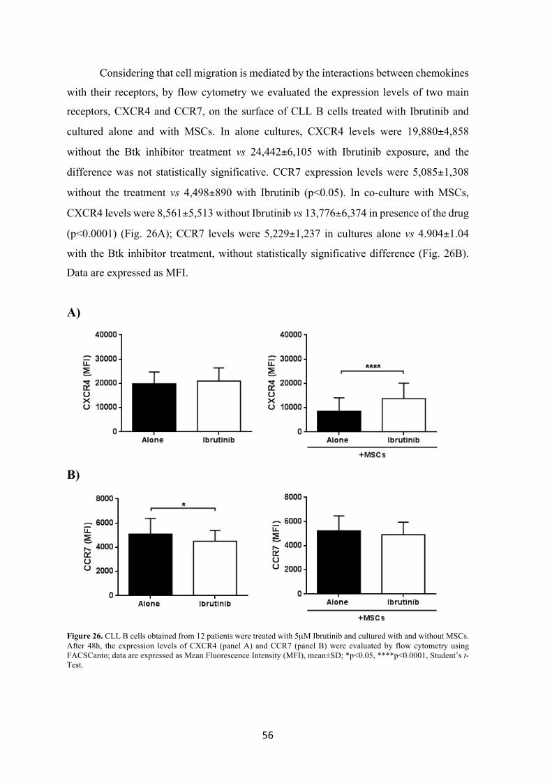
56
Considering that cell migration is mediated by the interactions between chemokines
with their receptors, by flow cytometry we evaluated the expression levels of two main
receptors, CXCR4 and CCR7, on the surface of CLL B cells treated with Ibrutinib and
cultured alone and with MSCs. In alone cultures, CXCR4 levels were 19,880±4,858
without the Btk inhibitor treatment vs 24,442±6,105 with Ibrutinib exposure, and the
difference was not statistically significative. CCR7 expression levels were 5,085±1,308
without the treatment vs 4,498±890 with Ibrutinib (p<0.05). In co-culture with MSCs,
CXCR4 levels were 8,561±5,513 without Ibrutinib vs 13,776±6,374 in presence of the drug
(p<0.0001) (Fig. 26A); CCR7 levels were 5,229±1,237 in cultures alone vs 4.904±1.04
with the Btk inhibitor treatment, without statistically significative difference (Fig. 26B).
Data are expressed as MFI.
A)
B)
Figure 26. CLL B cells obtained from 12 patients were treated with 5µM Ibrutinib and cultured with and without MSCs. After 48h, the expression levels of CXCR4 (panel A) and CCR7 (panel B) were evaluated by flow cytometry using FACSCanto; data are expressed as Mean Fluorescence Intensity (MFI), mean±SD; *p<0.05, ****p<0.0001, Student’s t-Test.

57
9. Ibrutinib treatment affects CLL B cell adhesion
Having observed that the treatment with Ibrutinib did not affect CLL B cell
movement in response to MSC stimuli, we also analyzed the adhesion of leukemic B cells
to MSCs after treatment with the Btk inhibitor since cell-cell contact with MSC is crucial
for CLL B cell survival. We found that the percentage of leukemic B cells adherent to
MSCs was significantly reduced by Ibrutinib (7.7%±3.8% alone vs 3.3%±2.4, p<0.05);
(Fig. 27).
Figure 27. Percentage of cell adherent to MSCs with and without Ibrutinib. CLL B cells from 7 patients were treated with and without 5µM Ibrutinib and incubated for 1h at 37°C in 5% CO2 in complete RPMI medium. Cells not adherent were removed by washing 3 times with RPMI medium. The layer of cells was detached by incubation with Accutase; cells were than stained with anti-CD19 APC and counted by flow cytometry; mean±SD *p<0.05, Student’s t-Test.
Using phase-contrast microscopy Olympus IX-81 we documented photographically that
CLL cells adhesion to MSCs was reduced after Ibrutinib treatment (Fig. 28)
Figure 28. Microscopy analysis of CLL B cell adhesion to MSCs with and without Ibrutinib. CLL cells from 7 patients were treated with or without 5µM Ibrutinib and incubated for 1h at 37°C in 5% CO2 in complete RPMI medium. CLL B cell adhesion to MSCs was documented photographically using phase-contrast microscopy Olympus IX-81.
Alone Ibrutinib
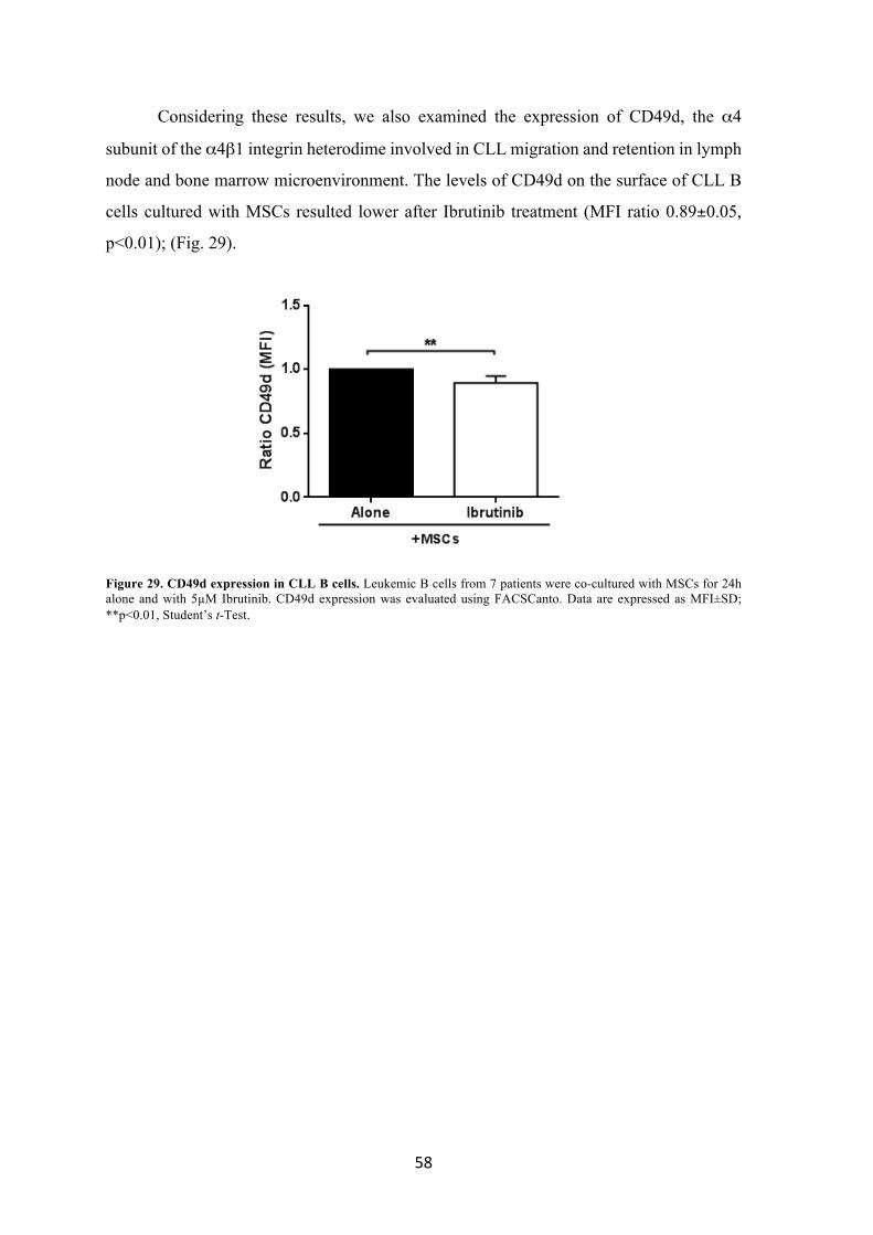
58
Considering these results, we also examined the expression of CD49d, the α4
subunit of the α4β1 integrin heterodime involved in CLL migration and retention in lymph
node and bone marrow microenvironment. The levels of CD49d on the surface of CLL B
cells cultured with MSCs resulted lower after Ibrutinib treatment (MFI ratio 0.89±0.05,
p<0.01); (Fig. 29).
Figure 29. CD49d expression in CLL B cells. Leukemic B cells from 7 patients were co-cultured with MSCs for 24h alone and with 5µM Ibrutinib. CD49d expression was evaluated using FACSCanto. Data are expressed as MFI±SD; **p<0.01, Student’s t-Test.

59
DISCUSSION
Chronic Lymphocytic Leukemia is the most common leukemia in adults and is
characterized by the accumulation of clonal CD19+/CD5+/CD23+ B lymphocytes, due to
uncontrolled growth and resistance to apoptosis. Intrinsic factors, such as genetic lesions,
anti-apoptotic proteins, and aberrant signaling networks within leukemia cells have long
been the main focus of CLL research. However, over the past years, it became increasingly
clear that external signals from the microenvironment make pivotal contributions to CLL
progression. In healthy subjects MSCs represent a small fraction of the stromal cell
population, about 0.01-0.0001% of mononuclear cells, but are the dominant stromal cell
population in CLL microenvironment. MSCs provide an attachment site and growth factors
for normal haematopoiesis and, both in CLL, are thought to function in a similar fashion,
creating a niche within the BM in which CLL B cells lodge and are nourished and protected
from cytotoxic agents (38).
In this project, MSCs from BM of 46 CLL patients were co-cultured with leukemic
B cells in order to mimic the neoplastic in vivo microenvironment. Our results demonstrated
that malignant B cells are susceptible to the antiapoptotic effect of MSCs, favouring
neoplastic B cell survival in vitro for at least 7 days. This evidence is relevant considering
that CLL B cells spontaneously undergo apoptosis once they are removed from the in vivo
microenvironment and placed in suspension cultures without the supportive stromal cells.
This effect was less relevant in normal B lymphocytes, suggesting the presence of a peculiar
receptor structure which allows the neoplastic B cell to respond to pro-survival stimuli
produced from the elements of the microenvironment in which it is localized. In fact,
leukemic B cell expresses specific chemokine and cytokine receptors and responds
selectively to soluble factors produced by the compounds of the microenvironment into the
active sites of disease (39). Despite CLL B cells are characterized by a typical phenotype
and cytogenetic abnormalities, the disease displays heterogeneous clinical courses,
suggesting that each malignant clone could present intrinsic features that affect the
interactions with the microenvironment. CLL natural history, including response to
treatment and drug resistance, is determined both by causal and influential genes and by
the relationships that malignant B cells entertain with their supportive microenvironments

60
(38). Therefore, studying the role of microenvironment we may provide essential strategies
to treat and eradicate the disease.
We observed a high variability in the viability of CLL B cells cultured with MSCs
and, to better understand these data, we analysed the cleavage pattern of the PARP protein,
indicator of caspase activity. The detection of PARP 89kDa fragment in CLL patients, after
7 days of co-culture with MSCs, revealed two subsets of CLL clones with different
sensitivity to MSCs pro-survival stimuli. The first group was classified as “dependent”
from microenvironmental signals, when CLL B cells underwent spontaneous apoptosis in
medium alone, but were rescued by the presence of MSCs. The second group, classified as
“independent” from microenvironmental stimuli, identified CLL clones whose viability
was high both when cultured in medium alone and in the presence of MSCs, indicating that
these leukemic cells were able to survive independently from pro-survival signals coming
from stromal cells. Further experiments allowed us to establish that the different behaviour
displayed from neoplastic clones is likely to be related to intrinsic features of neoplastic B
cells rather than to the variety of the microenvironment. These observations could be
relevant to identify patients more responsive to druggable targets on marrow
microenvironment and also to find putative new strategies for CLL therapy. In fact, it is
likely that a clone “dependent” from microenvironmental stimuli will be more easily
affected by a treatment that aims to interactions with it; on the other hand, a clone
“independent” from the signals of the microenvironment probably will not show a
particular sensitivity to therapies which target the CLL-microenvironment cross-talk.
The increase of malignant B cell viability in the presence of MSCs might be
mediated by soluble factors and/or cell-cell contact. Our data indicated that both cell-cell
contact and soluble factors are relevant for the survival of malignant clone. We observed
an increased migration of neoplastic B cells in the presence of the conditioned medium of
MSCs, demonstrating that MSCs from CLL patients are able to produce factors that
promote the recruitment of CLL B cell toward a favourable niche for the maintenance of
the leukemic clone. Considering that MSCs constitutively secrete a high amount of
chemokines, which organize CLL cell trafficking and homing (41), the identification of
humoral and cellular factors responsible for the pro-survival effect in vivo could be useful
also to find new therapeutical targets for CLL and possible promising approaches to
manage the disease.
CLL cell interactions with MSCs play a critical role in the disease pathogenesis, in
particular in CLL survival, homing, proliferation but also in treatment failure. The cross-

61
talk between MSCs and the leukemic B cell seems to play a key role in inhibiting apoptosis
induced by drugs. We demonstrated that the presence of MSCs protects leukemic B cells
from apoptosis after the in vivo exposure to Fludarabine and Cyclophosphamide, a
chemotherapy regimen considered as the gold standard for first-line treatment in CLL
patients (5). Furthermore, our results confirm the protective role of MSCs also performing
in vivo experiments, with leukemic B cells collected from patients after the first cycle of
FLU/Cy therapy. These data could partially explain the reason why in some CLL patients,
maybe with a neoplastic clone more “dependent” on microenvironment stimuli, it is more
difficult to obtain a complete or long-lasting response. These results, in fact, point out the
protective role of microenvironment not only toward malignant B cells in vitro but also on
apoptosis induced by the in vivo administration of chemotherapy, maybe causing therapy
refractoriness or an unsatisfactory response to treatment.
The management of CLL is undergoing profound changes; several new drugs have
been approved for CLL treatment and many others are in advanced clinical development in
the pipeline to be approved for this disease (6). The CLL microenvironment has gained
extensive attention during the last few years, thanks to the progressive understanding of the
mechanisms involved in CLL B cell proliferation and survival and to the introduction of
several novel small molecule inhibitors, which target the CLL-microenvironment cross-
talk (81). Different drugs, used in CLL treatment, have been demonstrated to induce in
vitro apoptosis of CLL B cells, whereas their effect is reduced when administered in vivo,
probably due to the presence of pro-survival signals coming from the surrounding
environment. Therefore, it is suitable to test the effect of new therapeutic agents in the
presence of microenvironmental compounds (such as MSCs) which might interfere with
the biological effects of the drugs, allowing the identification of subgroups of patients who
may benefit from treatments targeting the cross-talk with supportive cells at the sites of the
disease.
Considering the relevance of the BCR in the support of the neoplastic B cell, we
evaluated the role of MSCs during the treatment with Ibrutinib, a novel Btk inhibitor who
targets proteins essential in signaling transduction mediated by the BCR. We tested the
effect of the Btk inhibitor, known to reduce CLL B cell migration and to induce in vitro apoptosis (94), in our co-culture system. We found that Ibrutinib is able to induce leukemic
cell apoptosis independently from MSCs presence, confirming its potential high efficacy
in the treatment of CLL. By contrast, the Btk inhibitor did not affect B cell migration toward

62
a MSC-conditioned medium, rich in cytokines and chemokines, suggesting that malignant
B cells do not lose their ability to move toward a protective niche in presence of Ibrutinib.
To better understand these data, we studied some of the receptors involved in cell
migration. CLL B cells express on their surface high levels of chemokine receptors; in
particular CXCR4 and CCR7 are involved in BM and lymphoid tissues homing of the
neoplastic clone (95). We observed that malignant B cells, co-cultured with MSCs and
treated with Ibrutinib, displayed an increase of CXCR4 expression levels. The CXCR4
chemokine receptor is expressed at high levels on the surface of CLL B cells and mediated
chemotaxis, migration across vascular endothelium and actin polymerization in response
to CXCL12 gradients, produced by MSCs (49). CXCL12 also induces a direct pro-survival
effect on leukemic B cells. CXCR4 surface expression is regulated by its ligand CXCL12
via receptor endocytosis. In the presence of MSCs, in CLL B cells not treated with
Ibrutinib, CXCR4 is internalized into the cytoplasm, indicating the successful receptor-
ligand interaction. Although the exact mechanism of CXCR4 up-regulation is still not
completely clear, our results indicated that the use of kinase inhibitor could induce
modifications in chemokine receptors, in particular interfering with CXCR4-CXCL12 axis.
Instead the expression of CCR7, the receptor for both CCL21 and CCL19, correlates with
clinical lymphadenopathy, a clinical feature of more advanced disease (96) (97). Although
little is known about the mechanisms determining lymph node enlargement in CLL, some
studies suggested that CCR7 engagement by CCL21 and/or CCL19 stimulates CLL B cell
entry into lymph nodes (96). Considering that Ibrutinib in vivo treatment rapidly decrease
lymph node size, we evaluated CCR7 expression levels after the in vitro treatment with the
Btk inhibitor. Our results demonstrated lower CCR7 levels after Ibrutinib exposure,
suggesting a possible interaction with this receptor. Nevertheless, in co-culture with MSCs
CCR7 was not affected by the Btk inhibitor treatment, suggesting that, probably, the in vivo
effect of Ibrutinib on CCR7 is more complex and need further studies to be clarified.
Since the direct cell-cell contact with stromal cells is crucial for CLL B cell survival,
we tested the effect of Ibrutinib on neoplastic B cell adhesion to a MSCs layer. We found
a significant reduction of CLL B cells adhesion through a down-modulation of CD49d
expression, an integrin involved in anchoring cells to tissues via extracellular matrix. Our
results suggest that Ibrutinib is not only able to reduce CLL B cell viability, but that it could
also interfere with cell-cell contact, fundamental in cell survival but also as a first step in
cell migration. Therefore, we could suppose that, in presence of Ibrutinib, MSCs are not
able to further protect CLL B cells that could remain on peripheral blood and potentially

63
more exposed to pro-apoptotic stimuli. Ibrutinib is a potent drug and highly selective with
great results in CLL therapy; however, it does not completely eliminate the malignant clone
in CLL and results mainly in partial responses. A better understanding on the complexity
of the cross-talk between CLL cells and their microenvironment could help to define also
mechanisms of resistance to Ibrutinib and treatment failure, as well to plan randomized
clinical trials comparing new inhibitors of BCR signaling and their combinations with
standard chemo-immunotherapy.
In conclusion, this project demonstrated that MSCs co-culture represents a
reproducible in vitro system with functional similarities to in vivo bone marrow conditions,
pointing out that the heterogeneity of the disease is reflected also in CLL B cell capacity to
respond to favourable signals from MSCs (98). Our findings on the role of MSCs and their
effect on neoplastic B lymphocytes during chemotherapy exposure open a new scenario to
better identify the most effective drugs or drug combinations. Furthermore, we showed that
Ibrutinib treatment, inactivating enzymes in the BCR signaling pathway, which are
aberrantly activated in CLL, is able not only to inhibit CLL B cell proliferation and survival
but also to interact with the cross-talk with the stromal cells, a necessary condition for the
eradication of the disease. Additional in vivo and in vitro studies will accelerate the
development of these new concepts and will help to define the best drug combinations.

64

65
BIBLIOGRAPHY
1. Caligaris-Cappio F, Hamblin TJ. B-cell chronic lymphocytic leukemia: a bird of a different feather. J Clin Oncol (1999); 17: 399-408. 2. Kikushige Y, Ishikawa F, Miyamoto T, Shima T, Urata S, Yoshimoto G, Mori Y, Iino T, Yamauchi T, Eto T, Niiro H, Iwasaki H, Takenaka K, Akashi K. Self-renewing hematopoietic stem cell is the primary target in pathogenesis of human chronic lymphocytic leukemia. Cancer Cell (2011); 20: 246-256. 3. (Website), National Cancer Institute. Surveillance Epidemology and End Results Cancer Statistics review (2010). 4. Molica S. Sex differences in incidence and outcome of chronic lymphocytic leukemia patients. Leuk Lymphoma (2006); 47: 1477-1480. 5. Eichorst B, Robak T, Montserrat E, Ghia P, Hillmen P, Hallek M, Buske C. Chronic lymphocytic leukemia: ESMO Clinical Practice Guidelines for diagnosis, treatment and follow-up. Ann Oncol (2015); 26: 78-84. 6. Hallek M. Chronic lymphocytic leukemia: 2013 update on diagnosis, risk stratification and treatment. Am J Hematol (2013); 88: 804-816. 7. Hallek M, Cheson BD, Catovsky D, Caligaris-Capio F, Dighiero G, Dohner H, Hillmen P, Keating MJ, Montserrat E, Rai KR, Kipps TJ. Guidelines for the diagnosis and treatment of chronic lymphocytic leukemia: a report from the International Workshop on Chronic Lymphocyitic Leukemia updating the National Cancer Institute-Working Group 1996 guidelines. Blood (2008); 112: 5259. 8. Moreau EJ, Matutes E, A'Hern RP, Morilla AM, Morilla RM, Owusu-Ankomah KA, Seon BK, Catovsky D. Improvement of the chronic lymphocytic leukemia scoring system with the monoclonal antibody SN8 (CD79b). Am J Clin Pathol (1997); 108: 378-382. 9. Rawstron AC, Bennett FL, O'Connor SJ, Kwok M, Fenton JA, Plummer M, de Tute R, Owen RG, Richards SJ, Jack AS, Hillmen P. Monoclonal B-cell lymphocytosis and chronic lymphocyitc leukemia. N Engl J Med (2008); 111: 5446-5456. 10. Rozman C, Montserrat E. Chronic lymphocytic leukemia. N Engl J Med (1995); 333: 1052-1057. 11. Binet JL, Auquier A, Dighiero G, Chastang C, Piguet H, Goasguen J, Vaugier G, Potron G, Colona P, Oberling F, Thomas M, Tchernia G, Jacquillat C, Boivin P, Lesty C, Duault MT, Monconduit M, Belabbes S, Gremy F. A new prognostic classification of chronic lymphocytic leukemia derived from a multivariate survival analysis. Cancer (1981); 48: 198-206. 12. Rai KR, Sawitsky A, Cronkite EP, Chanana AD, Levy RN, Pasternack BS. Clinical staging of chronic lymphocytic leukemia. Blood (1975); 46: 219-234. 13. Damle RN, Wasil T, Fais F, Ghiotto F, Valetto A, Allen SL, Buchbinder A, Budman D, Dittmar K, Kolitz J, Lichtman SM, Schulman P, Vinciguerra VP, Rai KR, Ferrarini M, Chiorazzi N. Ig V gene mutation status and CD38 expression as novel prognostic indicators in chronic lymphocytic leukemia. Blood (1994); 94: 1840-1847. 14. S. Deaglio, T. Vaisitti, S. Aydin, L. Bergui, G. D'Arena, L. Bonello, P. Omede, M. Scatolini, O. Jaksic, G. Chiorino, D. Efremov, F. Malavasi. CD38 and ZAP-70 are functionally linked and mark CLL cells with high migratory potential. Blood (2007); 110: 4012-4021. 15. Gattei V, Bulian P, Del Principe MI, Zucchetto A, Maurillo L, Buccisano F, Bomben R, Dal-Bo M, Luciano F, Rossi FM, Degan M, Amadori S, Del Poeta G. Relevance of CD49d protein expression as overall survival and progressive disease prognosticator in chronic lymhocytic leukemia. Blood (2008); 111: 865-873.

66
16. Orchard JA, Ibbotson RE, Davis Z, Wiestner A, Rosenwald A, Thomas PW, Hamblin TJ, Staudt LM, Oscier DG. ZAP-70 expression and prognosis in chronic lymphocytic leukemia. Lancet (2004); 363: 105-111. 17. Döhner H, Stilgenbauer S, Benner A, Leupolt E, Kröber A, Bullinger L, Döhner K, Bentz M, Lichter P. Genomic aberrations and survival in chronic lymphocytic leukemia. N Engl J Med (2000); 343: 1910-1916. 18. Hallek M, Fischer K, Fingerle-Rowson G, Fink AM, Busch R, Mayer J, Hensel M, Hopfinger G, Hess G, von Grünhagen U, Bergmann M, Catalano J, Zinzani PL, Caligaris-Cappio F, Seymour JF, Berrebi A, Jäger U, Cazin B, Trneny M, Westermann A, Wendtner CM, Eichhorst BF, Staib P, Bühler A, Winkler D, Zenz T, Böttcher S, Ritgen M, Mendila M, Kneba M, Döhner H, Stilgenbauer S; International Group of Investigators; German Chronic Lymphocytic Leukaemia Study Group. Addition of rituximab to fludarabine and cyclophosphamide in patients with chronic lymphocytic leukemia: A randomised, open-label, phase 3 trial. Lancet (2010); 376: 1164-1174. 19. Zenz T, Vollmer D, Trbusek M, Smardova J, Benner A, Soussi T, Helfrich H, Heuberger M, Hoth P, Fuge M, Denzel T, Häbe S, Malcikova J, Kuglik P, Truong S, Patten N, Wu L, Oscier D, Ibbotson R, Gardiner A, Tracy I, Lin K, Pettitt A, Pospisilova S, Mayer J, Hallek M, Döhner H, Stilgenbauer S; European Research Initiative on CLL (ERIC). TP53 mutation profile in chronic lymphocytic leukemia: Evidence for a disease specific profile from a comprehnsive analysis of 268 mutations. Leukemia (2010); 24: 2072-2079. 20. Puente XS, Pinyol M, Quesada V, Conde L, Ordóñez GR, Villamor N, Escaramis G, Jares P, Beà S, González-Díaz M, Bassaganyas L, Baumann T, Juan M, López-Guerra M, Colomer D, Tubío JM, López C, Navarro A, Tornador C, Aymerich M, Rozman M, Hernández JM, Puente DA, Freije JM, Velasco G, Gutiérrez-Fernández A, Costa D, Carrió A, Guijarro S, Enjuanes A, Hernández L, Yagüe J, Nicolás P, Romeo-Casabona CM, Himmelbauer H, Castillo E, Dohm JC, de Sanjosé S, Piris MA, de Alava E, San Miguel J, Royo R, Gelpí JL, Torrents D, Orozco M, Pisano DG, Valencia A, Guigó R, Bayés M, Heath S, Gut M, Klatt P, Marshall J, Raine K, Stebbings LA, Futreal PA, Stratton MR, Campbell PJ, Gut I, López-Guillermo A, Estivill X, Montserrat E, López-Otín C, Campo E. Whole-genome sequencing identifies recurrent mutations in chronic lymphocytic leukaemia. Nature (2011); 475: 101-105. 21. Rossi D, Rasi S, Fabbri G, Spina V, Fangazio M, Forconi F, Marasca R, Laurenti L, Bruscaggin A, Cerri M, Monti S, Cresta S, Famà R, De Paoli L, Bulian P, Gattei V, Guarini A, Deaglio S, Capello D, Rabadan R, Pasqualucci L, Dalla-Favera R, Foà R, Gaidano G. Mutations of NOTCH1 are an independent predictor of survival in chronic lymphocytic leukemia. Blood (2012); 119: 521-529. 22. Wang L, Lawrence MS, Wan Y, Stojanov P, Sougnez C, Stevenson K, Werner L, Sivachenko A, DeLuca DS, Zhang L, Zhang W, Vartanov AR, Fernandes SM, Goldstein NR, Folco EG, Cibulskis K, Tesar B, Sievers QL, Shefler E, Gabriel S, Hacohen N, Reed R, Meyerson M, Golub TR, Lander ES, Neuberg D, Brown JR, Getz G, Wu CJ. SF3B1 and other novel cancer genes in chronic lymphocytic leukemia. N Engl J Med (2011); 119: 2854-2862. 23. Rossi D, Fangazio M, Rasi S, Vaisitti T, Monti S, Cresta S, Chiaretti S, Del Giudice I, Fabbri G, Bruscaggin A, Spina V, Deambrogi C, Marinelli M, Famà R, Greco M, Daniele G, Forconi F, Gattei V, Bertoni F, Deaglio S, Pasqualucci L, Guarini A, Dalla-Favera R, Foà R, Gaidano G. Disruption of BIRC3 associates with fludarabine chemorefractoriness in TP53 wild-type chronic lymphocytic leukemia. Blood (2012); 119: 2854-2862. 24. Keating MJ, Lerner S, Kantarjian H, Freireich EJ, O'Brien S. The serum β2microglobulin (β2m) level is more powerful than stage in predicting response and survival in chronic lymphocytic leukemia (CLL). Blood (1995); 86: 606a. 25. Sarfati M, Chevret S, Chastang C, Biron G, Stryckmans P, Delespesse G, Binet JL, Merle-Beral H, Bron D. Prognostic importance of serum soluble CD23 level in chronic lymphocytici leukemia. Blood (1996); 88: 4259-4264. 26. Magnac C, Porcher R, Davi F, Nataf J, Payelle-Brogard B, Tang RP, Oppezzo P, Lévy V, Dighiero G, Ajchenbaum-Cymbalista F. Predicitive value of serum thymidine kinase level for Ig-V mutational status in B-CLL. Leukemia (2003); 17: 133-137.

67
27. Mauro FR, Bandini G, Barosi G, Billio A, Brugiatelli M, Cuneo A, Lauria F, Liso V, Marchetti M, Meloni G, Montillo M, Zinzani P, Tura S. SIE, SIES, GITMO updated clinical recommendations for the management of chronic lymphocytic leukemia. Leuk Reser (2012); 36: 459-466. 28. Eichorst B, Robak T, Montserrat E, Ghia P, Hillmen P, Hallek M, Buske C. Chronic lymphocytic leukemia: ESMO Clinical Practice Guidelines for diagnosis, treatment and follow up. Ann Oncol (2011); 22: 50-54. 29. Stevenson FK, Krysov S, Davies AJ, Steele AJ, Packam G. B-cell receptor signaling in chronic lymphocytic leukemia. Blood (2011); 118: 4313-4320. 30. Collins RJ, Verschuer LA, Harmon BV, Prentice RL, Pope JH, Kerr JF. Spontaneous programmed death (apoptosis) of B-chronic lymphocytic leukaemia cells following their culture in vitro. Br J Haematol (1989); 71: 343-350. 31. Ghia P, Caligaris-Cappio F. The indispensable role of microenvironment in the natural history of low-grade B-cell neoplasms. Adv Cancer Res (2000); 79:157-173. 32. Helfrich H, Fuge M, Eldering E, Bühler A, Winkler D, Volden M, Kater AP, Mertens D, Te Raa D, Döhner H, Stilgenbauer S, Zenz T. DNA damage-induced transcriptional program in CLL: biological and diagnostic implications for functional p53 testing. Blood (2011); 117: 1622-1632. 33. Burger JA, Chiorazzi N. B cell receptor signaling in chronic lymphocytic leukemia. Trends Immunol (2013); 34: 592-601. 34. Scielzo C, Ghia P, Conti A, Bachi A, Guida G, Genua M, Alessio M, Caligaris-Cappio F. HS1 protein is differentially expressed in chronic lymphocytic leukemia patient subsets with good or poor prognosis. 115, 2005, J Clin Invest (2005); 115: 1644-1650. 35. Herishanu Y, Pérez-Galán P, Liu D, Biancotto A, Pittaluga S, Vire B, Gibellini F, Njuguna N, Lee E, Stennett L, Raghavachari N, Liu P, McCoy JP, Raffeld M, Stetler-Stevenson M, Yuan C, Sherry R, Arthur DC, Maric I, White T, Marti GE, Munson P, Wilson WH, Wiestner A. The lymph node microenvironment promotes B-cell receptor signaling, NF-kappaB activation, and tumor proliferation in chronic lymphocytic leukemia. Blood (2011); 117: 563-574. 36. Krysov S, Dias S, PAterson A, Mockridge CI, Potter KN, Smith KA, Ashton-Key M, Stevenson FK, Packham G. Surface IgM stimulation induces MEK1/2-dependent MYC expression in chronic lymphocytic leukemia cells. Blood (2012); 119: 170-179. 37. Morabito F, Cutrona G, Gentile M, Fabbi M, Matis S, Colombo M, Reverberi D, Megna M, Spriano M, Callea V, Vigna E, Rossi E, Lucia E, Festini G, Zupo S, Molica S, Neri A, Ferrarini M. Prognostic relevance of in vitro response to cell stimulation via surface IgD in binet stagea CLL. Br J Haematol (2010); 149: 160-163. 38. Caligaris-Capio F, Bertilaccio MT, Scielzo C. How the microenvironment wires the natural history of chronic lymphocytic leukemia. Semin Cancer Biol (2014); 24: 43-48. 39. Burger JA. Nurture versus Nature: The Microenvironment in Chronic Lymphocytic Leukemia. Hematology Am Soc Hematol Educ Program (2011); 96-103. 40. Tsukada N, Burger JA, Zvaifler NJ, Kipps TJ. Distinctive features of "nurselike" cells that differentiate in the context of chronic lymphocytic leukemia. Blood (2002); 99: 1030-1037. 41. Hacken ET, Burger JA. Microenvironment interactions and B-cell receptor signaling in Chronic Lymphocytic Leukemia: Implications for disease pathogenesis and treatment. Biochim Biophys Acta (2015); [Epub ahead of print]. 42. Maffei R, Bulgarelli J, Fiorcari S, Martinelli S, Castelli I, Deaglio S, Debbia G, Fontana M, Coluccio V, Bonacorsi G, Zucchini P, Narni F, Torelli G, Lupi M, Marasca R. Physical contact with endothelial cells

68
through beta1- and beta2-integrins rescues chronic lymphocytic leukemia cells frm spontaneous and drug-induced apoptosis and induces a peculiar gene expression profile in leukemic cells. Haematologica (2012); 97: 952-960. 43. Heinig K, Gatjen M, Grau M, Stache V, Anagnostopoulos I, Gerlach K, Niesner RA, Cseresnyes Z, Hauser AE, Lenz P, Hehlgans T, Brink R, Westermann J, Dorken B, Lipp M, Lenz G, Rehm A, Hopken UE. Access to follicular dendritic cells is a pivotal step in murine chronic lymphocytic leukemia B-cell activation and proliferation. Cancer Discov (2014); 4: 1448-1465. 44. Brusa D, Serra S, Coscia M, Rossi D, D'Arena G, Laurenti L, Jaksic O, Fedele G, Inghirami G, Gaidano G, Malavasi F, DEaglio S. The PD-1/PD-L1 axis contributes to T-cell dysfunction in chronic lymphocytic leukemia. Haematologica (2013); 98: 953-963. 45. Veuillen C, Aurran-Schleinitz T, Castellano R, Rey J, Mallet F, Orlanducci F, Pouyet L, Just-Landi S, Coso D, Ivanov V, Carcopino X, Bouabdallah R, Collette Y, Fauriat C, Olive D. Primary B-CLL resistance to NK cell cytotoxicity can be overcome in vitro and in vivo by priming NK cells and monoclonal antibody therapy. J Clin Immunol (2012); 32: 632-646. 46. Simmons PJ, Torok-Storb B. Identification of stromal cell precursors in human bone marrow by a novel monoclonal antibody, STRO-1. Blood (1991); 78: 55-62. 47. Lagneaux L, Delforge A, Bron D, De Bruyn C, Stryckmans P. Chronic lymphocytic leukemic B cells but not normal B cells are rescued from apoptosis by contact with normal bone marrow stromal cells. Blood (1998); 91: 2387-2396. 48. Ruan J, Hyjek E, Kermani P, Christos PJ, Hooper AT, Coleman M, Hempstead B, Leonard JP, Chadburn A, Rafii S. Magnitude of stromal hemangiogenesis correlates with histologic subtype of non-Hodgkin's lymphoma. Clin Cancer Res (2006); 12: 5622-5631. 49. Burger JA, Burger M, Kipps TJ. Chronic Lymphocytic Leukemia B Cells Express Functional CXCR4 Chemokine Receptors That Mediate Spontaneous Migration Beneath Bone Marrow Stromal Cells. Blood (1999); 94: 3658-3667. 50. Bagnara D, Kaufman MS, Calissano C, Marsilio S, Patten PE, Simone R, Chum P, Yan XJ, Allen SL, Kolitz JE, Baskar S, Rader C, Mellstedt H, Rabbani H, Lee A, Gregersen PK, Rai KR, Chiorazzi N. A novel adoptive transfer model of chronic lymphocytic leukemia suggests a key role for T lymphocytes in the disease. Blood (2011); 117: 5463-5472. 51. Purroy N, Abrisqueta P, Carabia J, Carpio C, Palacio C, Bosch F, Crespo M. Co-culture of primary CLL cells with bone marrow mesenchymal cells, CD40 ligand and CpG ODN promotes proliferation of chemoresistant CLL cells phenotypically comparable to those proliferating in vivo. Oncotarget (2015); 6: 7632-7643. 52. Marquez ME, Hernandez-Uzcategui O, Cornejo A, Vargas P, Da Costa O. Bone marrow stromal mesenchymal cells induce down regulation of CD20 expression ob B-CLL: implications for rituximab resistance in CLL. Br J Haematol (2015); 169: 211-218. 53. Jitschin R, Braun M, Qorrai M, Saul D, Le Blanc K, Zenz T, Mougiakakos D. Stromal cell-mediated glycolytic switch in CLL-cells involves Notch-c-Myc signaling. Blood (2015); 125: 3432-3436. 54. Lutzny G, Kocher T, Schmidt-Supprian M, Rudelius M, Klein-Hitpass L, Finch AJ, Durig J, Wagner M, Haferlach C, Kohlmann A, Schnittger S, Seifert M, Wanninger S, Zaborsky N, Oostendorp R, Ruland J, Leitgers M, Kuhnt T, Schafer Y, Lampl B, Peschel C, Egle A, Ringshausen I. Protein kinase c-dependent activation of NF-kappaB in stromal cells is indispensable for the survival of chronic lymphocytic leukemia B cells in vivo. Cancer Cell (2013); 23: 77-92.

69
55. Smith JR, Pochampally R, Perry A, Hsu SC, Prockop DJ. Isolation of a highly clonogenic and multipotential subfraction of adult stem cells from bone marrow stroma. Stem Cells (2004); 22: 823-831. 56. Jiang Y, Jahagirdar BN, Reinhardt RL, Schwartz RE, Keene CD, Ortiz-Gonzalez XR, Reyes M, Lenvik T, Lund T, Blackstad M, Du J, Aldrich S, Lisberg A, Low WC, Largaespada DA, Verfaillie CM. Pluripotency of mesenchymal stem cells derived from adult marrow. Nature (2002); 418: 41-49. 57. Kunkel SL, Strieter RM, Lindley IJ, Westwick J. Chemokines: new ligands, receptors and activities. Immunol Today (1995); 16: 559-561. 58. Bleul CC, Fuhlbrigge RC, Casasnovas JM, Aiuti A, Springer TA. A highly efficacious lymphocyte chemoattractant, stromal cell-derived factor (SDF-1). J Exp Med (1996); 184: 1101-1109. 59. Burger JA, Burger M, Kipps TJ. Chronic lymphocytic leukemia B cells express functional CXCR4 chemokine receptors that mediate spontaneous migration beneath bone marrow stromal cells. Blood (1999); 94: 3658-3667. 60. Vaisitti T, Aydin S, Rossi D, Cottino F, Bergui L, D'Arena G, Bonello L, Horenstein AL, Brennan P, Pepper C, Gaidano G, Malvasi F, Deaglio S. CD38 increases CXCL12-mediated signals and homing of chronic lymphocytic leukemia (B-CLL). Leukemia (2010); 24: 958-969. 61. O'Hayre M, Salanga CL, Kipps TJ, Messmer D, Dorrenstein PC, Handel TM. Elucidating the CXCL12/CXCR4 signaling network in chronic lymphocytic leukemia through phosphoproteomics analysis. PloS ONE (2010); 5: 11-16. 62. Burger JA, Quiroga MP, Hartmann E, Burkle A, Wierda WG, Keating MJ, Rosenwald A. High-level expression of the T-cell chemokines CCL3 and CCL4 by chronic lymhocytic leukemia B cells in nurselike cell cocoltures and after BCR stimulation. Blood (2009); 113: 3050-3058. 63. Kathleen JT, Lin K, Zuzel M, Cawley JC. The chemokine receptor CCR7 and α4 integrin are important for migration of chronic lymphocytic leukemia cells into lymph nodes. Blood (2002); 99: 2977-2984. 64. Cramer P, Hallek M. Prognostic factors in chronic lymphocytic leukemia-what do we need to know? Nat Rev Clin Oncol (2011); 8: 38-47. 65. Extermann M, Overcash J, Lymah GH, Parr J, Balducci L. Comorbidity and functional status are independent in older patients. J Clin Oncol (1998); 16: 1582-1587. 66. Plunkett W, Gandhi V, Huang P, Robertson LE, Yang LY, Gregoire V, Estey E, Keating MJ. Fludarabine: Pharmacokinetics, mechanisms of action, and rationale for combination therapies. Semin Oncol (1993); 20: 2-12. 67. Rai KR, Peterson BL, Appelbaum FR, Kolitz J, Elias L, Shepherd L, Hines J, Threatte GA, Larson RA, Cheson BD, Schiffer CA. Fludarabine compared with chlorambucil as primary therapy for chronic lymphocytic leukemia. N Engl J Med (2000); 343: 1750-1757. 68. Leporrier M, Chevret S, Cazin B, Boudjerra N, Feugier P, Desablens B, Rapp MJ, Jaubert J, Autrand C, Divine M, Dreyfus B, Maloum K, Travade P, Dighiero G, Binet JL, Chastang C; French Cooperative Group on Chronic Lymphocytic Leukemia. Randomized comparison of fludarabine, CAP, and CHOP in 983 previously untreated stage B and C chronic lymphocytic leukemia patients. Blood (2001); 98: 2319-2325. 69. Eichorst BF, Busch R, Hopfinger G, Pasold R, Hensel M, Steinbrecher C, Siehl S, Jäger U, Bergmann M, Stilgenbauer S, Schweighofer C, Wendtner CM, Döhner H, Brittinger G, Emmerich B, Hallek M; German CLL Study Group. Fludarabine plus cyclophosphamide versus fludarabine alone in first line therapy of younger patients with chronic lymphocyitic leukemia. Blood (2006); 107: 885-891.

70
70. Catovsky D, Richards S, Matuted E, Oscier D, Dyer MJ, Bezares RF, Pettitt AR, Hamblin T, Milligan DW, Child JA, Hamilton MS, Dearden CE, Smith AG, Bosanquet AG, Davis Z, Brito-Babapulle V, Else M, Wade R, Hillmen P; UK National Cancer Research Institute (NCRI) Haematological Oncology Clinical Studies Group; NCRI Chronic Lymphocytic Leukaemia Working Group. Assesment of fludarabine plus cyclophosphamide for patients with chronic lymphocytic leukaemia (the LRF CLL4 Trial): a randomized controlled trial. Lancet (2007); 370: 230-239. 71. Hallek M, Fischer K, Fingerle-Rowson, Fink AM, Busch R, Mayer J, Hensel M, Hopfinger G, Hess G, von Grünhagen U, Bergmann M, Catalano J, Zinzani PL, Caligaris-Cappio F, Seymour JF, Berrebi A, Jäger U, Cazin B, Trneny M, Westermann A, Wendtner CM, Eichhorst BF, Staib P, Bühler A, Winkler D, Zenz T, Böttcher S, Ritgen M, Mendila M, Kneba M, Döhner H, Stilgenbauer S; International Group of Investigators; German Chronic Lymphocytic Leukaemia Study Group. Addition of rituximab to fludarabine and cyclophosphamide in patients with chronic lymphocytic leukaemia: a randomised, open-label, phase 3 trial. Lancet (2010); 376: 1164-1174. 72. Fischer K, Cramer P, Busch R, Böttcher S, Bahlo J, Schubert J, Pflüger KH, Schott S, Goede V, Isfort S, von Tresckow J, Fink AM, Bühler A, Winkler D, Kreuzer KA, Staib P, Ritgen M, Kneba M, Döhner H, Eichhorst BF, Hallek M, Stilgenbauer S, Wendtner CM. Bendamustine in combination with rituximab for previously untreated patients with chronic lymphocytic leukemia: a multicenter phase II trial of the German Chronic Lymphocytic Leukemia Study Group. J Clin Oncol (2011); 29: 3559-3566. 73. Goede V, Fischer K, Busch R, Engelke A, Eichhorst B, Wendtner CM, Chagorova T, de la Serna J, Dilhuydy MS, Illmer T, Opat S, Owen CJ, Samoylova O, Kreuzer KA, Stilgenbauer S, Döhner H, Langerak AW, Ritgen M, Kneba M, Asikanius E, Humphrey K, Wenger M, Hallek M. Obinutuzumab plus chlorambucil in patients with CLL and coexisting conditions. N Engl J Med (2014); 370: 1101-1110. 74. McClanahan F, Gribben J. New insights into hematopoietic stem cell transplantation for chronic lymphocytic leukemia: a 2015 perspective. Clin Adv Hematol Oncol (2015); 13: 586-594. 75. Byrd JC, Brown JR, O'Brien, S Barrientos JC, Kay NE, Reddy NM, Coutre S, Tam CS, Mulligan SP, Jaeger U, Devereux S, Barr PM, Furman RR, Kipps TJ, Cymbalista F, Pocock C, Thornton P, Caligaris-Cappio F, Robak T, Delgado J, Schuster SJ, Montillo M, Schuh A, de Vos S, Gill D, Bloor A, Dearden C, Moreno C, Jones JJ, Chu AD, Fardis M, McGreivy J, Clow F, James DF, Hillmen P; RESONATE Investigators. Ibrutinib versus ofatumumab in previously treated chronic lymphoid leukemia. N Engl J Med (2014); 371: 213-223. 76. Furman RR, Sharman JP, Coutre SE, Cheson BD, Pagel JM, Hillmen P, Barrientos JC, Zelenetz AD, Kipps TJ, Flinn I, Ghia P, Eradat H, Ervin T, Lamanna N, Coiffier B, Pettitt AR, Ma S, Stilgenbauer S, Cramer P, Aiello M, Johnson DM, Miller LL, Li D, Jahn TM, Dansey RD, Hallek M, O'Brien SM. Idelalisib and rituximab in relapsed chronic lymphocytic leukemia. N Engl J Med (2014); 371: 213-223. 77. Dreger P, Schetelig J, Andersen N, Corradini P, van Gelder M, Gribben J, Kimby E, Michallet M, Moreno C, Stilgenbauer S, Montserrat E; European Research Initiative on CLL (ERIC) and the European Society for Blood and Marrow Transplantation (EBMT). Managing high-risk CLL during transition to a new treatment era: stem cell transplantation or novel agents? Blood (2014); 124: 3841-3849. 78. Jones JA, Byrd JC. How will B-cell-receptor-targeted therapies change future CLL-therapy? Blood (2014); 123: 1455-1460. 79. Scarfò L, Ghia P. Reprogramming cell death: BCL2 family inhibition in hematological malignancies. Immunol Lett (2013); 155: 36-39. 80. Niemann CU, Wiestner A. B-cell receptor signaling as a driver of lymphoma development and evolution. Semin Cancer Biol (2013); 23: 410-421. 81. Hacken ET, Burger JA. Molecular Pathways: Targeting the microenvironment in chronic lymphocytic leukemia-focus on the B-cell receptor. Clin Cancer Res (2014); 3: 548-556.

71
82. Tomlinson MG, Woods DB, McMahon M, Wahl MI, Witte ON, Kurosaki T, Bolen JB, Johnston JA. A conditional form of Bruton's tyrosyne kinase is sufficient to activate multiple downstream signaling pathways via PLC Gamma 2 in B cells. BMC Immunol (2001); 2: 4. 83. De Gorter DJ, Beuling EA, Kersseboom R, Middendorp S, van Gils JM, Hendriks RW, Pals ST, Spaargaren M. Bruton's tyrosine kinase and phospholipase Cgamma2 mediate chemokine-controlled B cell migration and homing. Immunity (2007); 26: 93-104. 84. Hoellenriegel J, Meadows SA, Sivina M, Wierda WG, Kantarjian H, Keating MJ, Giese N, O'Brien S, Yu A, Miller LL, Lannutti BJ, Burger JA. The phosphoinositide 3'-kinase delta inhibitor, CAL-101, inhibits B-cell receptor signaling and chemokine networks in chronic lymphocytic leukemia. Blood (2011); 118: 3603-3612. 85. Sharman JP, Coutre SE, Furman RR. Second interim analysis of a phase 3 study od idelalisib (ZYDELIG) plus rituximab (R) for relapsed chronic lymphocytic leukemia (CLL): efficacy analysis in patientsubpopulations with del(17p) and other adverse prognostic factors. 124, 2014, Blood (2014); 124: abstr 330. 86. Friedberg JW, Sharman J, Sweetenham J, Johnston PB, Vose JM, Lacasce A, Schaefer-Cutillo J, De Vos S, Sinha R, Leonard JP, Cripe LD, Gregory SA, Sterba MP, Lowe AM, Levy R, Shipp MA. Inhibition of Syk with fostamatinib disodium has significant activity in non-Hdgkin lymphoma and chronic lymphocytic leukemia. Blood (2010); 115: 2578-2585. 87. Herman SE, Gordon AL, Hertlein E, Ramanunni A, Zhang X, Jaglowski S, Flynn J, Jones J, Blum KA, Buggy JJ, Hamdy A, Johnson AJ, Byrd JC. Bruton tyrosine kinase represents a promising therapeutic target for treatment of chronic lymphocytic leukemia and is effectively targetd by PCI-32765. Blood (2011); 117: 6287-6296. 88. Davids MS, Brown JR. Ibrutinib: a first in class covalent inhibitor of Bruton's tyrosine kinase.Future Oncol (2014); 10: 957-967 89. Byrd JC, Furman RR, Coutre SE, Flinn IW, Burger JA, Blum KA, Grant B, Sharman JP, Coleman M, Wierda WG, JOnes JA, Zhao W, Heerema NA, Johnson AJ, Sukbuntherng J, Chang BY, Clow F, Hedrick E, Buggy JJ, James DF, O'Brien S. Targeting BTK with Ibrutinib in Relapsed Chronic Lymphocytic Leukemia. N Engl J Med (2013); 369: 32-42. 90. Cheson BD, Byrd JC, Rai KR, Kay NE, O'Brien SM, FLinn IW, Wiestner A, Kipps TJ. Novel targeted agents and the need to refine clinical end points in chronic lymphocytic leukemia. J Clin Oncol (2012); 30: 2820-2822. 91. McCaig AM, Cosimo E, Leach MT, Michie AM. Dasatinib inhibits B cell receptor signalling in chronic lymphocytic leukemia but novel combination approaches are required to overcome additional pro-survival microenvironmental signal. Br J Haematol (2011); 143: 199-211. 92. Chao DT, Korsmeyer SJ. BCL-2 family: regulators of cell death. Annu Rev Immunol (1998); 16: 395-419. 93. Souers AJ, Leverson JD, Boghaert ER, Ackler SL, Catron ND, Chen J, Dayton BD, Ding H, Enschede SH, Fairbrother WJ, Huang DC, Hymowitz SG, Jin S, Khaw SL, Kovar PJ, Lam LT, Lee J, Maecker HL, Marsh KC, Mason KD, Mitten MJ, Nimmer PM, Oleksijew A, Park CH, Park CM, Phillips DC, Roberts AW, Sampath D, Seymour JF, Smith ML, Sullivan GM, Tahir SK, Tse C, Wendt MD, Xiao Y, Xue JC, Zhang H, Humerickhouse RA, Rosenberg SH, Elmore SW. ABT-199, a potent and selective BCL-2 inhibitor, achieves antitumor activity while sparing platelets. Nat Med (2013); 19 202-208. 94. Herman SE, Gordon AL, Hertlein E, Ramanunni A, Zhang X, Jaglowski S, Flynn J, Jones J, Blum KA, Buggy JJ, Hamdy A, Johnson AJ, Byrd JC. Bruton tyrosine kinase represents a promising therapeutic target

72
for treatment of chronic lymphocytic leukemia and is effectively targeted by PCI-32765. Blood (2011); 117: 6287-6296. 95. Davids MS, Burger JA. Cell Trafficking in Chronic Lymphocytic Leukemia. Open J Hematol (2012); 3(s1). 96. Till KJ, Lin K, Zuzel M, Cawley JC. The chemokine receptor CCR7 and alpha4 integrin are important for migration of chronic lymphocytic leukemia cells into lymph nodes. Blood (2002); 99: 2977-2984. 97. Förster R, Davalos-Misslitz AC, Rot A. CCR7 and its ligands: balancing immunity and tolerance. Nat Rev Immunol (2008); 8: 362–371. 98. Trimarco V, Ave E, Facco M, Chiodin G, Frezzato F, MArtini V, Gattazzo C, Lessi F, Giorgi CA, Visentin A, Castelli M, Severin F, Zambello R, Piazza F, Semenzato G, Trentin L. Cross-talk between chronic lymphocytic leukemia (CLL) tumor B cells and mesenchymal stromal cells (MSCs): implications for neoplastic cell survival. Oncotarget (2015); 6: 42130-42149.