unit i - kngac.ac.in
TRANSCRIPT

CC4 – CELL AND MOLEULAR BIOLOGY
Subject Code: 18K3Z04
Unit I
PROKARYOTIC AND EUKARYOTIC CELL
Every living organism falls into one of two groups: eukaryotes or prokaryotes.
Cellular structure determines which group an organism belongs to.
Prokaryote definition
Prokaryotes are unicellular organisms that lack membrane-bound structures, the
most noteworthy of which is the nucleus. Prokaryotic cells tend to be small, simple cells,
measuring around 0.1-5 μm in diameter.
While prokaryotic cells do not have membrane-bound structures, they do have distinct
cellular regions. In prokaryotic cells, DNA bundles together in a region called the
nucleoid.
Prokaryotic cell features
Here is a breakdown of what you might find in a prokaryotic bacterial cell.
• Nucleoid: A central region of the cell that contains its DNA.
• Ribosome: Ribosomes are responsible for protein synthesis.
• Cell wall: The cell wall provides structure and protection from the outside environment.
Most bacteria have a rigid cell wall made from carbohydrates and proteins called
peptidoglycans.
• Cell membrane: Every prokaryote has a cell membrane, also known as the plasma
membrane, that separates the cell from the outside environment.
• Capsule: Some bacteria have a layer of carbohydrates that surrounds the cell wall called
the capsule. The capsule helps the bacterium attach to surfaces.
• Fimbriae: Fimbriae are thin, hair-like structures that help with cellular attachment.
• Pili: Pili are rod-shaped structures involved in multiple roles, including attachment and
DNA transfer.
• Flagella: Flagella are thin, tail-like structures that assist in movement.
Examples of prokaryotes
Bacteria and archaea are the two types of prokaryotes.
Do prokaryotes have mitochondria?
No, prokaryotes do not have mitochondria. Mitochondria are only found in eukaryotic
cells.
Eukaryote definition
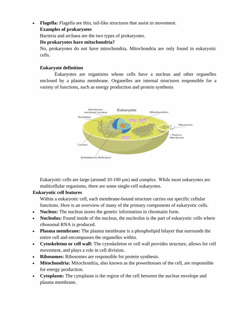
Eukaryotes are organisms whose cells have a nucleus and other organelles
enclosed by a plasma membrane. Organelles are internal structures responsible for a
variety of functions, such as energy production and protein synthesis
Eukaryotic cells are large (around 10-100 μm) and complex. While most eukaryotes are
multicellular organisms, there are some single-cell eukaryotes.
Eukaryotic cell features
Within a eukaryotic cell, each membrane-bound structure carries out specific cellular
functions. Here is an overview of many of the primary components of eukaryotic cells.
• Nucleus: The nucleus stores the genetic information in chromatin form.
• Nucleolus: Found inside of the nucleus, the nucleolus is the part of eukaryotic cells where
ribosomal RNA is produced.
• Plasma membrane: The plasma membrane is a phospholipid bilayer that surrounds the
entire cell and encompasses the organelles within.
• Cytoskeleton or cell wall: The cytoskeleton or cell wall provides structure, allows for cell
movement, and plays a role in cell division.
• Ribosomes: Ribosomes are responsible for protein synthesis.
• Mitochondria: Mitochondria, also known as the powerhouses of the cell, are responsible
for energy production.
• Cytoplasm: The cytoplasm is the region of the cell between the nuclear envelope and
plasma membrane.

• Cytosol: Cytosol is a gel-like substance within the cell that contains the organelles.
• Endoplasmic reticulum: The endoplasmic reticulum is an organelle dedicated to protein
maturation and transportation.
• Vesicles and vacuoles: Vesicles and vacuoles are membrane-bound sacs involved in
transportation and storage.
Other common organelles found in many, but not all, eukaryotes include the Golgi
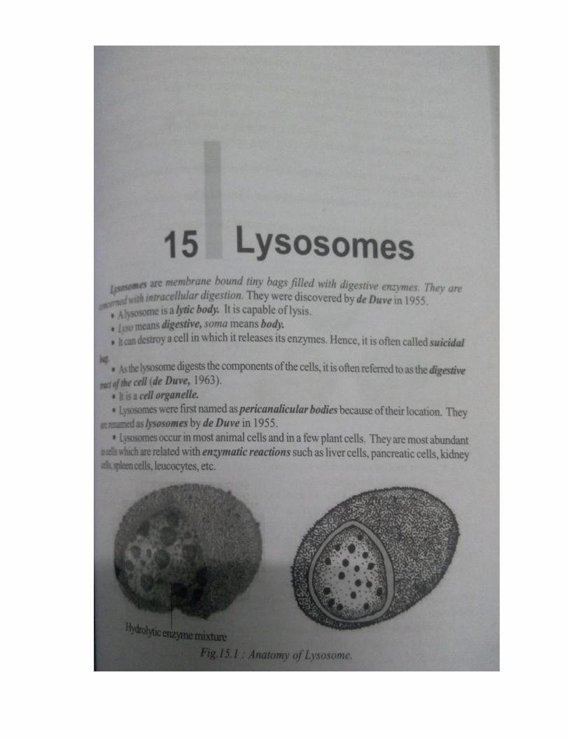
apparatus, chloroplasts and lysosomes.
Examples of eukaryotes
Animals, plants, fungi, algae and protozoans are all eukaryotes.
*******
PLASMA MEMBRANE - ULTRA STRUCTURE
Introduction
The plasma membrane (also known as the cell membrane or cytoplasmic
membrane) is a biological membrane that separates the interior of a cell from its outside
environment.
Plasma membrane composed of a phospholipid bilayer with embedded proteins,
the plasma membrane is selectively permeable to ions and organic molecules and
regulates the movement of substances in and out of cells. Plasma membranes must be very
flexible in order to allow certain cells, such as red blood cells and white blood cells, to
change shape as they pass through narrow capillaries.
The plasma membrane also plays a role in anchoring the cytoskeleton to provide
shape to the cell, and in attaching to the extracellular matrix and other cells to help group
cells together to form tissues. The membrane also maintains the cell potential.

UNIT MEMBRANE MODEL
The structure of the fatty acid tails of the phospholipids is important in determining
the properties of the membrane, and in particular, how fluid it is.
Saturated fatty acids have no double bonds (are saturated with hydrogens), so they are
relatively straight. Unsaturated fatty acids, on the other hand, contain one or more double
bonds, often resulting in a bend or kink. The saturated and unsaturated fatty acid tails of
phospholipids behave differently as temperature drops:
• At cooler temperatures, the straight tails of saturated fatty acids can pack tightly together,
making a dense and fairly rigid membrane.
• Phospholipids with unsaturated fatty acid tails cannot pack together as tightly because of
the bent structure of the tails. Because of this, a membrane containing unsaturated
phospholipids will stay fluid at lower temperatures than a membrane made of saturated
ones.
Most cell membranes contain a mixture of phospholipids, some with two saturated
(straight) tails and others with one saturated and one unsaturated (bent) tail. Many
organisms—fish are one example—can adjust physiologically to cold environments by
changing the proportion of unsaturated fatty acids in their membranes. For more
information about saturated and unsaturated fatty acids, see the article on lipids.
In addition to phospholipids, animals have an additional membrane component that helps
to maintain fluidity. Cholesterol, another type of lipid that is embedded among the
phospholipids of the membrane, helps to minimize the effects of temperature on fluidity.
At low temperatures, cholesterol increases fluidity by keeping phospholipids from packing
tightly together, while at high temperatures, it actually reduces fluidity^{3,4}3,4start
superscript, 3, comma, 4, end superscript. In this way, cholesterol expands the range of
temperatures at which a membrane maintains a functional, healthy fluidity.
FLUID MOSAIC MODEL
The currently accepted model for the structure of the plasma membrane, called
the fluid mosaic model, was first proposed in 1972. This model has evolved over time,

but it still provides a good basic description of the structure and behavior of membranes in
many cells.
According to the fluid mosaic model, the plasma membrane is a mosaic of
components—primarily, phospholipids, cholesterol, and proteins—that move freely and
fluidly in the plane of the membrane.
Source: Open Stax Biology.
Image of the plasma membrane, showing the phospholipid bilayer with peripheral and
integral membrane proteins, glycoproteins (proteins with a carbohydrate attached),
glycolipids (lipids with a carbohydrate attached), and cholesterol molecules.
The principal components of the plasma membrane are lipids (phospholipids and
cholesterol), proteins, and carbohydrate groups that are attached to some of the lipids and
proteins.
• A phospholipid is a lipid made of glycerol, two fatty acid tails, and a phosphate-linked
head group. Biological membranes usually involve two layers of phospholipids with their
tails pointing inward, an arrangement called a phospholipid bilayer.
• Cholesterol, another lipid composed of four fused carbon rings, is found alongside
phospholipids in the core of the membrane.
• Membrane proteins may extend partway into the plasma membrane, cross the membrane
entirely, or be loosely attached to its inside or outside face.
• Carbohydrate groups are present only on the outer surface of the plasma membrane and
are attached to proteins, forming glycoproteins, or lipids, forming glycolipids.
The proportions of proteins, lipids, and carbohydrates in the plasma membrane vary
between different types of cells. For a typical human cell, however, proteins account for
about 50 percent of the composition by mass, lipids (of all types) account for about 40
percent, and the remaining 10 percent comes from carbohydrates.

FUNCTIONS OF THE PLASMA MEMBRANE
A Physical Barrier
The plasma membrane surrounds all cells and physically separates the cytoplasm,
which is the material that makes up the cell, from the extracellular fluid outside the cell.
This protects all the components of the cell from the outside environment and allows
separate activities to occur inside and outside the cell.
The plasma membrane provides structural support to the cell. It tethers
the cytoskeleton, which is a network of protein filaments inside the cell that hold all the
parts of the cell in place. The cell wall is composed of molecules such as cellulose. It
provides additional support to the cell, and it is why plant cells do not burst like animal
cells do if too much water diffuses into them.
Selective Permeability
Plasma membranes are selectively permeable (or semi-permeable), meaning that
only certain molecules can pass through them. Water, oxygen, and carbon dioxide can
easily travel through the membrane. Generally, ions (e.g. sodium, potassium) and polar
molecules cannot pass through the membrane; they must go through specific channels or
pores in the membrane instead of freely diffusing through.
Endocytosis and Exocytosis
Endocytosis is when a cell ingests relatively larger contents than the single ions or
molecules that pass through channels. Through endocytosis, a cell can take in large
quantities of molecules or even whole bacteria from the extracellular fluid. Exocytosis is
when the cell releases these materials. The cell membrane plays an important role in both
of these processes. The shape of the membrane itself changes to allow molecules to enter
or exit the cell.
Cell Signaling
Another important function of the membrane is to facilitate communication and
signaling between cells. It does so through the use of various proteins and carbohydrates
in the membrane. Proteins on the cell “mark” that cell so that other cells can identify it.
The membrane also has receptors that allow it to carry out certain tasks when molecules
such as hormones bind to those receptors.
*******
CYTOPLASM- PHYSICAL AND BIOLOGICAL PROPERTIES
Definitions
The cytoplasm is a highly viscous (gel-like) substance enclosed within the cell
membrane. It is composed of water (about 85 percent), proteins (10 to 15 percent), lipids
(2 to 4 percent), nucleic acids, inorganic salts and polysaccharides in smaller amounts.
Depending on the cell, cytoplasm may also contain occasional granules of
inclusions (e.g. stored nutrients and pigments, etc). Apart from the cell membrane, which
encloses all cell components, a majority of cell organelles (ribosome, Golgi

apparatus, Endoplasmic Reticulum, etc) are located in the cytoplasm. For this reason, most
of the metabolic activities occur within the cytoplasm.
1. Physical properties:
The colloidal nature of cytoplasm is responsible for the various physical properties
a colloidal system may be defined as a system of liquid medium containing suspends
particles varying in diameter from 1/1,000,000 to 1/10,000 nm. Some of the physical
properties are follows.
Cytoplasm may be differentiated into a sol state and gel state. Sol state is the liquid
state and the gel state is the semi solid state. These two phases are interchangeable. This
capacity is known as phase reversal.
Elasticity:
Depending on the circumstances, the cytoplasm can extend or contract subject to a
certain limit. This is known as elasticity.
Cohesiveness:
The suspended particles in cytoplasm have mutual attraction, thus exhibiting
cohesiveness.
Contractility:
It is the capacity of peripheral cytoplasm to absorb or remove water from cell to
the exterior. This is manifested very well by guard cells.
Viscosity:
The suspended particles of cytoplasm are responsible for its viscous nature.
Brownian movement:
The suspended particles of cytoplasm are in a state of to and fro movement called
Brownian movement.
In addition to this, cytoplasm also exhibits movements (seen in the Plasmodium of
slime molds) and ‘cyclosis or streaming movements (seen in the leaf cells of Elodea).

2. Biological properties:
Irritability:
The irritability is the fundamental and inheritant property of the matrix.
Conductivity:
The conductivity is the process of conduction or transmission of excitation from
the place of its origin to the region of its reaction.
Movement:
The cytoplasmic matrix can perform movement due to cyclosis.
Growth:
Due to secretory or anabolic activities of the cell, new protoplasm continuously
increases in its volume.
Reproduction:
The cytoplasm has the property of asexual and sexual reproduction.
*******

Unit II