unit 11 anterior leg dorsum of foot. locate bones of the foot talus calcaneus cuboid navicular...
TRANSCRIPT

Unit 11
Anterior LegDorsum of Foot

Locate bones of the footTalus
Calcaneus
Cuboid
NavicularCuneiforms
5 Metatarsals
and 14
Phalanges
BonesPlate 523A
M
P
P
L I M
Lateral
TARSAL
TalusCalcaneus
CuboidNavicular
Cuneiforms
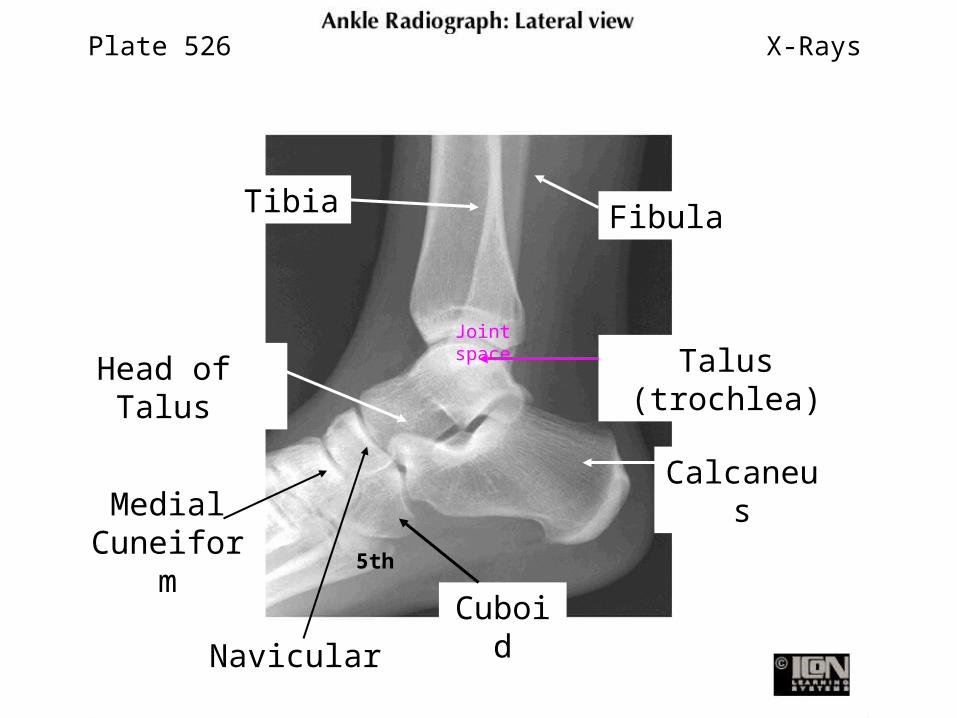
Plate 526
Joint space
X-Rays
Tibia Fibula
Talus (trochlea)
Head of Talus
CalcaneusMedial
Cuneiform
Navicular
5th
Cuboid

Identify the
Subtalar jointIdentify the
Transverse Tarsal joint
Standard method of surgical amputation
occurs at the transverse tarsal
joint
Plate 523B
N C
Bones
Planted
Inversion
Eversion
Transverse Tarsal JointTalocalcaneonavicul
arCalcaneocuboid
Calcaneus
Talus
Cuneiforms
Movements of:

Plate 526
Joint space
X-Rays
Tibia Fibula
Talus (trochlea)
Subtalar Joint space
CalcaneusMedial
Cuneiform
Navicular
5th
Cuboid
Transverse Tarsal
Joint

Plate 522B
Identify the deep fascia (crural) and the intermuscular
septa
They divide the leg into
three compartments; each has its
own nerve supply
Medial
T
FDeep Fascia of Leg
Posterior Intermuscular
Septum
Anterior Intermuscular
Septum
Interosseous
Membrane
A
L
P
Right Leg
Compartments
Anterior Compartment: Deep Fibular NerveLateral Compartment: Superficial Fibular
Nerve
Peroneal = Fibular
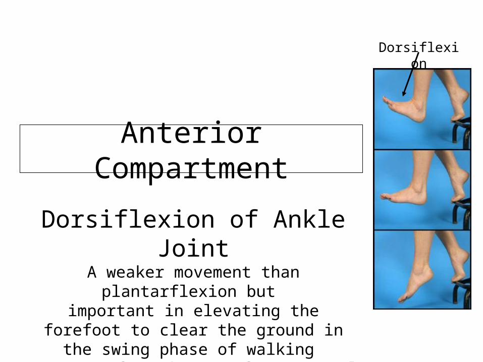
Anterior Compartment
Dorsiflexion of Ankle JointA weaker movement than plantarflexion
but important in elevating the forefoot to
clear the ground in the swing phase of walking
– range of 20 degrees from neutral
Dorsiflexion
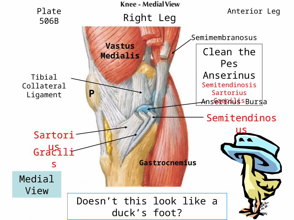
Clean the Pes
AnserinusSemitendinosis
SartoriusGracilis
Doesn’t this look like a duck’s foot?
Plate 506B Anterior Leg
P
Vastus Medialis
SartoriusGracilis
Semitendinosus
Semimembranosus
Tibial Collateral Ligament
Anserinus Bursa
Right Leg
Gastrocnemius
Medial View

Identify/CutSuperior Extensor
Retinacula Inferior Extensor
Retinacula
Plate 530
Retinacula prevent tendons
from bowstringing
Anterior Leg
Superior Extensor
Retinaculum
InferiorExtensor
Retinaculum
Synovial sheaths

Identify Muscles of Anterior
Compartment
Tibialis Anterior
Action:Dorsiflexio
nof foot and inversion of
foot
Plate 519
Read about shin splints
and compartment
syndromes pages 636 & 642, Moore
Anterior Leg
Tibialis AnteriorExtensor Hallicus Longus
Extensor Digitorum Longus
Fibularis Tertius
Tibialis Anterior
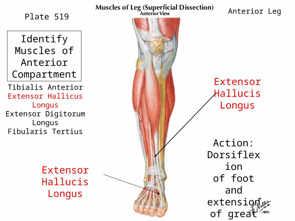
Extensor Hallucis Longus
Action:Dorsiflexio
nof foot and extension
of great toe
Plate 519
Extensor Hallucis Longus
Identify Muscles of Anterior
CompartmentTibialis Anterior
Extensor Hallicus LongusExtensor Digitorum
LongusFibularis Tertius
Anterior Leg

Extensor Digitorum
Longus
Action:Dorsiflexio
nof foot and extension of lateral four digits
Plate 519
Extensor Hood
Identify Muscles of Anterior
CompartmentTibialis Anterior
Extensor Hallicus LongusExtensor Digitorum
Longus Fibularis Tertius
Anterior Leg
Extensor Digitorum
Longus

Action:Dorsiflexio
nof foot and
aids in eversion
of foot
Fibularis Tertius
Really a part of Extensor Digitorum
Longus
Anterior Leg
Identify Fibularis Tertius
Fibularis Tertius
Extensor Digitorum Longus
Extensor Digitorum Longus
Plate 529A
Plate 519
5th
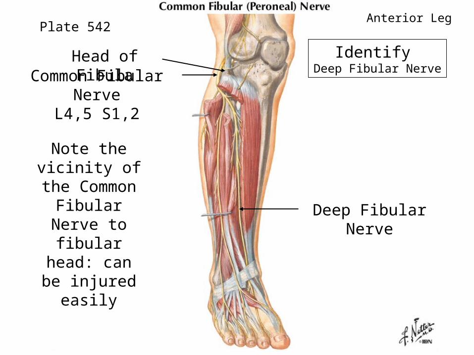
Identify Deep Fibular Nerve
Note the vicinity of
the Common Fibular
Nerve to fibular head:
can be injured easily
Plate 542
Deep Fibular Nerve
Common Fibular Nerve
L4,5 S1,2
Head of Fibula
Anterior Leg

Identify Anterior Tibial Artery
The Anterior Tibial Artery pierces and
then runs on the interosseous
membrane with the Deep Fibular
Nerve next to Tibialis Anterior
Plate 520 Anterior Leg
Anterior Tibial Artery
Anterior Tibial Artery

Dorsum of the Foot

Identify
ExtensorDigitorum Brevis
Helps extend the toes at MP and IP
joints
Plate 530
Extensor Hood
Dorsum of Foot
Extensor Retinaculum
Extensor Hallucis Longus
Extensor Digitorum BrevisExtensor Hallucis Brevis

Identify
Extensor Hallucis Brevis-part of EDB
Helps extend the big toe
Plate 530
Extensor Hood
Dorsum of Foot
Extensor Retinaculum
Extensor Hallucis Longus
Extensor Digitorum BrevisExtensor Hallucis Brevis

Identify 4 Dorsal Interossei or
DABS
There are also 3 PADS (plantar) on the bottom of
the foot
Plate 531
Check out extensor
hoods
Dorsum of Foot
EDB
DABS
All innervated by Lateral
Plantar Nerve (Tibial branch)
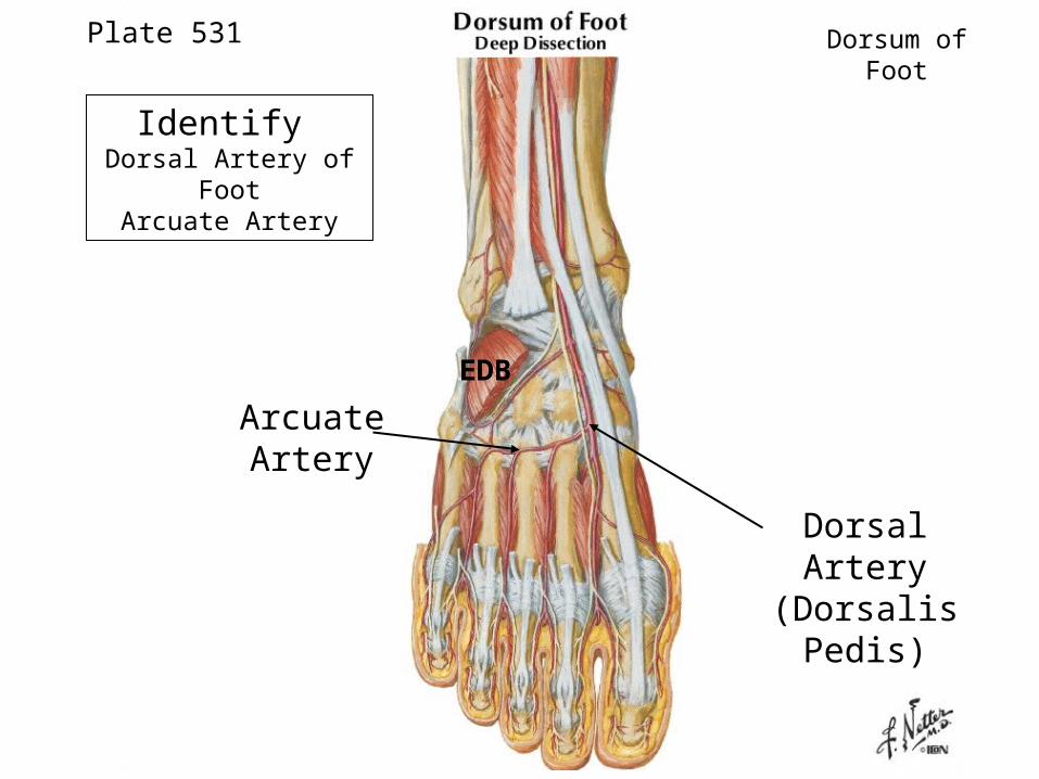
Plate 531 Dorsum of Foot
Dorsal Artery(Dorsalis
Pedis)
Arcuate Artery
EDB
Identify Dorsal Artery of
FootArcuate Artery

Follow Deep Fibular Nerve to cleft between big toe and 2nd
toe
“Ski boot”
syndrome p. 642
Plate 531 Dorsum of Foot
Deep Fibular Nerve
EDB
Both Dorsal artery and Deep
Fibular nerve are found lateral to
tendon of EHL
Lateral Compartment
Evertors
inversioneversion
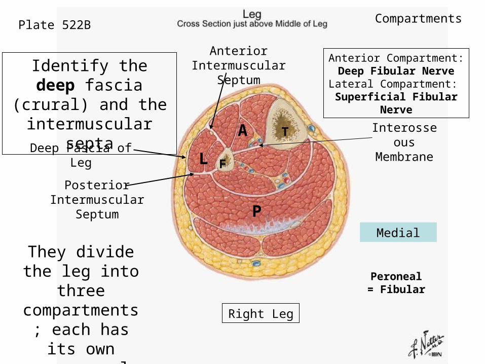
Plate 522B
Identify the deep fascia (crural) and the intermuscular
septa
They divide the leg into
three compartments; each has its
own nerve supply
Medial
T
FDeep Fascia of Leg
Posterior Intermuscular
Septum
Anterior Intermuscular
Septum
Interosseous
Membrane
A
L
P
Right Leg
Compartments
Anterior Compartment: Deep Fibular NerveLateral Compartment: Superficial Fibular
Nerve
Peroneal = Fibular
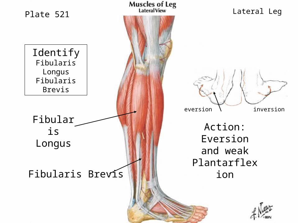
Identify Fibularis Longus Fibularis Brevis
Action: Eversion and
weak Plantarflexio
n
Plate 521
Fibularis Longus
Fibularis Brevis
Lateral Leg
inversioneversion

Identify Fibular
Retinacula
Plate 529A
Cut open the
retinacula to expose FL and FB
Lateral Leg
Fibular Retinacul
a
Fibularis Longus
Fibularis Brevis
Fibularis Longus
Fibularis Brevis
5th Metatarsal

Identify Superficial Fibular
Nerve
Plate 542
This nerve innervates the two
muscles in the lateral
compartment and then becomes cutaneous to
dorsum of foot
What vessel supplies
blood to the muscles in
lateral compartmen
t?
Lateral Leg
Superficial Fibular Nerve
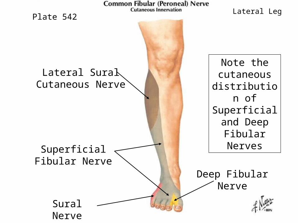
Note the cutaneous distribution
of Superficial and Deep
Fibular Nerves
Plate 542Lateral Leg
Lateral Sural Cutaneous Nerve
Superficial Fibular Nerve
Deep Fibular Nerve
Sural Nerve
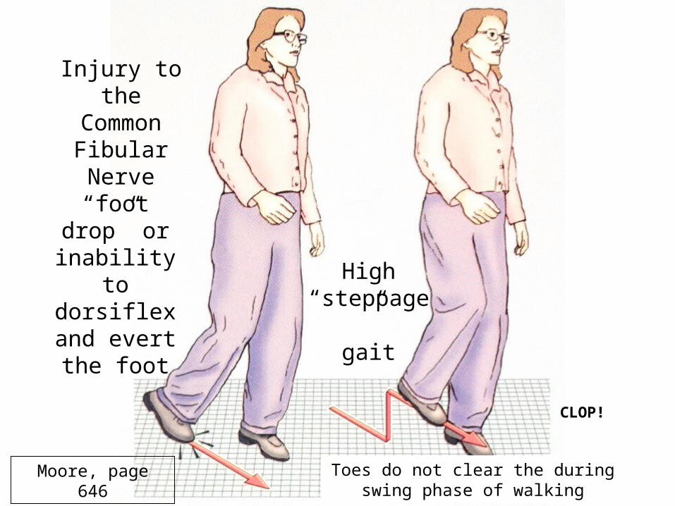
Injury to the
Common Fibular Nerve
“foot drop” or inability to dorsiflex and evert the foot
High “steppage
”gait
Toes do not clear the during swing phase of walking
Moore, page 646
CLOP!
Knee Joint
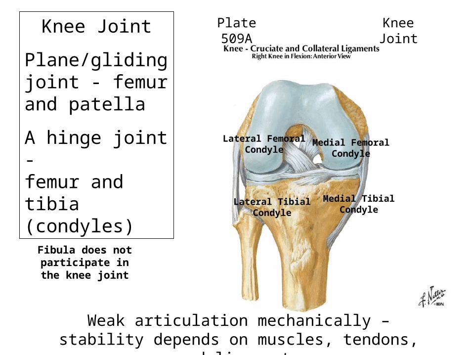
Knee Joint
Plane/gliding joint - femur and patella
A hinge joint - femur and tibia (condyles)
Weak articulation mechanically – stability depends on muscles, tendons, and ligaments
Plate 509A Knee Joint
Lateral Femoral Condyle
Medial Femoral Condyle
Medial Tibial Condyle
Lateral Tibial Condyle
Fibula does not participate in the knee joint

Plate 510
Lateral Condyle
Lateral Epicondyle
Medial Condyle
Medial Epicondyle
Head
Neck
Medial Condyle
Lateral Condyle
Femur
Tibia
Fibula
Right KneeKnee Joint
Patella

There are three major ligaments of the knee that strengthen it - the
knee joint itself is relatively unstable
because of the bony surfaces:
Patellar LigamentFibular Collateral LigamentTibial Collateral Ligament

Plastinized knee
or cadaver
Identify Patella Patellar
Ligament
Plate 507A
Quadriceps is the most
important muscle for stability of
joint
P
Patellar Ligamen
t
Knee Joint
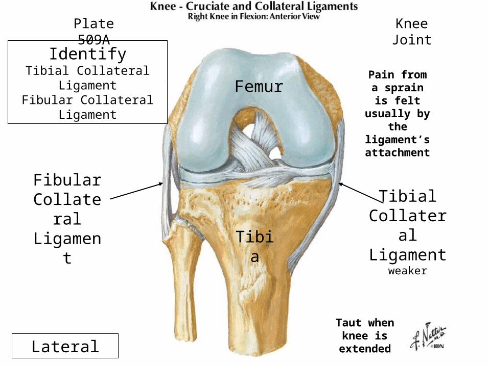
IdentifyTibial Collateral
LigamentFibular Collateral
Ligament
Plate 509A Knee Joint
Lateral
Fibular Collatera
l Ligamen
t
Tibial Collateral Ligament
weakerTibia
Femur
Taut when knee is
extended
Pain from a sprain is
felt usually by
the ligament’s attachmen
t

Identify Medial MeniscusLateral Meniscus
Note Popliteus tendon
The Medial Meniscus is
attached firmly to tibial
collateral ligament
Plate 509A
Act as shock
absorbers and deepen
socket
Knee Joint
Lateral
Lateral Meniscus Medial
Meniscus ©
Femur
Tibia
Where do meniscal tears
heal best?
Pain felt near joint
line
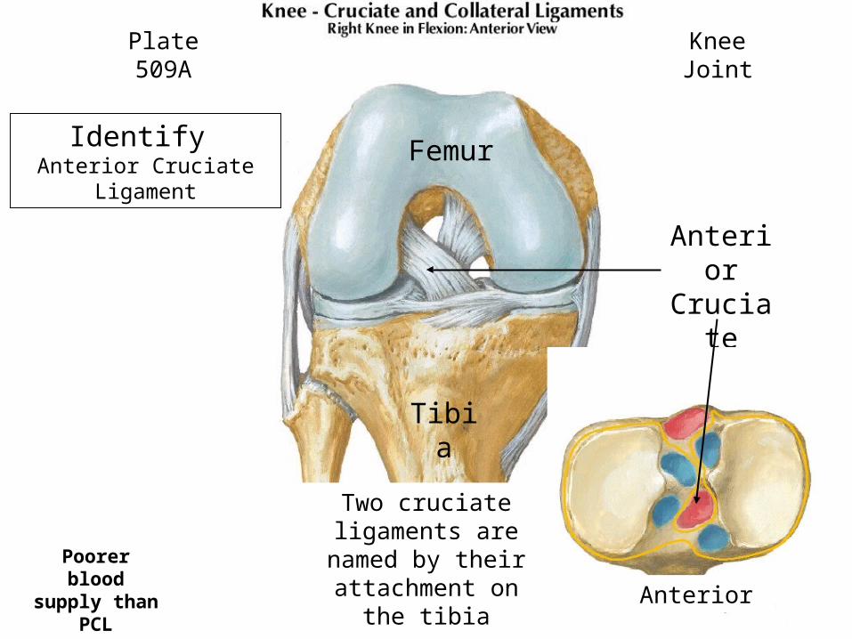
Identify Anterior Cruciate
Ligament
Plate 509A
Two cruciate ligaments are
named by their attachment on the
tibia
Knee Joint
Anterior Cruciat
e Ligame
nt
Anterior
Femur
Tibia
Poorer blood
supply than PCL

Resists posterior
displacement of femur on
tibia or hyperextension
Knee Joint

Use middle one third of patellar tendon to repair
Knee JointMoore, page 698

Identify Posterior Cruciate
Ligament
Plate 509B
Stronger of cruciate
ligaments
Knee Joint
Posterior Cruciate Ligament
Anterior
Lateral

Resists anterior
displacement of femur on
tibia and hyperflexion
Knee Joint
Tibia
Main stabilizer of femur when walking downhill

Knee Joint
Moore, page 698
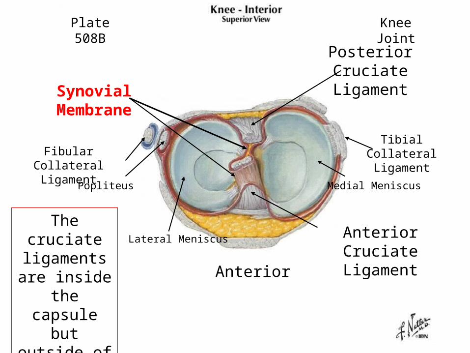
The cruciate
ligaments are inside
the capsule but outside
of the synovial
membrane
Anterior
Anterior Cruciate Ligament
Posterior Cruciate Ligament
Plate 508B Knee Joint
Synovial Membran
eFibular Collateral
Ligament
Tibial Collateral Ligament
Popliteus
Lateral Meniscus
Medial Meniscus
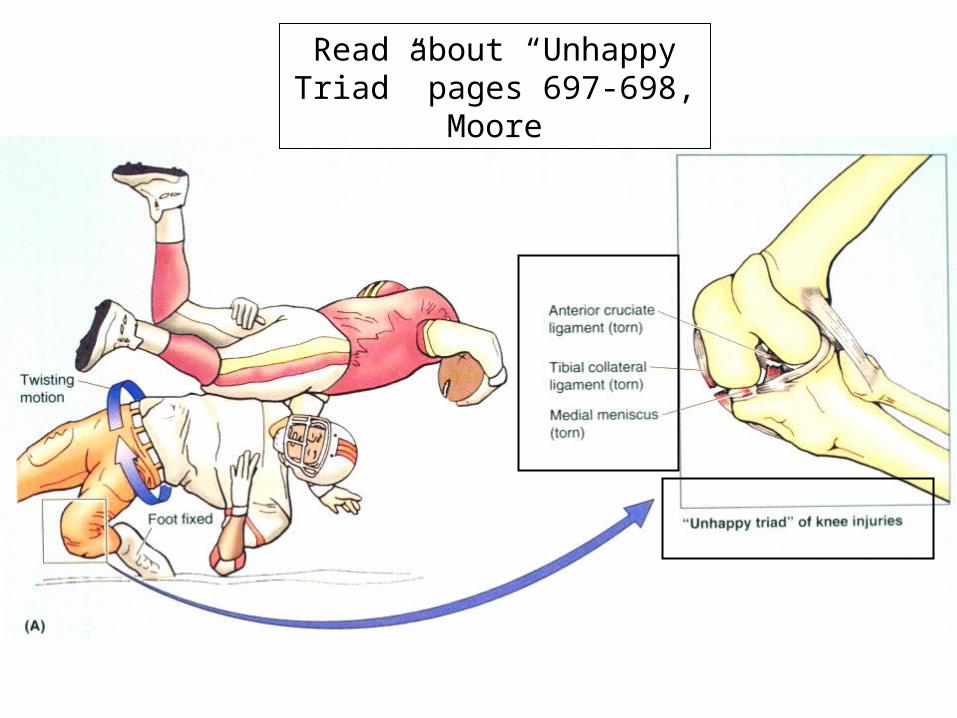
Read about “Unhappy Triad” pages 697-698,
Moore
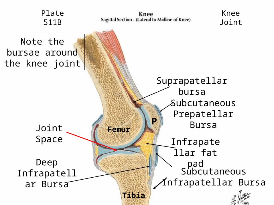
Note the bursae around the knee
joint
Plate 511B Knee Joint
Infrapatellar fat pad
Suprapatellar bursa
Subcutaneous Infrapatellar Bursa
Deep Infrapatellar
Bursa
Subcutaneous Prepatellar Bursa
Joint Space
Femur
Tibia
P
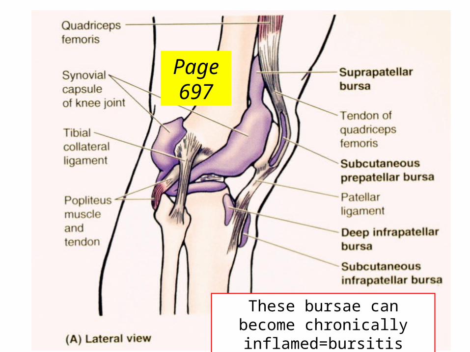
Page 697
These bursae can become chronically
inflamed=bursitis
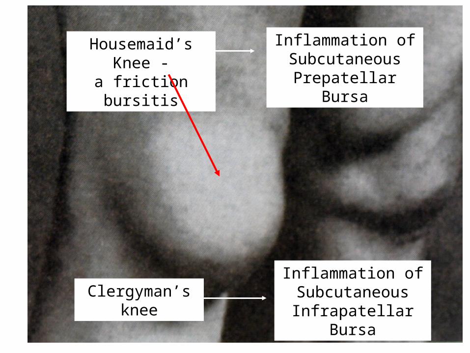
Housemaid’s Knee -
a friction bursitis
Inflammation of Subcutaneous
Prepatellar Bursa
Clergyman’s knee
Inflammation of Subcutaneous
Infrapatellar Bursa

Ankle Joint - Talocrural
HINGE JOINT
Joint between medial malleolus of tibia, lateral
malleolus of fibula and the talus
MOST COMMONLY INJURED JOINT IN THE BODY

Plate 526BLeft Ankle
Medial Malleolus
Lateral Malleolus
Tibia
Fibula
Talus
Talocrural Joint
Calcaneus
Talocrural joint

Identify Deltoid (Medial) Ligament – 4 parts1. Anterior Tibiotalar2. Tibionavicular3. Tibiocalcaneal4. Posterior Tibiotalar
Eversion sprain
Plate 527BTalocrural Joint
1
23
4
inversioneversion
Tibia
Calcaneus
Talus

Moore, Page 707
Pott’s fracture-dislocation occurs when the foot is forcibly everted: pulls on strong medial ligament
often tearing off the medial malleolus. Talus moves laterally shearing off the lateral malleolus or more
commonly, breaking the fibula.
Talocrural Joint
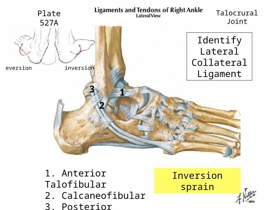
Identify Lateral
Collateral Ligament
Plate 527A Talocrural Joint
12
3
1. Anterior Talofibular 2. Calcaneofibular3. Posterior Talofibular
Inversion sprain
inversioneversion

Page 706, Moore
Inversion sprains are the most common
Anterior Talofibular Ligament is most often sprained
Talocrural Joint

Laboratory/Quiz