unique mutation portraits and frequent col2a1 gene alteration in
TRANSCRIPT
Research
Unique mutation portraits and frequent COL2A1 genealteration in chondrosarcomaYasushi Totoki,1 Akihiko Yoshida,2 Fumie Hosoda,1 Hiromi Nakamura,1 Natsuko Hama,1
Koichi Ogura,3 Aki Yoshida,4 Tomohiro Fujiwara,3 Yasuhito Arai,1 Junya Toguchida,5
Hitoshi Tsuda,2 Satoru Miyano,6 Akira Kawai,3 and Tatsuhiro Shibata1
1Division of Cancer Genomics, National Cancer Center Research Institute, Chuo-ku, Tokyo, 104-0045, Japan; 2Division of Pathology
and Clinical Laboratories, 3Division of Musculoskeletal Oncology, National Cancer Center Hospital, Chuo-ku, Tokyo, 104-0045, Japan;4Department of Orthopaedic Surgery, Okayama University Graduate School of Medicine, Dentistry and Pharmaceutical Sciences,
Okayama, 700-8558, Japan; 5Department of Tissue Regeneration, Institute for Frontier Medical Sciences, Kyoto University, Kyoto,
606-8507, Japan; 6Laboratory of DNA Informatics Analysis, Human Genome Center, The Institute of Medical Science, The University
of Tokyo, Minato-ku, Tokyo, 108-8639, Japan
Chondrosarcoma is the second most frequent malignant bone tumor. However, the etiological background of chon-drosarcomagenesis remains largely unknown, along with details on molecular alterations and potential therapeutic tar-gets. Massively parallel paired-end sequencing of whole genomes of 10 primary chondrosarcomas revealed that theprocess of accumulation of somatic mutations is homogeneous irrespective of the pathological subtype or the presence ofIDH1mutations, is unique among a range of cancer types, and shares significant commonalities with that of prostate cancer.Clusters of structural alterations localized within a single chromosome were observed in four cases. Combined with tar-geted resequencing of additional cartilaginous tumor cohorts, we identified somatic alterations of the COL2A1 gene, whichencodes an essential extracellular matrix protein in chondroskeletal development, in 19.3% of chondrosarcoma and 31.7%of enchondroma cases. Epigenetic regulators (IDH1 and YEATS2) and an activin/BMP signal component (ACVR2A) wererecurrently altered. Furthermore, a novel FN1-ACVR2A fusion transcript was observed in both chondrosarcoma andosteochondromatosis cases. With the characteristic accumulative process of somatic changes as a background, moleculardefects in chondrogenesis and aberrant epigenetic control are primarily causative of both benign and malignant carti-laginous tumors.
[Supplemental material is available for this article.]
Chondrosarcoma accounts for >20% of primary bone sarcomas
with an overall incidence rate estimated at approximately one in
200,000 (Whelan et al. 2012). The patients are mostly older than
50 yr and show male dominance. There are two common sub-
types: central and peripheral. Central chondrosarcoma pre-
dominates (;80%) and arises in the medullary cavity of the long
bone, while peripheral chondrosarcoma (;15%) develops from
the surface of the bone (Fletcher et al. 2002; Bov�ee et al. 2010).
Clinically, low-grade chondrosarcomas rarely metastasize and can
be managed with local resection. In contrast, high-grade chon-
drosarcomas often metastasize and are lethal in most cases. Since
the tumor cells exist in specific microenvironments such as low
vascularity and accumulated extracellular matrix, they are large-
ly resistant to conventional chemotherapy and radiotherapy.
Therefore, identification of new therapeutic targets is required for
this tumor.
Benign cartilage tumors (enchondroma and osteochondroma)
may progress to chondrosarcoma (Bov�ee et al. 2010). Mutations of
exostosin 1 (EXT1) and EXT2 genes are linked to hereditary and
sporadic osteochondromatosis and are also reported in chon-
drosarcoma (Hecht et al. 1997; Wuyts et al. 1998). EXT1 and EXT2
regulate proper heparan sulfate proteoglycan processing, and their
defects cause abnormal diffusion of hedgehog ligands (Koziel et al.
2004). Mutations in the PTH1R gene were also identified in
enchondroma, which disrupts the IHH–PTHLH feedback loop
and also induces constitutive hedgehog signaling (Hopyan et al.
2002). Consistently transgenic mice that express Gli2—a down-
stream effector of the hedgehog signal—in chondrocytes develop
cartilaginous lesions similar to human enchondromas (Hopyan
et al. 2002).
In addition to hedgehog signaling, alterations of other mo-
lecular pathways such as TP53/MDM2 (Wadayama et al. 1993;
Larramendy et al. 1997), AKT1 (Schrage et al. 2009), and CDK4/RB
(Asp et al. 2001) have been reported in chondrosarcomas. Recently,
frequent somatic mutations in isocitrate dehydrogenase 1 (IDH1)
and IDH2 have been identified in both enchondroma and central
chondrosarcoma (Amary et al. 2011a), and somatic mosaic IDH1/2
mutations are associated with multiple enchondromatosis (Amary
et al. 2011b; Pansuriya et al. 2011). These molecular and genetic
observations support a close association between benign osteo-
cartilaginous tumors and chondrosarcoma; however, the detailed
molecular events and etiological risk factors underlying chon-
drosarcomagenesis remain largely unknown.
� 2014 Totoki et al. This article is distributed exclusively by Cold SpringHarbor Laboratory Press for the first six months after the full-issue publicationdate (see http://genome.cshlp.org/site/misc/terms.xhtml). After six months, itis available under a Creative Commons License (Attribution-NonCommercial4.0 International), as described at http://creativecommons.org/licenses/by-nc/4.0/.
Corresponding author: [email protected] published online before print. Article, supplemental material, and pub-lication date are at http://www.genome.org/cgi/doi/10.1101/gr.160598.113.
24:000–000 Published by Cold Spring Harbor Laboratory Press; ISSN 1088-9051/14; www.genome.org Genome Research 1www.genome.org
Cold Spring Harbor Laboratory Press on April 4, 2018 - Published by genome.cshlp.orgDownloaded from
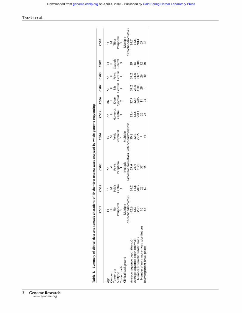
Table
1.
Summary
ofclinicaldata
andso
maticalterationsof10ch
ondrosarcomaca
sesanalyzedbywhole-genomesequencing
CS01
CS02
CS03
CS04
CS05
CS06
CS07
CS08
CS09
CS10
Age
14
32
58
43
62
86
50
58
54
33
Gender
FM
MM
FF
FF
FM
Tumorsite
Rib
Pelvis
Pelvis
Pelvis
Humerus
Knee
Rib
Pelvis
Scapula
Tibia
Subtype
Peripheral
Central
Peripheral
Peripheral
Central
Synovial
Central
Central
Central
Peripheral
Tumorgrade
12
11
32
22
31
Clin
icalbackground
Multiple
osteoch
ondromatosis
Multiple
osteoch
ondromatosis
Multiple
osteoch
ondromatosis
Multiple
osteoch
ondromatosis
Averagesequence
depth
(tumor)
42.6
34.2
27.6
30.8
33.4
37.7
37.2
37.2
29
24.7
Averagesequence
depth
(norm
al)
32.7
31.6
25.4
32.9
32.8
32.7
27.6
31.6
35
31.6
Numberofsomaticsubstitutions
3521
5005
6128
2215
5043
3705
4100
5326
2288
7014
Numberofnonsynonymoussubstitutions
10
26
37
726
11
29
26
12
27
Rearrangementbreakpoints
66
60
45
44
24
23
140
10
37
2 Genome Researchwww.genome.org
Totoki et al.
Cold Spring Harbor Laboratory Press on April 4, 2018 - Published by genome.cshlp.orgDownloaded from
Results
Whole-genome sequencing (WGS) of chondrosarcoma
Massively parallel paired-end sequencing of the whole genomes of
10 pairs of primary chondrosarcoma and matched normal muscle
tissues was performed. The cases included five central, four pe-
ripheral, and one synovial—a rare subtype (Table 1). The median
sequence coverage was 33.43 for tumor tissue and 31.43 for
normal tissue (Table 1). All peripheral cases were associated with
osteochondromatosis and harbored germline EXT1 or EXT2 mu-
tations (Supplemental Table S1). No IDH1/2 or other enchon-
dromatosis-associated gene mutations (PTH1R, PTPN11, or ACP5)
(Hopyan et al. 2002; Bowen et al. 2011; Briggs et al. 2011) were
observed in the germline genomes. In total, 44,345 somatic single-
nucleotide variations (SNVs; ranging from2215–7014 per genome,
1.55/Mb on average) and 4096 small insertions/deletions (indels,
ranging from 269–544 per genome) were identified (Fig. 1A; Sup-
plemental Table S2). The somatic point mutations included 211
nonsynonymous mutations (21.1 per genome on average) and 14
indels (1.4 per genome on average) in the coding regions (Sup-
plemental Table S3). More than 91% (78/85) of somatic sub-
stitutions and 63% (17/27) of somatic indels were validated by
Sanger sequencing. Thesemutationswere significantly enriched in
membranous proteins, especially those with transmembrane re-
ceptor activity (Supplemental Table S4).
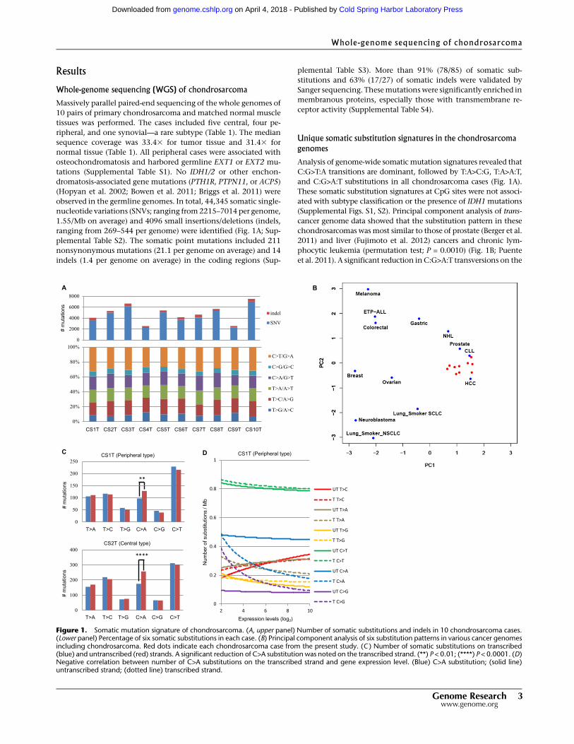
Unique somatic substitution signatures in the chondrosarcomagenomes
Analysis of genome-wide somatic mutation signatures revealed that
C:G>T:A transitions are dominant, followed by T:A>C:G, T:A>A:T,
and C:G>A:T substitutions in all chondrosarcoma cases (Fig. 1A).
These somatic substitution signatures at CpG sites were not associ-
ated with subtype classification or the presence of IDH1 mutations
(Supplemental Figs. S1, S2). Principal component analysis of trans-
cancer genome data showed that the substitution pattern in these
chondrosarcomas wasmost similar to those of prostate (Berger et al.
2011) and liver (Fujimoto et al. 2012) cancers and chronic lym-
phocytic leukemia (permutation test; P = 0.0010) (Fig. 1B; Puente
et al. 2011). A significant reduction inC:G>A:T transversions on the
Figure 1. Somatic mutation signature of chondrosarcoma. (A, upper panel) Number of somatic substitutions and indels in 10 chondrosarcoma cases.(Lower panel) Percentage of six somatic substitutions in each case. (B) Principal component analysis of six substitution patterns in various cancer genomesincluding chondrosarcoma. Red dots indicate each chondrosarcoma case from the present study. (C ) Number of somatic substitutions on transcribed(blue) and untranscribed (red) strands. A significant reduction of C>A substitution was noted on the transcribed strand. (**) P < 0.01; (****) P < 0.0001. (D)Negative correlation between number of C>A substitutions on the transcribed strand and gene expression level. (Blue) C>A substitution; (solid line)untranscribed strand; (dotted line) transcribed strand.
Whole-genome sequencing of chondrosarcoma
Genome Research 3www.genome.org
Cold Spring Harbor Laboratory Press on April 4, 2018 - Published by genome.cshlp.orgDownloaded from
transcribed strand was observed in both central (CS-2T, 7T, 8T, and
9T) and peripheral (CS-1T, 3T, and 10T) cases (Fig. 1C; Supplemental
Fig. S3), which correlated with gene expression level (Fig. 1D).
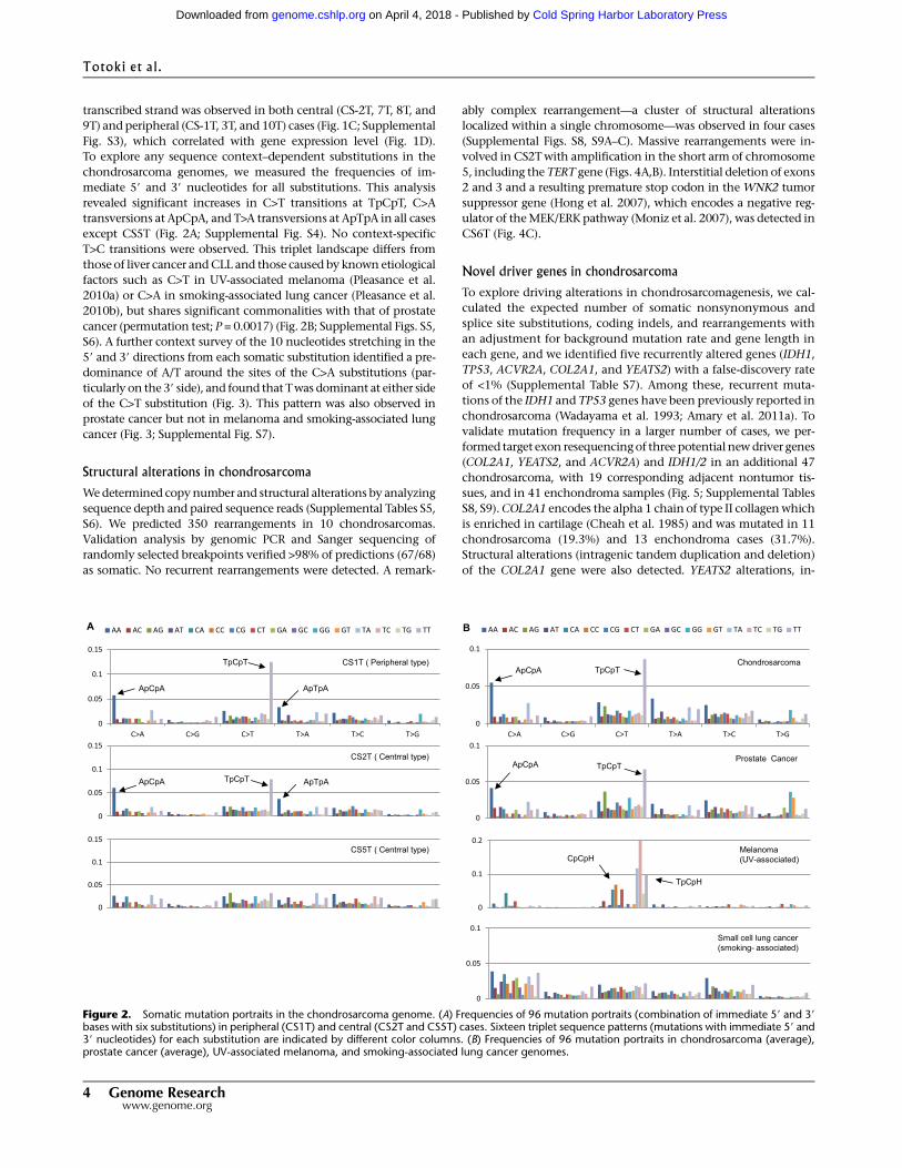
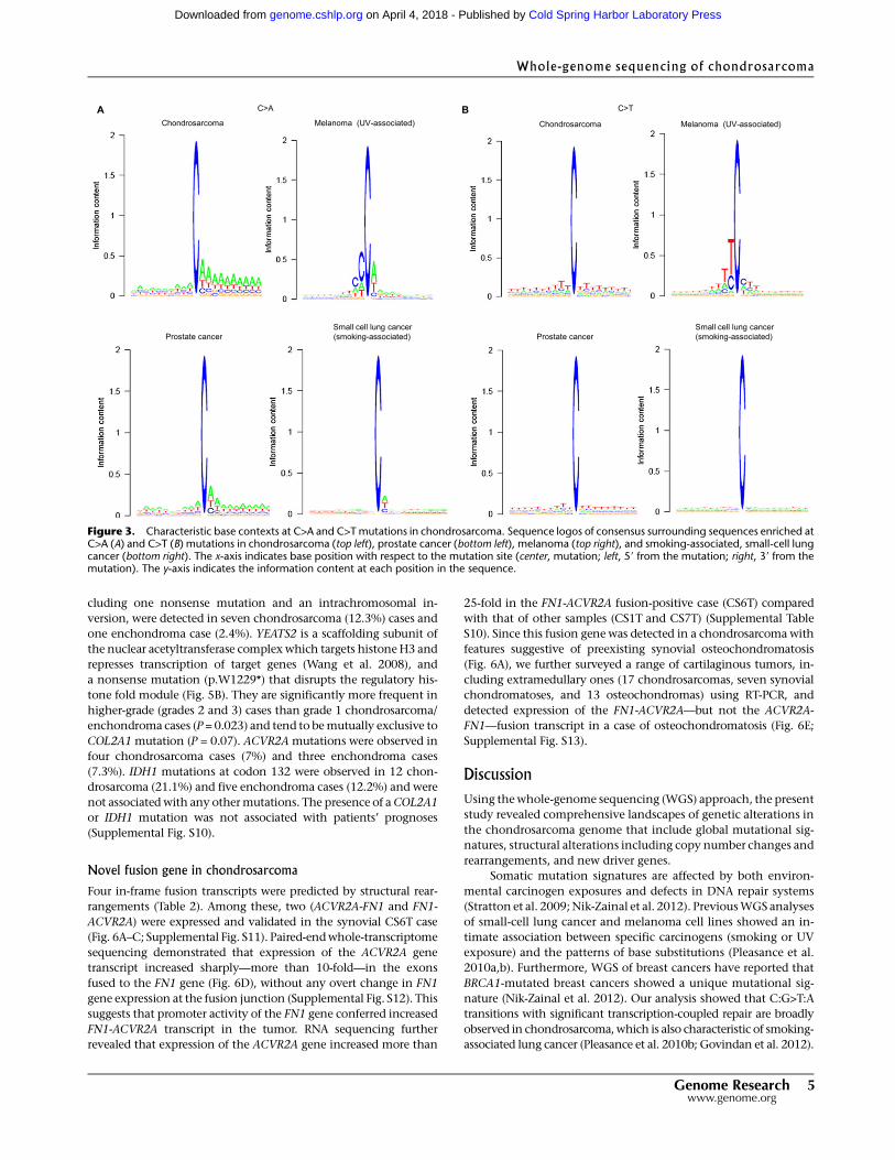
To explore any sequence context–dependent substitutions in the
chondrosarcoma genomes, we measured the frequencies of im-
mediate 59 and 39 nucleotides for all substitutions. This analysis
revealed significant increases in C>T transitions at TpCpT, C>A
transversions at ApCpA, and T>A transversions at ApTpA in all cases
except CS5T (Fig. 2A; Supplemental Fig. S4). No context-specific
T>C transitions were observed. This triplet landscape differs from
those of liver cancer andCLL and those caused by known etiological
factors such as C>T in UV-associated melanoma (Pleasance et al.
2010a) or C>A in smoking-associated lung cancer (Pleasance et al.
2010b), but shares significant commonalities with that of prostate
cancer (permutation test; P = 0.0017) (Fig. 2B; Supplemental Figs. S5,
S6). A further context survey of the 10 nucleotides stretching in the
59 and 39 directions from each somatic substitution identified a pre-
dominance of A/T around the sites of the C>A substitutions (par-
ticularly on the 39 side), and found that Twas dominant at either side
of the C>T substitution (Fig. 3). This pattern was also observed in
prostate cancer but not in melanoma and smoking-associated lung
cancer (Fig. 3; Supplemental Fig. S7).
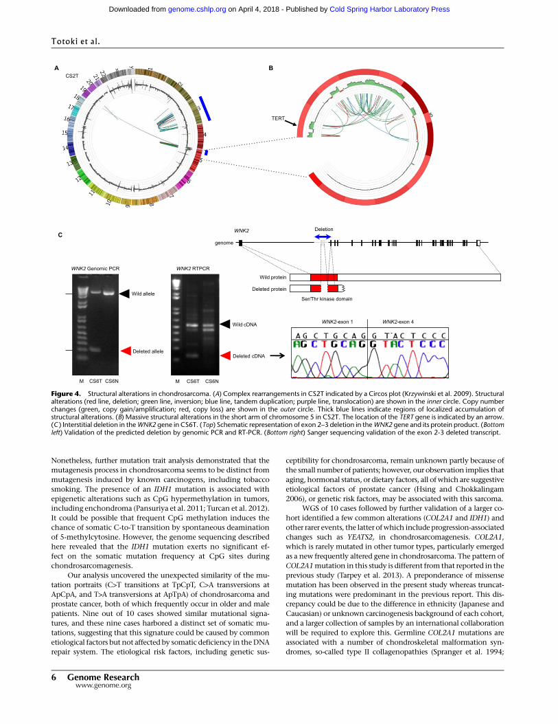
Structural alterations in chondrosarcoma
Wedetermined copynumber and structural alterations by analyzing
sequence depth and paired sequence reads (Supplemental Tables S5,
S6). We predicted 350 rearrangements in 10 chondrosarcomas.
Validation analysis by genomic PCR and Sanger sequencing of
randomly selected breakpoints verified >98% of predictions (67/68)
as somatic. No recurrent rearrangements were detected. A remark-
ably complex rearrangement—a cluster of structural alterations
localized within a single chromosome—was observed in four cases
(Supplemental Figs. S8, S9A–C). Massive rearrangements were in-
volved in CS2Twith amplification in the short arm of chromosome
5, including the TERT gene (Figs. 4A,B). Interstitial deletion of exons
2 and 3 and a resulting premature stop codon in the WNK2 tumor
suppressor gene (Hong et al. 2007), which encodes a negative reg-
ulator of theMEK/ERK pathway (Moniz et al. 2007), was detected in
CS6T (Fig. 4C).
Novel driver genes in chondrosarcoma
To explore driving alterations in chondrosarcomagenesis, we cal-
culated the expected number of somatic nonsynonymous and
splice site substitutions, coding indels, and rearrangements with
an adjustment for background mutation rate and gene length in
each gene, and we identified five recurrently altered genes (IDH1,
TP53, ACVR2A, COL2A1, and YEATS2) with a false-discovery rate
of <1% (Supplemental Table S7). Among these, recurrent muta-
tions of the IDH1 and TP53 genes have been previously reported in
chondrosarcoma (Wadayama et al. 1993; Amary et al. 2011a). To
validate mutation frequency in a larger number of cases, we per-
formed target exon resequencingof three potential newdriver genes
(COL2A1, YEATS2, and ACVR2A) and IDH1/2 in an additional 47
chondrosarcoma, with 19 corresponding adjacent nontumor tis-
sues, and in 41 enchondroma samples (Fig. 5; Supplemental Tables
S8, S9).COL2A1 encodes the alpha 1 chain of type II collagenwhich
is enriched in cartilage (Cheah et al. 1985) and was mutated in 11
chondrosarcoma (19.3%) and 13 enchondroma cases (31.7%).
Structural alterations (intragenic tandem duplication and deletion)
of the COL2A1 gene were also detected. YEATS2 alterations, in-
Figure 2. Somatic mutation portraits in the chondrosarcoma genome. (A) Frequencies of 96 mutation portraits (combination of immediate 59 and 39bases with six substitutions) in peripheral (CS1T) and central (CS2T and CS5T) cases. Sixteen triplet sequence patterns (mutations with immediate 59 and39 nucleotides) for each substitution are indicated by different color columns. (B) Frequencies of 96 mutation portraits in chondrosarcoma (average),prostate cancer (average), UV-associated melanoma, and smoking-associated lung cancer genomes.
Totoki et al.
4 Genome Researchwww.genome.org
Cold Spring Harbor Laboratory Press on April 4, 2018 - Published by genome.cshlp.orgDownloaded from
cluding one nonsense mutation and an intrachromosomal in-
version, were detected in seven chondrosarcoma (12.3%) cases and
one enchondroma case (2.4%). YEATS2 is a scaffolding subunit of
the nuclear acetyltransferase complexwhich targets histoneH3 and
represses transcription of target genes (Wang et al. 2008), and
a nonsense mutation (p.W1229*) that disrupts the regulatory his-
tone fold module (Fig. 5B). They are significantly more frequent in
higher-grade (grades 2 and 3) cases than grade 1 chondrosarcoma/
enchondroma cases (P = 0.023) and tend to bemutually exclusive to
COL2A1 mutation (P = 0.07). ACVR2A mutations were observed in
four chondrosarcoma cases (7%) and three enchondroma cases
(7.3%). IDH1 mutations at codon 132 were observed in 12 chon-
drosarcoma (21.1%) and five enchondroma cases (12.2%) and were
not associatedwith any othermutations. The presence of aCOL2A1
or IDH1 mutation was not associated with patients’ prognoses
(Supplemental Fig. S10).
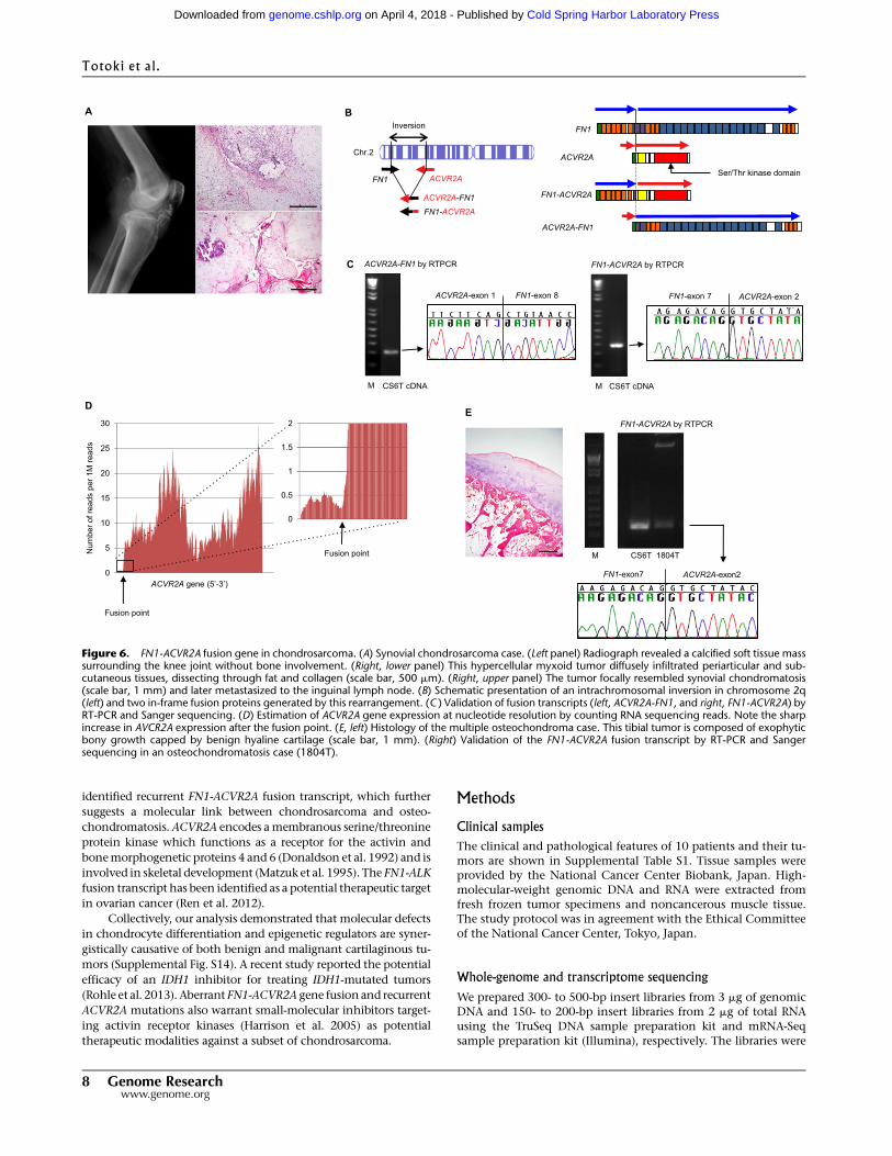
Novel fusion gene in chondrosarcoma
Four in-frame fusion transcripts were predicted by structural rear-
rangements (Table 2). Among these, two (ACVR2A-FN1 and FN1-
ACVR2A) were expressed and validated in the synovial CS6T case
(Fig. 6A–C; Supplemental Fig. S11). Paired-endwhole-transcriptome
sequencing demonstrated that expression of the ACVR2A gene
transcript increased sharply—more than 10-fold—in the exons
fused to the FN1 gene (Fig. 6D), without any overt change in FN1
gene expression at the fusion junction (Supplemental Fig. S12). This
suggests that promoter activity of the FN1 gene conferred increased
FN1-ACVR2A transcript in the tumor. RNA sequencing further
revealed that expression of the ACVR2A gene increased more than
25-fold in the FN1-ACVR2A fusion-positive case (CS6T) compared
with that of other samples (CS1T and CS7T) (Supplemental Table
S10). Since this fusion gene was detected in a chondrosarcoma with
features suggestive of preexisting synovial osteochondromatosis
(Fig. 6A), we further surveyed a range of cartilaginous tumors, in-
cluding extramedullary ones (17 chondrosarcomas, seven synovial
chondromatoses, and 13 osteochondromas) using RT-PCR, and
detected expression of the FN1-ACVR2A—but not the ACVR2A-
FN1—fusion transcript in a case of osteochondromatosis (Fig. 6E;
Supplemental Fig. S13).
DiscussionUsing thewhole-genome sequencing (WGS) approach, the present
study revealed comprehensive landscapes of genetic alterations in
the chondrosarcoma genome that include global mutational sig-
natures, structural alterations including copy number changes and
rearrangements, and new driver genes.
Somatic mutation signatures are affected by both environ-
mental carcinogen exposures and defects in DNA repair systems
(Stratton et al. 2009;Nik-Zainal et al. 2012). PreviousWGS analyses
of small-cell lung cancer and melanoma cell lines showed an in-
timate association between specific carcinogens (smoking or UV
exposure) and the patterns of base substitutions (Pleasance et al.
2010a,b). Furthermore, WGS of breast cancers have reported that
BRCA1-mutated breast cancers showed a unique mutational sig-
nature (Nik-Zainal et al. 2012). Our analysis showed that C:G>T:A
transitions with significant transcription-coupled repair are broadly
observed in chondrosarcoma,which is also characteristic of smoking-
associated lung cancer (Pleasance et al. 2010b; Govindan et al. 2012).
Figure 3. Characteristic base contexts at C>A and C>Tmutations in chondrosarcoma. Sequence logos of consensus surrounding sequences enriched atC>A (A) and C>T (B) mutations in chondrosarcoma (top left), prostate cancer (bottom left), melanoma (top right), and smoking-associated, small-cell lungcancer (bottom right). The x-axis indicates base position with respect to the mutation site (center, mutation; left, 59 from the mutation; right, 39 from themutation). The y-axis indicates the information content at each position in the sequence.
Whole-genome sequencing of chondrosarcoma
Genome Research 5www.genome.org
Cold Spring Harbor Laboratory Press on April 4, 2018 - Published by genome.cshlp.orgDownloaded from
Nonetheless, further mutation trait analysis demonstrated that the
mutagenesis process in chondrosarcoma seems to be distinct from
mutagenesis induced by known carcinogens, including tobacco
smoking. The presence of an IDH1 mutation is associated with
epigenetic alterations such as CpG hypermethylation in tumors,
including enchondroma (Pansuriya et al. 2011; Turcan et al. 2012).
It could be possible that frequent CpG methylation induces the
chance of somatic C-to-T transition by spontaneous deamination
of 5-methylcytosine. However, the genome sequencing described
here revealed that the IDH1 mutation exerts no significant ef-
fect on the somatic mutation frequency at CpG sites during
chondrosarcomagenesis.
Our analysis uncovered the unexpected similarity of the mu-
tation portraits (C>T transitions at TpCpT, C>A transversions at
ApCpA, and T>A transversions at ApTpA) of chondrosarcoma and
prostate cancer, both of which frequently occur in older and male
patients. Nine out of 10 cases showed similar mutational signa-
tures, and these nine cases harbored a distinct set of somatic mu-
tations, suggesting that this signature could be caused by common
etiological factors but not affected by somatic deficiency in theDNA
repair system. The etiological risk factors, including genetic sus-
ceptibility for chondrosarcoma, remain unknown partly because of
the small number of patients; however, our observation implies that
aging, hormonal status, or dietary factors, all of which are suggestive
etiological factors of prostate cancer (Hsing and Chokkalingam
2006), or genetic risk factors, may be associated with this sarcoma.
WGS of 10 cases followed by further validation of a larger co-
hort identified a few common alterations (COL2A1 and IDH1) and
other rarer events, the latter ofwhich include progression-associated
changes such as YEATS2, in chondrosarcomagenesis. COL2A1,
which is rarely mutated in other tumor types, particularly emerged
as a new frequently altered gene in chondrosarcoma. The pattern of
COL2A1mutation in this study is different from that reported in the
previous study (Tarpey et al. 2013). A preponderance of missense
mutation has been observed in the present study whereas truncat-
ing mutations were predominant in the previous report. This dis-
crepancy could be due to the difference in ethnicity (Japanese and
Caucasian) or unknown carcinogenesis background of each cohort,
and a larger collection of samples by an international collaboration
will be required to explore this. Germline COL2A1 mutations are
associated with a number of chondroskeletal malformation syn-
dromes, so-called type II collagenopathies (Spranger et al. 1994;
Figure 4. Structural alterations in chondrosarcoma. (A) Complex rearrangements in CS2T indicated by a Circos plot (Krzywinski et al. 2009). Structuralalterations (red line, deletion; green line, inversion; blue line, tandem duplication; purple line, translocation) are shown in the inner circle. Copy numberchanges (green, copy gain/amplification; red, copy loss) are shown in the outer circle. Thick blue lines indicate regions of localized accumulation ofstructural alterations. (B) Massive structural alterations in the short arm of chromosome 5 in CS2T. The location of the TERT gene is indicated by an arrow.(C ) Interstitial deletion in theWNK2 gene in CS6T. (Top) Schematic representation of exon 2–3 deletion in theWNK2 gene and its protein product. (Bottomleft) Validation of the predicted deletion by genomic PCR and RT-PCR. (Bottom right) Sanger sequencing validation of the exon 2-3 deleted transcript.
Totoki et al.
6 Genome Researchwww.genome.org
Cold Spring Harbor Laboratory Press on April 4, 2018 - Published by genome.cshlp.orgDownloaded from
Nishimura et al. 2005), including spondyloepiphyseal dysplasia
congenita, achondrogenesis type II, and osteoarthritis associated
with chondrodysplasia. Col2a1 mutant mice demonstrated growth
plate disorganization with reduced elaborate collagen fibrils (Esapa
et al. 2012). However, no clinical association between these con-
genital diseases and cartilaginous tumors has been reported so far.
The presence of frequent COL2A1 as well as IDH1/2 mutations in
both chondrosarcoma and enchondroma demonstrated by the
present and previous studies (Amary et al. 2011a) supports a model
of progression from enchondroma to chondrosarcoma. Our analy-
sis also identified that aberrations in the epigenetic regulators play
important roles in cartilaginous tumors. The presence of an IDH1
mutation is associated with CpG hypermethylation in tumors in-
cluding enchondroma (Pansuriya et al. 2011; Turcan et al. 2012).
YEATS2 is a scaffolding subunit of the nuclear acetyltransferase
complex and harbors a histone-like module that interacts with
TATA-binding protein and negatively regulates gene transcription
(Wang et al. 2008). Somaticmutations in theYEATS2 gene have also
been reported in lung (3.8%), colorectal (1.8%), and endometrial
(1.8%) cancers in the COSMIC database (Forbes et al. 2011).
No disease-specific common fusion gene was detected in our
cohort of chondrosarcomas.However, analysis of a synovial subtype
Figure 5. Mutation landscape of chondrosarcoma. (A) Enchondroma and chondrosarcoma samples were displayed with the presence of the IDH1/2,COL2A1, YEATS2, and ACVR2A genes; tumor subtype; and grade (G1–G3). The Q-value for mutated genes is shown on the right. (B) Distribution ofmutations and rearrangements in the coding regions of COL2A1, YEATS2, and ACVR2A genes. Black and blue arrowheads indicate missense mutations inchondrosarcoma and enchondroma, respectively. Red and green arrowheads indicate non-sense mutation and the position of intrachromosomal in-version. Asterisks indicate mutations verified as somatically acquired by sequencing the corresponding normal genome. Colored boxes indicate functionaldomains in each molecule (green, signal peptide; brown, von Willebrand factor type C domain; orange, collagen triple helix repeat; purple, fibrillarcollagen C-terminal domain; blue, coiled-coil domain; light green, YEATS family domain; gray, histone fold domain; yellow, activin types I and II receptordomain; black, transmembrane domain; red, serine/threonine protein kinase domain).
Table 2. Inframe fusion genes detected in chondrosarcoma
Fusion gene Sample Rearrangement type Chromosome 1 Position 1 Chromosome 2 Position 2
PDE1C–MACC1 CS01T Deletion chr7 20,201,367 chr7 32,263,702LIPI–FAM176C CS03T Inversion chr21 15,528,857 chr21 33,858,432ACVR2A–FN1 CS06T Inversion chr2 148,646,674 chr2 216,289,052FN1–ACVR2A CS06T Inversion chr2 148,646,754 chr2 216,289,134
Whole-genome sequencing of chondrosarcoma
Genome Research 7www.genome.org
Cold Spring Harbor Laboratory Press on April 4, 2018 - Published by genome.cshlp.orgDownloaded from
identified recurrent FN1-ACVR2A fusion transcript, which further
suggests a molecular link between chondrosarcoma and osteo-
chondromatosis.ACVR2A encodes amembranous serine/threonine
protein kinase which functions as a receptor for the activin and
bonemorphogenetic proteins 4 and 6 (Donaldson et al. 1992) and is
involved in skeletal development (Matzuk et al. 1995). The FN1-ALK
fusion transcript has been identified as a potential therapeutic target
in ovarian cancer (Ren et al. 2012).
Collectively, our analysis demonstrated that molecular defects
in chondrocyte differentiation and epigenetic regulators are syner-
gistically causative of both benign and malignant cartilaginous tu-
mors (Supplemental Fig. S14). A recent study reported the potential
efficacy of an IDH1 inhibitor for treating IDH1-mutated tumors
(Rohle et al. 2013). Aberrant FN1-ACVR2A gene fusion and recurrent
ACVR2A mutations also warrant small-molecular inhibitors target-
ing activin receptor kinases (Harrison et al. 2005) as potential
therapeutic modalities against a subset of chondrosarcoma.
Methods
Clinical samplesThe clinical and pathological features of 10 patients and their tu-mors are shown in Supplemental Table S1. Tissue samples wereprovided by the National Cancer Center Biobank, Japan. High-molecular-weight genomic DNA and RNA were extracted fromfresh frozen tumor specimens and noncancerous muscle tissue.The study protocol was in agreement with the Ethical Committeeof the National Cancer Center, Tokyo, Japan.
Whole-genome and transcriptome sequencing
We prepared 300- to 500-bp insert libraries from 3 mg of genomicDNA and 150- to 200-bp insert libraries from 2 mg of total RNAusing the TruSeq DNA sample preparation kit and mRNA-Seqsample preparation kit (Illumina), respectively. The libraries were
Figure 6. FN1-ACVR2A fusion gene in chondrosarcoma. (A) Synovial chondrosarcoma case. (Left panel) Radiograph revealed a calcified soft tissue masssurrounding the knee joint without bone involvement. (Right, lower panel) This hypercellular myxoid tumor diffusely infiltrated periarticular and sub-cutaneous tissues, dissecting through fat and collagen (scale bar, 500 mm). (Right, upper panel) The tumor focally resembled synovial chondromatosis(scale bar, 1 mm) and later metastasized to the inguinal lymph node. (B) Schematic presentation of an intrachromosomal inversion in chromosome 2q(left) and two in-frame fusion proteins generated by this rearrangement. (C ) Validation of fusion transcripts (left, ACVR2A-FN1, and right, FN1-ACVR2A) byRT-PCR and Sanger sequencing. (D) Estimation of ACVR2A gene expression at nucleotide resolution by counting RNA sequencing reads. Note the sharpincrease in AVCR2A expression after the fusion point. (E, left) Histology of the multiple osteochondroma case. This tibial tumor is composed of exophyticbony growth capped by benign hyaline cartilage (scale bar, 1 mm). (Right) Validation of the FN1-ACVR2A fusion transcript by RT-PCR and Sangersequencing in an osteochondromatosis case (1804T).
Totoki et al.
8 Genome Researchwww.genome.org
Cold Spring Harbor Laboratory Press on April 4, 2018 - Published by genome.cshlp.orgDownloaded from
subjected to paired-end sequencing of 100 bp on the HiSeq 2000(Illumina) according to the manufacturer’s instructions.
Detection of somatic point mutations and short indels
Paired-end reads were aligned to the human reference genome(GRCh37) using the Burrows-Wheeler aligner (BWA) (Li andDurbin2009). Probable PCRduplications, inwhichpaired-end reads alignedto the same genomic positions, were removed using SAMtools(Li et al. 2009) and a program developed in-house. To find somaticpoint mutations and short indels, SAMtools was applied withstringent confidence filtering conditions we developed (Totokiet al. 2011). The details of our filtering conditions are described inthe Supplemental Information.
Validation of candidate driver genes in an additional cohort
To validate the mutation frequencies of COL2A1, YEATS2, andACVR2A with recurrent mutations in benign and malignant car-tilaginous tumors, we amplified all protein-coding exons of thosegenes using formalin-fixed, paraffin-embedded (FFPE) DNA from47 chondrosarcoma, 19 corresponding adjacent nontumor tissues,and 41 enchondroma samples. Six sequencing libraries were pre-pared from 115 amplicon mixtures of pooled DNA from threechondrosarcomas, pooled normalDNA, and pooledDNA from twoenchondromas. The 115 amplicons covered a total of 20 kb ofcoding regions of the three genes. The six libraries were subjectedto paired-end sequencing of 100 bp using an Illumina GA IIx se-quencer. Paired-end reads were aligned to the human referencegenome (GRCh37) using BWA, and somatic mutations were calledusing SAMtools (Li et al. 2009) and programs developed in-house.All candidate 137 SNVs and 13 short indels for ACVR2A, COL2A1,and YEATS2 and themutational hot spots for IDH1 and IDH2werefurther verified in individual cases by the MassARRAY system(Sequenom). The primer sets, which include a pair of ampliconprimers and an extension primer for each SNV, were designed us-ing the MassARRAY Designer software (Sequenom) (SupplementalTable S11). The details of our filtering conditions of the mutationcall and the verification by theMassARRAY system are described inthe Supplemental Information.
Fusion gene validation by RT-PCR and sequencing
Total RNA was reverse-transcribed to cDNA using SuperScript III(Invitrogen). cDNA was subjected to PCR amplification using ExTaq (Takara Bio). The PCR products were directly sequenced inboth directions by Sanger sequencing using the BigDye Terminatorkit (Applied Biosystems).
Data accessSequence and mutation/indel data have been submitted to theEuropean Genome-phenome Archive (EGA; https://www.ebi.ac.uk/ega/) under accession number EGAS00001000505.
AcknowledgmentsWe thank Dr. Toshifumi Ozaki, Department of OrthopaedicSurgery, Okayama University Medical School, for samples; Drs.S. Hosokawa and H. Iwama, Sequenom, for MassARRAY data anal-ysis; and Ms. T. Urushidate, N. Okada, A. Kokubu, H. Shimizu, andS. Ohashi for technical assistance. The supercomputing resourceSHIROKANE was provided by the Human Genome Center at TheUniversity of Tokyo. This study was supported by the Program forPromotion of Fundamental Studies in Health Sciences from the
National Institute of Biomedical Innovation (NIBIO), NationalCancer Center Research and Development Funds (23-A-8 and 23-A-10), a Grant in Aid for Scientific Research B (22390296), anda Health Labor Sciences Research Grant. The National CancerCenter Biobank is supported by the National Cancer Center Re-search and Development Fund, Japan.
Author contributions: Conception and design by Y.T., A.Y., A.K.,and T.S. Study materials and patients provided by A.Y., K.O., A.Y.,T.F., and H.T. Data analysis and interpretations by Y.T., A.Y., F.H.,H.N., N.H., Y.A., J.T., S.M., and T.S. Y.T., A.Y., A.K., and T.S. wrotethe manuscript.
References
Amary MF, Bacsi K, Maggiani F, Damato S, Halai D, Berisha F, Pollock R,O’Donnell P, Grigoriadis A, Diss T, et al. 2011a. IDH1 and IDH2mutations are frequent events in central chondrosarcoma and centraland periosteal chondromas but not in other mesenchymal tumours.J Pathol 224: 334–343.
Amary MF, Damato S, Halai D, Eskandarpour M, Berisha F, Bonar F,McCarthy S, Fantin VR, Straley KS, Lobo S, et al. 2011b. Ollier diseaseand Maffucci syndrome are caused by somatic mosaic mutations ofIDH1 and IDH2. Nat Genet 43: 1262–1265.
Asp J, Inerot S, Block JA, Lindahl A. 2001. Alterations in the regulatorypathway involving p16, pRb and cdk4 in human chondrosarcoma.J Orthop Res 19: 149–154.
Berger MF, Lawrence MS, Demichelis F, Drier Y, Cibulskis K, SivachenkoAY, Sboner A, Esgueva R, Pflueger D, Sougnez C, et al. 2011. Thegenomic complexity of primary human prostate cancer. Nature 470:214–220.
Bov�ee JV, Hogendoorn PC, Wunder JS, Alman BA. 2010. Cartilage tumoursand bone development: molecular pathology and possible therapeutictargets. Nat Rev Cancer 10: 481–488.
Bowen ME, Boyden ED, Holm IA, Campos-Xavier B, Bonaf�e L, Superti-Furga A, Ikegawa S, Cormier-Daire V, Bov�ee JV, Pansuriya TC, et al.2011. Loss-of-function mutations in PTPN11 causemetachondromatosis, but not Ollier disease or Maffucci syndrome.PLoS Genet 7: e1002050.
Briggs TA, Rice GI, Daly S, Urquhart J, Gornall H, Bader-Meunier B, Baskar K,Baskar S, Baudouin V, Beresford MW, et al. 2011. Tartrate-resistant acidphosphatase deficiency causes a bone dysplasia with autoimmunity anda type I interferon expression signature. Nat Genet 43: 127–131.
Cheah KS, Stoker NG, Griffin JR, Grosveld FG, Solomon E. 1985.Identification and characterization of the human type II collagen gene(COL2A1). Proc Natl Acad Sci 82: 2555–2559.
Donaldson CJ,Mathews LS, ValeWW. 1992. Molecular cloning and bindingproperties of the human type II activin receptor. Biochem Biophys ResCommun 184: 310–316.
Esapa CT, Hough TA, Testori S, Head RA, Crane EA, Chan CP, Evans H,Bassett JH, Tylzanowski P, McNally EG, et al. 2012. A mouse model forspondyloepiphyseal dysplasia congenita with secondary osteoarthritisdue to a Col2a1 mutation. J Bone Miner Res 27: 413–428.
Fletcher CDM, Unni K, Mertens F. 2002. Pathology and genetics of tumours ofsoft tissues and bone. IARC Press, Lyon, France.
Forbes SA, Bindal N, Bamford S, Cole C, Kok CY, Beare D, Jia M, Shepherd R,Leung K, Menzies A, et al. 2011. COSMIC: mining complete cancergenomes in the Catalogue of Somatic Mutations in Cancer.Nucleic AcidsRes 39: D945–D950.
Fujimoto A, Totoki Y, Abe T, Boroevich KA, Hosoda F, Nguyen HH, Aoki M,Hosono N, Kubo M, Miya F, et al. 2012. Whole-genome sequencing ofliver cancers identifies etiological influences on mutation patterns andrecurrent mutations in chromatin regulators. Nat Genet 44: 760–764.
Govindan R, Ding L, GriffithM, Subramanian J, Dees ND, Kanchi KL,MaherCA, Fulton R, Fulton L, Wallis J, et al. 2012. Genomic landscape of non-small cell lung cancer in smokers and never-smokers. Cell 150: 1121–1134.
Harrison CA, Gray PC, Vale WW, Robertson DM. 2005. Antagonists ofactivin signaling: mechanisms and potential biological applications.Trends Endocrinol Metab 16: 73–78.
Hecht JT, Hogue D, Wang Y, Blanton SH, Wagner M, Strong LC, Raskind W,Hansen MF, Wells D. 1997. Hereditary multiple exostoses (EXT):mutational studies of familial EXT1 cases and EXT-associatedmalignancies. Am J Hum Genet 60: 80–86.
Hong C,Moorefield KS, Jun P, Aldape KD, Kharbanda S, Phillips HS, CostelloJF. 2007. Epigenome scans and cancer genome sequencing converge onWNK2, a kinase-independent suppressor of cell growth. Proc Natl AcadSci 104: 10974–10979.
Whole-genome sequencing of chondrosarcoma
Genome Research 9www.genome.org
Cold Spring Harbor Laboratory Press on April 4, 2018 - Published by genome.cshlp.orgDownloaded from
Hopyan S, Gokgoz N, Poon R, Gensure RC, Yu C, Cole WG, Bell RS, J€uppnerH, Andrulis IL, Wunder JS, et al. 2002. A mutant PTH/PTHrP type Ireceptor in enchondromatosis. Nat Genet 30: 306–310.
Hsing AW, Chokkalingam AP. 2006. Prostate cancer epidemiology. FrontBiosci 11: 1388–1413.
Koziel L, Kunath M, Kelly OG, Vortkamp A. 2004. Ext1-dependent heparansulfate regulates the range of Ihh signaling during endochondralossification. Dev Cell 6: 801–813.
KrzywinskiM, Schein J, Birol I, Connors J, Gascoyne R, HorsmanD, Jones SJ,Marra MA. 2009. Circos: an information aesthetic for comparativegenomics. Genome Res 19: 1639–1645.
Larramendy ML, Tarkkanen M, Valle J, Kivioja AH, Ervasti H, Karaharju E,Salmivalli T, Elomaa I, Knuutila S. 1997. Gains, losses, andamplifications of DNA sequences evaluated by comparative genomichybridization in chondrosarcomas. Am J Pathol 150: 685–691.
Li H, Durbin R. 2009. Fast and accurate short read alignment with Burrows-Wheeler transform. Bioinformatics 25: 1754–1760.
Li H, Handsaker B, Wysoker A, Fennell T, Ruan J, Homer N, Matth G,Abecasis G, Durbin R, 1000 Genome Project Data Processing Subgroup.2009. The sequence alignment/map format and SAMtools.Bioinformatics 25: 2078–2079.
Matzuk MM, Kumar TR, Bradley A. 1995. Different phenotypes for micedeficient in either activins or activin receptor type II.Nature374:356–360.
Moniz S, Ver�ıssimo F, Matos P, Braz~ao R, Silva E, Kotelevets L, Chastre E,Gespach C, Jordan P. 2007. Protein kinase WNK2 inhibits cellproliferation by negatively modulating the activation of MEK1/ERK1/2.Oncogene 26: 6071–6081.
Nik-Zainal S, Alexandrov LB,Wedge DC, Van Loo P, Greenman CD, Raine K,Jones D, Hinton J, Marshall J, Stebbings LA, et al. 2012. Mutationalprocesses molding the genomes of 21 breast cancers. Cell 149: 979–993.
Nishimura G, Haga N, Kitoh H, Tanaka Y, Sonoda T, KitamuraM, ShirahamaS, Itoh T, Nakashima E, Ohashi H, et al. 2005. The phenotypic spectrumof COL2A1 mutations. Hum Mutat 26: 36–43.
Pansuriya TC, van Eijk R, d’Adamo P, van Ruler MA, Kuijjer ML, Oosting J,Cleton-Jansen AM, van Oosterwijk JG, Verbeke SL, Meijer D, et al. 2011.Somatic mosaic IDH1 and IDH2 mutations are associated withenchondroma and spindle cell hemangioma in Ollier disease andMaffucci syndrome. Nat Genet 43: 1256–1261.
Pleasance ED, Stephens PJ, O’Meara S, McBride DJ, Meynert A, Jones D, LinML, Beare D, Lau KW,GreenmanC, et al. 2010a. A small-cell lung cancergenome with complex signatures of tobacco exposure.Nature 463: 184–190.
Pleasance ED, Cheetham RK, Stephens PJ, McBride DJ, Humphray SJ,Greenman CD, Varela I, LinML, Ord�o~nez GR, Bignell GR, et al. 2010b. Acomprehensive catalogue of somatic mutations from a human cancergenome. Nature 463: 191–196.
Puente XS, Pinyol M, Quesada V, Conde L, Ord�o~nez GR, Villamor N,Escaramis G, Jares P, Be�a S, Gonz�alez-D�ıazM, et al. 2011.Whole-genome
sequencing identifies recurrent mutations in chronic lymphocyticleukaemia. Nature 475: 101–105.
Ren H, Tan ZP, Zhu X, Crosby K, Haack H, Ren JM, Beausoleil S, Moritz A,Innocenti G, Rush J, et al. 2012. Identification of anaplastic lymphomakinase as a potential therapeutic target in ovarian cancer. Cancer Res 72:3312–3323.
Rohle D, Popovici-Muller J, Palaskas N, Turcan S, Grommes C, Campos C,Tsoi J, Clark O, Oldrini B, Komisopoulou E, et al. 2013. An inhibitor ofmutant IDH1 delays growth and promotes differentiation of gliomacells. Science 340: 626–630.
Schrage YM, Briaire-de Bruijn IH, deMirandaNF, vanOosterwijk J, TaminiauAH, van Wezel T, Hogendoorn PC, Bov�ee JV. 2009. Kinome profiling ofchondrosarcoma reveals SRC-pathway activity and dasatinib as optionfor treatment. Cancer Res 69: 6216–6222.
Spranger J, Winterpacht A, Zabel B. 1994. The type II collagenopathies:a spectrum of chondrodysplasias. Eur J Pediatr 153: 56–65.
StrattonMR,Campbell PJ, Futreal PA. 2009. The cancer genome.Nature 458:719–724.
Tarpey PS, Behjati S, Cooke SL, Loo PV, Wedge DC, Pillay N, Marshall J,Meara SO, Davies H, Zainal SN, et al. 2013. Frequent mutation of themajor cartilage collagen gene COL2A1 in chondrosarcoma. Nat Genet45: 923–926.
Totoki Y, Tatsuno K, Yamamoto S, Arai Y, Hosoda F, Ishikawa S, Tsutsumi S,Sonoda K, Totsuka H, Shirakihara T, et al. 2011. High-resolutioncharacterization of a hepatocellular carcinoma genome. Nat Genet 43:464–469.
Turcan S, Rohle D, Goenka A, Walsh LA, Fang F, Yilmaz E, Campos C,Fabius AW, Lu C, Ward PS, et al. 2012. IDH1 mutation is sufficient toestablish the glioma hypermethylator phenotype. Nature 483: 479–483.
Wadayama B, Toguchida J, Yamaguchi T, Sasaki MS, Yamamuro T. 1993. p53expression and its relationship to DNA alterations in bone and softtissue sarcomas. Br J Cancer 68: 1134–1139.
Wang YL, Faiola F, XuM, Pan S, Martinez E. 2008. Human ATAC Is a GCN5/PCAF-containing acetylase complex with a novel NC2-like histone foldmodule that interacts with the TATA-binding protein. J Biol Chem 283:33808–33815.
Whelan J, McTiernan A, Cooper N, Wong YK, Francis M, Vernon S, StraussSJ. 2012. Incidence and survival of malignant bone sarcomas in England1979–2007. Int J Cancer 131: E508–E517.
Wuyts W, Van Hul W, De Boulle K, Hendrickx J, Bakker E, Vanhoenacker F,Mollica F, L€udecke HJ, Sayli BS, Pazzaglia UE, et al. 1998. Mutations inthe EXT1 and EXT2 genes in hereditary multiple exostoses. Am J HumGenet 62: 346–354.
Received May 15, 2013; accepted in revised form June 23, 2014.
10 Genome Researchwww.genome.org
Totoki et al.
Cold Spring Harbor Laboratory Press on April 4, 2018 - Published by genome.cshlp.orgDownloaded from
10.1101/gr.160598.113Access the most recent version at doi: published online July 14, 2014Genome Res.
Yasushi Totoki, Akihiko Yoshida, Fumie Hosoda, et al. chondrosarcoma
gene alteration inCOL2A1Unique mutation portraits and frequent
Material
Supplemental
http://genome.cshlp.org/content/suppl/2014/07/15/gr.160598.113.DC1
P<P
Published online July 14, 2014 in advance of the print journal.
License
Commons Creative
.http://creativecommons.org/licenses/by-nc/4.0/described at a Creative Commons License (Attribution-NonCommercial 4.0 International), as
). After six months, it is available underhttp://genome.cshlp.org/site/misc/terms.xhtmlfirst six months after the full-issue publication date (see This article is distributed exclusively by Cold Spring Harbor Laboratory Press for the
ServiceEmail Alerting
click here.top right corner of the article or
Receive free email alerts when new articles cite this article - sign up in the box at the
object identifier (DOIs) and date of initial publication. by PubMed from initial publication. Citations to Advance online articles must include the digital publication). Advance online articles are citable and establish publication priority; they are indexedappeared in the paper journal (edited, typeset versions may be posted when available prior to final Advance online articles have been peer reviewed and accepted for publication but have not yet
http://genome.cshlp.org/subscriptionsgo to: Genome Research To subscribe to
© 2014 Totoki et al.; Published by Cold Spring Harbor Laboratory Press
Cold Spring Harbor Laboratory Press on April 4, 2018 - Published by genome.cshlp.orgDownloaded from